Improving on Nature: The Role of Nanomedicine in the ... · Martina Asprea, Giada Capecchi, Maria...
16
Historical Overview of the Role of Natural Products in Medicine Natural products from plants, animals, and minerals have played a key role in treating and preventing human diseases since ancient times and they still represent a significant source of modern drugs. Currently, the global market of natural products, which is mainly derived from the plant kingdom, is estimated at more than US$80 billion and it continues to grow. Herbal drugs and extracts sold as dietary supplements, food, or herbal medicinal products are the principal products, while single isolated constituents rep- resent a lesser proportion. In addition, according to the WHO, be- tween 65 and 80% of populations in developing countries pres- ently use medicinal plants as therapeutic remedies [1]. Indeed, natural products still represent a main source of drugs thanks to their enormous structural and chemical diversity to which synthetic libraries cannot compare [2]. Over time, drug discovery has undergone many transforma- tions, and in the last decade, it has become clear that the use of drugs targeting single sites have low therapeutic value against multifactorial and complex diseases, especially cancer and diabe- tes [3]. By contrast, diverse natural products can modulate multi- ple targets activating various signalling or functional pathways [4]. The tangible importance of natural products to the drug dis- covery process and their possible role in therapy is unquestion- able. Conversely, their efficacy is frequently scarce because of low hydrophilicity and an intrinsic dissolution rate, and/or physical or chemical instability. In addition, they can have low absorption, scarce biodistribution, first-pass metabolism, poor penetration and accumulation in the organs of the body, or trivial targeting ef- ficacy. Without a doubt, extracts have a generally better thera- peutic performance than single constituents. Occasionally, the constituents provide synergistic or simply additive action result- ing in an enhanced therapeutic value. Conversely, extracts are usually very complex mixtures, made up of molecules with differ- ent solubility and chemical structures. They are generally consid- ered poor drug candidates because they commonly need re- peated administrations or higher doses with respect to the single Authors Anna Rita Bilia, Vieri Piazzini, Clizia Guccione, Laura Risaliti, Martina Asprea, Giada Capecchi, Maria Camilla Bergonzi Affiliation Department of Chemistry, University of Florence, Sesto Fiorentino, Florence, Italy Key words nanomedicine, nanoparticles, micelles and vesicles, artemisinin, curcumin, salvianolic acid B, silymarin received September 25, 2016 revised January 16, 2017 accepted January 20, 2017 Bibliography DOI http://dx.doi.org/10.1055/s-0043-102949 Published online February 8, 2017 | Planta Med 2017; 83: 366–381 © Georg Thieme Verlag KG Stuttgart · New York | ISSN 0032‑0943 Correspondence Prof. Dr. Anna Rita Bilia Department of Chemistry, University of Florence via Ugo Schiff 6, 50121 Sesto Fiorentino, Florence, Italy Phone: + 39 055 4 57 37 08 [email protected] Supporting information available online at http://www.thieme-connect.de/products ABSTRACT Natural products have been used as a major source of drugs for millen- nia, and about half of the pharmaceuticals in use today are derived from natural products. However, their efficacy can be limited because of their low hydrophilicity and intrinsic dissolution rate(s), or physical/ chemical instability. In addition, they can present scarce absorption, poor pharmacokinetics and bioavailability, scarce biodistribution, first-pass metabolism, trivial penetration and accumulation in the or- gans of the body, or low targeting efficacy. Novel nanoformulations based on drug delivery systems, namely nanoparticles, micelles, and vesicles, offer significant promise in overcoming these limitations. Nowadays, nanomedicine is crucial in developing appropriate thera- peutic treatments of essential drugs, specifically antitumor and anti- parasistic agents (i.e., Taxol, vincristine, camptothecin, doxorubicin, artemisinin) and other emerging molecules with pleiotropic functions (i.e., resveratrol, curcumin, salvianolic acid B, honokiol). Additionally, the number of nanoformulations developed with flavonoids, in partic- ular rutin, quercetin, silymarin, and green tea catechins, is constantly increasing, and a significant number of publications have appeared in the last decade pertaining to nanoformulations based on extracts and essential oils. Most of these studies report very promising nanoformu- lations with sustained release and improved bioavailability at much lower doses than conventional preparations, and in many cases, also a better safety profile. Improving on Nature: The Role of Nanomedicine in the Development of Clinical Natural Drugs Reviews 366 Bilia AR et al. Improving on Nature: … Planta Med 2017; 83: 366–381 This document was downloaded for personal use only. Unauthorized distribution is strictly prohibited.
Transcript of Improving on Nature: The Role of Nanomedicine in the ... · Martina Asprea, Giada Capecchi, Maria...

Authors
Anna Rita Bilia, Vieri Piazzini, Clizia Guccione, Laura Risaliti,
Martina Asprea, Giada Capecchi, Maria Camilla Bergonzi
Affiliation
Department of Chemistry, University of Florence, Sesto Fiorentino,
Florence, Italy
Key words
nanomedicine, nanoparticles, micelles and vesicles, artemisinin,
curcumin, salvianolic acid B, silymarin
received September 25, 2016
revised January 16, 2017
accepted January 20, 2017
Bibliography
DOI http://dx.doi.org/10.1055/s-0043-102949
Published online February 8, 2017 | Planta Med 2017; 83: 366–381
© Georg Thieme Verlag KG Stuttgart · New York | ISSN 0032‑0943
Correspondence
Prof. Dr. Anna Rita Bilia
Department of Chemistry, University of Florence
via Ugo Schiff 6, 50121 Sesto Fiorentino, Florence, Italy
Phone: + 390554573708
Supporting information available online at
http://www.thieme-connect.de/products
ABSTRACT
Natural products have been used as a major source of drugs for millen-
nia, and about half of the pharmaceuticals in use today are derived
from natural products. However, their efficacy can be limited because
of their low hydrophilicity and intrinsic dissolution rate(s), or physical/
chemical instability. In addition, they can present scarce absorption,
poor pharmacokinetics and bioavailability, scarce biodistribution,
first-pass metabolism, trivial penetration and accumulation in the or-
gans of the body, or low targeting efficacy. Novel nanoformulations
based on drug delivery systems, namely nanoparticles, micelles, and
vesicles, offer significant promise in overcoming these limitations.
Nowadays, nanomedicine is crucial in developing appropriate thera-
peutic treatments of essential drugs, specifically antitumor and anti-
parasistic agents (i.e., Taxol, vincristine, camptothecin, doxorubicin,
artemisinin) and other emerging molecules with pleiotropic functions
(i.e., resveratrol, curcumin, salvianolic acid B, honokiol). Additionally,
the number of nanoformulations developed with flavonoids, in partic-
ular rutin, quercetin, silymarin, and green tea catechins, is constantly
increasing, and a significant number of publications have appeared in
the last decade pertaining to nanoformulations based on extracts and
essential oils. Most of these studies report very promising nanoformu-
lations with sustained release and improved bioavailability at much
lower doses than conventional preparations, and in many cases, also
a better safety profile.
Improving on Nature: The Role of Nanomedicine in the Developmentof Clinical Natural Drugs
Reviews
Thi
s do
cum
ent w
as d
ownl
oade
d fo
r pe
rson
al u
se o
nly.
Una
utho
rized
dis
trib
utio
n is
str
ictly
pro
hibi
ted.
Historical Overview of the Role of NaturalProducts in Medicine
Natural products from plants, animals, and minerals have played akey role in treating and preventing human diseases since ancienttimes and they still represent a significant source of moderndrugs. Currently, the global market of natural products, which ismainly derived from the plant kingdom, is estimated at more thanUS$80 billion and it continues to grow. Herbal drugs and extractssold as dietary supplements, food, or herbal medicinal productsare the principal products, while single isolated constituents rep-resent a lesser proportion. In addition, according to the WHO, be-tween 65 and 80% of populations in developing countries pres-ently use medicinal plants as therapeutic remedies [1].
Indeed, natural products still represent a main source of drugsthanks to their enormous structural and chemical diversity towhich synthetic libraries cannot compare [2].
Over time, drug discovery has undergone many transforma-tions, and in the last decade, it has become clear that the use of
366
drugs targeting single sites have low therapeutic value againstmultifactorial and complex diseases, especially cancer and diabe-tes [3]. By contrast, diverse natural products can modulate multi-ple targets activating various signalling or functional pathways[4].
The tangible importance of natural products to the drug dis-covery process and their possible role in therapy is unquestion-able. Conversely, their efficacy is frequently scarce because oflow hydrophilicity and an intrinsic dissolution rate, and/or physicalor chemical instability. In addition, they can have low absorption,scarce biodistribution, first-pass metabolism, poor penetrationand accumulation in the organs of the body, or trivial targeting ef-ficacy. Without a doubt, extracts have a generally better thera-peutic performance than single constituents. Occasionally, theconstituents provide synergistic or simply additive action result-ing in an enhanced therapeutic value. Conversely, extracts areusually very complex mixtures, made up of molecules with differ-ent solubility and chemical structures. They are generally consid-ered poor drug candidates because they commonly need re-peated administrations or higher doses with respect to the single
Bilia AR et al. Improving on Nature:… Planta Med 2017; 83: 366–381

ABBREVIATIONS
A‑CL artemisinin-loaded conventional liposomes
A‑PL artemisinin-loaded PEGylated liposomes
AC‑CL artemisinin-curcumin-loaded conventional
liposomes
AC‑PL artemisinin-curcumin-loaded pegylated
liposomes
AIDS acquired immune deficiency syndrome
ALT alanine aminotransferase
AP alkaline phosphatase
ART artemisinin
ART‑L artemisinin loaded in long circulating liposomes
ART‑LTf artemisinin loaded in long circulating liposomes
actively targeted with transferrin
ASGR asialoglycoprotein receptor
AST serum aspartate aminotransferase
AUC area under the plasma concentration-time curve
AUC(0→∞) the area under the curve from time 0 to infinity
BCS biopharmaceutics classification system
CD cyclodextrin
DHA dihydroartemisinin
EMA European Medicines Agency
EPR enhance permeation and retention effect
FDA Food and Drug Administration
Fol folate
GRAS generally recognized as safe
HP-β-CD hydroxypropyl-β-cyclodextrinHSV herpes simplex virus
i. p. intraperitoneal
MDR multidrug resistance
miRNA microribonucleic acid
NLCs nanostructured lipid carriers
PEG polyethylene glycol
PLA polylactic acid
PLGA polylactic glycolic acid
RES reticuloendothelial system
SLNs solid lipid nanoparticles
t(1/2β) elimination half-life
TEM transmission electron microscope
Tmax time at which the Cmax is observed
TOPO topoisomerase
WHO World Health Organization Thi
s do
cum
ent w
as d
ownl
oade
d fo
r pe
rson
al u
se o
nly.
Una
utho
rized
dis
trib
utio
n is
str
ictly
pro
hibi
ted.
constituents. Accordingly, pure constituents are easier to handle,even if they are more expensive to produce because of the isola-tion steps. The development of semisynthetic compounds or syn-thetic analogues, or the production of prodrugs represents a fur-ther strategy to optimize the performance of natural products,even if, as in many cases, these approaches were not satisfactory[5, 6]. The design and production of appropriate drug delivery sys-tems, in particular those of a nanosize, offers an additional andmore advanced approach to optimized bioavailability and/or sta-bility of isolated constituents and extracts. The prefix nano-
Bilia AR et al. Improving on Nature:… Planta Med 2017; 83: 366–381
comes from the ancient Greek να̃νος through the Latin nanusmeaning literally “dwarf” and, by extension, “very small”, namelybetween 50 and 300 nm. Numerous nanosized drug delivery sys-tems have already entered into clinical use. The most pressingchallenge for the near future is the design of multifunctional,structured materials able to target specific tissues or organs orcontaining functionalities to allow transport across biological bar-riers. These delivery systems are smartly designed to tag a varietyof chemical, molecular, and biological entities. A successful drugcarrier system needs to demonstrate optimal drug loading and re-lease properties, a long shelf life, and exert a much higher thera-peutic efficacy as well as lower side effects [7].
The promising therapeutic activities of many isolated naturalproducts or extracts have encouraged nanotechnologists to de-sign and formulate useful nanoformulations to improve solubility,stability, cellular uptake/internalization efficacy, specificity, toler-ability, and therapeutic index. Currently, nanotechnology has anenormous impact in medical technology, significantly improvingthe performance of drugs in terms of efficacy, safety, and patientcompliance. Some paradigmatic success stories concerning bothsingle constituents and refined/native extracts of nanoengineeredformulations are reported in this review.
Nanomedicine: Merely a Question of Size?
Bioavailability is defined by the EMA as “the rate and extent towhich the active ingredient or active moiety is absorbed from adrug product and becomes available at the site of action”. Bio-availability is usually assessed by determining the AUC (AUC-timerelationship after administration of a hypothetical drug.). After in-travenous administration, it is assumed that the given dose of thedrug is 100% bioavailable, since the drug is introduced directly in-to the systemic circulation. All other forms of systemic adminis-trations, including oral, intramuscular, subcutaneous, etc., gener-ally present a bioavailability of less than 100% [8]. In particular,after oral administration, the solubility, dissolution rate, gastroin-testinal permeability, first-pass metabolism, and susceptibility toefflux mechanisms are fundamental parameters that control rateand extent of drug absorption and its bioavailability. The BCS is ascientific classification of a drug substance based on its aqueoussolubility and intestinal permeability that correlates in vitro disso-lution and in vivo bioavailability of drug products (▶ Fig. 1).
The BCS takes into account two major factors: solubility andintestinal permeability, which governs the rate and extent of oraldrug absorption from solid dosage forms and, ultimately, theirbioavailability. For this reason, BCS is a fundamental tool in drugdevelopment, especially in the development of oral drug formula-tions. A drug is considered highly soluble when the higheststrength is soluble in 250mL (this volume is derived from typicalbioequivalence study protocols) or less of aqueous media over thepH range of 1.0–7.5; otherwise, the drug substance is consideredpoorly soluble. Permeability classification is based directly on theextent of intestinal absorption of a drug substance in humans orindirectly on the measurements of the mass transfer rate acrossthe intestinal membrane. A drug substance is considered highlypermeable when the extent of intestinal absorption is 90% or
367

▶ Fig. 1 BCS of drug products.
▶ Fig. 2 Total surface areas of conventional, micronized, and nano-sized drug powders.
Reviews
Thi
s do
cum
ent w
as d
ownl
oade
d fo
r pe
rson
al u
se o
nly.
Una
utho
rized
dis
trib
utio
n is
str
ictly
pro
hibi
ted.
higher based on mass balance or in comparison to an intravenousreference dose. BCS class I drugs are those molecules having highsolubility and high permeability and are considered prototypicaldrugs. BCS class II drugs have high permeability and studies arefocused on their solubility enhancement. Bioavailability of classIII drugs is permeability rate limited, but dissolution is likely to oc-cur rapidly. In case of BCS class IV, bioavailability is limited by bothdissolution as well as intestinal permeability, and they may sufferfrom an inadequate, or highly variable, rate and/or extent of drugabsorption (sometimes as a function of food in the stomach, i.e.,fed/fasted variability) [9]. Increased drug water solubility and per-meability could be achieved via different strategies, including thechemical approach to obtain more bioavailable semisyntheticcompounds or prodrugs and the technological approach in orderto develop proper drug delivery systems [10,11]. A simple and af-fordable technique to enhance solubility of a drug is to modify itsphysical characteristics by reducing particle size and/or modifyingits crystal habit. Apart from conventional micronizing techniques,particle technology now deals with various nanoparticle engineer-ing processes as promising methods to improve drug solubility.Nanopowders in the solid state (either amorphous or crystalline)have a typical size range of 10–1000 nm. Nanoscale materialshave far greater surface areas than similar masses of larger scalematerials, resulting in an increased dissolution. To quantify thepotential benefit of a nanosized powder to hugely improve theirsolubility, the following principles can be applied. A solid cube ofa material measuring 1mm per side (the typical size of conven-tional drugs) has 6mm2 of surface area. When the size is reducedto 10 µm, the total surface area is 600mm2, which becomes60000mm2 if the cube is 100 nm per side (▶ Fig. 2). Only a mini-mum amount of surfactants or hydrophilic polymers is added tothe formulation for steric and electrostatic surface stabilization.They form a thin coating that inhibits the formation of cakes orsimply the aggregation of the crystals/amorphous powders,which can severely affect the dissolution behavior. Nanopowders,both in the form of nanocrystals and amorphous systems, can, atleast theoretically, improve all common drug administrationroutes (oral, parenteral, transdermal, transmucosal, ocular, pul-monary), controlling the rate and extent of drug absorption and,ultimately, its bioavailability [12,13].
Indeed, the formulation of “nanosized” vectors offers addition-al advantages that are not achievable with simple nanopowders.In addition to the increased solubility (typical of nanopowders),these carriers can enhance photo- and chemical stability, bioavail-ability, and, as a consequence, efficacy. Nanovectors are also ca-pable of addressing several drug delivery problems, i.e., overcom-ing multidrug-resistance phenomena, penetrating cellular bar-riers that may limit the intended target site, and improving in vivoefficacy of the drug [14,15]. A nanotechnology approach couldbe attractive in modulating both drug pharmacokinetics and bio-distribution, thus decreasing potential side effects by leaving thenormal sensitive cells unharmed. Additionally, nanovectors couldmask unpleasant taste or limit the volatility of a molecule/extract(i.e., essential oil) [16,17].
Size, shape, charge, hydrophobic/hydrophilic properties, andsurface chemistry play astonishing roles in the ultimate successof nanocarriersʼ optimizing properties, namely, reactivity,
368
strength, electrical characteristics, and in vivo behavior, includingactive intracellular delivery and improved pharmacokinetic andpharmacodynamics profiles of drugs. In this way, some physiolog-ical parameters including hepatic filtration, tissue extravasation,tissue diffusion, and kidney excretion may be modified. A sche-matic representation of these factors is reported in ▶ Fig. 3.
The size of nanosystems is directly related to the cellular up-take rate (the best is 200–300 nm in diameter) and to the timethey remain in blood circulation (those having a diameter lessthan 10 nm are cleared via glomerular filtration in the kidneys)[18].
Surface modification of nanosystems can be achieved by coat-ing the exterior with mucoadhesive molecules (i.e., chitosan), en-hancing drug absorption, or using hydrophilic polymers, for ex-ample, PEG, which can limit opsonization by the cells of the RES.The nanocarriers that escape RES can circulate in the bloodstreamfor longer periods and have a greater chance of reaching the tar-geted tissues. It has been observed that under certain circum-stances, namely, inflammation/hypoxia, tumors, and infarct, the
Bilia AR et al. Improving on Nature:… Planta Med 2017; 83: 366–381

▶ Fig. 3 Roles of size, shape, charge, hydrophobic/hydrophilic character, and surface chemistry of nanocarriers to optimize in vivo behavior includ-ing active intracellular delivery and improved pharmacokinetics and pharmacodynamics of drugs, modifying some physiological parameters, in-cluding hepatic filtration, tissue extravasation, tissue diffusion, and kidney excretion.
▶ Fig. 4 The role of EPR effects for nanocarrier behavior in healthy tissues (a), their accumulation in inflamed or tumor tissues (b), and active drugtargeting of nanocarriers (c).
Thi
s do
cum
ent w
as d
ownl
oade
d fo
r pe
rson
al u
se o
nly.
Una
utho
rized
dis
trib
utio
n is
str
ictly
pro
hibi
ted.
endothelial lining of the blood vessel wall becomes more perme-able than in the physiological state of the tissue. Nanocarriers canbetter penetrate tumors or body inflamed regions, due to theirleaky constitution and pores ranging from 100–1000 nm in diam-eter (▶ Fig. 4).
An alternative strategy of passive targeting through the EPR ef-fect is decoration of the nanocarriers surface with targeting ele-ments to maximize their accumulation at the site of interest, de-
Bilia AR et al. Improving on Nature:… Planta Med 2017; 83: 366–381
fined as active drug targeting (▶ Fig. 4). This approach representsan ideal therapy to treat a disease conserving normal tissues andcells. A large body of evidence suggests that these targeting strat-egies could overcome drug resistance and side effects to the vitalorgans. Diverse targeting elements can be used to decorate thesurface of the nanocarriers, namely, antibodies, small molecules,proteins, and peptides (▶ Fig. 3) [19].
369
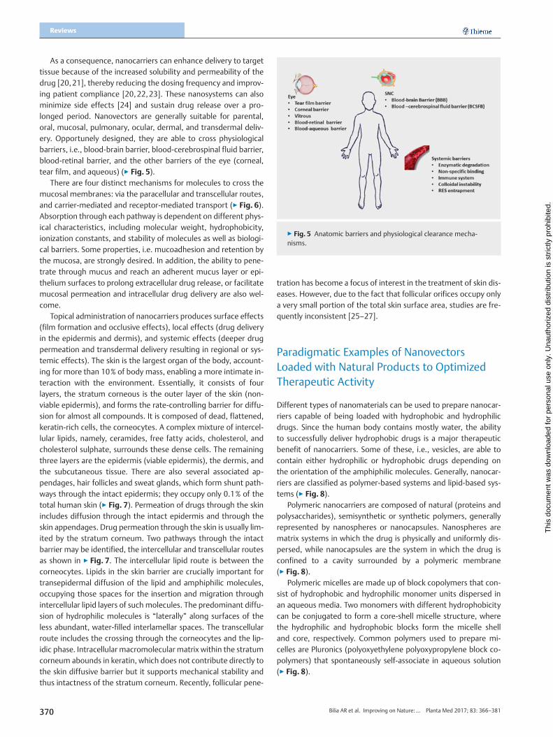
▶ Fig. 5 Anatomic barriers and physiological clearance mecha-nisms.
Reviews
Thi
s do
cum
ent w
as d
ownl
oade
d fo
r pe
rson
al u
se o
nly.
Una
utho
rized
dis
trib
utio
n is
str
ictly
pro
hibi
ted.
As a consequence, nanocarriers can enhance delivery to targettissue because of the increased solubility and permeability of thedrug [20,21], thereby reducing the dosing frequency and improv-ing patient compliance [20,22,23]. These nanosystems can alsominimize side effects [24] and sustain drug release over a pro-longed period. Nanovectors are generally suitable for parental,oral, mucosal, pulmonary, ocular, dermal, and transdermal deliv-ery. Opportunely designed, they are able to cross physiologicalbarriers, i.e., blood-brain barrier, blood-cerebrospinal fluid barrier,blood-retinal barrier, and the other barriers of the eye (corneal,tear film, and aqueous) (▶ Fig. 5).
There are four distinct mechanisms for molecules to cross themucosal membranes: via the paracellular and transcellular routes,and carrier-mediated and receptor-mediated transport (▶ Fig. 6).Absorption through each pathway is dependent on different phys-ical characteristics, including molecular weight, hydrophobicity,ionization constants, and stability of molecules as well as biologi-cal barriers. Some properties, i.e. mucoadhesion and retention bythe mucosa, are strongly desired. In addition, the ability to pene-trate through mucus and reach an adherent mucus layer or epi-thelium surfaces to prolong extracellular drug release, or facilitatemucosal permeation and intracellular drug delivery are also wel-come.
Topical administration of nanocarriers produces surface effects(film formation and occlusive effects), local effects (drug deliveryin the epidermis and dermis), and systemic effects (deeper drugpermeation and transdermal delivery resulting in regional or sys-temic effects). The skin is the largest organ of the body, account-ing for more than 10% of body mass, enabling a more intimate in-teraction with the environment. Essentially, it consists of fourlayers, the stratum corneous is the outer layer of the skin (non-viable epidermis), and forms the rate-controlling barrier for diffu-sion for almost all compounds. It is composed of dead, flattened,keratin-rich cells, the corneocytes. A complex mixture of intercel-lular lipids, namely, ceramides, free fatty acids, cholesterol, andcholesterol sulphate, surrounds these dense cells. The remainingthree layers are the epidermis (viable epidermis), the dermis, andthe subcutaneous tissue. There are also several associated ap-pendages, hair follicles and sweat glands, which form shunt path-ways through the intact epidermis; they occupy only 0.1% of thetotal human skin (▶ Fig. 7). Permeation of drugs through the skinincludes diffusion through the intact epidermis and through theskin appendages. Drug permeation through the skin is usually lim-ited by the stratum corneum. Two pathways through the intactbarrier may be identified, the intercellular and transcellular routesas shown in ▶ Fig. 7. The intercellular lipid route is between thecorneocytes. Lipids in the skin barrier are crucially important fortransepidermal diffusion of the lipid and amphiphilic molecules,occupying those spaces for the insertion and migration throughintercellular lipid layers of such molecules. The predominant diffu-sion of hydrophilic molecules is “laterally” along surfaces of theless abundant, water-filled interlamellar spaces. The transcellularroute includes the crossing through the corneocytes and the lip-idic phase. Intracellular macromolecular matrix within the stratumcorneum abounds in keratin, which does not contribute directly tothe skin diffusive barrier but it supports mechanical stability andthus intactness of the stratum corneum. Recently, follicular pene-
370
tration has become a focus of interest in the treatment of skin dis-eases. However, due to the fact that follicular orifices occupy onlya very small portion of the total skin surface area, studies are fre-quently inconsistent [25–27].
Paradigmatic Examples of NanovectorsLoaded with Natural Products to OptimizedTherapeutic Activity
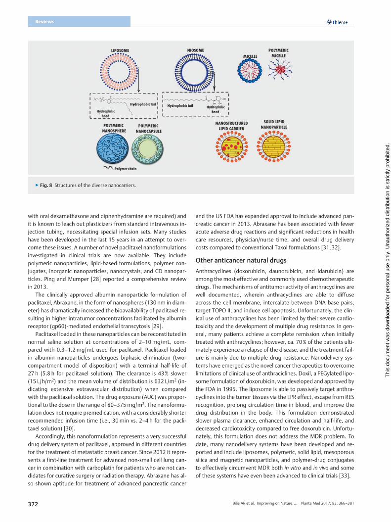
Different types of nanomaterials can be used to prepare nanocar-riers capable of being loaded with hydrophobic and hydrophilicdrugs. Since the human body contains mostly water, the abilityto successfully deliver hydrophobic drugs is a major therapeuticbenefit of nanocarriers. Some of these, i.e., vesicles, are able tocontain either hydrophilic or hydrophobic drugs depending onthe orientation of the amphiphilic molecules. Generally, nanocar-riers are classified as polymer-based systems and lipid-based sys-tems (▶ Fig. 8).
Polymeric nanocarriers are composed of natural (proteins andpolysaccharides), semisynthetic or synthetic polymers, generallyrepresented by nanospheres or nanocapsules. Nanospheres arematrix systems in which the drug is physically and uniformly dis-persed, while nanocapsules are the system in which the drug isconfined to a cavity surrounded by a polymeric membrane(▶ Fig. 8).
Polymeric micelles are made up of block copolymers that con-sist of hydrophobic and hydrophilic monomer units dispersed inan aqueous media. Two monomers with different hydrophobicitycan be conjugated to form a core-shell micelle structure, wherethe hydrophilic and hydrophobic blocks form the micelle shelland core, respectively. Common polymers used to prepare mi-celles are Pluronics (polyoxyethylene polyoxypropylene block co-polymers) that spontaneously self-associate in aqueous solution(▶ Fig. 8).
Bilia AR et al. Improving on Nature:… Planta Med 2017; 83: 366–381

▶ Fig. 6 Mechanisms of enhanced absorption of nanoparticles through mucosal epithelia.
▶ Fig. 7 Permeation mechanisms of nanocarriers in the skin.
Thi
s do
cum
ent w
as d
ownl
oade
d fo
r pe
rson
al u
se o
nly.
Una
utho
rized
dis
trib
utio
n is
str
ictly
pro
hibi
ted.
Lipid-based nanocarriers include nanometric-scaled emul-sions, namely, micro- and nanoemulsions, vesicles including lipo-somes and niosomes, micelles, and nanoparticles divided roughlyinto SLNs and NLCs (▶ Fig. 8). Vesicles and micelles are aggregatesof amphiphilic lipids that organize themselves spontaneously.Vesicles are characterized by bilayer structures that are suitableto load both hydrophilic and hydrophobic compounds. Micellescan only load lipophilic molecules in the lipid core. SLNs and NLCsare solid particles at room temperature and human body temper-atures. They present a lipid core, which makes these carriers suit-able for entrapment of lipophilic compounds. Lipids and surfac-tant agents are generally selected from a plethora of edible con-stituents and GRAS approved compounds.
Bilia AR et al. Improving on Nature:… Planta Med 2017; 83: 366–381
Paclitaxel (Taxol) nanocarriers: improved watersolubility, efficacy and safety profiles
Paclitaxel (Taxol), the most exciting plant-derived anticancer drugdiscovered in recent years, promotes the assembly of tubulin intomicrotubules. It was first isolated from the bark of the Pacific yew(Taxus brevifolia L.). It was approved for clinical use against ovariancancer in 1992 and against breast cancer in 1994. Nowadays, thedrug represents a first-line treatment for ovarian, breast, lung,and colon cancer and a second-line treatment for AIDS-relatedKaposiʼs sarcoma. It is so effective that some oncologists refer tothe period before 1994 as the “pre-Taxol” era for treating breastcancer. Taxol is typically administered intravenously, but due tothe low water solubility, the i. v. route of administration needspolyethoxylated castor oil (Cremophor EL, a non-ionic surfactant)and ethanol (50 :50) as cosolvents. Consequently, this formula-tion is associated with serious and dose-limiting toxicities. Cremo-phor EL itself causes hypersensitivity reactions (premedication
371
▶ Fig. 8 Structures of the diverse nanocarriers.
Reviews
Thi
s do
cum
ent w
as d
ownl
oade
d fo
r pe
rson
al u
se o
nly.
Una
utho
rized
dis
trib
utio
n is
str
ictly
pro
hibi
ted.
with oral dexamethasone and diphenhydramine are required) andit is known to leach out plasticizers from standard intravenous in-jection tubing, necessitating special infusion sets. Many studieshave been developed in the last 15 years in an attempt to over-come these issues. A number of novel paclitaxel nanoformulationsinvestigated in clinical trials are now available. They includepolymeric nanoparticles, lipid-based formulations, polymer con-jugates, inorganic nanoparticles, nanocrystals, and CD nanopar-ticles. Ping and Mumper [28] reported a comprehensive reviewin 2013.
The clinically approved albumin nanoparticle formulation ofpaclitaxel, Abraxane, in the form of nanospheres (130 nm in diam-eter) has dramatically increased the bioavailability of paclitaxel re-sulting in higher intratumor concentrations facilitated by albuminreceptor (gp60)-mediated endothelial transcytosis [29].
Paclitaxel loaded in these nanoparticles can be reconstituted innormal saline solution at concentrations of 2–10mg/mL, com-pared with 0.3–1.2mg/mL used for paclitaxel. Paclitaxel loadedin albumin nanoparticles undergoes biphasic elimination (two-compartment model of disposition) with a terminal half-life of27 h (5.8 h for paclitaxel solution). The clearance is 43% slower(15 L/h/m2) and the mean volume of distribution is 632 L/m2 (in-dicating extensive extravascular distribution) when comparedwith the paclitaxel solution. The drug exposure (AUC) was propor-tional to the dose in the range of 80–375mg/m2. The nanoformu-lation does not require premedication, with a considerably shorterrecommended infusion time (i.e., 30min vs. 2–4 h for the pacli-taxel solution) [30].
Accordingly, this nanoformulation represents a very successfuldrug delivery system of paclitaxel, approved in different countriesfor the treatment of metastatic breast cancer. Since 2012 it repre-sents a first-line treatment for advanced non-small cell lung can-cer in combination with carboplatin for patients who are not can-didates for curative surgery or radiation therapy. Abraxane has al-so shown aptitude for treatment of advanced pancreatic cancer
372
and the US FDA has expanded approval to include advanced pan-creatic cancer in 2013. Abraxane has been associated with feweracute adverse drug reactions and significant reductions in healthcare resources, physician/nurse time, and overall drug deliverycosts compared to conventional Taxol formulations [31,32].
Other anticancer natural drugs
Anthracyclines (doxorubicin, daunorubicin, and idarubicin) areamong the most effective and commonly used chemotherapeuticdrugs. The mechanisms of antitumor activity of anthracyclines arewell documented, wherein anthracyclines are able to diffuseacross the cell membrane, intercalate between DNA base pairs,target TOPO II, and induce cell apoptosis. Unfortunately, the clin-ical use of anthracyclines has been limited by their severe cardio-toxicity and the development of multiple drug resistance. In gen-eral, many patients achieve a complete remission when initiallytreated with anthracyclines; however, ca. 70% of the patients ulti-mately experience a relapse of the disease, and the treatment fail-ure is mainly due to multiple drug resistance. Nanodelivery sys-tems have emerged as the novel cancer therapeutics to overcomelimitations of clinical use of anthraciclines. Doxil, a PEGylated lipo-some formulation of doxorubicin, was developed and approved bythe FDA in 1995. The liposome is able to passively target anthra-cyclines into the tumor tissues via the EPR effect, escape from RESrecognition, prolong circulation time in blood, and improve thedrug distribution in the body. This formulation demonstratedslower plasma clearance, enhanced circulation and half-life, anddecreased cardiotoxicity compared to free doxorubicin. Unfortu-nately, this formulation does not address the MDR problem. Todate, many nanodelivery systems have been developed and re-ported and include liposomes, polymeric, solid lipid, mesoporoussilica and magnetic nanoparticles, and polymer-drug conjugatesto effectively circumvent MDR both in vitro and in vivo and someof these systems have even been advanced to clinical trials [33].
Bilia AR et al. Improving on Nature:… Planta Med 2017; 83: 366–381

Thi
s do
cum
ent w
as d
ownl
oade
d fo
r pe
rson
al u
se o
nly.
Una
utho
rized
dis
trib
utio
n is
str
ictly
pro
hibi
ted.
Vincristine and vinblastine are alkaloids from Catharanthusroseus (periwinkle), a plant of the Apocynaceae family. They arecurrently approved anticancer drugs inducing tubulin to form spi-ral polymers at physiological protein concentrations, thus inter-fering in the formation of microtubules. Vinblastine and vincris-tine are used for childrenʼs leukemia and in the treatment ofHodgkinʼs disease. In particular, vincristine sulphate is used totreat Wilmʼs tumor, neuroblastoma, breast cancer, rhabdomyo-sarcoma, and osteogenic sarcoma. In 2012, the FDA approved ananoformulation of vincristine sulphate loaded in liposome (Mar-qibo) for the treatment of adult patients with Philadelphia chro-mosome-negative (Ph-) acute lymphoblastic leukemia. Vincristinesulphate administered as a liposomal formulation exhibits a lowerclearance and higher AUC compared with conventional drug for-mulations. Other nanoformulations based on vincristine sulphateliposomes have been approved for the treatment of relapsedaggressive non-Hodgkinʼs lymphoma and other cancers. Advan-tages of liposomes over the existing conventional preparationsare the increased blood circulation time, the intensification drugaccumulation in the tumor, prolonged efficacy over an extendedperiod [34,35].
Multifunctional nanoparticles, based on pH-sensitive PLGA-PEG-Fol and cell penetrating peptide R7-conjugated PLGA‑PEG,have been prepared for targeting vincristine sulphate to tumorsand overcoming MDR. In this study, the pH-triggered vincristinerelease was 65.6% during 8 h at pH 5.0, but only 35.8% at pH 7.4,demonstrating that a large amount of vincristine was releasedrapidly in a weak acidic environment. These innovative nanopar-ticles significantly enhance cellular uptake and cytotoxicity inMCF-7 and MCF-7/Adr cells when compared to the nanoparticlessolely modified by Fol or R7. With Fol receptor-mediated endo-cytosis and strong intracellular penetration, vincristine-Fol/R7NPs increased drug accumulation in resistant tumor cells by es-caping P-glycoprotein-mediated drug efflux. In vivo imaging sug-gested the active targeting attributed to pH sensitivity, and theFol receptor-mediated effect could improve tumor-targeting effi-cacy. Indeed, vincristine-Fol/R7 NPs exhibited the strongest anti-tumor efficacy in vivo. Therefore, Fol/R7 NPs are effective nanocar-riers for delivering antitumor drugs and overcoming MDR [36].
Camptothecin is a quinoline alkaloid derived from the bark,wood, and fruit of the Asian tree Camptotheca acuminata L.(Nyssaceae). Drs. Wall and Wani discovered camptothecin in1966 and the National Cancer Institute investigated its potentialantitumor activity as a selective inhibitor of DNATOPO I. Renewedresearch led to the development of water-soluble analogues ofcamptothecin that also retained the anticancer activity, thus over-coming one of the major limitations of clinical use. Irinotecan andtopotecan were approved in 1996 by the FDA for the treatment ofcolon, lung, breast, and ovarian cancers. Due to major limitationsof these derivatives, several nanoparticulate formulations are cur-rently being developed with the aim of improving the therapeuticindex of these compounds. Lipid-based formulations of campto-thecin, both liposomal and polymeric micelles, have been pro-duced and tested for parenteral administration in the treatmentof ovarian and colorectal cancers. These nanoformulations enablethe delivery of higher amounts of drug to target tissues by virtue
Bilia AR et al. Improving on Nature:… Planta Med 2017; 83: 366–381
of the pharmacokinetic properties of the carrier and reduce toxic-ity to nontarget tissues [37,38].
Artemisinin nanocarriers: improved water solubility,stability, and efficacy
Artemisinin (qinghaosu) is a unique sesquiterpene lactone with anendoperoxide moiety isolated from Artemisia annua L. (qinghao,Asteraceae). Discovered by Youyou Tu in 1972 it is currently rec-ommended by the WHO in combination with other antimalarialdrugs to treat drug-resistant Plasmodium falciparum strains, cere-bral malaria, and malaria in children. Beyond malaria, artemisininand its derivatives (artemisinins) exert significant activities to-wards other protozoans (Leishmania, Trypanosoma, amoebas, Neo-spora caninum, and Eimeria tenella), trematodes (Schistosom a, liv-er flukes), and viruses (human cytomegalovirus, hepatitis B and Cviruses). Recently, the activity of artemisinin and its derivativesagainst cancer, bacteria, and fungi have also been proved. Thetherapeutic value of artemisinins is limited due to poor waterand oil solubility, a low bioavailability after oral administration be-cause of rapid degradation by the liver, and a short (ca. 2.5 h) half-life [39]. Therefore, there is an urgent need to develop new arte-misinin formulations to increase its bioavailability, selectivity, andtherapeutic application.
Artemisinin-loaded conventional and PEG (PEGylated) deco-rated liposomes were proposed as nanocarriers to increase bio-pharmaceutical properties of the drug. The pharmacokinetic pro-file and the main pharmacokinetic parameters of the carriers wereevaluated in healthy mice after i. p. administration. Free artemisi-nin was rapidly cleared from plasma and hardly detected 1 h afteradministration. Conversely, both liposomal formulations showeda much longer blood circulation time than free artemisinin; arte-misinin was still detectable after 3 and 24 h of administration, re-spectively, for conventional and PEGylated liposomes. AUC0–24 hvalues were increased by approximately six times in both liposo-mal formulations in comparison with free artemisinin. A strong ef-fect of formulation on the half-life of artemisinin was enhanced bymore than 5-fold by the incorporation of PEG into liposomes.Liposomes loaded with artemisinin, especially the long circulatingvesicles, could really represent a new strategy for developingsmart, well tolerated, and efficacious therapeutic nanocarriers totreat tumors, but it could also be very useful to treat parasitic dis-eases [39].
The antimalarial properties of artemisinin liposomes and arte-misinin plus curcumin liposomes have also been investigated inPlasmodium berghei NK-65 infected mice, a suitable model forstudying malaria because the infection has structural, physiologi-cal, and life cycle analogies with the human disease. Mice weretreated with artemisinin at a dosage of 50mg/kg/day or artemisi-nin plus curcumin administered at a dosage of 100mg/kg/day.Artemisinin began to decrease parasitemia levels only 7 days afterthe start of the treatment and it appeared to have a fluctuanttrend in blood concentration, which is reflected in the antimalarialeffectiveness (Fig. 1S, Supporting Information). By contrast, treat-ments with A‑CL, AC‑CL, A‑PL, and AC‑PL appeared to have an im-mediate antimalarial effect. All formulations cured malaria-in-fected mice within the same post-inoculation period. Additionally,all formulations showed less variability in artemisinin plasma con-
373

Reviews
Thi
s do
cum
ent w
as d
ownl
oade
d fo
r pe
rson
al u
se o
nly.
Una
utho
rized
dis
trib
utio
n is
str
ictly
pro
hibi
ted.
centrations, which suggests that A‑CL, AC‑CL, A‑PL, and AC‑PLgive a modified release of the drug(s) and, consequently, a con-stant antimalarial effect during time. In particular, A‑PL seems togive the most pronounced and statistically significant therapeuticeffect in this murine model of malaria. The enhanced permanencyin the blood of A‑PL suggests the use of these nanosystems assuitable passive targeted carriers for parasitic infections [40].
In a further study by the same scientists, dihydroartemisinin,one of the most potent anticancer artemisinin-like compoundsdue to its apoptotic effects, was formulated in liposomes. Besidesits effectiveness, dihydroartemisinin is a poorly water-solubledrug with low bioavailability and a low half-life (34–90min),therefore, the development of new formulations of dihydroarte-misinin that enable quick availability to the body is in great need.Conventional and PEGylated liposomes were loaded with di-hydroartemisinin. A higher internalization occurred in the conven-tional formulation with respect to the stealth one, suggesting thatthe hydrophilic steric barrier of PEG molecules can reduce cellularuptake. Flow cytometry analysis was used also an alternative tech-nique for rapid size determination of liposomes. Cytotoxicity stud-ies in the MCF-7 cell line confirmed the absence of toxicity in blankformulations and suggested that liposomes may be suitable car-riers for delivery of DHA, avoiding the use of formulations basedon organic solvents [41].
The same group of researchers investigated the antitumorproperties of artemisinin using a novel actively targeted nanocar-rier, a PEGylated nanoliposome decorated with transferrin. Hence,transferrin receptors are largely expressed in cancer cells wherethe iron content is higher than in normal cells. Artemisinin-loadedliposomes were investigated for their cell uptake and cytotoxicityproperties (Fig. 2S, Supporting Information) using the HCT-8 cellline, selected among several cell lines because of transferrin re-ceptor overexpression. The results confirmed the enhanced deliv-ery of artemisinin loaded in liposomes actively targeted withtransferrin in comparison with the other artemisinin-loaded lipo-somes and an improved cytotoxicity [42].
A recent review of artemisinin-based nanocarriers describesliposomes, niosomes, micelles, solid lipid nanocarriers, nanostruc-tured lipid carriers, nanoparticles, fullerenes, and nanotubes withdifferent therapeutic applications. These nanovectors offer signif-icant promise in improving the half-life and controlled release,better permeability, resistance to metabolic modification, andhighly specific site targeted delivery [43].
Curcumin-loaded nanoparticles: increased efficacyof a plethora of activities
Curcumin is the major bioactive constituent of the rhizome of tur-meric (Curcuma longa L., Zingiberaceae), an Indian traditionalherbal medicine. Isolated in 1815, its chemical structure, deter-mined in 1910, is diferuloylmethane [1,7-bis(4-hydroxy-3-me-thoxyphenyl)-1,6-heptadiene-3, 5 dione] and it possesses anti-angiogenic, antiproliferative, antitumorigenic, antioxidant, andanti-inflammatory properties in both in vitro and in vivo studies.Due to its multitargeting ability in various pathological conditions,it is very stimulating in its translation into a therapeutic agent.Like many other lipophilic small molecules, curcumin also has lim-itations for its efficient use in clinical scenarios in order to treat
374
disease conditions. These include the low hydrophilicity and in-trinsic dissolution rate(s), low physical and chemical instability,rapid metabolization with low nanomolar levels of the parentcompound and its glucuronide and sulphate conjugates found inthe peripheral or portal circulation, low absorption, poor pharma-cokinetics and bioavailability, and low penetration and targetingefficacy.
During the last decade, an extraordinary and astonishing num-ber (ca. one thousand) of nanoformulations have been undertak-en to enhance curcumin use in in vitro, in vivo, preclinical, and clin-ical settings that involve the use of nanogels, and a plethora of lip-id and hydrophilic nanocarriers, i.e., polymeric nanoparticles, lipo-somes, micelles, nanoemulsions, CD complexes, nanodisks, nano-fibers, solid lipid nanoparticles, and solid nanodispersions. Thespecific roles and advantages of each delivery system are reportedin a very comprehensive and recent review [44]. Many of these ef-forts initially dealt with improved bioavailability, but newer for-mulations pay attention to efficient targeting of curcumin at thediseased area with the aid of antibody, aptamer, and peptide me-diation [44].
Biodegradable PEGylated polyester nanoparticles loaded withcurcumin were investigated for their bioavailability using a Caco-2 cell model to characterize the cellular transport pathway and de-termine the effect of polymer architecture including PEG chainlength and core material on its cellular interaction and transcellu-lar transport. PEG chain length (from 2000 to 5000 Da) and corematerial (PLA/PLGA) hardly affected the cellular interaction andthe intracellular itinerary of the nanoparticles. However, in thecase of transcellular transport, the maximal transcellular trans-port efficiency for its payload was achieved by the PEG5000-PLA40000 nanoparticles, which present higher drug loading ca-pacity and slower drug release. The findings revealed the cellularinteraction mechanism of PEGylated polyester nanoparticles andprovided evidence for the role of polymer architectures in modu-lating the transcellular permeability of the agents loaded by thenanoparticles, and would be helpful in improving carrier designto enhance drug delivery [45]. Kundu et al. [46] investigated anti-glioma activity of curcumin-loaded lipid nanoparticles and its en-hanced bioavailability in brain tissue for effective glioblastomatherapy. Curcumin-loaded nanoparticles inhibited cellular prolif-eration, migration, and invasion along with a higher percentageof cell cycle arrest and telomerase inhibition, leading to a greaterpercentage of apoptotic cell death in glioma cells compared withnative curcumin. An in vivo study demonstrated an enhanced bio-availability of curcumin in blood serum and brain tissue whenbeing delivered by curcumin-loaded glyceryl monooleate nano-particles compared with native curcumin in a rat model. A studyperformed by Kakkar et al. [47] pursued to improve the oral bio-availability of curcumin by incorporating it into solid lipid nano-particles. The developed nanoparticles (size ca. 130 nm) exhibitedprolonged drug release in vitro. In vivo pharmacokinetic studiesrevealed that after oral administration (50mg/kg, 25mg/kg,12.5mg/kg, and 1mg/kg), a significant improvement in oral bio-availability was achieved by the nanoparticles compared to freecurcumin (by 39, 32, 59, and 155 times at 50mg/kg, 25mg/kg,12.5mg/kg, and 1mg/kg doses, respectively). An in vitro test,the so-called parallel artificial membrane permeability assay
Bilia AR et al. Improving on Nature:… Planta Med 2017; 83: 366–381

Thi
s do
cum
ent w
as d
ownl
oade
d fo
r pe
rson
al u
se o
nly.
Una
utho
rized
dis
trib
utio
n is
str
ictly
pro
hibi
ted.
(PAMPA), has also been reported to evaluate permeation of curcu-min loaded in different nanocarriers developed for oral adminis-tration. Righeschi et al. [48] focused the studies on the develop-ment and characterization of solid lipid nanoparticles for the en-capsulation of curcumin for oral administration using GRAS ex-cipients, i.e., Compritol and lecithin. The particle size was lowerthan 300 nm with a drug entrapment efficiency of 80%. PAMPAtests showed a considerable increase of curcumin permeatedwhen formulated as solid lipid nanoparticles. X‑ray studies and amodified release profile suggested that curcumin molecules aresolubilized into the solid lipid matrix. A further approach to im-prove the solubility and the stability and oral uptake of curcuminwas the formulation of o/w microemulsions using food gradecomponents. Three microemulsions were developed and charac-terized, stabilized by non-ionic surfactants Cremophor EL, Tween20, Tween 80, or lecithin and contained a variety of oils, namely,olive oil, wheat germ oil, and vitamin E. The oral absorption of cur-cumin microemulsions was investigated in vitro using PAMPA. Theoptimal formulation consisted of vitamin E (3.3 g/100 g), Tween20 (53.8 g/100 g), ethanol (6.6 g/100 g), and water (36.3 g/100 g), obtaining a percentage of permeation through the artifi-cial membrane of about 70% [49]. Dendrosomal curcumin, apolymeric micelle of 142 nm with constant physical and chemicalstability, synthesized by esterification of oleoyl chloride (0.01mol)and polyethylene glycol 400 (0.01mol) was developed and testedfor the inhibition on the proliferation of U87MG cells, a cellularmodel of glioblastoma. Evaluation was made by considering mas-ter genes of pluripotency and regulatory miRNA. Methylthiazoltetrazolium assay and flow cytometry were used to detect theantiproliferative effects of dendrosomal curcumin. Annexin-V-FLUOS and caspase assay were used to quantify apoptosis. Thesensitivity of U87MG cells as well as hBMSC and HFSF‑PI3 cells toDNC, free curcumin, and empty dendrosome were studied usingMTT assay. The nanoparticle significantly suppressed the prolifer-ation of U87MG cells in a time- and dose-dependent manner(Fig. 3S, Supporting Information). The half maximal inhibitoryconcentration (IC50) of dendrosoma for U87MG cells was 20 µMafter 24 h (Fig. 3S A, Supporting Information) and 48 h (Fig. 3S B,Supporting Information), which declined to 10 µM at 72 h(p < 0.001) (Fig. 3S C, Supporting Information). However, the via-bility of U87MG was not affected by free curcumin only at 72 h.Dendrosome increased the water solubility and entry of curcuminto cells without any toxic effects. As indicated in Fig. S3, Support-ing Information, dendrosomes induced apoptosis in a time- anddose-dependent manner. Studying the inhibitory effects of den-drosome on hBMSC showed that these cells are affected by thenanocarrier as well as cancerous cells and in a concentration high-er than in U87MG cells. Fig. 4S A, Supporting Information, showsthe effects of dendrosome on cell viability of hBMSC after 24 h ofdrug exposure. Cell viability declined to 67% after treatment with25 µM dendrosome (p < 0.01) and to 35 and 31% after treatmentwith 30 µM and 35 µM dendrosome, respectively (p < 0.001). Theloss of cell viability was also determined after 48 hours of treat-ment with DNC (Fig. 4S B, Supporting Information). The viabilityof HFSF‑PI3 was not significantly affected due to treatment andat determined IC50 for U87MG lines, no inhibitory effect was de-tected on these cells (p > 0.05); however, at 25 µM concentration,
Bilia AR et al. Improving on Nature:… Planta Med 2017; 83: 366–381
the viability of the cells decreased to 50% (Fig. 5S, Supporting In-formation) (p < 0.01). Therefore, in concentrations suppressive forcancer cells, no harmful effects connected to dendrosome wereobserved in stem cells and normal fibroblast cells, showing thesafety of this formulation as an anticancer treatment agent onnormal cells [50]. Finally, Bhawana et al. [51] developed curcu-min-loaded polymeric nanoparticles, (2–40 nm) with antimicro-bial activity. Efficacy was evaluated using a microplate dilutiontechnique against Streptococcus aureus, Bacillus subtilis, Escherichiacoli, Pseudomonas aeruginosa, Penicillium notatum, and Aspergillumniger. The water solubility and small size of curcumin nanopar-ticles enhanced antimicrobial activity when compared to free cur-cumin. The antibacterial activity was more pronounced than itsantifungal activity. Among bacteria, gram-positive strains weremore sensitive. TEM analysis revealed that when the nanoparticleswere introduced into a bacterium, they completely destroyed thecell wall, resulting in bacterial cell death. A recent review reportedthe therapeutic application of different natural products, mainlypolyphenols, including curcumin in rheumatoid arthritis therapy.The review highlighted how nano/submicron vectors can be use-ful in increasing bioavailability and efficacy of the natural prod-ucts [52].
Resveratrol-loaded nanocarriers to improve stabilityand efficacy
Resveratrol (3,4′,5-trihydroxy-trans-stilbene) is a natural poly-phenolic compound abundant in grapes, peanuts, and red wine.First detected in grape vines (Vitis vinifera L., Vitaceae), especiallywhen infected with Botrytis cinerea, resveratrol is synthesized al-most entirely in the skin and its content is at its maximum just be-fore the grapes reach maturity. Therefore, resveratrolʼs highestconcentration is in the skin and seeds of grapes (50–100 µg pergram, corresponding to 5–10% of their biomass). Like the major-ity of polyphenols, it has a rapid and extensive metabolism withthe formation of several resveratrol glucuronides and sulphates.In humans, about 70% of orally administered resveratrol (25mg)is rapidly (< 30min) absorbed and metabolized, with a peak plas-ma level of ca. 2 µM of resveratrol metabolites and a half-life of 9–10 h. Furthermore, there is a significant person-to-person variabil-ity in drug absorption and metabolic processes. The extent towhich the human colon can absorb and metabolize resveratrol de-pends on the hepatic function and on the metabolic activity of thelocal intestinal microflora [53].
Resveratrol has emerged as one of the most promising natu-rally occurring compounds with great preventive and therapeuticefficacies. Studies have confirmed its potential in the preventionof coronary disease and neurodegenerative pathologies. Resvera-trol has been reported to elicit many cellular responses, includingcell cycle arrest, differentiation, and apoptosis, and to inhibit thegrowth of several types of cancer, principally prostate and coloncancers. Resveratrol and its analogues present pharmacologicalsafety and may be used in combination with other agents to en-hance therapeutic efficacy and minimize toxicity. However, intrin-sic features that lead to low bioavailability, low water solubility,and instability have compromised its wide array of activity, andnumerous nanoformulations have been currently investigated toenhance its efficacy.
375

Reviews
Thi
s do
cum
ent w
as d
ownl
oade
d fo
r pe
rson
al u
se o
nly.
Una
utho
rized
dis
trib
utio
n is
str
ictly
pro
hibi
ted.
Shao et al. [54] developed mPEG poly(epsiloncaprolactone)-based nanoparticles incorporating resveratrol and demonstrateda significantly higher rate of cell death as compared to an equiva-lent dose of free resveratrol in glioma cells. Resveratrol loaded insolid lipid nanoparticles resulted in decreasing cell proliferation,preventing skin cancer [54]. In another study, resveratrol nano-particles based on poly(epsiloncaprolactone) and poly(D,L-lactic-co-glycolic acid)-poly(ethylene glycol) were proven to strongly en-hance the uptake by PCa cell lines. In addition, when the PCa, DU-145, and LNCaP cell lines were treated with the nanosystems, allthree cell lines showed significantly elevated cytotoxicity com-pared to those treated with free resveratrol at different concentra-tions (from 10 µM to 40 µM). Remarkably, there was a consistentsensitivity of nano-resveratrol towards both the hormone sensi-tive LNCaP cells and androgen-independent DU-145 prostate can-cer cell lines [55].
In a further study, mitochondrial targeting resveratrol lipo-somes modified with a dequalinium polyethylene glycol-distea-roylphosphatidyl ethanolamine conjugate were developed. Thenanopartices induced apoptosis in both nonresistant and resistantcancer cells by the dissipating mitochondrial membrane potential.It also increased caspase-9 and caspase-3 activities. Significantantitumor efficacy was exerted by resveratrol liposomes in xeno-grafted resistant A549/cDDP cancers in nude mice and tumorspheroids by deep penetration [56]. In another study, the viabilityof HEK 293 cells and their photoprotection after UV‑B irradiationwas tested with free and liposomal resveratrol. Interestingly, cellviability was found to be decreased at a 100 µM concentration,and cell proliferation increased at 10 µM and achieved the mosteffective photoprotection. This study showed the effectivenessof resveratrol at 10 µM and also toxicity at higher concentrationsconsidering the changes in apoptotic features and cell shape andits detachment [57].
The trans-resveratrol-loaded lipid core nanocapsules were usedto test its antiglioma activity on the C6 glioma cell line in vitro andon brain implanted C6 cells in in vivo models. In vitro studies indi-cated that lipid nanostructured carriers loaded with resveratroldecreased the cell viability of C6 glioma cells to a much greaterextent when compared to resveratrol used in solution. In vivostudies of the nanoparticles also showed a marked decrease inthe size of the tumor, suggesting they could be used effectivelyin the treatment of gliomas [58]. A further study was carried outon several anti-inflammatory natural products, namely, carvacrol,thymol, resveratrol, pterostilbene, N-(3-oxo-dodecanoyl)-l-homo-serine lactone, caffeic acid, and caffeic acid phenethyl ester. All ofthe constituents were loaded in liposomes to improve their solu-bility, stability, and bioavailability, therefore addressing the par-ticular challenges associated with each of these natural agents.The chemical instability of resveratrol was improved by liposomeencapsulation, preventing the inactivation of cis-trans isomeriza-tion, and its intravenous administration inhibited tumor growthby approximately 70% in a murine tumor model, showing thatsimple solubilization can have important therapeutic benefits[59].
376
Nanovectors loaded with salvianolic acid:exploring other activities
Salvianolic acid B represents the most characteristic hydrophilicconstituent, Salvia miltiorrhiza Bunge (Lamiaceae), a very famousherbal drug of traditional Chinese medicine. In virtue of the fewside effects, extracts and single constituents are successfully usedin clinics in China, Korea, Japan, and other Asian countries for thetreatment of heart and cerebrovascular disease, hepatitis, hepato-cirrhosis, neurasthenic insomnia, cancer, chronic renal failure, anddysmenorrhea. Both extracts and single constituents have posi-tive effects in CNS neuronal injury and degeneration in several an-imal models [60].
Strong antioxidant activity of salvianolic acid B has also beenreported and it may be useful in the treatment of some severechronic diseases where there is an imbalance of reactive oxygenspecies formation and where intracellular reactive oxygen and ni-trogen species levels can cause severe cell damage and even celldeath. In particular, it can protect against oxidative stress as wellas the antioxidant superoxide dismutase and reduced activity ofglutathione, important determinants of neuropathological andbehavioral consequences in neuropathic pain. This is a chronic dis-ease defined by the WHO as an untreatable illness because thera-peutics are unsatisfactory in many cases and there is an urgentneed to discover and develop novel active drugs. Both conven-tional and long circulating liposomes loaded with salvianolic acidB were developed for parental administration due to the poorchemical stability and bioavailability of salvianolic acid B. Thesecarriers were submitted to pharmacological studies using thepaw pressure test in an animal model of neuropathic pain wherea peripheral mononeuropathy was produced by a chronic con-striction injury of the sciatic nerve. Salvianolic acid B was effectiveagainst mechanical hyperalgesia when administered intraperito-neally at a dose of 100mg/kg, 15min after administration.According to the in vivo studies, encapsulation, especially intoPEGylated liposomes, increased and prolonged the antihyperalge-sic activity 30min after i. p. administration and the effect was stillsignificant after 45min [61].
Honokiol loaded in nanocarriers: retardedelimination and prolonged residence timein the circulating system
Honokiol, 3′,5-di(2-propenyl)-1,1′-biphenyl-2,4′-diol, is a constit-uent of the Chinese medicinal plantMagnolia officinalis L. (Magno-liaceae). It has several pharmacological effects, including anti-in-flammatory, antithrombotic, antirheumatic, and antioxidant withanxiolytic, central nervous system depressant, and muscle relax-ant activities. In addition, it has potent antitumor activity. Again,this compoundʼs hydrophobic properties represent an obstacle,because high hydrophobicity prevents vascular administration.Stable liposomes containing an inclusion complex of honokiol inHP‐β‐CD used a molar ratio of honokiol/HP‐β‐CD/phosphatidilcholine 1 :2 : 2. The mean particle size was 123.5 nm, the zeta po-tential was − 25.6mV, and the EE was 91.09 ± 2.76%. The releaseprofile in vitro demonstrated that honokiol is released from theliposome with a sustained and slow profile. A pharmacokineticstudy revealed that honokiol-in-HP-β-CD-in-liposome significantlyretarded the elimination of honokiol and prolonged the residence
Bilia AR et al. Improving on Nature:… Planta Med 2017; 83: 366–381
Thi
s do
cum
ent w
as d
ownl
oade
d fo
r pe
rson
al u
se o
nly.
Una
utho
rized
dis
trib
utio
n is
str
ictly
pro
hibi
ted.
time in the circulating system. Honokiol-in-HP-β-CD‑in-liposomehad antiproliferative activity in A549 and HepG2 tumor cells com-pared with free honokiol [62].
In a further study, self-assembled pectin nanoparticles con-taining a complex of HP‐β‐CD and honokiol were prepared andevaluated for delivering honokiol to HepG2 cells. An investigationof the in vitro release indicated that the drug-loaded nanoparticlesexhibited a higher drug release rate than free honokiol and an ef-fective sustained release. Cytotoxicity, cell apoptosis, and cellularuptake studies further confirmed that the pectin nanoparticleswith galactose residues generated higher cytotoxicity than freehonokiol on HepG2 cells, which highly expressed ASGR. Neverthe-less, these findings were not observed in ASGR-negative A549cells under similar conditions. Therefore, pectin nanoparticlesloaded with honokiol complexed with HP-β-CD have a specific ac-tive targeting ability to ASGR-positive HepG2 cells and could beused as potential drug carriers in the treatment of liver-related tu-mors [63].
Nanoparticles loaded with honokiol were prepared with ther-mosensitive poly(ethylene glycol)-poly(epsilon-caprolactone)-poly(ethylene glycol) hydrogel to improve honokiol therapeuticefficacy on malignant pleural effusion-bearing mice. The hydrogelwas administered intrapleurally after Lewis lung carcinoma cell in-oculation. Honokiol-hydrogel reduced the number of pleural tu-mor foci, prolonging the survival time of MPE-bearing mice moreeffectively compared with the control groups. In addition, hono-kiol-hydrogel successfully inhibited angiogenesis as assessed byCD31 (p < 0.05). Histological analysis of pleural tumors exhibitedthat honokiol loaded in hydrogel led to an increased rate of apo-ptosis [64].
PEGylated liposomal honokiol with a particle size of ca. 100 nmhas been developed, and both pharmacokinetic properties andhuman plasma protein binding ability were investigated. Thepharmacokinetics properties were studied after intravenous ad-ministration in Balb/c mice. There were significant differences oft(1/2β) and AUC(0→∞) parameters. The mean t(1/2β) value ofPEGylated liposomal honokiol and free honokiol were 26.09minand 13.46min, respectively. The AUC(0→∞) ratio of PEGylatedliposomal honokiol to free honokiol was about 1.85-fold (219.24/118.68 µg/mL min). Honokiol at concentrations of 0.5, 8.0, and20 µg/mL in human plasma achieved a bound percentage from60 to 65%. The results suggested that PEGylated liposome im-proved honokiol solubility, increased the drug concentration inplasma, and decreased the clearance [65].
Flavonoids loaded in nanocarriersto enhance bioefficacy
Flavonoids are among the biggest group of polyphenols, widelydistributed in plants, especially in edible plants. Hence, flavonoidsrepresent the active constituents of many dietary supplementsand herbal remedies, as well as there is an increasing interest inthis class of polyphenols as functional ingredients of beverages,food grains, and dairy products. A plethora of evidence supportsthe health benefits and value of flavonoids, which can play a rolein physiological function treatment and in the prevention of dis-eases, particularly of degenerative conditions including cancers,cardiovascular, and neurodegenerative diseases. Conversely, vari-
Bilia AR et al. Improving on Nature:… Planta Med 2017; 83: 366–381
ous studies have also shown that flavonoids have some drawbacksafter oral administration, principally stability, bioavailability, andbioefficacy.
Water solubility and gastric stability are the major limiting fac-tors for flavonoids to pass the biological membrane and to be ab-sorbed systematically following oral administration. An enormousnumber of nanodelivery techniques, including nanoparticles,nanocapsules, micro- and nanoemulsions, micelles, vesicles, solidlipid nanoparticles, and nanostructured lipid capsules, were suc-cessfully developed for overcoming the delivery challenges of fla-vonoids, as recently reported in a review [66]. Most of the papersare related to pure epigallocatechin gallate, quercetin, rutin, api-genin, baicalein, puerarin, apigenin, and hesperetin using princi-pally biodegradable nanoparticles (natural or synthetic polymers),micelles, vesicles, and micro- and nanoemulsions. Results haveshown that mucoadhesive chitosan nanoparticles loaded with fla-vonoids were very useful in increasing flavonoid bioavailability. Incomparison to microdelivery systems, nanocarriers have some ad-vantages: they are more stable, provide more surface area, andhave the potential to enhance bioavailability by increasing the ab-sorption from enterocytes, by receptor-mediated endocytosis andtranscytosis, and by phagocytosis via specialized microfold cells.Finally, nanodelivery systems improve the controlled release ofthe encapsulated flavonoids. A recent review focused on nanocar-riers made of GRAS approved compounds and represent drivingformulating carriers for innovative and more effective formula-tions of functional foods, dietary supplements, and herbal medic-inal products [16].
Successful nanoformulations based on extracts
Extracts are complex mixtures of constituents having different po-larities and absorption properties and, in many cases, have goodactivity in in vitro assays, which is not reproducible in in vivo ex-periments. In some cases, constituents can be incompatible withother components in the formulation or have undesirable proper-ties, which, consequently, nanotechnological strategies have at-tempted to enhance their efficacy. The nanosystems can increaseselectivity and activity, protect against thermal- or photodegrada-tion, reduce side effects, promote sustained release of active con-stituents, or reduce the required dose, generally resulting in im-proved activity. Some recent reviews have focused on the nano-technology approach of extracts using different drug delivery sys-tems based on nanotechnology, principally polymeric nanopar-ticles, solid lipid nanoparticles, liquid crystal systems, vesicles,nanoemulsions, and microemulsions, in order to impart a formu-lationʼs most desirable properties. The most investigated are theextracts of ginseng, green tea, ginkgo, and milk thistle [66–68].
Nanocarriers based on sylimarinto optimize hepatic protection
Silymarin is one of the best-known hepatoprotective extracts ob-tained from the seeds of Silybum marianum L., commonly knownas milk thistle (Asteraceae). Since ancient times, this plant hasbeen used as an herbal cure for liver and biliary tract diseases. Ithas been reputed that milk thistle extracts safeguard and regen-erate liver cells in various diseases affecting liver, principally cir-rhosis, jaundice, and hepatitis. It exhibits strong antioxidant ac-
377
Reviews
Thi
s do
cum
ent w
as d
ownl
oade
d fo
r pe
rson
al u
se o
nly.
Una
utho
rized
dis
trib
utio
n is
str
ictly
pro
hibi
ted.
tion via free radical scavenging activity and inhibits lipid peroxida-tion. It also prevents the entry of harmful toxicants, primarilyheavy metals, pesticides, alcohols, medicines, and CCl4 in the liv-er, thereby protecting the liver cells from further damage. Sily-marin is a mixture of flavonolignans, which comprises silybin, iso-silybin, silydianin, and silychristine. It is poorly soluble in water andexerts its hepatoprotective action at an oral dose of 240–800mg/day in two or three divided doses. When administered orally, peakplasma concentration is reached in 2–4 h, with t1/2 = 6 h. Only 20–50% silymarin is absorbed from the gastrointestinal tract where itundergoes extensive enterohepatic circulation. Therefore, ab-sorption of silymarin from the gastrointestinal tract is low, makingbioavailability poor. Its poor bioavailability is mainly due to exten-sive metabolism, poor aqueous solubility, and rapid excretionthrough urine and bile as well as low permeability across intestinalepithelial cells. Silymarin represents a good candidate for nanofor-mulations. In 2011, a very interesting review concerning the re-cent published literature on various techniques for increasing thebioavailability of silymarin, including the nanotechnological ap-proach, was published [69]. A more recent study reported that si-lymarin-loaded solid nanoparticles, containing silymarin/castoroil/polyvinylpyrrolidone/Transcutol HP/Tween 80 at a weight ratioof 5/3/3/1.25/1.25, improved drug solubility about 1300-fold[70]. Additionally, after oral administration of 10mg/kg in rats,the nanoparticles showed a considerably shorter time to peakconcentration. In particular, the plasma concentrations of silyma-rin in nanoparticles between 0.25 and 1.5 h were impressivelyhigher when compared with a silymarin conventional formulation(1.27 and 1.44 times, respectively, p < 0.05). The AUC of the drugprovided by the nanoparticles was approximately 1.3-fold greaterthan that of the conventional formulation. On the other hand,Tmax for the nanoparticles was significantly shorter than that ofthe conventional formulation (p < 0.05). The plasma levelsachieved by the nanoparticles increased up to 22.85 ± 3.59 µg/mL at Tmax (1 h). Therefore, the enhanced oral bioavailability of si-lymarin in rats might be associated with a marked enhancementin the absorption rate of silymarin as a result of the increased dis-solution of the drug in the nanoparticles. In addition, the silyma-rin-loaded nanoparticles significantly reduced CCl4-induced hepa-totoxicity, indicating improved bioactivity compared with silyma-rin conventional formulation and the pure drug. Treatment withCCl4 significantly elevated the SAT activity, which was significantlyreduced by pretreatment with silymarin-loaded nanoparticles.The protective effects of silymarin-loaded nanoparticles on CCl4-induced acute liver toxicity were also observed by histopathologicexamination. Compared with healthy livers, which had normallobular architecture with a central vein and radiating hepaticcords, liver sections from CCl4-exposed rats showed necrosis ofhepatocytes, hepatocyte degeneration, and infiltration of inflam-matory cells. While the CCl4-induced destruction of liver architec-ture was not improved in rats treated with silymarin powder or theconventional formulation, the liver tissue from rats treated withsilymarin nanoparticles showed less severe damage surroundingthe central vein, suggesting superior hepatoprotective activity ofsilymarin nanoparticles over the drug powder or the commercialconventional formulation [70].
378
Nanocarriers based on Cuscuta chinensis extracts
Cuscuta chinensis Lam. (Convolvulaceae) is a commonly used tra-ditional Chinese medicine to nourish the liver and kidney. Theethanolic extract is generally administered, but due to the poorwater solubility of its major and characteristic constituents, flavo-noids and lignans, its absorption upon oral administration couldbe limited.
Nanoparticles based on the ethanolic extract were developedand evaluated for the hepatoprotective and antioxidant effectson acetaminophen-induced hepatotoxicity in rats. An oral doseof the extract at 125 and 250mg/kg and the extract loaded inthe nanoparticles at 25 and 50mg/kg showed a significant hepa-toprotective effect relatively to the same extent (p < 0.05) by re-ducing levels of AST, ALT, and AP. These biochemical assessmentswere supported by rat hepatic biopsy examinations. In addition,the antioxidant activities of the extract and the nanoparticles bothsignificantly increased superoxide dismutase, catalase, and gluta-thione peroxidase, and reduced malondialdehyde (p < 0.05).Moreover, the results also indicated that the hepatoprotectiveand antioxidant effects of 50mg/kg of the extract loaded in thenanoparticles were effectively better than 125mg/kg of the ex-tract (p < 0.05). An oral dose of the extract loaded in nanopar-ticles, five times less as the extract itself, could exhibit similar lev-els of outcomes [71].
Nanocarriers loaded with essential oilsto optimize their therapeutic use
Essential oils are complex blends of a variety of volatile moleculesincluding terpenoids, phenol components, and aliphatic constitu-ents, which have a strong interest in the pharmaceutical, sanitary,cosmetic, agricultural, and food industries. Since the Middle Ages,essential oils have been widely used for their bactericidal, viru-cidal, fungicidal, antiparasitical, insecticidal, and medicinal prop-erties including analgesic, sedative, anti-inflammatory, spasmo-lytic, and locally anesthetic remedies. Their nanoencapsulation indrug delivery systems has been proposed to have the capability ofdecreasing volatility and improving stability, water solubility, andefficacy of essential oil-based formulations by maintenance oftherapeutic effectiveness. Two categories of nanocarriers can beproposed: polymeric nanoparticulate formulations, extensivelystudied with significant improvement of the essential oil anti-microbial activity, and lipid carriers, including liposomes, solid lip-id nanoparticles, nanostructured lipid particles, and nano- andmicroemulsions.
A significantly large part of the current literature on the encap-sulation of essential oils deals with micrometric size capsules,which are used for the protection of the active compoundsagainst environmental factors (e.g., oxygen, light, moisture, andpH), to decrease oil volatility, and to transform the oil into a pow-der. Encapsulation in nanometric particles is an alternative forovercoming these problems but, additionally, due to the subcellu-lar size, may increase the cellular absorption mechanisms and in-crease bioefficacy, as reported in a comprehensive recent review[17].
Among the different studies described in the literature, the ef-fect of liposomal inclusion on the stability and in vitro antiherpeticactivity of Santolina insularis L. (Asteraceae) essential oil was very
Bilia AR et al. Improving on Nature:… Planta Med 2017; 83: 366–381
ocum
ent w
as d
ownl
oade
d fo
r pe
rson
al u
se o
nly.
Una
utho
rized
dis
trib
utio
n is
str
ictly
pro
hibi
ted.
remarkable. Pure essential oil cannot be use topically because ofits high volatility and enhancer penetration properties, which canresult in safety concerns, hence, a suitable formulation should beprepared. Liposomes successfully prevented its degradation andwere effective in inactivating HSV-1, and that activity is principallydue to direct virucidal effects [72].
The same authors also studied the antiherpetic activity of theessential oil of Artemisia arborescens L. (Asteraceae) loaded in lipo-somes. These carriers were very suitable to be incorporated ingood amounts of essential oil, and stability studies pointed outthat vesicle dispersions were very stable for at least six months.Neither oil leakage nor vesicle size alteration occurred during thisperiod. After one year of storage, oil retention was still good, butvesicle fusion was present. Antiviral assays demonstrated that theliposomal formulation enhanced its in vitro antiherpetic activity[73].
In a further study, the same author prepared solid lipid nano-particles loaded with A. arborescens essential oil and tested theantiherpetic activity. The nanoparticles entrapped the essentialoil in high yields and the mean particle size increased only slightlyafter two years of storage, indicating a high physical stability. Invitro antiviral assays showed that nanoparticle incorporation didnot affect the essential oil antiherpetic activity. The in vitro skinpermeation experiments demonstrated the capability of nanopar-ticles of greatly improving the oil accumulation into the skin. Oilpermeation occurred only when the oil was delivered from thecontrol solution [74].
Frankincense and myrrh are gum resins obtained from the gen-era Boswellia and Commiphora, respectively. Modern pharmaco-logical research has revealed that essential oils are the primary ef-fective components in frankincense and myrrh oil that exhibit abroad spectrum of biological activities, principally antimicrobial,anti-inflammatory, and antitumor activities. As with other essen-tial oils, the instability and poor water solubility result in poor oralbioavailability, which limits its clinical application. The compo-nents of frankincense and myrrh oil are very sensitive to light, air,and high temperature, and they stimulate the gastrointestinaltract, making these oils unsuitable for oral administration. A re-cent study has reported the successful preparation of solid lipidnanoparticles for the oral delivery of frankincense and myrrh es-sential oils [75].
Thi
s d
Concluding Remarks
Extensive research is going on in natural products and herbal ex-tracts to incorporate them into nano-drug delivery systems. Theapplication of nanotechnology to drug delivery has already had asignificant impact on many areas of medicine, and this approachis essential to developing appropriate therapeutic treatments ofsome essential drugs, principally antitumor and antiparasisticagents. Amazing nanoparticles loaded with paclitaxel, doxorubi-cin, vincristine, camptothecin, artemisinin, curcumin, resveratrol,honokiol, salvianolic acid B, green tea catechins, and silymarin areavailable on the market or are under clinical studies. Remarkableresults have also been obtained for nanoformulations based oncomplex mixtures of constituents, extracts, and essential oils,
Bilia AR et al. Improving on Nature:… Planta Med 2017; 83: 366–381
which may extend a formulationʼs action, and successfully com-bine active substances with different degrees of hydrophilicity/lipophilicity. So far, the results obtained from nanoencapsulatednatural products are very encouraging, generally having betterstability, sustained release, and improved bioavailability at muchlower doses, and increasing long-term safety of these constitu-ents. We have entered the era of nanomedicine with naturaldrugs, which is probably the most salient feature of the field anda decisive step toward the demonstration that natural drugs cancontinue to play a key role in modern clinics.
Supporting information
Data which represent parasitemia progression for mice groups in-fected with P. berghei and treated with ART loaded in lipososmes,in vitro cytotoxicity of free ART, ART‑L, and ART‑L‑Tf on HCT-8 co-lon cancer cells, and the effects of dendrosomal curcumin, freecurcumin, and empty dendrosome on the viability of U87MGcells, human bone marrow-derived stem cells, and HFSF‑PI3 cellsare available as Supporting Information.
Acknowledgements
The authors wish to thank Ente Cassa Risparmio di Firenze forkindly supporting part of the research.
Conflict of Interest
The authors declare no conflict of interest.
References
[1] Cameron A, Ewen M, Auton M, Abegunde D. WHO (World Health Orga-nization): The world medicines situation 2011: Medicines prices, avail-ability and affordability. Geneva, Switzerland. Available at http://www.who.int/medicines/areas/policy/world_medicines_situation/WMS_ch6_wPricing_v6.pdf. Accessed September 3, 2016
[2] BeghynT, Deprez-Poulain R, Willand N, Folleas B, Deprez B. Natural com-pounds: leads or ideas? Bioinspired molecules for drug discovery. ChemBiol Drug Des 2008; 72: 3–15
[3] Lu JJ, Pan W, Hu YJ, Wang YT. Multi-target drugs: the trend of drug re-search and development. PLoS One 2012; 7: e40262
[4] Morphy R. Selectively nonselective kinase inhibition: striking the rightbalance. J Med Chem 2009; 53: 1413–1437
[5] Mehta P, Shah R, Lohidasan S, Mahadik KR. Pharmacokinetic profile ofphytoconstituent (s) isolated from medicinal plants – A comprehensivereview. J Tradit Complement Med 2015; 5: 207–227
[6] Kesarwani K, Gupta R. Bioavailability enhancers of herbal origin: an over-view. Asian Pac J Trop Biomed 2013; 3: 253–266
[7] Couvreur P, Vauthier C. Nanotechnology: intelligent design to treat com-plex disease. Pharm Res 2006; 23: 1417–1450
[8] Committee for proprietary medicinal products (CPMP). Note forguidance on the investigation of bioavailability and bioequivalence.Available at http://www.ema.europa.eu/docs/en_GB/document_library/Scientific_guideline/2010/01/WC500070039.pdf. Accessed Sep-tember 3, 2016
[9] Wu CY, Benet LZ. Predicting drug disposition via application of BCS:transport/absorption/elimination interplay and development of a bio-
379

Reviews
Thi
s do
cum
ent w
as d
ownl
oade
d fo
r pe
rson
al u
se o
nly.
Una
utho
rized
dis
trib
utio
n is
str
ictly
pro
hibi
ted.
pharmaceutics drug disposition classification system. Pharm Res 2005;22: 11–23
[10] Fleisher D, Bong R, Stewart BH. Improved oral drug delivery: solubilitylimitations overcome by the use of prodrugs. Adv Drug Deliv Rev 1996;19: 115–130
[11] Stella VJ, Nti-Addae KW. Prodrug strategies to overcome poor watersolubility. Adv Drug Deliv Rev 2007; 59: 677–694
[12] Merisko-Liversidge E, Liversidge GG, Cooper ER. Nanosizing: a formula-tion approach for poorly-water-soluble compounds. Eur J Pharm Sci2003; 18: 113–120
[13] Möschwitzer JP. Drug nanocrystals in the commercial pharmaceuticaldevelopment process. Int J Pharm 2013; 453: 142–156
[14] Hans ML, Lowman AM. Biodegradable nanoparticles for drug deliveryand targeting. Curr Opin Solid State Mater Sci 2002; 6: 319–327
[15] Jabr-Milane LS, van Vlerken LE, Yadav S, Amiji MM. Multi-functionalnanocarriers to overcome tumor drug resistance. Cancer Treat Rev2008; 34: 592–602
[16] Bilia AR, Isacchi B, Righeschi C, Guccione C, Bergonzi MC. Flavonoidsloaded in nanocarriers: an opportunity to increase oral bioavailabilityand bioefficacy. Food Nutr Sci 2014; 5: 1212–1227
[17] Bilia AR, Guccione C, Isacchi B, Righeschi C, Firenzuoli F, Bergonzi MC.Essential oils loaded in nanosystems: a developing strategy for a success-ful therapeutic approach. Evid Based Complement Alternat Med 2014;2014: 651593
[18] Gaumet M, Vargas A, Gurny R, Delie F. Nanoparticles for drug delivery:the need for precision in reporting particle size parameters. Eur J PharmBiopharm 2008; 69: 1–9
[19] Nicolas J, Mura S, Brambilla D, Mackiewicz N, Couvreur P. Design, func-tionalization strategies and biomedical applications of targeted bio-degradable/biocompatible polymer-based nanocarriers for drug deliv-ery. Chem Soc Rev 2013; 42: 1147–1235
[20] Gelperina S, Kisich K, Iseman MD, Heifets L. The potential advantages ofnanoparticle drug delivery systems in chemotherapy of tuberculosis. AmJ Respir Crit Care Med 2005; 172: 1487–1490
[21] Kumar M, Misra A, Babbar AK, Mishra AK, Mishra P, Pathak K. Intranasalnanoemulsion based brain targeting drug delivery system of risperidone.Int J Pharm 2008; 358: 285–291
[22] Emerich DF, Thanos CG. The pinpoint promise of nanoparticle-baseddrug delivery and molecular diagnosis. Biomol Eng 2006; 23: 171–184
[23] Bawa R. Nanoparticle-based therapeutics in humans: a survey. Nano-techol Law Business 2008; 5: 135–155
[24] Zhang L, Gu FX, Chan JM, Wang AZ, Langer RS, Farokhzad OC. Nano-particles in medicine: therapeutic applications and developments. ClinPharmacol Ther 2008; 83: 761–769
[25] Mezei M, Gulasekharam V. Liposomes – a selective drug delivery systemfor the topical route of administration. Lotion dosage form. Life Sci1980; 26: 1473–1477
[26] Cevc G. Transfersomes, liposomes and other lipid suspensions on theskin: permeation enhancement, vesicle penetration, and transdermaldrug delivery. Crit Rev Ther Drug Carrier Syst 1996; 13: 257–388
[27] Gupta M, Agrawal U, Vyas SP. Nanocarrier-based topical drug delivery forthe treatment of skin diseases. Expert Opin Drug Deliv 2012; 9: 783–804
[28] Ping M, Mumper RJ. Paclitaxel nano-delivery systems: a comprehensivereview. J Nanomed Nanotechnol 2013; 18: 1000164
[29] Green MR, Manikhas GM, Orlov S, Afanasyev B, Makhson AM, Bhar P,Hawkins MJ. Abraxane, a novel cremophor-free, albumin-bound particleform of paclitaxel for the treatment of advanced non-small-cell lung can-cer. Ann Oncol 2006; 17: 1263–1268
[30] Gradishar WJ. Albumin-bound paclitaxel: a next-generation taxane.Expert Opin Pharmacother 2006; 7: 1041–1053
[31] Untch M, Jackisch C, Schneeweiss A, Conrad B, Aktas B, Denkert C,Eidtmann H, Wiebringhaus H, Kümmel S, Hilfrich J, Warm M, Paepke S,
380
Just M, Hanusch C, Hackmann J, Blohmer JU, Clemens M, Darb-EsfahaniS, Schmitt WD, Dan Costa S, Gerber B, Engels K, Nekljudova V, Loibl S,von Minckwitz G. Nab-paclitaxel versus solvent-based paclitaxel in neo-adjuvant chemotherapy for early breast cancer (GeparSepto-GBG 69): arandomised, phase 3 trial. Lancet Oncol 2016; 17: 345–356
[32] Dranitsaris G, Yu B, Wang L, Sun W, Zhou Y, King J, Kaura S, Zhang A,Yuan P. Abraxane® versus Taxol® for patients with advanced breast can-cer: A prospective time and motion analysis from a Chinese health careperspective. J Oncol Pharm Pract 2016; 22: 205–211
[33] Ma P, Mumper RJ, Barenholz Y. Anthracycline nano-delivery systems toovercome multiple drug resistance: a comprehensive review. J ControlRelease 2012; 160: 117–134
[34] Igor VZ, Norbert M, Quet-Fah A. Liposome-encapsulated vincristine, vin-blastine and vinorelbine: a comparative study of drug loading and reten-tion. J Control Release 2005; 104: 103–111
[35] Yang SH, Lin CC, Lin ZZ, Tseng YL, Hong RL. A phase 1 pharmacokineticstudy of liposomal vinorelbine in patients with advanced solid tumor.Invest New Drugs 2012; 30: 282–289
[36] Wang Y, Dou L, He H, Zhang Y, Shen Q. Multifunctional nanoparticles asnanocarrier for vincristine sulphate delivery to overcome tumor multi-drug resistance. Mol Pharm 2014; 11: 885–894
[37] Patankar N, Waterhouse D. Nano-particulate drug delivery systems forcamptothecins. Cancer Ther 2012; 8: 90–104
[38] Cuong NV, Hsieh MF, Huang CM. Recent development in nano-sized dos-age forms of plant alkaloid camptothecin-derived drugs. Recent PatAnticancer Drug Discov 2009; 24: 254–261
[39] Isacchi B, Arrigucci S, la Marca G, Bergonzi MC, Vannucchi MG, Novelli A,Bilia AR. Conventional and long-circulating liposomes of artemisinin:preparation, characterization, and pharmacokinetic profile in mice.J Liposome Res 2011; 21: 237–244
[40] Isacchi B, Bergonzi MC, Grazioso M, Righeschi C, Pietretti A, Severini C,Bilia AR. Artemisinin and artemisinin plus curcumin liposomal formula-tions: enhanced antimalarial efficacy against Plasmodium berghei-in-fected mice. Eur J Pharm Biopharm 2012; 80: 528–534
[41] Righeschi C, Coronnello M, Mastrantoni A, Isacchi B, Bergonzi MC, MiniE, Bilia AR. Strategy to provide a useful solution to effective delivery ofdihydroartemisinin: development, characterization and in vitro studiesof liposomal formulations. Colloids Surf B Biointerfaces 2014; 116:121–127
[42] Leto I, Coronnello M, Righeschi C, Bergonzi MC, Mini E, Bilia AR.Enhanced efficacy of artemisinin loaded in transferrin-conjugatedliposomes versus stealth liposomes against HCT‑8 colon cancer cells.ChemMedChem 2016; 11: 1745–1751
[43] Efferth T, Romero MR, Bilia AR, Osman AG, ElSohly M, Wink M, Bauer R,Khan I, Bergonzi MC, Marin JJG. Expanding the therapeutic spectrum ofartemisinin: activity against infectious diseases beyond malaria and nov-el pharmaceutical developments. World J Tradit Chin Med 2016; 2: 1–23
[44] Yallapu MM, Nagesh PK, Jaggi M, Chauhan SC. Therapeutic applicationsof curcumin nanoformulations. AAPS J 2015; 17: 1341–1356
[45] Song Q, Wang X, Hu Q, Huang M, Yao L, Qi H, Qiu Y, Jiang X, Chen J,Chen H, Gao X. Cellular internalization pathway and transcellular trans-port of pegylated polyester nanoparticles in Caco-2 cells. Int J Pharm2013; 445: 58–68
[46] Kundu P, Mohanty C, Sahoo SK. Antiglioma activity of curcumin-loadedlipid nanoparticles and its enhanced bioavailability in brain tissue for ef-fective glioblastoma therapy. Acta Biomater 2012; 8: 2670–2687
[47] Kakkar V, Singh S, Singla D, Kaur IP. Exploring solid lipid nanoparticles toenhance the oral bioavailability of curcumin. Mol Nutr Food Res 2011;55: 495–503
[48] Righeschi C, Bergonzi MC, Isacchi B, Bazzicalupi C, Gratteri P, Bilia AR.Enhanced curcumin permeability by SLN formulation: The PAMPA ap-proach. LWT-Food Sci Technol 2016; 66: 475–483
Bilia AR et al. Improving on Nature:… Planta Med 2017; 83: 366–381

Thi
s do
cum
ent w
as d
ownl
oade
d fo
r pe
rson
al u
se o
nly.
Una
utho
rized
dis
trib
utio
n is
str
ictly
pro
hibi
ted.
[49] Bergonzi MC, Hamdouch R, Mazzacuva F, Isacchi B, Bilia AR. Optimiza-tion, characterization and in vitro evaluation of curcumin microemul-sions. LWT-Food Sci Technol 2014; 59: 148–155
[50] Tahmasebi Mirgani M, Isacchi B, Sadeghizadeh M, Marra F, Bilia AR,Mowla SJ, Najafi F, Babaei E. Dendrosomal curcumin nanoformulationdownregulates pluripotency genes via miR‑145 activation in U87MGglioblastoma cells. Int J Nanomedicine 2014; 9: 403–417
[51] Bhawana I, Basniwal RK, Buttar HS, Jain VK, Jain N. Curcumin nanopar-ticles: preparation, characterization, and antimicrobial study. J AgricFood Chem 2011; 59: 2056–2061
[52] Rahman M, Beg S, Verma A, Al Abbasi FA, Anwar F, Saini S, Akhter S,Kumar V. Phytoconstituents as pharmacotherapeutics in rheumatoidarthritis: challenges and scope of nano/submicromedicine in its effec-tive delivery. J Pharm Pharmacol 2017; 69: 1–14
[53] Wenzel E, Somoza V. Metabolism and bioavailability of trans-resveratrol.Mol Nutr Food Res 2005; 49: 472–481
[54] Shao J, Li X, Lu X, Jiang C, Hu Y, Li Q, You Y, Fu Z. Enhanced growth inhi-bition effect of resveratrol incorporated into biodegradable nanopar-ticles against glioma cells is mediated by the induction of intracellularreactive oxygen species levels. Colloids Surf B Biointerfaces 2009; 72:40–47
[55] Sanna V, Siddiqui IA, Sechi M, Mukhtar H. Resveratrol loaded nanopar-ticles based on poly(epsiloncaprolactone) and poly(D,L-lactic-co-gly-colic acid)-poly(ethylene glycol) blend for prostate cancer treatment.Mol Pharm 2013; 10: 3871–3881
[56] Wang XX, Li YB, Yao HJ, Ju RJ, Zhang Y, Li RJ, Yu Y, Zhang L, Lu WL. Theuse of mitochondrial targeting resveratrol liposomes modified with adequalinium polyethylene glycol distearoylphosphatidyl ethanolamineconjugate to induce apoptosis in resistant lung cancer cells. Biomaterials2011; 32: 5673–5687
[57] Caddeo C, Teskac K, Sinico C, Kristl J. Effect of resveratrol incorporated inliposomes on proliferation and UVB protection of cells. Int J Pharm 2008;363: 183–191
[58] Figueiró F, Bernardi A, Frozza RL, Terroso T, Zanotto-Filho A, Jandrey EH,Moreira JC, Salbego CG, Edelweiss MI, Pohlmann AR, Guterres SS,Battastini AM. Resveratrol-loaded lipid-core nanocapsules treatmentreduces in vitro and in vivo glioma growth. J Biomed Nanotechnol 2013;9: 516–526
[59] Coimbra M, Isacchi B, van Bloois L, Torano JS, Ket A, Wu X, Broere F,Metselaar JM, Rijcken CJ, Storm G, Bilia AR, Schiffelers RM. Improving sol-ubility and chemical stability of natural compounds for medicinal use byincorporation into liposomes. Int J Pharm 2011; 416: 433–442
[60] Bonaccini L, Karioti K, Bergonzi MC, Bilia AR. Effects of Salvia miltiorrhizaon CNS neuronal injury and degeneration: a plausible complementaryrole of tanshinones and depsides. Planta Med 2015; 81: 1003–1016
[61] Isacchi B, Fabbri V, Galeotti N, Bergonzi MC, Karioti A, Ghelardini C,Vannucchi MG, Bilia AR. Salvianolic acid B and its liposomal formulations:anti-hyperalgesic activity in the treatment of neuropathic pain. EurJ Pharm Sci 2011; 44: 552–558
Bilia AR et al. Improving on Nature:… Planta Med 2017; 83: 366–381
[62] Wang X, Deng L, Cai L, Zhang X, Zheng H, Deng C, Duan X, Zhao X, WeiY, Chen L. Preparation, characterization, pharmacokinetics, and bioac-tivity of honokiol-in-hydroxypropyl-β-cyclodextrin-in-liposome. J PharmSci 2011; 100: 3357–3364
[63] Zhang Y, Chen T, Yuan P, Tian R, Hu W, Tang Y, Jia Y, Zhang L. Encapsu-lation of honokiol into self-assembled pectin nanoparticles for drug de-livery to HepG2 cells. Carbohydr Polym 2015; 133: 31–38
[64] Fang F, Gong C, Qian Z, Zhang X, Gou M, You C, Zhou L, Liu J, Zhang Y,Guo G, Gu Y, Luo F, Chen L, Zhao X, Wei Y. Honokiol nanoparticles inthermosensitive hydrogel: therapeutic effects on malignant pleural effu-sion. ACS Nano 2009; 3: 4080–4088
[65] Wang XH, Cai LL, Zhang XY, Deng LY, Zheng H, Deng CY, Wen JL, Zhao X,Wei YQ, Chen LJ. Improved solubility and pharmacokinetics of PEGylatedliposomal honokiol and human plasma protein binding ability of hono-kiol. Int J Pharm 2011; 410: 169–174
[66] Bonifácio BV, Silva PB, Ramos MA, Negri KM, Bauab TM, Chorilli M. Nano-technology-based drug delivery systems and herbal medicines: a review.Int J Nanomedicine 2014; 9: 1–15
[67] Gunasekaran T, Haile T, Nigusse T, Dhanaraju MD. Nanotechnology: aneffective tool for enhancing bioavailability and bioactivity of phytomed-icine. Asian Pac J Trop Biomed 2014; 4: S1–S7
[68] Alexander A, Ajazuddin, Patel RJ, Saraf S, Saraf S. Recent expansion ofpharmaceutical nanotechnologies and targeting strategies in the fieldof phytopharmaceuticals for the delivery of herbal extracts and bio-actives. J Control Release 2016; 241: 110–124
[69] Javed S, Kohli K, Ali M. Reassessing bioavailability of silymarin. AlternMed Rev 2011; 16: 239–249
[70] Yang KY, Hwang du H, Yousaf AM, Kim DW, Shin YJ, Bae ON, Kim YI, KimJO, Yong CS, Choi HG. Silymarin-loaded solid nanoparticles provide ex-cellent hepatic protection: physicochemical characterization and in vivoevaluation. Int J Nanomedicine 2013; 8: 3333–3343
[71] Yen FL, Wu TH, Lin LT, Cham TM, Lin CC. Nanoparticles formulation ofCuscuta chinensis prevents acetaminophen-induced hepatotoxicity inrats. Food Chem Toxicol 2008; 46: 1771–1777
[72] Valenti D, De Logu A, Loy G, Sinico C, Bonsignore L, Cottiglia F, Garau D,Fadda AM. Liposome-incorporated Santolina insularis essential oil: prep-aration, characterization and in vitro antiviral activity. J Liposome Res2001; 11: 73–90
[73] Sinico C, De Logu A, Lai F, Valenti D, Manconi M, Loy G, Bonsignore L,Fadda AM. Liposomal incorporation of Artemisia arborescens L. essentialoil and antiviral activity. Eur J Pharm Biopharm 2005; 59: 161–168
[74] Lai F, Sinico C, De Logu A, Zaru M, Müller RH, Fadda AM. SLN as a topicaldelivery system for Artemisia arborescens essential oil: in vitro antiviral ac-tivity and skin permeation study. Int J Nanomedicine 2007; 2: 419–425
[75] Shi F, Zhao JH, Liu Y, Wang Z, Zhang YT, Feng NP. Preparation and char-acterization of solid lipid nanoparticles loaded with frankincense andmyrrh oil. Int J Nanomedicine 2012; 7: 2033–2043
381
![Vol 102 - [Jerry Bergonzi]](https://static.fdocuments.net/doc/165x107/55cf9807550346d033951bac/vol-102-jerry-bergonzi.jpg)