Importance of Anatomical Landmarks on Axillary ......Importance of Anatomical Landmarks on Axillary...
26
11 Importance of Anatomical Landmarks on Axillary Neurovascular Territories for Surgery Nuket Gocmen Mas 1 , Hamit Selim Karabekir 2 , Mete Edizer 1 and Orhan Magden 1 1 Department of Anatomy, Faculty of Medicine, Dokuz Eylul University, Izmir, 2 Department of Neurosurgery, Faculty of Medicine, Afyon Kocatepe University, Afyonkarahisar, Turkey 1. Introduction The anatomy of axillary neurovascular architechture is very important for neurosurgeon, plastic and cardiovascular surgeons, and also radiologists to aid in diagnosis, treatment and planning surgical procedure. Walsh and Willar were firstly described brachial plexus (BP) anatomy in details from 1877 (Akboru et al, 2010). After rapid development of microsurgical approaches, variations and injuries of the plexus, their diagnosis and treatment were searched by many authors. Inspite of the belief that BP malformations together with the vascular malformations, variations of BP may be encountered without arterial or venous abnormalities. Variations of axillary vessels and BP are of importance for clinicians either the diagnostic interventions or the surgical applications. The knowledge of the anatomical variations of the vascular and BP can help to give explanation when encountering incomprehensible and extraordinary clinical signs. While planning flap surgery, the surface landmarks on axillary skin area and variations of the neurovasculatures are of significance for surgeons. Iatrogenic BP injuries have been reported during infraclavicular and transaxillary biopsy, general anesthesia and resection of space occupying lesions in axillary region. Cause of iatrogenic injuries include needle trauma and haematoma during central venous catheterization due to neural ischaemia may be encountered. The vein catheterization is more likely with multiple needle passes and generally affects BP (Zhang et al, 2011). Anatomical knowledge on peripheric nerves position of cords of BP is also important in order to prevent an iatrogenic nerve injury during traumatic lesions of humerus, postoperatively. In literature, limited cadaveric data revealed a close relationship between inserted screws which are used in fractures of the humerus and anatomical nerves direction is present (Ligsters et al 2008). The diagnosis and advanced surgical methods were described as neurolysis, nerve transfers and vascularized or non-vascularized nerve grafts combined with local and free muscle transfers for reanimation of the upper extremity and the axillary region in plexopathy cases with regaining functional outcomes. www.intechopen.com
Transcript of Importance of Anatomical Landmarks on Axillary ......Importance of Anatomical Landmarks on Axillary...
11
Importance of Anatomical Landmarks on Axillary Neurovascular Territories for Surgery
Nuket Gocmen Mas1, Hamit Selim Karabekir2, Mete Edizer1 and Orhan Magden1
1Department of Anatomy, Faculty of Medicine, Dokuz Eylul University, Izmir,
2Department of Neurosurgery, Faculty of Medicine, Afyon Kocatepe University, Afyonkarahisar,
Turkey
1. Introduction
The anatomy of axillary neurovascular architechture is very important for neurosurgeon,
plastic and cardiovascular surgeons, and also radiologists to aid in diagnosis, treatment and
planning surgical procedure. Walsh and Willar were firstly described brachial plexus (BP)
anatomy in details from 1877 (Akboru et al, 2010). After rapid development of microsurgical
approaches, variations and injuries of the plexus, their diagnosis and treatment were
searched by many authors. Inspite of the belief that BP malformations together with the
vascular malformations, variations of BP may be encountered without arterial or venous
abnormalities. Variations of axillary vessels and BP are of importance for clinicians either
the diagnostic interventions or the surgical applications. The knowledge of the anatomical
variations of the vascular and BP can help to give explanation when encountering
incomprehensible and extraordinary clinical signs. While planning flap surgery, the surface
landmarks on axillary skin area and variations of the neurovasculatures are of significance
for surgeons. Iatrogenic BP injuries have been reported during infraclavicular and
transaxillary biopsy, general anesthesia and resection of space occupying lesions in axillary
region. Cause of iatrogenic injuries include needle trauma and haematoma during central
venous catheterization due to neural ischaemia may be encountered. The vein
catheterization is more likely with multiple needle passes and generally affects BP (Zhang et
al, 2011).
Anatomical knowledge on peripheric nerves position of cords of BP is also important in
order to prevent an iatrogenic nerve injury during traumatic lesions of humerus,
postoperatively. In literature, limited cadaveric data revealed a close relationship between
inserted screws which are used in fractures of the humerus and anatomical nerves direction
is present (Ligsters et al 2008). The diagnosis and advanced surgical methods were
described as neurolysis, nerve transfers and vascularized or non-vascularized nerve grafts
combined with local and free muscle transfers for reanimation of the upper extremity and
the axillary region in plexopathy cases with regaining functional outcomes.
www.intechopen.com
Current Concepts in Plastic Surgery
212
2. Anatomy of axillary region
Anatomically, the axilla is a space between the medial side of the upper extremity and the lateral aspect of the chest wall. Many major neurovascular structures pass between the thorax and upper limb via the space (Standring et al., 2005, Moore & Dalley 1999). The usual anatomy of the axilla is clearly well-known to all operators, hovewer, anatomical variations of the region are not well defined, though they are neither uncommon nor few. Classically, the axilla covers the lateral branches of some intercostal nerves, the neurovascular structures such as axillary vein, artery and their branches, the infraclavicular part of BP and its pheripheric branches, loose adipose areolar connective tissue, some lymph nodes and vessels, and sometimes the axillary tail of the breast (Natsis et al, 2010).
Its appearance is such like a pyramid. Its obtused apex runs into the root of the neck which
is called cervico-axillary canal (Standring et al, 2005). The apex is circumscribed by the first
rib, the scapula and the clavicle. The anterior wall limits the pectoralis major and minor
muscles. The posterior wall overlies the subscapular muscles. The margin of the medial wall
is the serratus anterior muscle. The lateral wall is narrow and formed by the intertubercular
groove. The base overlies fascia of the axillary fossa and the skin. The posterior borders are
the latissimus dorsi and subscapularis muscles, the medial boundry is the serratus anterior
muscle and the anterior border is the pectoralis major and also lateral boundries are the
fascia of the axilla, chest wall and humerus. In literature there are some anomalies on the
axillary components. The most encountered variation on the axillary region is localization
anomaly of axillary arch. For instance, as a variant, the arch may originate from lateral
margin of the latissimus dorsi, lying across the axilla, and inserting into tendon of the
pectoralis major muscle nearby its humeral insertion point. Occasionally, variations are also
defined in the literature (McWhirter & Malyon 2008). In classical anatomy texts described on
the axillary arches inserting into the long head of the triceps muscle or the medial
intramuscular septum of the arm. The axillary arch does not show large significance in
clinically. When removing an axillary node, the presence of an axillary arch may confuse the
normal anatomical structure. As a mistake the arch for the free margin of the latissimus
dorsi muscle can give rise to the operator to dissect along the arch rather than the muscle in
a more proximal manner instead of usual application (Bartlett et al, 1981). The major risk of
unwitting injury to the BP and axillary vein may also cause an incomplete removing of
nodes, so the surgeons might be more superordinate in the axilla. If an axillary arch is
represented during a breast reconstructive surgery using a latissimus dorsi flap, it may need
to be seperated in the axilla because of causing a compression of the vascular pedicle
leading to failure of the flap (Kayvan et al, 2008; Bartlett et al, 1981).
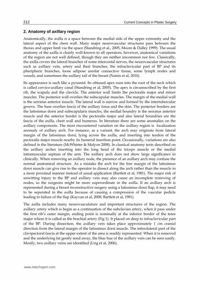
The axilla includes many neurovasculature and important structures of the region. The
axillary artery which is begin as a continuation of the subclavian artery, when it pass under
the first rib’s outer margin, ending point is nominally at the inferior border of the teres
major where it is called as the brachial artery (Fig 1). It placed on deep to infraclavicular part
of the BP. During dissection, the axillary vein takes place approximately 1 cm cranial
direction from the lateral margin of the latissimus dorsi muscle. The inferolateral part of the
clavipectoral fascia at the upper extent of the area is readily represented. When it is removed
and the underlying fat gently send away, the blue hue of the axillary vein can be seen easily.
Mostly, two axillary veins are identified (Ung et al, 2006).
www.intechopen.com
Importance of Anatomical Landmarks on Axillary Neurovascular Territories for Surgery
213
Fig. 1. The subclavian artery and its branches have been demonstrated. (SA: Subclavian artery, STA: Superior thoracic artery, TA: Thoracoacromial artery, PB: Pectoral branch of thoracoacromial artery, AB: Acromial branch of thoracoacromial artery, DB: Deltoid branch of thoracoacromial artery, LTA: Lateral thoracic artery, TDA: Thoracodorsal artery, CSA: Circumflex humeral artery, BA: Brachial artery, ACHA: Anterior circumflex humeral artery PCHA: Posterior circumflex humeral artery, 1: First part of axillary artery, 2: Second part of axillary artery, 3: Third part of axillary artery, I, I, III, IV have been indicated the first, the second, the third and the fourth costae) [Illustrated by Asc.Prof.Edizer M.].
A typical brief definition of the BP is usually found in classical anatomy textbooks as
follows: The BP composed of the anterior primary rami of C4 through T1 spinal nerves. Each
root which innervates a particular myotome and dermatome represent the anterior primary
rami of the spinal nerve. The roots placed between the anterior and middle scalene muscles.
The roots are come together and then form the trunks in the posterior cervical triangle. As
posteriorly and anteriorly divisions are formed by bifurcation of the trunks deep to the
clavicle. Primitive posterior musculature like extensor muscles are innervated by the
posterior divisions of the trunks. The primitive anterior musculature like the flexor muscles
are also innervated by the anterior divisions. The anterior or the posterior divisions come
together and form cords. The cords placed on the axillary region beneath the pectoralis
minor muscle and neighbouring of the axillary artery. The lateral cord which derived C4-C7,
is composed of the joining of the anterior divisions of the upper and middle trunks and is
called for its localization according to the axillary artery. The lateral pectoral nerve (C5-C7),
the musculocutaneous nerve (C4-C6) and lateral cord of the median nerve (C5-C7) derive
from the lateral cord of BP. The medial cord (C8-T1) is continuation as anterior division of
www.intechopen.com
Current Concepts in Plastic Surgery
214
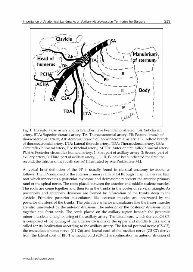
the lower trunk and is called for its localization according to the axillary artery. The medial
pectoral nerve (C8-T1), the medial cord of the median nerve (C6-C8), the ulnar nerve (C8-
T1), the medial antebrachial cutaneous nerve (C8-T1) and the medial brachial cutaneous
nerve (T1) are derived from the medial cord of BP. The posterior cord which is composed of
the fusion of the posterior divisions of the upper, middle and lower trunks, is also called for
its localization according to the axillary artery. The upper subscapular (C5-C6), the middle
subscapular (thoracodorsal) (C6-C8), the lower subscapular (C5-C6), the axillary (C5-C6)
and the radial (C5-T1) nerves come from the posterior trunk of BP (Fig 2).
Fig. 2. The brachial plexus and the peripheral nerves have been demonstrated (The pectoralis minor muscle which is originated from the coracoid process has been shown as projection by dotted line. AA: Axillary artery, AV: Axillary vein, DSN: Dorsal scapular nerve, SSN: Suprascapular nerve, SCN: Subclavius nerve, MCN: Musculocutaneous nerve, MN: Median nerve, RN: Radial nerve, AN: Axillary nerve, UN: Ulnar nerve, LTN: Long thoracic nerve, a: Inferior subscapular nerve, b: Thoracodorsal nerve, c: Superior subscapular nerve, d: Medial cutaneous nerve of forearm, e: Medial cutaneous nerve of arm, f: Superior subscapular nerve) [Illustrated by Asc.Prof. Edizer M.].
Infraclavicular part of BP has complex structure in axillary region. There are many
investigations on some variations in nerve contributions to the BP and vasculature of the
axillary region in literature (Loukas et al, 2010; Shaw et al, 1995). Understanding the variations
in the nerve distributions of the BP may assist both anatomists and surgeons for analysis of
normal anatomy, diagnosis and application of clinical conditions that involve the BP (Fig 3).
www.intechopen.com
Importance of Anatomical Landmarks on Axillary Neurovascular Territories for Surgery
215
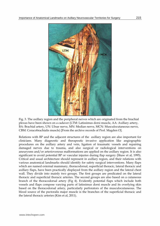
Fig. 3. The axillary region and the peripheral nerves which are originated from the brachial plexus have been shown on a cadaver (LTM: Latissimus dorsi muscle, AA: Axillary artery, BA: Brachial artery, UN: Ulnar nerve, MN: Median nerve, MCN: Musculocutaneous nerve, CBM: Coracobrachialis muscle) [From the archive records of Prof. Magden O].
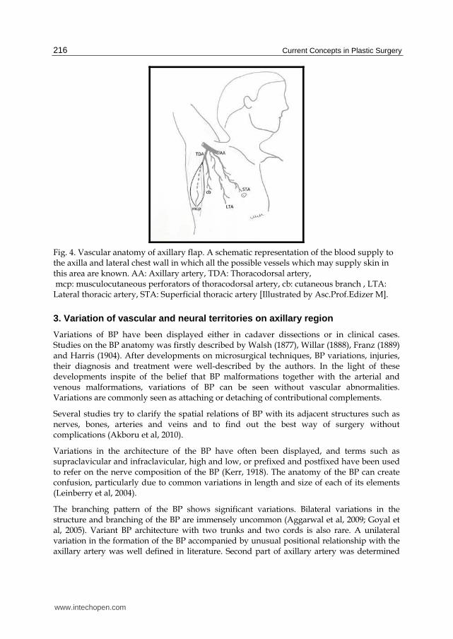
Relations with BP and the adjacent structures of the axillary region are also important for clinicians. Many diagnostic and therapeutic invasive application like angiographic procedures on the axillary artery and vein, ligation of traumatic vessels and repairing damaged nerves due to trauma, and also surgical or radiological interventions on aneurysms and/or arteriovenous malformations are applied on the axillary region. It is also significant to avoid potential BP or vascular injuries during flap surgery (Shaw et al, 1995). Critical and usual architecture should represent in axillary region, and their relations with various anatomical landmarks should identify for safety surgical interventions. Many flaps which are named external mammary, thoracodorsal, superficial thoracic, lateral thoracic and axillary flaps, have been practically displayed from the axillary region and the lateral chest wall. They divide into mainly two groups. The first groups are predicated on the lateral thoracic and superficial thoracic arteries. The second groups are also based on a cutaneous branch of the thoracodorsal artery (Fig 4). Evidently potential flaps which include both vessels and flaps compose varying parts of latissimus dorsi muscle and its overlying skin based on the thoracodorsal artery, particularly perforators of the musculocutaneous. The blood source of the pectoralis major muscle is the branches of the superficial thoracic and the lateral thoracic arteries (Kim et al, 2011).
www.intechopen.com
Current Concepts in Plastic Surgery
216
Fig. 4. Vascular anatomy of axillary flap. A schematic representation of the blood supply to the axilla and lateral chest wall in which all the possible vessels which may supply skin in this area are known. AA: Axillary artery, TDA: Thoracodorsal artery, mcp: musculocutaneous perforators of thoracodorsal artery, cb: cutaneous branch , LTA: Lateral thoracic artery, STA: Superficial thoracic artery [Illustrated by Asc.Prof.Edizer M].
3. Variation of vascular and neural territories on axillary region
Variations of BP have been displayed either in cadaver dissections or in clinical cases. Studies on the BP anatomy was firstly described by Walsh (1877), Willar (1888), Franz (1889) and Harris (1904). After developments on microsurgical techniques, BP variations, injuries, their diagnosis and treatment were well-described by the authors. In the light of these developments inspite of the belief that BP malformations together with the arterial and venous malformations, variations of BP can be seen without vascular abnormalities. Variations are commonly seen as attaching or detaching of contributional complements.
Several studies try to clarify the spatial relations of BP with its adjacent structures such as nerves, bones, arteries and veins and to find out the best way of surgery without complications (Akboru et al, 2010).
Variations in the architecture of the BP have often been displayed, and terms such as supraclavicular and infraclavicular, high and low, or prefixed and postfixed have been used to refer on the nerve composition of the BP (Kerr, 1918). The anatomy of the BP can create confusion, particularly due to common variations in length and size of each of its elements (Leinberry et al, 2004).
The branching pattern of the BP shows significant variations. Bilateral variations in the structure and branching of the BP are immensely uncommon (Aggarwal et al, 2009; Goyal et al, 2005). Variant BP architecture with two trunks and two cords is also rare. A unilateral variation in the formation of the BP accompanied by unusual positional relationship with the axillary artery was well defined in literature. Second part of axillary artery was determined
www.intechopen.com
Importance of Anatomical Landmarks on Axillary Neurovascular Territories for Surgery
217
lying inferomedial to the BP instead of passing between medial and lateral cords. In literature there was some authors observed branching pattern of the subscapular, lateral thoracic, and posterior circumflex humeral arteries, as well as those branches’ topographic relationships to the two terminal branches of the posterior cord of the BP (Olinger & Benninger, 2010).
Generally, the variations in formation, location, and courses of cords of the BP are defined in literature. These variations are divided into three groups. The first group is abnormal location of the cords. The second group is absence of the posterior cord. The third group is abnormal formation and course of the median nerve. As a variation, Pandey et al (2007) declarated absence of the posterior cord in their series and they defined the lateral cord and the medial root of the median nerve had received communicating branches from the posterior cord. However, as a rare variation, absence of the musculocutaneous nerve can be encountered (Song et al, 2003; Gumusburun et al, 2000). A rare constellation of multiple upper limb anomalies were declerated by Wadhwa et al in 2008. Variations in the branching pattern of the posterior cord are clinically important. This knowledge may help the anesthesiologists and the surgeons during operation. It is also significant for avoiding unexpanded injury of the nerves and the axillary artery during blocks, interpreting effects of nervous compressions, while repairing of the plexus injuries and other surgical procedures (Aggarwal et al, 2010; Muthoka et al, 2011; Johnson et al, 2010).
Iatrogenic BP damages have been shown during infraclavicular and transaxillary biopsy, general anesthesia and resection of tumours in axillary region. An ulnar nerve pressure palsy which is the most frequent positioning damage under general anaesthesia, may occure because of malpositioning of the patient. Cause of the damage may include needle trauma and haematoma during central venous catheterization due to neural ischaemia. The vein catheterization with multiple needle passes generally affects BP. Some damages, such as those correlated with uncommon variations, may not be preventable. However, many if not most cases are preventable by the way of a detailed anatomical knowledge on axillary region and displaying of situations in which peripheral nerves are primarily under risk (Zhang et al, 2011; Minville et al, 2006).
Injuries of the BP may affect in the axillary region due to blunt or penetrating traumas. Axillary artery injury might be accompanied with brachial plexus injury because of haematoma (Murata et al, 2008). Although the supraclavicular strecth injuries are more common than infraclavicular stretch injury lesions, the infraclavicular lesions can be treated technically more difficult than the supraclavicular. Because the infraclavicular injuries are related with a higher incidence of vascular and dislocation or fraction damages (Kim et al, 2004).
The variations have also value because a large range of diagnostic or therapeutic invasive procedures are carried out on the axillary artery or its branches (Reid et al, 1984; Mas et al, 2006). Previously, the variations in origins of the lateral thoracic, the superficial thoracic, the thoracodorsal, the axillary arteries were also described by many authors in details (Taylor et al, 1975; Harii et al, 1978; Rowsell et al, 1984; Anson et al, 1939; Ricbourg et al, 1975; Ricbourg 1975; Conink et al, 1976; Bhattacharya et al, 1990; Chandra et al, 1988; De Coninck et al, 1975; Baudet et al, 1976; Irigaray et al, 1979; Cabanié et al, 1980; Yang et al, 1983).
The axillary artery is a continuation of the subclavian artery, originates at the outer margin of the first rib, ending at the distal border of the teres major muscle. The pectoralis minor
www.intechopen.com
Current Concepts in Plastic Surgery
218
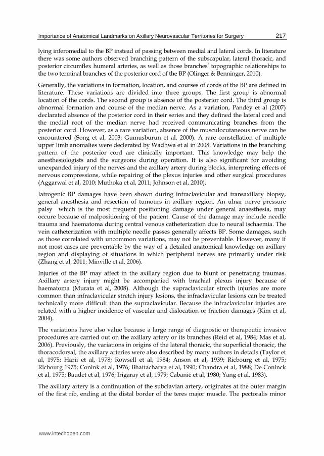
muscle crosses it and divides it into three parts as the first (proximal), the second (posterior) and the third (distal). The first part of the axillary artery is lie between the first rib and the upper margin of the pectoralis minor muscle. The first branch of the first part is the superior thoracic artery which harvests the first and second intercostal space and the upper part of the serratus anterior muscle. It anastomoses with the intercostal arteries. There were many variations on superior thoracic artery in literature. Pandley and Shukla implied that the superior thoracic artery arose from the thoracoacromial trunk in 16.8% cases of the right and 6.1% of the left axilla and the lateral thoracic artery in 39.8% cases of the right and 29.3% of the left axilla. Magden et al (2007) claimed that the superior thoracic artery was found out of the position as a variation. Differ from the knowledge of the classical textbook, it was originated from the first part of the axillary artery as the second branch. Instead of the superior thoracic artery, an aberrant independent origin of the serratus anterior branch as the first branch which originated directly from the first part of the axillary artery was presented in the case (Magden et al, 2007).
A B
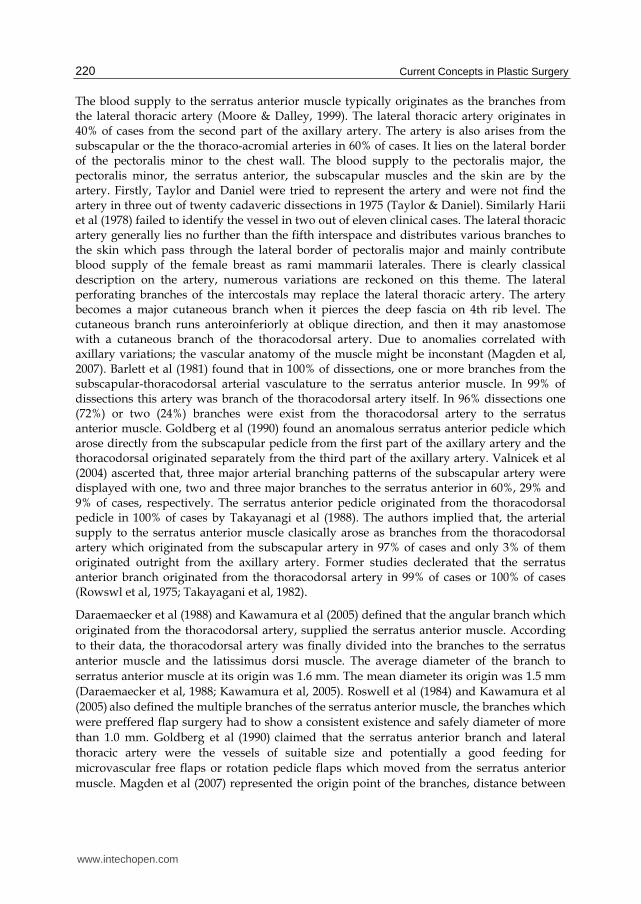
Fig. 5. A-B: Anomalous axillary artery tree has been presented.The serratus anterior vascular branch (BS) as the first branch (newly reported anomaly), the lateral thoracic-thoracodorsal common trunk (TLT) and the circumflex scapular artery arise directly from the axillary artery (CSA). The branches which supply the first external intercostal muscle are indicated by plus signs, the common slip arteries which arise from the serratus branch are indicated by asteriks. AA: Axillary Artery, BS: Branch to Serratus Anterior Muscle, STA: Superior Thoracic Artery, TLT: Lateral Thoracic-Thoracodorsal Trunk, LTA: Lateral Thoracic Artery, TDA: Thoracodorsal Artery, CSA: Circumflex Scapular Artery, LTN: Long Thoracic Nerve, TDN: Thoracodorsal Nerve, ASM: Anterior Serratus Muscle, PMM: Pectoralis Major Muscle, PMiM: Pectoralis Minor Muscle, LDM: Latissimus Dorsi Muscle, SSM: Subscapular Muscle, FR: First Rib, C: Clavicle. [A: From the archive records of Prof. Magden O, the case was published in International Journal of Morphology at 2007, B: Illustrated by Prof.Magden O].
The second part of the axillary artery locates deep to the pectoralis minor muscle. The lateral cord of the BP places on laterally to the artery, the medial cord is medial to it, and the posterior cord is also posterior to it. The second part of the axillary artery has two branches as the thoracoacromial and the lateral thoracic arteries. Generally, the long thoracic artery which is originates directly from the second part of the axillary artery courses along the thoracic wall superficially to the serratus anterior muscle and branches of the blood supply to
www.intechopen.com
Importance of Anatomical Landmarks on Axillary Neurovascular Territories for Surgery
219
the muscle (Moore & Dalley, 1999; Magden et al, 2007). Magden et al defined a common trunk containing the lateral thoracic artery and the thoracodorsal arteries that originate together from the second third of the axillary artery. They suggested to call the arteries “a lateral thoracic- thoracodorsal” common trunk. In literature, the thoracodorsal artery was described different origin and percentage as a branch of the subscapular artery in 97% of the cases by Goldberg et al (1990) and 94% of the cases by Roswell et al or a branch of the lateral thoracic artery in 1% of the cases (Magden et al. 2007). Roswell et al found that in 24% of dissections, the thoracodorsal artery gave two branches to the serratus anterior muscle; one of them was 1 mm in diameter and the other one was on average 2 mm in diameter (Roswell et al. 1984). According to Magden et al all of the branches which harvested to the serratus anterior muscle had diameter of more than 1.0 mm and each had average diameters large enough for anastomosis, so they considered that these branches were safe to use as the pedicles of flaps.
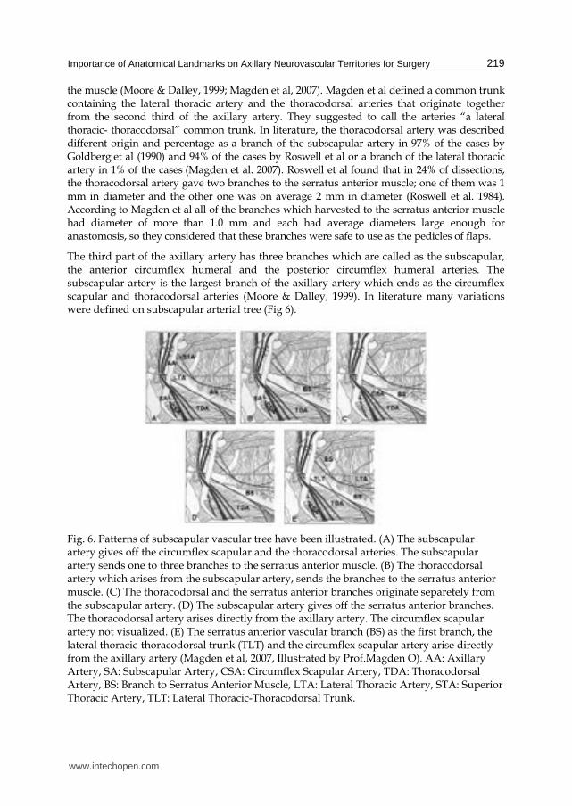
The third part of the axillary artery has three branches which are called as the subscapular, the anterior circumflex humeral and the posterior circumflex humeral arteries. The subscapular artery is the largest branch of the axillary artery which ends as the circumflex scapular and thoracodorsal arteries (Moore & Dalley, 1999). In literature many variations were defined on subscapular arterial tree (Fig 6).
Fig. 6. Patterns of subscapular vascular tree have been illustrated. (A) The subscapular artery gives off the circumflex scapular and the thoracodorsal arteries. The subscapular artery sends one to three branches to the serratus anterior muscle. (B) The thoracodorsal artery which arises from the subscapular artery, sends the branches to the serratus anterior muscle. (C) The thoracodorsal and the serratus anterior branches originate separetely from the subscapular artery. (D) The subscapular artery gives off the serratus anterior branches. The thoracodorsal artery arises directly from the axillary artery. The circumflex scapular artery not visualized. (E) The serratus anterior vascular branch (BS) as the first branch, the lateral thoracic-thoracodorsal trunk (TLT) and the circumflex scapular artery arise directly from the axillary artery (Magden et al, 2007, Illustrated by Prof.Magden O). AA: Axillary Artery, SA: Subscapular Artery, CSA: Circumflex Scapular Artery, TDA: Thoracodorsal Artery, BS: Branch to Serratus Anterior Muscle, LTA: Lateral Thoracic Artery, STA: Superior Thoracic Artery, TLT: Lateral Thoracic-Thoracodorsal Trunk.
www.intechopen.com
Current Concepts in Plastic Surgery
220
The blood supply to the serratus anterior muscle typically originates as the branches from the lateral thoracic artery (Moore & Dalley, 1999). The lateral thoracic artery originates in 40% of cases from the second part of the axillary artery. The artery is also arises from the subscapular or the the thoraco-acromial arteries in 60% of cases. It lies on the lateral border of the pectoralis minor to the chest wall. The blood supply to the pectoralis major, the pectoralis minor, the serratus anterior, the subscapular muscles and the skin are by the artery. Firstly, Taylor and Daniel were tried to represent the artery and were not find the artery in three out of twenty cadaveric dissections in 1975 (Taylor & Daniel). Similarly Harii et al (1978) failed to identify the vessel in two out of eleven clinical cases. The lateral thoracic artery generally lies no further than the fifth interspace and distributes various branches to the skin which pass through the lateral border of pectoralis major and mainly contribute blood supply of the female breast as rami mammarii laterales. There is clearly classical description on the artery, numerous variations are reckoned on this theme. The lateral perforating branches of the intercostals may replace the lateral thoracic artery. The artery becomes a major cutaneous branch when it pierces the deep fascia on 4th rib level. The cutaneous branch runs anteroinferiorly at oblique direction, and then it may anastomose with a cutaneous branch of the thoracodorsal artery. Due to anomalies correlated with axillary variations; the vascular anatomy of the muscle might be inconstant (Magden et al, 2007). Barlett et al (1981) found that in 100% of dissections, one or more branches from the subscapular-thoracodorsal arterial vasculature to the serratus anterior muscle. In 99% of dissections this artery was branch of the thoracodorsal artery itself. In 96% dissections one (72%) or two (24%) branches were exist from the thoracodorsal artery to the serratus anterior muscle. Goldberg et al (1990) found an anomalous serratus anterior pedicle which arose directly from the subscapular pedicle from the first part of the axillary artery and the thoracodorsal originated separately from the third part of the axillary artery. Valnicek et al (2004) ascerted that, three major arterial branching patterns of the subscapular artery were displayed with one, two and three major branches to the serratus anterior in 60%, 29% and 9% of cases, respectively. The serratus anterior pedicle originated from the thoracodorsal pedicle in 100% of cases by Takayanagi et al (1988). The authors implied that, the arterial supply to the serratus anterior muscle clasically arose as branches from the thoracodorsal artery which originated from the subscapular artery in 97% of cases and only 3% of them originated outright from the axillary artery. Former studies declerated that the serratus anterior branch originated from the thoracodorsal artery in 99% of cases or 100% of cases (Rowswl et al, 1975; Takayagani et al, 1982).
Daraemaecker et al (1988) and Kawamura et al (2005) defined that the angular branch which
originated from the thoracodorsal artery, supplied the serratus anterior muscle. According
to their data, the thoracodorsal artery was finally divided into the branches to the serratus
anterior muscle and the latissimus dorsi muscle. The average diameter of the branch to
serratus anterior muscle at its origin was 1.6 mm. The mean diameter its origin was 1.5 mm
(Daraemaecker et al, 1988; Kawamura et al, 2005). Roswell et al (1984) and Kawamura et al
(2005) also defined the multiple branches of the serratus anterior muscle, the branches which
were preffered flap surgery had to show a consistent existence and safely diameter of more
than 1.0 mm. Goldberg et al (1990) claimed that the serratus anterior branch and lateral
thoracic artery were the vessels of suitable size and potentially a good feeding for
microvascular free flaps or rotation pedicle flaps which moved from the serratus anterior
muscle. Magden et al (2007) represented the origin point of the branches, distance between
www.intechopen.com
Importance of Anatomical Landmarks on Axillary Neurovascular Territories for Surgery
221
the branches and the surgical reference points, the diameters, the courses, and supplying
pattern of the serratus anterior muscle in the case report. Five through nine slips were
harvested by one (40%), two (50%), or three (10%) branches from the thoracodorsal artery
(Cuadros et al, 1995). Godat et al (2004) defined a different arterial architecture to the lower
part of the serratus muscle. Basically, a single serratus artery which was the terminal branch
of the thoracodorsal artery had multiply branches, each lying between two neighbourhood
serratus slips. Differ from the knowledge of the classical textbook; Magden et al (2007)
found an aberrant independent origin of the serratus branch which arose directly from the
first part of the axillary artery was presented as a first branch. The inferior part of the
serratus anterior muscle was also harvested by the serratus anterior branch. They also
defined that the superior and middle part of the serratus anterior muscle which was
supplied by the lateral thoracic artery as “a lateral thoracic-thoracodorsal common trunk",
arose from the axillary artery (Magden et al, 2007).
In classical textbook, the subscapular artery, about 4 cm from its origin, it gives off the
circumflex scapular artery, usually larger than continuation of the subscapular artery
(Moore & Dalley, 1999; Williams & Warwick, 1989). Sometimes, the circumflex scapular
artery may originate directly from the axillary artery in 14.7% of cases (Valnicek et al, 2004).
Goldberg et al (1990) noted that the serratus anterior branch arose directly from subscapular
artery from the proximal third of the axillary artery and the thoracodorsal artery originated
separately from the distal third of the axillary artery as variations.
Several authors declerated that a branch or branches to the serratus anterior muscle arose
from the thoracodorsal artery which originated from the subscapular artery in all of the
cases (Tobin et al, 1990; Barlett et al, 19981). Magden et al (2007) declerated that they could
not encountered the subscapular artery in their case as a variation.
The data on the variations is of anatomical and surgical interest. The results on axillary tree
confirm the anatomical safety of the serratus anterior flaps to eradicate ambiguity due to
variations of the axillary artery. Detailed knowledge on variations of axillary architecture
can prevent damage on the branches of the axillary artery during elevating serratus anterior
flaps. It is also important to understand anatomic characteristics on neigbourhood area of
axillary region (Magden et al, 2007). Magden et al offered to great care should be taken
during flap surgery for moving the axillary artery and its branches as completely. The
superficial thoracic artery which is placed on in front of the lateral part of pectoralis major,
is a cutaneous artery of anterolateral thoracic wall. Adachi and Manchot both firstly defined
it (Anson et al, 1939). Anson et al (1939) called it as the accessory lateral thoracic artery but
its course was not same from that of the lateral thoracic in that it was a direct cutaneous
artery purely supplying the skin and run on apex of the lateral part of pectoralis major
muscle. It originated from the proximal part of the brachial artery or distal part of the
axillary arter in the 5 cm segment associated to the lower border of insertion of the tendo
pectoralis major muscle. It might lie down to 6th intercostal space and 26 cm down the
anterior thoracic wall. Rowsell et al described the cutaneous branches which was originating
from directly the axillary artery in 7% of cases in 1984. Salmon (1936) also defined it
originating from the lateral thoracic or the subscapular arteries, but did not see to detect in
excess of 8 cm in length. Harii et al (1978) also described the artery as giving off a cutaneous
branch to the medial side of the upper extremity and the upper lateral quadrant of the breast.
www.intechopen.com
Current Concepts in Plastic Surgery
222
In the literature many authors defined importance of the anatomical landmarks for describing relationship of the BP with the glenoid labrum because the procedures for stabilizing the shoulder and to success the placement of the sutures to the glenoid rim either through the bone or with suture hinges.
According to these cadaveric studies, the musculocutaneous, axillary, and subscapular nerves locate on inferior and lateral to the coracoid process. The musculocutaneous nerve is especially at risk while using the arthroscopy on the shoulder. This nerve originates from the lateral cord of the brachial plexus and pierces the coracobrachialis four to five centimeters inferior to the coracoid process. Lateral and inferior to the coracoid process, the superior and inferior subscapular nerves innervate the subscapularis and the teres major muscles. The brachial plexus and the axillary vessels, placed medial to the coracoid process, have no risk as long as the surgical approach is made lateral to the coracoid process.
Pedicled serratus anterior muscle flap wrapping around the brachial plexus is also used as a treatment for severe axillary neuropathic pain and distress due to neuroma formation. Many surgeons tried to find new techniques with good results for the treatment of the severe neuropathic pain syndromes arise from the brachial plexus. For the brachial plexus reconstruction microneurolysis, interposition nerve grafts, direct end to end repair or vascularized grafting required. In the light of the authors’ experimental studies the techniques to repair injured nerves like neurolysis, nerve grafting, nerve transfer, nerve coaptation, direct muscle neurotization, fascicular transfer and end to side neuroraphy are discussed.
The BP injuries commonly are seen with multisystem trauma. The main mechanism of the BP injury is extreme traction of the nerves or direct impact. Upward traction results generally in the lesion of the lower cervical nerve roots like C8 and T1 whereas the downward traction results in the lesion of the upper cervical nerve roots. Paralysis of the shoulder, arm, and/or hand with parasthesias and altered sensation were the common symptoms of the BP injuries. The temperature and color of the limb may be changed because of the autonomic nervous system damage. The main opinion of the BP injuries treatment depends on the mechanism and the time of the trauma. Acute BP trauma in the axillary region with vascular trauma is a great challenge for the surgeon for restoring the upper extremity function. For these cases interdisciplinary operative and postoperative approach is essential to obtain the best results.
For the diagnostic interventions and surgical procedures variation of the axillary nerves and vessels are also significant for surgeons and clinicians. With varying frequency iatrogenic axillary neurovasculatory injuries during surgical interventions on the shoulder have been described previously. These complications may involve axillary vessels, axillary nerve, median nerve, ulnar nerve, radial nerve and or musculocutaneous nerve.
4. Pedicled flap elevation
Clinically, the branches of the axillary artery used in pedicled flap transferred applications. The lateral thoracic artery originates from the second part of the axillary artery, revert the lateral border of the tendo pectoralis minor muscle and runs the lateral margin of the muscle for nearly 4-5 cm before extend along under of the pectoralis major muscle. Therein, it associated with the pectoral branch of the thoraco-acromial artery and by perforators which
www.intechopen.com
Importance of Anatomical Landmarks on Axillary Neurovascular Territories for Surgery
223
traverse round the lateral margin of pectoralis major muscle and harvest to the lateral part of the breast in female. It may lie along the community between the pectoral branch and the lateral thoracic artery. A pedicled flap from the lateral thoracic wall was well defined and named the lateral thoracic region flap by Bhattacharya (1990). The flap is based on more than just the lateral thoracic artery. There may also be a supply from the acromio-thoracic axis by its pectoral margin of pectoral branch which gives off terminal branches around the lateral margin of pectoralis major to over the superficial fascia and which also association with the lateral thoracic artery.
The surface landmarks of the ventral margin of the flap are of importance. The surgeon should start at the 3rd intercostal space and continue throughout a line dropped vertically downwards from the coracoid process, crossing the infero-lateral margin of the pectoralis major and passing over the serratus anterior and external oblique muscles to a point not more than 3 cm below the costal border. The dorsal incision should be at the 3rd interspace again, at a point 2 cm medial to the lateral margin of latissimus dorsi and runs vertically downwards crossing infero-lateral margin of the muscle and lies to not more than 3 cm below the costal border. The ventral and dorsal borders may be converged by a curve such that the end of the flap follows nearly 5 cm below the costal border in the mid-axillary line.
During elevating the flap special attention is represented the fascia and vessels for up to 1 cm from beneath the infero-lateral margin of pectoralis major muscle.
The subaxillary flap which is named the smaller pedicled flap is used for reconstruction of the opposite hand. The flap is based on a single artery and measures up to 7 cm x 20 cm (Chandra et al, 1988).
The intercostal arteries give off lateral cutaneous branches and posterior branches. Their anterior branches are given off either before or after the lateral branch crosses the deep fascia and lie antero-inferiorly, for a distance of 2-4 cm, along the the fibres of external oblique muscle together with the lateral cutaneous nerves and then distribute under skin. Generally, great variation on the branch is normally seen. The lateral cutaneous branch itself is intensive and is often replaced by many large musculocutaneous perforators. In the 3th, 4th and 5th interspaces the artery are so small as to be not significant. In the lower spaces the perforators are larger but one or more may be absent in which case the perforator in the directly neighbourhood space above or below compensates for the insufficiency. There are also three or four musculocutaneous perforators through external oblique muscle originating from intercostal, subcostal and lumbal arteries.
The thoracoacromial artery originates from the second part of the axillary artery, run round the medial border of tendo pectoralis minor muscle, pierces the claviopectoral fascia. Then it divides into four branches as acromial, clavicular, deltoid and pectoral. The branches supply the ventral part of deltoid muscle, pectoralis major muscle, and the cutaneous tissue in the region over the claviopectoral fascia. The pectoral branch is the largest branch and gives off a small artery to pectoralis minor muscle before run on the deep of pectoralis major muscle after piercing the muscle. In there, it comminicates with branches of the perforators of the internal thoracic artery where have run through the medial ends of the intercostal spaces. Musculocutaneous perforators are sent which supply overlying skin. The acromial or deltoid branches of the thoraco-acromial artery which send a major direct cutaneous branch which supplies out in the underskin fat tissue for an uncertain distance and rarely more
www.intechopen.com
Current Concepts in Plastic Surgery
224
than 10 cm traverse the thoracic wall and 7.5 cm below the clavicle. The branches of the artery which comminicates with those of the 2nd and 3rd intercostal perforators from the internal thoracic artery were encountered in up to nearly 60% of cases. Variations on the branching pattern of the thoracoacromial artery were studied by Reid & Taylor (1984) in over 100 cadaveric specimens. Classically, the subscapular artery originates from the third part of the axillar artery and lie down behind the axillary vein occasionally sends the posterior of circumflex artery. At a level between 0.5 and 5 cm under its origin locus it bifurcates as the circumflex scapular artery and the thoracodorsal artery. The thoracodorsal artery sends a cutaneous branch in nearly 75% of cases, before it pierces and supplies the latissimus dorsi muscle. The cutaneous branch originates between approximately 0.5-2 cm beyond the bifurcation of the subscapular artery. Hence, the utmost feasible length of the vascular pedicle is supplied by the subscapular and thoracodorsal arteries as variable between 1 and 7 cm. A further content to the vascularized pedicle is founded by the cutaneous branch itself, which is either of a long type or a short type, making the total length of the anastomosable vascular trunk between nearly 3-10 cm long with a mean of 6 cm (Olinger & Benninger, 2010; Magden et al, 2007; Valnicek et al, 2004, Reid & Taylor, 1984).
The thoracodorsal artery sends a cutaneous branch which may be used as a feasible flap from the lateral thoracic wall. The termed thoracodorsal axillary flap is developed from both the arterial supply and the topographical site, but flaps based on the artery occasionally termed another name in the literature. The feasibility of harvesting a flap based on the artery was first studied morphologically by De Conick et al (1975) and Taylor & Daniel (1975). Free flaps transfer was firstly defined by Baudet et al (1976) and Irigary et al (1979). The latter case was performed in a child where the authors defined the feasible flap for microvascular transfer of the large size of the thoracodorsal artery.
The subaxillary pedicled flap was found by Cabanie et al (1980). Chandra et al (1988) was firstly standardized the design of a convienent pedicled flap based on the artery. A wider and longer version of the pedicled flap, based on the cutaneous branch of the thoracodorsal artery together with any of the other artery may be present on the lateral thoracic wall. Chandra et al (1988) described that the artery pierces to the deep fascia at the level of the 4th intercostal space in the posterior axillary line; thereafter becoming more superficial as it lies downward nearly 1 cm ventral and parallel to free margin of the latissimus dorsi muscle.
The venous drainage of the flap is by a small cutaneous vein which drains into the two venae comicantes accompaning with the thoracodorsal artery. The nerve innervation of the flap is the lateral cutaneous branches of the 3rd and 4th intercostal nerves. The thoracodorsal nerve which is the motor nerve to latissimus dorsi lies ventral to the thoracodorsal artery (Chandra et al, 1988; Cabanie et al, 1980).
5. Planning surgical procedure
According to surgeons, in planning the axillary pedicled flap, the operator may use the
horizontal continuation of a line drawn through the nipples to surface mark the base of the
flap. Then, the long axis of the flap has its centre line 1 cm ventral and parallel to the margin
of the latissimus dorsi muscle. The largest flap described by Chandra was 7 x 20 cm and all
donor sites enable of direct closure (1988).
www.intechopen.com
Importance of Anatomical Landmarks on Axillary Neurovascular Territories for Surgery
225
Due to variation of the site of the cutaneous branch which may even be not present, it is best to dissect the axilla first and restore the anatomy. If the cutaneous branch is too small or totally absent, then the muscle branch together with a piece of the margin of latissimus dorsi muscle might be needed to support the flap and outline of the flap may be in the dorsal of the positions with a strong cutaneous branch the flap may be positioned more ventrally.
For the cutaneous branch flap, the structure is surrounded by the margin of pectoralis major muscle ventrally, by a line 2 cm medial to the margin of latissimus dorsi muscle dorsally, and by the 8th intercostal space caudally (Chandra et al, 1988; Silverberg et al, 2003).
6. Surgery
There’s different approaches to the axillary region for different pathologies of these area. The main approaches those use at the literature and we use in our daily practice were summerized at this section. After patient placed supine to operating table donor side elevated 450 and patient’s arm abducted 1300 for preparing free skin flap. For exposure of the axillary vessels a mid axillary incision is done. The incision slightly ventrally forms axilla downward along the outerside of the pectoralis major muscle and flap’s anterior border incison follows a line slopping inferiorly and obliqually. The anterior board of the flap lies from the 3rd intercostal space and dropped vertically downwards from coracoid process and then after crossing the inferolateral border of the pectoralis major muscle and reached over the serratus anterior muscle at a point which is not more then 3 cm below the costal margin. At the distal end of the flap the incison curves round and passes up over the edge of latissimus dorsi muscle. The posterior incision starts at a point 2 cm lateral border of latissimus dorsi muscle from the 3rd intercostal space level and passes vertically at a point not extends more then 3 cm below the costal border and downwards crossing the inferolateral border of the muscle. At the mid-axillary line 5 cm below the costal border the anterior and posterior borders of the curve of the flap may be jointed.
Pedicled flap is a different type of axillary flap which has a base at the level of 4th costa is marked by a line to nipples with the arm by the same side and the centre line of the flap is approximately 1 cm ventral to the free edge of the latissimus dorsi muscle.
For the breast cancer cases the axillary dissection elicites detailed information related with
the nerves, vessels, nodal status and the topography of the muscles. Local control of the
axillary disease and establishing of systemic adjuvant therapy are dependent on axillary
surgery additionally. When lymph nodes are removed from the axial region in operations
for primer mammary carsinoma or other pathologies like lymphadenopathy, lipoma,
sabeceous cyst, lipodsytrophy, hidradenitis suppurativa, vascular malformations and
metastatic carcinomas, the anatomical relationship of the vessels and nerves in the axilla are
important, so the positions of major structures are significant for surgeons (De Cholnoky
1951, Silverberg et al, 2003). The favored recipient vessels for microvasculary breast
reconstruction have differency from the thoracodorsal to internal mammary vessels because
of the deep location and bad exposure of the vessels in the axilla and the other technical
difficulties. Many authors generally used the same arm adduction maneuver during
microvascular anastomoses in the axillary region and compared that with conventional
abducted arm position regarding the exposure of the vessels, the operation time and the
position of the surgeon and his assistant (Gravvanis et al, 2008).
www.intechopen.com
Current Concepts in Plastic Surgery
226
Recently improved reconstructive and microsurgical outcomes of the cases with brachial vascular and plexus injuries related traumatic BP palsy, avulsion, rupture, haematomas or tumors on axillary region was mentioned in the literature.
Several anatomical studies and a few surgical studies are determined the anatomical variations of the anterior serratus, the pectoralis major and the latissimus dorsi muscles. During the axillary lymphadenectomy for breast cancer all of these muscle anomalies are significant clinically. If there’s a muscle anomaly pass through the surgical field the surgery may be affected so the border of the surgery can be changed. There have been few studies discovering these anomalous muscles of the axillary region (Natsis et al, 2010).
During axillary lymph nodes dissections due to breast cancer the detailed defination of the anatomical landmarks of the axillary region is very important. Facilitating to access to the axillary fossa in the cases of the tumor, the patient is positioned as supine with a rolled towel under the ipsilateral scapula. For surgical approach there is no consensus for axillary lymph node dissection surgery. The main goal of the lymph node dissection of axillary region is remove all of the lymph node-bearing tissue. This contains all of the lymphatic bearing tissue posterior, anterior, inferior and superior to the axillary vessels. The dissection limits extend approximately at the level of the areola inferiorly from the level of the thoracodorsal nerve insertion to the latissimus dorsi muscle to superiorly the subclavius muscle. The medial limit of the dissection is medial to the edge of the pectoralis minor muscle (the level of rib), extending laterally to the margin of the latissimus dorsi muscle.
There are transverse (a), U-shaped (b) and extended S-shaped (c) incision choices; a; extending form the border of the latissimus dorsi muscle to the edge of the pectoralis major; b or c incisions following the contour of the pectoralis major into the axillary apex and down the border of the latissimus dorsi muscle. C type incision can be done from the behind of the pectoralis major muscle and start from the posterior under the level of the axillary hairline and then inferior of the anterior edge of the latissimus dorsi muscle (Dzwierzynski, 2010).
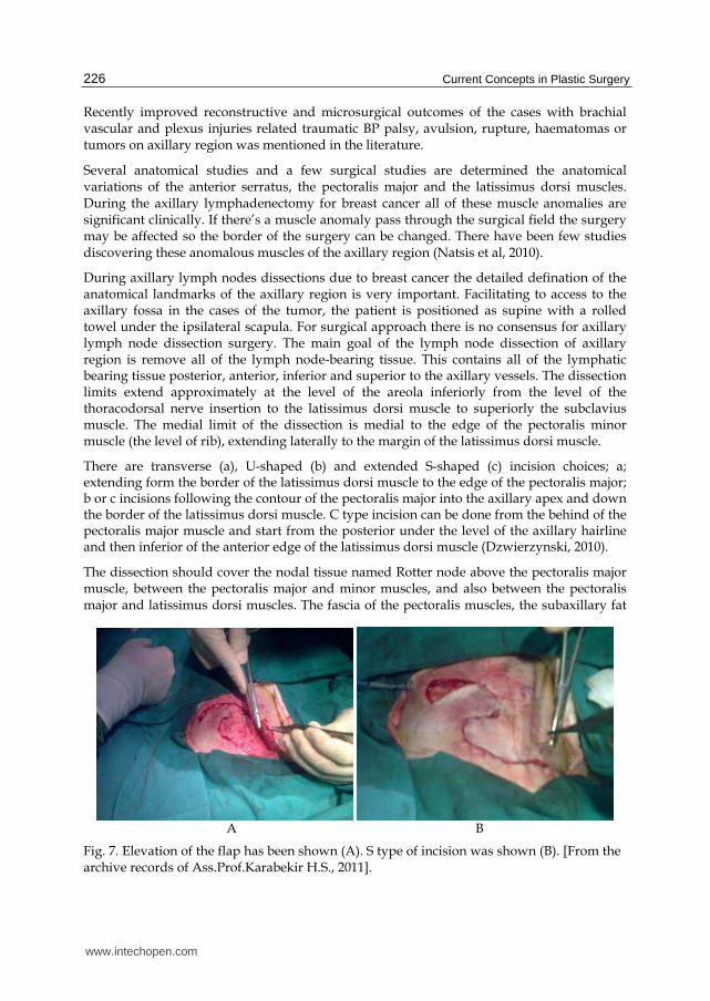
The dissection should cover the nodal tissue named Rotter node above the pectoralis major muscle, between the pectoralis major and minor muscles, and also between the pectoralis major and latissimus dorsi muscles. The fascia of the pectoralis muscles, the subaxillary fat
A B
Fig. 7. Elevation of the flap has been shown (A). S type of incision was shown (B). [From the archive records of Ass.Prof.Karabekir H.S., 2011].
www.intechopen.com
Importance of Anatomical Landmarks on Axillary Neurovascular Territories for Surgery
227
pad, the interpectoral space, and lateral thoracic wall is also removed. The intercostal brachial nerves should be dissected, but the long thoracic and thoracodorsal nerves must be saved unless overly involved in tumor.
The axilla is frequently affected in severe burns that involve the body and the upper arm. Due to its unfavorable contour the acute treatment of the deep axillary burns still occured a difficult problem beacuse of a significant skin graft loss post-burn axillary contractures in the patients. The axillary disabilities deeply affect hand function because it affects the strategic positioning of the hand. Full hand motion is actually useless as the hand can not be positioned for the best function when significant contracture of this joint occurs. Because of the complex anatomy of the axilla which has been characterized as a unique three dimensional pyramid shape, the surgical correction of the axillary burn contracture has remained a forciable challenge. The restoration of the function of axilla is the goal of reconstructive surgery. The axillary surgery also may affect shoulder and hand surgery (Asuku et al, 2008; Larson et al, 1971; Robson & Smith, 1990; Yang, 2005; Hallock, 1993; Button et al, 2010).
Some cadaver studies have implied to define muscles of adjacent area of the axillary region.
They also provide neurovasculary support in their usage as a flap for covering defects of
the region. For flap survival recognition of any vascular and neural variation is very
important to select the appropriate pedicle. In the literature the different type of surgical
approach techniques are described for reaching axillary region. The best known type is
curved upward incion, which is made just under the hairbearing region of the axilla. A
diagonal incision made across the axilla and this incision extended for many centimeters in
the space between the anterior and posterior axillary line, axillary fossa was represented by
protecting neurovascular structures and lymph node of the axilla.
During axillary surgery a subclavicular mid-axillary incision may be performed then the skin and subcutaneous tissue are removed to present the axillary vessels and infraclavicular part of BP after clavipectoral fascia seperated, unilaterally. Dissection is carry on anteriorly and medially behind the anterior axillary fold until the lateral margin of pectoralis major is represented. The lateral border of the latter is easily represented as its muscle bundles lie upward to downward in relation to the horizontally lying fibres of serratus anterior. Although no surgical landmark represents the caudal extent of the dissection, lateral muscle boundry of the latissimus dorsi marks the lateral extent of the dissection. When the intercostobrachial nerve is represented as it crosses the lateral border of latissimus dorsi muscle, the surgeon should be aware that the axillary vein is approximately 1 cm to upward direction. Many times, double axillary veins are seen and the most inferior vessel indicates the limit of the dissection. The fat pad is retracted to inferior while axillar dissection dissected too far. The inferolateral part of the clavipectoral fascia at the upper extention of the dissection is easily seen. When clavipectoral fascia is seperated together with the axillary sheath, the axillary vessels and the brachial plexus are exposed. The musculocutaneous nerve is particularly at risk while arthroscopic approach to the shoulder. This nerve originates from the lateral cord of the brachial plexus and enters the coracobrachialis four to five centimeters distal to the coracoid process. Lateral and inferior to the coracoid process, the superior and inferior subscapular nerves innervate the subscapularis and the teres major muscles. The fragility of the axillary nerve has been well known to during shoulder arthroscopic and open surgical interventions. Iatrogenic injuries to the axillary nerve during
www.intechopen.com
Current Concepts in Plastic Surgery
228
surgical applications on the axillary region have been declerated previously with varying frequency (Lögters et al, 2008). Kulkarni et al (1992) have studied the course of the axillary nerve in the deltoid muscle. The nerve was found to extent 2.2–2.6 cm. above the midpoint on the vertical plane of the deltoid muscle, and parity was drawn to give the certain course of the nerve in this muscle (Kulkarni et al, 1992). Tubbs et al (2001) have described surgical landmarks for the proximal portion of the axillary nerve and defined an anatomical “triangle” within which the axillary nerve was recognized in all cadavers and evaluated the relationship of the axillary nerve with the musculocutaneous nerve. A triangle configuration of anatomic area that contains the axillary neurovascular bundle, containing the proximal part of the axillary nerve was recognized for the anterior approach. The coracoid process constituted the apex of this triangle while the medial rim of the coracobrachialis and the lateral border of the pectoralis minor were developing its lateral and medial margins, respectively.
A virtual horizontal line associating the superior margin of the tendon of the latissimus
dorsi to the coracobrachialis constituted the floor of the triangle. According to result of a
cadaveric study by Apaydin et al (2007), it was suggessted that the axillary nerve was an
average of 3.7 cm. away from the coracoid (Apaydin et al, 2007). The result in concordance
with the results of the study by Lo et al (2004), which defined this distance as mean, 30.3
mm, Tubbs et al (Tubbs et al, 2001; Apaydin et al, 2010; Apaydin et al, 2007). Surgeons
should be taken care, during dissection, to evade injury of the axillary nerve, which pierces
the deep surface of the deltoid approximately five centimeters lateral to the acromion.
The longest distance from the mid-acromion to the lower border of the axillary nerve was
approximately 80-85 milimeters with the arm forward abduct. Some authors claimed that
the distance of the axillary nerve to the mid-acromion in neutral and 90 degree vertical
abduction, and found it to be 61 milimeters and 45 milimeters. Several authors examined the
posterior deltoid splitting approach, and displayed the distance from the axillary nerve to
the posterolateral corner of the acromion to be 65 milimeters in neutral and decreased to 51
milimeters in 90 degree vertical abduction and 46 milimeters in 30 degree extension. They
also found the distances at 45 degree pronation and 45 degree supination to be 62 and 61
milimeters (Tubbs et al, 2001; Apaydin et al, 2010; Robinson et al, 2007; Zlotolow et al, 2006).
It must be noted that smaller branches of the axillary nerve may pierce to the deltoid as close approximately one or one and a half centimeter lateral to acromion. Also the axillary nerve is nearly six centimeters inferior to the apex of the humeral head. The nerve is placed high on the axilla, dorsal to the axillary artery and ventral to the subscapularis muscle. It presents the posterior cord at the level of the lower margin of the pectoralis minor muscle and travers lateral and dorsally. The axillary nerve is initially lateral to the radial nerve, dorsal to the axillary artery, and ventral to the subscapularis muscle; in the lower margin of this muscle, it curves backward and runs through the quadrangular space with the posterior circumflex humeral artery, where it bifurcates anterior and posterior branches.
Alternatively, a long deltopectoral incision may be made from the deltopectoral triangle to the axilla. Then, the skin may be elevated and laid down to both sides by making perpendicular incisions to the first incision. The pectoralis major and the clavicular part of the deltoid are displayed by cutting the deltopectoral fascia. Then the humeral insertion of the pectoralis major is cut and elevated medially. At this step, pectoralis minor and the
www.intechopen.com
Importance of Anatomical Landmarks on Axillary Neurovascular Territories for Surgery
229
axillary neurovascular bundle that is placed lateral to this muscle are exposed. Tendon of the pectoralis minor should be cared for disturbance. Then, the fascicles of the brachial plexus, the axillary vessels, and their branches are represented.
Skin and bony anatomical landmarks of the axillary region have crucial significance for the surgeons who interested in microsurgery and reconstructive surgery.
Our anatomical evaluations and representing objective criterias of the landmarks on the axillary region may support the literature for safety surgical approach.
7. Case report
A 22-year-old woman presented with severe rightsided pain affecting the shoulder, arm and
neck. She also experienced numbness in her rightupper extremity. There was history of
trauma before three months as traffic accident. The patient’s complaints were not alleviated
by analgesic treatment during three months. On physical examination the range of motion
of the shoulder was limited and more painful in overhead movements. On palpation she
had tenderness in the shoulder, axillary region, clavicle and neck. Shoulder and neck
radiographs did not reveal any pathology. Generalized numbness and pain on the right
extremity suggested a probable diagnosis of brachial plexus injury or cervical
radiculopathy. The magnetic resonance imaging (MRI) studies of the shoulder, neck and
cranium did not reveal any pathology, but we detected radicular hyperintensity at C7 and
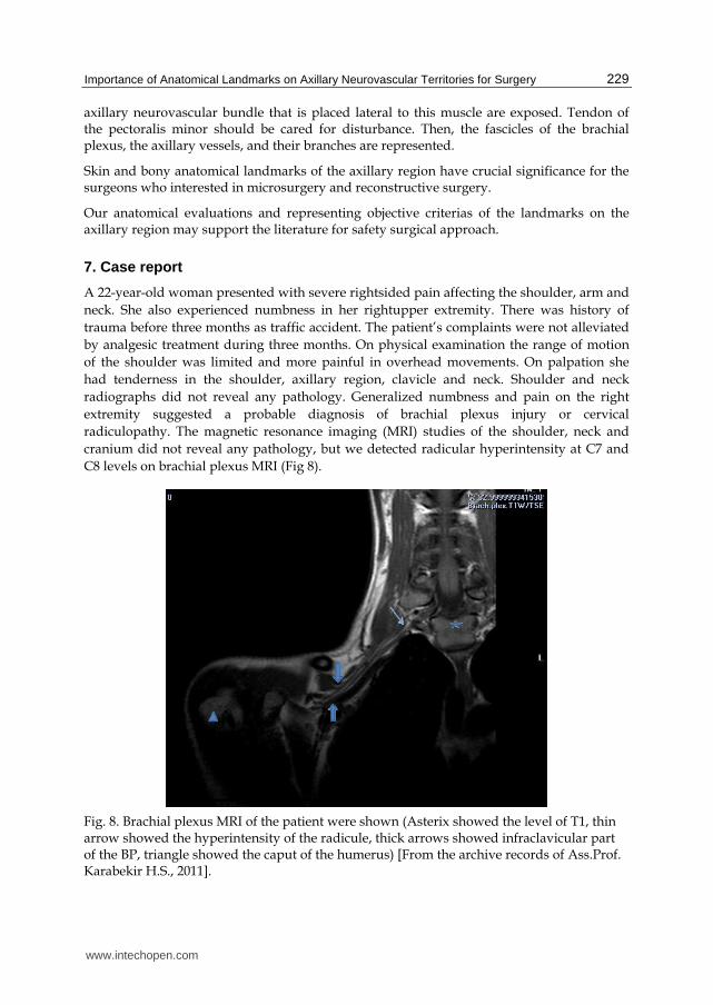
C8 levels on brachial plexus MRI (Fig 8).
Fig. 8. Brachial plexus MRI of the patient were shown (Asterix showed the level of T1, thin arrow showed the hyperintensity of the radicule, thick arrows showed infraclavicular part of the BP, triangle showed the caput of the humerus) [From the archive records of Ass.Prof. Karabekir H.S., 2011].
www.intechopen.com
Current Concepts in Plastic Surgery
230
The hyperintensity revealed as edema. EMG examination revealed as plexopathy. The pain
of the patient diagnosed as neuropathic pain, because of persistant pain during three
mounths. Analgesic, anti-inflammatory and antidepressant therapies were arranged with
the combination of gabapentine. After two weeks medical treatment, the patient had still
severe shoulder, arm and forearm persistant pain. So, operation was offered to the patient,
but she did not accept the surgery. Activity limitation and a new analgesic management
were rearranged.
8. References
Aggarwal, A., Harjeet, K., Sahni, D., Aggarwall, A. (2009). “Bilateral multiple complex
variations in the formation and branching pattern of brachial plexus.” Surg Radiol
Anat. Nov; 31(9):723-31.
Aggarwal, A., Puri, N., Aggarwal, A.K., Harjeet, K., Sahni, D. (2010). “Anatomical variation
in formation of brachial plexus and its branching.” Surg Radiol Anat. Nov; 32(9):891-
4.
Akboru, I.M., Solmaz, I., Secer, H.I., Izci, Y., Daneyemez, M. (2010). “The surgical anatomy
of the brachial plexus.” Turkish Neurosurgery Vol: 20, No: 2, 142-50.
Anson, B.J., Wright, R.R., Wolfer, J.A. (1939). “Blood supply of the mammary gland.”
Surgery, Gynaecology and Obstetrics. 69; 468-73.
Apaydin, N., Uz, A., Bozkurt, M., Elhan, A. (2007). “The anatomic relationships of the
axillary nerve and surgical landmarks for its localization from the anterior aspect of
the shoulder.” Clin Anat. Apr;20(3):273-7.
Apaydin, N., Tubbs, R.S., Loukas, M., Duparc, F. (2010). “Review of the surgical anatomy of
the axillary nerve and the anatomic basis of its iatrogenic and traumatic injury.”
Surg Radiol Anat. Mar; 32(3):193-201.
Asuku, M.E., Ibrahim, A., Ijekeye, F.O. (2008). “Post-burn axillary contractures in pediatric
patients: a retrospective survey of management and outcome.” Burns.
Dec;34(8):1190-95.
Bartlett, S. P., May, J.W., Yaremchuk, M. J. (1981). “The latissimus dorsi muscle: Fresh
cadaver study of the primary neurovascular pedicle.” Plast. Reconstr. Surg. May;
67(5)631-6.
Baudet, J., Guimberteau, J.C., Nascimento, E. (1976). “Successful clinical transfer of two free
thoraco-dorsal axillary flaps.” Plast Reconstr. Surg. Dec; 58(6): 680-8.
Bhattacharya, S., Bhagia, S.P., Bhatnagar, S.K., Chandra, R. (1990). “The lateral thoracic
region flap.” Br J Plast Surg. Mar; 43(5): 162-8.
Button, J., Scott, J., Taghizadeh, R., Weiler-Mithoff, E., Hart, A.M. (2010). “Shoulder function
following autologous latissimus dorsi breast reconstruction. A prospective three
year observational study comparing quilting and non-quilting donor site
techniques.” J Plast Reconstr Aesthet Surg. Sep;63(9):1505-12.
Cabanié, H., Garbé, J.F., Guimberteau, J.C. (1980). “Anatomical basis of the thoracodorsal
axillary flap with respect to its transfer by means of microvascular surgery”.
Anatomia Clinica. 2: 65-73.
Chandra, R., Kumar, P., Abdi, S.H.M. (1988). “The subaxillary pedicled flap.” British Journal
of Plastic Surgery. 41: 69-173.
www.intechopen.com
Importance of Anatomical Landmarks on Axillary Neurovascular Territories for Surgery
231
Conink, A., Vanderlinden, E., Boecks, W. (1976). “The thoracodorsal skin flap: A possible
donor site in distant transfer of island flaps by microvascular anastomosis.” Chir.
Plastica (Berlin); 3: 283-91.
Cuadros, C.L., Driscoll, C.L., Rothkopf, D.M. (1995). “The anatomy of the lower serratus
anterior muscle: a fresh cadaver study.” Plast Reconstr Surg Jan;95(1):93-7.
Daraemaecker, R., Thienen, C. V., Lejour, M., Dor, P. (1988). “The serratus anterior-scapular
free flap: A new osteomuscular unit for reconstruction after radical head and neck
surgery.” In proceedings of the second international conference on head neck
cancer, Boston. Mass, July 31-August 5.
De Cholnoky, T. (1951). “Accessory breast tissue in the axilla”. NY State J Med.
Oct;51(19):2245-48.
De Coninck, A., Boeckx, W., Vanderlinden, E., Claessen, G. (1975). Autocraft with vascular
microsutures. Anatomy of donor site. Ann. Chir. Plast 20(2): 163-70.
Dzwierzynski, W.W. (2010). “Complete lymph node dissection for regional nodal
metastasis.” Clin Plast Surg Jan; 37(1):113-25.
Godat, D.M., Sanger, J.R., Lifchez, S.D., Recinos, R.F., Yan, J.G., Godat, M.R., Ramirez, C.E.,
Matloub, H.S. (2004). “Detailed neurovascular anatomy of the serratus anterior
muscle: implications for a functional muscle flap with multiple independent force
vectors.” Plast Reconstr Surg Jul; 114(1):21-9; discussion 30-1.
Goldberg, J.A., Lineaweaver, W.C., Buncke, H.J. (1990). “An aberrant independent origin of
the serratus anterior pedicle”. Ann Plast Surg Dec; 25(6):487-90.
Goyal, N., Harjeet, Gupta, M. (2005). “Bilateral variant contributions in the formation of
median nerve.” Surg Radiol Anat Dec; 27(6):562-5.
Gravvanis, A., Caulfield, R.H., Ramakrishnan, V., Niranjan, N. (2008). “Recipient vessel
exposure in the axilla during microvascular breast reconstruction.” J Reconstr
Microsurg Nov; 24(8):595-8.
Gumusburun, E., Adiguzel, E. (2000). “A variation of the brachial plexus characterized by
the absence of the musculocutaneous nerve: a case report.” E.Surg Radiol Anat
22(1):63-5.
Hallock, G.G. (1993). “A systematic approach to flap selection for the axillary burn
contracture.” J Burn Care Rehabil May-Jun; 14(3):343-7.
Harii, I., Torii, S., Sekiguchi, J. (1978). “The free lateral thoracic flap.” Plast Reconst Surg
Aug;62(2):212-22.
Irigaray, A., Roncagliolo, A., Fossati, G. (1979). “Transfer of a free lateral thoracic flap in a
child: Case report.” Plast Reconst Surg Aug; 64(2):259-63.
Johnson, E.O., Vekris, M., Demesticha, T., Soucacos, P.N. (2010). “Neuroanatomy of the
brachial plexus: normal and variant anatomy of its formation”. Surg Radiol Anat
Mar; 32(3):291-7.
Kawamura, K., Yajima, H., Kobata, Y., Shigematsu, K., Takakura, Y. (2005). “Anatomy of Y-
shaped configurations in the subscapular arterial system and clinical application to
harvesting flow-through flaps”. Plast Reconstr Surg Sep 15;116(4): 1082-89.
Kerr, A.T. (1918). “The brachial plexus of nerves in man, the variations in its formations and
branches.” Am J Anat 23:285-395.
www.intechopen.com
Current Concepts in Plastic Surgery
232
Kim, J.T., Ng, SW., Naidu, S., Kim, J., Kim, Y.H. (2011). “Lateral thoracic perforator flap:
Additional perforator flap option from the lateral thoracic region” J Plast Reconst
Aesthet Surg Dec; 64(12): 1596-602.
Kim, D.H., Murovic, J.A., Tiel, R.L., Kline, D.G. (2004). “Infraclavicular brachial plexus
stretch injury.” Neurosurg Focus May 15;16(5):E4.
Kulkarni, R.R., Nandedkar, A.N., Mysorekar, V.R. (1992). “Position of the axillary nerve in
the deltoid muscle.” Anat Rec Feb; 232(2):316-7.
Larson, D.L., Abston, S., Evans, E.B., Dobrkovsky, M., Linares, H.A. (1971). “Techniques for
decreasing scar formation and contractures in the burned patient.” J Trauma Oct;
11(10):807-23.
Leinberry, C.F., Wehbé, M.A. (2004). “Brachial plexus anatomy.” Hand Clin Feb; 20(1):1-5.
Lögters, T.T., Wild, M., Windolf, J., Linhart, W. (2008). “Axillary nerve palsy after retrograde
humeral nailing: clinical confirmation of an anatomical fear.” Arch Orthop Trauma
Surg 128 (12):1431–1435.
Magden, O., Gocmen-Mas, N., Caglar, B. (2007). “Multiple Variations in the Axillary Arterial
Tree Relevant to Plastic Surgery: A Case Report”. Int J. Morphol. 25(2):357-61.
McWhirter, D., Malyon, A. (2008). “The axillary arch: a rare but recognised variation in
axillary anatomy.” J Plast Reconstr Aesthet Surg Sep; 61(9):1124-6.
Minville, V., Fourcade, O., Idabouk, L., Claassen, J., Chassery, C., Nguyen, L., Pourrut, J.C.,
Benhamou, D. (2006). “Infraclavicular brachial plexus block versus humeral block
in trauma patients: a comparison of patient comfort”. Anesth Analg Mar; 102(3):912-
5.
Moore, L.K., Dalley, A.F.(1999) Anatomy 4 th.ed. Lippincott Williams & Wilkins. 703.
Murata, K., Maeda, M., Yoshida, A., Yajima, H., Okuchi, K. (2008). “Axillary artery injury
combined with delayed brachial plexus palsy due to compressive hematoma in a
young patient: a case report”. Journal Brachial Plex Peripher Nerve Inj Mar; 28; 3:9.
Muthoka, J.M., Sinkeet, S.R., Shahbal, S.H., Matakwa, L.C., Ogeng'o, J.A. (2011). “Variations
in branching of the posterior cord of brachial plexus in a Kenyan population”. J
Brachial Plex Peripher Nerve Inj Jun 7; 6:1.
Natsis, K., Vlasis, K., Totlis, T., Paraskevas, G., Noussios, G., Skandalakis, P., Koebke, J.
(2010). “Abnormal muscles that may affect axillary lymphadenectomy: surgical
anatomy”. Breast Cancer Res Treat. Feb; 120(1):77-82.
Mas, N., Pelin, C. Zagyapan, R., Bahar, H. (2006). “Unusual relation of the median nerve
with the accessory head of the biceps brachii muscle: An original case report.” Int. J.
Morphol 24(4):561-64.
Olinger, A., Benninger, B. (2010). “Branching patterns of the lateral thoracic, subscapular,
and posterior circumflex humeral arteries and their relationship to the posterior
cord of the brachial plexus”. Clin Anat May; 23(4):407-12.
Pandey, S.K., Shukla, V.K. (2007). “Anatomical variations of the cords of brachial plexus and
the median nerve.” Clin Anat Mar; 20(2):150-56.
Reid, C.D., Taylor, G.I. (1984). “The vascular territory of the acromiothoracic axis.” Br J Plast
Surg 37:194-212.
www.intechopen.com
Importance of Anatomical Landmarks on Axillary Neurovascular Territories for Surgery
233
Ricbourg, B., Lassau, J.P., Violette, A.M., Merland, J.J. (1975). “A propos de l'artère
mammaria externe. Origine, territoire et intérêt pour les transplants cutanés libres.”
Archives d'Anatomie Pathologique 23:317-22.
Ricbourg, B. (1975). "Un nouveau site donneur pour transplant cutanè: le territorie
mammaria externe.” Lettre d'information du Groupe d'Advancement pour la
Microchirurgie 2: 1-9.
Robinson, C.M., Khan, L., Akhtar, A., Whittaker, R. ( 2007). “The extended deltoid-splitting
approach to the proximal humerus” J Orthop Trauma 21:657-62
Robson, M.C., Smith, D.J. (1990). “Burned hand. In: Jurkiewicz MJ, Krizek TJ, Mathes SJ,
Ariyan S, editors.” Plastic Surgery; principles and practice. St. Louis: CV Mosby; 781-
802.
Rowsell, A.R., Davies, D.M., Eisenberg, N., Taylor, G.I. (1984). “The anatomy of the
subscapular-thoracodorsal arteries system: study of 100 cadavers dissections.” Br J
Plast Surg Oct; 37(4): 574-6.
Song, W.C., Jung, H.S., Kim, H.J., Shin, C., Lee, B.Y., Koh, K.S. (2003). “A variation of the
musculocutaneous nerve absent.” Yonsei Med J Dec 30;44(6):1110-3.
Silverberg, M.A., Rahman, M.Z. (2003). “Axillary breast tissue mistaken for suppurative
hidradenitis: an avoidable error.” J Emerg Med Jul; 25(1):51-5.
Standring, S., Ellis, H., Healy, J.C., Johnson, D., Williams, A., Collins, P. (2005). Gray’s
Anatomy (ed 39). London, Churchill Livingstone.
Taylor, G.I., Daniel, R.K. (1975). “The anatomy of several free flap donor sites.” Plast Reconst
Surg Sep; 56(3):243-53.
Takayanagi, S., Ohtsuka, M., Tsukie, T. (1988). “Use of the latissimus dorsi and the serratus
anterior muscles as a combined flap.” Ann Plast Surg Apr; 20(4):333-9.
Tobin, G. R., Moberg, A., Ringberg, A., Netscher, D. (1990). “Mandibular-facial
reconstruction with segmentally split serratus anterior composite flaps.” Clin Plast
Surg Oct; 17(4): 633-72.
Tubbs, R.S., Oakes, W.J., Blount, J.P., Elton, S., Salter, G., Grabb, P.A. (2001). “Surgical
landmarks for the proximal portion of the axillary nerve. Neurosurg Dec; 95(6):998-
1000.
Ung, O., Tan, M., Chua, B., Barraclough, B. (2006). “Complete axillary dissection: a
technique that still has relevance in contemporary management of breast cancer.”
Anz J Surg Jun; 76(6):518-21.
Valnicek, S.M., Mosher, M., Hopkins, J.K., Rockwell, W.B. (2004). “The subscapular arterial
tree as a source of microvascular arterial grafts.” Plast Reconstr Surg Jun;
113(7):2001-05.
Wadhwa, S., Vasudeva, N., Kaul, J.M. (2008). “A rare constellation of multiple upper limb
anomalies.” Folia Morphol (Warsz) Nov; 67(4):236-39.
Williams, P.L. Warwick, R. (1989). Gray’s Anatomy. 37th ed. Churchill-Livingstone, London,
England; 611.
Yang, J-Y. (2005). Reconstruction of axillary contracture. In: McCauley RL, editor. Functional
and aestheticreconstruction of burned patients. New York: Taylor and Francis; 367-78.
Yang, Z.N., Shih, R., Chao, L., Shih, T.S. (1983). “Free transplantation of sub-axillary lateral
thoracodorsal flap in burn surgery.” Burns Incl Therm Inj Feb; 10(3):164-69.
www.intechopen.com

Current Concepts in Plastic Surgery
234
Zhang, J., Moore, A.E., Stringer, M.D. (2011). “Iatrogenic upper limb nerve injuries: a
systematic review.” ANZ J Surg Apr; 81(4); 227-36.
Zlotolow, D.A., Catalano, L.W. 3rd., Barron, O.A., Glickel, S.Z. (2006). “ Surgical exposures
of the humerus.” J Am Acad Orthop Surg Dec; 14(13):754-65.
www.intechopen.com
Current Concepts in Plastic SurgeryEdited by Dr. Frank Agullo
ISBN 978-953-51-0398-1Hard cover, 264 pagesPublisher InTechPublished online 23, March, 2012Published in print edition March, 2012
InTech EuropeUniversity Campus STeP Ri Slavka Krautzeka 83/A 51000 Rijeka, Croatia Phone: +385 (51) 770 447 Fax: +385 (51) 686 166www.intechopen.com
InTech ChinaUnit 405, Office Block, Hotel Equatorial Shanghai No.65, Yan An Road (West), Shanghai, 200040, China
Phone: +86-21-62489820 Fax: +86-21-62489821
Plastic surgery continues to be a rapidly growing field in medicine. There have been multiple recentadvancements in the field. Specifically, there has been a continuously growing interest in fat grafting, bodycontouring, minimally invasive surgery, and plastic surgery education. At the same time, there have beencontinued advances and modifications in surgical techniques, which translate into better and improved resultsfor our patients while increasing safety and efficacy. The title of the book is Current Concepts in PlasticSurgery and, as such, it highlights some of the "hot topics" in recent years. We have invited renownedspecialists from around the world to share their valued expertise and experience. Most of the chapters willexpose the reader to multiple techniques for achieving desired results, with emphasis on the author's preferredmethodology.
How to referenceIn order to correctly reference this scholarly work, feel free to copy and paste the following:
Nuket Gocmen Mas, Hamit Selim Karabekir, Mete Edizer and Orhan Magden (2012). Importance ofAnatomical Landmarks on Axillary Neurovascular Territories for Surgery, Current Concepts in Plastic Surgery,Dr. Frank Agullo (Ed.), ISBN: 978-953-51-0398-1, InTech, Available from:http://www.intechopen.com/books/current-concepts-in-plastic-surgery/importance-of-anatomical-landmarks-on-axillary-neurovascular-territories-for-surgery

© 2012 The Author(s). Licensee IntechOpen. This is an open access articledistributed under the terms of the Creative Commons Attribution 3.0License, which permits unrestricted use, distribution, and reproduction inany medium, provided the original work is properly cited.