Implantation in the resorbed posterior mandible using ... · It was therefore proposed an original...
6
POSEIDO. 2013;1(2) Bypass of the mandibular nerve 125 ISSN 2307-5295, Published by the POSEIDO Organization & Foundation under a Creative Commons Attribution-NonCommercial-NoDerivs 3.0 Unported (CC BY-NC-ND 3.0) License. Clinical case letter Implantation in the resorbed posterior mandible using computerized planning and surgical guides to bypass the mandibular nerve: a 2-year follow-up Ziv Mazor Private Practice, Ra’anana, Israel. Corresponding author: Ziv Mazor, [email protected] Submitted April 9 th , 2013; accepted after minor corrections on April 25 th , 2013. 1. Introduction The treatment of the resorbed posterior mandible remains a challenge in modern implant dentistry. After tooth removal, the centrifuge bone resorption is always very significant, and often accelerated by the mechanical constraints related to removable prostheses. The presence of the large vascular-nervous pedicle - the mandibular alveolar nerve - within the mandible body reduces significantly the volume of implantable bone and remains the main obstacle to implantation in the resorbed posterior mandible. Even if the bone quality at the mandible is often high due to its strong cortical organization, this characteristic becomes a real inconvenient for all bone grafting techniques: it is difficult to promote a full long-term integration of a bone graft on the very cortical mandible bone. As bone regeneration or grafting is difficult in this area, another approach for the treatment of the resorbed mandible is to perform a transposition of the alveolar nerve prior to implant placement [1]. This technique offers good results [2], particularly with the non-traumatic modern instruments such as piezosurgical lancets [3,4], but it remains a difficult technique that requires an experienced skillful surgeon. Moreover, this technique implies the destruction of a bone layer along the mandible (to get access to the nerve) and several cases of mandibular fracture after nerve transposition and implantation were reported in the literature [5,6]. In some cases, another clinical option could be to bypass the mandibular alveolar nerve using an accurate planning of the implant placement around the nerve. This option has never been published or evaluated before, and the objective of this article is to describe a first evaluation of this therapeutic strategy. 2. Materials/methods and results A 68-year old male patient required an oral rehabilitation of the lower right mandible. The patient was in good general condition and non-smoker. The alveolar ridges were already significantly resorbed. After careful X-ray evaluation, it was noticed a residual alveolar bone height of 5-6mm above the mandibular nerve (Figure 1A). With this height, it was uncertain to perform a proper direct implantation above the nerve without damaging the pedicle, even with short implants [7]. Moreover, the presence of a residual second molar with severe periodontal resorption and the general shape of the mandible body reduced even more the bone volume available for a classical implantation in the physiological axis.
Transcript of Implantation in the resorbed posterior mandible using ... · It was therefore proposed an original...

POSEIDO. 2013;1(2) Bypass of the mandibular nerve
125
ISSN 2307-5295, Published by the POSEIDO Organization & Foundation
under a Creative Commons Attribution-NonCommercial-NoDerivs 3.0 Unported (CC BY-NC-ND 3.0) License.
Clinical case letter Implantation in the resorbed posterior mandible using computerized planning and surgical guides to bypass the mandibular nerve: a 2-year follow-up Ziv Mazor Private Practice, Ra’anana, Israel. Corresponding author: Ziv Mazor, [email protected] Submitted April 9th, 2013; accepted after minor corrections on April 25th, 2013.
1. Introduction
The treatment of the resorbed posterior mandible remains a challenge in modern implant dentistry. After tooth removal, the centrifuge bone resorption is always very significant, and often accelerated by the mechanical constraints related to removable prostheses. The presence of the large vascular-nervous pedicle - the mandibular alveolar nerve - within the mandible body reduces significantly the volume of implantable bone and remains the main obstacle to implantation in the resorbed posterior mandible.
Even if the bone quality at the mandible is often high due to its strong cortical organization, this characteristic becomes a real inconvenient for all bone grafting techniques: it is difficult to promote a full long-term integration of a bone graft on the very cortical mandible bone. As bone regeneration or grafting is difficult in this area, another approach for the treatment of the resorbed mandible is to perform a transposition of the alveolar nerve prior to implant placement [1]. This technique offers good results [2], particularly with the non-traumatic modern instruments such as piezosurgical lancets [3,4], but it remains a difficult technique that requires an experienced skillful surgeon. Moreover, this technique implies the destruction of a bone layer along the mandible (to get access to the nerve) and several cases of mandibular fracture after nerve transposition and implantation were reported in the literature [5,6].
In some cases, another clinical option could be to bypass the mandibular alveolar nerve using an accurate planning of the implant placement around the nerve. This option has never been published or evaluated before, and the objective of this article is to describe a first evaluation of this therapeutic strategy.
2. Materials/methods and results A 68-year old male patient required an oral rehabilitation of the lower right mandible. The patient was in good general condition and non-smoker. The alveolar ridges were already significantly resorbed. After careful X-ray evaluation, it was noticed a residual alveolar bone height of 5-6mm above the mandibular nerve (Figure 1A). With this height, it was uncertain to perform a proper direct implantation above the nerve without damaging the pedicle, even with short implants [7]. Moreover, the presence of a residual second molar with severe periodontal resorption and the general shape of the mandible body reduced even more the bone volume available for a classical implantation in the physiological axis.

126 Clinical case letter: Mazor Z (2013)
ISSN 2307-5295, Published by the POSEIDO Organization & Foundation
under a Creative Commons Attribution-NonCommercial-NoDerivs 3.0 Unported (CC BY-NC-ND 3.0) License.
The use of this residual height was however not fully compromised. This patient mandible posterior body had some interesting characteristics in terms of width and shape, as it seemed possible to insert implants with different axes to avoid the main nerve canal (Figure 1B). It was therefore proposed an original treatment plan with a direct implantation of standard implants bypassing the mandibular nerve: in this strategy, the anterior implant could be placed lingual to the nerve and the two distal implants placed buccal to the canal (Figure 1C).
Figure 1. Radiographic analysis and planning. (A) On the CT scanner, the residual bone height above the mandibular nerve was only 5-6mm in the right posterior alveolar ridge. (B) The positioning of each dental implant was modeled with the software, using non conventional implantation axis bypassing the mandibular nerve. (C) The position of the 3 implants was selected on the Simplant software to remain laterally to the mandibular nerve. (D) The axis of each implant was chosen to use all the residual bone volume available and to respect the future orientation of the occlusion forces. (E) The position of the implant collars was also directed by the needs of the future implant-supported bridge. (F) A final surgical guide was then modeled based on the implant and prosthetic digital planning.
POSEIDO. 2013;1(2) Bypass of the mandibular nerve
127
ISSN 2307-5295, Published by the POSEIDO Organization & Foundation
under a Creative Commons Attribution-NonCommercial-NoDerivs 3.0 Unported (CC BY-NC-ND 3.0) License.
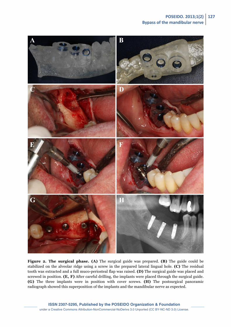
Figure 2. The surgical phase. (A) The surgical guide was prepared. (B) The guide could be stabilized on the alveolar ridge using a screw in the prepared lateral lingual hole. (C) The residual tooth was extracted and a full muco-periosteal flap was raised. (D) The surgical guide was placed and screwed in position. (E, F) After careful drilling, the implants were placed through the surgical guide. (G) The three implants were in position with cover screws. (H) The postsurgical panoramic radiograph showed this superposition of the implants and the mandibular nerve as expected.

128 Clinical case letter: Mazor Z (2013)
ISSN 2307-5295, Published by the POSEIDO Organization & Foundation
under a Creative Commons Attribution-NonCommercial-NoDerivs 3.0 Unported (CC BY-NC-ND 3.0) License.
Figure 3. Follow-up. (A, B, C, D) Three months after surgery, a CT scanner was performed and confirmed the exact positioning of each implant. The distal (A) and central (B) implants were bypassing the nerve in vestibular, the mesial implant was bypassing the nerve in lingual (C). The peri-implant bone tissue was stable (D). (E) The implants were then uncovered and trans-gingival screws were placed prior to prosthetic rehabilitation. (F) Two years after surgery, the clinical and radiological follow-up confirmed a stable clinical result.
The treatment planning was first accurately modeled. Computerized Tomography scanner DICOM data were converted through the Simplant software (Materialise Dental NV, Leuven, Belgium) in order to visualize the treatment planning. The second right molar was scheduled for extraction during the surgical procedure. Three implants of small diameter (3.25mm) and normal length (13mm) could be placed with specific axes around the nerve, by using all the residual bone volume available (Figure 1D). Care was taken to keep axes compatible with the construction of the functional and stable prosthesis (Figure 1E). This model was then used to order a surgical guide (Figure 1F), in order to place the implants exactly in the best position around the nerve and without damaging the mandible cortical plates. The surgical guide included metal sleeves for implant drills and insertion. The guide was also designed with a fixation screw to guaranty the fixation of the guide in correct position during the surgery (Figures 2A, 2B).
Surgical treatment was carried out under conscious sedation. Local anesthesia was administered and full muco-periostal flap was elevated to have a direct access to the underlying bone ridge. The second right molar was extracted and the site was carefully curetted (Figure 2C). The surgical guide was then placed and fixated with a 2mm diameter screw (Figure 2D). Implant osteotomies were done according to the surgical plan. Three implants (Biomet 3I, Palm Beach Gardens, FL, USA) with a diameter of 3.25mm and with Certain internal connection were placed (Figures 2E, 2F, 2G). Standard sutures were done to close the surgical site. Healing was uneventful. No change in the lip or chin sensibility was noticed after surgery. The post-operatory panoramic X-ray showed the superposition of the implants and nerve on the picture (Figure 2H). Three months after surgery, a 3D examination was performed and showed that the implants were indeed in the

POSEIDO. 2013;1(2) Bypass of the mandibular nerve
129
ISSN 2307-5295, Published by the POSEIDO Organization & Foundation
under a Creative Commons Attribution-NonCommercial-NoDerivs 3.0 Unported (CC BY-NC-ND 3.0) License.
axis that was planned on the computer, all around the nerve canal (Figures 3A to 3D). The implants were then uncovered (Figure 3E) for the connection of the trans-gingival screws and the preparation of the implant-supported bridge. Two years after surgery, the clinical and radiographic follow-up confirmed the stability of the implants (Figure 3F).
3. Discussion In this clinical situation, several treatment options could have been proposed.
However, no approach could be considered as a conventional treatment plan, as there is no consensus or perfect solution for these cases. With this kind of residual height, all options remain quite experimental.
The possibility of nerve transposition technique was not indicated here [2] as there was too much bone remaining around the nerve, and the access to the nerve would be too damaging for the mandible: this technique is clearly to be kept for more extreme situations.
The development of short implants is particularly significant in this field nowadays and this approach offers excellent results [7,8]. The use of short implants was possible but limited in this case, as the residual bone heights (with 1mm security above the nerve) along the alveolar ridge were below the minimum 6mm that are required to place properly short implants [7]. With the available heights, it was uncertain to perform a proper direct implantation above the nerve without damaging the pedicle.
The last option would be the addition of bone graft or the bone regeneration before or during implantation. In this case, as most of the bone resorption was in height and not in width, the addition of a bone graft would be even more uncertain, as mandible bone reconstruction in height is often considered the most difficult treatment in implant dentistry. Therefore the use of the computer-based planning was probably the safest approach here.
Computer-based planning has been developed and advocated since many years in this field [9]. It is not only helpful for the accurate placement of implants, but also as an instrument of pre-surgical analysis of bone densities [10] or of pre-modeling and visualization of a complex bone reconstructive surgery [11]. However, its development is limited by the time and cost the use of this technique is requiring. The planning is time-consuming for the practitioner, the software and the order of the custom-made surgical guide are expensive, while the real need of this approach is mostly limited to complex cases. If this approach is interesting for full-arch bridges [12] or the placement of pterygoid or zygoma implants [13], it has a limited relevance for small cases, particularly with experienced practitioners. However in this case, this instrument was very useful to solve a relatively simple but difficult situation.
Even if the CT scan and the software give a feeling of security to the surgeon, it should always be reminded that the accuracy of this planning is not 100% guaranteed, due to the physical limits of the X-ray scanner exam, computer mathematical reconstruction and the potential artifacts that can arise during the phases (for example an imperceptible movement of the patient during the scanner)[14]. Therefore, the guide cannot replace a serious experience and surgical skills in case something unexpected in perceived during the surgery. Moreover it is very important to remain careful and reasonable during the treatment planning and keep adequate distances of securities between the wished implant position and the anatomical structures such as the nerve or the cortical plates. Indeed, the perforation of the cortical plate can also have severe consequences if a hemorrhage under the tongue is provoked.

130 Clinical case letter: Mazor Z (2013)
ISSN 2307-5295, Published by the POSEIDO Organization & Foundation
under a Creative Commons Attribution-NonCommercial-NoDerivs 3.0 Unported (CC BY-NC-ND 3.0) License.
As a conclusion, this innovative approach cannot be used for all cases, but should be kept in mind as a safe option if all other techniques are considered inadequate and if the anatomy of the patient allows this bypass.
Disclosure of interests The author has no conflict of interest to report.
Acknowledgement The author wants to thank Prof. David M. Dohan Ehrenfest for his help during the preparation of this manuscript.
References [1] Jensen J, Reiche-Fischel O, Sindet-Pedersen S. Nerve transposition and implant placement in the atrophic posterior mandibular alveolar ridge. J Oral Maxillofac Surg. 1994;52(7):662-8; discussion 9-70. [2] Lorean A, Kablan F, Mazor Z, Mijiritsky E, Russe P, Barbu H, Levin L. Inferior alveolar nerve transposition and reposition for dental implant placement in edentulous or partially edentulous mandibles: a multicenter retrospective study. Int J Oral Maxillofac Surg. 2013;42(5):656-9. [3] Leclercq P, Zenati C, Amr S, Dohan DM. Ultrasonic bone cut part 1: State-of-the-art technologies and common applications. J Oral Maxillofac Surg. 2008;66(1):177-82. [4] Leclercq P, Zenati C, Dohan DM. Ultrasonic bone cut part 2: State-of-the-art specific clinical applications. J Oral Maxillofac Surg. 2008;66(1):183-8. [5] Kan JY, Lozada JL, Boyne PJ, Goodacre CJ, Rungcharassaeng K. Mandibular fracture after endosseous implant placement in conjunction with inferior alveolar nerve transposition: a patient treatment report. Int J Oral Maxillofac Implants. 1997;12(5):655-9. [6] Karlis V, Bae RD, Glickman RS. Mandibular fracture as a complication of inferior alveolar nerve transposition and placement of endosseous implants: a case report. Implant Dent. 2003;12(3):211-6. [7] Srinivasan M, Vazquez L, Rieder P, Moraguez O, Bernard JP, Belser UC. Survival rates of short (6 mm) micro-rough surface implants: a review of literature and meta-analysis. Clin Oral Implants Res. 2013;In Press. [8] Draenert FG, Sagheb K, Baumgardt K, Kammerer PW. Retrospective analysis of survival rates and marginal bone loss on short implants in the mandible. Clin Oral Implants Res. 2012;23(9):1063-9. [9] Tardieu PB, Vrielinck L, Escolano E, Henne M, Tardieu AL. Computer-assisted implant placement: scan template, simplant, surgiguide, and SAFE system. Int J Periodontics Restorative Dent. 2007;27(2):141-9. [10] Norton MR, Gamble C. Bone classification: an objective scale of bone density using the computerized tomography scan. Clin Oral Implants Res. 2001;12(1):79-84. [11] Pascual D, Dumont N, Breton P, Cheynet F, Chossegros C. [Modelization of bone graft reconstruction for posterior mandibular segment using the Simplant(R) software]. Rev Stomatol Chir Maxillofac. 2012;113(2):108-14. [12] Amorfini L, Storelli S, Romeo E. Rehabilitation of a dentate mandible requiring a full arch rehabilitation. Immediate loading of a fixed complete denture on 8 implants placed with a bone-supported surgical computer-planned guide: a case report. J Oral Implantol. 2011;37(Spec No):106-13. [13] Vrielinck L, Politis C, Schepers S, Pauwels M, Naert I. Image-based planning and clinical validation of zygoma and pterygoid implant placement in patients with severe bone atrophy using customized drill guides. Preliminary results from a prospective clinical follow-up study. Int J Oral Maxillofac Surg. 2003;32(1):7-14. [14] Maloney K, Bastidas J, Freeman K, Olson TR, Kraut RA. Cone beam computed tomography and SimPlant materialize dental software versus direct measurement of the width and height of the posterior mandible: an anatomic study. J Oral Maxillofac Surg. 2011;69(7):1923-9.
This article can be cited as: Mazor Z. Implantation in the resorbed posterior mandible using computerized planning and surgical guides to bypass the mandibular nerve: a 2-year follow-up. POSEIDO. 2013;1(2):125-30.