Implant-supported fixed restoration of post-traumatic mandibular … · The Journal of Advanced...
6
The Journal of Advanced Prosthodontics 67 Implant-supported fixed restoration of post-traumatic mandibular defect accompanied with skin grafting: A clinical report Kwantae Noh, DMD, MSD, PhD, Woo-Jin Choi, DMD, MSD, Ahran Pae*, DMD, MSD, PhD Department of Prosthodontics, School of Dentistry, Kyung Hee University, Seoul, Republic of Korea Traumatic defects are mostly accompanied by hard and soft tissue loss. This report describes the surgical and prosthetic treatment of a patient with post-traumatic mandibular defect. A split-thickness skin graft was performed prior to implant placement and prefabricated acrylic stent was placed to hold the graft in place. The esthetic and functional demands of the patient were fulfilled by implant-supported screw-retained fixed prosthesis using CAD-CAM technology. [ J Adv Prosthodont 2013;5:67-72] KEY WORDS: Dental implants; Skin grafting; Stents; CAD-CAM; Zirconium oxide INTRODUCTION The prosthodontic rehabilitation of partially and com- pletely edentulous patients using implants is a biocompat- ible and predictable treatment modality. 1-3 Dental implants can successfully be used to rehabilitate maxillofacial defects. 4 Defects due to trauma often cause hard and soft tissue loss and result in esthetic problems and functional disorders. 5 Rehabilitation of the traumatic defect is a sig- nificant challenge for the prosthodontist and surgeon. The goals of treatment are restoration of the esthetics, structural integrity, and function. The importance of precision of fit for implant resto- rations has been reported by several authors. 6-8 Framework misfit causes stress to implant frameworks and the surrounding bone. 7 Screw-retained implant pros- thesis frameworks cast by dental alloys involve certain risks such as distortion and stress from heating and cool- ing during lost-wax technique. 7,8 To obtain passive fit, it is Corresponding author: Ahran Pae Department of Prosthodontics, School of Dentistry, Kyung Hee University, 1 Hoegi-Dong, Dongdaemun-Gu, Seoul, 130-701, Republic of Korea Tel. 8229589349: e-mail, [email protected] Received October 10, 2012 / Last Revision December 6, 2012 / Accepted December 12, 2012 © 2013 The Korean Academy of Prosthodontics This is an Open Access article distributed under the terms of the Creative Commons Attribution Non-Commercial License (http://creativecommons. org/licenses/by-nc/3.0) which permits unrestricted non-commercial use, distribution, and reproduction in any medium, provided the original work is properly cited. often necessary to section and solder the framework. Several other methods for improving passive fit were introduced, including modified casting technique, spark- erosion, and laser welding. However, these methods are technique sensitive and may cause errors. Recently, milled zirconium oxide frameworks using computer–aided design and computer-aided manufactur- ing (CAD-CAM) technology are widely used in implant dentistry, which present better fit than framework made by traditional casting techniques. 9 Zirconium oxide has good physicochemical properties such as, low thermal conductivity, high corrosion rsistance, and high flexural strength (900-1200 MPa). 10,11 In addition, zirconium oxide present enhanced biocompatibility. 10-12 The cost related to the considerable use of high noble metal alloys for frame- works can be reduced by using milled zirconium oxide framework with CAD-CAM technology. Problems associ- ated with the casting method, such as distortion and shrinkage also can be avoided. It was reported that a sufficient amount of keratin- ized tissue around implants was important for long-term peri-implant health. 13 Split-thickness skin grafts at the sur- gically reconstructive site are often needed for enhancing the peri-implant soft tissue condition. 14 At the time of surgery, to keep the split thickness skin graft in close approximation to the recipient site during the initial stage is important. 15 Without close contact, the graft may not adhere to the recipient site because a hematoma may form between the skin and periosteum. Therefore, the device or mold to hold skin graft in place, such as bol- sters and stents, are required. 16 http://dx.doi.org/10.4047/jap.2013.5.1.67 http://jap.or.kr J Adv Prosthodont 2013;5:67-72
Transcript of Implant-supported fixed restoration of post-traumatic mandibular … · The Journal of Advanced...

The Journal of Advanced Prosthodontics 67
Implant-supported fixed restoration of post-traumatic mandibular defect accompanied with skin grafting: A clinical report
Kwantae Noh, DMD, MSD, PhD, Woo-Jin Choi, DMD, MSD, Ahran Pae*, DMD, MSD, PhDDepartment of Prosthodontics, School of Dentistry, Kyung Hee University, Seoul, Republic of Korea
Traumatic defects are mostly accompanied by hard and soft tissue loss. This report describes the surgical and prosthetic treatment of a patient with post-traumatic mandibular defect. A split-thickness skin graft was performed prior to implant placement and prefabricated acrylic stent was placed to hold the graft in place. The esthetic and functional demands of the patient were fulfilled by implant-supported screw-retained fixed prosthesis using CAD-CAM technology. [ J Adv Prosthodont 2013;5:67-72]
KEY WORDS: Dental implants; Skin grafting; Stents; CAD-CAM; Zirconium oxide
INTRODUCTION
The prosthodontic rehabilitation of partially and com-pletely edentulous patients using implants is a biocompat-ible and predictable treatment modality.1-3 Dental implants can successfully be used to rehabilitate maxillofacial defects.4 Defects due to trauma often cause hard and soft tissue loss and result in esthetic problems and functional disorders.5 Rehabilitation of the traumatic defect is a sig-nificant challenge for the prosthodontist and surgeon. The goals of treatment are restoration of the esthetics, structural integrity, and function.
The importance of precision of fit for implant resto-ra t ions has been repor ted by severa l authors. 6-8 Framework misfit causes stress to implant frameworks and the surrounding bone.7 Screw-retained implant pros-thesis frameworks cast by dental alloys involve certain risks such as distortion and stress from heating and cool-ing during lost-wax technique.7,8 To obtain passive fit, it is
Corresponding author: Ahran PaeDepartment of Prosthodontics, School of Dentistry, Kyung Hee University, 1 Hoegi-Dong, Dongdaemun-Gu, Seoul, 130-701, Republic of KoreaTel. 8229589349: e-mail, [email protected] October 10, 2012 / Last Revision December 6, 2012 / Accepted December 12, 2012
© 2013 The Korean Academy of ProsthodonticsThis is an Open Access article distributed under the terms of the Creative Commons Attribution Non-Commercial License (http://creativecommons.org/licenses/by-nc/3.0) which permits unrestricted non-commercial use, distribution, and reproduction in any medium, provided the original work is properly cited.
often necessary to section and solder the framework. Several other methods for improving passive fit were introduced, including modified casting technique, spark-erosion, and laser welding. However, these methods are technique sensitive and may cause errors.
Recently, milled zirconium oxide frameworks using computer–aided design and computer-aided manufactur-ing (CAD-CAM) technology are widely used in implant dentistry, which present better fit than framework made by traditional casting techniques.9 Zirconium oxide has good physicochemical properties such as, low thermal conductivity, high corrosion rsistance, and high flexural strength (900-1200 MPa).10,11 In addition, zirconium oxide present enhanced biocompatibility.10-12 The cost related to the considerable use of high noble metal alloys for frame-works can be reduced by using milled zirconium oxide framework with CAD-CAM technology. Problems associ-ated with the casting method, such as distortion and shrinkage also can be avoided.
It was reported that a sufficient amount of keratin-ized tissue around implants was important for long-term peri-implant health.13 Split-thickness skin grafts at the sur-gically reconstructive site are often needed for enhancing the peri-implant soft tissue condition.14 At the time of surgery, to keep the split thickness skin graft in close approximation to the recipient site during the initial stage is important.15 Without close contact, the graft may not adhere to the recipient site because a hematoma may form between the skin and periosteum. Therefore, the device or mold to hold skin graft in place, such as bol-sters and stents, are required.16
http://dx.doi.org/10.4047/jap.2013.5.1.67http://jap.or.kr J Adv Prosthodont 2013;5:67-72

68
This clinical report describes the use of prefabricated acrylic stent after split thickness skin graft and implant-supported screw-retained fixed restorations using CAD-CAM technology of a patient who presented unilateral mandibular defect after trauma.
CASE REPORT
A 54-year-old man was referred to the Department of Prosthodontics at Kyung Hee University to restore the mandibular left missing area. The patient had been in a motor vehicle accident about 20 years ago and had received a number of reconstruction surgeries due to the mandibular traumatic defect of the left side.
Comprehensive intraoral and radiographic examina-tion revealed significant bone loss of the mandibular left side. The floor of the mouth was firm, and lingual vesti-bule of left side was very shallow. The patient also pre-sented shallow mandibular buccal vestibule and insuffi-cient amount of keratinized tissue. The premolars and molars on the left mandible were missing. Mandibular
central, lateral incisor and canine on the left side were shown to be hopeless (Figs. 1, 2).
Hard and soft tissue loss was needed to be restored by prosthetic rehabilitation. Several treatment options were suggested and discussed with the patient. Removable den-tal prosthesis was not considered as a treatment option because of poor tissue condition and lack of support. In spite of alveolar bone loss, inserting dental implants in the alveolar ridge was possible. Also the patient was expecting fixed restoration. Therefore, implant-supported fixed prosthesis was planned. A treatment plan was designed for fabrication of a fixed prosthesis supported by 4 dental implants.
Prior to implant surgery, a split-thickness skin graft combined with vestibuloplasty at the defect site was planned due to lack of keratinized tissue and shallow ves-tibule. To improve graft adherence, an intraoral stent was fabricated. Preliminary cast of type III stone (New Plaston; GC Co., Tokyo, Japan) of the mandible was pre-pared, and the left buccal vestibule was trimmed to the expected depth (Fig. 3). The stent was processed in clear acrylic resin (Vertex Rapid Simplified; Vertex Dental, Zeist, Netherlands), which was extended to the expected vestibular depth (Fig. 4A).
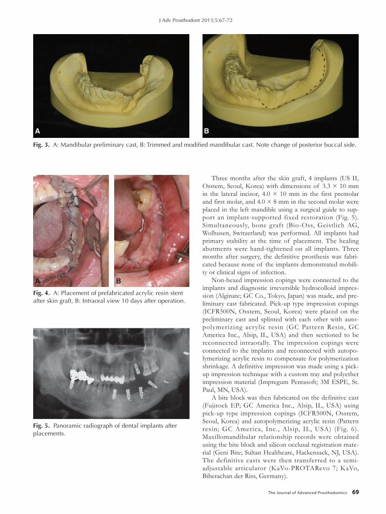
One month after the extraction of mandibular lateral incisor and canine on the left side, a split-thickness skin graft (12 cm × 5 cm) was harvested from the right upper thigh and secured with 3-0 silk sutured. Then stent was positioned on the grafted site and ligatured into the man-dibular anterior teeth to stabilize the stent (Fig. 4B). The patient was in loss of posterior support on the left side for the healing period. This allowed better adaptation and healing of the skin graft to the underlying recipient site. The stent was removed 10 days after operation. There were no complications in the graft recipient or donor sites. The interim denture was then relined with tissue conditioning material (Soft-liner; GC Co., Tokyo, Japan).
Fig. 1. Pretreatment panoramic radiograph. The patient received a reconstructive surgery about 20 years ago due to trauma.
A B
Fig. 2. Pretreatment intraoral condition. A: Frontal view, B: Mandibular occlusal view. Note the shallow buccal vestibule and lack of keratinized tissue of left posterior mandible.
Implant-supported fixed restoration of post-traumatic mandibular defect accompanied with skin grafting: A clinical report
The Journal of Advanced Prosthodontics 69
J Adv Prosthodont 2013;5:67-72
Three months after the skin graft, 4 implants (US II, Osstem, Seoul, Korea) with dimensions of 3.3 × 10 mm in the lateral incisor, 4.0 × 10 mm in the first premolar and first molar, and 4.0 × 8 mm in the second molar were placed in the left mandible using a surgical guide to sup-port an implant-supported fixed restoration (Fig. 5). Simultaneously, bone graft (Bio-Oss, Geistlich AG, Wolhusen, Switzerland) was performed. All implants had primary stability at the time of placement. The healing abutments were hand-tightened on all implants. Three months after surgery, the definitive prosthesis was fabri-cated because none of the implants demonstrated mobili-ty or clinical signs of infection.
Non-hexed impression copings were connected to the implants and diagnostic irreversible hydrocolloid impres-sion (Alginate; GC Co., Tokyo, Japan) was made, and pre-liminary cast fabricated. Pick-up type impression copings (ICFR500N, Osstem, Seoul, Korea) were placed on the preliminary cast and splinted with each other with auto-polymerizing acrylic resin (GC Pattern Resin, GC America Inc., Alsip, IL, USA) and then sectioned to be reconnected intraorally. The impression copings were connected to the implants and reconnected with autopo-lymerizing acrylic resin to compensate for polymerization shrinkage. A definitive impression was made using a pick-up impression technique with a custom tray and polyether impression material (Impregum Pentasoft; 3M ESPE, St. Paul, MN, USA).
A bite block was then fabricated on the definitive cast (Fujirock EP; GC America Inc., Alsip, IL, USA) using pick-up type impression copings (ICFR500N, Osstem, Seoul, Korea) and autopolymerizing acrylic resin (Pattern resin; GC America, Inc., Alsip, IL, USA) (Fig. 6). Maxillomandibular relationship records were obtained using the bite block and silicon occlusal registration mate-rial (Geni Bite; Sultan Healthcare, Hackensack, NJ, USA). The definitive casts were then transferred to a semi-adjustable articulator (KaVo-PROTARevo 7; KaVo, Biberachan der Riss, Germany).
A B
Fig. 3. A: Mandibular preliminary cast, B: Trimmed and modified mandibular cast. Note change of posterior buccal side.
Fig. 5. Panoramic radiograph of dental implants after placements.
Fig. 4. A: Placement of prefabricated acrylic resin stent after skin graft, B: Intraoral view 10 days after operation.
A B

70
Implant-supported fixed restoration of post-traumatic mandibular defect accompanied with skin grafting: A clinical report
Non-hexed temporary abutments (PSM100, Osstem) were connected to the implant analogs (FAM300, FAR300, Osstem) on the definitive cast and full contour wax-up was made. Full contour waxing was then cut back for veneering porcelain and converted into an acrylic res-in framework (GC Pattern resin; GC America Inc.) using putty index (Lab Putty; Contene/Whaledent, Inc., Cuyahoga Falls, OH, USA) to be scanned into the com-puter. This acrylic prosthesis was then copied into zirco-nia prosthesis (Prettau, Zirkonzahn, South Tyrol-Gais, Italy) with a computer-controlled milling machine (Zirkograph 025 ECO, Zirkozahn).
At the next appointment, passive fit of the screw-retained zirconia framework was evaluated intraorally (Fig. 7). Periapical radiograph were made for verifying of the implant-abutment interface. Then the zirconia frame-work was veneered with feldspathic porcelain (Creation Zi-F; Creation Willi Geller Intl GmbH, Meiningen, Austria). Individual cement-type zirconia crowns (LAVA, 3M ESPE, ST. Paul, MN, USA) were designed in the left premolar and first molar region (Fig. 8). Maxillary first molar was made of conventional screw-type metal ceram-ic restoration.
At the appointment of insertion, the definitive screw retained mandibular zirconia prosthesis was evaluated intraorally, and the abutment screws were tightened to the torque of 35 Ncm. The patient was satisfied with the esthetic and functional end results of the definitive resto-rations (Fig. 9).
Fig. 7. Zirconium oxide framework for the mandibular implant-supported prosthesis. Note the increased keratinized tissue and vestibular depth of left buccal side.
Fig. 8. Mandibular screw-retained implant-supported restorations with two cementable zirconium oxide crowns and maxillary implant crown.
A B
Fig. 9. Definitive prosthesis. A: Facial view, B: Occlusal view.
Fig. 6. Bite block was fabricated on definitive cast using pick-up type impression copings and autopolymerizing acrylic resin.
The Journal of Advanced Prosthodontics 71
J Adv Prosthodont 2013;5:67-72
The screw access holes were sealed with photopoly-merizing provisional material (Fermit; Ivoclar Vivadent, Schann, Liechtenstein) and composite resin (Z100 Restorative; 3M ESPE). The single zirconium oxide crowns were cemented to the framework with provisional cement (Tempbond; Kerr Corporation, Orange, CA, USA).
The patient was instructed on maintenance of inter-proximal gingival health with the use of Super-floss (Oral-B Inc., Iowa City, IA, USA) and Water Jet (Water Pik Inc., Fort Collins, CO, USA). The patient presented satisfactory oral hygiene for the 12 months follow-up peri-od. No clinical complications were observed during the follow-up period (Fig. 10).
DISCUSSION
For the surgical procedure of skin graft, intimate contact between the split-thickness skin graft and the underlying periosteum in vestibuloplasty is crucial. Failure of the skin graft may be due to circumstances that negate inti-mate contact of the graft to the recipient site.15 A stent used for the skin graft must hold the split thickness skin graft in close approximation to the underlying periosteum for the initial healing period. Furthermore, minimum bulk is necessary to increase patient’s comfort when the stent is placed. The prefabricated acrylic stent used in this case successfully held the graft in place and patient’s discom-fort was minimal during the initial stage.
Combinations of different restorative materials have been reported to be used in rehabilitation of partially and complete edentulous arches with implant-supported pros-thesis.17-19 Metal-resin implant-fixed complete dental pros-thesis can be used for this case. However, this type of res-toration may pose problems, such as loosening of the acrylic teeth and wear of the occluding surface over time. Replacement of teeth and maintenance of the prostheses are often needed.17
In this case, zirconium oxide milled framework using CAD/CAM technology was used for screw-type implant supported restoration. Although failures in bonding
between core and veneering porcelain have frequently occurred,20 there are advantages of this technology, including reduction in laboratory time and less technique sensitivity compared with traditional lost-wax tech-niques.21 The maxillary first molar was restored with con-ventional screw-type metal ceramic restoration. However, implant-supported fixed prosthesis with long span of the mandibular defect area, as in this situation, often requires prosthetic challenges such as sectioning and soldering. However, these extra procedures may be eliminated with zirconium oxide milled framework with CAD/CAM tech-nology. Esthetics was enhanced as individual cementable all-ceramic restorations were fabricated for implants with unfavorable positions for screw-type restorations. Considering the maintenance of the shallow mandibular vestibule and skin grafted area, rehabilitation with all-ceramic implant-supported prosthesis may provide a high predictability.
CONCLUSION
This clinical report describes the oral rehabilitation of a patient with post-traumatic mandibular defect. The patient underwent split-thickness skin graft, implant placement, and fabrication of implant-supported fixed prostheses using CAD/CAM technology. A multidisci-plinary approach was crucial for the optimum result. The definitive prosthetic rehabilitation improved esthetics and function of the patient and presented favorable prognosis.
REFERENCES
1. Zarb GA, Schmitt A. The longitudinal clinical effectiveness of osseointegrated dental implants: the Toronto study. Part III: Problems and complications encountered. J Prosthet Dent 1990;64:185-94.
2. Zarb GA, Schmitt A. The longitudinal clinical effectiveness of osseointegrated dental implants: the Toronto Study. Part II: The prosthetic results. J Prosthet Dent 1990;64:53-61.
3. Adell R, Eriksson B, Lekholm U, Brånemark PI, Jemt T. Long-term follow-up study of osseointegrated implants in the treatment of totally edentulous jaws. Int J Oral Maxillofac Implants 1990;5:347-59.
4. Landes CA. Zygoma implant-supported midfacial prosthet-ic rehabilitation: a 4-year follow-up study including assess-ment of quality of life. Clin Oral Implants Res 2005;16: 313-25.
5. Tjellstrom A, Jansson K, Brånemark PI. Craniofacial de-fects in advanced osseointegration surgery. In: Worthington P, Brånemark PI, eds. Advanced osseointegration surgery: maxillofacial applications. Chicago; Quintessence; 1992. p. 263-312.
6. Kallus T, Bessing C. Loose gold screws frequently occur in full-arch fixed prostheses supported by osseointegrated im-plants after 5 years. Int J Oral Maxillofac Implants 1994; 9:169-78.
7. Jemt T, Lekholm U. Measurements of bone and frame-
Fig. 10. Panoramic radiograph 12 months after placement of definitive prosthesis.

72
Implant-supported fixed restoration of post-traumatic mandibular defect accompanied with skin grafting: A clinical report
work deformations induced by misfit of implant super-structures. A pilot study in rabbits. Clin Oral Implants Res 1998;9:272-80.
8. Jemt T, Lie A. Accuracy of implant-supported prostheses in the edentulous jaw: analysis of precision of fit between cast gold-alloy frameworks and master casts by means of a three-dimensional photogrammetric technique. Clin Oral Implants Res 1995;6:172-80.
9. Drago C, Saldar riaga RL, Domagala D, Almasri R. Volumetric determination of the amount of misfit in CAD/CAM and cast implant frameworks: a multicenter laboratory study. Int J Oral Maxillofac Implants 2010;25: 920-9.
10. Piconi C, Maccauro G. Zirconia as a ceramic biomaterial. Biomaterials 1999;20:1-25.
11. Manicone PF, Rossi Iommetti P, Raffaelli L. An overview of zirconia ceramics: basic properties and clinical applica-tions. J Dent 2007;35:819-26.
12. Lee BC, Jung GY, Kim DJ, Han JS. Initial bacterial adhe-sion on resin, titanium and zirconia in vitro. J Adv Prosthodont 2011;3:81-4.
13. Schrott AR, Jimenez M, Hwang JW, Fiorellini J, Weber HP. Five-year evaluation of the influence of keratinized mucosa on peri-implant soft-tissue health and stability around im-plants supporting full-arch mandibular fixed prostheses. Clin Oral Implants Res 2009;20:1170-7.
14. Baima RF. Implant-supported restoration of a mandibular reconstruction with an osteocutaneous microvascular free flap: a clinical report. J Prosthodont 1995;4:150-9.
15. Firtell DN, Oatis GW, Curtis TA, Sugg WE Jr. A stent for a split-thickness skin graft vestibuloplasty. J Prosthet Dent 1976;36:204-10.
16. Tomsett KL, Chambers MS, Martin JW, Gillenwater AM, Lemon JC. A technique for the fabrication of an immediate mandibular surgical stent securing a skin graft. J Prosthet Dent 2005;93:395-7.
17. Purcell BA, McGlumphy EA, Holloway JA, Beck FM. Prosthetic complications in mandibular metal-resin im-plant-fixed complete dental prostheses: a 5- to 9-year analy-sis. Int J Oral Maxillofac Implants 2008;23:847-57.
18. Bozini T, Petridis H, Garefis K, Garefis P. A meta-analysis of prosthodontic complication rates of implant-supported fixed dental prostheses in edentulous patients after an ob-servation period of at least 5 years. Int J Oral Maxillofac Implants 2011; 26:304-18.
19. Larsson C, Vult von Steyern P, Nilner K. A prospective study of implant-supported full-arch yttria-stabilized te-tragonal zirconia polycrystal mandibular fixed dental pros-theses: three-year results. Int J Prosthodont 2010;23:364-9.
20. Triwatana P, Nagaviroj N, Tulapornchai C. Clinical perfor-mance and failures of zirconia-based fixed partial dentures: a review literature. J Adv Prosthodont 2012;4:76-83.
21. Reshad M, Cascione D, Aalam AA. Fabrication of the man-dibular implant-supported fixed restoration using CAD/CAM technology: a clinical report. J Prosthet Dent 2009; 102:271-8.


![Evaluation of Two Different Attachment Systems Used with ... · Implant-retained overdentures is extremely valuable [4,5]. Rehabilitation with mandibular implant-tissue-supported](https://static.fdocuments.net/doc/165x107/5e6a2f66f71f2340c57bdc1c/evaluation-of-two-different-attachment-systems-used-with-implant-retained-overdentures.jpg)