IMPACTO DO CONSUMO DE POLPA DE AÇAÍ PARA A...
99
UNIVERSIDADE ESTADUAL DE CAMPINAS Faculdade de Engenharia de Alimentos NATHALIA MEDINA DOS SANTOS IMPACTO DO CONSUMO DE POLPA DE AÇAÍ PARA A PREVENÇÃO DO COMPROMETIMENTO COGNITIVO IMPACT OF AÇAI PULP CONSUMPTION FOR THE PREVENTION OF COGNITIVE IMPAIRMENT CAMPINAS 2018
Transcript of IMPACTO DO CONSUMO DE POLPA DE AÇAÍ PARA A...

1
UNIVERSIDADE ESTADUAL DE CAMPINAS Faculdade de Engenharia de Alimentos
NATHALIA MEDINA DOS SANTOS
IMPACTO DO CONSUMO DE POLPA DE AÇAÍ PARA A PREVENÇÃO DO COMPROMETIMENTO COGNITIVO
IMPACT OF AÇAI PULP CONSUMPTION FOR THE PREVENTION OF COGNITIVE IMPAIRMENT
CAMPINAS 2018
2
NATHALIA MEDINA DOS SANTOS
IMPACTO DO CONSUMO DE POLPA DE AÇAÍ PARA A PREVENÇÃO DO
COMPROMETIMENTO COGNITIVO
IMPACT OF AÇAI PULP CONSUMPTION FOR THE PREVENTION OF COGNITIVE IMPAIRMENT
Dissertação apresentada à Faculdade de Engenharia de Alimentos da Universidade Estadual de Campinas como parte dos requisitos exigidos para a obtenção do título de Mestra em Alimentos e Nutrição, na Área de Nutrição Experimental e aplicada à Tecnologia de Alimentos. Dissertation presented to the Faculty of Food Engineering of the University of Campinas in partial fulfillment of the requirements for the degree of Master in Food and Nutrition the area of Experimental Nutrition and Nutrition applied to Food Technology.
Orientador: Prof. Dr. Mário Roberto Maróstica Júnior ESTE EXEMPLAR CORRESPONDE À VERSÃO FINAL DEFENDIDA PELA ALUNA NATHALIA MEDINA DOS SANTOS E ORIENTADA PELO PROF. DR. MÁRIO ROBERTO MARÓSTICA JÚNIOR
CAMPINAS 2018

Agência(s) de fomento e nº(s) de processo(s): CNPq, 132230/2016-0
Ficha catalográficaUniversidade Estadual de Campinas
Biblioteca da Faculdade de Engenharia de AlimentosClaudia Aparecida Romano - CRB 8/5816
Santos, Nathalia Medina dos, 1992- Sa59i SanImpacto do consumo de polpa de açaí para a prevenção do
comprometimento cognitivo / Nathalia Medina dos Santos. – Campinas, SP :[s.n.], 2018.
SanOrientador: Mário Roberto Maróstica Junior. SanDissertação (mestrado) – Universidade Estadual de Campinas, Faculdade
de Engenharia de Alimentos.
San1. Obesidade. 2. Cognição. 3. Compostos bioativos. 4. Açaí. 5. Hipocampo.
I. Maróstica Junior, Mário Roberto. II. Universidade Estadual de Campinas.Faculdade de Engenharia de Alimentos. III. Título.
Informações para Biblioteca Digital
Título em outro idioma: Impact of açai pulp consumption for the prevention of cognitiveimpairmentPalavras-chave em inglês:ObesityCognitionBioactive compoundsHippocampusÁrea de concentração: Nutrição Experimental e Aplicada à Tecnologia de AlimentosTitulação: Mestra em Alimentos e NutriçãoBanca examinadora:Mário Roberto Maróstica Junior [Orientador]César Renato SartoriElisvânia Freitas dos SantosData de defesa: 22-03-2018Programa de Pós-Graduação: Alimentos e Nutrição
Powered by TCPDF (www.tcpdf.org)

4
COMISSÃO EXAMINADORA
_________________________________________________ Prof. Dr. Mário Roberto Maróstica Júnior – Orientador
Universidade Estadual de Campinas
________________________________________________ Prof. Dr. César Renato Sartori – Membro titular
Universidade Estadual de Campinas
________________________________________________ Profª. Drª. Elisvânia Freitas dos Santos – Membro titular
Universidade Federal de Mato Grosso do Sul
A ata de defesa, com as respectivas assinaturas dos membros, encontra-se no processo de vida acadêmica do aluno.

5
Dedico aos que, como eu, nunca deixaram
de acreditar e lutar pelos seus sonhos.
6
AGRADECIMENTOS
À Deus, por ter me protegido e guiado por todo o meu caminho.
À minha mãe, Ana Cristina, pelo amor incondicional, por ter me dado forças
pra acreditar que eu conseguiria até nos momentos mais difíceis e por lutar todas as
batalhas ao meu lado.
À minha família, por tanto se orgulhar de mim e por toda a compreensão para
lidar com minha ausência.
À minha maior parceira, Ângela Batista, que se dedicou à me ensinar tanto
desde o início, me deu o exemplo de como ser uma profissional e amiga em todos
os momentos. Pela companhia todos os dias e noites no laboratório. Muito obrigada.
Ao meu orientador, Mário Maróstica, por ter confiado um trabalho desta
grandeza em minhas mãos e ter dado todo o suporte que eu precisava para
executá-lo.
Aos professores Maria Alice e César Sartori, por terem aberto as portas de
seus laboratórios para a realização deste trabalho e todas as orientações que
engrandeceram o mesmo.
À todos os membros da banca examinadora por terem dedicado seu tempo
em colaborar com a melhoria deste trabalho.
Às minhas parceiras de laboratório, os dias não seriam os mesmos sem
vocês. Obrigada por todas as risadas ao meu lado e todos os conselhos que me
deram. Espero caminharmos sempre unidas.
À Monique Mendonça, pela paciência, didática e dedicação em me ensinar e
auxiliar nos meses que passei em seu laboratório.
À Soely Reis, nossa técnica do laboratório, por estar sempre disponível para
ajudar em qualquer assunto e pela amizade e parceria nestes anos.
À FEA e à Unicamp por serem a porta de entrada para o meu crescimento
profissional. Ao CNPq por ter subsidiado a minha bolsa de estudos, possibilitando
minha permanência tão longe de casa.
À todos os meus amigos externos à Unicamp, por terem tornado essa
caminhada mais agradável e inesquecível. Perto ou longe, vocês são o meu porto
seguro.
Esta dissertação de mestrado não poderia chegar a bom porto sem o precioso
apoio de todos vocês. Muito obrigada.

7
RESUMO
A epidemia da obesidade vem se tornando cada vez mais um problema de saúde
pública em todo o mundo. Além das consequências à saúde já muito bem
fundamentadas, como as doenças cardiovasculares, alguns tipos de câncer,
síndrome metabólica, diabetes do tipo 2, mais recentemente, também tem sido
associada à insuficiências cognitivas e demência. O consumo de alimentos naturais
seria alternativa para minimizar e/ou reverter os efeitos da dieta contemporânea, rica
em açúcares simples e gordura saturada, que acompanha o ganho de peso da
população mundial. O açaí é um fruto tipicamente brasileiro que ganhou muita
atenção devido ao seu alto potencial antioxidante. O fruto conta com grande
quantidade de compostos bioativos de interesse. Assim, nosso foi investigar estes
compostos e seus efeitos na prevenção da obesidade e doenças associadas, como
a resistência periférica à insulina, e o desempenho cognitivo em camundongos. Para
isto, polpas comerciais de açaí adquiridas em Manaus-AM foram liofilizadas e
analisadas quanto à sua composição de macronutrientes, antioxidantes, compostos
fenólicos, carotenoides e composição lipídica. A polpa seca foi adicionada à dietas
normo e hiperlipídicas e administradas por 10 semanas à camundongos adultos. Na
antepenúltima semana de ensaio os animais passaram por um teste de tolerância à
glicose, na penúltima por um teste de tolerância à insulina e na última semana de
ensaio, por um teste de cognição. Ao final do experimento, os animais foram
sacrificados e foram coletados o soro, tecido adiposo e hipocampo e avaliou-se a
adiposidade, estresse oxidativo, resistência à insulina periférica e via da sinalização
da fosforilação da proteína tau estabilizante de microtúbulo. A dieta hiperlipídica
promoveu maior ganho de peso corporal, maior resistência à insulina periférica,
diminuiu a atividade de enzimas do metabolismo antioxidante, aumentou a
fosforilação da tau no hipocampo e implicou em prejuízo do desempenho cognitivo
dos animais. A suplementação com a polpa de açaí promoveu maior sensibilidade
periférica à insulina nos animais obesos, menor peso relativo dos tecidos adiposos
viscerais, e, apesar de ter sido observada apenas uma tendência de menor
fosforilação da tau, estes animais mostraram melhor desempenho no teste de
reconhecimento de objeto novo. Em conclusão, a suplementação de dietas com a
polpa de açaí preveniu o ganho de gordura visceral, a resistência à insulina e indicou
efeito neuroprotetor, minimizando os déficits cognitivos.

8
ABSTRACT
The obesity epidemy has increasingly become a public health problem throughout
the world. In addition to the already well-founded health consequences such as
cardiovascular diseases, cancer, metabolic syndrome and type 2 diabetes and more
recently, cognitive impairment and dementia have also been associated with
overweight. The consumption of natural foods would be an alternative to minimize
and / or reverse the effects of the contemporary diet, rich in simple sugars and
saturated fatty acids, which follows the weight gain of the world population. Açai is a
Brazilian fruit that has gained a lot of attention due to its high antioxidant potential
and large amount of bioactive compounds. Thus, our objective was to investigate
these compounds and their effects on the prevention of obesity and associated
diseases, such as peripheral insulin resistance and cognitive impairment in mice. For
this purpose, commercial açai pulps purchased in Manaus-AM were lyophilized and
analyzed regarding its composition of macronutrients, antioxidants, phenolic
compounds, carotenoids and fatty acids content. The freezed-dried pulp was added
to normal and high-fat diets and administered to adult mice for 10 weeks. In the
antepenultimate experimental week, the animals underwent a glucose tolerance test,
and in the penultimate week, an insulin tolerance test and in the last week, a
cognition test. At the end of the experiment, animals were sacrificed and the serum,
adipose tissue and hippocampus were collected for adiposity, oxidative stress,
peripheral insulin resistance and phosphorylation of the tau microtubule stabilizing
protein evaluation. The high-fat diet was able to promote greater body weight gain,
greater resistance to peripheral insulin, decreased enzyme activity of the antioxidant
metabolism of the animals, increased tau phosphorylation in the hippocampus,
which implied impaired cognitive performance in the test of recognition of new object
by the animals. However, supplementation with açai pulp improved peripheral insulin
sensitivity in obese animals, lower relative-to-body weight of visceral adipose tissues,
and although only a trend of lower phosphorylation of tau was observed, these
animals showed better performance in the new object recognition test. In conclusion,
dietary supplementation with açai pulp prevented visceral fat gain, peripheral insulin
resistance and indicated neuroprotective effect, preventing cognitive impairment.

9
SUMÁRIO
INTRODUÇÃO GERAL.......................................................................................... 11
OBJETIVOS........................................................................................................... 13
CAPÍTULO 1........................................................................................................... 14
REVISÃO DE LITERATURA.................................................................................. 15
1 OBESIDADE: ETIOLOGIA E CONSEQUÊNCIAS PARA A SAÚDE................. 15
1.1 Fatores epidemiológicos da obesidade........................................................ 15
1.2 Tecido adiposo, inflamação e resistência à insulina..................................... 16
1.3 Produção de adipocinas na obesidade......................................................... 17
2 O PAPEL DA OBESIDADE NO DESENVOLVIMENTO DE DOENÇAS
NEURODEGENERATIVAS................................................................................
18
2.1 Doença de Alzheimer e obesidade............................................................... 18
2.2 Consequências da obesidade nos marcadores cognitivos........................... 19
2.3 Utilização do teste de reconhecimento de objeto novo como medida de
memória em modelos animais............................................................................
23
3 ATUAÇÃO DOS COMPOSTOS BIOATIVOS NA SAÚDE................................. 24
3.1 Frutas vermelhas e seus compostos bioativos............................................. 24
3.2 Mecanismos de ação na resistência à insulina e neurodegeneração.......... 25
4 AÇAÍ................................................................................................................... 27
REFERÊNCIAS...................................................................................................... 31
CAPÍTULO 2 ......................................................................................................... 40
Artigo original: CONSUMO DE POLPA DE AÇAÍ MELHORA FUNÇÕES
COGNITIVAS E SENSIBILIDADE PERIFÉRICA À INSULINA EM
CAMUNDONGOS ALIMENTADOS COM DIETA HIPERLIPÍDICA........................
42
ABSTRACT ........................................................................................................... 43
1 Introduction......................................................................................................... 44
2 Material and methods....................................................................................... 45
2.1 Açai pulp characterization............................................................................ 45
2.1.1 Bioactive compounds................................................................................. 45
2.1.1.1 Non-polar bioactive compounds............................................................. 46
2.1.1.2 Polar bioactive compounds..................................................................... 47
2.1.2 Antioxidant capacity................................................................................... 48

10
2.2 Animal experimentation................................................................................ 49
2.2.1 Novel Object Recognition test................................................................... 50
2.2.2 Glucose Tolerance Test and Insulin Tolerance Test................................. 51
2.2.3 Adiponectin................................................................................................ 52
2.2.4 Antioxidant enzyme activities..................................................................... 52
2.2.5 Western blotting......................................................................................... 52
2.3 Statistics....................................................................................................... 53
3 Results................................................................................................................ 53
3.1 Chemical characterization of açai pulp......................................................... 53
3.2 Mice's food and energy intake, body and tissue weights.............................. 57
3.3 Glucose Tolerance Test, peripheral insulin resistance and fasting
adiponectin......................................................................................................... 58
3.4 Antioxidant defense...................................................................................... 60
3.5 Hippocampal protein quantification............................................................... 60
3.6 Novel Object Recognition............................................................................. 62
4 Discussion.......................................................................................................... 62
5 References......................................................................................................... 67
CAPÍTULO 3........................................................................................................... 80
CONCLUSÃO GERAL............................................................................................ 81
CAPÍTULO 4........................................................................................................... 82
REFERÊNCIAS GERAL......................................................................................... 83
ANEXO 1................................................................................................................ 98
ANEXO 2................................................................................................................ 99
11
INTRODUÇÃO GERAL
O sobrepeso e a obesidade são apontados como problemas de saúde
pública e corroboram com o aumento na prevalência de doenças crônicas
metabólicas e degenerativas em todo o mundo (WHO, 2003). Nos últimos anos, o
aumento da proporção de indivíduos com sobrepeso na população tem crescido de
forma expressiva nas nações desenvolvidas (BHUROSY; JEEWON, 2014). Este
fenômeno tem se replicado nas nações em desenvolvimento, como o Brasil, onde o
percentual dos indivíduos considerados obesos vem crescendo nas últimas décadas
(FERREIRA; MAGALHÃES, 2006).
De acordo com dados da VIGITEL (Vigilância de Fatores de Risco e
Proteção para Doenças Cardiovasculares por Inquérito Telefônico) de 2016, 53,8%
dos brasileiros estão acima do peso e quase 19% já tem um quadro de obesidade
estabelecido, de acordo com o índice de massa corporal (IMC); o número de obesos
no Brasil cresceu 60% em dez anos, passando de 11,8% em 2006 para 18,9% em
2016 e o sobrepeso cresceu 23,6%, passando de 42,6% em 2006 para 53,8% em
2016 (BRASIL, 2017). Estes dados voltam a atenção também para o índice de
mortalidade por doenças crônicas não transmissíveis, que são responsáveis por
72% de óbitos no país, sendo a obesidade importante fator de risco de
desenvolvimento de doenças como o diabetes (Ministério da Saúde, 2015).
Fatores genéticos, ambientais e psicossociais são determinantes no
desenvolvimento da obesidade (KOPELMAN, 2000). No entanto, as mudanças
ocorridas no estilo de vida nos últimos anos sugerem que esta condição se deve
principalmente ao desequilíbrio energético resultante do aporte calórico da dieta
superior às necessidades energéticas individuais, aliado ao sedentarismo
(FRANKLIN; KANALEY, 2009). A alimentação de indivíduos obesos é
majoritariamente baseada em alimentos caracterizados pelo alto teor de gorduras
saturadas, açúcares simples, com pouca ou nenhuma inclusão de vegetais
(BUETTNER et al., 2007; GARDNER; RHODES, 2009). Desta forma, medidas para
controlar a origem destas doenças crônicas por meio da alimentação vêm sendo
cada vez mais o foco de investigações científicas.
A obesidade está associada a um processo inflamatório de baixo grau,
porém crônico, que por sua vez é o principal responsável pela instalação da
resistência à insulina (RI) (POSEY et al., 2009). Danos na sinalização da insulina

12
são fundamentais para o desenvolvimento de diabetes mellitus tipo 2 (DM2), e estão
fortemente associados com a síndrome metabólica (MOLLER; KAUFMAN, 2005),
neurodegeneração, e perdas na cognição, sintomas que predispõe o surgimento da
doença de Alzheimer (CORREIA et al., 2012; STEEN et al., 2005).
As frutas vermelhas ou “berries” possuem uma concentração expressiva
de polifenóis e pigmentos responsáveis por ação anti-obesogênica,
antidiabetogênica e melhora dos quadros de RI e inflamação (DRAGANO et al.,
2013). A capacidade dos compostos fenólicos em atravessarem a barreira
hematoencefálica (BHE) propicia sua atividade neuroprotetora, permitindo ação
direta no tecido nervoso (MILBURY; KALT 2010; SHUKITT-HALE, 2012). A atividade
protetora destes compostos é relacionada, principalmente à sua capacidade de
combater radicais livres e o estresse oxidativo, que está aumentado nos processos
patológicos (KIM et al., 2010; GUO et al., 2012).
O açaí (Euterpe sp.) é um fruto tipicamente brasileiro, mais propriamente
da Amazônia, e vem sendo consumido cada vez mais por todas as outras regiões do
país e no mundo. Muita atenção lhe é dada devido à sua capacidade antioxidante e
possível papel como alimento ou ingrediente funcional (SCHAUSS et al., 2006;
COÏSSON et al., 2005). Esta fruta fornece vários compostos antioxidantes tais como
polifenóis, carotenoides, e ácido ascórbico (SCHAUSS et al., 2006a). Estudos in
vitro demonstraram que por possuir expressiva capacidade antioxidante, o açaí
possui atividade hipocolesterolemiante in vivo, além de inibir a peroxidação lípidica
em seres humanos (SCHAUSS et al., 2006; PACHECO-PALENCIA et al., 2008;
SOUZA et al., 2012; SCHAUSS et al., 2009).
No presente trabalho, realizou-se o estudo da associação da ingestão de
uma dieta hiperlipídica com a polpa de açaí, rica em fibras dietéticas, ácidos graxos
insaturados e polifenóis, que são compostos associados com melhoras de
parâmetros de saúde como exposto acima, com o objetivo de descobrir o potencial
desta fruta em prevenir, além de marcadores metabólicos responsáveis por
obesidade e RI, os fatores de risco para doença de Alzheimer relacionados à
obesidade.

13
OBJETIVOS
O objetivo geral deste trabalho foi investigar em camundongos o efeito da
ingestão de polpa liofilizada de açaí (PLA) sobre o controle e prevenção de eventos
fisiopatológicos relacionados à obesidade, com foco no comprometimento cognitivo.
Como objetivos específicos, este trabalho apresentou os seguintes fins:
• Avaliar características físico-químicas da polpa liofilizada de açaí, bem como
seu valor nutricional e compostos bioativos;
• Comparar efeitos do consumo de dietas normolipídicas e hiperlipídicas sem e
com adição de 2% de PLA;
• Avaliar o efeito funcional da ingestão da PLA sobre a resistência à insulina,
obesidade e cognição e relacionar estes parâmetros;
• Analisar a eficácia do consumo de polpa liofilizada de açaí em atenuar os
efeitos nocivos de dieta rica em gordura sobre o estresse oxidativo e glicemia
dos animais;
• Determinar efeito da suplementação de PLA na prevenção de déficits
cognitivos induzidos pela ingestão de dieta hiperlipídica nas proteínas de uma
das vias de ativação da fosforilação da tau no hipocampo dos animais.

14
CAPÍTULO 1
REVISÃO DE LITERATURA

15
CAPÍTULO 1
REVISÃO DE LITERATURA
1 OBESIDADE: ETIOLOGIA E CONSEQUÊNCIAS PARA A SAÚDE
1.1 Fatores epidemiológicos da obesidade
A obesidade vem sendo considerada por muitos uma epidemia do século
XXI e pode ser definida como um peso corporal desproporcional para a altura com
acumulação excessiva de tecido adiposo, geralmente acompanhada de inflamação
sistêmica leve e crônica (SELLAYAH; CAGAMPANG; COX, 2014). Apesar de ser
uma doença multifatorial, que envolve fatores genéticos e endócrinos, também está
associada a fatores comportamentais como a ingestão excessiva de dietas ricas em
gorduras saturadas e açúcares e o baixo nível de atividade física (MENDONÇA,
2004). Estudos mostram que cerca de 12% da ingestão diária de energia dos
adultos americanos vem de gorduras saturadas e 13% vem dos açúcares
adicionados (MICHA et al., 2014). Este número é significantemente mais alto do que
o recomendado (de 5-10%) pelo Departamento de Agricultura dos EUA (USA, 2015).
Não surpreendentemente, os EUA são hoje o país com maior prevalência de
sobrepeso e obesidade no mundo (OECD, 2016).
Estes dados são preocupantes, sendo a obesidade uma doença crônica
de difícil tratamento e, apesar de décadas de esforços para retardar o progresso da
epidemia, atualmente 39% da população mundial está obesa ou com excesso de
peso (WHO, 2015). No Brasil, pesquisa realizada em 2016 pela VIGITEL (Vigilância
de Fatores de Risco e Proteção para Doenças Cardiovasculares por Inquérito
Telefônico) revela que 53,8% dos brasileiros estão acima do peso e quase 19% já
tem um quadro de obesidade estabelecido, de acordo com o IMC (BRASIL, 2017). A
situação é ainda mais alarmante quando se avalia a evolução do sobrepeso e
obesidade ao longo dos anos (2006 – sobrepeso: 43% e obesidade: 11%), sendo
observado aumento dessas no decorrer do tempo (BRASIL, 2017).
A epidemia de obesidade tem implicações psicossociais e econômicas
relevantes, representando uma importante fonte de custos de Saúde Pública. Em

16
2002, os Estados Unidos tiveram um gasto de em média 92,6 bilhões de dólares
com a obesidade e o sobrepeso, o equivalente a 9% do total das despesas com
saúde no país (NGUYEN; EL-SERAG, 2010). Além de ser por si só, um problema de
saúde pública mundial, as consequencias da obesidade para a saúde são inúmeras.
A obesidade é um fator de risco para diversas doenças como as doenças
cardiovasculares, diversos tipos de câncer, síndrome metabólica, DM2 e, mais
recentemente, também tem sido associada a insuficiências cognitivas e demência
(MILLER; SPENCER, 2014). Desta maneira, diversos estudos vem sendo
elaborados nesta área com a intenção de impulsionar o desenvolvimento de novas
abordagens para a compreensão, prevenção e combate aos fatores de risco para
estas doenças.
1.2 Tecido adiposo, inflamação e resistência à insulina
O tecido adiposo é um órgão endócrino metabolicamente ativo, que se
comunica com o sistema nervoso central (SNC) e com tecidos periféricos por meio
da secreção de adipocinas (p. ex. leptina, adiponectina), e citocinas (p. ex. TNFα, IL-
1β, IL-6 e quimiocinas) que regulam vários processos metabólicos, bem como a
homeostase energética, sensibilização à insulina e resposta imuno-inflamatória
(OTTAVIANI; MALAGOLI; FRANCESCHI, 2011). O consumo calórico excessivo
ocasiona um aumento do acúmulo de triglicerídeos nos adipócitos (hipertrofia) e, em
algumas situações, em número (hiperplasia) dos adipócitos, gerando uma disfunção
nestes. Os adipócitos hipertrofiados secretam agentes pró-inflamatórios promovendo
inflamação sistêmica de baixo grau (KENNEDY et al., 2008).
O aumento de tecido adiposo pode aumentar também a infiltração de
citocinas pró-inflamatórias (p. ex. TNFα, IL-6) após estabelecido quadro inflamatório
no tecido devido à infiltração de macrófagos, característica da obesidade
(OTTAVIANI; MALAGOLI; FRANCESCHI, 2011). Várias serina/treonina quinases
são ativadas por citocinas pró-inflamatórias que, juntamente com os altos níveis de
ácidos graxos livres circulantes, contribuem para a inibição da sinalização da
insulina ao propagar sinais de fosforilação do substrato 1 do receptor da insulina
(IRS-1) em serina (MILANSKI et al., 2012). Sendo assim, o aumento de células
adiposas e a infiltração por macrófagos no tecido adiposo são os principais
17
contribuintes para a inflamação e a resistência à insulina (RI). As citocinas
produzidas podem atingir a circulação e prejudicar a propagação do sinal da insulina
em outros órgãos como fígado e músculos (HOTAMISLIGIL, 2006).
A RI é considerada um estado metabólico no qual há diminuição da
capacidade da insulina endógena ou exógena em estimular a utilização celular de
glicose e manter normais as suas respostas metabólicas (REAVEN, 2005). Como
mecanismo compensatório, as células β-pancreáticas passam a produzir mais
insulina a fim de manter a homeostase glicêmica, ocorrendo a falência progressiva
destas células. A RI, causada pela obesidade, combinada com secreção insuficiente
de insulina pelas células β-pancreáticas são a principal causa de DM2 (JOHNSON;
OLEFSKY, 2013; KAHN; HULL; UTZSCHNEIDER, 2006; KUSMINSKI et al., 2009).
1.3 Produção de adipocinas na obesidade
A leptina regula o comportamento alimentar quando sinaliza estímulos
anorexigênicos (supressores da fome) no hipotálamo e possui ainda características
pró-inflamatórias (FRIEDMAN; HALAAS, 1998; TILG; MOSCHEN, 2006). Na
obesidade a produção das adipocinas encontra-se desequilibrada devido a
inflamação e hipertrofia do tecido (TILG; MOSCHEN, 2006). Os níveis de leptina no
sangue correlacionam-se positivamente com a massa adiposa, sendo que, nos
indivíduos obesos, ocorre uma alta produção de leptina, condição conhecida por
hiperleptinemia, que induz a falhas na sua sinalização, indicando a ocorrência de
resistência à leptina, e consequentemente menor atuação de neurotransmissores
anorexigênicos, agravando o quadro de obesidade (FRIEDMAN; HALAAS, 1998;
MYERS; COWLEY; MÜNZBERG, 2008).
A adiponectina é outra adipocina secretada pelo tecido adiposo. Possui
características anti-inflamatórias, sendo capaz de interagir, em condições normais,
com células imunes como macrófagos, onde suprime a produção e secreção de
TNFα e IL-6 e monócitos, aumentando sua produção de citocinas anti-inflamatórias
(GARAULET et al., 2007). Ela melhora a sensibilidade à insulina, oxidação da
gordura muscular e dissipação energética (JUGE-AUBRY; HENRICHOT; MEIER,
2005). A ação sobre a sensibilidade à insulina da adiponectina pode envolver a
ativação da proteína quinase dependente de AMP (AMPK), conhecida por regular a

18
produção de glicose no fígado pela diminuição da expressão de enzimas da
neoglicogênese (KERSHAW; FLIER, 2004). Os níveis de adiponectina no plasma e
no tecido adiposo diminuem em indivíduos obesos, onde é inibida tanto pelos fatores
pró-inflamatórios, como TNFα e IL-6, bem como por hipóxia e pelo estresse oxidativo
(HOSOGAI et al., 2007).
2 O PAPEL DA OBESIDADE NO DESENVOLVIMENTO DE DOENÇAS NEURODEGENERATIVAS
2.1 Doença de Alzheimer e obesidade
A doença de Alzheimer (DA) é a sexta principal causa de morte nos
Estados Unidos, com cerca de 5,5 milhões de pessoas com a doença (MURPHY et
al., 2016). É o subtipo mais comum de demência, com uma prevalência global de
10-30% na população acima dos 65 anos de idade, representando de 60% a 90% de
todas as demências. Estima-se que havia 46,8 milhões de pessoas em todo o
mundo vivendo com demência em 2015 e que este número alcançará 131,5 milhões
em 2050 (PRINCE et al., 2016). Com o envelhecimento da população e o
consequente aumento na incidência da DA, esta doença tornou-se um desafio para
o sistema de saúde, com um custo estimado anual de US$ 259 bilhões (REITZ;
MAYEUX, 2014).
A DA é neurodegenerativa e progressiva, diagnosticada principalmente
por suas características clínicas, incluindo comprometimento progressivo da função
cognitiva, orientação e atividade motora, distúrbios da fala, dificuldade em
reconhecer ou identificar objetos e pessoas (MASTERS et al., 2015). As
características histopatológicas da DA incluem as chamadas placas senis e os
emaranhados neurofibrilares (NFT) formados, respectivamente, por deposição do
peptídeo amiloide-β agregado (Aβ), um produto de clivagem da proteína precursora
da amiloide (APP), e da agregação da proteína estabilizante de microtúbulo tau (tau)
hiperfosforilada. Juntamente com processos associados, como inflamação e
estresse oxidativo, estas características patológicas contribuem para a perda da
integridade sináptica e neurodegeneração progressiva (BLENNOW; DE LEON;
ZETTERBERG, 2006).

19
A alta ingestão de gordura e a obesidade têm sido associadas com a
patogênese da neurodegeneração e DA (LUCHSINGER et al., 2002). Estudos
sugerem que a obesidade e o aumento do consumo de dietas com alto teor de
gordura aumentam o risco de desenvolvimento de demência (ELIAS et al., 2003;
KALMIJN et al., 1997; SOLFRIZZI; PANZA; CAPURSO, 2003). Em 1990,
Greenwood e Winocur publicaram um dos primeiros estudos que revelou os efeitos
do consumo de dieta rica em ácidos graxos saturados (SFA) na aprendizagem e
memória em ratos (GREENWOOD; WINOCUR, 1990).
A RI e o DM2 estão intimamente relacionados ao surgimento de doenças
neurodegenerativas e ao desenvolvimento de demência, sendo este o principal
motivo pelo qual o excesso de peso e a obesidade são considerados fatores de risco
para o desenvolvimento de DA (CHENG et al., 2012; HENI et al., 2015). Além disso,
níveis elevados de citocinas pró-inflamatórias aumentam a inflamação, o que, por
sua vez, causa déficit cognitivo (UCHOA; MOSER; PIKE, 2016).
2.2 Consequências da obesidade nos marcadores cognitivos
Estudos com modelos de obesidade induzida por dieta em roedores
demonstram comprometimento no desempenho da memória e na aprendizagem
(BOITARD et al., 2014; KOTHARI et al., 2017; SAH et al., 2017). Esses efeitos são
atribuídos principalmente à inflamação e alteração da ação da insulina no cérebro
(DE FELICE; FERREIRA, 2014; PISTELL et al., 2010). A inflamação cerebral é
caracterizada por níveis elevados de citocinas inflamatórias segregadas por células
locais (células da micróglia) (HARRY; KRAFT, 2008; MANDREKAR; LANDRETH,
2010). Além disso, as citocinas pró-inflamatórias secretadas em alta quantidade pelo
tecido adiposo na obesidade podem ter acesso ao cérebro através da BHE e
complementar a resposta inflamatória local, resultando em plasticidade sináptica
reduzida e neurogênese prejudicada (KILIAAN; ARNOLDUSSEN; GUSTAFSON,
2014; ZHENG; ZHOU; WANG, 2016).
As células do cérebro não dependem totalmente de insulina para o
fornecimento de glicose, na medida em que possuem outros meios para sua
obtenção. Em contraste com os tecidos periféricos, o cérebro é considerado um
órgão insensível à insulina porque o GLUT-4 (transportador de glicose dependente

20
de insulina) está presente em baixo nível em algumas regiões do cérebro em
comparação com as outras isoformas, GLUT-1 e GLUT-3, e não parece ser
significativamente regulado pela insulina (BLÁZQUEZ et al., 2014).
Além disso, a insulina é produzida quase exclusivamente pelas células β-
pancreáticas e, sendo uma proteína, supunha-se que não poderia atravessar a BHE,
o que sustentava a ideia de que o SNC era um tecido insensível à insulina (BANKS;
OWEN; ERICKSON, 2012). No entanto, receptores da insulina (IR) e seus
substratos podem ser encontrados em diversas regiões do cérebro. A insulina é
transportada pela BHE por um sistema de transporte saturável. Um número
crescente de funções não metabólicas para a insulina no SNC estão sendo
descobertas em regiões relacionadas com a memória e aprendizagem: hipocampo,
amígdala e córtex frontal (BANKS; OWEN; ERICKSON, 2012; ZHAO; ALKON,
2001). Nestas regiões, o estímulo da insulina é importante para funções como a
sobrevida de neurônios, expressão da tau, metabolismo energético, função
mitocondrial, atividades sinápticas e processo cognitivo (DE LA MONTE, 2008;
MESSIER; TEUTENBERG, 2005).
Em uma condição normal, com a sinalização da insulina, o IRS é
fosforilado em tirosina e transmite sinais intracelulares de ativação da via da
fosfatidil-inositol 3-kinase (PI3K)- proteína kinase B (PKB/AKT). A PI3K estimula o
transporte de glicose e inibe a apoptose ativando PKB/AKT, que inibe a glicogênio
sintase quinase-3β (GSK-3β) em Serina 9, uma importante quinase de fosforilação
da tau (DUDEK; DATTA, 1997; LEE; KIM, 2007). Na RI, o bloqueio da propagação
do sinal da insulina inativa a quinase AKT, que se torna incapaz de fosforilar GSK3-β
e bloquear a sua ação de fosforilar a tau e impedir sua agregação e consequente
formação de NFT (CORREIA et al., 2012; JOLIVALT et al., 2008; SCHUBERT et al.,
2003).
Na obesidade, os níveis elevados de citocinas pró-inflamatórias podem
bloquear a sinalização de insulina intracelular, afetando o IRS-1 ao propagar sinais
de fosforilação em serina (DE FELICE; FERREIRA, 2014). A expressão e/ou ação
diminuída de IRS-1 no hipocampo foram identificadas em indivíduos com DA
(TALBOT et al., 2012). Além disso, indivíduos portadores de DA tem a taxa de
líquido cefalorraquidiano / insulina periférica reduzida em comparação à indivíduos
saudáveis, indicando redução da ação da insulina no SNC (FREIHERR et al., 2013;
21
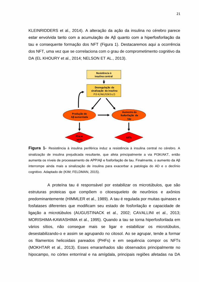
KLEINRIDDERS et al., 2014). A alteração da ação da insulina no cérebro parece
estar envolvida tanto com a acumulação de Aβ quanto com a hiperfosforilação da
tau e consequente formação dos NFT (Figura 1). Destacaremos aqui a ocorrência
dos NFT, uma vez que se correlaciona com o grau de comprometimento cognitivo da
DA (EL KHOURY et al., 2014; NELSON ET AL., 2013).
Figura 1- Resistência à insulina periférica induz a resistência à insulina central no cérebro. A
sinalização de insulina prejudicada resultante, que afeta principalmente a via PI3K/AKT, então
aumenta os níveis de processamento de APP/Aβ e fosforilação de tau. Finalmente, o aumento da Aβ
interrompe ainda mais a sinalização de insulina para exacerbar a patologia do AD e o declínio
cognitivo. Adaptado de (KIM; FELDMAN, 2015).
A proteína tau é responsável por estabilizar os microtúbulos, que são
estruturas proteicas que compõem o citoesqueleto de neurônios e axônios
predominantemente (HIMMLER et al., 1989). A tau é regulada por muitas quinases e
fosfatases diferentes que modificam seu estado de fosforilação e capacidade de
ligação a microtúbulos (AUGUSTINACK et al., 2002; CAVALLINI et al., 2013;
MORISHIMA-KAWASHIMA et al., 1995). Quando a tau se torna hiperfosforilada em
vários sítios, não consegue mais se ligar e estabilizar os microtúbulos,
desestabilizando-o e assim se agrupando no citosol. Ao se agrupar, tende a formar
os filamentos helicoidais pareados (PHFs) e em sequência compor os NFTs
(MOKHTAR et al., 2013). Esses emaranhados são observados principalmente no
hipocampo, no córtex entorrinal e na amígdala, principais regiões afetadas na DA
Resistência à insulina central
Desregulação dasinalização da insulina
PI3-K/Akt/GSK3α/β
Produção de Aβ aumentada
Aumento da fosforilação da
tau
Placas senis
NFTs
22
(LEWIS et al., 2000). O axônio é degenerado com a deterioração do citoesqueleto,
perdendo a capacidade de manter as conexões e sinapses dos neurônios
(MATTSON, 1995).
Os NFTs são uma característica clássica da tauopatia observada não
somente na DA, mas em outras doenças neurodegenerativas como demência
frontotemporal (FTD), doença de Parkinson, entre outras (KOSIK; JOACHIM;
SELKOE, 1986; WILLIAMS, 2006; WOOD et al., 1986). Embora não esteja claro se
as NFTs induzem diretamente a neurodegeneração, sua presença está associada à
morte neuronal (KRIL et al., 2002). Como a sinalização de insulina pode modular a
fosforilação da proteína tau, uma falha da sua ação no cérebro poderia levar à
diminuição da função neuronal e sinaptogênese (KLEINRIDDERS et al., 2014).
Assim, o desenvolvimento de RI especifica do cérebro fundamenta a ideia de que a
doença de Alzheimer seja um terceiro tipo de diabetes (DE LA MONTE, 2008).
Em um mecanismo adicional, falhas na sinalização da leptina também
podem levar à hiperfosforilação da tau e à neurodegeneração (LEE, 2011). Apesar
dos altos níveis periféricos circulantes de leptina, os níveis de leptina do líquido
cefalorraquidiano também parecem estar mais baixos na obesidade, sugerindo o
transporte prejudicado da leptina através da BHE e um mecanismo local de
resistência à leptina (FARR; TSOUKAS; MANTZOROS, 2015). Os receptores de
leptina são encontrados não apenas no hipotálamo, que é o principal sítio de ação
da leptina na regulação do peso corporal, mas também são expressos no córtex e
no hipocampo, duas áreas principais afetadas em DA (HÅKANSSON; MEISTER,
1998). Nas células neuronais, demonstrou-se que a leptina reduz significativamente
a fosforilação de tau em várias regiões vulneráveis do cérebro e melhora a patologia
do cérebro relacionada à Aβ e à tau (GRECO et al., 2008). Em modelos de ratos
com DA, a leptina parece melhorar a neurogênese do hipocampo pela proliferação
de precursores neuronais e atenuar a neurodegeneração induzida por Aβ (GRECO
et al., 2010). Por fim, a leptina parece ter efeitos neurotróficos e neuroprotetores
agudos no modelo de camundongo transgênico APP/PS1 (PÉREZ-GONZÁLEZ et
al., 2011).

23
2.3 Utilização do teste de reconhecimento de objeto novo como medida de memória em modelos animais
Os modelos animais de memória são o objeto de muitas publicações
científicas, pelo menos desde o século XX (GALLAGHER, 1997; MORRIS et al.,
1982). Nos seres humanos, a memória é acessada através de linguagem falada ou
escrita, enquanto que nos animais, as funções cognitivas devem ser acessadas
através de diferentes tipos de comportamentos em muitos modelos específicos de
memória e aprendizado (ANTUNES; BIALA, 2012).
O teste de reconhecimento de objeto novo (NOR) é usado para avaliar a
cognição, particularmente a memória de reconhecimento, em modelos de distúrbios
do SNC em roedores. O teste explora o instinto natural dos roedores por ter
interesse pela novidade e tornou-se um modelo amplamente utilizado para a
investigação de alterações de memória (BAXTER, 2010). A medida do
reconhecimento de objeto é influenciada pela diferença entre o tempo gasto com um
novo objeto e o tempo gasto com um objeto familiar (ENNACEUR; DELACOUR,
1988). O teste é composto por três fases: habituação, familiarização e fase de teste.
Na fase de habituação, cada animal explora livremente uma arena na ausência de
objetos e em seguida retorna para sua gaiola. Durante a fase de familiarização, o
animal é colocado na arena contendo dois objetos idênticos e deixado explorá-los
durante alguns minutos. Para evitar a coerção na exploração dos objetos, os
roedores são colocados de costas para o objeto. Após um intervalo de retenção,
durante a fase de teste, o animal é colocado novamente na arena com dois objetos,
mas agora um é idêntico ao anterior e o outro é novo (Figura 2) (ENNACEUR, 2010;
ENNACEUR; DELACOUR, 1988; GASKIN et al., 2010; HAMMOND; TULL;
STACKMAN, 2004; TAGLIALATELA et al., 2009). Tanto na fase de familiarização
como na fase de teste, os objetos estão localizados em cantos opostos e simétricos
da arena e a localização do objeto novo versus familiar é contrabalançada
(HAMMOND; TULL; STACKMAN, 2004). Os roedores saudáveis passam mais
tempo explorando o novo objeto durante os primeiros minutos da fase de teste em
detrimento do antigo, sendo que se o contrário acontece, há algum indicativo de
comprometimento cognitivo (ENNACEUR, 2010).

24
Figura 2 – Teste de reconhecimento de objetos (NOR): os animais são deixados em uma caixa para
explorar o ambiente livremente sem objetos na fase de habituação; na fase de familiarização dois
objetos idênticos são colocados na arena para que os animais explorem livremente. Na terceira fase
(fase de teste) um dos objetos é trocar por um objeto novo e a preferência por um dos objetos em
relação ao tempo total de exploração é medida.
O hipocampo é fundamental para a memória de reconhecimento de
objetos e, se houver lesões nesta estrutura, ocorrerá comprometimento moderado
da memória anterógrada – a capacidade de armazenar novas informações a partir
de um determinado momento (BROADBENT et al., 2010). Embora o hipocampo não
desempenhe um papel direto na discriminação das diferentes características de
cada objeto, é fundamental como um “detector de novidades” devido ao seu papel
na comparação de informações armazenadas anteriormente com os novos aspectos
recebidos de uma situação particular (CLARKE et al., 2010).
3 ATUAÇÃO DOS COMPOSTOS BIOATIVOS NA SAÚDE
3.1 Frutas vermelhas e seus compostos bioativos
As plantas produzem grande quantidade de compostos fenólicos. De
acordo com as suas estruturas químicas, estes polifenóis são divididos em várias
famílias, uma das quais é a família dos flavonoides (MANACH et al., 2004). As
plantas comestíveis, bem como alimentos e bebidas derivadas delas, proporcionam
à dieta humana generosas quantidades de flavonoides (até 1g/dia), por exemplo
(CROZIER; JAGANATH; CLIFFORD, 2007). Os flavonoides têm uma estrutura
química de base constituída por dois anéis de benzeno ligados através de um anel
de pirano heterocíclico. Os padrões substituintes do grupo hidroxila fornecem os
centros de reação. Os flavonoides podem ser divididos em várias subfamílias de
acordo com o grau de oxidação do heterociclo de oxigênio e os padrões de
25
substituição, por exemplo, flavonas, flavonóis, isoflavonas, antocianinas, flavonoides,
e flavanonas (CROZIER; JAGANATH; CLIFFORD, 2007; JAGANATH; CROZIER,
2011).
As antocianinas são responsáveis pela pigmentação vermelha, azul e
violeta de flores, frutos, folhas, sementes e raízes. Pertencem à grande classe dos
compostos fenólicos, sendo classificadas secundariamente como flavonoides,
devido à estrutura característica de sua cadeia carbônica, na qual dois anéis
aromáticos estão separados por um anel heterocíclico (KONG et al., 2003). São
compostos de rápida absorção e metabolização, associados não só a ações
antioxidantes, como a ações anti-inflamatórias, anti-obesogênicas e sensibilizadora
à insulina in vivo (DRAGANO et al., 2013).
3.2 Mecanismos de ação na resistência à insulina e neurodegeneração
Mecanismos de ação contra a RI: a cianidina-3-glicosídeo (C3G) pode
interferir na produção hepática de glicose, que é um fator importante no diabetes.
Reduz a expressão de TNF-α e outras citocinas pró-inflamatórias no fígado e tecido
adiposo, além de inibir a ativação de JNK. Ao aumentar a fosforilação de AKT, a
C3G estimula a diminuição na produção hepática de glicose pela diminuição de
FoxO1 no núcleo dos adipócitos e hepatócitos (DRAGANO et al., 2013; GUO et al.,
2012). Sob estímulo da C3G, a AKT fosforilada pode ainda ativar a proteína AS160.
Esta ativa a translocação do transportador de glicose GLUT4 até a membrana para
captação da glicose em células dos tecidos adiposo e muscular (LETO; SALTIEL,
2012). Como um mecanismo paralelo, antocianinas também podem doar elétrons e
neutralizar os radicais livres, impedindo a ativação de JNK pelo estresse oxidativo
(PRIOR, 2003).
Em ratos alimentados com uma dieta de alto teor de frutose, a
suplementação com polifenóis “não flavonoides” também mostrou aumento na
expressão de RNAm e nos níveis de proteínas de sinalização de insulina (por
exemplo: receptores de insulina, IRS-1 e 2, PI3K, AKT e transportadores de glicose
GLUT 1 e 4) (QIN et al., 2010).
Mecanismos de ação nos núcleos centrais de cognição: as frutas
vermelhas, ricas em flavonoides, especialmente antocianinas, tem sido associadas à

26
prevenção e desaceleração de doenças neurodegenerativas. Atividades anti-
inflamatórias neurais e sistêmicas de flavonoides têm sido relatadas e podem ser
associadas com a prevenção de muitas doenças crônicas (DEVORE et al., 2012).
Cada vez mais tem crescido as evidências de que o consumo de
alimentos ricos em flavonoides pode influenciar positivamente a função cognitiva,
bem como inibir a progressão da DA e/ou reverter os déficits cognitivos em modelos
animais, especialmente roedores. Estes dados sugerem que estes compostos
bioativos tenham potencial terapêutico na prevenção e/ou tratamento da demência
(BATISTA et al., 2017; CAREY et al., 2017; CAREY; GOMES; SHUKITT-HALE,
2014; WILLIAMS; SPENCER, 2012). O maior consumo de “berries” e antocianinas,
bem como flavonoides totais por mulheres mais velhas, foi associado à progressão
mais lenta de declínio cognitivo (DEVORE et al., 2012).
Trabalhos com modelos experimentais sugerem que frutas vermelhas
ricas em antocianinas atenuam a peroxidação lipídica e elevaram o status
antioxidante no cérebro de animais obesos, mesmo com diminuição na massa
cerebral (BATISTA et al., 2014). Adicionalmente, após a suplementação de mirtilo
selvagem, rico em flavonoides e antocianinas, houve significante melhora na
capacidade de memória e aprendizado de roedores durante teste cognitivo de
esquiva-passiva (PAPANDREOU et al., 2009). Para corroborar estes resultados, os
autores observaram diminuição na atividade de acetilcolinesterase (uma enzima que
quebra a acetilcolina, neurotransmissor encontrado no cérebro), aumento nos níveis
de glutationa e redução da peroxidação lipídica no cérebro dos camundongos
(PAPANDREOU et al., 2009). O mirtilo também induziu diminuição nos níveis de
substâncias pró-oxidantes, de apoptose e neurotoxicidade no tecido cerebral
causado por adição de galactose em dieta de ratos experimentais (ÇOBAN et al.,
2014).
Em relação ao mecanismo molecular de atuação destes compostos,
sabe-se que os flavonoides podem se ligar a receptores ou mediadores de vias de
sinalização da PI3K, exercendo neuroproteção por suprimir a ativação transcricional
da FoxO1 para genes apoptóticos (Fas ligante), além de inibir a hiperfosforilação da
tau mediada por GSK3β, mantendo a função dos axônios (WILLIAMS; SPENCER,
2012). As vias de ativação da inflamação, da leptina e AMPK nos centros nervosos,
também são outro ponto de ação dos flavonoides nos centros de controle da

27
cognição. Estes podem sensibilizar a ação da insulina, e aumentar a taxa de
fosforilação da GSK3β e impedir a fosforilação da tau (GRECO et al., 2008, 2009a,
2009b; JEON et al., 2012). Outros mecanismos de atuação dos flavonoides nos
núcleos centrais são relatados no que diz respeito à inibição da produção da
proteína Aβ, impedindo a formação das placas senis (DRAGICEVIC et al., 2011).
4 AÇAÍ
O açaí (Euterpe sp.) é uma fruta típica da Amazônia, com grande
ocorrência e importância econômica no estado brasileiro do Pará. O açaizeiro é uma
palmeira delgada e de múltiplas dimensões, amplamente distribuída nas planícies de
inundação do estuário amazônico (MUÑIZ-MIRET et al., 1996). Seu fruto é
altamente perecível, portanto é predominantemente consumido e comercializado
como polpas congeladas e outros produtos industriais, que passam por várias
etapas de processamento, como pasteurização, congelamento e diluição
(PACHECO-PALENCIA; HAWKEN; TALCOTT, 2007). A polpa comestível de açaí é
comumente macerada com água para produzir uma bebida espessa e roxa de
textura cremosa, aparência oleosa e sabor característico, o “vinho” - nome dado à
polpa do açaí diluída em pouca água - tradicional na cultura dos povos da região
(MUÑIZ-MIRET et al., 1996).
Devido à sua natureza altamente perecível, o consumo e a
comercialização do açaí eram restritos a apenas um nível regional no Brasil. No
entanto, o aumento do interesse internacional e a expansão da sua distribuição
tornaram a polpa de açaí e vários produtos de varejo feitos de polpa de açaí
amplamente disponíveis para o público em geral. Além de ser uma fruta altamente
energética, o açaí é reconhecido por suas propriedades funcionais para uso em
produtos nutricionais e alimentos, devido à sua alta atividade antioxidante,
relacionada ao alto teor antocianico e fenólico (COÏSSON et al., 2005; SCHAUSS et
al., 2006b).
Duas espécies de açaí são cultivadas e apresentam grande valor
econômico para os moradores da região Amazônica: Euterpe precatoria e Euterpe
oleracea. A principal diferença entre elas é seu padrão de crescimento. A primeira é
uma palmeira de um único caule (estipe, como é chamado), e a E. oleracea é uma

28
palmeira multicaule, com cerca de treze plantas (Figura 3). A E. precatoria atinge,
em média, 15 a 35 m de altura e 10 a 20 cm de diâmetro, enquanto a E. oleracea
alcança 15 a 20 m de altura e 12 a 18 cm de diâmetro. O cacho pode ter peso entre
3 e 8 kg, sendo que 70% destes corresponde ao peso dos frutos. Os frutos são
drupas de forma globosa, roxo escuro, com diâmetro de aproximadamente 1,7 cm e
peso entre 2 e 3 g, sendo cerca de 7% polpa (Figura 4). Seu fruto maduro tem
coloração de púrpura a quase preta e podem ser colhidos o ano todo, mas
apresentam melhor qualidade organoléptica de agosto a dezembro (LORENZI; DE
SOUZA, 1996; ROGEZ, 2000).
Figura 3 – Hábito de crescimento monocaule da palmeira de E. Precatoria (A) e
multicaule da palmeira de E. Oleracea (B). Fotos: Google Imagens; reprodução.
Muita atenção é dada ao açaí devido ao seu potencial antioxidante e seu
possível papel como alimento funcional. O açaí apresenta entre seus constituintes
nutricionais alto teor de energia, devido principalmente à presença de lipídios,
tornando-o um importante alimento de suprimento energético. Ao contrário da
maioria das frutas e bagas, ou “berries”, ele é fonte de ácidos graxos
monoinsaturados, principalmente o oleico, mas encontra-se também quantidades
consideráveis dos ácidos graxos essenciais poli-insaturados, como o linoleico e
linolênico (YUYAMA et al., 2011).
A) B)

29
Figura 4 – Frutos inteiros e descerrados de E. Precatoria (A) e E. Oleracea (B). Fotos:
Frutas Nativas da Amazônia. Fonte: http://frutasnativasdaamazonia.blogspot.com.br.
Além de seu valor nutricional como fonte de energia, é expressivo o seu
teor de fibra alimentar e de antocianinas. Entre as antocianinas, cianidina-3-O-
rutinosídeo e cianidina-3-O-glicosídeo são relatadas como as principais constituintes
deste fruto (GALLORI et al., 2004; PACHECO-PALENCIA; HAWKEN; TALCOTT,
2007; SCHAUSS et al., 2006a). Estudos realizados têm associado o fruto à, além da
ação antioxidante, atividade pró-apoptótica em células cancerígenas,
hipocolesterolemiante e anti-inflamatória em modelos in vitro e in vivo (CHOI et al.,
2017; DE OLIVEIRA et al., 2015; DIAS et al., 2015; SCHAUSS et al., 2006b; SILVA
et al., 2014) . Além disso, o açaí foi relacionado com a proteção de células neuronais
in vitro contra o estresse oxidativo induzido e ataques inflamatórios de regiões
envolvidas tanto na função cerebral geral, quanto na memória e cognição e na
reversão dos efeitos prejudiciais do envelhecimento no comportamento motor e
cognitivo em animais idosos (CAREY et al., 2017; CAREY; GOMES; SHUKITT-
HALE, 2014). Não surpreendentemente, os povos indígenas das áreas nativas
frequentemente utilizam este fruto na medicina popular (SCHRECKINGER et al.,
2010). A Tabela 1 ilustra os compostos e quantidades encontradas no fruto de açaí
já relatados na literatura.
Tabela 1- Composição química, físico-química e atividade antioxidante do açaí.
Composto/Atividade Parte da
fruta
Valor Método Ref.
Umidade (g%) Polpa* (b. u.) 4,92 AOAC 1
Proteínas (g%) Polpa* (b. u.) 8,13 IAL 1
A) B)
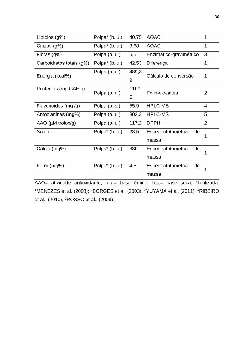
30
Lipídios (g%) Polpa* (b. u.) 40,75 AOAC 1
Cinzas (g%) Polpa* (b. u.) 3,68 AOAC 1
Fibras (g%) Polpa (b. u.) 5,5 Enzimático-gravimétrico 3
Carboidratos totais (g%) Polpa* (b. u.) 42,53 Diferença 1
Energia (kcal%) Polpa (b. u.) 489,3
9 Cálculo de conversão 1
Polifenóis (mg GAE/g) Polpa (b. u.)
1109.
5 Folin-ciocalteu 2
Flavonoides (mg /g) Polpa (b. s.) 55,9 HPLC-MS 4
Antocianinas (mg%) Polpa (b. u.) 303,3 HPLC-MS 5
AAO (µM trolox/g) Polpa (b. u.) 117,2 DPPH 2
Sódio Polpa* (b. u.) 28,5 Espectrofotometria de
massa 1
Cálcio (mg%) Polpa* (b. u.) 330 Espectrofotometria de
massa 1
Ferro (mg%) Polpa* (b. u.) 4,5 Espectrofotometria de
massa 1
AAO= atividade antioxidante; b.u.= base úmida; b.s.= base seca; *liofilizada.
1MENEZES et al. (2008); 2BORGES et al. (2003); 3YUYAMA et al. (2011); 4RIBEIRO
et al., (2010); 5ROSSO et al., (2008).
31
REFERÊNCIAS
ANTUNES, M.; BIALA, G. The novel object recognition memory: Neurobiology, test procedure, and its modificationsCognitive Processing, 2012.
AUGUSTINACK, J. C. et al. Specific tau phosphorylation sites correlate with severity of neuronal cytopathology in Alzheimer’s disease. Acta Neuropathologica, v. 103, n. 1, p. 26–35, 2002.
BANKS, W. A.; OWEN, J. B.; ERICKSON, M. A. Insulin in the brain: There and back againPharmacology and Therapeutics, 2012.
BATISTA, Â. G. et al. Intake of jaboticaba peel attenuates oxidative stress in tissues and reduces circulating saturated lipids of rats with high-fat diet-induced obesity. Journal of Functional Foods, v. 6, n. 1, p. 450–461, 2014.
BATISTA, Â. G. et al. Jaboticaba berry peel intake prevents insulin-resistance-induced tau phosphorylation in mice. Molecular Nutrition and Food Research, v. 61, n. 10, 2017.
BAXTER, M. G. “I’ve seen it all before”: explaining age-related impairments in object recognition. Theoretical comment on Burke et al. (2010). Behavioral neuroscience, v. 124, n. 5, p. 706–709, 2010.
BHUROSY, T.; JEEWON, R. Overweight and obesity epidemic in developing countries: a problem with diet, physical activity, or socioeconomic status? TheScientificWorldJournal, v. 2014, p. 964236, 2014.
BLÁZQUEZ, E. et al. Insulin in the brain: Its pathophysiological implications for states related with central insulin resistance, type 2 diabetes and alzheimer’s diseaseFrontiers in Endocrinology, 2014.
BLENNOW, K.; DE LEON, M. J.; ZETTERBERG, H. Alzheimer’s disease. Lancet, v. 368, n. 9533, p. 387–403, 2006.
BOITARD, C. et al. Impairment of hippocampal-dependent memory induced by juvenile high-fat diet intake is associated with enhanced hippocampal inflammation in rats. Brain, Behavior, and Immunity, v. 40, p. 9–17, 2014.
BRASIL. VIGILÂNCIA DE FATORES DE RISCO E PROTEÇÃO PARA DOENÇAS CRÔNICAS POR INQUÉRITO TELEFÔNICO - VIGITEL 2016Ministério da Saúde. Secretaria de Vigilância em Saúde. Departamento de Vigilância de Doenças e Agravos não Transmissíveis e Promoção da Saúde. [s.l: s.n.]. Disponível em: <www.saude.gov.br/svs>.
BROADBENT, N. J. et al. Object recognition memory and the rodent hippocampus. Learning & Memory, v. 17, n. 1, p. 5–11, 2010.
CAREY, A. N. et al. Blueberry supplementation attenuates microglia activation and
32
increases neuroplasticity in mice consuming a high-fat diet. Nutritional Neuroscience, p. 1–11, 21 Sep. 2017.
CAREY, A. N.; GOMES, S. M.; SHUKITT-HALE, B. Blueberry supplementation improves memory in middle-aged mice fed a high-fat diet. Journal of Agricultural and Food Chemistry, v. 62, n. 18, p. 3972–3978, 2014.
CAVALLINI, A. et al. An unbiased approach to identifying tau kinases that phosphorylate tau at sites associated with alzheimer disease. Journal of Biological Chemistry, v. 288, n. 32, p. 23331–23347, 2013.
CHENG, G. et al. Diabetes as a risk factor for dementia and mild cognitive impairment: A meta-analysis of longitudinal studiesInternal Medicine Journal, 2012.
CHOI, Y. J. et al. Açaí Berries Inhibit Colon Tumorigenesis in Azoxymethane/Dextran Sulfate Sodium-Treated Mice. Gut and Liver, v. 11, n. 2, p. 243–252, 2017.
CLARKE, J. R. et al. Plastic modifications induced by object recognition memory processing. Proc Natl Acad Sci U S A, v. 107, n. 6, p. 2652–2657, 2010.
ÇOBAN, J. et al. Blueberry treatment attenuates D-galactose-induced oxidative stress and tissue damage in rat liver. Geriatrics and Gerontology International, v. 14, n. 2, p. 490–497, 2014.
COÏSSON, J. D. et al. Euterpe oleracea juice as a functional pigment for yogurt. Food Research International. Anais...2005
CORREIA, S. C. et al. Insulin signaling, glucose metabolism and mitochondria: Major players in Alzheimer’s disease and diabetes interrelationBrain Research, 2012.
CROZIER, A.; JAGANATH, I. B.; CLIFFORD, M. N. Phenols, Polyphenols and Tannins: An Overview. In: Plant Secondary Metabolites: Occurrence, Structure and Role in the Human Diet. [s.l: s.n.]. p. 1–24.
DE FELICE, F. G.; FERREIRA, S. T. Inflammation, defective insulin signaling, and mitochondrial dysfunction as common molecular denominators connecting type 2 diabetes to Alzheimer DiseaseDiabetes, 2014.
DE LA MONTE, S. M. Alzheimer’s Disease Is Type 3 Diabetes—Evidence Reviewed. Journal of Diabetes Science and Technology J Diabetes Sci Technol J Diabetes Sci Technol, v. 22, n. 6, p. 1101–1113, 2008.
DE OLIVEIRA, P. R. B. et al. Euterpe oleracea Mart.-derived polyphenols protect mice from diet-induced obesity and fatty liver by regulating hepatic lipogenesis and cholesterol excretion. PLoS ONE, v. 10, n. 12, 2015.
DEVORE, E. E. et al. Dietary intakes of berries and flavonoids in relation to cognitive decline. Annals of Neurology, v. 72, n. 1, p. 135–143, 2012.
DIAS, M. M. DOS S. et al. Anti-inflammatory activity of polyphenolics from açai

33
(Euterpe oleracea Martius) in intestinal myofibroblasts CCD-18Co cells. Food Funct., v. 6, n. 10, p. 3249–3256, 2015.
DRAGANO, N. R. V. et al. Freeze-dried jaboticaba peel powder improves insulin sensitivity in high-fat-fed mice. British Journal of Nutrition, v. 110, n. 3, p. 447–455, 2013.
DRAGICEVIC, N. et al. Green tea epigallocatechin-3-gallate (EGCG) and other flavonoids reduce Alzheimer’s amyloid-induced mitochondrial dysfunction. Journal of Alzheimer’s Disease, v. 26, n. 3, p. 507–521, 2011.
DUDEK, H.; DATTA, S. R. Regulation of neuronal survival by the serine-threonine protein kinase Akt. Science, v. 275, n. 5300, p. 661, 1997.
EL KHOURY, N. B. et al. Insulin dysfunction and Tau pathology. Frontiers in Cellular Neuroscience, v. 8, 2014.
ELIAS, M. F. et al. Lower cognitive function in the presence of obesity and hypertension: The Framingham heart study. International Journal of Obesity, v. 27, n. 2, p. 260–268, 2003.
ENNACEUR, A. One-trial object recognition in rats and mice: Methodological and theoretical issuesBehavioural Brain Research, 2010.
ENNACEUR, A.; DELACOUR, J. A new one - trial test for neurobiological studies of memory in rats . 1 " Behavioral data. Behavioural Brain Research, v. 31, p. 47–59, 1988.
FARR, O. M.; TSOUKAS, M. A.; MANTZOROS, C. S. Leptin and the brain: Influences on brain development, cognitive functioning and psychiatric disorders. Metabolism: Clinical and Experimental, v. 64, n. 1, p. 114–130, 2015.
FREIHERR, J. et al. Intranasal insulin as a treatment for alzheimer’s disease: A review of basic research and clinical evidenceCNS Drugs, 2013.
FRIEDMAN, J. M.; HALAAS, J. L. Leptin and the regulation of body weight in mammalsNature, 1998.
GALLAGHER, M. Animal models of memory impairment. Philosophical transactions of the Royal Society of London. Series B, Biological sciences, v. 352, n. 1362, p. 1711–1717, 1997.
GALLORI, S. et al. Polyphenolic Constituents of Fruit Pulp of Euterpe oleracea Mart. (Acai palm). Chromatographia, v. 59, n. 11–12, p. 739–743, 2004.
GARAULET, M. et al. Adiponectin, the controversial hormone. Public Health Nutrition, v. 10, n. 10A, 2007.
GASKIN, S. et al. Object familiarization and novel-object preference in rats. Behavioural Processes, v. 83, n. 1, p. 61–71, 2010.
GRECO, S. J. et al. Leptin reduces Alzheimer’s disease-related tau phosphorylation

34
in neuronal cells. Biochemical and Biophysical Research Communications, v. 376, n. 3, p. 536–541, 2008.
GRECO, S. J. et al. Leptin inhibits glycogen synthase kinase-3beta to prevent tau phosphorylation in neuronal cells. Neuroscience letters, v. 455, n. 3, p. 191–4, 2009a.
GRECO, S. J. et al. Leptin regulates tau phosphorylation and amyloid through AMPK in neuronal cells. Biochemical and Biophysical Research Communications, v. 380, n. 1, p. 98–104, 2009b.
GRECO, S. J. et al. Leptin reduces pathology and improves memory in a transgenic mouse model of Alzheimer’s disease. Journal of Alzheimer’s disease : JAD, v. 19, n. 4, p. 1155–67, 2010.
GREENWOOD, C. E.; WINOCUR, G. Learning and memory impairment in rats fed a high saturated fat diet. Behavioral and neural biology, v. 53, p. 74–87, 1990.
GUO, H. et al. Cyanidin 3-glucoside attenuates obesity-associated insulin resistance and hepatic steatosis in high-fat diet-fed and db/db mice via the transcription factor FoxO1. Journal of Nutritional Biochemistry, v. 23, n. 4, p. 349–360, 2012.
HÅKANSSON, M. L.; MEISTER, B. Transcription factor STAT3 in leptin target neurons of the rat hypothalamus. Neuroendocrinology, v. 68, n. 6, p. 420–427, 1998.
HAMMOND, R. S.; TULL, L. E.; STACKMAN, R. W. On the delay-dependent involvement of the hippocampus in object recognition memory. Neurobiology of Learning and Memory, v. 82, n. 1, p. 26–34, 2004.
HARRY, G. J.; KRAFT, A. D. Neuroinflammation and microglia: considerations and approaches for neurotoxicity assessment. Expert Opinion on Drug Metabolism & Toxicology, v. 4, n. 10, p. 1265–1277, 2008.
HENI, M. et al. Impaired insulin action in the human brain: Causes and metabolic consequencesNature Reviews Endocrinology, 2015.
HIMMLER, A. et al. Tau consists of a set of proteins with repeated C-terminal microtubule-binding domains and variable N-terminal domains. Molecular and cellular biology, v. 9, n. 4, p. 1381–8, 1989.
HOSOGAI, N. et al. Adipose tissue hypoxia in obesity and its impact on adipocytokine dysregulation. Diabetes, v. 56, n. 4, p. 901–911, 2007.
HOTAMISLIGIL, G. S. Inflammation and metabolic disordersNature, 2006.
JAGANATH, I. B.; CROZIER, A. Flavonoid Biosynthesis. In: Plant Metabolism and Biotechnology. [s.l: s.n.]. p. 293–320.
JEON, B. T. et al. Resveratrol attenuates obesity-associated peripheral and central inflammation and improves memory deficit in mice fed a high-fat diet. Diabetes, v. 61, n. 6, p. 1444–1454, 2012.
35
JOHNSON, A. M. F.; OLEFSKY, J. M. The origins and drivers of insulin resistanceCell, 2013.
JOLIVALT, C. G. et al. Defective insulin signaling pathway and increased glycogen synthase kinase-3 activity in the brain of diabetic mice: Parallels with Alzheimer’s disease and correction by insulin. Journal of Neuroscience Research, v. 86, n. 15, p. 3265–3274, 2008.
JUGE-AUBRY, C. E.; HENRICHOT, E.; MEIER, C. A. Adipose tissue: A regulator of inflammationBest Practice and Research: Clinical Endocrinology and Metabolism, 2005.
KAHN, S. E.; HULL, R. L.; UTZSCHNEIDER, K. M. Mechanisms linking obesity to insulin resistance and type 2 diabetes. Nature, v. 444, n. 7121, p. 840–846, 2006.
KALMIJN, S. et al. Dietary fat intake and the risk of incident dementia in the Rotterdam Study. Annals of neurology, v. 42, n. 5, p. 776–782, 1997.
KENNEDY, A. et al. Saturated Fatty Acid-Mediated Inflammation and Insulin Resistance in Adipose Tissue: Mechanisms of Action and Implications. Journal of Nutrition, v. 139, n. 1, p. 1–4, 2008.
KERSHAW, E. E.; FLIER, J. S. Adipose tissue as an endocrine organ. The Journal of clinical endocrinology and metabolism, v. 89, n. 6, p. 2548–56, 2004.
KILIAAN, A. J.; ARNOLDUSSEN, I. A. C.; GUSTAFSON, D. R. Adipokines: A link between obesity and dementia?The Lancet Neurology, 2014.
KIM, B.; FELDMAN, E. L. Insulin resistance as a key link for the increased risk of cognitive impairment in the metabolic syndromeExperimental & molecular medicine, 2015.
KLEINRIDDERS, A. et al. Insulin action in brain regulates systemic metabolism and brain function. Diabetes. Anais...2014
KONG, J.-M. et al. Analysis and biological activities of anthocyanins. Phytochemistry, v. 64, p. 923–933, 2003.
KOSIK, K. S.; JOACHIM, C. L.; SELKOE, D. J. Microtubule-associated protein tau (tau) is a major antigenic component of paired helical filaments in Alzheimer disease. Proceedings of the National Academy of Sciences of the United States of America, v. 83, n. 11, p. 4044–8, 1986.
KOTHARI, V. et al. High fat diet induces brain insulin resistance and cognitive impairment in mice. Biochimica et Biophysica Acta (BBA) - Molecular Basis of Disease, v. 1863, n. 2, p. 499–508, 2017.
KRIL, J. J. et al. Neuron loss from the hippocampus of Alzheimer’s disease exceeds extracellular neurofibrillary tangle formation. Acta Neuropathologica, v. 103, n. 4, p. 370–376, 2002.
KUSMINSKI, C. M. et al. Diabetes and apoptosis: LipotoxicityApoptosis, 2009.

36
LEE, E. B. Obesity, leptin, and Alzheimer’s diseaseAnnals of the New York Academy of Sciences, 2011.
LEE, J.; KIM, M. S. The role of GSK3 in glucose homeostasis and the development of insulin resistance. Diabetes Research and Clinical Practice, v. 77, n. 3 SUPPL., 2007.
LETO, D.; SALTIEL, A. R. Regulation of glucose transport by insulin: Traffic control of GLUT4Nature Reviews Molecular Cell Biology, 2012.
LEWIS, J. et al. Neurofibrillary tangles, amyotrophy and progressive motor disturbance in mice expressing mutant (P301L)tau protein. Nature Genetics, v. 25, n. 4, p. 402–405, 2000.
LORENZI, H.; DE SOUZA, H. M. Palmeiras no Brasil: nativas e exóticas. [s.l.] Editora Plantarum, 1996.
LUCHSINGER, J. A et al. Caloric intake and the risk of Alzheimer disease. Archives of neurology, v. 59, n. 8, p. 1258–1263, 2002.
MANACH, C. et al. Polyphenols: Food sources and bioavailabilityAmerican Journal of Clinical Nutrition, 2004.
MANDREKAR, S.; LANDRETH, G. E. Microglia and inflammation in Alzheimer’s disease. CNS & neurological disorders drug targets, v. 9, n. 2, p. 156–67, 2010.
MASTERS, C. L. et al. Alzheimer’s diseaseNature Reviews Disease Primers, 2015.
MATTSON, M. P. Degenerative and protective signaling mechanisms in the neurofibrillary pathology of AD. Neurobiology of Aging, v. 16, n. 3, p. 447–457, 1995.
MENDONÇA, C. P. Dietary and physical activity factors as determinants of the increase in overweight/obesity in BrazilCadernos de saude publica / Ministerio da Saude, Fundacao Oswaldo Cruz, Escola Nacional de Saude Publica, 2004. Disponível em: <http://ovidsp.ovid.com/ovidweb.cgi?T=JS&PAGE=reference&D=emed9&NEWS=N&AN=39695101>
MESSIER, C.; TEUTENBERG, K. The role of insulin, insulin growth factor, and insulin-degrading enzyme in brain aging and Alzheimer’s diseaseNeural Plasticity, 2005.
MICHA, R. et al. Global, regional, and national consumption levels of dietary fats and oils in 1990 and 2010: a systematic analysis including 266 country-specific nutrition surveys. BMJ, v. 348, n. apr14 18, p. g2272–g2272, 2014.
MILANSKI, M. et al. Inhibition of hypothalamic inflammation reverses diet-induced insulin resistance in the liver. Diabetes, v. 61, n. 6, p. 1455–1462, 2012.
MILLER, A. A.; SPENCER, S. J. Obesity and neuroinflammation: A pathway to

37
cognitive impairment. Brain, Behavior, and Immunity, v. 42, p. 10–21, 2014.
MOKHTAR, S. H. et al. The beta-amyloid protein of alzheimer’s disease: Communication breakdown by modifying the neuronal cytoskeleton. International Journal of Alzheimer’s Disease, v. 2013, 2013.
MORISHIMA-KAWASHIMA, M. et al. Proline-directed and non-proline-directed phosphorylation of PHF-tau. Journal of Biological Chemistry, v. 270, n. 2, p. 823–829, 1995.
MORRIS, R. G. M. et al. Place navigation impaired in rats with hippocampal lesions. Nature, v. 297, n. 5868, p. 681–683, 1982.
MUÑIZ-MIRET, N. et al. The economic value of managing the açaí palm (Euterpe oleracea Mart.) in the floodplains of the Amazon estuary, Pará, Brazil. Forest Ecology and Management, v. 87, n. 1, p. 163–173, 1996.
MURPHY, S. L. et al. Deaths: Final Data for 2013. National vital statistics reports : from the Centers for Disease Control and Prevention, National Center for Health Statistics, National Vital Statistics System, v. 64, n. 2, p. 1–119, 2016.
MYERS, M. G.; COWLEY, M. A.; MÜNZBERG, H. Mechanisms of Leptin Action and Leptin Resistance. Annual Review of Physiology, v. 70, n. 1, p. 537–556, 2008.
NELSON ET AL. Correlation of Alzheimer Disease Neuropathologic Changes With Cognitive Status: A Review of the Literature. J Neuropathol Exp Neurol. 2012 May ; 71(5): 362–381. doi:10.1097/NEN.0b013e31825018f7, v. 71, n. 5, p. 362–381, 2013.
NGUYEN, D. M.; EL-SERAG, H. B. The Epidemiology of Obesity. Gastroenterology Clinics of North America, v. 39, n. 1, p. 1–7, 2010.
OECD. OECD Health Statistics 2015. OECD Publishing, p. 2013–2015, 2016.
OTTAVIANI, E.; MALAGOLI, D.; FRANCESCHI, C. The evolution of the adipose tissue: A neglected enigmaGeneral and Comparative Endocrinology, 2011.
PACHECO-PALENCIA, L. A.; HAWKEN, P.; TALCOTT, S. T. Phytochemical, antioxidant and pigment stability of açai (Euterpe oleracea Mart.) as affected by clarification, ascorbic acid fortification and storage. Food Research International, v. 40, n. 5, p. 620–628, 2007.
PAPANDREOU, M. A. et al. Effect of a polyphenol-rich wild blueberry extract on cognitive performance of mice, brain antioxidant markers and acetylcholinesterase activity. Behavioural Brain Research, v. 198, n. 2, p. 352–358, 2009.
PÉREZ-GONZÁLEZ, R. et al. Leptin induces proliferation of neuronal progenitors and neuroprotection in a mouse model of alzheimer’s disease. Journal of Alzheimer’s Disease, v. 24, n. SUPPL. 2, p. 17–25, 2011.
PISTELL, P. J. et al. Cognitive impairment following high fat diet consumption is associated with brain inflammation. Journal of Neuroimmunology, v. 219, n. 1–2, p.

38
25–32, 2010.
POSEY, K. A. et al. Hypothalamic proinflammatory lipid accumulation, inflammation, and insulin resistance in rats fed a high-fat diet. American journal of physiology. Endocrinology and metabolism, v. 296, n. 5, p. E1003-12, 2009.
PRINCE, M. et al. World Alzheimer Report 2016 Improving healthcare for people living with dementia. Coverage, Quality and costs now and in the future. Alzheimer’s Disease International (ADI), p. 1–140, 2016.
PRIOR, R. L. Fruits and vegetables in the prevention of cellular oxidative damage. American Journal of Clinical Nutrition. Anais...2003
QIN, B. et al. Green tea polyphenols improve cardiac muscle mrna and protein levels of signal pathways related to insulin and lipid metabolism and inflammation in insulin-resistant rats. Molecular Nutrition and Food Research, v. 54, n. SUPPL. 1, 2010.
REAVEN, G. M. THE INSULIN RESISTANCE SYNDROME: Definition and Dietary Approaches to Treatment. Annual Review of Nutrition, v. 25, n. 1, p. 391–406, 2005.
REITZ, C.; MAYEUX, R. Alzheimer disease: Epidemiology, diagnostic criteria, risk factors and biomarkersBiochemical Pharmacology, 2014.
ROGEZ, H. Açaí: preparo, composição e melhoramento da conservação. [s.l.] EDUFPA, 2000.
SAH, S. K. et al. Effect of high-fat diet on cognitive impairment in triple-transgenic mice model of Alzheimer’s disease. Biochemical and Biophysical Research Communications, 2017.
SCHAUSS, A. G. et al. Phytochemical and nutrient composition of the freeze-dried amazonian palm berry, Euterpe oleraceae Mart. (Acai). Journal of Agricultural and Food Chemistry, v. 54, n. 22, p. 8598–8603, 2006a.
SCHAUSS, A. G. et al. Antioxidant Capacity and Other Bioactivities of the Freeze-Dried Amazonian Palm Berry, Euterpe oleraceae Mart. (Acai). Journal of Agricultural and Food Chemistry, v. 54, n. 22, p. 8604–8610, 2006b.
SCHRECKINGER, M. E. et al. Berries from South America: A Comprehensive Review on Chemistry, Health Potential, and Commercialization. Journal of Medicinal Food, v. 13, n. 2, p. 233–246, 2010.
SCHUBERT, M. et al. Insulin receptor substrate-2 deficiency impairs brain growth and promotes tau phosphorylation. The Journal of neuroscience : the official journal of the Society for Neuroscience, v. 23, n. 18, p. 7084–7092, 2003.
SELLAYAH, D.; CAGAMPANG, F. R.; COX, R. D. On the evolutionary origins of obesity: A new hypothesisEndocrinology, 2014.
SILVA, D. F. et al. Cytotoxic effects of Euterpe oleracea Mart. in malignant cell lines. BMC Complementary and Alternative Medicine, v. 14, n. 1, p. 175, 2014.
39
SOLFRIZZI, V.; PANZA, F.; CAPURSO, A. The role of diet in cognitive decline. Journal of neural transmission (Vienna, Austria : 1996), v. 110, n. 1, p. 95–110, 2003.
TAGLIALATELA, G. et al. Intermediate- and long-term recognition memory deficits in Tg2576 mice are reversed with acute calcineurin inhibition. Behavioural Brain Research, v. 200, n. 1, p. 95–99, 2009.
TALBOT, K. et al. Demonstrated brain insulin resistance in Alzheimer’s disease patients is associated with IGF-1 resistance, IRS-1 dysregulation, and cognitive decline. Journal of Clinical Investigation, v. 122, n. 4, p. 1316–1338, 2012.
TILG, H.; MOSCHEN, A. R. Adipocytokines: Mediators linking adipose tissue, inflammation and immunity. Nature Reviews Immunology, v. 6, n. 10, p. 772–783, 2006.
UCHOA, M. F.; MOSER, V. A.; PIKE, C. J. Interactions between inflammation, sex steroids, and Alzheimer’s disease risk factorsFrontiers in Neuroendocrinology, 2016.
USA DEPARTMENT OF AGRICULTURE AND DEPARTMENT OF HEALTH AND HUMAN SERVICES. 2015 – 2020 Dietary Guidelines for Americans. 2015 – 2020 Dietary Guidelines for Americans (8th edition), p. 18, 2015.
WILLIAMS, D. R. Tauopathies: Classification and clinical update on neurodegenerative diseases associated with microtubule-associated protein tauInternal Medicine Journal, 2006.
WILLIAMS, R. J.; SPENCER, J. P. E. Flavonoids, cognition, and dementia: Actions, mechanisms, and potential therapeutic utility for Alzheimer diseaseFree Radical Biology and Medicine, 2012.
WOOD, J. G. et al. Neurofibrillary tangles of Alzheimer disease share antigenic determinants with the axonal microtubule-associated protein tau (tau). Proceedings of the National Academy of Sciences of the United States of America, v. 83, n. 11, p. 4040–3, 1986.
YUYAMA, L. K. O. et al. Caracterização físico-química do suco de açaí de Euterpe precatoria Mart. oriundo de diferentes ecossistemas amazônicos. Acta Amazonica, v. 41, n. 4, p. 545–552, 2011.
ZHAO, W. Q.; ALKON, D. L. Role of insulin and insulin receptor in learning and memory. Molecular and cellular endocrinology, v. 177, n. 1–2, p. 125–34, 2001.
ZHENG, C.; ZHOU, X.-W.; WANG, J.-Z. The dual roles of cytokines in Alzheimer’s disease: update on interleukins, TNF-α, TGF-β and IFN-γ. Translational Neurodegeneration, v. 5, n. 1, p. 7, 2016.

40
CAPÍTULO 2
Artigo Original: CONSUMO DE POLPA DE AÇAÍ MELHORA FUNÇÕES
COGNITIVAS E SENSIBILIDADE PERIFÉRICA À INSULINA EM CAMUNDONGOS
ALIMENTADOS COM DIETA HIPERLIPÍDICA
41
CAPÍTULO 2
Artigo Original: CONSUMO DE POLPA DE AÇAÍ MELHORA FUNÇÕES
COGNITIVAS E SENSIBILIDADE PERIFÉRICA À INSULINA EM CAMUNDONGOS
ALIMENTADOS COM DIETA HIPERLIPÍDICA
Nathalia Medina dos Santos, Ângela Giovana Batista, Monique Culturato Padilha
Mendonça, Célio Fernando Figueiredo Angolini, Renato Grimaldi, César Renato
Sartori, Maria Alice da Cruz-Höfling and Mário Roberto Maróstica Júnior. Açai pulp
intake improves cognitive functions and peripheral insulin sensitivity in high-fat diet-
fed mice. A ser submetido à revista Journal of Functional Foods, Elsevier, ISSN:
1756-4646.
Vide autorização CEUA no Anexo 1.
42
Research article
Açai pulp intake improves cognitive functions and peripheral insulin sensitivity in high-fat diet-fed mice
Nathalia Medina dos Santos1, Ângela Giovana Batista2, Monique Culturato Padilha
Mendonça3, Célio Fernando Figueiredo Angolini4, Renato Grimaldi5, César Renato
Sartori6, Maria Alice da Cruz-Höfling3 and Mário Roberto Maróstica Júnior1.
1School of Food Engineering, Department of Food and Nutrition, University of Campinas, Campinas,
SP, Brazil.
2Faculty of Food and Nutrition, Federal University of Santa Maria, Pameira das Missões, Rio Grande
do Sul, RS, Brazil.
3Institute of Biology, Department of Biochemistry and Tissue Biology, University of Campinas,
Campinas, SP, Brazil.
4School of Food Engineering, Department of Food Science, University of Campinas, Campinas, SP,
Brazil.
5Department of Food Technology, Faculty of Food Engineering, University of Campinas, Campinas,
SP, Brazil.
6Institute of Biology, Department of Department of Structural and Functional Biology, University of
Campinas, Brazil.
*Corresponding author: Mário Roberto Maróstica Júnior Contact details:
Departamento de Alimentos e Nutrição. Rua Monteiro Lobato, 80. Cidade
Universitária. Campinas, SP, Brasil. Zip Code: 13083-862. E-mail:
Abbreviations: AD: Alzheimer's disease; AKT: protein kinase B; AP: açai pulp; CAT:
catalase; EAT: epidiymal adipose tissue; GSH: reduced glutathione; GSK3-β:
Glycogen synthase kinase 3-β; IRS: insulin receptor substrate; MAT: mesenteric
adipose tissue; NOR: novel object recognition; NFT: neurofibrillary tangles; RAT:
retroperitoneal adipose tissue; SOD: superoxide dismutase; Tau: microtubule-
associated protein tau;

43
ABSTRACT
Açai is a Brazilian berry rich in flavonoids, anthocyanins and other bioactive
compounds involved in mechanisms related to several benefits to health, such as
oleic acid. Many evidences indicate that obesity and insulin resistance constitute risk
factors for development of neurodegenerative diseases. (Poly)phenol compounds are
under study for its role in protecting effects against neural injuries and degeneration.
In this regard, an Amazonian açai pulp (AP) was characterized due to its proximate
composition, phenolic compounds, carotenoids, and antioxidant capacity. Then, we
induced obesity and insulin resistance with a high-fat diet (H group) in mice and
compared to a normal-fat standard diet (N group). We have supplemented the both
diets with 2% AP and investigated proofs of peripheral insulin resistance, recognition
memory, tau protein stability via AKT/GSK3-β signaling, and oxidative stress in
hippocampus. The high-fat diet supplemented with AP (HA group) did not prevent
weight gain, but improved peripheral insulin sensitivity and phosphorylation of
AKT/GSK3-β in hippocampus, although no significant differences were observed in
tau phosphorylation. The animals fed high-fat diets with AP showed better
performance in novel object recognition test (NOR) in comparison to the H group.
Catalase activity and reduced glutathione (GSH) values were improved in the HA
mice. These results suggested that the supplementation of AP can attenuate the
effects of a high-fat diet consumption in peripheral insulin resistance and improve
cognitive behavior, but the mechanisms underlying this finding doesn’t seems to
depend of tau phosphorylation.
Keywords: Euterpe sp.; super-fruit; neurodegeneration; anthocyanins; obesity.

44
1. Introduction
A long-term consumption of a high-energy, high-fat diets in middle or late life
has been associated with an increased risk of cognitive deficits and neurological
diseases such as dementia like Alzheimer’s disease (AD) (Freeman et al., 2014;
Kothari et al., 2017; Morris & Tangney, 2014; Spencer et al., 2017). Obesity and its
resulting morbidities, such as diabetes mellitus type 2, midlife hypertension and
sedentarism are primary preventable environmental risk factors associated to
emergence of AD beyond old age and genetic factors (Norton et al., 2014).
Consuming a high-fat diet may cause central nervous system dysfunction through
multiple mechanisms, not completely elucidated so far.
The intake of high-fat diets, rich in saturated fatty acids, has been associated
to behavioral deficits and increased phosphorylation of tau (microtubule-associated
tau protein) in mouse (Kothari et al., 2017). Both insulin and leptin resistance due to
obese conditions are pointed out as possible causes in the context (Kim & Feldman,
2015; Lee, 2011).
Glycogen synthase kinase-3β (GSK-3β) is the main tau kinase in the brain and
in abnormal conditions, it is responsible for tau hyperphosphorylation and
consequent formation of neurofibrillary tangles (NFT), an important marker of
neuronal alterations in AD (Kaytor & Orr, 2002). Normally, this kinase is inactivated
when phosphorylated at Serine 9 by other kinases, such as AKT (protein kinase B)
(Kaytor & Orr, 2002; Schubert et al., 2004). Leptin appears to play a role in reduction
of tau phosphorylation mediated through AMPK, AKT and p38 pathways (Greco et al,
2009a). AMPK is also known to interact with GSK-3β, and may be a link to the
AKT/GSK-3β activation (Greco et al., 2008; 2009b). In the same way, in insulin
resistance condition, the inhibition of insulin signal propagation inactivates AKT,
impeding the signaling cascade to GSK3-β phosphorylation and activating tau
phosphorylation (Schubert et al., 2003, 2004).
Berry fruits contain (poly)phenols such as flavonoids and anthocyanins
involved in several mechanisms related to prevention of insulin resistance, obesity
and cognitive impairment (Guo et al., 2012). The anthocyanins and metabolites have
been shown to enter the brain, and their concentrations could be correlated with
improved cognitive performance (Gutierres et al., 2014; Qin et al., 2013; Youdim et

45
al., 2003). In aged animals, the intake of polyphenol-rich fruits was able to reverse or
delay age-related impairment when cognitive test challenges were performed (Malin
et al., 2011; Shukitt-Hale et al., 2017). Recent studies indicate that diets
supplemented with blueberries and jaboticaba (a Brazilian berry), for example, can
prevent some of the neuronal deleterious effects caused by high-fat diet consumption
(Batista et al., 2017; Carey et al., 2017).
Açai is a black-purple berry fruit native from Brazil, extensively studied due to
its nutritional, phytochemical composition and impressive antioxidant activity and
anti-inflammatory effects (De Oliveira et al., 2015; Dias et al., 2015; Schaus et al.,
2006a). These properties were responsible for açai's high popularity in North America
and European countries, where is known as “super-fruit”. Açai contains a variety of
polyphenols, mainly anthocyanins and other flavonoids, responsible for its dark
purple color and to its antioxidant activity. Furthermore, this berry is a great source of
omega 9 (oleic acid) and dietary fibers (Smith et al., 2012; Luo et al., 2012; Pacheco-
Palencia et al., 2009; Schauss et al., 2006) . In regards to this study, açai was once
related to protection of brain cultured cells against induced oxidative stress and
inflammatory insults in regions involved in overall brain function, memory and
cognition, and in reversing the detrimental effects of aging on motor and cognitive
behavior in aged animals (Carey et al., 2017).
These data led us to investigate the effect of açai pulp intake in the prevention
of memory and cognitive impairment resulted of a high-fat diet intake, as well as to
investigate hippocampal markers associated with tau phosphorylation. Furthermore,
peripheral insulin resistance and markers of oxidative stress were explored in this
study.
2. Material and methods
2.1 Açai pulp characterization
An Amazonian açai (Euterpe sp.) frozen pulp was obtained from a local
market in Manaus-AM, Brazil. The açai pulp was freeze-dried to obtain a powder
(AP). The analysis of macronutrients, total protein, moisture and ash was performed
according to the methods described by the Association of Official Analytical Chemists

46
(AOAC, 2012). The total lipids were determined according to the Bligh & Dyer (1959)
method, the total and insoluble dietary fibers were determined through the
enzymatic-gravimetric method, according to the AOAC (1995) and the soluble fibers
were calculated by difference between both. The concentration of carbohydrates was
obtained as the subtraction between 100 and the total percentage of the other
components.
A hexane extract was made in order to obtain a non-polar extract. For this, the
AP samples were extracted by successive maceration with hexane at room
temperature using a shaking water bath (170 rpm) for 1 hour at room temperature
(22 °C ± 2). The solution obtained was then filtered, and extracted twice, combined
and vacuum-concentrated in a rotary evaporator (T < 37 °C) to a final volume of 50
ml (0.1g mL-1) (Prior et al., 2003). The extraction procedure was conducted in
triplicate. The extract was then shaken in an Erlenmeyer flask containing isopropanol
and hexane, then transferred to a separation funnel and gently washed with water, 3
times. The hexane extract was filtered in sodium sulfate and filled with hexane.
Using the same procedure as for the non-polar extraction, a polar fraction was
obtained, extracting the remaining residue of açai pulp with successive maceration in
0.1% HCl in methanol in order to analyze the phenolic compounds. The solution
obtained was then filtered, combined and vacuum-concentrated (T < 38 °C) to a final
volume of 50 mL (0.1g mL-1) (Degenhardt, Knapp, & Winterhalter, 2000; Kammerer,
Carle, & Schieber, 2004).
2.1.1 Bioactive compounds
2.1.1.1 Non-polar bioactive compounds
To determine the total carotenoid content, the non-polar extract and blank
(hexane) were read at 450 nm in the microplate reader and the calculation was made
as described in a standard method (Higby, 1962).
The fatty acid composition was determined using an Agilent 6850 Series gas
chromatography (GC) system (Agilent Technologies, Santa Clara, CA, USA)
equipped with a capillary column. The samples were prepared following esterification
according to a method described by Hartman & Lago (1973). Fatty acid methyl esters

47
were separated using AOCS method Ce 2-66 (AOCS, 2009), employing a 60-m DB-
23 capillary column (50% cyanopropyl methylpolysiloxane; Agilent Technologies)
with an internal diameter of 0.25 mm, coated with a 0.25 mm film. Chromatography
was performed under the following conditions: heating at 110 oC for 5 min in an oven,
followed by heating to 215 oC at a rate of 5 oC/min and holding at 215 oC for 24 min.
The detector temperature was 280 oC, the injector temperature was 250 oC, helium
was used as the carrier gas, the split ratio was 1:50, and the injection volume was
1.0 µL. The qualitative composition was determined via area normalization and was
expressed as the mass percentage.
2.1.1.2 Polar bioactive compounds
Polar extract was diluted 100 times by pipetting 10 µL in 990 µL water. The
liquid chromatographic – mass spectrometry (LC-MS) analysis was performed in
negative ion mode using a High Performance Liquid Chromatography (HPLC,
Hewlett Packard, Agilent Technologies 1290 series, City, country) coupled to a Q-
ToF iFunnel 6550 mass spectrometer fitted with an electrospray ionization (ESI)
source, using a Poroshell 120 SB-Aq 2.7 μm column (2.1x100 mm, Agilent). Mobile
phase A was H2O containing 0.1% formic acid; and mobile phase B was acetonitrile
containing 0.1% formic acid. The samples were eluted with a flow rate of 0.45 mL
min-1 and the following linear gradient: 0 - 1 min, 5% B; 1 - 10 min, 5% B to 18% B;
10 -13 min 18% B to 70% B; 13 - 15 min, 70% B to 100% B; 15 - 17 min, 100% B
and 3 min of post time at 5% B to column equilibration. The mass spectrometer
parameters used were: VCap 3000 V; fragmentor voltage at 150 V; OCT 1RF Vpp at
750 V; Gas Temperature at 290 oC; Sheath Gas Temperature at 350 oC; drying gas
at 12 L min-1. Mass spectra were acquired in profile mode and the acquisition range
was 100 - 1500 m/z. Data were treated using Agilent MassHunter Qualitative
Analysis B 0.7 software.
The total phenolic content was determined by the Folin-Ciocalteu method,
adapted from Swain & Hillis (1959). The absorbance was measured at 725 nm and
the results were expressed as gallic acid equivalents (GAE mg g-1).
The monomeric anthocyanins were quantified according to the pH-differential
method described by Wrolstad (1976). The anthocyanin content was calculated as
cyanidin 3-glucoside equivalent (C3G mg 100g-1).

48
The flavonoids contents were determined by colorimetric reaction with
aluminum chloride as described by Zhishen, Mengcheng, & Jianming (1999).
Catechin was used as standard, the absorbance was read at 510 nm and the results
were expressed as mg catechin g-1 (mg CE g-1).
2.1.2 Antioxidant capacity
The H-ORAC (hydrophilic oxygen radical absorbance capacity) (Ou et al.,
2013) test was carried out adding 20 µL of diluted methanolic extract or standard
solutions, 125 µL of fluorescein diluted in phosphate buffer (pH 7.4), and 25 µL of
AAPH (2,2-azobis (2-methylpropionamidine) dihydrochloride) to black microplates, in
the dark. Trolox ((±)-6-hydroxy-2,5,7,8-tetramethylchromane-2-carboxylic acid) was
used as standard and a microplate reader with fluorescent filters: excitation
wavelength, 485 nm; emission wavelength, 520 nm and the fluorescence was read
each 1 min, for 80 min. The H-ORAC values were expressed in µmol trolox
equivalent (µmol TE) per gram of AP by using the standard curves for each assay.
Lipophilic ORAC (L-ORAC) was based on the Ou et al., (2013) method. The
non-polar extract of AP was diluted in a 7% randomly methylated β-cyclodextrin
(RMCD) (RMCD Trappsol®) solution made in 50% aqueous acetone (v/v). After
aliquoting appropriate diluted samples in dark microplates, fluorescein and AAPH
diluted in phosphate buffer (7.4 pH) were quickly added to the plate. The microplate
was read as in H-ORAC. The area under the curve was calculated as described in a
previous work (Dávalos, Gómez-Cordovés, & Bartolomé, 2004). Values were
expressed as μmol trolox equivalent (TE). The total-ORAC (T-ORAC) was assessed
by the sum of H-ORAC values and L-ORAC values.
The ferric reducing ability of the methanolic extract was determined using the
FRAP method (Rufino et al., 2010). The FRAP reagent was prepared in the dark,
using 0.3 M acetate buffer (pH 3.6), 10 mM TPTZ in a 40 mM HCl solution and 20
mM FeCl3 (at proportions of 10:1:1). The sample or standard solutions, water and
FRAP reagent were mixed and incubated in an oven for 30 min at 37 °C. The
samples and trolox standard curve were read at 595 nm.
These analyses were done in the microplate reader (Synergy HT, Biotek, Winooski,
USA) spectrophotometer with Gen5™ 2.0 data analysis software.

49
2.2 Animal experimentation
This research followed the Animal Experimentation Ethical Principles adopted
by the Brazilian Society of Laboratory Animal Science (SBCAL) and was approved by
the Institutional Committee for Ethics in Animal Use (CEUA/UNICAMP, protocol
#4212-1/2016). Fifty two 8-weeks old male Swiss mice (Mus musculus, weight 38.6 ±
2.9 g) obtained from the Multidisciplinary Center of Biological Investigation
(CEMIB/UNICAMP) and fed with standard semi-purified diet AIN-93G (American
Institute of Nutrition) (Reeves et al., 1993) were allocated in individual polypropylene
cages, with free access to food and water, controlled relative humidity (60%–70%)
and temperature (22 ± 2°C) and standard inverted 12/12 h dark/light cycles. The
animals remained in acclimatization for 7 days receiving a semi-purified maintenance
diet based on the AIN-93M (Reeves et al., 1993). Then they were randomly assigned
into four groups (n = 13) according to the offered diet (Table 1) : a) normal-fat diet
(AIN-93M) - N group; b) normal-fat diet with 2% AP (w/w) – NA group; c) high-fat diet
(60% total calories from fat) – H group and d) high-fat diet containing 2% AP (w/w) -
HA group. The dose of AP in the experimental diets were chosen based on its
anthocyanin content according to previous studies using anthocyanin-rich berry
(Batista et al., 2013, 2014). Furthermore, other research groups have also used 2%
açai in the diet with the purpose to study metabolic diseases (Carey et al., 2017;
Souza et al., 2010; Poulose et al., 2017). The diets were replaced at intervals of 2
days to prevent fat and anthocyanin oxidation. Food intake was monitored every 2
days and weight gain weekly.
Table 1. Composition of experimental diets
Ingredient (g kg -1) N NA H HA
Casein (84,59%
protein) 140.67 140.67 140.67 140.67
Corn starch 465.25 461.13* 264.99* 260.81*
Maltodextrin 154.85 153.48* 88.18* 86.80*
Sucrose 99.90 99.02* 56.89* 56.01*
Soy oil 40.00 31.60# 40.00 31.60#

50
Cellulose 50.00 44.77** 50.00 44.77**
Lard - - 310.00 310.00
Mineral mix 35.00 35.00 35.00 35.00
Vitaminic mix 10.00 10.00 10.00 10.00
L-Cystine 1.80 1.80 1.80 1.80
Choline bitartrate 2.50 2.50 2.50 2.50
tert-Butylhydroquinone 0.008 0.008 0.008 0.008
Açai pulp - 20.00 - 20.00
Caloriesa 3717.22 ±
28.67
3695.28 ±
52.10
5294.92 ±
21.51
5238.02 ±
79.76
N: normal-fat diet fed group; NA: normal-fat diet plus 2% AP group; H: high-fat diet
fed group; HA: high-fat diet containing 2% AP group. *The carbohydrate ingredients
were adapted according to the lard content and/or the addition of freeze-dried açai
pulp (AP) powder. #The soy oil content was adapted and discounted according to the
amount of total lipids of AP. **The cellulose content was adapted and discounted
according to the amount of total dietary fibers of AP. aCalories values (kcal kg-1) were
calculated considering the conversion factors of Atwater.
At the end of the 10th week, the animals were euthanized after injection of
ketamine chloride (100 mg kg-1) and xylazine chloride (10 mg kg-1) in a concentration
of 2:1 (v v-1). After euthanizing, brains were rapidly removed, washed in a 0.9%
saline solution, weighed, frozen in liquid nitrogen and kept at -80°C until posterior
analyses.
2.2.1 Novel Object Recognition test
Non-spatial declarative memory was assessed in novel object recognition test
(NOR) at the 8th week of experiment (n =10). The NOR apparatus was located in a
separate dark room containing two red-light lamp bulbs. A video camera was
suspended above the center of the arena, and the image was projected to a monitor
to allow the experimenter to observe the animal’s behavior in an adjacent room. The

51
apparatus used for the test was an open field arena (30 cm × 30 cm × 40 cm) made
of coated wood. The walls were white and the floor was black. The objects used were
a tower of Lego® bricks and a Falcon tube filled with colored sand. All objects were
fixed to the floor with velcro, 15 cm apart and 5 cm from the wall. The test began
between 9:00 and 10:00 a.m. and was divided in three phases: during the habituation
phase, mice individually explored the open field freely for five minutes. In the
familiarization phase, two identical objects were placed diagonally in the open field.
The mice were placed in the arena facing away from the objects and were allowed
to explore the objects for five minutes. Twenty-four hours later (during the test phase)
the mice was introduced into the field again. In this phase, two objects were placed at
the same position as in the familiarization phase and one of the familiar objects was
replaced with a novel object. The animals were allowed to explore freely both the
objects for five minutes. The positions of objects in the test phase were
counterbalanced between animals to avoid positional biases. After each phase
session, the animals were returned to their home cages and the arena and the
objects were cleaned with 70% ethanol. All the test phases were recorded and later
analyzed using stopwatches by blinded observers. Exploration was defined as direct
contact of the nose or front paws with the object. Time spent exploring each object
during the test phase was calculated as a recognition index (RI): a percent of time
spent exploring the familiar (TF) or the novel object (TN) relative to the total time spent
exploring both objects [RI = TN or TF/(TN + TF)] (Carey et al., 2009). The NOR task
evaluates the rodent’s ability to recognize a novel object in the environment. Since
the NOR’s methodology assesses the rodent’s natural preference for novel objects, a
higher percentage of time spent with the novel object is considered an index of
improvement in learning and memory performance using the hippocampus (Carey et
al., 2009).
2.2.2 Glucose Tolerance Test and Insulin Tolerance Test
A glucose tolerance test (GTT) was performed in 6 h-fasted mice in the 9th
week of experiment by intraperitoneally (ip) injecting a solution of D-glucose (2 g kg-
1). Blood glucose levels were measured with an FreeStyle Lite glucometer (Abbott
Diabetes Care, Alameda, CA, USA) using appropriate test strips. The venous blood

52
was collected via tail vein before (0 min) and 30, 60, 90 and 120 min after glucose
injection.
The insulin tolerance test (ITT) was carried out in 6 h-fasted mice on the 10th
week of experiment. The blood sample was collected via tail vein and the glucose
levels were measured before (0 min) and 15, 30, 45 and 60 min after an ip-injection
of saline solution (0.9%) containing 0.75 units kg-1 insulin (Novolin R, Novo Nordisk
Bagsvaerd, DK).
In the last day of experiment (70th day) the fasting (6 h) blood glucose was
also measured via tail vein using the glucometer.
2.2.3 Adiponectin
Fasting (6 h) serum adiponectin was assessed using a specific ELISA kit (Cat.
#EZMADP-60K, Millipore, St. Charles, Missouri, USA).
2.2.4 Antioxidant enzyme activities
The hippocampus dissection was performed as described in a previous study
(Batista et al., 2017). Approximately 100 mg mL-1 of tissue were homogenized in
phosphate buffer (PB), pH 7.4 and the homogenates were used for antioxidant status
analyses described below. The protein concentrations of the tissue homogenates
were determined using the Bradford method (Bradford, 1976).
The measurement of reduced glutathione (GSH) and superoxide dismutase
(SOD) values in tissue homogenates and serum was conducted as detailed in a
previous study (Batista et al., 2014). For catalase (CAT) activity, we performed the
methodology described in Batista et al (2017).
2.2.5 Western Blotting
Western blotting was performed in hippocampal homogenates (n = 5/ group).
The dissected hippocampi were homogenized in a protein extraction cocktail (10
mmol L-1 ethylenediamine tetraacetic acid, 2 mmol L-1 phenylmethane sulfonyl-
fluoride, 100 mmol L-1 NaF, 10 mmol L-1 sodium pyrophosphate, 10 mmol L-1 NaVO4,
10 μg mL-1 aprotinin and 100 mmol L-1 Tris, pH 7.4). Cellular protein was quantified
by Bradford assay (Bio-Rad, Hercules, CA, USA), then 40 µg of the cleared lysates

53
were separated on 8% (AKT, GSK3-β and SYP) or 12% (tau) SDS-PAGE and
electrotransferred onto nitrocellulose membrane (BioRad). After electrotransfer, the
membranes were prior incubated with 5% bovine serum albumin to block non-
specific antigenic sites followed by washing with TBS-T (0.1% Tris-buffered saline
with 0.05% Tween 20, pH 7.4). Subsequently, the membranes were incubated with
primary antibodies overnight to assess the protein level of: p-AKT 1/2/3 (Ser 473)-R,
p-GSK-3β (Ser 9), p-tau (Thr 205), SYP (D-4), IRS-1 (E-12), AKT 1/2/3 (H-136),
GSK-3β (H-76), tau (TAU-5) from Santa Cruz Biotechnology (Santa Cruz, CA, USA).
A molecular-weight standard was used and ran concurrently on each gel for accurate
determination of the proper molecular weight for each antibody (BioRad #1610374).
Then, the membranes were washed with TBS-T and incubated with appropriate
secondary antibody. The β-actin antibody (Sigma-Aldrich) was used as internal
control and the results were expressed as a ratio of p-proteins/total proteins in case
of the phosphorylated ones or normalized by β-actin in case of synaptophysin (SYP).
Membranes were exposed to chemiluminescence solution (Super Signal West Pico
Chemiluminescent Substrate; Pierce Biotechnology, Rockford, IL, USA) for 3 min to
detect reactive bands, and captured using Gene Gnome equipment and the
GeneSys image acquisition software (Syngene Bio Imaging, Cambridge, UK). Blot
band densitometry was acquired using the NIH ImageJ 1.45s software (Wayne
Rasband, NIH, Bethesda, MD, USA).
2.3 Statistics
GraphPad Prism 5.0 (GraphPad Software, Inc. La Jolla, CA, USA) was
employed for statistical analysis. Weekly weight gain, GTT and ITT tests were
analyzed using two-way ANOVA followed by Bonferroni test (p < 0.05). The Student’s
t test (p < 0.05) was used to compare group pairs according to the diet relations (N ×
H; N × NA and H × HA).
3. Results
3.1 Chemical characterization of açai pulp
The results of macronutrients composition, bioactive compounds and
antioxidant capacity of freezed-dried açai pulp are displayed in Table 2. The AP

54
showed high energy content (598.38 kcal 100g-1) mainly due to its fat content which
corresponded to 63.18% of the calories contained in the pulp, whereas only 31.5%
are equivalent to the energy coming from the carbohydrates. The total dietary fiber
correspond to 27,36% of the pulp.
The AP powder contains functional compounds of interest and have shown
great antioxidant capacity as observed in the Folin–Ciocalteau, total flavonoids,
monomeric anthocyanins, FRAP and H-ORAC methods (Table 3). In addition, some
phenolic compounds were identified in the AP by LC-MS analysis, such as cyanidin-
3-rutinoside, catechin and cyanidin-3-glucoside (Table 3).
Regarding non-polar compounds, fatty acids, carotenoids and L-ORAC was
assessed demonstrating the major fatty acids in AP as oleic, palmitic, linoleic and
stearic acids by CG analysis and interesting amounts of carotenoids corroborating to
the total antioxidant capacity of açai (Table 4).
Table 2. Macronutrients, phenolics and antioxidants of açai pulp*.
Component Value**
Moisture (g 100 g-1) 0.97 ± 0.04
Protein (N x 6,25) (g 100 g-1) 7.85 ± 0.21
Lipids (g 100 g-1) 42.01 ± 0.95
Ash (g 100 g-1) 1.95 ± 0.09
Total carbohydrates (g 100 g-1) 47.20 ± 0.80
Total dietary fibers (g 100 g-1) 27.36 ± 0.79
Soluble dietary fibers (g 100 g-1) 8.75 ± 0.13
Insoluble dietary fibers (g 100 g-1) 18.61 ± 0.46
Total (poly)phenols (mg 100 g-1) 3906.87 ± 180.16
Total flavonoids (mg 100 g-1) 2365.84 ± 82.25
Monomeric anthocyanins (mg 100 g-1) 1573.45 ± 28.29
Total carotenoids (mg 100 g-1) 5.53 ± 0.007

55
Antioxidant capacity
H-ORAC (µmol TE g-1) 664.16 ± 15.66
L-ORAC (µmol TE g-1) 7.71 ± 0.24
T_ORAC (µmol TE g-1) 671.88 ± 15.68
FRAP (µmol TE g-1) 407.44 ± 5.38
*Freeze-dried samples. **Expressed as wet weight of samples.
Table 3. Phenolic compounds identified in AP methanolic extract using HPLC-ESI(−)-
MS/MS.
# Name Formula Precursor RT RA
(%)
MS/MS
1 Unknown C21H24O1
1
451.122 0.63 1.19 304/174
2 Citric acid C6 H8O7 191.020 0.784 2.16 158/103
3 (Homo)2-citrate C8H12O7 219.051 1.524 2.41 201/166/73
4 Dihydroxybenzoic
acid
C7H6O4 153.019 1.759 4.83 137/123/119/69
5 Catechin-O-β-D-
dlucopyranoside
C21H24O1
1
451.125 3.491 1.84 289/137/271
6 Catechin# C15H14O6 289.072 3.875 8.99 289/245/151/123/
109
7 Unknown C27H32O1
6
611.162 4.249 0.13 -
8 Kaempferol-O-3?-
rutinoside
C27H30O1
5
593.150 5.228 0.2 593/430/284
9 Epicatechin# C15H14O6 289.071 5.24 0.74 289/174/109
10 Unknown C21H22O1
1
449.108 5.263 0.12 -
11 Cyanidin-O-
rutinoside
hydration*
C27H32O1
6
611.164 5.771 9.61 611/475/285/149
12 Cyanidin-O-
glucoside
C21H20O1
1
447.094 6.023 2.14 -
13 Cyanidin-O-
rutinoside
hydration*
C27H32O1
6
611.162 6.563 14.14 611/475/285
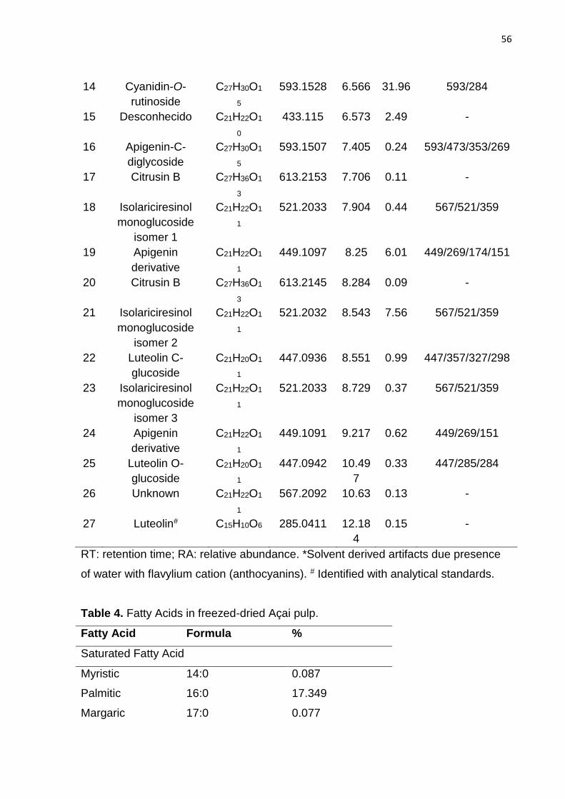
56
14 Cyanidin-O-
rutinoside
C27H30O1
5
593.1528 6.566 31.96 593/284
15 Desconhecido C21H22O1
0
433.115 6.573 2.49 -
16 Apigenin-C-
diglycoside
C27H30O1
5
593.1507 7.405 0.24 593/473/353/269
17 Citrusin B C27H36O1
3
613.2153 7.706 0.11 -
18 Isolariciresinol
monoglucoside
isomer 1
C21H22O1
1
521.2033 7.904 0.44 567/521/359
19 Apigenin
derivative
C21H22O1
1
449.1097 8.25 6.01 449/269/174/151
20 Citrusin B C27H36O1
3
613.2145 8.284 0.09 -
21 Isolariciresinol
monoglucoside
isomer 2
C21H22O1
1
521.2032 8.543 7.56 567/521/359
22 Luteolin C-
glucoside
C21H20O1
1
447.0936 8.551 0.99 447/357/327/298
23 Isolariciresinol
monoglucoside
isomer 3
C21H22O1
1
521.2033 8.729 0.37 567/521/359
24 Apigenin
derivative
C21H22O1
1
449.1091 9.217 0.62 449/269/151
25 Luteolin O-
glucoside
C21H20O1
1
447.0942 10.49
7
0.33 447/285/284
26 Unknown C21H22O1
1
567.2092 10.63 0.13 -
27 Luteolin# C15H10O6 285.0411 12.18
4
0.15 -
RT: retention time; RA: relative abundance. *Solvent derived artifacts due presence
of water with flavylium cation (anthocyanins). # Identified with analytical standards.
Table 4. Fatty Acids in freezed-dried Açai pulp.
Fatty Acid Formula %
Saturated Fatty Acid
Myristic 14:0 0.087
Palmitic 16:0 17.349
Margaric 17:0 0.077

57
Stearic 18:0 3.103
Eicosanoic 20:0 0.312
Behenic 22:0 0.052
Lignoceric 24:0 0.059
Total 21.039
Monounsaturated Fatty Acid
Palmitoleic 16:1 0.374
Margaroleic 17:1 0.071
Oleic 18:1 69.519
Gadoleic 20:1 0.158
Total 70.122
Polyunsaturated Fatty Acids
Linoleic 18:2 8.039
Linolenic 18:3 0.793
Total 8.832
3.2 Mice's food and energy intake, body and tissue weights
The animals fed the high-fat diets showed higher body weights from the fourth
experimental week (p < 0.01). The NA group had lower body weight gain (p < 0.05)
than the control group (N) (Table 5), even though the total food and calorie intake
were similar between these groups (Table 5). The mice fed the normal-fat diets had a
higher food intake in comparison to the high-fat diets-fed groups, but the opposite
was observed concerning the total energy intake (Table 5).
Regarding to the adipose tissue pad weight (EAT, RAT and MAT), the intake
of the high-fat diet increased the fat mass in H group in comparison to N (Table 5).
Although, even if the body weight was not different (p < 0.05) between H and HA
groups, the EAT and MAT weights were smaller in the HA-mice, when compared to H
(23.6% and 19.9% reduction, respectively) but no differences were found for RAT
weight. The relative brain weight was lower in the high-fat diet-fed mice than the
normal-fat group. No changes were observed in the relative liver weight.

58
Table 5. Growth, food intake parameters and tissue weights ratio (g 100 g-1 total
body weight) of the mice after 10 experimental weeks.
Parameter N NA H HA
Final body weight 52.56 ± 4.45 48.54 ± 4.48• 59.85 ± 4.92### 57.97 ± 4.55
Total weight gain 14.01 ± 3.13 12.31 ± 2.22• 20.46 ± 3.42### 19.12 ± 4.04
Food intake
407.23 ± 35.87 384.55 ± 37.28
293.22 ±
39.65### 282.87 ± 70.71
Calorie intake 1487.34 ±
112.71
1400.88 ±
122.13
1601.26 ±
151.10#
1535.47 ±
383.17
Brain 0.657 ± 0.020 0.704 ± 0.014 0.591 ± 0.017# 0.579 ± 0.010
Liver 4.632 ± 0.236 4.299 ± 0.131 5.159 ± 0.421 4.854 ± 0.266
EAT 4.312 ± 0.17 4.534 ± 0.26 5.370 ± 0.31## 4.104 ± 0.25**
RAT 1.527 ± 0.09 1.764 ± 0.13 2.530 ± 0.38# 2.446 ± 0.23
MAT 1.791 ± 0.09 1.678 ± 0.12 2.879 ± 0.16### 2.361 ± 0.13*
N: normal-fat diet fed group; NA: normal-fat diet plus 2% FAP group; H: high-fat diet-fed
group; HA: high-fat diet containing 2% FAP group. Final body weight, total weight gain and
food intake (g); Calorie intake (kcal); EAT: epididymal adipose tissue; RAT: retroperitoneal
adipose tissue; MAT: mesenteric adipose tissue. Values are expressed as mean ± SEM, n =
13. Data were analyzed using analysis of variance followed by Student’s t test. #Indicates
significant differences between groups N and H; •Indicates significant differences between
groups N and NA; *Indicates significant differences between groups H and HA according to
Student’s t test •, # or *p < 0.05; ## or **p < 0.01; and ### < 0.001.
3.3 Glucose Tolerance Test, peripheral insulin resistance and fasting adiponectin
In the GTT and ITT tests, the H group showed the highest (p < 0.05) AUC in
response to both glucose and insulin injection, when compared to the N group
(Figure 1). The AUC values for blood glucose levels during the GTT did not differ
between the high-fat diet-fed groups, which demonstrate that glucose intolerance
was not prevented by the intake of HA-diet. However, the blood glucose response
after insulin injection in ITT was lower in HA versus H, suggesting a better insulin
sensitivity in the HA-fed animals (Figure 1). Furthermore, fasting glucose and fasting
adiponectin in the last experimental day was higher in H in comparison to N and HA
groups (p < 0.05; Figure 2).

59
Figure 1 - Glycemic response in GTT test (a); AUC response in GTT test (b);
Glycemic response in ITT test (c); AUC response in ITT test (d); Fasting blood
glucose levels (e). N: normal-fat diet-fed group; NA: normal-fat diet plus 2% FAP
group; H: high-fat diet fed group; HA: high-fat diet containing 2% FAP group. In (a)
and (c) data were analyzed by two-way ANOVA and the Bonferroni post-hoc test. In
(b), (d) and (e) the Student’s t test was applied. * Indicates difference between N and
H groups and #indicates difference between H and HA groups. * and # Indicates p <
0.05, ** p < 0.01 and *** and ### p < 0.001, n = 6.
(a) Glucose response in GTT
0 50 100 1500
100
200
300
400
500N
H
NA
HA** **
***
Minutes after glucose injection
Gly
ce
mia
(m
g d
L-1
)
(b) AUC of GTT
NNA H
HA
0
10000
20000
30000
40000
50000
***
AU
C
(e) Fasting glucose
NNA H
HA
0
50
100
150
200
** **
mg
dL
-1
(d) AUC of ITT
NNA H
HA
0
2000
4000
6000
8000
10000
*** **A
UC
(c) Glucose response in ITT
0 15 30 45 600
50
100
150
200
250
NA
N
H
HA
** ***
***
***
****
###
###
#
#
Minutes after insulin injection
Gly
ce
mia
(m
g d
L-1
)
(f) Fasting adiponectin
NNA H
HA
0
3
6
9
12
**
**
(µg
mL
-1)

60
3.4 Antioxidant defense
The hippocampal and serum catalase activity in the high-fat diet-fed mice were
lower than those fed the normal-fat diet (Table 6). However, the addition of AP in
high-fat diet seemed to play a protective role in the hippocampus and serum showing
an improvement in the CAT activity (Table 6). Nevertheless, the AP-supplemented
groups showed an increase in hippocampal and serum (NA) levels, when compared
to their respective control groups. The SOD activity was lower in H group in
comparison to the normal fat diet-fed group and the addition of AP in this diet
improved the antioxidant activity of this enzyme in NA group. GSH hippocampus HA
H
Table 6. Antioxidant enzyme activity and reduced glutathione contents in the mice
groups after 10 experimental weeks.
Enzyme N NA H HA
Hippocampus
CAT 3.62 ± 0.13 4.05 ± 0.32 2.80 ± 0.31## 3.82 ± 0.60*
GSH 174.23 ± 11.67 219.11 ± 16.77* 158.03 ± 19.68 199.41 ± 26.74
SOD 4.54 ± 0.12 5.39 ± 0.18* 4.52 ± 0.48 4.28 ± 0.24
Serum
CAT 19.53 ± 1.73 19.58 ± 1.10 14.94 ± 1.34# 21.00 ± 1.78*
GSH 133.27 ± 2.41 128.74 ± 6.19 118.68 ± 6.89# 138.70 ± 6.41
SOD 19.42 ± 1.32 18.85 ± 0.76 14.25 ± 1.51# 15.74 ± 0.95
N: normal-fat diet fed group; NA: normal-fat diet plus 2% AP group; H: high-fat
diet fed group; HA: high-fat diet containing 2% AP group. CAT: catalase (nmol
min-1 mg-1 formed formaldehyde); GSH: reduced glutathione (nmol mg-1); SOD:
superoxide dismutase (U mg-1); #Indicates significant differences between groups
N and H; *Indicates significant differences between groups H and HA according
to Student’s t test # or *p < 0.05 and ## p < 0.01. Values expressed as means ±
SEM, n = 5.
3.5 Hippocampal protein quantification
The AKT/GSK3-β/tau axis was assessed in the hippocampus of the mice, a region of

61
the brain involved in learning and memory. The higher phosphorylation of AKT in the
groups N and HA relative to the H group improved the phosphorylation and
inactivation of GSK3-β in serine 9 in the HA group. A trend of lower phosphorylation
of GSK3-β was observed in H group in comparison to N (p = 0.067). Tau
phosphorylation in Thr 205 was increased in the hippocampus of H group when
compared to N, indicating the deleterious effect of the diet. A trend of lower
phosphorylation of tau in Thr 205 was observed in the group fed HA diet (p = 0.079)
(Figure 2). No differences were observed in synaptophysin (SYP).
Figure 2 – AKT phosphorylation (a); GSK3-β phosphorylation (b); tau phosphorylation
(c); synaptophysin (d). N: normal-fat diet fed group; NA: normal-fat diet plus 2% AP
group; H: high-fat diet fed group; HA: high-fat diet containing 2% AP group. The results
are shown as a percentage of the control (N group as 100%) Data were analyzed using
analysis of variance followed by Students t test. *Indicates p < 0.05 and **p < 0.01
between N × H and H × HA groups, n = 5.
SYP
β-actin
(d) SYP
NNA H
HA
0
50
100
150
Arb
itra
ry u
nits
(b) p-GSK3- / GSK3-
NNA H
HA
0
50
100
150
200
*
Arb
itra
ry u
nits
p-GSK3-β
GSK3-β
(a) p-AKT / AKT
NNA H
HA
0
50
100
150
200 **
*
Arb
itra
ry u
nits
p-AKT
AKT
(c) p-tau / tau
NNA H
HA
0
50
100
150
200 *
Arb
itra
ry u
nits
p-tau
tau

62
3.6 Novel Object Recognition
The high-fat group demonstrated significantly decrease in recognition index
(RI) when exposed to novel object compared to N group, failing to show a preference
for the novel object, suggesting a deficit in object recognition memory in this test.
Although, HA-mice showed preference for novel object over the familiar ones (p <
0.05), suggesting improvement in object recognition memory when compared to
high-fat diet-fed mice (H) (Figure 3).
Figure 3 - Long-term recognition memory assessed by the novel object recognition
(NOR) test. Recognition Index: a percent of time spent exploring the familiar or the
novel object relative to the total time spent exploring both objects. N: normal-fat diet
fed group; NA: normal-fat diet plus 2% FAP group; H: high-fat diet fed group; HA:
high-fat diet containing 2% FAP group. *Indicates p < 0.05 according to analysis of
variance followed by Student’s t test.
4. Discussion
Many studies have demonstrated the effects of açai intake on health,
highlighting anti-inflammatory, antioxidant, antitumoral and anti-obesogenic
properties (Choi et al., 2017; De Oliveira et al., 2015; Dias et al., 2015; Schauss, Wu,
Prior, Ou, Huang, et al., 2006; Silva et al., 2014). However, there are few studies
relating açai intake effects on cognition. Since the past decade, obesity, marked by
the consumption of a diet rich in saturated fatty acids and simple sugar, has been
related as a risk factor for both insulin resistance and cognitive impairment (Beydoun,
Beydoun, & Wang, 2008; Leboucher et al., 2013). In this study, we showed that the
NOR - Recognition Index
NNA H
HA
0
20
40
60
80* *
Pre
fere
nce
fo
r N
ove
l o
bje
ct
(%)

63
addition of açai pulp to a high-fat diet prevented peripheral insulin resistance, visceral
fat mass gain and improved cognitive behavior in mice.
Açai is known by its great amount of bioactive compounds like (poly)phenols,
especially flavonoids and anthocyanins as predominant phytochemicals. Cyanidin 3-
rutinoside, one of the identified compounds in this study, was cited as the major
anthocyanin in açai (1.25 – 1.93 mg g-1), followed by cyanidin-3-glucoside (0.95 –
1.17 mg g-1) (Pacheco-Palencia, Duncan, & Talcott, 2009; Schauss et al., 2006).
Both anthocyanins are extensively related to health benefits in other animals models,
in vitro and also in human trials (Ereminas et al., 2017; Karlsen et al., 2007; Qin et
al., 2009; Thilavech et al., 2017; You et al., 2017). Furthermore, we found
monounsaturated fatty acid (MUFA) (oleic acid, omega-9) as the main fraction of the
total lipids in açai pulp. Regarding dietary fiber, the insoluble fraction was the
predominant. Also its content of sugars is very low (Schauss et al., 2006). These
findings are similar to other studies that characterized açai; however it is possible to
find some divergences into different species (Smith et al., 2012; Luo et al., 2012;
Schauss et al., 2006).
Although açai supplementation did not prevent weight gain in the high-fat
group, we observed a reduction in adipose tissue pad weights (MAT and EAT). The
prevention of fat mass gain has also been observed after administration of 2% açai
pulp in rodents being associated or not with total body weight loss (De Oliveira et al.,
2015; Guerra et al., 2015). A previous report evaluated the consumption of different
types of berries and showed increased total weight gain and liver weight in rodents
after intake of 20% açai in a high-fat diet. However, the same study related açai
intake to increased lean body mass percentage and the relative size of the
epididymal fat pad was lower in group receiving açai, when compared to the high-fat
group (Heyman et al., 2014). Açai was the berry who had the highest fat content
among all the assessed berries in the mentioned study (Heyman et al., 2014), and
the 20% supplementation represents a total of 3.46 g 100 g-1 in diet of palmitic acid in
the diet. In this context, we chose smaller dose of açai, since an elevated
consumption of palmitic acid was once related to increase of body weight and liver
triglycerides in mice, which could explain their findings (Wit et al., 2012).
The adipose tissue is responsible for the production of adipokines such as
leptin and adiponectin, but in obesity, due to the hypertrophy and consequent tissue

64
inflammation, the production of these cytokines is unbalanced (Tilg & Moschen,
2006; Zhang & Scarpace, 2006). Adiponectin has a potent insulin-sensitizing effect
and in obesity both adiponectin release and adiponectin receptors are down-
regulated. The up-regulation of this adipokine and adiponectin receptor, as well as its
functions is one of the mechanism of most interest in terms of prevention of obesity-
linked insulin resistance (Yadav et al., 2013). In our study, we showed a lower
peripheral insulin resistance and elevated levels of adiponectin in serum of the mice
that consumed the HA-diet. The C3G anthocyanin was once reported to stimulate the
expression of adiponectin by the adipose tissue probably because its involvement in
the acetylation of FoxO1, which is a transcription factor that regulates the adiponectin
production (Liu et al., 2014).
A recent study has demonstrated that the treatment of obese mice with an
açai aqueous extract (dose) also attenuated insulin resistance and ameliorated
adiponectin levels in serum with no alteration the body weight gain (Carey, Miller, et
al., 2017). Even that the whole pulp fruit, including the lipid fraction, was used in our
experimental diets, our results corroborate the cited study, evidencing that the
proportion of palmitic acid of the supplementation (0.34 g 100 g-1) was not harmful
according to the analyzed parameters. Furthermore, as we demonstrated, açai has
oleic acid as the main fatty acid in its composition. Studies demonstrated that the
addition of MUFAs to the diet of healthy subjects improved insulin sensitivity (Vessby
et al., 2001). The consumption of MUFAs has also prevented postprandial decrease
in peripheral adiponectin gene expression and insulin resistance in insulin-resistant
subjects (Paniagua González et al., 2007). Thus, the fatty fraction might be the
differential advantage from açai in relation to other berries.
Diets rich in fruits and their (poly)phenols have been used in many studies
targeting the improvement of brain functions such as learning and memory (Jeon et
al., 2012). Flavonoid and anthocyanin-rich foods were shown to be effective in
reversing deficits in learning and memory, which could be affected by aging or a
long-term consumption of high-fat diets (Batista et al., 2017; Carey et al., 2017;
Carey et al., 2014). Our research group has demonstrated that the intake of a
Brazilian berry concomitantly to a high-fat diet could prevent the impairment in
hippocampal function (Batista et al., 2017). The present study extended these
searches to açai pulp. We showed that the consumption of HA-diet increased the

65
phosphorylation of AKT, leading to a higher phosphorylation and consequent
inactivation of GSK3-β and a trend to lower tau phosphorylation at the Thr 205 site in
the hippocampus of the mice. In an insulin resistance status, caused by obesity,
AKT’s propagation signals is inactivated (Greco et al., 2009). The AKT kinase is
responsible for phosphorylating GSK3-β, which when phosphorylated is inactive on
phosphorylating tau, preventing subsequent formation of NFT, a classic hallmark of
tauopathies observed in many neurodegenerative diseases (Kaytor & Orr, 2002). A
higher activation of IRS in hippocampal tissue was associated to a reduced tau
phosphorylation in the same model of mice fed a high-fat diet containing jaboticaba
berry, which indicated a better central insulin sensitivity in those animals when
compared to the control high-fat group (Batista et al., 2017; Schubert et al., 2003).
This could be a possible mechanism to explain the increase of AKT phosphorylation
in the HA group, since the insulin receptor can activate PI3K leading to
phosphorylation and activation of AKT (Duan, Li, & Rui, 2004).
Many sites of phosphorylation of tau are been under recent research to
discover the mechanisms of tauopathies . Tau binds to microtubules which provides
a physical pathway for axonal transporters and support cellular structure in neuronal
cells, and is regulated by many different kinases and phosphatases (Himmler et al.,
1989). Its hyperphosphorylation compromises the capability to bind and to stabilize
microtubules, as well as to aggregate when the equilibrium is harmed, resulting in
the formation of higher order structures, such as NFTs (Köpke et al., 1993). In the
present study, we evaluated the Thr 205 site and showed no significant decrease in
phosphorylation of supplemented HA-group compared to high-fat group. However,
the inactivation of GSK3-β could prevent tau’s phosphorylation in other sites such as
Ser 199, Ser 202, Ser 396, Thr 231, Thr 181, among others documented elsewhere
(García-Font et al., 2016; Liang et al., 2016; Sawmiller et al., 2016; Song et al.,
2016).
Although tau’s phosphorylation did not decrease, the group fed the high-fat
diet with açai pulp had a better behavior in NOR test relative to its control, presenting
an improvement in object recognition memory, denunciating a prevention of cognitive
impairment induced by the diet. The hippocampus plays a key role in comparing
previously stored information with new incoming aspects of one particular situation,
and is important for recognition memory as a "novelty detector"; if there are lesions in
66
this structure, it will occur moderate and reliable memory impairment (Broadbent et
al., 2010; Clarke et al., 2010).
The better performance in NOR test by the mice fed the high-fat diet
supplemented with açai might also have been related to other mechanisms. For
example, the expression of neurotrophins like brain-derived neurotrophic factor
(BDNF) through the ERK/CREB signaling were associated to the consumption of
berries and flavonoids (Rendeiro et al., 2014; Williams et al., 2008). Studies have
suggested that flavonoids induces both ERK1/2 and CREB activation (Maher,
Akaishi, & Abe, 2006; Schroeter et al., 2007). The CREB’s activation, in its turn, is
involved with the expression of genes, such as BDNF (Tao et al., 1998). This
neurothrophin appears to regulate neuronal function and memory (via the PI3
kinase/AKT/mTOR signaling pathway), leading to an increase translational efficacy
and capacity (Bekinschtein et al., 2007; Wullschleger, Loewith, & Hall, 2006).
Previous data showed açai’s administration efficacy in protect brain cell culture
from oxidative stress and inflammatory insults in critical neuronal regions involved in
memory, cognition and overall brain function (Carey et al., 2017). In a different model
of cognitive/memory impairment, the consumption of açai has reversed the
deleterious effects of aging on motor and cognitive behavior, as well as improved
working memory assessed by the Morris water maze (a test of spatial memory)
(Carey et al., 2017; Poulose et al., 2017). However, this is the first report about how
the intake of açai pulp behaves in the prevention of hippocampal impairment induced
by a high-fat diet in middle-aged mice investigating the tau phosphorylation and the
NOR test.
We have also shown an elevated activity of CAT and a trend of increase in
GSH values in the hippocampus of the HA group in comparison to the H group,
indicating increase of the activation of the local antioxidant defense system. The CNS
is especially vulnerable to free radical damage because of its lipid content. The brain
also have a high oxygen consumption rate and a lower relative amount of antioxidant
enzymes compared with other tissues (Coyle & Puttfarcken, 1993). An increased
brain oxidative stress seems to have an important role in cognitive impairment
caused by normal aging and neurodegenerative diseases (Padurariu et al., 2013)
and the administration of diets containing components like (poly)phenols and other
antioxidant compounds has been shown to improve such deficit, since the antioxidant

67
enzymes play a role in protection of cells against the oxidative stress (Batista et al.,
2017; Veberic et al., 2015).
In conclusion, our results indicate that the supplementation of the high-fat
diet with 2% açai pulp showed beneficial effects in prevention of the deleterious
effects in peripheral insulin resistance and in cognitive impairment related to obesity.
The improvement in AKT and GSK3-β phosphorylation seen in this group
corroborates these findings and suggests more investigation of other protein
signaling pathways in the hippocampus, like the CREB/BDNF, as well as other
phosphorylation sites of tau. Thus, the cognitive improvements seen in NOR test by
the animals that received the high-fat diet supplemented with açai pulp could be
better elucidated.
Acknowledgements
MRMJ acknowledges CNPq (301108/2016-1) and FAPESP (2015/50333-1) for
financial support.
5. References
AOAC. (1995). Official methods of analysis of AOAC International. Association of
Official Analysis Chemists International.
https://doi.org/10.3109/15563657608988149
AOAC. (2012). Official Methods of Analysis of AOAC International. Association of
Official Analysis Chemists International.
AOCS. (2009). AOCS Official Method Cd 1c-85. Calculated iodine value. Official
Methods and Recommended Practices of the American Oil Chemists’ Society.
Batista, Â. G., Lenquiste, S. A., Cazarin, C. B. B., da Silva, J. K., Luiz-Ferreira, A.,
Bogusz, S., … Maróstica, M. R. (2014). Intake of jaboticaba peel attenuates
oxidative stress in tissues and reduces circulating saturated lipids of rats with
high-fat diet-induced obesity. Journal of Functional Foods, 6(1), 450–461.
https://doi.org/10.1016/j.jff.2013.11.011
Batista, Â. G., LENQUISTE, S. A., MOLDENHAUER, C., GODOY, J. T., REIS, S. M.
P. M., & JÚNIOR, M. R. M. (2013). Jaboticaba ( Myrciaria jaboticaba ( Vell .)

68
Berg .) peel improved triglycerides excretion and hepatic lipid peroxidation in
high-fat-fed rats Consumo de casca de jabuticaba ( Myrciaria jaboticaba, 26(5),
571–581.
Batista, Â. G., Soares, E. S., Mendonça, M. C. P., da Silva, J. K., Dionísio, A. P.,
Sartori, C. R., … Maróstica Júnior, M. R. (2017). Jaboticaba berry peel intake
prevents insulin-resistance-induced tau phosphorylation in mice. Molecular
Nutrition and Food Research, 61(10). https://doi.org/10.1002/mnfr.201600952
Bekinschtein, P., Katche, C., Slipczuk, L. N., Igaz, L. M., Cammarota, M., Izquierdo,
I., & Medina, J. H. (2007). mTOR signaling in the hippocampus is necessary for
memory formation. Neurobiology of Learning and Memory, 87(2), 303–307.
https://doi.org/10.1016/j.nlm.2006.08.007
Beydoun, M. A., Beydoun, H. A., & Wang, Y. (2008). Obesity and central obesity as
risk factors for incident dementia and its subtypes: A systematic review and
meta-analysis. Obesity Reviews. https://doi.org/10.1111/j.1467-
789X.2008.00473.x
Bligh, E. G., & Dyer, W. J. (1959). A rapid method of total lipid extration and
purification. Canadian Journal of Biochemistry and Physiology, 37(8), 911–917.
https://doi.org/10.1139/o59-099
Bradford, M. M. (1976). A rapid and sensitive method for the quantitation of
microgram quantities of protein using the principle of protein dye binding.
Analytical Biochemistry, 72, 248–254. https://doi.org/10.1016/0003-
2697(76)90527-3
Broadbent, N. J., Gaskin, S., Squire, L. R., & Clark, R. E. (2010). Object recognition
memory and the rodent hippocampus. Learning & Memory, 17(1), 5–11.
https://doi.org/10.1101/lm.1650110
Carey, A. N., Gildawie, K. R., Rovnak, A., Thangthaeng, N., Fisher, D. R., & Shukitt-
Hale, B. (2017). Blueberry supplementation attenuates microglia activation and
increases neuroplasticity in mice consuming a high-fat diet. Nutritional
Neuroscience, 1–11. https://doi.org/10.1080/1028415X.2017.1376472
Carey, A. N., Gomes, S. M., & Shukitt-Hale, B. (2014). Blueberry supplementation

69
improves memory in middle-aged mice fed a high-fat diet. Journal of Agricultural
and Food Chemistry, 62(18), 3972–3978. https://doi.org/10.1021/jf404565s
Carey, A. N., Lyons, A. M., Shay, C. F., Dunton, O., & McLaughlin, J. P. (2009).
Endogenous kappa opioid activation mediates stress-induced deficits in learning
and memory. The Journal of Neuroscience : The Official Journal of the Society
for Neuroscience, 29(13), 4293–300. https://doi.org/10.1523/JNEUROSCI.6146-
08.2009
Carey, A. N., Miller, M. G., Fisher, D. R., Bielinski, D. F., Gilman, C. K., Poulose, S.
M., & Shukitt-Hale, B. (2017). Dietary supplementation with the polyphenol-rich
açaí pulps (Euterpe oleracea Mart. and Euterpe precatoria Mart.) improves
cognition in aged rats and attenuates inflammatory signaling in BV-2 microglial
cells. Nutritional Neuroscience, 20(4), 238–245.
https://doi.org/10.1080/1028415X.2015.1115213
Choi, Y. J., Choi, Y. J., Kim, N., Nam, R. H., Lee, S., Lee, H. S., … Lee, D. H. (2017).
Açaí Berries Inhibit Colon Tumorigenesis in Azoxymethane/Dextran Sulfate
Sodium-Treated Mice. Gut and Liver, 11(2), 243–252.
https://doi.org/10.5009/gnl16068
Clarke, J. R., Cammarota, M., Gruart, A., Izquierdo, I., & Delgado-Garcia, J. M.
(2010). Plastic modifications induced by object recognition memory processing.
Proc Natl Acad Sci U S A, 107(6), 2652–2657. https://doi.org/0915059107
[pii]\r10.1073/pnas.0915059107
Coyle, J. T., & Puttfarcken, P. (1993). Oxidative stress, glutamate, and
neurodegenerative disorders. Science (New York, N.Y.), 262(5134), 689–95.
https://doi.org/10.1126/science.7901908
Dávalos, A., Gómez-Cordovés, C., & Bartolomé, B. (2004). Extending Applicability of
the Oxygen Radical Absorbance Capacity (ORAC-Fluorescein) Assay. Journal of
Agricultural and Food Chemistry, 52(1), 48–54. https://doi.org/10.1021/jf0305231
De Oliveira, P. R. B., Da Costa, C. A., De Bem, G. F., Cordeiro, V. S. C., Santos, I.
B., De Carvalho, L. C. R. M., … Resende, A. C. (2015). Euterpe oleracea Mart.-
derived polyphenols protect mice from diet-induced obesity and fatty liver by
regulating hepatic lipogenesis and cholesterol excretion. PLoS ONE, 10(12).

70
https://doi.org/10.1371/journal.pone.0143721
de Wit, N., Derrien, M., Bosch-Vermeulen, H., Oosterink, E., Keshtkar, S., Duval, C.,
… van der Meer, R. (2012). Saturated fat stimulates obesity and hepatic
steatosis and affects gut microbiota composition by an enhanced overflow of
dietary fat to the distal intestine. AJP: Gastrointestinal and Liver Physiology,
303(5), G589–G599. https://doi.org/10.1152/ajpgi.00488.2011
Degenhardt, A., Knapp, H., & Winterhalter, P. (2000). Separation and purification of
anthocyanins by high-speed countercurrent chromatography and screening for
antioxidant activity. Journal of Agricultural and Food Chemistry, 48(2), 338–343.
https://doi.org/10.1021/jf990876t
Dias, M. M. dos S., Martino, H. S. D., Noratto, G., Roque-Andrade, A., Stringheta, P.
C., Talcott, S., … Mertens-Talcott, S. U. (2015). Anti-inflammatory activity of
polyphenolics from açai (Euterpe oleracea Martius) in intestinal myofibroblasts
CCD-18Co cells. Food Funct., 6(10), 3249–3256.
https://doi.org/10.1039/C5FO00278H
Duan, C., Li, M., & Rui, L. (2004). SH2-B promotes insulin receptor substrate 1
(IRS1)- and IRS2-mediated activation of the phosphatidylinositol 3-kinase
pathway in response to leptin. Journal of Biological Chemistry, 279(42), 43684–
43691. https://doi.org/10.1074/jbc.M408495200
E. Smith, R., Eaker, J., Tran, K., Goerger, M., Wycoff, W., U. O. Sabaa-Srur, A., & M.
da Silva Menezes, E. (2012). Insoluble Solids in Brazilian and Floridian Acai
(Euterpe oleraceae Mart.). The Natural Products Journale, 2(2), 95–98.
https://doi.org/10.2174/2210315511202020095
Ereminas, G., Majiene, D., Sidlauskas, K., Jakstas, V., Ivanauskas, L., Vaitiekaitis,
G., & Liobikas, J. (2017). Neuroprotective properties of anthocyanidin glycosides
against H2O2-induced glial cell death are modulated by their different stability
and antioxidant activity in vitro. Biomedicine and Pharmacotherapy, 94, 188–
196. https://doi.org/10.1016/j.biopha.2017.07.077
Freeman, L. R., Haley-Zitlin, V., Rosenberger, D. S., & Granholm, A.-C. (2014).
Damaging effects of a high-fat diet to the brain and cognition: A review of
proposed mechanisms. Nutritional Neuroscience, 17(6), 241–251.
71
https://doi.org/10.1179/1476830513Y.0000000092
García-Font, N., Hayour, H., Belfaitah, A., Pedraz, J., Moraleda, I., Iriepa, I., … Oset-
Gasque, M. J. (2016). Potent anticholinesterasic and neuroprotective
pyranotacrines as inhibitors of beta-amyloid aggregation, oxidative stress and
tau-phosphorylation for Alzheimer’s disease. European Journal of Medicinal
Chemistry, 118, 178–192. https://doi.org/10.1016/j.ejmech.2016.04.023
Greco, S. J., Sarkar, S., Casadesus, G., Zhu, X., Smith, M. A., Ashford, J. W., …
Tezapsidis, N. (2009). Leptin inhibits glycogen synthase kinase-3beta to prevent
tau phosphorylation in neuronal cells. Neuroscience Letters, 455(3), 191–4.
https://doi.org/10.1016/j.neulet.2009.03.066
Greco, S. J., Sarkar, S., Johnston, J. M., & Tezapsidis, N. (2009). Leptin regulates
tau phosphorylation and amyloid through AMPK in neuronal cells. Biochemical
and Biophysical Research Communications, 380(1), 98–104.
https://doi.org/10.1016/j.bbrc.2009.01.041
Greco, S. J., Sarkar, S., Johnston, J. M., Zhu, X., Su, B., Casadesus, G., …
Tezapsidis, N. (2008). Leptin reduces Alzheimer’s disease-related tau
phosphorylation in neuronal cells. Biochemical and Biophysical Research
Communications, 376(3), 536–541. https://doi.org/10.1016/j.bbrc.2008.09.026
Guerra, J. F. da C., Maciel, P. S., de Abreu, I. C. M. E., Pereira, R. R., Silva, M.,
Cardoso, L. de M., … Pedrosa, M. L. (2015). Dietary açai attenuates hepatic
steatosis via adiponectin-mediated effects on lipid metabolism in high-fat diet
mice. Journal of Functional Foods, 14, 192–202.
https://doi.org/10.1016/j.jff.2015.01.025
Guo, H., Xia, M., Zou, T., Ling, W., Zhong, R., & Zhang, W. (2012). Cyanidin 3-
glucoside attenuates obesity-associated insulin resistance and hepatic steatosis
in high-fat diet-fed and db/db mice via the transcription factor FoxO1. Journal of
Nutritional Biochemistry, 23(4), 349–360.
https://doi.org/10.1016/j.jnutbio.2010.12.013
Gutierres, J. M., Carvalho, F. B., Schetinger, M. R. C., Marisco, P., Agostinho, P.,
Rodrigues, M., … Spanevello, R. (2014). Anthocyanins restore behavioral and
biochemical changes caused by streptozotocin-induced sporadic dementia of
72
Alzheimer’s type. Life Sciences, 96(1–2), 7–17.
https://doi.org/10.1016/j.lfs.2013.11.014
Hartman, L., & Lago, R. C. A. (1973). Rapid preparation of fatty acid methyl esters
from lipids. Lab. Pract., 22(January), 475–476,494.
Heyman, L., Axling, U., Blanco, N., Sterner, O., Holm, C., & Berger, K. (2014).
Evaluation of beneficial metabolic effects of berries in high-fat fed C57BL/6J
mice. Journal of Nutrition and Metabolism, 2014.
https://doi.org/10.1155/2014/403041
Higby, W. K. (1962). A Simplified Method for Determination of Some Aspects of the
Carotenoid Distribution in Natural and Carotene???Fortified Orange Juice.
Journal of Food Science, 27(1), 42–49. https://doi.org/10.1111/j.1365-
2621.1962.tb00055.x
Himmler, A., Drechsel, D., Kirschner, M. W., & Martin, D. W. (1989). Tau consists of
a set of proteins with repeated C-terminal microtubule-binding domains and
variable N-terminal domains. Molecular and Cellular Biology, 9(4), 1381–8.
https://doi.org/10.1128/MCB.9.4.1381.Updated
Jeon, B. T., Jeong, E. A., Shin, H. J., Lee, Y., Lee, D. H., Kim, H. J., … Roh, G. S.
(2012). Resveratrol attenuates obesity-associated peripheral and central
inflammation and improves memory deficit in mice fed a high-fat diet. Diabetes,
61(6), 1444–1454. https://doi.org/10.2337/db11-1498
Kammerer, D., Carle, R., & Schieber, A. (2004). Quantification of anthocyanins in
black carrot extracts (Daucus carota ssp. sativus var. atrorubens Alef.) and
evaluation of their color properties. European Food Research and Technology,
219(5), 479–486. https://doi.org/10.1007/s00217-004-0976-4
Karlsen, A., Retterstøl, L., Laake, P., Paur, I., Bøhn, S. K., Sandvik, L., & Blomhoff,
R. (2007). Anthocyanins inhibit nuclear factor-kappaB activation in monocytes
and reduce plasma concentrations of pro-inflammatory mediators in healthy
adults. The Journal of Nutrition, 137(8), 1951–4.
https://doi.org/papers3://publication/uuid/829A150F-976C-4854-98D1-
D5D9F1DD7B25

73
Kaytor, M. D., & Orr, H. T. (2002). The GSK3β signaling cascade and
neurodegenerative disease. Current Opinion in Neurobiology.
https://doi.org/10.1016/S0959-4388(02)00320-3
Kim, B., & Feldman, E. L. (2015). Insulin resistance as a key link for the increased
risk of cognitive impairment in the metabolic syndrome. Experimental &
Molecular Medicine. https://doi.org/10.1038/emm.2015.3
Köpke, E., Tung, Y. C., Shaikh, S., Alonso, A. D. C., Iqbal, K., & Grundke-Iqbal, I.
(1993). Microtubule-associated protein tau: Abnormal phosphorylation of a non-
paired helical filament pool in Alzheimer disease. Journal of Biological
Chemistry, 268(32), 24374–24384. https://doi.org/10.1016/j.bbrc.2011.11.056
Kothari, V., Luo, Y., Tornabene, T., O’Neill, A. M., Greene, M. W., Geetha, T., &
Babu, J. R. (2017). High fat diet induces brain insulin resistance and cognitive
impairment in mice. Biochimica et Biophysica Acta (BBA) - Molecular Basis of
Disease, 1863(2), 499–508. https://doi.org/10.1016/j.bbadis.2016.10.006
Leboucher, A., Laurent, C., Fernandez-Gomez, F.-J., Burnouf, S., Troquier, L.,
Eddarkaoui, S., … Blum, D. (2013). Detrimental effects of diet-induced obesity
on $τ$ pathology are independent of insulin resistance in $τ$ transgenic mice.
Diabetes, 62(May), 1681–1688. https://doi.org/10.2337/db12-0866
Lee, E. B. (2011). Obesity, leptin, and Alzheimer’s disease. Annals of the New York
Academy of Sciences. https://doi.org/10.1111/j.1749-6632.2011.06274.x
Liang, Z., Zhang, B., Su, W. W., Williams, P. G., & Li, Q. X. (2016). C-
Glycosylflavones Alleviate Tau Phosphorylation and Amyloid Neurotoxicity
through GSK3β Inhibition. ACS Chemical Neuroscience, 7(7), 912–923.
https://doi.org/10.1021/acschemneuro.6b00059
Liu, Y., Li, D., Zhang, Y., Sun, R., & Xia, M. (2014). Anthocyanin increases
adiponectin secretion and protects against diabetes-related endothelial
dysfunction. AJP: Endocrinology and Metabolism, 306(8), E975–E988.
https://doi.org/10.1152/ajpendo.00699.2013
Luo, R., Tran, K., A. Levine, R., M. Nickols, S., M. Monroe, D., U. O. Sabaa-Srur, A.,
& E. Smith, R. (2012). Distinguishing Components in Brazilian Acai (Euterpe

74
oleraceae Mart.) and in Products Obtained in the USA by Using NMR. The
Natural Products Journale, 2(2), 86–94.
https://doi.org/10.2174/2210315511202020086
Maher, P., Akaishi, T., & Abe, K. (2006). Flavonoid fisetin promotes ERK-dependent
long-term potentiation and enhances memory. Proceedings of the National
Academy of Sciences, 103(44), 16568–16573.
https://doi.org/10.1073/pnas.0607822103
Malin, D. H., Lee, D. R., Goyarzu, P., Chang, Y. H., Ennis, L. J., Beckett, E., …
Joseph, J. A. (2011). Short-term blueberry-enriched diet prevents and reverses
object recognition memory loss in aging rats. Nutrition, 27(3), 338–342.
https://doi.org/10.1016/j.nut.2010.05.001
Morris, M. C., & Tangney, C. C. (2014). Dietary fat composition and dementia risk.
Neurobiology of Aging. https://doi.org/10.1016/j.neurobiolaging.2014.03.038
Norton, S., Matthews, F. E., Barnes, D. E., Yaffe, K., & Brayne, C. (2014). Potential
for primary prevention of Alzheimer’s disease: An analysis of population-based
data. The Lancet Neurology, 13(8), 788–794. https://doi.org/10.1016/S1474-
4422(14)70136-X
Oliveira de Souza, M., Silva, M., Silva, M. E., de Paula Oliveira, R., & Pedrosa, M. L.
(2010). Diet supplementation with acai (Euterpe oleracea Mart.) pulp improves
biomarkers of oxidative stress and the serum lipid profile in rats. Nutrition, 26(7–
8), 804–810. https://doi.org/10.1016/j.nut.2009.09.007
Ou, B., Chang, T., Huang, D., & Prior, R. L. (2013). Determination of total antioxidant
capacity by oxygen radical absorbance capacity (ORAC) using fluorescein as
the fluorescence probe: First action 2012.23. Journal of AOAC International,
96(6), 1372–1376. https://doi.org/10.5740/jaoacint.13-175
Pacheco-Palencia, L. A., Duncan, C. E., & Talcott, S. T. (2009). Phytochemical
composition and thermal stability of two commercial açai species, Euterpe
oleracea and Euterpe precatoria. Food Chemistry, 115(4), 1199–1205.
https://doi.org/10.1016/j.foodchem.2009.01.034
Padurariu, M., Ciobica, A., Lefter, R., Serban, I. L., Stefanescu, C., & Chirita, R.

75
(2013). The oxidative stress hypothesis in Alzheimer’s disease. Psychiatria
Danubina. https://doi.org/10.1111/head.12009
Paniagua González, J. A., Gallego De La Sacristana, A., Romero, I., Vidal-Puig, A.,
Latre, J. M., Sanchez, E., … Perez-Jimenez, F. (2007). Monounsaturated fat-rich
diet prevents central body fat distribution and decreases postprandial
adiponectin expression induced by a carbohydrate-rich diet in insulin-resistant
subjects. Diabetes Care, 30(7), 1717–1723. https://doi.org/10.2337/dc06-2220
Poulose, S. M., Bielinski, D. F., Carey, A., Schauss, A. G., & Shukitt-Hale, B. (2017).
Modulation of oxidative stress, inflammation, autophagy and expression of Nrf2
in hippocampus and frontal cortex of rats fed with açaí-enriched diets. Nutritional
Neuroscience, 20(5), 305–315. https://doi.org/10.1080/1028415X.2015.1125654
Prior, R. L., Hoang, H., Gu, L., Wu, X., Bacchiocca, M., Howard, L., … Jacob, R.
(2003). Assays for hydrophilic and lipophilic antioxidant capacity (oxygen radical
absorbance capacity (ORACFL)) of plasma and other biological and food
samples. Journal of Agricultural and Food Chemistry, 51(11), 3273–3279.
https://doi.org/10.1021/jf0262256
Qin, L., Zhang, J., & Qin, M. (2013). Protective effect of cyanidin 3-O-glucoside on
beta-amyloid peptide-induced cognitive impairment in rats. Neuroscience
Letters, 534(1), 285–288. https://doi.org/10.1016/j.neulet.2012.12.023
Qin, Y., Xia, M., Ma, J., Hao, Y. T., Liu, J., Mou, H. Y., … Ling, W. H. (2009).
Anthocyanin supplementation improves serum LDL- and HDL-cholesterol
concentrations associated with the inhibition of cholesteryl ester transfer protein
in dyslipidemic subjects. American Journal of Clinical Nutrition, 90(3), 485–492.
https://doi.org/10.3945/ajcn.2009.27814
Reeves, Nielsen, F. H., & Fahey, G. C. (1993). AIN-93 Purified Diets for Laboratory
Rodents: Final Report of the American Institute of Nutrition Ad Hoc Writing
Committee on the Reformulation of the AIN-76A Rodent Diet. The Journal of
Nutrition, 123(11), 1939–1951. https://doi.org/10.1017/CBO9781107415324.004
Rendeiro, C., Foley, A., Lau, V. C., Ring, R., Rodriguez-Mateos, A., Vauzour, D., …
Spencer, J. P. E. (2014). A role for hippocampal PSA-NCAM and NMDA-NR2B
receptor function in flavonoid-induced spatial memory improvements in young

76
rats. Neuropharmacology, 79, 335–344.
https://doi.org/10.1016/j.neuropharm.2013.12.003
Rufino, M. do S. M., Alves, R. E., de Brito, E. S., Pérez-Jiménez, J., Saura-Calixto,
F., & Mancini-Filho, J. (2010). Bioactive compounds and antioxidant capacities
of 18 non-traditional tropical fruits from Brazil. Food Chemistry, 121(4), 996–
1002. https://doi.org/10.1016/j.foodchem.2010.01.037
Sawmiller, D., Habib, A., Li, S., Darlington, D., Hou, H., Tian, J., … Tan, J. (2016).
Diosmin reduces cerebral Aβ levels, tau hyperphosphorylation,
neuroinflammation, and cognitive impairment in the 3xTg-AD mice. Journal of
Neuroimmunology, 299, 98–106. https://doi.org/10.1016/j.jneuroim.2016.08.018
Schauss, A. G., Wu, X., Prior, R. L., Ou, B., Huang, D., Owens, J., … Shanbrom, E.
(2006). Antioxidant Capacity and Other Bioactivities of the Freeze-Dried
Amazonian Palm Berry, Euterpe oleraceae Mart. (Acai). Journal of Agricultural
and Food Chemistry, 54(22), 8604–8610. https://doi.org/10.1021/jf0609779
Schauss, A. G., Wu, X., Prior, R. L., Ou, B., Patel, D., Huang, D., & Kababick, J. P.
(2006). Phytochemical and nutrient composition of the freeze-dried amazonian
palm berry, Euterpe oleraceae Mart. (Acai). Journal of Agricultural and Food
Chemistry, 54(22), 8598–8603. https://doi.org/10.1021/jf060976g
Schroeter, H., Bahia, P., Spencer, J. P. E., Sheppard, O., Rattray, M., Cadenas, E.,
… Williams, R. J. (2007). (-)Epicatechin stimulates ERK-dependent cyclic AMP
response element activity and up-regulates GluR2 in cortical neurons. Journal of
Neurochemistry, 101(6), 1596–1606. https://doi.org/10.1111/j.1471-
4159.2006.04434.x
Schubert, M., Brazil, D. P., Burks, D. J., Kushner, J. a, Ye, J., Flint, C. L., … White,
M. F. (2003). Insulin receptor substrate-2 deficiency impairs brain growth and
promotes tau phosphorylation. The Journal of Neuroscience : The Official
Journal of the Society for Neuroscience, 23(18), 7084–7092.
https://doi.org/23/18/7084 [pii]
Schubert, M., Gautam, D., Surjo, D., Ueki, K., Baudler, S., Schubert, D., … Brüning,
J. C. (2004). Role for neuronal insulin resistance in neurodegenerative diseases.
Proceedings of the National Academy of Sciences of the United States of

77
America, 101(9), 3100–5. https://doi.org/10.1073/pnas.0308724101
Shukitt-Hale, B., Thangthaeng, N., Kelly, M. E., Smith, D. E., & Miller, M. G. (2017).
Raspberry differentially improves age-related declines in psychomotor function
dependent on baseline motor ability. Food & Function.
https://doi.org/10.1039/c7fo00894e
Silva, D. F., Vidal, F. C. B., Santos, D., Costa, M. C. P., Morgado-Díaz, J. A., do
Desterro Soares Brandão Nascimen, M., & de Moura, R. S. (2014). Cytotoxic
effects of Euterpe oleracea Mart. in malignant cell lines. BMC Complementary
and Alternative Medicine, 14(1), 175. https://doi.org/10.1186/1472-6882-14-175
Song, Y.-X., Miao, J.-Y., Qiang, M., He, R.-Q., Wang, X.-M., & Li, W.-W. (2016).
Icariin protects SH-SY5Y cells from formaldehyde-induced injury through
suppression of Tau phosphorylation. Chinese Journal of Integrative Medicine,
22(6), 430–437. https://doi.org/10.1007/s11655-015-2116-3
Spencer, S. J., Korosi, A., Layé, S., Shukitt-Hale, B., & Barrientos, R. M. (2017).
Food for thought: how nutrition impacts cognition and emotion. Npj Science of
Food, 1(1), 7. https://doi.org/10.1038/s41538-017-0008-y
Swain, T., & Hillis, W. E. (1959). The phenolic constituents of Prunus domestica. I.—
The quantitative analysis of phenolic constituents. Journal of the Science of
Food and Agriculture, 10(1), 63–68. https://doi.org/10.1002/jsfa.2740100110
Tao, X., Finkbeiner, S., Arnold, D. B., Shaywitz, A. J., & Greenberg, M. E. (1998).
Ca2+influx regulates BDNF transcription by a CREB family transcription factor-
dependent mechanism. Neuron, 20(4), 709–726. https://doi.org/10.1016/S0896-
6273(00)81010-7
Thilavech, T., Abeywardena, M. Y., Adams, M., Dallimore, J., & Adisakwattana, S.
(2017). Naturally occurring anthocyanin cyanidin-3-rutinoside possesses
inherent vasorelaxant actions and prevents methylglyoxal-induced vascular
dysfunction in rat aorta and mesenteric arterial bed. Biomedicine &
Pharmacotherapy = Biomedecine & Pharmacotherapie, 95, 1251–1259.
https://doi.org/10.1016/j.biopha.2017.09.053
Tilg, H., & Moschen, A. R. (2006). Adipocytokines: Mediators linking adipose tissue,

78
inflammation and immunity. Nature Reviews Immunology, 6(10), 772–783.
https://doi.org/10.1038/nri1937
Veberic, R., Slatnar, A., Bizjak, J., Stampar, F., & Mikulic-Petkovsek, M. (2015).
Anthocyanin composition of different wild and cultivated berry species. LWT -
Food Science and Technology, 60(1), 509–517.
https://doi.org/10.1016/j.lwt.2014.08.033
Vessby, B., Uusitupa, M., Hermansen, K., Riccardi, G., Rivellese, A. A., Tapsell, L.
C., … Storlien, L. H. (2001). Substituting dietary saturated for monounsaturated
fat impairs insulin sensitivity in healthy men and women: The KANWU study.
Diabetologia, 44(3), 312–319. https://doi.org/10.1007/s001250051620
Williams, C. M., El Mohsen, M. A., Vauzour, D., Rendeiro, C., Butler, L. T., Ellis, J. A.,
… Spencer, J. P. E. (2008). Blueberry-induced changes in spatial working
memory correlate with changes in hippocampal CREB phosphorylation and
brain-derived neurotrophic factor (BDNF) levels. Free Radical Biology and
Medicine, 45(3), 295–305. https://doi.org/10.1016/j.freeradbiomed.2008.04.008
Wrolstad, R. E. (1976). Color and Pigment Analyses in Fruit Products. Oregon State
University. Retrieved from
https://books.google.com.br/books?id=AsYmGwAACAAJ
Wullschleger, S., Loewith, R., & Hall, M. N. (2006). TOR signaling in growth and
metabolism. Cell. https://doi.org/10.1016/j.cell.2006.01.016
Yadav, A., Kataria, M. A., Saini, V., & Yadav, A. (2013). Role of leptin and
adiponectin in insulin resistance. Clinica Chimica Acta.
https://doi.org/10.1016/j.cca.2012.12.007
You, Y., Yuan, X., Liu, X., Liang, C., Meng, M., Huang, Y., … Zhan, J. (2017).
Cyanidin-3-glucoside increases whole body energy metabolism by upregulating
brown adipose tissue mitochondrial function. Molecular Nutrition and Food
Research, 61(11). https://doi.org/10.1002/mnfr.201700261
Youdim, K. A., Dobbie, M. S., Kuhnle, G., Proteggente, A. R., Abbott, N. J., & Rice-
Evans, C. (2003). Interaction between flavonoids and the blood-brain barrier: in
vitro studies. Journal of Neurochemistry, 85(1), 180–192.

79
https://doi.org/10.1046/j.1471-4159.2003.01652.x
Zhang, Y., & Scarpace, P. J. (2006). The role of leptin in leptin resistance and
obesity. Physiology and Behavior, 88(3), 249–256.
https://doi.org/10.1016/j.physbeh.2006.05.038
Zhishen, J., Mengcheng, T., & Jianming, W. (1999). The determination of flavonoid
contents in mulberry and their scavenging effects on superoxide radicals. Food
Chemistry, 64(4), 555–559. https://doi.org/10.1016/S0308-8146(98)00102-2
80
CAPÍTULO 3
CONCLUSÃO GERAL

81
CONCLUSÃO GERAL
A polpa de açaí tem ganhado cada vez mais espaço na alimentação da
população brasileira. Sua composição, rica em compostos bioativos como os
polifenóis, antocianinas, flavonoides e carotenoides, lhe garantem alta capacidade
antioxidante, que vem sendo extensivamente explorada na literatura em diversos
parâmetros de saúde. Entre seus atributos, esta fruta se destaca pela sua alta
contribuição energética devido ao elevado teor de lipídios, principalmente ácidos
graxos monoinsaturados ômega-9 e pela grande quantidade de fibras dietéticas,
principalmente as de característica insolúvel.
Ao ser adicionada à dieta hiperlipídica de camundongos, a polpa
liofilizada de açaí não induziu alteração no consumo de dieta e no peso dos animais,
mas os depósitos de gordura visceral dos mesmos estavam diminuídos. A
suplementação com PA promoveu menor glicemia de jejum e maior sensibilidade
periférica à ação da insulina durante o teste de tolerância à insulina pelos animais
alimentados com dieta hiperlipídica, não havendo alteração no teste de tolerância à
glicose. Houve um aumento nos níveis séricos de adiponectina nestes animais,
prevenindo o efeito deletério do excesso de gordura na dieta.
No hipocampo foi observado uma melhora na fosforilação de enzimas
envolvidas com a fosforilação da tau, entretanto esta não apresentou resultado
significativo no grupo HA. O teste de reconhecimento de objetos revelou que os
mesmos animais tiveram um melhor desempenho de memória, indicando que os
compostos do açaí podem ter tido uma ação independente da fosforilação da tau.
Em suma, o consumo de açaí juntamente com uma dieta rica em gordura
foi capaz de atenuar alguns efeitos deletérios à saúde e não promoveu nenhum
efeito negativo à saúde, dentre os parâmetros estudados. Os resultados obtidos
instigam novas pesquisas envolvendo outras vias metabólicas para completa
elucidação dos efeitos aqui encontrados e constatação do papel do açaí como
alimento funcional.

82
CAPÍTULO 4
REFERÊNCIAS GERAL
83
REFERÊNCIAS GERAL
ANTUNES, M.; BIALA, G. The novel object recognition memory: Neurobiology, test
procedure, and its modifications. Cognitive Processing, 2012.
AOAC. Official Methods of Analysis of AOAC International. 1995.
AOAC. Official methods of analysis of AOAC International. 2012. Disponível em:
<http://www.sidalc.net/cgi-
bin/wxis.exe/?IsisScript=agrissa.xis&method=post&formato=2&cantidad=1&expresio
n=mfn=002093>.
AOCS. AOCS Official Method Cd 1c-85. Calculated iodine value. Official
Methods and Recommended Practices of the American Oil Chemists’ Society,
2009.
AUGUSTINACK, J. C. et al. Specific tau phosphorylation sites correlate with severity
of neuronal cytopathology in Alzheimer’s disease. Acta Neuropathologica, v. 103,
n. 1, p. 26–35, 2002.
BANKS, W. A.; OWEN, J. B.; ERICKSON, M. A. Insulin in the brain: There and back
again. Pharmacology and Therapeutics, 2012.
BATISTA, Â. G. et al. Intake of jaboticaba peel attenuates oxidative stress in tissues
and reduces circulating saturated lipids of rats with high-fat diet-induced obesity.
Journal of Functional Foods, v. 6, n. 1, p. 450–461, 2014.
BATISTA, Â. G. et al . Jaboticaba (Myrciaria jaboticaba (Vell.) Berg.) peel improved
triglycerides excretion and hepatic lipid peroxidation in high-fat-fed rats. Revista de
Nutrição, Campinas , v. 26, n. 5, p. 571-581, Oct. 2013 .
BATISTA, Â. G. et al. Jaboticaba berry peel intake prevents insulin-resistance-
induced tau phosphorylation in mice. Molecular Nutrition and Food Research, v.
61, n. 10, 2017.
BAXTER, M. G. “I’ve seen it all before”: explaining age-related impairments in object
recognition. Theoretical comment on Burke et al. Behavioral neuroscience, v. 124,
n. 5, p. 706–709, 2010.
BEKINSCHTEIN, P. et al. mTOR signaling in the hippocampus is necessary for
memory formation. Neurobiology of Learning and Memory, v. 87, n. 2, p. 303–307,
2007.
BEYDOUN, M. A.; BEYDOUN, H. A.; WANG, Y. Obesity and central obesity as risk
factors for incident dementia and its subtypes: A systematic review and meta-
analysis. Obesity Reviews, 2008.
84
BHUROSY, T.; JEEWON, R. Overweight and obesity epidemic in developing
countries: a problem with diet, physical activity, or socioeconomic status? The
Scientific World Journal, v. 2014, p. 964236, 2014.
BLÁZQUEZ, E. et al. Insulin in the brain: Its pathophysiological implications for states
related with central insulin resistance, type 2 diabetes and alzheimer’s disease.
Frontiers in Endocrinology, 2014.
BLENNOW, K.; DE LEON, M. J.; ZETTERBERG, H. Alzheimer’s disease. Lancet, v.
368, n. 9533, p. 387–403, 2006.
BLIGH, E. G.; DYER, W. J. A rapid method of total lipid extration and purification.
Canadian Journal of Biochemistry and Physiology, v. 37, n. 8, p. 911–917, 1959.
BOITARD, C. et al. Impairment of hippocampal-dependent memory induced by
juvenile high-fat diet intake is associated with enhanced hippocampal inflammation in
rats. Brain, Behavior, and Immunity, v. 40, p. 9–17, 2014.
BRADFORD, M. M. A rapid and sensitive method for the quantitation of microgram
quantities of protein using the principle of protein dye binding. Analytical
Biochemistry, v. 72, p. 248–254, 1976.
BRASIL. VIGILÂNCIA DE FATORES DE RISCO E PROTEÇÃO PARA DOENÇAS
CRÔNICAS POR INQUÉRITO TELEFÔNICO - VIGITEL 2016. Ministério da Saúde.
Secretaria de Vigilância em Saúde. Departamento de Vigilância de Doenças e
Agravos não Transmissíveis e Promoção da Saúde. Disponível em:
<www.saude.gov.br/svs>.
BROADBENT, N. J. et al. Object recognition memory and the rodent hippocampus.
Learning & Memory, v. 17, n. 1, p. 5–11, 2010.
CAREY, A. N. et al. Blueberry supplementation attenuates microglia activation and
increases neuroplasticity in mice consuming a high-fat diet. Nutritional
Neuroscience, p. 1–11, 21 Sep. 2017b.
CAREY, A. N. et al. Dietary supplementation with the polyphenol-rich açaí pulps
(Euterpe oleracea Mart. and Euterpe precatoria Mart.) improves cognition in aged
rats and attenuates inflammatory signaling in BV-2 microglial cells. Nutritional
Neuroscience, v. 20, n. 4, p. 238–245, 2017a.
CAREY, A. N. et al. Endogenous kappa opioid activation mediates stress-induced
deficits in learning and memory. The Journal of neuroscience : the official journal
of the Society for Neuroscience, v. 29, n. 13, p. 4293–300, 2009.
CAREY, A. N.; GOMES, S. M.; SHUKITT-HALE, B. Blueberry supplementation
improves memory in middle-aged mice fed a high-fat diet. Journal of Agricultural
and Food Chemistry, v. 62, n. 18, p. 3972–3978, 2014.

85
CAVALLINI, A. et al. An unbiased approach to identifying tau kinases that
phosphorylate tau at sites associated with alzheimer disease. Journal of Biological
Chemistry, v. 288, n. 32, p. 23331–23347, 2013.
CHENG, G. et al. Diabetes as a risk factor for dementia and mild cognitive
impairment: A meta-analysis of longitudinal studies. Internal Medicine Journal,
2012.
CHOI, Y. J. et al. Açaí Berries Inhibit Colon Tumorigenesis in Azoxymethane/Dextran
Sulfate Sodium-Treated Mice. Gut and Liver, v. 11, n. 2, p. 243–252, 2017.
CLARKE, J. R. et al. Plastic modifications induced by object recognition memory
processing. Proc Natl Acad Sci U S A, v. 107, n. 6, p. 2652–2657, 2010.
ÇOBAN, J. et al. Blueberry treatment attenuates D-galactose-induced oxidative
stress and tissue damage in rat liver. Geriatrics and Gerontology International, v.
14, n. 2, p. 490–497, 2014.
COÏSSON, J. D. et al. Euterpe oleracea juice as a functional pigment for yogurt.
Food Research International. Anais...2005
CORREIA, S. C. et al. Insulin signaling, glucose metabolism and mitochondria: Major
players in Alzheimer’s disease and diabetes interrelation. Brain Research, 2012.
COYLE, J. T.; PUTTFARCKEN, P. Oxidative stress, glutamate, and
neurodegenerative disorders. Science (New York, N.Y.), v. 262, n. 5134, p. 689–95,
1993.
CROZIER, A.; JAGANATH, I. B.; CLIFFORD, M. N. Phenols, Polyphenols and
Tannins: An Overview. In: Plant Secondary Metabolites: Occurrence, Structure
and Role in the Human Diet. p. 1–24, 2007.
DÁVALOS, A.; GÓMEZ-CORDOVÉS, C.; BARTOLOMÉ, B. Extending Applicability of
the Oxygen Radical Absorbance Capacity (ORAC-Fluorescein) Assay. Journal of
Agricultural and Food Chemistry, v. 52, n. 1, p. 48–54, 2004.
DE FELICE, F. G.; FERREIRA, S. T. Inflammation, defective insulin signaling, and
mitochondrial dysfunction as common molecular denominators connecting type 2
diabetes to Alzheimer Disease. Diabetes, 2014.
DE LA MONTE, S. M. Alzheimer’s Disease Is Type 3 Diabetes—Evidence Reviewed.
Journal of Diabetes Science and Technology, v. 22, n. 6, p. 1101–1113, 2008.
DE OLIVEIRA, P. R. B. et al. Euterpe oleracea Mart.-derived polyphenols protect
mice from diet-induced obesity and fatty liver by regulating hepatic lipogenesis and
cholesterol excretion. PLoS ONE, v. 10, n. 12, 2015.
DE WIT, N. et al. Saturated fat stimulates obesity and hepatic steatosis and affects
gut microbiota composition by an enhanced overflow of dietary fat to the distal

86
intestine. AJP: Gastrointestinal and Liver Physiology, v. 303, n. 5, p. G589–G599,
2012.
DEGENHARDT, A.; KNAPP, H.; WINTERHALTER, P. Separation and purification of
anthocyanins by high-speed countercurrent chromatography and screening for
antioxidant activity. Journal of Agricultural and Food Chemistry, v. 48, n. 2, p.
338–343, 2000.
DEVORE, E. E. et al. Dietary intakes of berries and flavonoids in relation to cognitive
decline. Annals of Neurology, v. 72, n. 1, p. 135–143, 2012.
DIAS, M. M. DOS S. et al. Anti-inflammatory activity of polyphenolics from açai
(Euterpe oleracea Martius) in intestinal myofibroblasts CCD-18Co cells. Food
Function, v. 6, n. 10, p. 3249–3256, 2015.
DRAGANO, N. R. V. et al. Freeze-dried jaboticaba peel powder improves insulin
sensitivity in high-fat-fed mice. British Journal of Nutrition, v. 110, n. 3, p. 447–455,
2013.
DRAGICEVIC, N. et al. Green tea epigallocatechin-3-gallate (EGCG) and other
flavonoids reduce Alzheimer’s amyloid-induced mitochondrial dysfunction. Journal of
Alzheimer’s Disease, v. 26, n. 3, p. 507–521, 2011.
DUAN, C.; LI, M.; RUI, L. SH2-B promotes insulin receptor substrate 1 (IRS1)- and
IRS2-mediated activation of the phosphatidylinositol 3-kinase pathway in response to
leptin. Journal of Biological Chemistry, v. 279, n. 42, p. 43684–43691, 2004.
DUDEK, H.; DATTA, S. R. Regulation of neuronal survival by the serine-threonine
protein kinase Akt. Science, v. 275, n. 5300, p. 661, 1997.
E. SMITH, R. et al. Insoluble Solids in Brazilian and Floridian Acai (Euterpe
oleraceae Mart.). The Natural Products Journal, v. 2, n. 2, p. 95–98, 2012.
EL KHOURY, N. B. et al. Insulin dysfunction and Tau pathology. Frontiers in
Cellular Neuroscience, v. 8, 2014.
ELIAS, M. F. et al. Lower cognitive function in the presence of obesity and
hypertension: The Framingham heart study. International Journal of Obesity, v. 27,
n. 2, p. 260–268, 2003.
ENNACEUR, A. One-trial object recognition in rats and mice: Methodological and
theoretical issues. Behavioural Brain Research, 2010.
ENNACEUR, A.; DELACOUR, J. A new one - trial test for neurobiological studies of
memory in rats . 1 " Behavioral data. Behavioural Brain Research, v. 31, p. 47–59,
1988.
EREMINAS, G. et al. Neuroprotective properties of anthocyanidin glycosides against
H2O2-induced glial cell death are modulated by their different stability and

87
antioxidant activity in vitro. Biomedicine and Pharmacotherapy, v. 94, p. 188–196,
2017.
FARR, O. M.; TSOUKAS, M. A.; MANTZOROS, C. S. Leptin and the brain:
Influences on brain development, cognitive functioning and psychiatric disorders.
Metabolism: Clinical and Experimental, v. 64, n. 1, p. 114–130, 2015.
FREEMAN, L. R. et al. Damaging effects of a high-fat diet to the brain and cognition:
A review of proposed mechanisms. Nutritional Neuroscience, v. 17, n. 6, p. 241–
251, 2014.
FREIHERR, J. et al. Intranasal insulin as a treatment for alzheimer’s disease: A
review of basic research and clinical evidence. CNS Drugs, 2013.
FRIEDMAN, J. M.; HALAAS, J. L. Leptin and the regulation of body weight in
mammals. Nature, 1998.
GALLAGHER, M. Animal models of memory impairment. Philosophical transactions
of the Royal Society of London. Series B, Biological sciences, v. 352, n. 1362, p.
1711–1717, 1997.
GALLORI, S. et al. Polyphenolic Constituents of Fruit Pulp of Euterpe oleracea Mart.
(Acai palm). Chromatographia, v. 59, n. 11–12, p. 739–743, 2004.
GARAULET, M. et al. Adiponectin, the controversial hormone. Public Health
Nutrition, v. 10, n. 10A, 2007.
GARCÍA-FONT, N. et al. Potent anticholinesterasic and neuroprotective
pyranotacrines as inhibitors of beta-amyloid aggregation, oxidative stress and tau-
phosphorylation for Alzheimer’s disease. European Journal of Medicinal
Chemistry, v. 118, p. 178–192, 2016.
GASKIN, S. et al. Object familiarization and novel-object preference in rats.
Behavioural Processes, v. 83, n. 1, p. 61–71, 2010.
GRECO, S. J. et al. Leptin inhibits glycogen synthase kinase-3beta to prevent tau
phosphorylation in neuronal cells. Neuroscience letters, v. 455, n. 3, p. 191–4,
2009a.
GRECO, S. J. et al. Leptin reduces Alzheimer’s disease-related tau phosphorylation
in neuronal cells. Biochemical and Biophysical Research Communications, v.
376, n. 3, p. 536–541, 2008.
GRECO, S. J. et al. Leptin reduces pathology and improves memory in a transgenic
mouse model of Alzheimer’s disease. Journal of Alzheimer’s disease : JAD, v. 19,
n. 4, p. 1155–67, 2010.
88
GRECO, S. J. et al. Leptin regulates tau phosphorylation and amyloid through AMPK
in neuronal cells. Biochemical and Biophysical Research Communications, v.
380, n. 1, p. 98–104, 2009b.
GREENWOOD, C. E.; WINOCUR, G. Learning and memory impairment in rats fed a
high saturated fat diet. Behavioral and neural biology, v. 53, p. 74–87, 1990.
GUERRA, J. F. DA C. et al. Dietary açai attenuates hepatic steatosis via adiponectin-
mediated effects on lipid metabolism in high-fat diet mice. Journal of Functional
Foods, v. 14, p. 192–202, 2015.
GUO, H. et al. Cyanidin 3-glucoside attenuates obesity-associated insulin resistance
and hepatic steatosis in high-fat diet-fed and db/db mice via the transcription factor
FoxO1. Journal of Nutritional Biochemistry, v. 23, n. 4, p. 349–360, 2012.
GUTIERRES, J. M. et al. Anthocyanins restore behavioral and biochemical changes
caused by streptozotocin-induced sporadic dementia of Alzheimer’s type. Life
Sciences, v. 96, n. 1–2, p. 7–17, 2014.
HÅKANSSON, M. L.; MEISTER, B. Transcription factor STAT3 in leptin target
neurons of the rat hypothalamus. Neuroendocrinology, v. 68, n. 6, p. 420–427,
1998.
HAMMOND, R. S.; TULL, L. E.; STACKMAN, R. W. On the delay-dependent
involvement of the hippocampus in object recognition memory. Neurobiology of
Learning and Memory, v. 82, n. 1, p. 26–34, 2004.
HARRY, G. J.; KRAFT, A. D. Neuroinflammation and microglia: considerations and
approaches for neurotoxicity assessment. Expert Opinion on Drug Metabolism &
Toxicology, v. 4, n. 10, p. 1265–1277, 2008.
HARTMAN, L.; LAGO, R. C. A. Rapid preparation of fatty acid methyl esters from
lipids. Lab. Pract., v. 22, n. January, p. 475–476,494, 1973.
HENI, M. et al. Impaired insulin action in the human brain: Causes and metabolic
consequences. Nature Reviews Endocrinology, 2015.
HEYMAN, L. et al. Evaluation of beneficial metabolic effects of berries in high-fat fed
C57BL/6J mice. Journal of Nutrition and Metabolism, v. 2014, 2014.
HIGBY, W. K. A Simplified Method for Determination of Some Aspects of the
Carotenoid Distribution in Natural and Carotene???Fortified Orange Juice. Journal
of Food Science, v. 27, n. 1, p. 42–49, 1962.
HIMMLER, A. et al. Tau consists of a set of proteins with repeated C-terminal
microtubule-binding domains and variable N-terminal domains. Molecular and
cellular biology, v. 9, n. 4, p. 1381–8, 1989.

89
HOSOGAI, N. et al. Adipose tissue hypoxia in obesity and its impact on
adipocytokine dysregulation. Diabetes, v. 56, n. 4, p. 901–911, 2007.
HOTAMISLIGIL, G. S. Inflammation and metabolic disorders. Nature, 2006.
JAGANATH, I. B.; CROZIER, A. Flavonoid Biosynthesis. In: Plant Metabolism and
Biotechnology. p. 293–320, 2011.
JEON, B. T. et al. Resveratrol attenuates obesity-associated peripheral and central
inflammation and improves memory deficit in mice fed a high-fat diet. Diabetes, v. 61,
n. 6, p. 1444–1454, 2012.
JOHNSON, A. M. F.; OLEFSKY, J. M. The origins and drivers of insulin resistance.
Cell, 2013.
JOLIVALT, C. G. et al. Defective insulin signaling pathway and increased glycogen
synthase kinase-3 activity in the brain of diabetic mice: Parallels with Alzheimer’s
disease and correction by insulin. Journal of Neuroscience Research, v. 86, n. 15,
p. 3265–3274, 2008.
JUGE-AUBRY, C. E.; HENRICHOT, E.; MEIER, C. A. Adipose tissue: A regulator of
inflammation. Best Practice and Research: Clinical Endocrinology and
Metabolism, 2005.
KAHN, S. E.; HULL, R. L.; UTZSCHNEIDER, K. M. Mechanisms linking obesity to
insulin resistance and type 2 diabetes. Nature, v. 444, n. 7121, p. 840–846, 2006.
KALMIJN, S. et al. Dietary fat intake and the risk of incident dementia in the
Rotterdam Study. Annals of neurology, v. 42, n. 5, p. 776–782, 1997.
KAMMERER, D.; CARLE, R.; SCHIEBER, A. Quantification of anthocyanins in black
carrot extracts (Daucus carota ssp. sativus var. atrorubens Alef.) and evaluation of
their color properties. European Food Research and Technology, v. 219, n. 5, p.
479–486, 2004.
KARLSEN, A. et al. Anthocyanins inhibit nuclear factor-kappaB activation in
monocytes and reduce plasma concentrations of pro-inflammatory mediators in
healthy adults. The Journal of nutrition, v. 137, n. 8, p. 1951–4, 2007.
KAYTOR, M. D.; ORR, H. T. The GSK3β signaling cascade and neurodegenerative
disease. Current Opinion in Neurobiology, 2002.
KENNEDY, A. et al. Saturated Fatty Acid-Mediated Inflammation and Insulin
Resistance in Adipose Tissue: Mechanisms of Action and Implications. Journal of
Nutrition, v. 139, n. 1, p. 1–4, 2008.
KERSHAW, E. E.; FLIER, J. S. Adipose tissue as an endocrine organ. The Journal
of clinical endocrinology and metabolism, v. 89, n. 6, p. 2548–56, 2004.

90
KILIAAN, A. J.; ARNOLDUSSEN, I. A. C.; GUSTAFSON, D. R. Adipokines: A link
between obesity and dementia? The Lancet Neurology, 2014.
KIM, B.; FELDMAN, E. L. Insulin resistance as a key link for the increased risk of
cognitive impairment in the metabolic syndrome. Experimental & molecular
medicine, 2015.
KLEINRIDDERS, A. et al. Insulin action in brain regulates systemic metabolism and
brain function. Diabetes. Anais...2014
KONG, J.-M. et al. Analysis and biological activities of anthocyanins.
Phytochemistry, v. 64, p. 923–933, 2003.
KÖPKE, E. et al. Microtubule-associated protein tau: Abnormal phosphorylation of a
non-paired helical filament pool in Alzheimer disease. Journal of Biological
Chemistry, v. 268, n. 32, p. 24374–24384, 1993.
KOSIK, K. S.; JOACHIM, C. L.; SELKOE, D. J. Microtubule-associated protein tau
(tau) is a major antigenic component of paired helical filaments in Alzheimer disease.
Proceedings of the National Academy of Sciences of the United States of
America, v. 83, n. 11, p. 4044–8, 1986.
KOTHARI, V. et al. High fat diet induces brain insulin resistance and cognitive
impairment in mice. Biochimica et Biophysica Acta (BBA) - Molecular Basis of
Disease, v. 1863, n. 2, p. 499–508, 2017.
KRIL, J. J. et al. Neuron loss from the hippocampus of Alzheimer’s disease exceeds
extracellular neurofibrillary tangle formation. Acta Neuropathologica, v. 103, n. 4, p.
370–376, 2002.
KUSMINSKI, C. M. et al. Diabetes and apoptosis: Lipotoxicity. Apoptosis, 2009.
LEBOUCHER, A. et al. Detrimental effects of diet-induced obesity on $τ$ pathology
are independent of insulin resistance in $τ$ transgenic mice. Diabetes, v. 62, n. May,
p. 1681–1688, 2013.
LEE, E. B. Obesity, leptin, and Alzheimer’s disease. Annals of the New York
Academy of Sciences, 2011.
LEE, J.; KIM, M. S. The role of GSK3 in glucose homeostasis and the development
of insulin resistance. Diabetes Research and Clinical Practice, v. 77, n. 3 SUPPL.,
2007.
LETO, D.; SALTIEL, A. R. Regulation of glucose transport by insulin: Traffic control of
GLUT4. Nature Reviews Molecular Cell Biology, 2012.
LEWIS, J. et al. Neurofibrillary tangles, amyotrophy and progressive motor
disturbance in mice expressing mutant (P301L)tau protein. Nature Genetics, v. 25,
n. 4, p. 402–405, 2000.

91
LIANG, Z. et al. C-Glycosylflavones Alleviate Tau Phosphorylation and Amyloid
Neurotoxicity through GSK3β Inhibition. ACS Chemical Neuroscience, v. 7, n. 7, p.
912–923, 2016.
LIU, Y. et al. Anthocyanin increases adiponectin secretion and protects against
diabetes-related endothelial dysfunction. AJP: Endocrinology and Metabolism, v.
306, n. 8, p. E975–E988, 2014.
LORENZI, H.; DE SOUZA, H. M. Palmeiras no Brasil: nativas e exóticas. Editora
Plantarum, 1996.
LUCHSINGER, J. A et al. Caloric intake and the risk of Alzheimer disease. Archives
of neurology, v. 59, n. 8, p. 1258–1263, 2002.
LUO, R. et al. Distinguishing Components in Brazilian Acai (Euterpe oleraceae Mart.)
and in Products Obtained in the USA by Using NMR. The Natural Products
Journal, v. 2, n. 2, p. 86–94, 2012.
MAHER, P.; AKAISHI, T.; ABE, K. Flavonoid fisetin promotes ERK-dependent long-
term potentiation and enhances memory. Proceedings of the National Academy of
Sciences, v. 103, n. 44, p. 16568–16573, 2006.
MALIN, D. H. et al. Short-term blueberry-enriched diet prevents and reverses object
recognition memory loss in aging rats. Nutrition, v. 27, n. 3, p. 338–342, 2011.
MANACH, C. et al. Polyphenols: Food sources and bioavailability. American
Journal of Clinical Nutrition, 2004.
MANDREKAR, S.; LANDRETH, G. E. Microglia and inflammation in Alzheimer’s
disease. CNS & neurological disorders drug targets, v. 9, n. 2, p. 156–67, 2010.
MASTERS, C. L. et al. Alzheimer’s disease. Nature Reviews Disease Primers,
2015.
MATTSON, M. P. Degenerative and protective signaling mechanisms in the
neurofibrillary pathology of AD. Neurobiology of Aging, v. 16, n. 3, p. 447–457,
1995.
MENDONÇA, C. P. Dietary and physical activity factors as determinants of the
increase in overweight/obesity in Brazil. Cadernos de saude publica / Ministerio
da Saude, Fundacao Oswaldo Cruz, Escola Nacional de Saude Publica, 2004.
Disponível em:
<http://ovidsp.ovid.com/ovidweb.cgi?T=JS&PAGE=reference&D=emed9&NEWS=N&
AN=39695101>
MESSIER, C.; TEUTENBERG, K. The role of insulin, insulin growth factor, and
insulin-degrading enzyme in brain aging and Alzheimer’s disease. Neural Plasticity,
2005.

92
MICHA, R. et al. Global, regional, and national consumption levels of dietary fats and
oils in 1990 and 2010: a systematic analysis including 266 country-specific nutrition
surveys. BMJ, v. 348, n. apr14 18, p. g2272–g2272, 2014.
MILANSKI, M. et al. Inhibition of hypothalamic inflammation reverses diet-induced
insulin resistance in the liver. Diabetes, v. 61, n. 6, p. 1455–1462, 2012.
MILLER, A. A.; SPENCER, S. J. Obesity and neuroinflammation: A pathway to
cognitive impairment. Brain, Behavior, and Immunity, v. 42, p. 10–21, 2014.
MOKHTAR, S. H. et al. The beta-amyloid protein of alzheimer’s disease:
Communication breakdown by modifying the neuronal cytoskeleton. International
Journal of Alzheimer’s Disease, v. 2013, 2013.
MORISHIMA-KAWASHIMA, M. et al. Proline-directed and non-proline-directed
phosphorylation of PHF-tau. Journal of Biological Chemistry, v. 270, n. 2, p. 823–
829, 1995.
MORRIS, M. C.; TANGNEY, C. C. Dietary fat composition and dementia risk.
Neurobiology of Aging, 2014.
MORRIS, R. G. M. et al. Place navigation impaired in rats with hippocampal lesions.
Nature, v. 297, n. 5868, p. 681–683, 1982.
MUÑIZ-MIRET, N. et al. The economic value of managing the açaí palm (Euterpe
oleracea Mart.) in the floodplains of the Amazon estuary, Pará, Brazil. Forest
Ecology and Management, v. 87, n. 1, p. 163–173, 1996.
MURPHY, S. L. et al. Deaths: Final Data for 2013. National vital statistics reports :
from the Centers for Disease Control and Prevention, National Center for Health
Statistics, National Vital Statistics System, v. 64, n. 2, p. 1–119, 2016.
MYERS, M. G.; COWLEY, M. A.; MÜNZBERG, H. Mechanisms of Leptin Action and
Leptin Resistance. Annual Review of Physiology, v. 70, n. 1, p. 537–556, 2008.
NELSON ET AL. Correlation of Alzheimer Disease Neuropathologic Changes With
Cognitive Status: A Review of the Literature. J Neuropathol Exp Neurol. 2012 May ;
71(5): 362–381. doi:10.1097/NEN.0b013e31825018f7, v. 71, n. 5, p. 362–381, 2013.
NGUYEN, D. M.; EL-SERAG, H. B. The Epidemiology of Obesity. Gastroenterology
Clinics of North America, v. 39, n. 1, p. 1–7, 2010.
NORTON, S. et al. Potential for primary prevention of Alzheimer’s disease: An
analysis of population-based data. The Lancet Neurology, v. 13, n. 8, p. 788–794,
2014.
OECD. OECD Health Statistics 2015. OECD Publishing, p. 2013–2015, 2016.

93
OLIVEIRA DE SOUZA, M. et al. Diet supplementation with acai (Euterpe oleracea
Mart.) pulp improves biomarkers of oxidative stress and the serum lipid profile in rats.
Nutrition, v. 26, n. 7–8, p. 804–810, 2010.
OTTAVIANI, E.; MALAGOLI, D.; FRANCESCHI, C. The evolution of the adipose
tissue: A neglected enigma. General and Comparative Endocrinology, 2011.
OU, B. et al. Determination of total antioxidant capacity by oxygen radical
absorbance capacity (ORAC) using fluorescein as the fluorescence probe: First
action 2012.23. Journal of AOAC International, v. 96, n. 6, p. 1372–1376, 2013.
PACHECO-PALENCIA, L. A.; DUNCAN, C. E.; TALCOTT, S. T. Phytochemical
composition and thermal stability of two commercial açai species, Euterpe oleracea
and Euterpe precatoria. Food Chemistry, v. 115, n. 4, p. 1199–1205, 2009.
PACHECO-PALENCIA, L. A.; HAWKEN, P.; TALCOTT, S. T. Phytochemical,
antioxidant and pigment stability of açai (Euterpe oleracea Mart.) as affected by
clarification, ascorbic acid fortification and storage. Food Research International, v.
40, n. 5, p. 620–628, 2007.
PADURARIU, M. et al. The oxidative stress hypothesis in Alzheimer’s disease.
Psychiatria Danubina, 2013.
PANIAGUA GONZÁLEZ, J. A. et al. Monounsaturated fat-rich diet prevents central
body fat distribution and decreases postprandial adiponectin expression induced by a
carbohydrate-rich diet in insulin-resistant subjects. Diabetes Care, v. 30, n. 7, p.
1717–1723, 2007.
PAPANDREOU, M. A. et al. Effect of a polyphenol-rich wild blueberry extract on
cognitive performance of mice, brain antioxidant markers and acetylcholinesterase
activity. Behavioural Brain Research, v. 198, n. 2, p. 352–358, 2009.
PÉREZ-GONZÁLEZ, R. et al. Leptin induces proliferation of neuronal progenitors
and neuroprotection in a mouse model of alzheimer’s disease. Journal of
Alzheimer’s Disease, v. 24, n. SUPPL. 2, p. 17–25, 2011.
PISTELL, P. J. et al. Cognitive impairment following high fat diet consumption is
associated with brain inflammation. Journal of Neuroimmunology, v. 219, n. 1–2, p.
25–32, 2010.
POSEY, K. A. et al. Hypothalamic proinflammatory lipid accumulation, inflammation,
and insulin resistance in rats fed a high-fat diet. American journal of physiology.
Endocrinology and metabolism, v. 296, n. 5, p. E1003-12, 2009.
POULOSE, S. M. et al. Modulation of oxidative stress, inflammation, autophagy and
expression of Nrf2 in hippocampus and frontal cortex of rats fed with açaí-enriched
diets. Nutritional Neuroscience, v. 20, n. 5, p. 305–315, 2017.

94
PRINCE, M. et al. World Alzheimer Report 2016 Improving healthcare for people
living with dementia. Coverage, Quality and costs now and in the future. Alzheimer’s
Disease International (ADI), p. 1–140, 2016.
PRIOR, R. L. et al. Assays for hydrophilic and lipophilic antioxidant capacity (oxygen
radical absorbance capacity (ORACFL)) of plasma and other biological and food
samples. Journal of Agricultural and Food Chemistry, v. 51, n. 11, p. 3273–3279,
2003.
PRIOR, R. L. Fruits and vegetables in the prevention of cellular oxidative damage.
American Journal of Clinical Nutrition. Anais...2003
QIN, B. et al. Green tea polyphenols improve cardiac muscle mrna and protein levels
of signal pathways related to insulin and lipid metabolism and inflammation in insulin-
resistant rats. Molecular Nutrition and Food Research, v. 54, n. SUPPL. 1, 2010.
QIN, L.; ZHANG, J.; QIN, M. Protective effect of cyanidin 3-O-glucoside on beta-
amyloid peptide-induced cognitive impairment in rats. Neuroscience Letters, v. 534,
n. 1, p. 285–288, 2013.
QIN, Y. et al. Anthocyanin supplementation improves serum LDL- and HDL-
cholesterol concentrations associated with the inhibition of cholesteryl ester transfer
protein in dyslipidemic subjects. American Journal of Clinical Nutrition, v. 90, n. 3,
p. 485–492, 2009.
REAVEN, G. M. THE INSULIN RESISTANCE SYNDROME: Definition and Dietary
Approaches to Treatment. Annual Review of Nutrition, v. 25, n. 1, p. 391–406,
2005.
REEVES; NIELSEN, F. H.; FAHEY, G. C. AIN-93 Purified Diets for Laboratory
Rodents: Final Report of the American Institute of Nutrition Ad Hoc Writing
Committee on the Reformulation of the AIN-76A Rodent Diet. The Journal of
nutrition, v. 123, n. 11, p. 1939–1951, 1993.
REITZ, C.; MAYEUX, R. Alzheimer disease: Epidemiology, diagnostic criteria, risk
factors and biomarkers. Biochemical Pharmacology, 2014.
RENDEIRO, C. et al. A role for hippocampal PSA-NCAM and NMDA-NR2B receptor
function in flavonoid-induced spatial memory improvements in young rats.
Neuropharmacology, v. 79, p. 335–344, 2014.
ROGEZ, H. Açaí: preparo, composição e melhoramento da conservação.
EDUFPA, 2000.
RUFINO, M. DO S. M. et al. Bioactive compounds and antioxidant capacities of 18
non-traditional tropical fruits from Brazil. Food Chemistry, v. 121, n. 4, p. 996–1002,
2010.
95
SAH, S. K. et al. Effect of high-fat diet on cognitive impairment in triple-transgenic
mice model of Alzheimer’s disease. Biochemical and Biophysical Research
Communications, 2017.
SAWMILLER, D. et al. Diosmin reduces cerebral Aβ levels, tau
hyperphosphorylation, neuroinflammation, and cognitive impairment in the 3xTg-AD
mice. Journal of Neuroimmunology, v. 299, p. 98–106, 2016.
SCHAUSS, A. G. et al. Antioxidant Capacity and Other Bioactivities of the Freeze-
Dried Amazonian Palm Berry, Euterpe oleraceae Mart. (Acai). Journal of
Agricultural and Food Chemistry, v. 54, n. 22, p. 8604–8610, 2006b.
SCHAUSS, A. G. et al. Phytochemical and nutrient composition of the freeze-dried
amazonian palm berry, Euterpe oleraceae Mart. (Acai). Journal of Agricultural and
Food Chemistry, v. 54, n. 22, p. 8598–8603, 2006a.
SCHRECKINGER, M. E. et al. Berries from South America: A Comprehensive
Review on Chemistry, Health Potential, and Commercialization. Journal of
Medicinal Food, v. 13, n. 2, p. 233–246, 2010.
SCHROETER, H. et al. (-)Epicatechin stimulates ERK-dependent cyclic AMP
response element activity and up-regulates GluR2 in cortical neurons. Journal of
Neurochemistry, v. 101, n. 6, p. 1596–1606, 2007.
SCHUBERT, M. et al. Insulin receptor substrate-2 deficiency impairs brain growth
and promotes tau phosphorylation. The Journal of neuroscience : the official
journal of the Society for Neuroscience, v. 23, n. 18, p. 7084–7092, 2003.
SCHUBERT, M. et al. Role for neuronal insulin resistance in neurodegenerative
diseases. Proceedings of the National Academy of Sciences of the United
States of America, v. 101, n. 9, p. 3100–5, 2004.
SELLAYAH, D.; CAGAMPANG, F. R.; COX, R. D. On the evolutionary origins of
obesity: A new hypothesis. Endocrinology, 2014.
SHUKITT-HALE, B. et al. Raspberry differentially improves age-related declines in
psychomotor function dependent on baseline motor ability. Food & function, Nov.
2017.
SILVA, D. F. et al. Cytotoxic effects of Euterpe oleracea Mart. in malignant cell lines.
BMC Complementary and Alternative Medicine, v. 14, n. 1, p. 175, 2014.
SOLFRIZZI, V.; PANZA, F.; CAPURSO, A. The role of diet in cognitive decline.
Journal of neural transmission (Vienna, Austria : 1996), v. 110, n. 1, p. 95–110,
2003.
SONG, Y.-X. et al. Icariin protects SH-SY5Y cells from formaldehyde-induced injury
through suppression of Tau phosphorylation. Chinese journal of integrative
medicine, v. 22, n. 6, p. 430–437, Jun. 2016.
96
SPENCER, S. J. et al. Food for thought: how nutrition impacts cognition and emotion.
Science of Food, v. 1, n. 1, p. 7, 2017.
SWAIN, T.; HILLIS, W. E. The phenolic constituents of Prunus domestica. I.—The
quantitative analysis of phenolic constituents. Journal of the Science of Food and
Agriculture, v. 10, n. 1, p. 63–68, 1959.
TAGLIALATELA, G. et al. Intermediate- and long-term recognition memory deficits in
Tg2576 mice are reversed with acute calcineurin inhibition. Behavioural Brain
Research, v. 200, n. 1, p. 95–99, 2009.
TALBOT, K. et al. Demonstrated brain insulin resistance in Alzheimer’s disease
patients is associated with IGF-1 resistance, IRS-1 dysregulation, and cognitive
decline. Journal of Clinical Investigation, v. 122, n. 4, p. 1316–1338, 2012.
TAO, X. et al. Ca2+influx regulates BDNF transcription by a CREB family
transcription factor-dependent mechanism. Neuron, v. 20, n. 4, p. 709–726, 1998.
THILAVECH, T. et al. Naturally occurring anthocyanin cyanidin-3-rutinoside
possesses inherent vasorelaxant actions and prevents methylglyoxal-induced
vascular dysfunction in rat aorta and mesenteric arterial bed. Biomedicine &
pharmacotherapy, v. 95, p. 1251–1259, Nov. 2017.
TILG, H.; MOSCHEN, A. R. Adipocytokines: Mediators linking adipose tissue,
inflammation and immunity. Nature Reviews Immunology, v. 6, n. 10, p. 772–783,
2006.
UCHOA, M. F.; MOSER, V. A.; PIKE, C. J. Interactions between inflammation, sex
steroids, and Alzheimer’s disease risk factors. Frontiers in Neuroendocrinology,
2016.
USA DEPARTMENT OF AGRICULTURE AND DEPARTMENT OF HEALTH AND
HUMAN SERVICES. 2015 – 2020 Dietary Guidelines for Americans. 2015 – 2020
Dietary Guidelines for Americans (8th edition), p. 18, 2015.
VEBERIC, R. et al. Anthocyanin composition of different wild and cultivated berry
species. LWT - Food Science and Technology, v. 60, n. 1, p. 509–517, 2015.
VESSBY, B. et al. Substituting dietary saturated for monounsaturated fat impairs
insulin sensitivity in healthy men and women: The KANWU study. Diabetologia, v.
44, n. 3, p. 312–319, 2001.
WILLIAMS, C. M. et al. Blueberry-induced changes in spatial working memory
correlate with changes in hippocampal CREB phosphorylation and brain-derived
neurotrophic factor (BDNF) levels. Free Radical Biology and Medicine, v. 45, n. 3,
p. 295–305, 2008.

97
WILLIAMS, D. R. Tauopathies: Classification and clinical update on
neurodegenerative diseases associated with microtubule-associated protein tau.
Internal Medicine Journal, 2006.
WILLIAMS, R. J.; SPENCER, J. P. E. Flavonoids, cognition, and dementia: Actions,
mechanisms, and potential therapeutic utility for Alzheimer disease. Free Radical
Biology and Medicine, 2012.
WOOD, J. G. et al. Neurofibrillary tangles of Alzheimer disease share antigenic
determinants with the axonal microtubule-associated protein tau (tau). Proceedings
of the National Academy of Sciences of the United States of America, v. 83, n.
11, p. 4040–3, 1986.
WROLSTAD, R. E. Color and Pigment Analyses in Fruit Products. Oregon State
University, 1976.
WULLSCHLEGER, S.; LOEWITH, R.; HALL, M. N. TOR signaling in growth and
metabolism. Cell, 2006.
YADAV, A. et al. Role of leptin and adiponectin in insulin resistance. Clinica
Chimica Acta, 2013.
YOU, Y. et al. Cyanidin-3-glucoside increases whole body energy metabolism by
upregulating brown adipose tissue mitochondrial function. Molecular Nutrition and
Food Research, v. 61, n. 11, 2017.
YOUDIM, K. A. et al. Interaction between flavonoids and the blood-brain barrier: in
vitro studies. Journal of Neurochemistry, v. 85, n. 1, p. 180–192, 2003.
YUYAMA, L. K. O. et al. Caracterização físico-química do suco de açaí de Euterpe
precatoria Mart. oriundo de diferentes ecossistemas amazônicos. Acta Amazonica,
v. 41, n. 4, p. 545–552, 2011.
ZHANG, Y.; SCARPACE, P. J. The role of leptin in leptin resistance and obesity.
Physiology and Behavior, v. 88, n. 3, p. 249–256, 2006.
ZHAO, W. Q.; ALKON, D. L. Role of insulin and insulin receptor in learning and
memory. Molecular and cellular endocrinology, v. 177, n. 1–2, p. 125–34, 2001.
ZHENG, C.; ZHOU, X.-W.; WANG, J.-Z. The dual roles of cytokines in Alzheimer’s
disease: update on interleukins, TNF-α, TGF-β and IFN-γ. Translational
Neurodegeneration, v. 5, n. 1, p. 7, 2016.
ZHISHEN, J.; MENGCHENG, T.; JIANMING, W. The determination of flavonoid
contents in mulberry and their scavenging effects on superoxide radicals. Food
Chemistry, v. 64, n. 4, p. 555–559, 1999.

98
ANEXO 1
Autorização CEUA

99
ANEXO 2
Comprovante de cadastro de acesso no SisGen (Sistema Nacional de Gestão do
Patrimônio Genético e do Conhecimento Tradicional Associado)