Impacted Maxillary Canines: Facilitating Eruption or...
20
52 JDO 49 iAOI CASE REPORT Impacted Maxillary Canines: Facilitating Eruption or Surgical Uncovering Abstract A 11y10m female presented in the late mixed dentition stage, as the premolars were beginning to erupt. There was severe anterior crowding in both arches, and the maxillary canines were impacted. One year later the right maxillary canine erupted in a high, blocked out position. After extracting the deciduous canines and opening space as needed, the right canine spontaneously erupted into an acceptable alignment, but the left canine remained impacted. Cone-beam computed tomography (CBCT) accurately displayed the position of the impaction, and the overlying tissue was surgically removed to allow the upper left canine to erupt. The Discrepancy Index (DI) for this complex malocclusion was 15, and the Impaction Specific Assessment System (iSAS) score was an additional 15 points, for a total DI of 30. A passive self-ligating appliance, supplemented with bite turbos on the lower first molars, was used to alleviate the cross-bite of both upper lateral incisors. After 40 months of active treatment, the cast-radiograph evaluation (CRE) score was a marginal 31 points, primarily due to buccolingual inclinations and lack of intermaxillary occlusal contacts. Superimposition of cephalometric tracings showed that the ANB was reduced 1° but the mandibular plane angle increased ~1.5°. The latter resulted in a more feminine facial pattern. Follow-up photographs 1 year and 10 months after treatment revealed that both facial esthetics and occlusion were stable. (J Digital Orthod 2018;49:52-71) Key words: Impacted upper canine, open window surgery, impaction’s Specific Assessment System (iSAS) , iDI, iCRE History and Etiology An 11-year-10-month-old female presented with a severely crowded dentition (Figs. 1-3). Intraoral examination revealed a recently erupted upper left first premolar, but the other primary maxillary molars and a left canine were retained. The anterior dentition was severely crowded, both upper lateral incisors were in cross-bite, and the upper canines were unerupted. In the absence of obvious anomalies and pathology in the maxillary arch, the assumed etiology for the impactions was crowding and abnormal paths of eruption. There was no evidence of contributing oral habits or temporomandibular dysfunction. A pleasing alignment was achieved, as shown in Figs. 4-9. Diagnosis Skeletal: • Skeletal Class I: SNA 80°, SNB 76°, ANB 4° • Mandibular Plane Angle: SN-MP 37.5°, FMA 30.5° Dental: • Molar Relationships: End-on Class II on both sides
Transcript of Impacted Maxillary Canines: Facilitating Eruption or...

52
JDO 49 iAOI CASE REPORT
Impacted Maxillary Canines:
Facilitating Eruption or Surgical Uncovering
Abstract A 11y10m female presented in the late mixed dentition stage, as the premolars were beginning to erupt. There was severe anterior crowding in both arches, and the maxillary canines were impacted. One year later the right maxillary canine erupted in a high, blocked out position. After extracting the deciduous canines and opening space as needed, the right canine spontaneously erupted into an acceptable alignment, but the left canine remained impacted. Cone-beam computed tomography (CBCT) accurately displayed the position of the impaction, and the overlying tissue was surgically removed to allow the upper left canine to erupt. The Discrepancy Index (DI) for this complex malocclusion was 15, and the Impaction Specific Assessment System (iSAS) score was an additional 15 points, for a total DI of 30.
A passive self-ligating appliance, supplemented with bite turbos on the lower � rst molars, was used to alleviate the cross-bite of both upper lateral incisors. After 40 months of active treatment, the cast-radiograph evaluation (CRE) score was a marginal 31 points, primarily due to buccolingual inclinations and lack of intermaxillary occlusal contacts. Superimposition of cephalometric tracings showed that the ANB was reduced 1° but the mandibular plane angle increased ~1.5°. The latter resulted in a more feminine facial pattern. Follow-up photographs 1 year and 10 months after treatment revealed that both facial esthetics and occlusion were stable. (J Digital Orthod 2018;49:52-71)
Key words:Impacted upper canine, open window surgery, impaction’s Speci� c Assessment System (iSAS) , iDI, iCRE
History and Etiology
An 11-year-10-month-old female presented with a severely crowded dentition (Figs. 1-3). Intraoral examination revealed a recently erupted upper left first premolar, but the other primary maxillary molars and a left canine were retained. The anterior dentition was severely crowded, both upper lateral incisors were in cross-bite, and the upper canines were unerupted. In the absence of obvious anomalies and pathology in the maxillary arch, the assumed etiology for the impactions was crowding and abnormal paths of eruption.
There was no evidence of contributing oral habits or temporomandibular dysfunction. A pleasing alignment was achieved, as shown in Figs. 4-9.
Diagnosis
Skeletal:
• Skeletal Class I: SNA 80°, SNB 76°, ANB 4°
• Mandibular Plane Angle: SN-MP 37.5°, FMA 30.5°
Dental:
• Molar Relationships: End-on Class II on both sides

53
Impacted Maxillary Canines JDO 49
Dr. Linda Tseng,Lecturer, Beethoven Orthodontic Course (Left)
Dr. Chris Chang, Founder, Beethoven Orthodontic Center
Publisher, Journal of Digital Orthodontics (Center)
Dr. W. Eugene Roberts,Editor-in-chief, Journal of Digital Orthodontics (Right)
█ Fig. 2: Pre-treatment intraoral photographs
█ Fig. 1: Pre-treatment facial photographs
█ Fig. 3: Pre-treatment study models (casts)
█ Fig. 4: Post-treatment facial photographs
█ Fig. 5: Post-treatment intraoral photographs
█ Fig. 6: Post-treatment study models (casts)

54
JDO 49 iAOI CASE REPORT
█ Fig. 7:Pre-treatment cephalometric (above) and panoramic (below) radiographs
█ Fig. 8:Post-treatment cephalometric (above) and panoramic (below) radiographs
█ Fig. 9: Superimposed tracings of the pre-treatment (black) and post-treatment (red) cephalometric radiographs show the dental and skeletal changes during treatment. See text for details.

55
Impacted Maxillary Canines JDO 49
CEPHALOMETRIC SUMMARY
SKELETAL ANALYSIS
PRE-Tx POST-Tx DIFF.
SNA˚ (82º) 80° 78° 2° SNB˚ (80º) 76° 75° 1° ANB˚ (2º) 4° 3° 1° SN-MP˚ (32º) 37.5° 39° 1.5° FMA˚ (25º) 30.5° 32° 1.5° DENTAL ANALYSIS
U1 To NA mm (4 mm) 3 mm 4 mm 1 mm U1 To SN˚ (104º) 104° 109° 5° L1 To NB mm (4 mm) 6 mm 6 mm 0 mm L1 To MP˚ (90º) 95° 95° 0° FACIAL ANALYSIS
E-LINE UL (2-3 mm) 0 mm -1 mm 1 mm E-LINE LL (1-2 mm) 1.5 mm 0 mm 1.5 mm █ Table 1: Cephalometric summary
• Crowding: >10mm space deficiency for the upper arch,
~7mm in the lower arch.
• Cross-Bite: Both maxillary lateral incisors
• Impactions: Both maxillary canines
Facial:
• Profi le: Convex but within normal limits (WNL)
• Summary: Symmetry
• Incisal Exposure: WNL when smiling
The ABO Discrepancy Index (DI) was 15, and an additional 15 points were scored for the difficult position of the canine impaction, so the total DI was 30 as shown in the subsequent worksheet.
Specific Objectives of Treatment
The principal treatment objectives were: 1. Correct intermaxillary anterior crowding, 2. Open space for the impacted maxillary canines, 3. Expose and align the impactions, and 4. Achieve an ideal intermaxillary alignment.
Maxilla (all three planes):
• A - P: Allow for normal expression of growth
• Vertical: Allow for normal expression of growth
• Transverse: Maintain
Mandible (all three planes):
• A - P: Allow for normal expression of growth
• Vertical: Allow for normal expression of growth
• Transverse: Maintain
Maxillary Dentition:
• A - P: Retract incisors to correct overjet
• Vertical: Maintain
• Inter-molar / Inter-canine Width: Expand as
needed to relieve crowding
Mandibular Dentition:
• A - P: Maintain
• Vertical: Extrude
• Inter-molar / Inter-canine Width: Expand as
needed to relieve crowding
Facial Aesthetics:
• Maintain
56
JDO 49 iAOI CASE REPORT
Treatment Plan
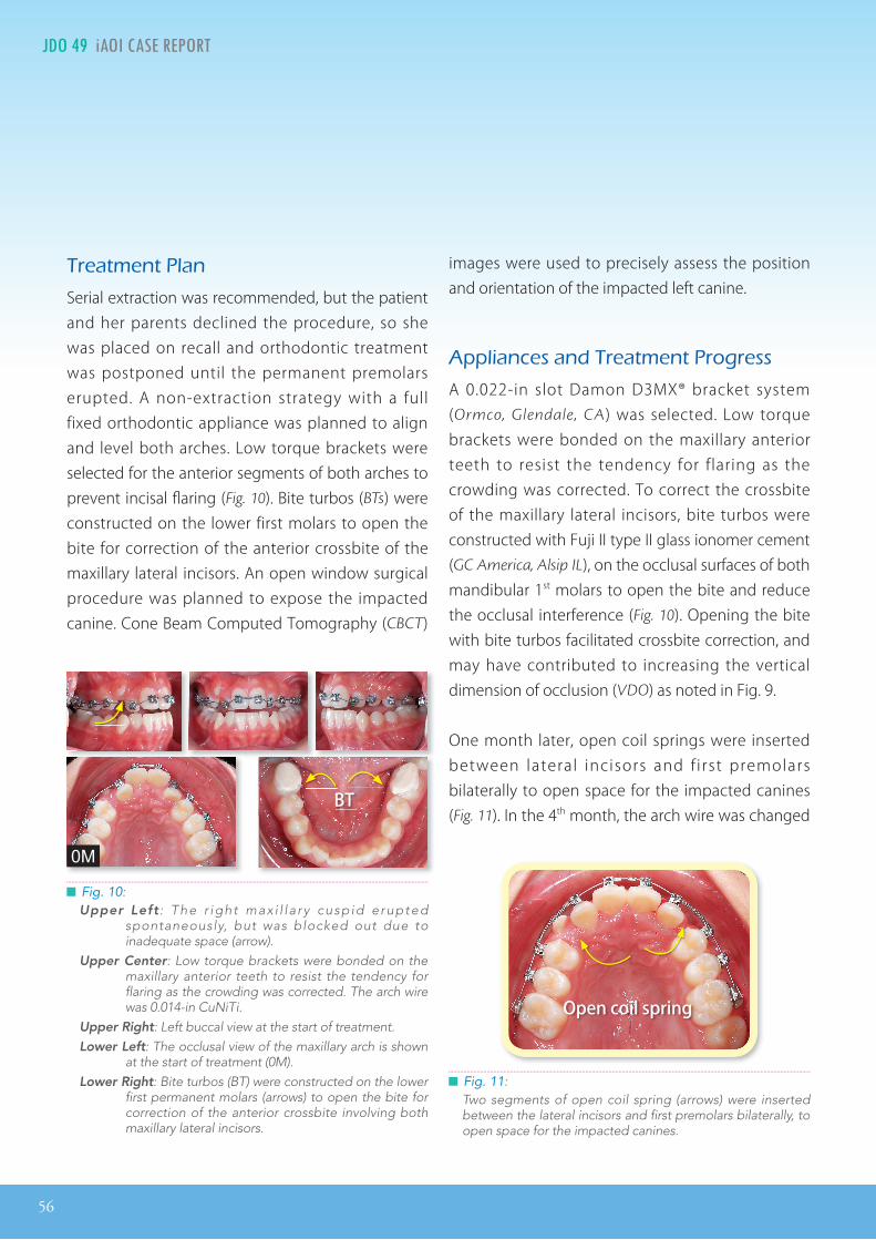
Serial extraction was recommended, but the patient and her parents declined the procedure, so she was placed on recall and orthodontic treatment was postponed until the permanent premolars erupted. A non-extraction strategy with a full fixed orthodontic appliance was planned to align and level both arches. Low torque brackets were selected for the anterior segments of both arches to prevent incisal fl aring (Fig. 10). Bite turbos (BTs) were constructed on the lower first molars to open the bite for correction of the anterior crossbite of the maxillary lateral incisors. An open window surgical procedure was planned to expose the impacted canine. Cone Beam Computed Tomography (CBCT)
images were used to precisely assess the position and orientation of the impacted left canine.
Appliances and Treatment Progress
A 0.022-in slot Damon D3MX® bracket system (Ormco, Glendale, CA) was selected. Low torque brackets were bonded on the maxillary anterior teeth to resist the tendency for flaring as the crowding was corrected. To correct the crossbite of the maxillary lateral incisors, bite turbos were constructed with Fuji II type II glass ionomer cement (GC America, Alsip IL), on the occlusal surfaces of both mandibular 1st molars to open the bite and reduce the occlusal interference (Fig. 10). Opening the bite with bite turbos facilitated crossbite correction, and may have contributed to increasing the vertical dimension of occlusion (VDO) as noted in Fig. 9.
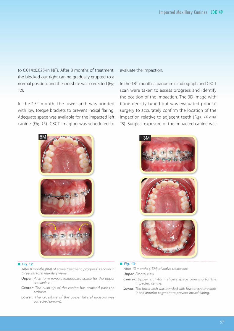
One month later, open coil springs were inserted between lateral incisors and f irst premolars bilaterally to open space for the impacted canines (Fig. 11). In the 4th month, the arch wire was changed
0M
Open coil spring
█ Fig. 10: Upper Left : The r ight max i l la ry cusp id erupted
spontaneously, but was blocked out due to inadequate space (arrow).
Upper Center: Low torque brackets were bonded on the maxillary anterior teeth to resist the tendency for flaring as the crowding was corrected. The arch wire was 0.014-in CuNiTi.
Upper Right: Left buccal view at the start of treatment.Lower Left: The occlusal view of the maxillary arch is shown
at the start of treatment (0M). Lower Right: Bite turbos (BT) were constructed on the lower
first permanent molars (arrows) to open the bite for correction of the anterior crossbite involving both maxillary lateral incisors.
█ Fig. 11: Two segments of open coil spring (arrows) were inserted between the lateral incisors and first premolars bilaterally, to open space for the impacted canines.
BT
57
Impacted Maxillary Canines JDO 49
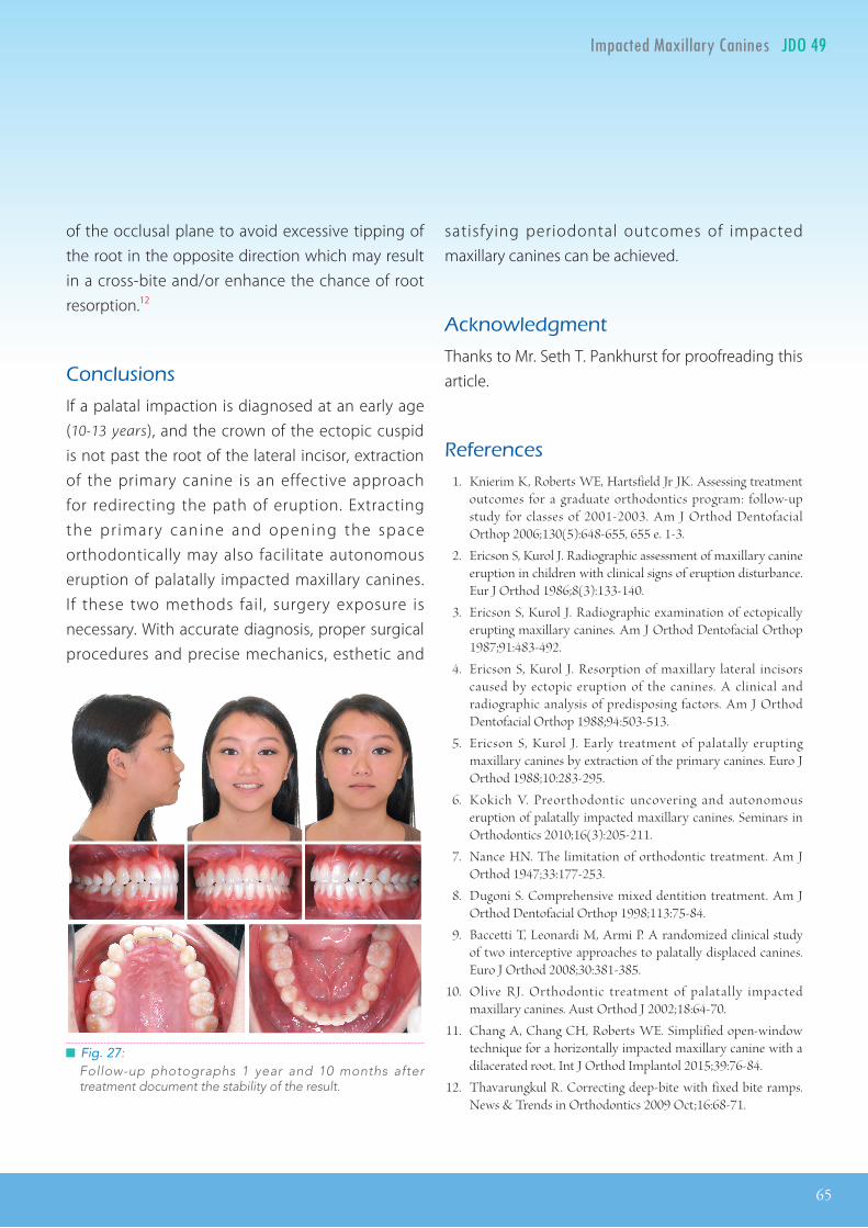
to 0.014x0.025-in NiTi. After 8 months of treatment, the blocked out right canine gradually erupted to a normal position, and the crossbite was corrected (Fig.
12).
In the 13th month, the lower arch was bonded with low torque brackets to prevent incisal flaring. Adequate space was available for the impacted left canine (Fig. 13). CBCT imaging was scheduled to
evaluate the impaction.
In the 18th month, a panoramic radiograph and CBCT scan were taken to assess progress and identify the position of the impaction. The 3D image with bone density tuned out was evaluated prior to surgery to accurately confirm the location of the impaction relative to adjacent teeth (Figs. 14 and
15). Surgical exposure of the impacted canine was
8M 13M
█ Fig. 12: After 8 months (8M) of active treatment, progress is shown in three intraoral maxillary views: Upper: Arch form reveals inadequate space for the upper
left canine. Center: The cusp tip of the canine has erupted past the
archwire. Lower: The crossbite of the upper lateral incisors was
corrected (arrows).
█ Fig. 13: After 13 months (13M) of active treatment: Upper: Frontal view Center: Upper arch-form shows space opening for the
impacted canine. Lower: The lower arch was bonded with low torque brackets
in the anterior segment to prevent incisal flaring.

58
JDO 49 iAOI CASE REPORT
performed by removing the overlying soft tissue, and the wound was covered with surgical dressing (Fig. 16). Three months after surgery, the upper left canine erupted spontaneously, and an eyelet was bonded onto the labial surface. A 0.013-in NiTi arch wire aligned the previously impacted tooth (Fig.
17), and the movement was documented with a series of intraoral photographs (Fig. 18). After seven months of traction, soft tissue accumulated on the buccal surface of the canine, and a gingivectomy was performed with a diode laser. One month later, a low torque bracket was bonded on the maxillary left canine (Fig. 19). In the 33rd month, the upper
█ Fig. 14: Left and Center: 3D imaging with bone density tuned-out documents the position of the impaction to adjacent teeth. Right: Slice views are useful for determining the thickness of the bone covering the impaction.
18M
18M
█ Fig. 15: At 18 months (18M) a panoramic radiograph documents progress prior to uncovering the impaction.
█ Fig. 16: At 18 months (18M) soft tissue covering the impaction was removed with electrosurgery (upper). The wound was covered with surgical dressing (lower).

59
Impacted Maxillary Canines JDO 49
29M 31M
█ Fig. 17: At 22 months (22M) into treatment and 4 months (4M) after surgical uncovering, an eyelet was bonded on the erupted canine and traction was applied with an 0.013-in NiTi arch wire (left). Arch alignment is near complete and bite turbos were bonded on the lower first molars (right).
█ Fig. 18: Left: At 23 months (23M) and 1 month (1M) after initiation traction Middle: At 24 months (24M) the canine is uprighting into its proper position in the arch. Right: At 29 months (29M) and 7 months (7M) after initiation of traction the previously impacted canine is entering the arch-
form (arrow).
█ Fig. 19: At 29 months (29M) of treatment and seven months of buccal traction on the upper left canine, soft tissue accumulated on the buccal side of the tooth (left). A diode laser was used to remove excessive gingiva relative to the mucogingival junction (black line, center). One month later, a low torque bracket was bonded on the maxillary left canine (right).
23M 24M 29M
1m 7m
22M
4m

60
JDO 49 iAOI CASE REPORT
arch wire was changed to 0.014x0.025-in NiTi. A panoramic radiograph was exposed to evaluate bracket positions relative to axial inclinations of all teeth, and they were repositioned as needed. In the 37th month, a 0.017x0.025-in TMA archwire was placed in the upper arch. An elastic (Bear 1/4-in, 4.5-
oz) was applied from the upper left canine to the lower left canine and the adjacent first premolar to close the open contact (Fig. 20). One month later, wire bending and palatal reduction was performed on the maxillary left canine for detailing the occlusion during the final stage of treatment
(Fig. 21). After 40 months of active treatment, all appliances were removed. Fixed anterior retainers and clear overlays were delivered for both arches (Fig. 22).
Results Achieved
Maxilla (all three planes):
• A - P: Retracted
• Vertical: Maintained
• Transverse: Maintained
Mandible (all three planes):
• A - P: Retracted
• Vertical: Increased (posterior rotation)
• Transverse: Maintained
█ Fig. 22: After 40 months (40M) of active treatment, all appliances were removed, fixed anterior retainers were bonded on the lingual surfaces of individual teeth in both arches.
4.5-oz
█ Fig. 20: A triangular elastic (Bear ¼ -in, 4.5-oz) was used to close the open contact in the left canine area.
█ Fig. 21: Palatal reduction of the palatal enamel surface was performed on the upper left canine. Archwire adjustment was utilized to finish the intermaxillary detailing.
38M
40M

61
Impacted Maxillary Canines JDO 49
Maxillary Dentition
• A - P: Anterior incisors retracted and slightly flared
• Vertical: Molars extruded
• Inter-molar / Inter-canine Width: Crowding
corrected with arch expansion
Mandibular Dentition
• A - P: Maintained
• Vertical: Molars and incisors were extruded
• Inter-molar / Inter-canine Width: Maintained
Facial Aesthetics:
• Facial convexity and lip protrusion WNL
Retention
Fixed lingual retainers were bonded on all maxillary incisors, and from canine to canine in the mandibular arch. Clear overlays were delivered for each arch. The patient was instructed to wear them full time for the first month and nights only thereafter. Instructions were provided for the home hygiene as well as for maintenance of the retainers.
Final Evaluation of the Treatment
Cephalometric superimpositions (Fig. 9) and analysis (Table 1) document vertical growth and posterior rotation of the mandible. The mandible increased in length about 10mm (Fig . 9), and the upper dentition was retracted. The upper incisor to SN angle increased from 104° to 109°. That fl aring eff ect resulted from correction of the crowding and the space-opening effect of the coil springs. Extruded molars in both arches and the lower incisors were
related to expression of mandibular growth, and possibly due to the posterior bite turbos. Posterior rotation of mandible resulted in a 1.5° increase in the mandibular plane angle. Although lower facial height increased, photographs after treatment (Fig.
4) show lip competence, as well as similar facial convexity and lip protrusion, compared to the pre-treatment records. Overall, the posterior rotation of the mandible was advantageous for maintaining a more feminine profi le.
The ABO Cast-Radiograph Evaluat ion (CRE ) score was 30 points, which slightly exceeds the ideal range (upper limit of 26 points). The major discrepancies were occlusal contacts (10 points), buccolingual inclination (7 points), marginal ridges (4 points), alignment/rotations (3 points), and distal tipping of the upper second molars (Fig. 23). If these discrepancies had been discovered with prefinish records,1 the finish CRE score could have been substantially improved with bracket repositioning and archwire adjustments. Overall, the dentition was well aligned, and the patient was satisfi ed with the result.
█ Fig. 23: Distal tipping of the upper second molars were noted after debonding. This problem could be corrected by repositioning the brackets as shown.

62
JDO 49 iAOI CASE REPORT
64%
91%
Discussion
1. Impacted Maxillary Canine
Other than the mandibular third molar, the maxillary canine is the most commonly impacted tooth with an incidence from 1-2.5%.2 Conventional r ad iog raphy i n 2D r equ i r e s two o r t h ree conventional intraoral films, exposed at different projections, to reliably assess the position and eruption pathway of impacted canines in most children. The optimal age for radiologic investigation is 10 to 13 years.3 Genetics is thought to be the primary factor, but delayed exfoliation of the primary canine, lack of space and ectopic path of eruption may also contribute. The prevalence of labial or palatal impaction is 15% and 85% respectively. The incidence rate in females is 3.2 times that of males. Palatally impacted canines are five times more common in Europeans compared to Asians.2-5
Root resorption of permanent incisors may be related to ectopic eruption of the maxillary canines4 and cysts have also been reported for untreated impactions. Extraction of the primary canine may favorably infl uence the path of eruption. The success rate differs according to the impacted canine position at the start of treatment, mesial or distal to the midline of the lateral incisor in the panoramic radiograph (Fig. 24). With late diagnosis, crowding, root resorption and/or horizontal path of eruption, surgical exposure with active orthodontic extrusion is the treatment of choice.5 For the patient in the present study (Figs. 10 and 25), the right upper canine was positioned distal to the midline of the lateral incisor. It erupted spontaneously, but was subsequently blocked out. The left upper canine was
11y10m
13y2m
█ Fig. 24: The success rate for normalization for the maxillary permanent canine eruption by extracting the deciduous canine and opening space for the impaction is related to the overlap of the unerupted canine relative to the incisor roots at the start of treatment: 91% if the overlap involves only the distal aspect of the lateral incisor root, and drops to 64% if the overlap is more than half of the central incisor root.
█ Fig. 25: Relative to the pretreatment view of the maxillary arch pretreatment (11y10m), the right canine erupts in a high position because it is blocked-out, but the left canine is impacted and its space is closed.

63
Impacted Maxillary Canines JDO 49
positioned mesial to the midline of the lateral incisor, and thus deeply imbedded in the palate. After the primary canine was lost, the space closed due to distal shift of the lateral incisor and mesial shift of the posterior teeth. Fixed orthodontic appliances were the optimal option.5,6
2. Leeway Space
Leeway space is the arch-length diff erence between the combined mesiodistal width of the deciduous cuspid and molars and their successors.7 Usually the total width of these three primary teeth is greater than that of their permanent successors by ~1.6-2.5mm per side in the lower arch and ~0.7-1.5mm per side in the upper arch. Girls usually have larger Leeway spaces than boys, probably because males usually have larger crown dimensions for all teeth.7 Preserving Leeway space helps relieve crowding.8 During late mixed dentition, a 5mm space defi ciency was measured in the lower anteriors of the present patient. The crowding was relieved spontaneously after the lower permanent premolars erupted, but this phenomenon was not observed in maxillary dentition (Figs. 25 and 26). The space for the maxillary left canine was probably lost due to a mesial shift of maxilla buccal segment. Baccetti et al.9 found significant mesial movement of the upper first molars (about 2.5mm) occurred both in untreated and primary canine extraction patients when the canines were palatally displaced.
3. Bracket Selection
Non-extraction leveling and aligning of a crowded dentition usually results in incisal flaring, which is
intensifi ed by the use of open coil springs to regain space for impacted canines.10 Bonding low torque brackets in the anterior segments of both arches decreases the fl aring tendency. Lateral force to move a palatally impacted canine into the arch is likely to tip the crown labially, so a low torque bracket is indicated to upright the canine once it is aligned in the arch.
4. Surgical Exposure
Another approach to facilitate autonomous eruption of the ectopically positioned maxillary cuspid is to open space orthodontically by separating the lateral incisor and premolar.10 If the impacted canine does not begin to erupt (Fig. 13), surgical exposure is needed. There are three important issues to
█ Fig. 26: Crowding in lower anterior region corrected spontaneously after the permanent premolars erupted (circle), apparently due to favorable utilization of Leeway space.

64
JDO 49 iAOI CASE REPORT
Open Window Pre-ortho. Uncovering Tech Kokich
Flap elevation N Y
Tooth uncovering cut a hole over the crown cut a hole on the flap
Suture N Y
surgical dressing 3 days 3 months
Chang
guarantee a successful result: accurate diagnosis, proper surgery and precise mechanics.
Accurate Diagnosis
With conventional apical fi lms, the buccal object rule is used to identify the labiolingual position of the impaction,2,3 but 3D imaging with a CBCT scan is the only common procedure for precisely locating the impaction relative to the adjacent teeth. The slice views are useful for determining the thickness of the bone covering the impaction (Fig. 14). These useful insights can help avert injury to adjacent structures.
Proper Surgery
A well defi ned surgical procedure produces effi cient alignment of a previously impacted tooth.11 The precise location of the impaction was identifi ed on the CBCT image. Crown position was marked with a sharp explorer that penetrated the soft tissue. Instead of a scalpel, a dental electrosurgical unit (ESU) was used to remove the covering soft tissue (Fig. 16) to control bleeding and provide a more
clear surgical fi eld. The most expedient way to align a previously impacted canine is to remove bone in the planned path of traction.11 The “osteo-bur” is more efficient than osteoclasts. However, for the present patient, bone was not removed in the path of traction. Nine months was required to align the left maxillary canine. Removing bone in the path of traction would have saved time.
Precise Mechanics
If the soft tissue and bone covering the crown of an upper canine impaction are carefully removed, the tooth will spontaneously erupt into the oral cavity.6 Furthermore, the bone level and periodontal attachment of the adjacent teeth are more healthy compared with the closed eruption technique. However, spontaneous eruption of an impaction in adults may take more than one year. To control treatment time, the present authors prefer active traction with a palatal screw to erupt the impaction into the palate, and then apply lateral force to move the tooth into the arch. It is important to initiate traction before the canine crown passes the level
█ Table 2:
Comparison of the Chang and Kokich methods for uncovering impactions. The crown signifi es the superior procedure at each step.
65
Impacted Maxillary Canines JDO 49
of the occlusal plane to avoid excessive tipping of the root in the opposite direction which may result in a cross-bite and/or enhance the chance of root resorption.12
Conclusions
If a palatal impaction is diagnosed at an early age (10-13 years), and the crown of the ectopic cuspid is not past the root of the lateral incisor, extraction of the primary canine is an effective approach for redirecting the path of eruption. Extracting the pr imary canine and opening the space orthodontically may also facilitate autonomous eruption of palatally impacted maxillary canines. If these two methods fail, surgery exposure is necessary. With accurate diagnosis, proper surgical procedures and precise mechanics, esthetic and
satisfying periodontal outcomes of impacted maxillary canines can be achieved.
Acknowledgment
Thanks to Mr. Seth T. Pankhurst for proofreading this article.
References
1. Knierim K, Roberts WE, Hartsfield Jr JK. Assessing treatment outcomes for a graduate orthodontics program: follow-up study for classes of 2001-2003. Am J Orthod Dentofacial Orthop 2006;130(5):648-655, 655 e. 1-3.
2. Ericson S, Kurol J. Radiographic assessment of maxillary canine eruption in children with clinical signs of eruption disturbance. Eur J Orthod 1986;8(3):133-140.
3. Ericson S, Kurol J. Radiographic examination of ectopically erupting maxillary canines. Am J Orthod Dentofacial Orthop 1987;91:483-492.
4. Ericson S, Kurol J. Resorption of maxillary lateral incisors caused by ectopic eruption of the canines. A clinical and radiographic analysis of predisposing factors. Am J Orthod Dentofacial Orthop 1988;94:503-513.
5. Ericson S, Kurol J. Early treatment of palatally erupting maxillary canines by extraction of the primary canines. Euro J Orthod 1988;10:283-295.
6. Kokich V. Preorthodontic uncovering and autonomous eruption of palatally impacted maxillary canines. Seminars in Orthodontics 2010;16(3):205-211.
7. Nance HN. The limitation of orthodontic treatment. Am J Orthod 1947;33:177-253.
8. Dugoni S. Comprehensive mixed dentition treatment. Am J Orthod Dentofacial Orthop 1998;113:75-84.
9. Baccetti T, Leonardi M, Armi P. A randomized clinical study of two interceptive approaches to palatally displaced canines. Euro J Orthod 2008;30:381-385.
10. Olive RJ. Orthodontic treatment of palatally impacted maxillary canines. Aust Orthod J 2002;18:64-70.
11. Chang A, Chang CH, Roberts WE. Simplified open-window technique for a horizontally impacted maxillary canine with a dilacerated root. Int J Orthod Implantol 2015;39:76-84.
12. Thavarungkul R. Correcting deep-bite with fixed bite ramps. News & Trends in Orthodontics 2009 Oct;16:68-71.
█ Fig. 27: Follow-up photographs 1 year and 10 months after treatment document the stability of the result.

66
JDO 49 iAOI CASE REPORT
OVERJET
0 mm. (edge-to-edge) = 1 pt.1 – 3 mm. = 0 pts.3.1 – 5 mm. = 2 pts.5.1 – 7 mm. = 3 pts.7.1 – 9 mm. = 4 pts.> 9 mm. = 5 pts.
Negative OJ (x-bite) 1 pt. per mm. per tooth =
OVERBITE
0 – 3 mm. = 0 pts.3.1 – 5 mm. = 2 pts.5.1 – 7 mm. = 3 pts.Impinging (100%) = 5 pts.
ANTERIOR OPEN BITE
0 mm. (edge-to-edge), 1 pt. per tooth
then 1 pt. per additional full mm. per tooth
LATERAL OPEN BITE
2 pts. per mm. per tooth
CROWDING (only one arch)
1 – 3 mm. = 1 pt.3.1 – 5 mm. = 2 pts.5.1 – 7 mm. = 4 pts.> 7 mm. = 7 pts.
OCCLUSION
Class I to end on = 0 pts.End on Class II or III = 2 pts. per side pts.
Full Class II or III = 4 pts. per side pts.
Beyond Class II or III = 1 pt. per mm. pts.pts. additional
Total =
Total =
Total =
Total =
Total =
Total = 0
TOTAL D.I.D.I. SCORECORE
8
LINGUAL POSTERIOR X-BITE
1 pt. per tooth Total =
BUCCAL POSTERIOR X-BITE
2 pts. per tooth Total =
CEPHALOMETRICS (See Instructions)
ANB ≥ 6° or ≤ -2° = 4 pts.
SN-MP
≥ 38° = 2 pts.
Each degree > 38° x 2 pts. =
≤ 26° = 1 pt.
Each degree < 26° x 1 pt. =
1 to MP ≥ 99° = 1 pt.
Each degree > 99° x 1 pt. =
OTHER (See Instructions)
Supernumerary teeth x 1 pt. =
Ankylosis of perm. teeth x 2 pts. =
Anomalous morphology x 2 pts. =
Impaction (except 3rd molars)rd molars)rd x 2 pts. =
Midline discrepancy (≥3mm) @ 2 pts. =
Missing teeth (except 3rd molars)rd molars)rd x 1 pts. =
Missing teeth, congenital x 2 pts. =
Spacing (4 or more, per arch) x 2 pts. =
Spacing (Mx cent. diastema ≥ 2mm) @ 2 pts. =
Tooth transposition x 2 pts. =
Skeletal asymmetry (nonsurgical tx) @ 3 pts. =
Addl. treatment complexities x 2 pts. =
Identify:
Each degree > 6° x 1 pt. =
Each degree < -2° x 1 pt. =
Total =
Total =
15+15=3015+15=3015+15=30
4
00
00
00
77
0
00
15
0
iiDI =15DI =15
1
1515 15
4
40404
Discrepancy Index Worksheet

67
Impacted Maxillary Canines JDO 49
67
1
iDI impaction Discrepancy Index
1. Angulation of the impaction to the midline in degree
2. Vertical distance from the occlusal plane
3. Mesiodistal position of the impaction tip
Grade 1 : 0º ~ 15º = 1 pt.Grade 2 : 16º ~ 30º = 2 pts.Grade 3 : ≥30º = 3 pts.
Grade 1 : Below the level of the CEJ = 1 pt.Grade 2 : Above the CEJ, but less than halfway up the root = 2 pts.Grade 3 : More than halfway up the root, but less than the full root length = 3 pts.Grade 4 : Above the full length of the root = 4 pts.
Grade 1 : No horizontal overlap = 1 pt.Grade 2 : Less than half the root width = 2 pts.Grade 3 : More than half, but less than the whole root width = 3 pts.Grade 4 : Complete overlap of root width or more = 4 pts.
Total Score: = 15Total = 3
Total = 4
Total = 3

68
JDO 49 iAOI CASE REPORT
iDI impaction Discrepancy Index
4. Anterior-posterior position of the impaction root apex
5. Root resorption of the adjacent tooth
6. Age relative to the completion of root formation
7. Labial or palatal position of the impaction
Grade 1 : Above the region of the canine position = 1 pt.Grade 2 : Above the upper first premolar region = 2 pts.Grade 3 : Above the upper second premolar region = 3 pts.
Palatal impaction = 1 pt.Labial impaction = 2 pts.
Normal apical contour = 0 pt.Apical irregularity, same length as pretreatment = 1 pt.Apical root resorption of less than 2 mm = 2 pts.Apical root resorption more than 2 mm, less than one third original root length = 3 pts.Apical root resorption more than one third original root length = 4 pts.
< 9 y/o ( Before Central incisor root completed ) = 0 pt.9 ~ 11 y/o ( Before Lateral incisor root completed ) = 1 pt.12~13 y/o ( Before 1st premolar root completed ) = 2 pts.> 13 y/o ( Canine root completed ) = 3 pts.
Total = 2
Total = 2
Total = 1
Total = 0

69
Impacted Maxillary Canines JDO 49
INSTRUCTIONS: Place score beside each deficient tooth and enter total score for each parameter in the white box. Mark extracted teeth with “X”. Second molars should be in occlusion.
Alignment/Rotations
Marginal Ridges
Buccolingual Inclination
Overjet
Occlusal Contacts
Occlusal Relationships
Interproximal Contacts
Root Angulation
4 4
3
7
3 3
10
0
3
0
1
1
1
Total CRE Score 30+1=31
1
11
2
1
1
11
1
11
11 1
1 1
2211 111122
iCRE=1
12
1
1
1
Cast-Radiograph Evaluation

70
JDO 49 iAOI CASE REPORT
12
5 4
12
5 4
1 234
5,6712
5 4
1 234
5,67
12
5 4
1 234
5,67
1. Gingival esthetic score
iCRE impaction Cast-Radiograph Evaluation
Total Score: = 1
2. Root resorption of the recovered and adjacent teeth
12
5 4
1 234
5,67 1. M & D Papillae 0 1 2
2. Keratinized Gingiva 0 1 2
3. Curvature of Gingival Margin 0 1 2
4. Level of Gingival Margin 0 1 2
5. Root Convexity ( Torque ) 0 1 2
6. Scar Formation 0 1 2
7. Keratinized Gingival Exists 0 1 2
1. M & D Papilla 0 1 2
2. Keratinized Gingiva 0 1 2
3. Curvature of Gingival Margin 0 1 2
4. Level of Gingival Margin 0 1 2
5. Root Convexity ( Torque ) 0 1 2
6. Scar Formation 0 1 2
7. Keratinized Gingival Exists 0 1 2
Total = 1
Total = 0Normal apical contour 0
Apical irregularity, same length aspretreatment 1
Apical root resorption of less than 2 mm 2
Apical root resorption more than 2 mm,less than one third original root length 3
Apical root resorption more than onethird original root length 4
Normal apical contour 0
Apical irregularity, same length aspretreatment 1
Apical root resorption of less than 2 mm 2
Apical root resorption more than 2 mm,less than one third original root length 3
Apical root resorption more than onethird original root length 4

71
Impacted Maxillary Canines JDO 49
12 35 4
4
1 2
3
5
1
2
34 6
12 34
56
12 35 4
4
1 2
3
5
1
2
34 6
12 34
56 12 3
5 4
4
1 2
3
5
1
2
34 6
12 34
56
1. Pink Esthetic Score
IBOI Pink & White Esthetic Score (Before Surgical Crown Lengthening)
Total Score: = 5
2. White Esthetic Score ( for Micro-esthetics )
12 35 4
4
1 2
3
5
1
2
34 6
12 34
56
1. M & D Papillae 0 1 2
2. Keratinized Gingiva 0 1 2
3. Curvature of Gingival Margin 0 1 2
4. Level of Gingival Margin 0 1 2
5. Root Convexity ( Torque ) 0 1 2
6. Scar Formation 0 1 2
1. Midline 0 1 2
2. Incisor Curve 0 1 2
3. Axial Inclination (5°, 8°, 10°) 0 1 2
4. Contact Area (50%, 40%, 30%) 0 1 2
5. Tooth Proportion (1:0.8) 0 1 2
6. Tooth to Tooth Proportion 0 1 2
1. M & D Papilla 0 1 2
2. Keratinized Gingiva 0 1 2
3. Curvature of Gingival Margin 0 1 2
4. Level of Gingival Margin 0 1 2
5. Root Convexity ( Torque ) 0 1 2
6. Scar Formation 0 1 2
1. Midline 0 1 2
2. Incisor Curve 0 1 2
3. Axial Inclination (5°, 8°, 10°) 0 1 2
4. Contact Area (50%, 40%, 30%) 0 1 2
5. Tooth Proportion (1:0.8) 0 1 2
6. Tooth to Tooth Proportion 0 1 2
Total = 2
Total = 3