Impact of Recombinant Human Bone OPYRIGHT...
12
400 Volume 16, Number 3, 2001 Impact of Recombinant Human Bone Morphogenetic Protein-2 on Residual Ridge Resorption After Tooth Extraction: An Experimental Study in the Rat Khairul Matin, BDS, PhD 1 /Hiroaki Nakamura, DDS, PhD 2 /Kazuharu Irie, DDS, PhD 3 / Hidehiro Ozawa, DDS, PhD 4 /Sadakazu Ejiri, DDS, PhD 5 Residual ridge resorption begins following tooth extraction and continuously reduces alveolar bone vol- ume, potentially creating a significant problem in dental implant treatment. In this study, the role of recombinant human bone morphogenetic protein-2 (rhBMP-2) in residual ridge resorption after tooth extraction was investigated. A polylactic acid/polyglycolic acid copolymer–coated gelatin sponge car- rier was implanted with or without rhBMP-2 (1 µg) in the mesial root sockets after removal of maxillary first molars in male Wistar rats. Fine structural and histomorphologic analyses were conducted 3 to 84 days after implantation. Direct bone formation was first observed after 5 days on the rhBMP-2 side, which was transformed into cortical alveolar ridge with a smooth periosteal layer by 84 days, whereas the control side displayed slower healing. Bone histomorphometry revealed greater total bone area and increased bone height after 14, 28, 56, and 84 days on the rhBMP-2 side compared to the control side, and differences were significant after 14, 28, and 56 days. Larger numbers of proliferating cells and densely populated differentiating mesenchymal cells were observed on the rhBMP-2 side than on the control side in the early stage, and chondrogenesis was not observed. The findings indicate that rhBMP-2 may stimulate proliferation and differentiation of mesenchymal cells in the rat maxillary root socket to preserve cortical bone volume in the socket without any evidence of chondrogenesis. (INT J ORAL MAXILLOFAC IMPLANTS 2001;16:400–411) Key words: absorbable gelatin sponge, alveolar bone, bone morphogenetic proteins, bone resorption R esorption of alveolar bone that occurs following tooth extraction is termed residual ridge resorp- tion. In humans, an average residual ridge resorp- tion of 2 mm occurs during the first 2 months after tooth extraction, with and/or without dentures, and a further 2 mm of resorption takes place by 1 year after tooth extraction with dentures. 1 Over the last 4 decades, numerous efforts have been made to either prevent residual ridge resorption or to regen- erate alveolar bone mass. Various bone regeneration methods have been or are being investigated and clinically applied in both the dental and orthopedic fields. Methods include autografts, allografts, osteoinduction by cytokines or growth factors, osteopromotion by the guided bone regeneration (GBR) technique, osteopromotion by low-power laser, immediate placement of dental implants, and combinations of these. 2–9 Recombinant human bone morphogenetic pro- tein-2 (rhBMP-2) is a synthetic protein that has been widely tested for bone induction and regeneration. Previous reports have shown that this novel protein molecule induces bone formation at ectopic sites and trephine defects in animal models. 10,11 As a member of the transforming growth factor- (TGF-) super 1 Researcher, Former PhD Student, Department of Fixed Pros- thetic Dentistry, Niigata University School of Dentistry, Niigata, Japan; Currently, Assistant Professor, Bangladesh Medical Stud- ies and Research Institute, Dhaka, Bangladesh. 2 Associate Professor, Department of Oral Anatomy, Okayama Uni- versity School of Dentistry, Okayama, Japan. 3 Lecturer, Department of Oral Anatomy, Health Sciences Univer- sity of Hokkaido, Hokkaido, Japan. 4 Director and Professor, Institute for Dental Science and Depart- ment of Oral Anatomy, Matumoto Dental University, Shiojiri, Japan. 5 Associate Professor, Department of Oral Anatomy, Niigata Uni- versity School of Dentistry, Niigata, Japan. Reprint requests: Prof Hidehiro Ozawa, Institute for Dental Sci- ence and Department of Oral Anatomy, Matumoto Dental Univer- sity, 1780 Gobara, Hirooka, Shiojiri, Nganano, Japan. Fax +81- 263-51-2192. COPYRIGHT © 2001 BY QUINTESSENCE PUBLISHING CO, INC. PRINTING OF THIS DOCUMENT IS RESTRICTED TO PERSONAL USE ONLY . NO PART OF THIS ARTICLE MAY BE REPRODUCED OR TRANSMITTED IN ANY FORM WITH- OUT WRITTEN PERMISSION FROM THE PUBLISHER.
Transcript of Impact of Recombinant Human Bone OPYRIGHT...

400 Volume 16, Number 3, 2001
Impact of Recombinant Human Bone Morphogenetic Protein-2 on Residual Ridge
Resorption After Tooth Extraction: An Experimental Study in the Rat
Khairul Matin, BDS, PhD1/Hiroaki Nakamura, DDS, PhD2/Kazuharu Irie, DDS, PhD3/Hidehiro Ozawa, DDS, PhD4/Sadakazu Ejiri, DDS, PhD5
Residual ridge resorption begins following tooth extraction and continuously reduces alveolar bone vol-ume, potentially creating a significant problem in dental implant treatment. In this study, the role ofrecombinant human bone morphogenetic protein-2 (rhBMP-2) in residual ridge resorption after toothextraction was investigated. A polylactic acid/polyglycolic acid copolymer–coated gelatin sponge car-rier was implanted with or without rhBMP-2 (1 µg) in the mesial root sockets after removal of maxillaryfirst molars in male Wistar rats. Fine structural and histomorphologic analyses were conducted 3 to 84days after implantation. Direct bone formation was first observed after 5 days on the rhBMP-2 side,which was transformed into cortical alveolar ridge with a smooth periosteal layer by 84 days, whereasthe control side displayed slower healing. Bone histomorphometry revealed greater total bone areaand increased bone height after 14, 28, 56, and 84 days on the rhBMP-2 side compared to the controlside, and differences were significant after 14, 28, and 56 days. Larger numbers of proliferating cellsand densely populated differentiating mesenchymal cells were observed on the rhBMP-2 side than onthe control side in the early stage, and chondrogenesis was not observed. The findings indicate thatrhBMP-2 may stimulate proliferation and differentiation of mesenchymal cells in the rat maxillary rootsocket to preserve cortical bone volume in the socket without any evidence of chondrogenesis. (INT JORAL MAXILLOFAC IMPLANTS 2001;16:400–411)
Key words: absorbable gelatin sponge, alveolar bone, bone morphogenetic proteins, bone resorption
Resorption of alveolar bone that occurs followingtooth extraction is termed residual ridge resorp-
tion. In humans, an average residual ridge resorp-tion of 2 mm occurs during the first 2 months after
tooth extraction, with and/or without dentures, anda further 2 mm of resorption takes place by 1 yearafter tooth extraction with dentures.1 Over the last4 decades, numerous efforts have been made toeither prevent residual ridge resorption or to regen-erate alveolar bone mass. Various bone regenerationmethods have been or are being investigated andclinically applied in both the dental and orthopedicfields. Methods include autografts, allografts,osteoinduction by cytokines or growth factors,osteopromotion by the guided bone regeneration(GBR) technique, osteopromotion by low-powerlaser, immediate placement of dental implants, andcombinations of these.2–9
Recombinant human bone morphogenetic pro-tein-2 (rhBMP-2) is a synthetic protein that has beenwidely tested for bone induction and regeneration.Previous reports have shown that this novel proteinmolecule induces bone formation at ectopic sites andtrephine defects in animal models.10,11 As a memberof the transforming growth factor-� (TGF-�) super
1Researcher, Former PhD Student, Department of Fixed Pros-thetic Dentistry, Niigata University School of Dentistry, Niigata,Japan; Currently, Assistant Professor, Bangladesh Medical Stud-ies and Research Institute, Dhaka, Bangladesh.
2Associate Professor, Department of Oral Anatomy, Okayama Uni-versity School of Dentistry, Okayama, Japan.
3Lecturer, Department of Oral Anatomy, Health Sciences Univer-sity of Hokkaido, Hokkaido, Japan.
4Director and Professor, Institute for Dental Science and Depart-ment of Oral Anatomy, Matumoto Dental University, Shiojiri,Japan.
5Associate Professor, Department of Oral Anatomy, Niigata Uni-versity School of Dentistry, Niigata, Japan.
Reprint requests: Prof Hidehiro Ozawa, Institute for Dental Sci-ence and Department of Oral Anatomy, Matumoto Dental Univer-sity, 1780 Gobara, Hirooka, Shiojiri, Nganano, Japan. Fax +81-263-51-2192.
CO
PY
RIG
HT
©2001 B
YQ
UIN
TE
SS
EN
CE
PU
BLIS
HIN
GC
O, IN
C. PR
INT
ING
OF
TH
ISD
OC
UM
EN
TIS
RE
ST
RIC
TE
DT
OP
ER
SO
NA
LU
SE
ON
LY. NO
PA
RT
OF
TH
ISA
RT
ICLE
MA
YB
ER
EP
RO
DU
CE
DO
RT
RA
NS
MIT
TE
DIN
AN
YF
OR
MW
ITH-
OU
TW
RIT
TE
NP
ER
MIS
SIO
NF
RO
MT
HE
PU
BLIS
HE
R.

The International Journal of Oral & Maxillofacial Implants 401
MATIN ET AL
family, rhBMP-2 has displayed its potential in differ-entiating mesenchymal precursors into cartilage,bone, and cementum-forming cells.12–14 Demonstra-tion of its effectiveness in regenerating alveolar boneand cementum by expanded polytetrafluoroethylene(e-PTFE) membrane and rhBMP-2 has also led toits use in alveolar bone regeneration.14 However,reports of attempts at immediate alveolar boneregeneration following tooth extraction for prostho-dontic rehabilitation purposes are limited.6,9,15
A simple, moldable, and effective carrier forrhBMP-2 for dental use remains to be identified. Inthis experiment, a polylactic acid and polyglycolicacid copolymer–coated gelatin sponge (PLGA/GS)was used as a carrier for rhBMP-2 to preserve alve-olar bone volume after maxillary tooth extraction inthe rat.
Bone morphogenetic proteins (BMPs) in generalinduce a complex cascade of chemotaxis, prolifera-tion, and cellular differentiation that closely resem-bles the normal process of embryonic bone forma-tion, including both intramembranous andendochondral bone.16 Cartilage phenotype expres-sion has been observed during intramembranousbone formation in the sockets of rats, and endo-chondral bone formation by rhBMP-2 has beenobserved in rat mandibular defects.17,18 Therefore,it is of interest to confirm rhBMP-2 behavior dur-ing socket healing.
This experiment investigated the regenerationprocess with and without rhBMP-2 by fine structural,histologic, histomorphometric, and histochemicalmethods. The effect of rhBMP-2 on osteoblastic cellproliferation was examined immunohistochemically.
MATERIALS AND METHODS
Animals and MaterialsThe protocol for this animal experiment wasapproved by the Committee on Guidelines for Ani-mal Experimentation of Niigata University Schoolof Dentistry, Niigata, Japan. Six-week-old maleWistar rats (170 to 190 g, Charles River Laboratory,Yokohama, Japan) were housed under similar condi-tions (22°C room temperature, 40% humidity, anda 12-hour daylight cycle) and were fed commercialrat food (MF, Oriental Yeast, Tokyo, Japan) and tapwater ad libitum.
A gelatin sponge (GS) (Yamanouchi Pharmaceu-tical Industries, Tokyo, Japan) coated with polylacticacid/polyglycolic acid copolymer (PLGA) was usedas the rhBMP-2 carrier.19 The molar ratio of PLGApolymers was 1:1, and the weight ratio of PLGA:GSwas 4:1, with approximately 90% porosity.20
Tooth Extraction and rhBMP-2 AdministrationOn the day of operation, buffer-diluted rhBMP-2(40 µg/100 µL, Yamanouchi Pharmaceutical Indus-tries; buffer contained L-glutamic acid, sodium chlo-ride, glycine, glucose, and Tween 80) was infiltratedinto the PLGA/GS and allowed to adsorb with thecarrier for 30 minutes. The PLGA/GS was cut intopieces approximately 1.5�1.5�1.5 mm, each ofwhich contained approximately 1 µg of rhBMP-2.Other pieces of PLGA/GS were soaked in bufferonly and cut similarly into smaller pieces. The ratswere anesthetized by diethyl ether inhalation fol-lowed by intraperitoneal (IP) administration of 0.5mL/100 g body weight 8% chloral hydrate (WakoPure Chemical Industries, Osaka, Japan). Then 2%xylocaine (Astra Japan, Osaka, Japan) was adminis-tered locally and the maxillary first molars were gen-tly extracted. Buffer containing PLGA/GS wasgrafted into the upper part of the anterior root of 1socket as control. The contralateral socket was simi-larly filled with the PLGA/GS containing rhBMP-2.Sockets were closed by suturing the gingiva.
Tissue ProcessingThe rats were sacrificed at 3, 5, 7, 14, 21, 28, 56,and 84 days after the operation (8 rats/group, exceptfor the 21-day group, which had only 4 rats; total n= 60). A total of 12 rats were injected with 5-bro-modeoxyuridine (BrdU) (Sigma Chemical, St.Louis, MO) 1 hour before sacrifice at 3, 5, and 7days after operation. They were anesthetized with adiethyl ether inhaler and intraperitoneal injection of0.4 mL/kg body weight pentobarbital sodium (50mg/mL, Abbott Laboratories, Abbott Park, IL) thenperfused through the left ventricle with Ringer’ssolution and the fixatives as described below.
Scanning Electron MicroscopyFine structural analyses were performed by scanningelectron microscopy (SEM) (Hitachi S-570, Tokyo,Japan). Rats were perfused with 2% paraformalde-hyde and 2.5% glutaraldehyde in 0.05 mol/L cacody-late buffer solution, then immersed in the same fixa-tive for 3 hours at 4°C. Soft tissues were removed byimmersing the specimen in 5% sodium hypochloritesolution for 15 hours (the solution was changed at 3-hour intervals). Specimens were washed for 12 hoursin distilled water and dehydrated in serially gradedethanol. They were then immersed in isoamyl acetateas the transitional fluid for about 10 minutes, whichwas replaced by carbon dioxide in a criticalpoint–dryer (HCP-1, Hitachi); specimens were thengold-coated by an ion coater (IB-3, Eiko Engineer-ing, Ibaraki, Japan). The maxillae were examined bySEM at 20 kV in a palatobuccal direction (Fig 1a).
CO
PY
RIG
HT
©2001 B
YQ
UIN
TE
SS
EN
CE
PU
BLIS
HIN
GC
O, IN
C. PR
INT
ING
OF
TH
ISD
OC
UM
EN
TIS
RE
ST
RIC
TE
DT
OP
ER
SO
NA
LU
SE
ON
LY. NO
PA
RT
OF
TH
ISA
RT
ICLE
MA
YB
ER
EP
RO
DU
CE
DO
RT
RA
NS
MIT
TE
DIN
AN
YF
OR
MW
ITH-
OU
TW
RIT
TE
NP
ER
MIS
SIO
NF
RO
MT
HE
PU
BLIS
HE
R.

402 Volume 16, Number 3, 2001
MATIN ET AL
Histology For histologic preparation, the rats were fixed with4% paraformaldehyde and 0.5% glutaraldehyde in a0.05 mol/L cacodylate buffer. Maxillae were dis-sected, decalcified in 10% EDTA-sodium solutionfor 20 days, dehydrated in serially graded alcohols,and embedded in paraffin. Serial sections of 5 µm,cut through the mesial foramen (Fig 1a), werestained with hematoxylin-eosin (H&E).
Bone Histomorphometry Bone histomorphometry was performed at 14, 28,56, 84 days after operation (n = 5 for each group,except for controls at 14 days and both sides at 84days, where n = 4). H&E-stained serial sections cutthrough the mesial foramen (Fig 1a) were selectedfor the analysis. Photomicrographs were magnified10 times with a profile projector (V-12, Nikon,Nippon Kagaku, Japan). As described by the dia-gram (Fig 1b), the total bone area (old bone + newbone), area of the carrier, bone marrow area, andother soft tissue areas inside the examined zonewere traced on white paper and color-painted sepa-rately. With a cable camera, the traced images weredigitized into a computerized image analyzer (IBAS2000, Kontron M14, Munich, Germany). Binaryimage processing was performed in the computer tomeasure all above-mentioned areas and alveolarbone heights. Alveolar bone heights were calibratedby measuring the distance between the horizontalbars on vertical lines at points M, N, O, and P asdescribed in Fig 1b. Means and standard deviationsfor all data were calculated by a personal computer.Data on respective items for rhBMP-2 groups andcontrol groups were compared by paired Student ttests, and P values < .05 were considered significant.
Enzyme Histochemistry Cryosections (16 µm) from 3-, 5-, 7-, and 14-dayspecimens were obtained by a Minotome (Damon,Needham, MA); embedded with O.C.T. Compound(Tissue-Tek, Miles, Elkhart, IN); and used fordetection of alkaline phosphatase (ALPase) activity.The sections were incubated according to the azo-dye method for 7 minutes at 37°C.
ImmunohistochemistrySome paraffin sections from the BrdU-injected ratswere used for immunohistochemical localization ofBrdU by incubation with anti-BrdU monoclonalantibody (Mab anti-BrdU)21 (Biodesign Interna-tional, Kennebunk, ME) with methanol solutioncontaining 0.3% hydrogen peroxide for 20 minutesat room temperature to block endogenous peroxi-dase. The sections were then incubated with 2 mol/L
hydrochloric acid for 30 minutes at 37°C to dena-tured DNA and neutralized with 0.1 mol/L sodiumborate solution (pH 8.5) for 10 minutes. Nonspecificbinding sites were blocked with 5% lowfat milk for15 minutes at room temp. Sections were incubatedwith MAb anti-BrdU (dilution 1:500) overnight at4°C. They were further incubated with biotinylatedanti-mouse gamma G immunoglobulins (HistofineSAB-PO(M) kit, Nichirei Corporation, Tokyo,Japan) for 30 minutes, then with peroxidase-conju-gated streptavidin (Histofine) for 15 minutes. Colorwas developed with a 3,3’ diaminobenzidine (DAB)solution for 5 minutes at room temperature andcounterstained with Goldner-Masson light green oreosin. They were photographed by a Vanox-S micro-scope (Olympus, Tokyo, Japan) at a magnification of�50 in 2 locations per section (Fig 1c), and BrdU-positive cells were counted.
RESULTS
Effect on Gross AnatomyThe union of gingiva occurred at the mesial half ofthe whole socket within 7 days. The distal halfremained open for about 14 days. From 14 to 28days, a slight elevation could be recognized in themesial root region on the rhBMP-2 side.
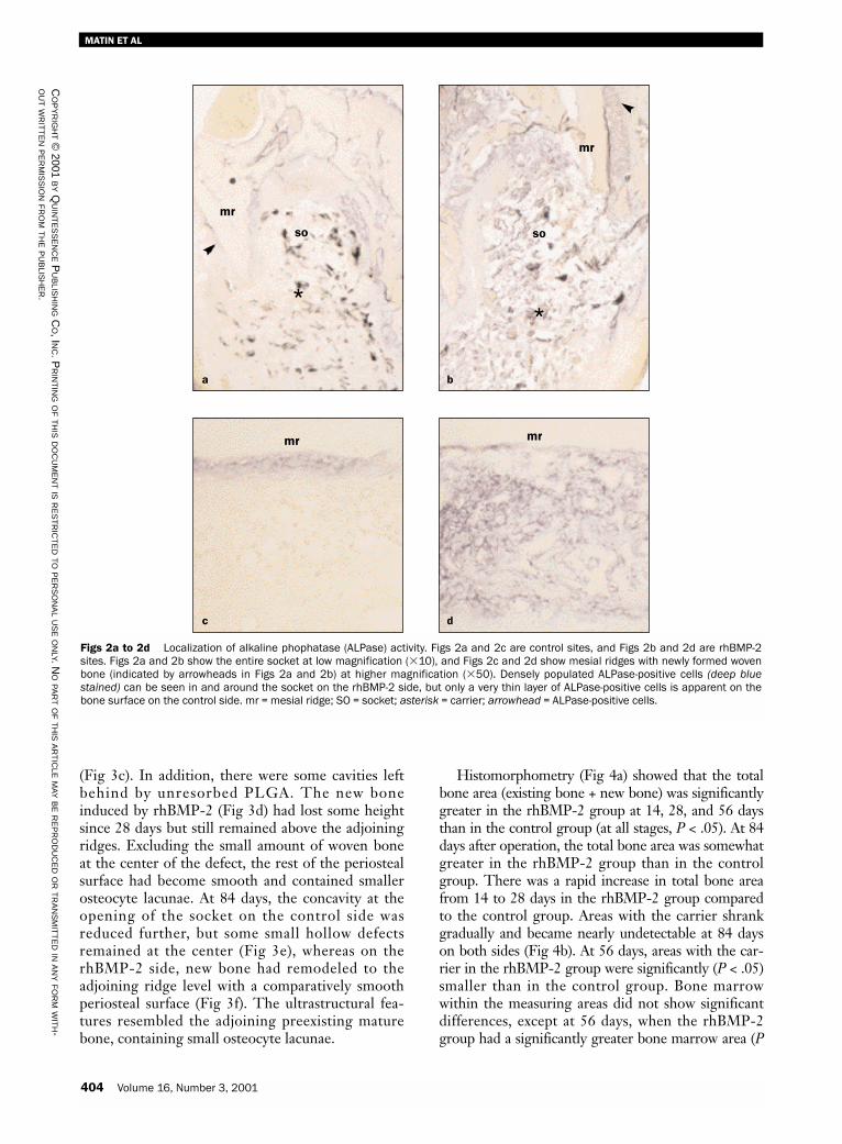
Effect on Differentiation and OsteogenesisAt 5 days after surgery in the rhBMP-2 sockets,more densely populated ALPase-positive osteoblas-tic cells were observed throughout the socket thanin the control sockets (Figs 2a and 2b). Some differ-entiating cells were present in the ridge area on thecontrol side along the old bone, but there werefewer than on the rhBMP-2 side (Figs 2c and 2d).Formation of woven bone with densely populatedALPase-positive osteoblastic cells was marked at themesial ridge area in rhBMP-2 sites (Fig 2d). Nochondrogenesis could be detected during the above-mentioned histochemical studies either inside oroutside the extraction socket.
Effect on Bone Formation and Adaptation Different paces and patterns were observed insocket healing between the control and rhBMP-2groups. Clear and distinctive features could be rec-ognized by SEM observation. Large cavities wereseen in the control, indicating healing that wasslower than in the rhBMP-2–treated sockets (Fig3a). Dome-shaped new bone in the woven stage wasobserved at 28 days on the rhBMP-2 side (Fig 3b).At 56 days postsurgery, healing in the control sideoccurred slowly, with new bone yet to fill the socket
CO
PY
RIG
HT
©2001 B
YQ
UIN
TE
SS
EN
CE
PU
BLIS
HIN
GC
O, IN
C. PR
INT
ING
OF
TH
ISD
OC
UM
EN
TIS
RE
ST
RIC
TE
DT
OP
ER
SO
NA
LU
SE
ON
LY. NO
PA
RT
OF
TH
ISA
RT
ICLE
MA
YB
ER
EP
RO
DU
CE
DO
RT
RA
NS
MIT
TE
DIN
AN
YF
OR
MW
ITH-
OU
TW
RIT
TE
NP
ER
MIS
SIO
NF
RO
MT
HE
PU
BLIS
HE
R.

The International Journal of Oral & Maxillofacial Implants 403
MATIN ET AL
Fig 1a Scanning electron microscopy (SEM) of a rat maxilla; palatobuccal viewshowing extraction socket of the 1st molar. The mesial root socket is outlined by thesquare. The arrow indicates the mesial foramen through which sagittal paraffin sec-tions were cut for bone histomorphometry. SO = socket; mr = mesial ridge; M2 = sec-ond molar; bar = 570 µm.
Fig 1b Diagrammatic representation of a paraffin section ofthe mesial root socket. Points M, P, Q, and R define the areaselected for histomorphometric analysis. The M-R line was drawnmedial to the mesial ridge, just distal to the muscle attachmentand parallel to the mesial wall of the socket. A parallel line P-Qwas drawn 1.8 mm distal to the M-R line, corresponding to thedistal wall of the socket. The M-P line was drawn along the baseof the socket. Q-R represents the oral border of the alveolar bone.Points M, N, O, and P were added by keeping an equal originaldistance of 0.6 mm, and alveolar bone height was measuredbetween the horizontal bars on the lines that extend downwardfrom these points.
Fig 1c Diagram of same section. The squares show the loca-tions selected for BrdU-positive cell count at an original magnifi-cation of �50.
P O N M
so
mr
R
Q
so
mr
M2
somr
CO
PY
RIG
HT
©2001 B
YQ
UIN
TE
SS
EN
CE
PU
BLIS
HIN
GC
O, IN
C. PR
INT
ING
OF
TH
ISD
OC
UM
EN
TIS
RE
ST
RIC
TE
DT
OP
ER
SO
NA
LU
SE
ON
LY. NO
PA
RT
OF
TH
ISA
RT
ICLE
MA
YB
ER
EP
RO
DU
CE
DO
RT
RA
NS
MIT
TE
DIN
AN
YF
OR
MW
ITH-
OU
TW
RIT
TE
NP
ER
MIS
SIO
NF
RO
MT
HE
PU
BLIS
HE
R.
404 Volume 16, Number 3, 2001
MATIN ET AL
(Fig 3c). In addition, there were some cavities leftbehind by unresorbed PLGA. The new boneinduced by rhBMP-2 (Fig 3d) had lost some heightsince 28 days but still remained above the adjoiningridges. Excluding the small amount of woven boneat the center of the defect, the rest of the periostealsurface had become smooth and contained smallerosteocyte lacunae. At 84 days, the concavity at theopening of the socket on the control side wasreduced further, but some small hollow defectsremained at the center (Fig 3e), whereas on therhBMP-2 side, new bone had remodeled to theadjoining ridge level with a comparatively smoothperiosteal surface (Fig 3f). The ultrastructural fea-tures resembled the adjoining preexisting maturebone, containing small osteocyte lacunae.
Histomorphometry (Fig 4a) showed that the totalbone area (existing bone + new bone) was significantlygreater in the rhBMP-2 group at 14, 28, and 56 daysthan in the control group (at all stages, P < .05). At 84days after operation, the total bone area was somewhatgreater in the rhBMP-2 group than in the controlgroup. There was a rapid increase in total bone areafrom 14 to 28 days in the rhBMP-2 group comparedto the control group. Areas with the carrier shrankgradually and became nearly undetectable at 84 dayson both sides (Fig 4b). At 56 days, areas with the car-rier in the rhBMP-2 group were significantly (P < .05)smaller than in the control group. Bone marrowwithin the measuring areas did not show significantdifferences, except at 56 days, when the rhBMP-2group had a significantly greater bone marrow area (P
Figs 2a to 2d Localization of alkaline phophatase (ALPase) activity. Figs 2a and 2c are control sites, and Figs 2b and 2d are rhBMP-2sites. Figs 2a and 2b show the entire socket at low magnification (�10), and Figs 2c and 2d show mesial ridges with newly formed wovenbone (indicated by arrowheads in Figs 2a and 2b) at higher magnification (�50). Densely populated ALPase-positive cells (deep bluestained) can be seen in and around the socket on the rhBMP-2 side, but only a very thin layer of ALPase-positive cells is apparent on thebone surface on the control side. mr = mesial ridge; SO = socket; asterisk = carrier; arrowhead = ALPase-positive cells.
a b
c d
mr
so
*
mr
so
*
mr mr
CO
PY
RIG
HT
©2001 B
YQ
UIN
TE
SS
EN
CE
PU
BLIS
HIN
GC
O, IN
C. PR
INT
ING
OF
TH
ISD
OC
UM
EN
TIS
RE
ST
RIC
TE
DT
OP
ER
SO
NA
LU
SE
ON
LY. NO
PA
RT
OF
TH
ISA
RT
ICLE
MA
YB
ER
EP
RO
DU
CE
DO
RT
RA
NS
MIT
TE
DIN
AN
YF
OR
MW
ITH-
OU
TW
RIT
TE
NP
ER
MIS
SIO
NF
RO
MT
HE
PU
BLIS
HE
R.

The International Journal of Oral & Maxillofacial Implants 405
MATIN ET AL
Figs 3a to 3f Scanning electron micrographs of the mesial root socket as shown by the square in Fig 1a. The figures at left are controlsites, and the figures at right are rhBMP-2 sites.
Figs 3a and 3b Specimens at 28 days. (Left) A large cavity in the socket indicates delayed healing on the control side.(Right) Dome-shaped new bone formation is apparent at the opening of the socket in the rhBMP-2 side. Part of the dome,adjoining the ridges, appears to be transforming into compact form but otherwise looks spongy. mr = mesial ridge; SO =socket; bar = 340 µm.
Figs 3c and 3d Specimens at 56 days. (Left) In the control side, socket healing occurred from the base, but a depressionbetween the buccal and palatal ridge remains, and some small cavities are also seen at the opening of the socket. (Right)Convex-shaped new bone is transforming into compact bone entirely on the rhBMP-2 side. mr = mesial ridge; SO = socket;bar = 340 µm.
Figs 3e and 3f Specimens at 84 days. The surface of the alveolar bone has become flat across the entire mesial rootsocket on the rhBMP-2 side (right), whereas the control side (left) remains concave with some small cavities. mr = mesialridge; SO = socket; bar = 340 µm.
mr
so
mr
so
mrsomr
so
mrso
mr so
CO
PY
RIG
HT
©2001 B
YQ
UIN
TE
SS
EN
CE
PU
BLIS
HIN
GC
O, IN
C. PR
INT
ING
OF
TH
ISD
OC
UM
EN
TIS
RE
ST
RIC
TE
DT
OP
ER
SO
NA
LU
SE
ON
LY. NO
PA
RT
OF
TH
ISA
RT
ICLE
MA
YB
ER
EP
RO
DU
CE
DO
RT
RA
NS
MIT
TE
DIN
AN
YF
OR
MW
ITH-
OU
TW
RIT
TE
NP
ER
MIS
SIO
NF
RO
MT
HE
PU
BLIS
HE
R.

406 Volume 16, Number 3, 2001
MATIN ET AL
< .05) (Fig 4c). In the early stages, soft tissue areaswere almost the same size in both the rhBMP-2 andcontrol groups. The areas gradually shrank over timeup to 56 days, and from this time they reached a steadystate, particularly on the rhBMP-2 side (Fig 4d).
Alveolar bone heights (Figs 5a to 5d) were signifi-cantly greater in the rhBMP-2 group at 14, 28, and56 days compared to the control group (at all times, P< .05). Here also there was a rapid increase in alveolarbone height from 14 to 28 days in rhBMP-2 com-pared to the control. At 84 days postsurgery, slightlyincreased alveolar bone heights were detected in therhBMP-2 side versus the control side (Fig 5d).
In the histologic examination, no major differ-ences between the control and rhBMP-2 sides couldbe recognized in H&E-stained paraffin sections at 3days after surgery (Figs 6a and 6b). Periodontal liga-ments and fibroblasts were seen along the wall and atthe bottom of the socket, and some inflammatorycells were also seen around the gelatin sponge. At 14days, a small amount of new bone could be seeninside the socket on the control side (Fig 6c). Newbone extended mostly toward the oral epithelium,and islands of new bone were formed between thecarrier particles on the rhBMP-2 side (Fig 6d). His-tologic examination at 84 days after surgery showed a
Figs 4a to 4d Graphs representing histomorphometry at different time stages measured as described in Fig 1b (n = 4 for 14-day controlsand 84 days in both groups; n = 5 for all other groups).
Fig 4a Total bone area. This was significantly greater in therhBMP-2 group at 14, 28, and 56 days than in the control group.At 84 days, the rhBMP-2 side still had more bone, but bone areaincreased over time in both groups. *P < .05.
Fig 4b Area occupied by the carrier, which was graduallyresorbed by 56 days in the rhBMP-2 group. *P < .05.
Fig 4c Bone marrow area. At 56 days bone marrow area in thecontrol group was significantly smaller than in the rhBMP-2group; otherwise, both groups maintained almost the same bonemarrow area. *P < .05.
Fig 4d Soft tissue area other than bone marrow. This showedgradual reduction with time and became steady by 56 days in therhBMP-2 group and by 84 days in the control group.
3.0
2.5
2.0
1.5
1.0
0.5
0.0
14 28 56 84
Time (days)
Tota
l bon
e ar
ea (m
m2)
*
**
Control rhBMP-20.7
0.6
0.5
0.4
0.3
0.2
0.1
0.0
14 28 56 84
Time (days)
Area
of c
arrie
r (m
m2)
*
*
Control rhBMP-2
0.50
0.45
0.40
0.35
0.30
0.25
0.20
0.15
0.10
0.05
0.0014 28 56 84
Time (days)
Bon
e m
arro
w a
rea
(mm
2)
*
Control rhBMP-2
1.2
1.0
0.8
0.6
0.4
0.2
0.014 28 56 84
Time (days)
Sof
t tis
sue
area
(mm
2)
Control rhBMP-2
CO
PY
RIG
HT
©2001 B
YQ
UIN
TE
SS
EN
CE
PU
BLIS
HIN
GC
O, IN
C. PR
INT
ING
OF
TH
ISD
OC
UM
EN
TIS
RE
ST
RIC
TE
DT
OP
ER
SO
NA
LU
SE
ON
LY. NO
PA
RT
OF
TH
ISA
RT
ICLE
MA
YB
ER
EP
RO
DU
CE
DO
RT
RA
NS
MIT
TE
DIN
AN
YF
OR
MW
ITH-
OU
TW
RIT
TE
NP
ER
MIS
SIO
NF
RO
MT
HE
PU
BLIS
HE
R.

The International Journal of Oral & Maxillofacial Implants 407
MATIN ET AL
rough periosteal surface and very small amounts ofcarrier particle on the control side (Fig 6e), while asmooth periosteal surface was observed on therhBMP-2 side (Fig 6f). Thick cortical bone formed atthe opening of the socket connecting the adjoiningridges on both sides, with a thicker layer forming inthe rhBMP-2 side versus the control (Figs 6e and 6f).
Effect on ProliferationBrdU-positive immature mesenchymal cells wereobserved on the bone surface at the ridge area onboth sides at 3 days (Figs 7a and 7b). More prolifer-ating cells were detected on the rhBMP-2 side. At
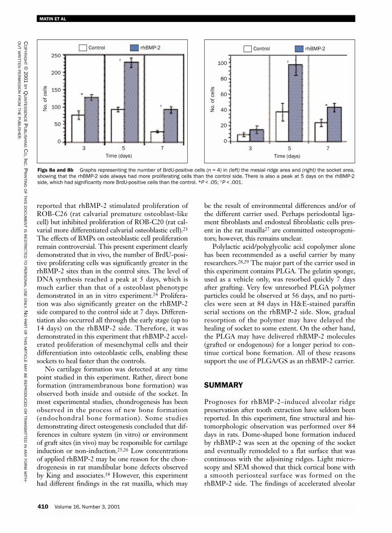
higher magnifications of the ridge area at 5 days,BrdU-positive mesenchymal cells on the controlside (Fig 7a inset) and BrdU-positive osteoblast-likecells on the rhBMP-2 side (Fig 7b inset) were seennear the mesial ridge. Freshly deposited bone matrixwas also seen around them on the rhBMP-2 sideonly. Both in the socket area (Figs 7c and 7d) and inthe ridge area, there were significantly greater num-bers (P < .001) of BrdU-positive cells in the rhBMP-2 group compared to the control group at 5 days(Figs 8a and 8b). The greatest number of BrdU-pos-itive cells was observed on the rhBMP-2 side at theridge area at 5 days. Although proliferation was
Figs 5a to 5d Graphs representing alveolar bone height measured at 4 points as described in Fig 1b.
Fig 5a At 14 days, alveolar bone height was significantlyincreased in the rhBMP-2 group compared to controls at M point,O point, and P point (P point was the peak point, at 2.9 mm). *P <.05.
Fig 5b At 28 days, alveolar bone height remained significantlygreater in the rhBMP-2 group than in the control group at N point,O point, and P point. The height had increased at the N point since14 days, but it had decreased at O point and P point. *P < .05.
Fig 5c At 56 days, alveolar bone height was further reduced inrhBMP-2 group but still remained significantly greater at O pointand P point. *P < .05.
Fig 5d At 84 days, the difference in alveolar bone heightsbetween the 2 groups was reduced.
3.0
3.5
2.5
2.0
1.5
1.0
0.5
0.0M point N point O point P point
Alve
olar
bon
e he
ight
(mm
) **
Control rhBMP-2
3.0
3.5
2.5
2.0
1.5
1.0
0.5
0.0M point N point O point P point
Alve
olar
bon
e he
ight
(mm
)
**
Control rhBMP-2
3.0
3.5
2.5
2.0
1.5
1.0
0.5
0.0M point N point O point P point
Alve
olar
bon
e he
ight
(mm
)
*
*
Control rhBMP-2
3.0
3.5
2.5
2.0
1.5
1.0
0.5
0.0M point N point O point P point
Alve
olar
bon
e he
ight
(mm
)
Control rhBMP-2
*
*
CO
PY
RIG
HT
©2001 B
YQ
UIN
TE
SS
EN
CE
PU
BLIS
HIN
GC
O, IN
C. PR
INT
ING
OF
TH
ISD
OC
UM
EN
TIS
RE
ST
RIC
TE
DT
OP
ER
SO
NA
LU
SE
ON
LY. NO
PA
RT
OF
TH
ISA
RT
ICLE
MA
YB
ER
EP
RO
DU
CE
DO
RT
RA
NS
MIT
TE
DIN
AN
YF
OR
MW
ITH-
OU
TW
RIT
TE
NP
ER
MIS
SIO
NF
RO
MT
HE
PU
BLIS
HE
R.

408 Volume 16, Number 3, 2001
MATIN ET AL
Figs 6a to 6e Hematoxylin and eosin (H&E) stained specimens viewed under light microscopy (�10).
Figs 6a and 6b (Left) Control specimenand (right) rhBMP-2 specimen at 3 days.There is no remarkable dif ferencebetween the 2 groups. In both sides, cellinfiltration inside the carrier particles isscant but appears to be condensing alongborder. mr = mesial ridge; SO = socket;asterisk = carrier.
Figs 6c and 6d (Left) Control specimenand (right) rhBMP-2 specimen at 14 days.A small amount of new bone formationoccurred inside the socket on the controlside. On the rhBMP-2 side, new bone for-mation occurred inside the entire socketin between carrier particles and extendedtoward the oral epithelium. mr = mesialridge; SO = socket; asterisk = carrier.
Figs 6e and 6f (Left) Control specimenand (right) rhBMP-2 specimen at 84 days.A small amount of connective tissue andsome bone marrow spaces can beobserved inside the matured bone onboth sides. Very small amounts of carrierparticles and a rough periodontal surfaceare visible on the control side, while asmooth periosteal surface can be seen onthe rhBMP-2 side. A thick cortical layer isvisible on both sides, with the rhBMP-2side being the thicker of the two. mr =mesial ridge; SO = socket; asterisk = car-rier; arrowheads = periosteal border of thenew bone.
mrso
*
somr
*
mr so
*
somr
*
*
mr
soso
mr
CO
PY
RIG
HT
©2001 B
YQ
UIN
TE
SS
EN
CE
PU
BLIS
HIN
GC
O, IN
C. PR
INT
ING
OF
TH
ISD
OC
UM
EN
TIS
RE
ST
RIC
TE
DT
OP
ER
SO
NA
LU
SE
ON
LY. NO
PA
RT
OF
TH
ISA
RT
ICLE
MA
YB
ER
EP
RO
DU
CE
DO
RT
RA
NS
MIT
TE
DIN
AN
YF
OR
MW
ITH-
OU
TW
RIT
TE
NP
ER
MIS
SIO
NF
RO
MT
HE
PU
BLIS
HE
R.

The International Journal of Oral & Maxillofacial Implants 409
MATIN ET AL
down-regulated at 7 days on both sides, the rhBMP-2 side had significantly greater numbers (P < .05) ofBrdU-positive cells (Figs 8a and 8b). Additionally,no cartilage formation could be detected during thisproliferation stage. Chondrogenesis was not seen atany stage under H&E-stained light microscopy.
DISCUSSION
Socket healing occurred in both the control andrhBMP-2 groups. In the control sites, healingoccurred at a comparatively slower rate than in therhBMP-2 sites. Sockets on the rhBMP-2 side healedfrom both inside the socket cavity and from theadjoining ridges (crest of the socket wall). In thisexperiment, bone formation was first recognized
outside the carrier on the mesial ridge at 5 days (Fig2b). BrdU-positive and ALPase-positive cells werealso recognized inside the carrier at the same stage.At 28 days, the new bone formed a dome at theridge, which was transformed into cortical bone witha smoother periosteal surface by 84 days. Bone histo-morphometry also showed that the total bone areaand alveolar height were significantly greater in therhBMP-2 group than in the control group up to 56days. However, by 84 days, there were no significantdifferences in both parameters between the 2 groups.A smoother periosteal surface and smaller osteocytelacunae indicated an increased maturation rate ofwoven bone in rhBMP-2 sites than in control sites.
Stimulatory effects of BMPs on osteoblastic cellproliferation have been observed in some in vitroexperiments.22 In contrast, one in vitro experiment
Figs 7a to 7d Immunohistochemical localization of proliferating cells by 5-Bromo-2’-deoxyuridine (BrdU) monoclonal antibody.
Figs 7a and 7b (Left) Control specimen and (right) rhBMP-2 specimen at 3 days, showing BrdU-positive cells (nucleus stained brown) atthe ridge area, with comparatively more on the rhBMP-2 side (�50). Inset photomicrographs are higher magnifications (�400) of ridgearea at 5 days. BrdU-positive mesenchymal cells are seen near the mesial ridge on the control side. BrdU-positive osteoblast-like cells areseen near the ridge, along with freshly formed bone matrix (right, inset). mr = mesial ridge; SO = socket, asterisks = carrier; large arrow-head = BrdU-positive cells; arrow = mesenchymal cells; small arrowhead = osteoblast-like cells; ma = bone matrix.
Figs 7c and 7d (Left) Control specimen and (right) rhBMP-2 specimen showing BrdU-positive cells in the socket area at 5 days. Herealso, BrdU-positive cells are more numerous on the rhBMP-2 side than on the control side. mr = mesial ridge; SO = socket; asterisk = car-rier; arrowheads = BrdU-positive cells.
**
mr mr
so
*
mr
so
*
mr
CO
PY
RIG
HT
©2001 B
YQ
UIN
TE
SS
EN
CE
PU
BLIS
HIN
GC
O, IN
C. PR
INT
ING
OF
TH
ISD
OC
UM
EN
TIS
RE
ST
RIC
TE
DT
OP
ER
SO
NA
LU
SE
ON
LY. NO
PA
RT
OF
TH
ISA
RT
ICLE
MA
YB
ER
EP
RO
DU
CE
DO
RT
RA
NS
MIT
TE
DIN
AN
YF
OR
MW
ITH-
OU
TW
RIT
TE
NP
ER
MIS
SIO
NF
RO
MT
HE
PU
BLIS
HE
R.
410 Volume 16, Number 3, 2001
MATIN ET AL
reported that rhBMP-2 stimulated proliferation ofROB-C26 (rat calvarial premature osteoblast-likecell) but inhibited proliferation of ROB-C20 (rat cal-varial more differentiated calvarial osteoblastic cell).23
The effects of BMPs on osteoblastic cell proliferationremain controversial. This present experiment clearlydemonstrated that in vivo, the number of BrdU-posi-tive proliferating cells was significantly greater in therhBMP-2 sites than in the control sites. The level ofDNA synthesis reached a peak at 5 days, which ismuch earlier than that of a osteoblast phenotypedemonstrated in an in vitro experiment.24 Prolifera-tion was also significantly greater on the rhBMP-2side compared to the control side at 7 days. Differen-tiation also occurred all through the early stage (up to14 days) on the rhBMP-2 side. Therefore, it wasdemonstrated in this experiment that rhBMP-2 accel-erated proliferation of mesenchymal cells and theirdifferentiation into osteoblastic cells, enabling thesesockets to heal faster than the controls.
No cartilage formation was detected at any timepoint studied in this experiment. Rather, direct boneformation (intramembranous bone formation) wasobserved both inside and outside of the socket. Inmost experimental studies, chondrogenesis has beenobserved in the process of new bone formation(endochondral bone formation). Some studiesdemonstrating direct osteogenesis concluded that dif-ferences in culture system (in vitro) or environmentof graft sites (in vivo) may be responsible for cartilageinduction or non-induction.25,26 Low concentrationsof applied rhBMP-2 may be one reason for the chon-drogenesis in rat mandibular bone defects observedby King and associates.18 However, this experimenthad different findings in the rat maxilla, which may
be the result of environmental differences and/or ofthe different carrier used. Perhaps periodontal liga-ment fibroblasts and endosteal fibroblastic cells pres-ent in the rat maxilla27 are committed osteoprogeni-tors; however, this remains unclear.
Polylactic acid/polyglycolic acid copolymer alonehas been recommended as a useful carrier by manyresearchers.28,29 The major part of the carrier used inthis experiment contains PLGA. The gelatin sponge,used as a vehicle only, was resorbed quickly 7 daysafter grafting. Very few unresorbed PLGA polymerparticles could be observed at 56 days, and no parti-cles were seen at 84 days in H&E-stained paraffinserial sections on the rhBMP-2 side. Slow, gradualresorption of the polymer may have delayed thehealing of socket to some extent. On the other hand,the PLGA may have delivered rhBMP-2 molecules(grafted or endogenous) for a longer period to con-tinue cortical bone formation. All of these reasonssupport the use of PLGA/GS as an rhBMP-2 carrier.
SUMMARY
Prognoses for rhBMP-2–induced alveolar ridgepreservation after tooth extraction have seldom beenreported. In this experiment, fine structural and his-tomorphologic observation was performed over 84days in rats. Dome-shaped bone formation inducedby rhBMP-2 was seen at the opening of the socketand eventually remodeled to a flat surface that wascontinuous with the adjoining ridges. Light micro-scopy and SEM showed that thick cortical bone witha smooth periosteal surface was formed on therhBMP-2 side. The findings of accelerated alveolar
Figs 8a and 8b Graphs representing the number of BrdU-positive cells (n = 4) in (left) the mesial ridge area and (right) the socket area,showing that the rhBMP-2 side always had more proliferating cells than the control side. There is also a peak at 5 days on the rhBMP-2side, which had significantly more BrdU-positive cells than the control. *P < .05; †P < .001.
250
200
150
100
50
03 5 7
Time (days)
*
Control rhBMP-2
100
80
60
40
20
03 5 7
Time (days)
Control rhBMP-2
†
†
†
*
No.
of c
ells
No.
of c
ells
CO
PY
RIG
HT
©2001 B
YQ
UIN
TE
SS
EN
CE
PU
BLIS
HIN
GC
O, IN
C. PR
INT
ING
OF
TH
ISD
OC
UM
EN
TIS
RE
ST
RIC
TE
DT
OP
ER
SO
NA
LU
SE
ON
LY. NO
PA
RT
OF
TH
ISA
RT
ICLE
MA
YB
ER
EP
RO
DU
CE
DO
RT
RA
NS
MIT
TE
DIN
AN
YF
OR
MW
ITH-
OU
TW
RIT
TE
NP
ER
MIS
SIO
NF
RO
MT
HE
PU
BLIS
HE
R.

bone formation by rhBMP-2 and adaptation with thepreexisting bone to create cortical alveolar ridge bonein rats suggest that the rhBMP-2 with PLGA/GScarrier may be useful in aiding socket healing.
ACKNOWLEDGMENT
This investigation was supported by Ministry of Education, Sci-ence, Sports and Culture of Japan. The authors would like tooffer their gratitude to the late Professor Haruka Kusakari ofthe Department of Fixed Prosthodontics, Niigata UniversitySchool of Dentistry, for professional support and advice. TherhBMP-2 and the carrier were kindly provided by YamanouchiPharmaceutical Industries, Tokyo, Japan.
REFERENCES
1. Carlsson GE, Persson G. Morphologic changes of themandible after extraction and wearing of dentures. A longi-tudinal, clinical, and x-ray cephalometric study covering 5years. Odontologisk Rev 1967;18:27–54.
2. Breine U, Brånemark P-I. Reconstruction of alveolar jawbone. An experimental and clinical study of immediate andpreformed autologous bone grafts in combination withosseointegrated implants. Scand J Plast Reconstr Surg 1980;14:23–48.
3. Urist MR. Bone formation by autoinduction. Science 1965;150:893–899.
4. Wozney JM, Rosen V, Celeste AJ, et al. Novel regulators ofbone formation: Molecular clones and activities. Science1988;242:1528–1534.
5. Schenk RK, Buser D, Hardwick WR, Dahlin C. Healingpattern of bone regeneration in membrane-protecteddefects. A histologic study in the canine mandible. Int J OralMaxillofac Implants 1994;9:13–29.
6. Howell TH, Fiorellini J, Jones A, et al. A feasibility studyevaluating rhBMP-2/absorbable collagen sponge device forlocal alveolar ridge preservation or augmentation. Int J Peri-odontics Restorative Dent 1997;17:125–139.
7. Rutherford RB, Sampath TK, Rueger DC, Taylor TD. Useof bovine osteogenic protein to promote rapid osseointegra-tion of endosseous dental implants. Int J Oral MaxillofacImplants 1992;7:297–301.
8. Lindhe A, Hedner E. Recombinant bone morphogeneticprotein-2 enhances bone healing, guided by osteopromotivee-PTFE membranes: An experimental study in rats. CalcifTissue Int 1995;56:549–553.
9. Cochran DL, Jones AA, Lilly LC, Fiorellini JP, Howell H.Evaluation of recombinant human bone morphogenetic pro-tein-2 in oral applications including the use of endosseousimplants: 3-year results of a pilot study in humans. J Perio-dontol 2000;71:1241–1257.
10. Alpaslan C, Irie K, Takahashi K, et al. Long-term evaluationof recombinant human bone morphogenic protein-2induced bone formation with a biologic and synthetic deliv-ery system. Br J Oral Maxillofac Surg 1996;34:414–418.
11. Toriumi DM, Kotler HS, Luxenberg DP, Holtrop ME,Wang EA. Mandibular reconstruction with a recombinantbone-inducing factor. Arch Otolaryngol Head Neck Surg1991;117:1101–1112.
12. Hoshi K, Amizuka N, Sakou T, Kurokawa T, Ozawa H.Fibroblasts of spinal ligaments pathologically differentiateinto chondrocytes induced by human bone morphogeneticprotein-2: Morphological examinations for ossification ofspinal ligaments. Bone 1997;21:155–162.
13. Wang EA, Rosen V, D’Alessandro JS, et al. Recombinanthuman bone morphogenic protein induces bone formation.(cartilage induction). Proc Natl Acad Sci 1990;87:2220–2224.
14. Sigurdsson TJ, Tatakis DN, Lee MB, Wikesjo UM. Perio-dontal regenerative potential of space-providing expandedpolytetrafluoroethylene membranes and recombinant humanbone morphogenic proteins. J Periodont Res 1995;66:511–521.
15. Cook SD, Salkeld SL, Rueger DC. Evaluation of recombi-nant osteogenic protein-1 (rhOP-1) placed with dentalimplants in fresh extraction sites. J Oral Implantol 1995;21:281–289.
16. Reddi AH, Huggins C. Biochemical sequences in the trans-formation of normal fibroblasts in adolescent rats. Proc NatlAcad Sci USA 1972;69:1601–1605.
17. Ting K, Petropulos LA, Iwatsu M, Nishimura I. Altered car-tilage phenotype expressed during intramembranous boneformation. J Bone Miner Res 1993;8:1377–1386.
18. King GN, King N, Cruchley AT, Wozney JM, Hughes FJ.Recombinant human bone morphogenetic protein-2 pro-motes wound healing in rat periodontal fenestration defects.J Dent Res 1997;76:1460–1470.
19. Correll JT, Prentice HR, Wise EC. Biologic investigationsof a new absorbable sponge. Surg Gynecol Obstet 1945;81:585–589.
20. Kinoshita A, Oda S, Takahashi K, Yokota S, Ishikawa I.Periodontal regeneration by application of recombinanthuman bone morphogenetic protein-2 to horizontal circum-ferential defects created by experimental periodontitis inbeagle dogs. J Periodontol 1997;68:103–109.
21. Gratzner HG. Monoclonal antibody to 5-bromo and 5-iododeoxyuridine: A new reagent for detection of DNAreplication. Science 1982;218:474–475.
22. Chen TL, Bates RL, Dudley A, Hammonds RG Jr, AmentoEP. Bone morphogenetic protein-2b stimulation of growthand osteogenic phenotypes in rats osteoblast-like cells: Com-parison with TGF-�. J Bone Miner Res 1991;6:1387–1393.
23. Yamaguchi A, Katagiri T, Ikeda T, Wozney JM, Rosen V,Wang EA. Recombinant human bone morphogenetic pro-tein-2 stimulates osteoblastic maturation and inhibits myo-genic differentiation in vitro. J Cell Biol 1991;113:681–687.
24. Stein GS, Lian JB. Molecular mechanisms mediating prolif-eration/differentiation interrelationships during progressivedevelopment of the osteoblast phenotype. Endocrinol Rev1993;14:424–442.
25. Iwasaki M, Nakahara H, Nakase T, et al. Bone morpho-genetic protein-2 stimulates osteogenesis but does not affectchondrogenesis in osteochondrogenic differentiation ofperiosteoum-derived cells. J Bone Miner Res 1991;9:1195–1204.
26. Rosen V, Thies RS. The BMP proteins in bone formationand repair. Trends Genet 1992;8:97–102.
27. Lin WL, McCulloch CAG, Cho MI. Differentiation ofperiodontal ligament fibroblasts into osteoblasts duringsocket healing after tooth extraction in the rat. Anat Rec1994;240:492–509.
28. Hollinger JO, Battistone GC. Biodegradable bone repairmaterials. Synthetic polymers and ceramics. Clin OrthopRel Res 1986;207:209–305.
29. Baum BJ, Mooney DJ. The impact of tissue engineering ondentistry. J Am Dent Assoc 2000;131:309–318.
The International Journal of Oral & Maxillofacial Implants 411
MATIN ET AL
CO
PY
RIG
HT
©2001 B
YQ
UIN
TE
SS
EN
CE
PU
BLIS
HIN
GC
O, IN
C. PR
INT
ING
OF
TH
ISD
OC
UM
EN
TIS
RE
ST
RIC
TE
DT
OP
ER
SO
NA
LU
SE
ON
LY. NO
PA
RT
OF
TH
ISA
RT
ICLE
MA
YB
ER
EP
RO
DU
CE
DO
RT
RA
NS
MIT
TE
DIN
AN
YF
OR
MW
ITH-
OU
TW
RIT
TE
NP
ER
MIS
SIO
NF
RO
MT
HE
PU
BLIS
HE
R.