Impact of 64 multi-detector computed tomography for the evaluation of aortic paraprosthetic...
6
Journal of Cardiology (2011) 58, 294—299 a va i la b le at www.sciencedirect.com j our na l ho me page: www.elsevier.com/locate/jjcc Original article Impact of 64 multi-detector computed tomography for the evaluation of aortic paraprosthetic regurgitation Masahiko Hara (MD) a,∗ , Masami Nishino (MD, PhD, FJCC) a , Masayuki Taniike (MD, PhD) a , Nobuhiko Makino (MD, PhD) a , Hiroyasu Kato (MD) a , Yasuyuki Egami (MD) a , Ryu Shutta (MD) a , Jun Tanouchi (MD, PhD, FJCC) a , Toshihiro Hunatsu (MD, PhD) b , Kazuhiro Taniguchi (MD, PhD) b , Yoshio Yamada (MD, PhD, FJCC) a a Division of Cardiology, Osaka Rosai Hospital, Sakai, Osaka, Japan b Department of Cardiovascular Surgery, Osaka Rosai Hospital, Sakai, Osaka, Japan Received 6 March 2011; received in revised form 24 June 2011; accepted 11 August 2011 Available online 15 September 2011 KEYWORDS Anatomical regurgitation orifice; Aortic paraprosthetic regurgitation; Computed tomography; Prosthetic heart valve Summary Background: Multi-detector computed tomography (MDCT) has been used to provide diagnostic information after heart valve replacements such as prosthetic valve dysfunction. However, only few data are available about aortic paraprosthetic regurgitation (APR). The aim of this study is to assess the feasibility, accuracy, and reproducibility of the existence of APR using contrast-enhanced 64-row MDCT. Methods: We retrospectively evaluated 20 consecutive patients who underwent both 64-row MDCT and two-dimensional transthoracic echocardiography (2D-TTE) after aortic valve replace- ment. The presence of APR was evaluated with 64-row MDCT, and validated with 2D-TTE, two-dimensional transesophageal echocardiography (2D-TEE), or intraoperative findings if avail- able. If APR was present, we also evaluated paraprosthetic anatomical regurgitation orifice (PARO) area to quantify the prognostic impact of APR or for surgical planning such as closure device sizing. Results: Overall, 12 of 20 valves showed beam-hardening artifact (BHA) which made the reliable evaluation of APR difficult. The presence of artifact seemed to depend on valve types. Among 8 patients who did not show BHA, there were perfect agreements between MDCT and 2D-TTE, 2D-TEE, or intra-operative findings about APR. There were excellent inter-observer agreements in the evaluation of APR and PARO area. PARO area was consistent with the echocardiographic severity of APR in this study. ∗ Corresponding author at: Division of Cardiology, Osaka Rosai Hospital, 1179-3 Nagasone-cho, Kita-ku, Sakai, Osaka 591-8025, Japan. Tel.: +81 72 252 3561; fax: +81 72 250 5492. E-mail address: [email protected] (M. Hara). 0914-5087/$ — see front matter © 2011 Japanese College of Cardiology. Published by Elsevier Ltd. All rights reserved. doi:10.1016/j.jjcc.2011.08.002
-
Upload
masahiko-hara -
Category
Documents
-
view
213 -
download
1
Transcript of Impact of 64 multi-detector computed tomography for the evaluation of aortic paraprosthetic...

J
O
Ifr
MMHJK
a
b
RA
T
0d
ournal of Cardiology (2011) 58, 294—299
a va i la b le at www.sc iencedi rec t .com
j our na l ho me page: www.elsev ier .com/ locate / j j cc
riginal article
mpact of 64 multi-detector computed tomographyor the evaluation of aortic paraprostheticegurgitation
asahiko Hara (MD)a,∗, Masami Nishino (MD, PhD, FJCC)a,asayuki Taniike (MD, PhD)a, Nobuhiko Makino (MD, PhD)a,iroyasu Kato (MD)a, Yasuyuki Egami (MD)a, Ryu Shutta (MD)a,un Tanouchi (MD, PhD, FJCC)a, Toshihiro Hunatsu (MD, PhD)b,azuhiro Taniguchi (MD, PhD)b, Yoshio Yamada (MD, PhD, FJCC)a
Division of Cardiology, Osaka Rosai Hospital, Sakai, Osaka, JapanDepartment of Cardiovascular Surgery, Osaka Rosai Hospital, Sakai, Osaka, Japan
eceived 6 March 2011; received in revised form 24 June 2011; accepted 11 August 2011vailable online 15 September 2011
KEYWORDSAnatomicalregurgitation orifice;Aortic paraprostheticregurgitation;Computedtomography;Prosthetic heart valve
SummaryBackground: Multi-detector computed tomography (MDCT) has been used to provide diagnosticinformation after heart valve replacements such as prosthetic valve dysfunction. However,only few data are available about aortic paraprosthetic regurgitation (APR). The aim of thisstudy is to assess the feasibility, accuracy, and reproducibility of the existence of APR usingcontrast-enhanced 64-row MDCT.Methods: We retrospectively evaluated 20 consecutive patients who underwent both 64-rowMDCT and two-dimensional transthoracic echocardiography (2D-TTE) after aortic valve replace-ment. The presence of APR was evaluated with 64-row MDCT, and validated with 2D-TTE,two-dimensional transesophageal echocardiography (2D-TEE), or intraoperative findings if avail-able. If APR was present, we also evaluated paraprosthetic anatomical regurgitation orifice(PARO) area to quantify the prognostic impact of APR or for surgical planning such as closuredevice sizing.
Results: Overall, 12 of 20 valves showed beam-hardening artifact (BHA) which made the reliableevaluation of APR difficult. The presence of artifact seemed to depend on valve types. Among8 patients who did not show BHA, there were perfect agreements between MDCT and 2D-TTE,2D-TEE, or intra-operative findings about APR. There were excellent inter-observer agreementsin the evaluation of APR and PARO area. PARO area was consistent with the echocardiographicseverity of APR in this study.∗ Corresponding author at: Division of Cardiology, Osaka Rosai Hospital, 1179-3 Nagasone-cho, Kita-ku, Sakai, Osaka 591-8025, Japan.el.: +81 72 252 3561; fax: +81 72 250 5492.
E-mail address: [email protected] (M. Hara).
914-5087/$ — see front matter © 2011 Japanese College of Cardiology. Published by Elsevier Ltd. All rights reserved.oi:10.1016/j.jjcc.2011.08.002

RuMDCT assessment of APR 295
Conclusions: Our retrospective data suggest that MDCT could be a reliable technique for theevaluation of APR after On-X standard or SJM standard valve replacement. MDCT can become anovel quantitative tool for the evaluation of PARO area.© 2011 Japanese College of Cardiology. Published by Elsevier Ltd. All rights reserved.
ovep8vafr
E
TcMafawtcaca
C
DCGaTSud1aeieaawdssfor subsequent off-line analysis.
Introduction
Aortic valve replacement is an effective and promisingtreatment in patients with significant aortic valvular heartdiseases [1]. Aortic paraprosthetic regurgitation (APR) is oneof the rare complications after aortic valve replacement,however, some patients require further surgical or tran-scatheter closure of APR because it could be life-threatening[2—5]. Two-dimensional echocardiography has long beenused as an essential method for the assessment of thesecomplications after heart valve replacement [3—7]. How-ever, it is difficult to accurately evaluate the exact locationof the paraprosthetic leaks and size of the defects using two-dimensional echocardiography partly due to mechanical orreverberation artifacts [3—7].
Recently, multi-detector computed tomography (MDCT)has been used to provide diagnostic information after heartvalve replacements such as prosthetic valve dysfunction[8—11]. However, only case reports are available aboutAPR detected by MDCT [8,9,12]. The aim of this study is toassess the feasibility, accuracy, and reproducibility of theexistence of APR using contrast-enhanced 64-row MDCT,and compared to already standardized two-dimensionalechocardiographic evaluation. We also measured theparaprosthetic anatomical regurgitation orifice (PARO) areato quantify the prognostic impact of APR or for surgicalplanning such as closure device sizing.
Methods
Study patients
We retrospectively enrolled 20 consecutive patientswho underwent both 64-row MDCT and two-dimensionaltransthoracic echocardiography (2D-TTE) after aortic valvereplacement between September 2007 and December 2009.Fifteen patients underwent mechanical valve replacement(6 On-X standard, On-X Life Technologies Inc., Austin,TX, USA; 2 SJM standard, 5 SJM HP, and 1 SJM Regentvalves, St. Jude Medical Inc., St. Paul, MN, USA; 1 ATS,ATS Medical Inc., Minneapolis, MN, USA) and the remaining5 patients underwent biological valve replacement (1 CEPand 4 Magna valves, Edwards Lifesciences, Irvine, CA, USA).Patients with suspected APR by 2D-TTE also underwent two-dimensional transesophageal echocardiography (2D-TEE)for additional evaluation of APR. This study includes onepreviously published case report of APR [12]. The mean timedifference between MDCT and 2D-TTE was 39 ± 48 days,
and patients who showed any changes in clinical statusduring the interval were excluded from the study. First,we evaluated the presence of beam-hardening artifact(BHA) which was defined as a black shining line projectingE
Da
utward from the prosthetics because the artifact impairsisualization around the prosthetics and makes the precisevaluation of APR difficult (Fig. 1). Then, we evaluated theresence of APR and PARO area using 64-row MDCT among
of 20 prosthetics without BHA. The presence of APR wasalidated with 2D-TTE, 2D-TEE, or intraoperative findings ifvailable (Table 1). Written informed consent was obtainedrom all patients before CT evaluation, and the institutionaleview board approved this study.
chocardiographic evaluation
wo-dimensional echocardiography was performed usingommercially available ultrasound system (Aplio, Toshibaedical, Tokyo, Japan) by an experienced medical doctornd sonographer. The consensus-based findings were usedor the analysis. The existence and severity of APR weressessed with Doppler echocardiography. We defined APRhen the flow jet signal was identified outside of the pros-
hetic reverberations at diastole. The severity of APR waslassified into 3 categories (mild, moderate, and severe) in
comprehensive manner using regurgitation jet area, venaontracta, regurgitation duration, and left ventricular sizenalyses [13].
omputed tomography acquisition
ata were acquired with a 64 multi-detector row dual sourceT (SOMATOM Definition, Siemens Medical Systems, Munich,ermany) with a detector collimation width of 0.6 mm and
gantry rotation time of 0.33 s within a single breath hold.ube voltage was fixed at 120 kV with tube current of 850 mA.can direction was craniocaudal. Electrocardiogram mod-lation was not used. The timing of data acquisition wasetermined with a bolus tracking method with a threshold of50 Hounsfield Unit by setting region of interest to ascendingorta at the bifurcation level of pulmonary artery. Contrastnhancement was obtained using nonionic contrast mediumnjection (Iopamirone 370 mgI/dL, Bayer Health Care, Lev-rkusen, Germany) from antecubital venous approach with
20-gauge cannula. Contrast medium was administered at flow rate of 4 mL/s. A beta-blocker (40 mg of metoprorol)as given before the examination at attending physicians’iscretion because bradycardia may worsen heart failureymptoms with aortic regurgitation. Images were recon-tructed from 0 to 90% of the RR interval with 10% intervals
valuation of APR
ata analysis was performed off-line using commerciallyvailable workstation (AquariusNetStation, Terarecon Inc.,
296 M. Hara et al.
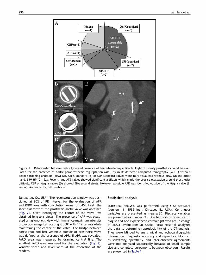
Figure 1 Relationship between valve type and presence of beam-hardening artifacts. Eight of twenty prosthetics could be eval-uated for the presence of aortic paraprosthetic regurgitation (APR) by multi-detector computed tomography (MDCT) withoutbeam-hardening artifacts (BHA) (A). On-X standard (B) or SJM standard valves were fully visualized without BHA. On the otherhand, SJM HP (C), SJM Regent, and ATS valves showed significant artifacts which made the precise evaluation around prostheticsd oweva
Stas(oapmawPsWr
S
S(vaootTi
ifficult. CEP or Magna valves (D) showed BHA around struts. Hrrow). Ao, aorta; LV, left ventricle.
an Mateo, CA, USA). The reconstruction window was posi-ioned at 90% of RR interval for the evaluation of APRnd PARO area with convolution kernel of B45f. First, thehort-axis view of the prosthetic aortic valve was obtainedFig. 2). After identifying the center of the valve, webtained long-axis views. The presence of APR was evalu-ted using long-axis view with 1 mm slice maximum intensityrojection image by rotating it 360◦ with 1◦ intervals whileaintaining the center of the valve. The bridge between
ortic root and left ventricle outside of prosthetic valveas defined as the presence of APR. If APR was revealed,
ARO area was measured by drawing central line. Themallest PARO area was used for the evaluation (Fig. 2).indow width and level were at the discretion of the
eaders.
awsa
er, possible APR was identified outside of the Magna valve (E,
tatistical analysis
tatistical analysis was performed using SPSS softwareversion 11, SPSS Inc., Chicago, IL, USA). Continuousariables are presented as mean ± SD. Discrete variablesre presented as number (%). One fellowship-trained cardi-logist and one experienced cardiologist who are in chargef MDCT evaluations at Osaka Rosai Hospital analyzedhe data to determine reproducibility of the CT analysis.hey were blinded to any clinical and echocardiographic
nformation. Diagnostic accuracy and reproducibility such
s sensitivity, specificity, and inter-observer agreementsere not analyzed statistically because of small sampleize and complete agreements between observers. Resultsre presented in Table 1.

RuMDCT assessment of APR 297
Table 1 MDCT, 2D-E and operative findings after aortic valve replacement.
Case Age Sex Prosthetics APR MDCT APR 2D-E Re-ope PARO1 PARO2 Duration (days)
1 73 Male On-X standard No No No 0 0 3712 65 Male On-X standard No No No 0 0 283 69 Female On-X standard No No No 0 0 4984 72 Male On-X standard No No No 0 0 205 36 Male On-X standard No No No 0 0 336 67 Male On-X standard No No No 0 0 2317 61 Female SJM standard Yes Severe Yes 48.4 43.6 45518 56 Male SJM standard Yes Severe Yes 22.6 21.7 4938
Patients with suspected aortic paraprosthetic regurgitation (APR) by two-dimensional transthoracic echocardiography (2D-TTE) alsounderwent two-dimensional transesophageal echocardiography (2D-TEE) for additional evaluation of APR. The presence of APR wasdetermined in a comprehensive way with 2D-TTE and 2D-TEE findings. 2D-E, 2D-TTE with or without 2D-TEE; duration, duration from
grap
D
IeaTa
A
WddhspermrTdSeatr
P
Wocs
(ni
aortic valve replacement; MDCT, multi-detector computed tomo(mm2); Re-ope, re-operation after MDCT.
Results
Fig. 1 shows the relationship between valve type and thepresence of BHA. Eight of twenty prosthetics could be evalu-ated by MDCT without BHA. The presence of artifact seemedto depend on valve types. On-X standard or SJM standardvalves were fully visualized without BHA. On the other hand,SJM HP, SJM Regent, and ATS valves showed significant arti-facts which made the precise evaluation around prostheticsdifficult. CEP or Magna valves showed BHA around struts(Fig. 1).
Eight prosthetics without BHA were then evaluated forthe presence of APR, and results were compared with 2D-TTE, 2D-TEE, or intraoperative findings if available (Table 1).In these 8 patients, age was 62 ± 12 years old, 6 (75%)were male, body surface area was 1.7 ± 0.1 m2, estimatedglomerular filtration rate was 81 ± 19 mL/min/1.73 m2, con-trast medium used for MDCT was 71 ± 6 mL, heart rate duringMDCT was 70 ± 18 bpm, left ventricular end-diastolic volumewas 169 ± 67 mL, end-systolic volume was 86 ± 45 mL, andleft ventricular ejection fraction was 49 ± 14%. As shownin Table 1, there were perfect agreements between MDCTand two-dimensional echocardiographic findings about APR.Besides, intra-operative findings were also completely com-patible with MDCT findings in cases 7 and 8 (Fig. 2).There were excellent inter-observer agreements betweenobserver 1 and 2 about the evaluation of APR and PARO area.
Two patients with APR showed some causes of tissuefragility around the prosthetics. The patient of case 7 inTable 1 underwent aortic valve replacement because ofsevere aortic regurgitation due to aortitis, and the patient ofcase 8 in Table 1 underwent aortic and mitral valve replace-ment because of severe aortic and mitral regurgitation dueto infectious endocarditis [12]. On the other hand, none ofthe remaining 6 patients in Table 1 showed any causes oftissue fragility at the day of valve replacement.
In addition, possible APR was identified outside of theMagna valve even with BHA around struts in one case(Fig. 1E). The presence of APR was validated with two-
dimensional echocardiography, and the possible PARO areawas 11.74 mm2 by observer 1 and 10.41 mm2 by observer 2which is consistent with two-dimensional echocardiographicfindings of moderate APR in this case.coar
hy; PARO, paraprosthetic anatomical regurgitation orifice area
iscussion
n this study, we newly proposed the methodology for thevaluation of APR by MDCT. We demonstrated that MDCTccurately identifies the existence of APR compared to 2D-TE, 2D-TEE, or intra-operative findings if available. PAROrea could be quantified with good reproducibility.
rtifact and valve type
e demonstrated that the presence of artifact seemed toepend on valve types (Fig. 1). On-X standard or SJM stan-ard valves were fully visualized without BHA. On the otherand, SJM HP, SJM Regent, ATS, CEP, and Magna valveshowed BHA which made the precise evaluation aroundrosthetics difficult. Tsai et al. reported that suture loos-ning could be evaluated using MDCT after one aortic valveeplacement and 3 mitral valve replacements with On-Xechanical valves [9]. On the other hand, Chenot et al.
eported that CEP shows BHA around metal struts [14].hese reports support our data. Even though SJM stan-ard valve was assessable without BHA by MDCT, SJM HP orJM Regent valves showed BHA. We speculated that newlyquipped MP35N (Nickel—Cobalt—Chromium—Molybdenum)lloy in suturing cuff of SJM HP or Regent valves may affecthe BHA. Thus, we think that architectural componentsather than prosthetic valve design may cause BHA.
ARO area by MDCT
e evaluated PARO area to quantify the prognostic impactf APR, and revealed that PARO area assessed by MDCT wasonsistent with the echocardiographic severity of APR in thistudy.
Conventionally, effective regurgitation orifice areaEROA) has been applied for the quantitative assessment ofative aortic valvular regurgitation in addition to the qual-tative assessment such as regurgitation jet area or vena
ontracta [13]. EROA < 10 mm2 is considered as mild, EROAf 10—20 mm2 as mild to moderate, EROA of 20—30 mm2s moderate to severe, and EROA > 30 mm2 as severe aorticegurgitation [13]. However, quantitative two-dimensional

298 M. Hara et al.
Figure 2 Evaluation of aortic paraprosthetic regurgitation (APR). A 56-year-old male with SJM standard valve was evaluated (case8 in Table 1). First, short-axis view of the prosthetic aortic valve was obtained (A). After identifying the center of the valve, weobtained long-axis views. The presence of APR was evaluated using long-axis view with 1 mm slice maximum intensity projectionimage by rotating it 360◦ with 1◦ intervals while maintaining the center of the valve (B). The bridge between aortic root and leftventricle outside of prosthetic valve was defined as the presence of APR (B, arrow). If APR was revealed, paraprosthetic anatomicalregurgitation orifice (PARO) area was measured by drawing central line (B, dotted line). The smallest PARO area was used for theevaluation (C). Volume-rendering image of APR viewed from ascending aorta was reconstructed (D). The multi-detector computedtomography findings were completely compatible with operative findings (E). Lower PARO was distorted in order to enlarge thev leftp k; LV
ervp[iwurgT
MYahore
iewing field by tweezers and association between aorta andresence of APR. Ao, aorta; LA, left atrium; LMT, left main trun
chocardiographic assessment of APR is difficult becauseeverberation artifact makes it difficult to analyze flow con-ergence [13]. 3D-TTE has the potential to evaluate theresence of APR but its resolution is not sufficient so far6,7]. Because the minimum spatial resolution of this MDCTs 0.33 mm according to the manufacturer specifications,e considered that it is possible to assess APR severity
sing PARO area which may require at least 1 mm2 spatialesolution referred to the EROA classification of aortic regur-itation by 2D-TTE [13]. Patients with severe APR (case 7 inable 1) and moderate to severe APR (case 8 in Table 1) bytwoi
ventricle could not be visualized well. Arrows indicate the, left ventricle; RCA, right coronary artery.
DCT needed surgical treatment in order to manage Nework Heart Association class 3 heart failure symptoms. Inddition, possible mild to moderate APR with BHA (Fig. 1E)as been followed-up carefully for 7 months medically with-ut any worsening and operation. Even though we shouldemember that BHA around struts may obscure the pres-nce of additional APR in the case of Fig. 1, this case implies
hat the presence of APR could be evaluated in some casesith BHA. Although the sample size is very small, clinicalutcomes shown in this study are consistent with APR sever-ty classification by MDCT. Thus, we think the PARO area
[
[
[
[
[
RuMDCT assessment of APR
assessed by MDCT might be accurate [15], and is applica-ble for assessing prognostic impact of APR and for surgicalplanning such as closure device sizing.
Clinical implications
Our data suggest that MDCT is a reliable technique forthe evaluation of APR and PARO area after aortic valvereplacements with On-X standard or SJM standard valves.Thus, we believe that MDCT should be applied for addi-tional evaluation and surgical planning in conjunction withechocardiography if APR is suspected after On-X standardor SJM standard valve replacement. Next-generation MDCT-assessable prosthetic valves should develop, especially forpatients who showed inflammatory diseases which may leadto tissue fragility around the prosthetics.
Study limitations
Our study has several limitations. First, it was a single cen-ter retrospective study which has small sample size partlybecause APR is uncommon. Second, we enrolled consec-utive CT examinations, but not consecutive patients withaortic valve replacement. It may lead to a biased result.Finally, 2D-TTE was used as a gold standard in this study. Ithas some limitations in the evaluation of prosthetic valvesalthough we also used 2D-TEE or operative findings if avail-able [6,7,13].
Conclusions
Our retrospective data suggest that MDCT could be a reliabletechnique for the evaluation of APR and PARO area after On-X standard or SJM standard valve replacement. Further largescale prospective study is needed to confirm our conclusion.
References
[1] Bonow RO, Carabello BA, Chatterjee K, de Leon AC Jr, FaxonDP, Freed MD, Gaasch WH, Lytle BW, Nishimura RA, O’Gara PT,O’Rourke RA, Otto CM, Shah PM, Shanewise JS. 2006 WritingCommittee Members, et al. 2008 Focused update incorporatedinto the ACC/AHA 2006 guidelines for the management ofpatients with valvular heart disease: a report of the Amer-ican College of Cardiology/American Heart Association TaskForce on Practice Guidelines (Writing Committee to Revise the1998 Guidelines for the Management of Patients With Valvu-
lar Heart Disease): endorsed by the Society of CardiovascularAnesthesiologists, Society for Cardiovascular Angiography andInterventions, and Society of Thoracic Surgeons. Circulation2008;118:e523—661.[
299
[2] Hammermeister KE, Sethi GK, Henderson WG, Oprian C, Kim T,Rahimtoola S. A comparison of outcomes in men 11 years afterheart-valve replacement with a mechanical valve or biopros-thesis. Veterans Affairs Cooperative Study on Valvular HeartDisease. N Engl J Med 1993;328:1289—96.
[3] Rallidis LS, Moyssakis IE, Ikonomidis I, Nihoyannopoulos P. Nat-ural history of early aortic paraprosthetic regurgitation: afive-year follow-up. Am Heart J 1999;138:351—7.
[4] O’Rourke DJ, Palac RT, Malenka DJ, Marrin CA, ArbuckleBE, Plehn JF. Outcome of mild periprosthetic regurgitationdetected by intraoperative transesophageal echocardiography.J Am Coll Cardiol 2001;38:163—6.
[5] Latson LA. Transcatheter closure of paraprosthetic valve leaksafter surgical mitral and aortic valve replacements. Expert RevCardiovasc Ther 2009;7:507—14.
[6] Singh P, Inamdar V, Hage FG, Kodali V, Karakus G, SuwanjutahT, Hsiung MC, Nanda NC. Usefulness of live/real time three-dimensional transthoracic echocardiography in evaluation ofprosthetic valve function. Echocardiography 2009;26:1236—49.
[7] Shiota T. 3D echocardiography: the present and the future. JCardiol 2008;52:169—85.
[8] Konen E, Goitein O, Feinberg MS, Eshet Y, Raanani E, Rimon U,Di-Segni E. The role of ECG-gated MDCT in the evaluation ofaortic and mitral mechanical valves: initial experience. Am JRoentgenol 2008;191:26—31.
[9] Tsai IC, Lin YK, Chang Y, Fu YC, Wang CC, Hsieh SR, Wei HJ, TsaiHW, Jan SL, Wang KY, Chen MC, Chen CC. Correctness of multi-detector-row computed tomography for diagnosing mechanicalprosthetic heart valve disorders using operative findings as agold standard. Eur Radiol 2009;19:857—67.
10] LaBounty TM, Agarwal PP, Chughtai A, Kazerooni EA, Wiza-uer E, Bach DS. Hemodynamic and functional assessment ofmechanical aortic valves using combined echocardiographyand multidetector computed tomography. J Cardiovasc ComputTomogr 2009;3:161—7.
11] LaBounty TM, Agarwal PP, Chughtai A, Bach DS, WizauerE, Kazerooni EA. Evaluation of mechanical heart valve sizeand function with ECG-gated 64-MDCT. Am J Roentgenol2009;193:W389—96.
12] Toda K, Taniguchi K. Periprosthetic leakage identified by multi-slice computed tomography. Ann Thorac Surg 2010;89:310.
13] Zoghbi WA, Enriquez-Sarano M, Foster E, Grayburn PA, KraftCD, Levine RA, Nihoyannopoulos P, Otto CM, Quinones MA,Rakowski H, Stewart WJ, Waggoner A, Weissman NJ, AmericanSociety of Echocardiography. Recommendations for evalua-tion of the severity of native valvular regurgitation withtwo-dimensional and Doppler echocardiography. J Am SocEchocardiogr 2003;16:777—802.
14] Chenot F, Montant P, Goffinet C, Pasquet A, VancraeynestD, Coche E, Vanoverschelde JL, Gerber BL. Evaluation ofanatomic valve opening and leaflet morphology in aortic valvebioprosthesis by using multidetector CT: comparison withtransthoracic echocardiography. Radiology 2010;255:377—85.
15] Vural M, Ucar O, Celebi OO, Cicekcioglu H, Durmaz HA, SelviNA, Koparal S, Aydogdu S. Evaluation of effective regurgitantorifice area of mitral valvular regurgitation by multislice car-diac computed tomography. J Cardiol 2010;56:236—9.