Immunology of the GI Tract - NASPGHAN
29
Immunology of the GI Tract a brief overview Esi Lamousé-Smith, MD, PhD Morgan Stanley Children’s Hospital of New York Columbia University [email protected] Content Reviewer: W. Allan Walker, MD
Transcript of Immunology of the GI Tract - NASPGHAN

Immunology of the GI Tracta brief overview
Esi Lamousé-Smith, MD, PhDMorgan Stanley Children’s Hospital of New York
Columbia [email protected]
Content Reviewer:
W. Allan Walker, MD

NASPGHAN Physiology Education Series
Series Editors:Christine Waasdorp Hurtado, MD, MSCS, [email protected] Kamin, [email protected]

Objectives
• Understand the organization of the intestinal immune tissues
• Understand the immune cell populations and their distribution in the intestinal tract
• Understand the role of sIgA to immune barrier function and regulation
• Understand how oral tolerance develops
• Understand the the importance of the intestinal flora in immunity

GALT
• The gastrointestinal associated lymphoid tissue (GALT) serves its function as
– a barrier against pathogen entry and spread within the host,
– an inductive site for innate and adaptive immune responses
– site for immune cell expansion and survival

GALT sites of immune induction
Antigens transported by EC and captured by DC are presented to naïve T and B cells inducing their activation, proliferation, and differentiation in the:– Peyers Patches
– Microfold cells (M cells)
– Mesenteric Lymph nodes
– Isolated lymphoid follicles (ILF)
– CryptopatchesReprinted from Nature Reviews Immunology Volume 8, Cerutti, A. The regulation of IgA class switching, p421-434, Figure 4. Copyright (2008) with permission from Nature Publishing Group.

GALT sites for effector immunity
• Following activation, effectorcells selectively up-regulate chemokine and adhesion molecules for homing to the GIT mucosa– CCR9: binds CCL25 which is
only expressed by EC in the GIT and thymus
– Α4β7: binds MADCAM-1 expressed by mucosal endothelial cells
• The tissues where effectorimmune function occurs are: – The lamina propria– The intra-epithelial cell
compartment
Reprinted from Nature Reviews Immunology Volume 8, Cerutti, A. The regulation of IgA class switching, p421-434, Figure 4. Copyright (2008) with permission from Nature Publishing Group.

GALT Effector Cells
Lamina Propria Cells
• Immunoglobulin A (IgA) secreting plasma cells
• Activated B cells
• T cells (CD4+>>CD8+)
• macrophages
• dendritic cells (DCs)
• innate lymphoid cells (ILCs)
Intraepithelial Cells
• Function based upon distribution along the length of the intestinal tract
• Exhibit an activated “antigen experienced” phenotype
• Majority are CD8αα+ and TCRγδ+ T cells

Epithelial Cells
• provide both a physical and immune barrier in the GIT
• at the “front line” of immune recognition in the GIT
• express extra and intracellular pattern recognition receptors (PRR) for the pathogen associated molecular patterns (PAMPs) expressed by bacteria and viral species

PRRs and PAMPs
• MDP= muramyldipeptide
• DAP= D-glutamyl-meso-diaminopimelicacid
• Not shown:
– The RIG-I intracellular receptors bind viral dsRNA
Reprinted from Nature Reviews Microbiology Volume 5, Kaufman, S.H.E. The contribution of immunology to the rational design of novel antibacterial vaccines,p491-504, Figure 2. Copyright (2007) with permission from Nature Publishing Group.

GIT epithelial cells recognize microbes binding to pattern recognition receptors
• Engagement by PAMPs triggers intracellular signaling cascade
• Subsequent gene transcription and production of cytokines & chemokines
• Recruit immune cells to fortify the EC response and limit microbial expansion or invasion
• Responding immune cells from the GALT include innate effectors (neutrophils, macrophages, dendritic cells, eosinophils, mast cells) and adaptive effectors (T cells and B cells)
Reprinted with permission from Proceedings of the National Academy of Sciences. Wells, JM et al, Epithelial crosstalk at the microbiota-mucosal interface, Figure 11. 108: 4607-14, 2011.
IgA
• IgA is the most abundant antibody isotype in the serum and in mucosal sites– 40 mg/kg or ∼3 g is produced
in an adult human per day!
• Contributes to maintenance of intestinal epithelial barrier function
• Requires epithelial pIgRexpression and transcriptional regulation of J-chain production http://www.buddycom.com/cells/immun/igxara/index.html
J= Joining chain, required for EC transport of IgA dimersSC= secretory chain, prevents proteolytic damage of IgA

Functions of IgA
• Confines commensal bacteria to the mucus layer of the intestinal lumen • Binds to invasive pathogens • Neutralizes microbial toxins and other inflammatory microbial products • Neutralization of antigens and pathogens in epithelial cell endosomes • Uptake of luminal antigens• Transport of antigens from the LP into the lumen
Figure reprinted from Nature Reviews Microbiology. Strungell, RA and Wijburg, ODLC, The role of secretory antibodies in infection immunity, Figure 3. 8: 656-67. Copyright 2010 with permission from Nature Publishing Group.

IgA Deficiency
• Deficiency is not severely detrimental
• Compensation by other antibody isotypes
– intestinal sIgM also binds pIgR for translocation into the intestinal lumen and other innate pathways
• However, sIgA deficiency may influence the development of autoimmunity and allergy
– limits innate immune responses,
– modulates intestinal regulatory T cells,
– regulates the commensal flora

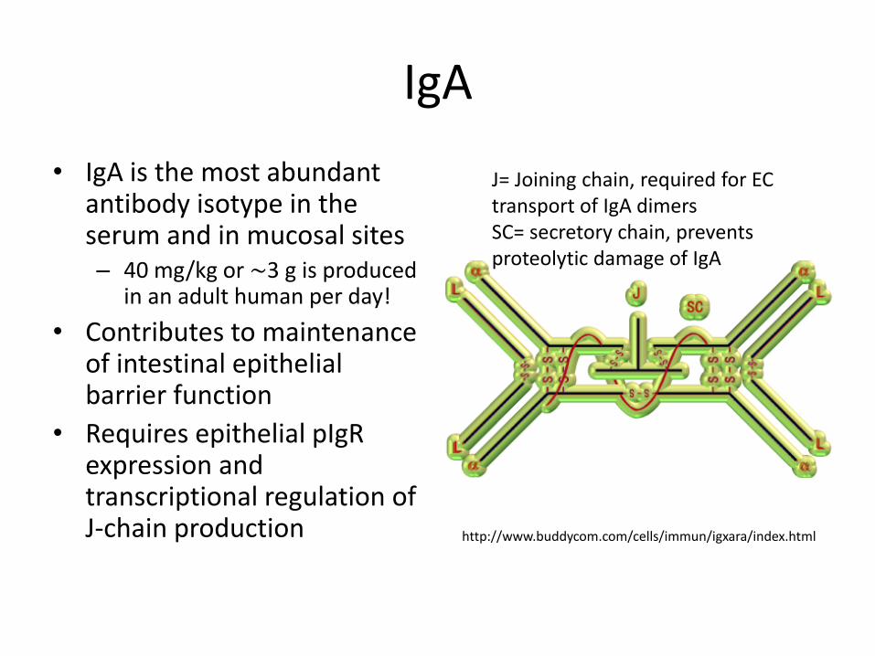
• We are a “super-organsim”
• The human gastrointestinal tract hosts a vast microbiome– >1000 different species; >majority non-culturable
• Establishes during the first week and stabilizes within the first 3-5 years of life
• Cooperative relationships have established through co-evolution to maintain homeostasis
Bacteria (~1000 species)
• Firmicutes (Ruminococcus, Clostridium, Eubacteria)
• Bacteroidetes(Bacteroides)
• Actinobacteria(Bifidobacteria)
• Proteobacteria(Enterobacteraceae)
• Fusobacteria
Viruses
FungiFigure reprinted from EMBO Reports Volume 7, O’Hara, AM & Shanahan, F. The gut flora as a forgotten organ, p688-693, Figure 1. Copyright (2006) with permission from Nature Publishing Group.
Combined genome 150 fold size of human genome
The GIM a diverse community

Immune tissue development requiresnormal gut colonization
Reprinted from Nature Review Immunology Volume 4, Macpherson, AJ and Harris, NL. Interactions between commensal intestinal bacteria and the immune system, p478-485, Figure 2. Copyright (2004) with permission from Nature Publishing Group.

Oral tolerance
• Induction of mucosal and systemic non-responsiveness to dietary antigens
• Active immune response that depends upon
– tolerogenic dendritic cells (make IL-10),
– CD4+CD25+T regulatory cells (make TGFβ & IL-10)
• Mechanisms of tolerance:
– Ignorance (no/low costimulatory signals)
– Inhibition/Anergy (inhibitory signals)

Uptake of food antigens occurs via
• Transcytosis through M cells,
• Paracellular diffusion or transcytocis across intestinal villi ECs,
• Uptake of exosomes (MHC Class II loading compartments fused with endosomes) by lamina propria DC,
• Luminal capture by CXCR31high
DC/macrophages.

GIT mucosal defenses balance immune responsiveness to its flora
• Commensal non-pathogenic & pathogenic gut microbial species are recognized by pattern recognition receptors (e.g. TLR, NOD)
• Several mechanisms are involved in attenuating inflammatory responses against the commensal flora: – Different levels and cellular distribution of TLRs on epithelial cells (EC)
• NOD2 modulation of TLR signaling (thus mutations of NOD2 may contribute to IBD)
– Containment of luminal flora and defense against microbial translocation• Mucin production• Elaboration of paneth cell defensins• IgA secretion
– Commensal bacteria mediated inhibition of NF-kβ signaling (likely through soluble mediators released by bacteria)
– Inhibition of innate and adaptive immune responses by • TSLP (thymic stromal lymphopoietin) production by enterocytes• Regulatory T cells• Tolerogenic dendritic cells

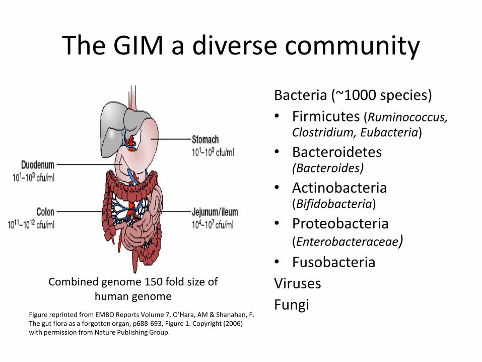
Dysbiosis
• Generally describes the intestinal flora when it has been altered from a healthy equilibrium or baseline
• Combined with other environmental and genetic influences likely contributes to the development of intestinal and systemic diseases:– IBD
– Diabetes
– Metabolic syndrome and obesity
– Food allergy
Dysbiosis and IBD
Reprinted from Gastroenterology Volume 139, B. Sartor, Genetics and Environmental Interactions Shape the Intestinal Microbiome to Promote Inflammatory Bowel Disease Versus Mucosal Homeostasis, pages 1816-1819, Figure 1. Copyright (2010) with permission from the author and Elsevier.

Summary Points
• The goal of GIT immunity is to recognize and maintain a healthy microbiome while also recognizing and expelling microbes that cause disease.
• GIT immune function derives from the immune cells and their interactions within the gastrointestinal associated lymphoid tissue, or GALT.
• The GALT includes innate immune function, provided by cells that quickly respond to threats and re-establish equilibrium.
• The GALT also includes adaptive immune function, provided by cells that respond to threats and generate a mucosal immune response that is specific and robust.
• Cross talk between innate and adaptive systems is crucial for avoiding unnecessary or ineffective inflammatory responses to ingested antigens and the re

Summary Points- 2
• Immune induction occurs in peyers patches and mesenteric lymph notes. Specialized cells in the mucosa overlying the patches are integral participants in the adaptive immune response.
• Immune effector function occurs in the lamina propria, and includes specific lymphocytes and plasma cells that product IgA.
• IgA serves to neutralize toxins and invasive organisms while also maintaining keeping commensal organisms in check.

Summary Points-3
• GIT microbiome has evolved to benefit us, and we have evolved to benefit its constituents.
• Derangements in the GIT microbiome are associated with inflammatory, allergic, and metabolic diseases.
• Oral tolerance of ingested protein is established by interaction between particular antigen presenting cells and modulatory T cells. Derangements can result in allergy.
Case #1
• 12 year old male presents with a one year history of– Stunted growth– Diarrheal stools– Occasional oral apthous sores
• What is the likely diagnosis?• What deficiency may be noted on histologic
examination of ileal biopsies?• Which identified PRR or cytokine gene mutations
could contribute to his diagnosis?
Case #2
• 3 year old toddler develops lip swelling, shortness of breath, and blotchy skin rash a few hours after eating a strawberry
• What is the likely diagnosis? And what immune process has broken down?
• What maternal factors would protect against development of this diagnosis?
• Which mucosal immune compartment and cell population contributes to regulated immunity against the strawberry?

Case #1
• 12 year old male presents with a one year history of– Stunted growth– Diarrheal stools– Occasional oral apthous sores
• What is the likely diagnosis? – Crohn’s Disease• What deficiency may be noted on histologic examination of ileal
biopsies? Paneth Cells• Which identified PRR or cytokine gene mutations could contribute
to his diagnosis? NOD2, IL-10, IL23/Th17 axis. Defects in the expression of these gene products contribute to driving chronic mucosal inflammation by impairing host control of the intestinal flora (NOD2) or by resulting in over-exuberant inflammatory responses against relatively innocuous gut bacteria (IL23/Th17 axis) or by allowing normal inflammatory responses to proceed unchecked due to ineffective negative regulation (IL10).

Case #2
• 3 year old toddler develops lip swelling, shortness of breath, and blotchy skin rash a few hours after eating a strawberry
• What is the likely diagnosis? Food Allergy• And what immune process has broken down? Oral
tolerance• What maternal factors would protect against development
of this diagnosis? Breast feeding and transfer of maternal secretory IgA
• Which mucosal immune compartment and cell population contributes to regulated immunity against the strawberry? Mesenteric lymph nodes, Foxp3 T regulatory cells, and IgA B cells

Please send any questions or comments to:
or