Week 13: Antimicrobial Drugs Tuesday, June 9, 2015 Biochemistry, Microbiology and Immunology.
1
Biochemistry Immunology
Immunotechnology-I
Paper : 16 Immunology Module : 11 Immunotechnology-I
Principal Investigator
Dr. Sunil Kumar Khare,Professor
Dept. of Chemistry,
I.I.T. Delhi
Content Reviewer:
Dr. M.N.Gupta, Emeritus Professor
Dept. of Biochemical Engg. and
Biotechnology, I.I.T. Delhi
Paper Coordinator
and
Content Writer
Dr. Prashant Mishra, Professor
Dept. of Biochemical Engg. and
Biotechnology, I.I.T. Delhi

2
Biochemistry Immunology
Immunotechnology-I
Description of Module
Subject Name Biochemstry
Paper Name 16 Immunology
Module Name/Title 11 Immunotechnology-I
Dr. Vijaya Khader Dr. MC Varadaraj

3
Biochemistry Immunology
Immunotechnology-I
1. Objectives
To understand:
a.) What experimental techniques are used to understand the functioning of the immune
system
b.) What techniques are based upon cells/molecules related to the immune system
c.) How some of these techniques are now the basis of invaluable diagnostic tests/design
of biosensors for analytes/infectious agents.
2. Concept Map
3. Description

4
Biochemistry Immunology
Immunotechnology-I
Immunization
Immunology started with efforts to understand how our bodies respond to infectious agents
and what we can do to avoid/minimize consequences of this invasion. However, right in the
early years, immunologists developed protocols to raise antibodies in experimental animals
against known (well defined or ill defined) antigen preparation.
Choice of the experimental animals
1. The animal should be healthy and as far removed as possible on the evolutionary
tree from the source of the antigen. As we learnt in the module on antigens and epitopes,
“foreignness” is one essential criterion for antigenicity
2. Depending upon the amount of anti-serum required, the animal with appropriate size
has to be chosen.
Figure 1

5
Biochemistry Immunology
Immunotechnology-I
Figure 2
Adjuvants
Adjuvants are substances which when mixed with an immunogen or antigen (before
injection), results in a better immune response from the host. In practical terms, higher
amount of antibody are obtained in the antiserum.
6
Biochemistry Immunology
Immunotechnology-I
In cases, the idea is to study the primary response and the secondary response
separately; the adjuvant should not be used during immunization. This is because the
adjuvants function by avoiding dispersal of the antigen rapidly from the injection site and
helping the antigen persist for a larger time in the tissues.
Jules Freund described water-in-mineral oil emulsions. Herbert in 1968 confirmed that
antigens injected along with such an emulsion persisted upto 544 days. On the other hand
oil-in-water emulsions were nowhere good. Such emulsions are called Freund’s incomplete
adjuvant. There is a complete Freund’s adjuvant also described which additionally has
dried heat killed bacteria such as Mycobacterium tuberculosis. The presence of glycolipid in
this also enhances cell mediated immunity and activity of the other associated immune
system members such as macrophages.
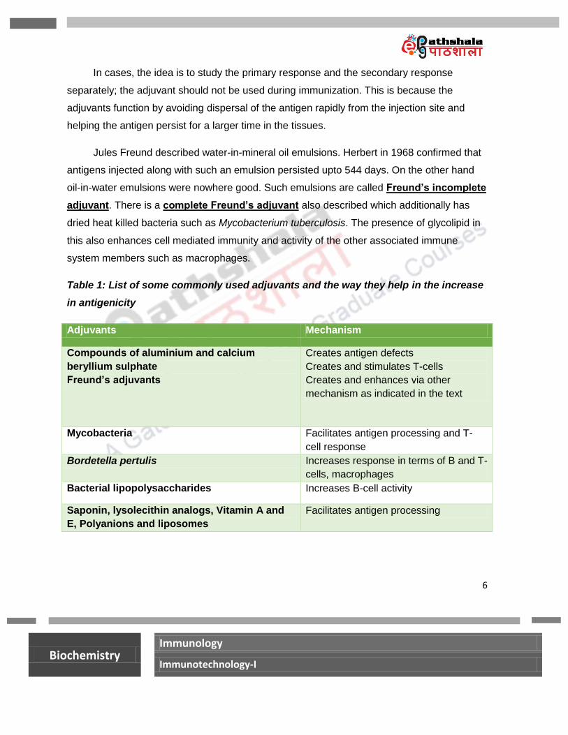
Table 1: List of some commonly used adjuvants and the way they help in the increase
in antigenicity
Adjuvants Mechanism
Compounds of aluminium and calcium
beryllium sulphate
Freund’s adjuvants
Creates antigen defects
Creates and stimulates T-cells
Creates and enhances via other
mechanism as indicated in the text
Mycobacteria Facilitates antigen processing and T-
cell response
Bordetella pertulis Increases response in terms of B and T-
cells, macrophages
Bacterial lipopolysaccharides Increases B-cell activity
Saponin, lysolecithin analogs, Vitamin A and
E, Polyanions and liposomes
Facilitates antigen processing
7
Biochemistry Immunology
Immunotechnology-I
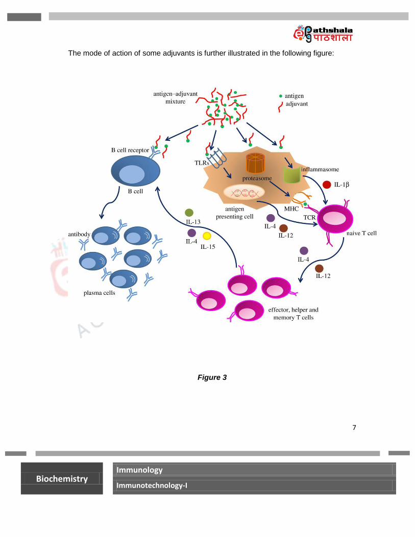
The mode of action of some adjuvants is further illustrated in the following figure:
Figure 3
8
Biochemistry Immunology
Immunotechnology-I
Freund‟s complete adjuvant is not used in humans as it produces chronic inflammation
around emulsion depots.
Many adjuvants are specifically used for stimulation of pathological conditions in
animals. For example, Complete Freund‟s adjuvant can induce autoimmunity.
Finally, many immunoenhancing drugs can also be used as adjuvants. For example,
levamisole, Isoprinosine and Avridine.
The Precipitin Reactions
The antigen molecules combine with the antibody molecules in a serum within few minutes.
When univalent haptens are used as antigens, these antigen-antibody complexes remain in
soluble form. Same is the case if monovalent antibody fragments Fab are used in place of
antibody molecules.
If multivalent antigens and atleast a bivalent antibody molecule is used, after 12-24 h, a
precipitate becomes visible. This precipitate results from the formation of a lattice like
structure. This is called precipitin reaction.
At the equivalence zone, the precipitate is maximum. In this equivalence zone, most of the
antigen and antibody molecules are part of this precipitin complex and do not exist free.

9
Biochemistry Immunology
Immunotechnology-I
Figure 4

10
Biochemistry Immunology
Immunotechnology-I
The precipitation was studied by Kraus in 1897. Kraus also established that this test works
only with homologous antiserum.
This technique can measure the potency of different antisera against a given antigen. It can
also establish relative heterogeneity of the given antigen-antibody pairs.
The precipitate can be weighed after centrifugation and thus the method adapts itself to
quantification.
Figure 5
The mechanism of precipitate formation was studied by Marrack, Heidelberg and Kendall.
The interlocking three dimensional lattice after a critical size cannot remain in solution.
11
Biochemistry Immunology
Immunotechnology-I
Sedimentation rate of the lattice α V x (ρ – ρ0) x g
Where: V= volume of the complex ρ = density of the particle/complex ρ0 = density of the solvent medium
Protein antigens in the molecular weight range of 40 kDa-160 kDa tend to result in
precipitin curve with a sharp peak. Denatured proteins, some viruses, polysaccharides on the
other hand tend to result in precipitin curve with a broad maxima.
Amino acid composition at the antigen binding site of the antibody plays an important
role in forming this lattice. Similarly, highly charged antigen molecules disrupt this lattice
formation due to electrostatic repulsion and do not form well defined precipitin curve.
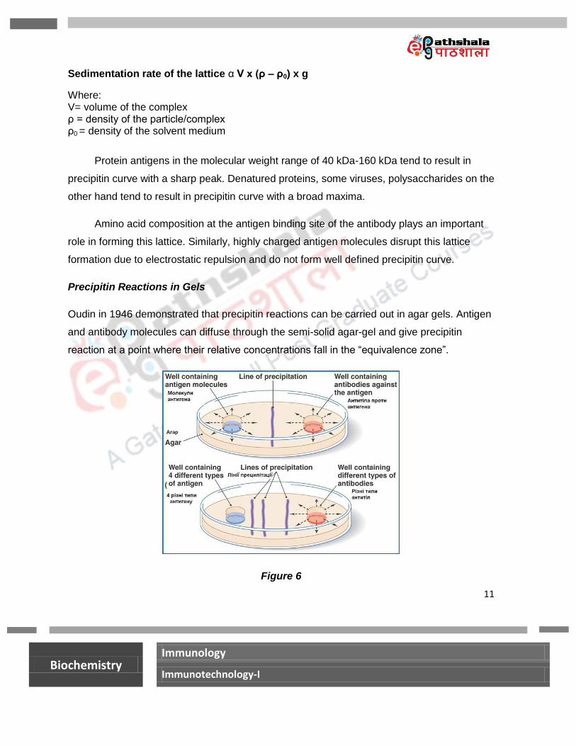
Precipitin Reactions in Gels
Oudin in 1946 demonstrated that precipitin reactions can be carried out in agar gels. Antigen
and antibody molecules can diffuse through the semi-solid agar-gel and give precipitin
reaction at a point where their relative concentrations fall in the “equivalence zone”.
Figure 6

12
Biochemistry Immunology
Immunotechnology-I
Figure 7
Agar gels are poured into the petriplates and allowed to set. Generally 1-2% agar in buffer at
pH 7-8.5 is used. Wells are then punched into the gel and the test solutions of antigen (Ag)
and antibody (Ab, generally in the centre) are added in different wells. The solutions diffuse
out and where Ag and Ab meet they bind to each other, crosslink and precipitate leaving a
line of precipitation. Depending upon the different types of antigens which react with the
antibody, different lines of precipitation emerge.
The double diffusion method developed by Ouchterlony is very useful in establishing the
identity and non-identity in antigen and antibody samples.

13
Biochemistry Immunology
Immunotechnology-I
Figure 8
The double diffusion technique may be used to determine the relationship between antigens
and a particular test antibody. Three basic patterns appear. In the case (A) (that is reaction
of identity) the precipitin arcs formed between the antibody and the two test antigens fuse
indicating that the antibody is precipitating identical epitopes in each preparation. In the case
(B) (that the case of non –identity), the antibody recognizes the two different antigens and
develops two different precipitation lines which intersect. In case (C) (that is the case of
partial identity) the antibody recognizes that the two antigens share the same epitope but the
second antibody also has an additional epitope. Thus a spur appears.
These reactions can also be done in agar plates as seen earlier. The precipitin bands can be
better visualized by washing the gel to remove the soluble proteins and then staining the
precipitin arcs with a protein stain such as Coomassie Blue.
Immunoelectrophoresis
This essentially has two steps:
1.) Separation of various antigens in a sample by electrophoresis in agar gel

14
Biochemistry Immunology
Immunotechnology-I
2.) Double diffusion analysis with the corresponding sera containing antibody for various
antigens
Immunoelectrophoresis was developed by Grabar and Williams in the 1950s. It is
very useful clinically. The antibodyconcentration in the range of 3-20 µg/ mL of the
antiserum can be detected qualitatively.
Rocket Electrophoresis
This is a variation of Immunoelectrophoresis and is also called Laurell technique. It is a
single step method as the antibody containing the anti-serum is incorporated in the agar
gel itself. The precipitin arcs have the shape of rockets. The pH is chosen so that the
antibodies contained in the gel are immobile and the antigens are negatively charged.
The precipitin arcs have the shape of rockets. The height of the rocket is proportional to
the antigen concentration. The sensitivity of this method is 10-20 fold more than the
double diffusion method.

15
Biochemistry Immunology
Immunotechnology-I
Figure 11
A slight modification of rocket electrophoresis: 2 D method
Figure 12
16
Biochemistry Immunology
Immunotechnology-I
Agglutination Reactions
If the antigens are in the form of cells (such as RBCs) or particles (antigens immobilized on
latex beads), the reaction between these multivalent „antigens‟ and bivalent or multivalent
antibody forms clumps. The clump formation is maximum in the equivalence zone. However,
in the quantitative version, generally serial dilution technique is used to determine maximum
dilution of the antiserum at which clump formation can be detected.

17
Biochemistry Immunology
Immunotechnology-I
Figure 13
The clump formation is the result of agglutination of the antigen via crosslinking with the
antibody molecules.
The highest dilution at which agglutination/clump formation is observed is called the titre
value.

18
Biochemistry Immunology
Immunotechnology-I
The agglutination tests are semi-quantitative but are sensitive enough to detect antibody
concentration of 0.001 µg/ mL of the serum.
The method provides a simple, rapid and sensitive way of identifying type of RBCs, bacteria,
fungi and other cells.
Summary
In this lecture we learnt about:
a.) Role of adjuvant in immunization
b.) The precipitin reactions in solution
c.) The precipitin reactions in agar gels
d.) Immunoelectrophoretic techniques
e.) Agglutination reactions