Immunological considerations for cell therapy using human embryonic stem cell derivatives
12
Immunological considerations for cell therapy using human embryonic stem cell derivatives Micha Drukker, Institute for Stem Cell Biology and Regenerative Medicine, Stanford University School of Medicine, Stanford, CA 94305–5323, USA Table of Contents 1. Introduction ............................................................................... 2 2. Expression of histocompatibility antigens on u/dhESCs .......................................... 2 3. Expression of T-cell regulating signals in u/dhESCs ............................................. 4 4. T-cell response against u/dhESCs measured by functional assays ................................... 5 5. Interaction of natural killer cells with hESCs ................................................... 6 6. Generation of patient-specific isogenic hESC lines .............................................. 7 7. Conclusions ............................................................................... 9 8. Acknowledgments ......................................................................... 9 9. References ................................................................................ 9 Abstract The isolation of human embryonic stem cells (hESCs) and their directed differentiation into a variety of tissue specific cell types offers a novel approach for production of cellular transplants. Still, successful translation of the therapeutic promise of hESCs to clinical practice depends heavily on purification of beneficial cell populations as well as on prevention of immune rejection. This review focuses on the immunological antigens that are expressed on differentiated progenies of hESCs and on the adaptive and innate immune processes that may target their implanted derivatives. The risk of immune rejection has driven the development of methods that may be used for preparation of patient-specific histocompatible pluripotent cell lines. It is hoped that pluripotent cell lines that will be generated following somatic cell nuclear transfer into oocytes or zygotes, oocyte parthenogenesis or introduction of reprogramming factors will be largely protected from immune rejection. These techniques are presented and the remaining immunological challenges for creating tolerated implants using these approaches are discussed. *Edited by Diane Mathis and Jerome Ritz. Last revised June 27, 2008. Published December 16, 2008. This chapter should be cited as: Drukker, M., Immunological considerations for cell therapy using human embryonic stem cell derivatives (December 16, 2008), StemBook, ed. The Stem Cell Research Community, StemBook, doi/10.3824/stembook.1.14.1, http://www.stembook.org. Copyright: C 2008 Micha Drukker. This is an open-access article distributed under the terms of the Creative Commons Attribution License, which permits unrestricted use, distribution, and reproduction in any medium, provided the original work is properly cited. Correspondence should be addressed to E.mail: M. Drukker. ([email protected]) 1 stembook.org
Transcript of Immunological considerations for cell therapy using human embryonic stem cell derivatives

Immunological considerations forcell therapy using human embryonic
stem cell derivativesMicha Drukker, Institute for Stem Cell Biology and RegenerativeMedicine, Stanford University School of Medicine, Stanford, CA94305–5323, USA
Table of Contents1. Introduction . . . . . . . . . . . . . . . . . . . . . . . . . . . . . . . . . . . . . . . . . . . . . . . . . . . . . . . . . . . . . . . . . . . . . . . . . . . . . . . 22. Expression of histocompatibility antigens on u/dhESCs . . . . . . . . . . . . . . . . . . . . . . . . . . . . . . . . . . . . . . . . . . 23. Expression of T-cell regulating signals in u/dhESCs . . . . . . . . . . . . . . . . . . . . . . . . . . . . . . . . . . . . . . . . . . . . . 44. T-cell response against u/dhESCs measured by functional assays . . . . . . . . . . . . . . . . . . . . . . . . . . . . . . . . . . . 55. Interaction of natural killer cells with hESCs . . . . . . . . . . . . . . . . . . . . . . . . . . . . . . . . . . . . . . . . . . . . . . . . . . . 66. Generation of patient-specific isogenic hESC lines . . . . . . . . . . . . . . . . . . . . . . . . . . . . . . . . . . . . . . . . . . . . . . 77. Conclusions . . . . . . . . . . . . . . . . . . . . . . . . . . . . . . . . . . . . . . . . . . . . . . . . . . . . . . . . . . . . . . . . . . . . . . . . . . . . . . . 98. Acknowledgments . . . . . . . . . . . . . . . . . . . . . . . . . . . . . . . . . . . . . . . . . . . . . . . . . . . . . . . . . . . . . . . . . . . . . . . . . 99. References . . . . . . . . . . . . . . . . . . . . . . . . . . . . . . . . . . . . . . . . . . . . . . . . . . . . . . . . . . . . . . . . . . . . . . . . . . . . . . . . 9
Abstract
The isolation of human embryonic stem cells (hESCs) and their directed differentiation into a varietyof tissue specific cell types offers a novel approach for production of cellular transplants. Still, successfultranslation of the therapeutic promise of hESCs to clinical practice depends heavily on purification of beneficialcell populations as well as on prevention of immune rejection. This review focuses on the immunologicalantigens that are expressed on differentiated progenies of hESCs and on the adaptive and innate immuneprocesses that may target their implanted derivatives. The risk of immune rejection has driven the developmentof methods that may be used for preparation of patient-specific histocompatible pluripotent cell lines. It ishoped that pluripotent cell lines that will be generated following somatic cell nuclear transfer into oocytesor zygotes, oocyte parthenogenesis or introduction of reprogramming factors will be largely protected fromimmune rejection. These techniques are presented and the remaining immunological challenges for creatingtolerated implants using these approaches are discussed.
*Edited by Diane Mathis and Jerome Ritz. Last revised June 27, 2008. Published December 16, 2008. This chapter should be cited as: Drukker, M.,Immunological considerations for cell therapy using human embryonic stem cell derivatives (December 16, 2008), StemBook, ed. The Stem CellResearch Community, StemBook, doi/10.3824/stembook.1.14.1, http://www.stembook.org.
Copyright: C© 2008 Micha Drukker. This is an open-access article distributed under the terms of the Creative Commons Attribution License, whichpermits unrestricted use, distribution, and reproduction in any medium, provided the original work is properly cited.
Correspondence should be addressed to E.mail: M. Drukker. ([email protected])
1
stembook.org

Immunological considerations for cell therapy using human embryonic stem cell derivatives
1. Introduction
Since the early days of modern medicine it has been known that transplantation of tissues from one individual toanother results in immune rejection, except when performed between identical twins. Extensive research done in thepast decades has uncovered that differences in protein sequences between individuals are the primary targets for theimmune response against allogeneic (genetically non-identical) grafts. These molecules were subsequently defined ashistocompatibility antigens as they determine whether implanted cells are accepted or rejected by the immune system.Today, the risk of organ rejection remains high despite efforts to match haplotypes of histocompatibility antigens aswell as use of immunosuppressive therapies.
Human embryonic stem cells (hESCs) can be propagated in high numbers in vitro, and have the potential todifferentiate into a myriad of cell types (Reubinoff et al., 2000; Thomson et al., 1998). The application of these cellsfor organ restoration is of particular importance due to limited availability of donated organs. However, it is unclearwhether differentiated hESCs will be recognized by the immune system following implantation in humans. Thereforeit is imperative to investigate the immune response against differentiated hESCs for successful clinical translation.
The purpose of this review is to describe the current knowledge of antigenicity and immunogenicity of hESCs andtheir derivatives. While antigenicity refers only to the capacity of cells to express histocompatibility antigens, immuno-genicity is a broader term for the potential to elicit immune response through additional factors, such as co-stimulatorymolecules. This review summarizes our current view of the histocompatibility antigens and immunomodulatingmolecules that are expressed on undifferentiated and differentiated hESCs (u/dhESCs). In addition, adaptive andinnate immune processes that may develop against the cells following recognition of histocompatibility antigens willbe discussed. As rejection processes could severely limit the use of hESC-derived transplants, multiple approaches arebeing developed to alleviate these risks. I will focus on techniques for derivation of isogenic (genetically identical)hESC lines and discuss the immunological benefits and remaining challenges for their potential application.
2. Expression of histocompatibility antigens on u/dhESCs
Histocompatibility antigens are classically divided into major histocompatibility complex (MHC) molecules,minor histocompatibility complex antigens (mHAgs; Roopenian et al., 2002) and ABO blood group antigens (Watkins,2001). The prominent mechanism of graft rejection is initiated by recognition of the graft’s MHC molecules byhost T cells (Lechler et al., 2005). Under normal circumstances, MHC molecules present processed cellular andextra-cellular antigens to circulating T cells. Since T cells that recognize self-antigens are depleted in the thymus,circulating T cells respond only to foreign antigens bound by MHC molecules. This situation changes dramaticallyfollowing transplantation of cells expressing allelic variants of MHC proteins to unmatched recipient. Due to similarityin structure, foreign MHC molecules may interact with the recipient’s T cells, evidently “mimicking” self-MHCmolecules presenting foreign antigens (Suchin et al., 2001). As a result, “direct allorecognition” (see Figure 1A)between host T cells and transplanted cells leads to sensitization, proliferation and cytotoxic response against thegraft (Morelli and Thomson, 2003). This response is initiated primarily by “passenger” dendritic cells (DCs) thatmigrate from transplanted organs to lymph nodes of the recipient where they allostimulate T cells. Importantly, theinitiation, propagation and termination of direct alloresponse depend also on additional stimulatory and inhibitorysignals provided by the transplanted cells as well as the site of transplantation and extent of graft vascularization(discussed below). Still, it is predominantly acute and unless treated with immunosuppressive drugs results in rapidgraft failure.
Although immunosuppressive therapy slows immune rejection processes, a large proportion of tissue graftssuccumb to chronic rejection that results in graft failure. Chronic immune responses are thought to result primarilyfrom processing of donor antigens by the recipient’s or the donor’s professional antigen presenting cells (APCs), suchas DCs and macrophages, and their presentation to T cells (Briscoe and Sayegh, 2002). This “indirect allorecognition”(see Figure 1B) pathway is largely analogous to physiologic antigen processing and is assumed to mediate graftrejection mainly by generation of alloantibodies and allospecific cytotoxic T cells (Bolton et al., 2008). As virtually allcirculating antigens are ingested and processed by APCs, chronic immune processes could target numerous proteinsthat differ in structure between donor cells and the recipient. The majority of these processes are aimed at MHCproteins due to their extreme polymorphism, but any other polymorphic antigens, collectively known as mHAgs, maybe targeted.
To reduce the immune response against allografts, efforts are being made to match as many MHC alleles aspossible prior to organ transplantation. However, it is particularly hard to match MHC haplotypes between genetically
2
stembook.org

Immunological considerations for cell therapy using human embryonic stem cell derivatives
FasTCR
PeptideantigenDonor
MHC
TCR
Donor MHCderivedantigen Cytotoxicity
towards graft
Cellulardeath
Cellular death(transplantedorgan)
Stimulationof B cells andalloantibody
release
RecipientMHC
RecipientDC
RecipientT cell
RecipientT cell
Immunologicallyprivileged
site
DC fromgraft
Direct allorecognition
Indirect allorecognition
A
B
FasligandCD80/CD86 CD28
CD80/CD86 CD28
Stimulationand proliferation
Stimulationand proliferation
Apoptosis(cell death)
DonorMHC
Granulerelease
Antigenuptake
Figure 1. Proposed pathways of interaction between transplanted hESC derivatives and the immune system. T cells recognize alloantigens by twodistinct pathways known as the direct and indirect. (A) Direct allorecognition occurs when recipient T cells recognize intact allogeneic MHC molecules(together with bound endogenous peptide) on the surface of donor APCs, such as DCs (left). FasL expression on target cells (mainly in immunoprivilegedsites) can prevent immune responses by inducing apoptosis in Fas-bearing leukocytes (right). (B) During indirect allorecognition, host APCs process donorderived proteins, primarily MHCs and present them in self-MHC molecules to recipient T cells. (A, B) Co-stimulatory interaction between CD80/CD86molecules on APCs with their receptor CD28 on T cells is essential for sensitization and propagation of immune responses. TCR T cell receptor.
unrelated individuals since there are up to several hundreds alleles encoding for the three MHC class I (MHC-I) genesand four pairs of MHC class II (MHC-II) genes. Both MHC classes are known to affect transplantation outcomes,since MHC-I molecules are ubiquitously expressed and MHC-II expressing hematopoietic and DCs are present inmost tissues.
To evaluate the antigenic properties of u/d hESCs, the cell-surface expression of both MHC classes was tested viaflorescence-activated cell sorter (FACS). It was found that undifferentiated cells express relatively low levels of MHC-Imolecules (Draper et al., 2002; Drukker et al., 2002) and do not express MHC-II molecules (Drukker et al., 2002),similarly to the inner cell mass (ICM) of human blastocyst stage embryos from which ESC are derived (Jurisicovaet al., 1996). When treated with interferon-γ (IFN-γ ), a pro inflammatory cytokine, MHC-I levels were substantiallyupregulated while MHC-II molecules remained undetected. Aggregation induced differentiation using embryoid body(EB) formation caused approximately 10-fold increase in MHC-I expression on the cell surface, and application ofIFNs induced approximately 100-fold increase. Furthermore, MHC-I expression levels reached somatic levels on
3
stembook.org

Immunological considerations for cell therapy using human embryonic stem cell derivatives
No granulerelease
No granulerelease
C Activationreceptor
Activationreceptor
ActivationreceptorLigand
Ligand
Granule release(Cytotoxicity)
RecipientT cellD
MHCclass I
MHC
Targetcell
Weak or nostimulation
NKcell
NKcell
NKcell
MHCclass I
Inhibitoryreceptor
Inhibitoryreceptor
Inhibitoryreceptor
Donor cell(hESC-derived transplanted cell
e.g. cardiomyocyte)
TCR
CD28
Figure 1. Proposed pathways of interaction between transplanted hESC derivatives and the immune system continued. (C). Natural killer (NK) cellactivation is controlled by integration of signals from activatory and inhibitory receptors. Inhibitory NK cell receptors recognize self-MHC-I molecules thatrestrain their activation (left). When unimpeded by the inhibitory receptors, binding of NK cell activation receptors to their ligands on target cells results in NKcell stimulation (right). (D) As most differentiated hESCs do not express co-stimulatory molecules (right) they may be protected from direct allorecognition(but their antigens can presumably initiate chronic immune responses through the indirect pathway). When MHC-I expression is low and lysis ligands areabsent, hESC derivatives may be protected from NK-cell response (left). Cardiomyocytes derived from hESCs could potentially be used for restoration ofheart functions (center). TCR T cell receptor.
IFN-γ treated teratoma cells (Drukker et al., 2002). MHC-II molecules remained below detection level under theseconditions, however, other differentiation protocols have reported expression of these molecules, especially duringhematopoietic and DC differentiation (Senju et al., 2007; Slukvin et al., 2006; Zhan et al., 2004). These data suggestthat differentiated hESCs would express high levels of MHC-I following transplantation, while only a small numberof differentiation protocols could promote expression of MHC-II (see Table 1).
The status of mHAgs expression on u/dhESCs has not been directly examined to date, but their expression isexpected. One group of mHAgs, which could be easily avoided by transplanting male hESC lines only to males, arederived from genes encoded by the Y chromosome, termed the HY antigens (Wang et al., 1995). It is also reasonablethat matching ubiquitously expressed mHAgs such as, HA-3, -4, -6, -7 and -8 (Brickner et al., 2001; de Bueger et al.,1992) would improve survival of hESC-derived grafts and in cases where hESC-derived hematopoietic stem cells aretransplanted, HA-1 and -2 should be matched as they are restricted to cells of this lineage (de Bueger et al., 1992).In addition, although the mitochondrial genome is relatively small, efforts should be made to match mitochondrialhaplotypes that were shown to encode for mHAgs (discussed below). Finally, it is important to investigate whetherhESC-derived embryonic antigens would constitute an additional class of mHAgs to which recipients are not tolerant,since they are not significantly expressed in adults. One group of histocompatibility antigens that was not yet examinedin u/dhESCs is the ABO blood group antigens. Since matching can easily prevent immune response against ABOantigens, it will not be discussed here further.
3. Expression of T-cell regulating signals in u/dhESCs
Apart from MHC interaction, co-stimulatory molecules are vital signals for the initiation and propagation ofT-cell mediated immune responses. The CD80 and CD86 (B7.1 and B7.2, respectively) ligands are of special importance
4
stembook.org
Immunological considerations for cell therapy using human embryonic stem cell derivatives
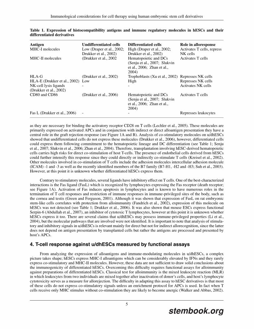
Table 1. Expression of histocompatibility antigens and immune regulatory molecules in hESCs and theirdifferentiated derivatives
Antigen Undifferentiated cells Differentiated cells Role in alloresponseMHC-I molecules Low (Draper et al., 2002;
Drukker et al., 2002)High (Draper et al., 2002;Drukker et al., 2002)
Activates T cells, repressNK cells
MHC-II molecules (Drukker et al., 2002 Hematopoietic and DCs(Senju et al., 2007; Slukvinet al., 2006; Zhan et al.,2004)
Activates T cells
HLA-G (Drukker et al., 2002) Trophoblasts (Xu et al., 2002) Represses NK cellsHLA-E (Drukker et al., 2002) Low High Represses NK cellsNK-cell lysis ligands(Drukker et al., 2002)
- - Activates NK cells
CD80 and CD86 (Drukker et al., 2006) Hematopoietic and DCs(Senju et al., 2007; Slukvinet al., 2006; Zhan et al.,2004)
Activates T cells
Fas L (Drukker et al., 2006) - - Represses leukocytes
as they are necessary for binding the activatory receptor CD28 on T cells (Lechler et al., 2005). These molecules areprimarily expressed on activated APCs and in conjunction with indirect or direct alloantigen presentation they have acentral role in the graft rejection response (see Figure 1A and B). Analysis of co-stimulatory molecules on u/dhESCsshowed that undifferentiated cells do not express these molecules (Drukker et al., 2006), however, differentiated cellscould express them following commitment to the hematopoietic lineage and DC differentiation (see Table 1; Senjuet al., 2007; Slukvin et al., 2006; Zhan et al., 2004). Therefore, transplantation involving hESC-derived hematopoieticcells carries high risks for direct co-stimulation of host T-cells. The presence of endothelial cells derived from hESCscould further intensify this response since they could directly or indirectly co-stimulate T cells (Kreisel et al., 2002).Other molecules involved in co-stimulation of T cells include the adhesion molecules intercellular adhesion molecule(ICAM) -1 and -3 as well as the recently identified members of the B7 family (B7-H1, -H2 and -H3; Suh et al., 2003).However, at this point it is unknown whether differentiated hESCs express them.
Contrary to stimulatory molecules, several ligands have inhibitory effect on T cells. One of the best-characterizedinteractions is the Fas ligand (FasL) which is recognized by lymphocytes expressing the Fas receptor (death receptor;see Figure 1A). Activation of Fas induces apoptosis in lymphocytes and is known to have numerous roles in thetermination of T cell responses and restriction of immune responses in immune-privileged sites of the body, such asthe cornea and testis (Green and Ferguson, 2001). Although it was shown that expression of FasL on rat embryonicstem-like cells correlates with protection from alloimmunity (Fandrich et al., 2002), expression of this molecule onhESCs was not detected (see Table 1; Drukker et al., 2006). It was also shown that mouse ESCs express functionalSerpin-6 (Abdullah et al., 2007), an inhibitor of cytotoxic T lymphocytes, however at this point it is unknown whetherhESCs express it too. There are several claims that u/dhESCs may possess immune-privileged properties (Li et al.,2004), but the molecular pathways that are involved were not identified. It is important to note that analysis of stimula-tory and inhibitory signals in u/dhESCs is relevant mainly for direct but not for indirect allorecognition, since the latterdoes not depend on antigen presentation by transplanted cells but rather the antigens are processed and presented byhost’s APCs.
4. T-cell response against u/dhESCs measured by functional assays
From analyzing the expression of alloantigens and immune-modulating molecules in u/dhESCs, a complexpicture takes shape; hESCs express MHC-I alloantigens which can be considerably elevated by IFNs and they rarelyexpress co-stimulatory and MHC-II molecules. However, these data are not sufficient to draw solid conclusions aboutthe immunogenicity of differentiated hESCs. Overcoming this difficulty requires functional assays for alloimmunityagainst preparations of differentiated hESCs. Classical test for alloimmunity is the mixed leukocyte reaction (MLR)in which leukocytes from two individuals are mixed together after inactivation of donor’s cells, and host’s lymphocytecytotoxicity serves as a measure for allorejection. The difficulty in adapting this assay to hESC derivatives is that mostof these cells do not express co-stimulatory signals unless an enrichment protocol for APCs is used. In fact when Tcells receive only MHC stimulus without co-stimulation they are likely to become anergic (Walker and Abbas, 2002).
5
stembook.org

Immunological considerations for cell therapy using human embryonic stem cell derivatives
To bypass the need for co-stimulation in MLR one could stimulate the recipient’s T cells by lymphocytes expressingthe same HLA alleles as the hESC line in question. Using this approach, it was shown that IFN stimulated u/dhESCsare rejected by T cells following incubation with the sensitized T cells (Drukker et al., 2006). These data imply that Tcells may efficiently reject transplanted derivatives of hESCs if sufficient stimulation is provided. It is important to notethat purified populations of therapeutic cells should be examined using similar assays in order to evaluate the expectedintensity of the immune response against them. In addition, these assays are largely biased towards determining thepotency of direct alloresponses, hence, other experimental procedures should be developed in order to further evaluatethe potential of indirect alloresponses.
The most reliable assays for post-transplantation immunity are those performed in vivo. The difficulty with thedevelopment of animal models for tests of hESC-immunity is that they should carry functional human immune cellsand provide the necessary environments for human immune responses. Together with colleagues, we have previouslyused the mouse trimera model (Reisner and Dagan, 1998) to test the human alloresponse against u/dhESCs (Drukkeret al., 2006). In short, preparation of this model starts by adoptive transfer of bone marrow from severe combinedimmunodeficient (SCID) mice into normal irradiated mouse recipients. Then mice are infused with human peripheralblood mononuclear cells (PBMCs) that serve as the human immune components (Dekel et al., 2003; Drukker, 2006).Later, human leukocyte alloresponse against other transplanted human cells and tissues can be evaluated. Using thismodel we observed that undifferentiated hESCs developed into teratomas and were not significantly rejected over thecourse of one month. Furthermore, transplantation of EBs, advanced teratoma fragments and a teratoma-derived cellline were also not rejected in this model. On the other hand, human skin grafts and transplanted human cell lineswere showing clear signs of rejection indicating potent humanized immune response by the trimera model (Drukkeret al., 2006). These data indicate that hESCs and their early derivatives are functionally less immunogenic thanadult tissues.
There is considerable evidences to support the idea that hESCs like other embryonic cells are immunologicallyimmature: the cells express relatively low levels of MHC molecules unless stimulated by IFNs and do not presentco-stimulatory signals (see Figure 1D). Similarly, it has been shown recently that various fetal organs are resistant toimmune rejection since they have low immune stimulatory potential (Dekel et al., 2003). Another factor that couldinfluence immune evasion is the contribution of the grafted cells to neo-vascularization. As endothelial cells can alsodirectly present MHC and co-stimulatory signals to the host T cells (Kreisel et al., 2002), their presence in the graftmight have a negative effect on graft survival (Libby and Pober, 2001). At this point it is unclear to which extenthESC-derived cells give rise to endothelial cells following transplantation and additional experiments are warrantedto examine the influence of these cells on transplant survival.
5. Interaction of natural killer cells with hESCs
Natural killer (NK) cells are cytotoxic lymphocytes possessing innate immune activities and can also affectadaptive immune responses. NK-cell mediated cell lysis is regulated by the balance of stimulatory and inhibitorysignals that they recognize on target cells (Raulet, 2006). The prominent NK-cell inhibitory ligands include membersof the MHC-I family as well as the non-classical human leukocyte antigens (HLA)-E and –G (see Figure 1C). AsNK cells lyse cells that lack MHC-I expression, it is possible that u/dhESCs which express low level of MHC-I andHLA-E molecules would be targeted. However, when tested in vitro, activated human NK cells did not effectively lyseu/dhESCs (Drukker et al., 2002). Similarly, mouse NK cells showed no response towards u/dhESCs in vivo (Drukkeret al., 2006). Thus far, no known NK-cell suppressive signal such as HLA-G molecules that protect embryonictrophoblasts from maternal NK cells (Kovats et al., 1990), have been shown to contribute to inhibition of NKcells by hESCs. Another explanation for the resistance of hESCs to lysis by NK cells may be due to lack ofstimulatory signals for NK cells (see Figure 1C). Although these signals are still not well-defined, recombinantproteins of soluble NK-cell activating receptors and IgGs can be used to evaluate the expression of activating ligandson target cells. Indeed, it was demonstrated that expression of stimulating molecules for NK-cell activating receptorsis very low or below detection level on u/dhESCs (see Table 1; Drukker et al., 2002). These data suggest thatundifferentiated hESCs are protected from NK cells by virtue of not expressing lysis signals (see Figure 1D). It isalso possible that the low expression of MHC-I is sufficient for protection and/or they express alternative inhibitorysignals.
While human NK cell-response against hESC-derived teratomas seems to be minimal (Drukker et al., 2006),mouse NK cells were shown to reject mouse ESC-derived hematopoietic progenitors in vivo. Rideout et al., reportedthat these progenitors engrafted in mice lacking T, B and NK cells but not in Rag2-/- mice that lack only T and Bcells (Rideout et al., 2002). They also demonstrated that an antibody-mediated depletion of NK cells in Rag2-null
6
stembook.org

Immunological considerations for cell therapy using human embryonic stem cell derivatives
Table 2. Proposed pathways for derivation of patient-specific hESC lines
Method Cell sources Genderspecificity
Immunologicalconsiderations fortransplantation
Demonstrate in
Mouse HumanParthenogenesis Metaphase-II
oocytes(Revazova et al.,2007)
Female Preferentially,MHC heterozygotelines should be used
+ +
Somatic cellnuclear transfer
Oocytes(Hochedlingerand Jaenisch,2003)
+ Somaticnucleus
No MitochondrialmHAgs mightinduce immuneresponse
+ −
Zygotes (Egliet al., 2007)
Inducedpluripotency
Embryonic andadult fibroblasts(Takahashi et al.,2007; Yu et al.,2007)
No Unknown + +
mice enhances hematopoietic progenitor engraftment. Since embryonic hematopoietic progenitors express low levelof MHC-II molecules compared to adult hematopoietic progenitors (Huang and Auerbach, 1993), it is generallythought that NK cells lyse the embryonic cells as they are not sufficiently repressed. These data raise the possi-bility that in some cases MHC matched hESC-derivatives may be at risk of rejection by the patient’s NK cells.Further studies are required to determine whether NK-cell response will be a significant obstacle for engraftment ofhESC-derivatives.
6. Generation of patient-specific isogenic hESC lines
Although hESCs derivatives seem to have reduced capacity to stimulate alloimmune responses, they are notconsidered as immune-privileged. Various approaches have been suggested to help alleviate the risk of rejection(Bradley et al., 2002; Drukker and Benvenisty, 2004), including genetic manipulation of MHC expression in thecells, induction of hematopoietic tolerance towards the cells and personalized production of isogenic cell lines. Of allthese approaches, the last couple of years have been highly prolific for the latter – the production of patient specificpluripotent cell lines. There are currently three proposed techniques for derivation of such cell lines (summarized inTable 2):
1. Somatic cell nuclear transfer (SCNT): it has been shown extensively in mice that ESCs could be derivedfollowing SCNT into enucleated oocytes that are allowed to develop to blastocyst stage (Hochedlinger andJaenisch, 2003). Similarly, it was shown recently that ESCs could be derived following SCNT into zygotesthat had their chromosomes removed (Egli et al., 2007). It has been proposed that differentiated tissues fromsuch “tailor-made” cell lines would not be rejected by the immune system of the nucleus-donor since theyare genetically identical, except for mitochondrial DNA (mtDNA). Even though there is still no solid prooffor the isolation of reprogrammed hESCs from cloned embryos, it is likely that overcoming several technicaldifficulties would allow the derivation of such cells in humans too.
2. Parthenogenesis: it has been shown for several mammalian species that activation of metaphase oocytes bychemical reagents can initiate development into blastocyst stage diploid parthenogenetic embryos. Protocolsfor ESC-derivation from such pseudo-zygotes were developed for mice (Allen et al., 1994) and recently humanembryos. It has been shown by Revazova et al. that about half of the human embryos that were chemicallyactivated, proceeded to the blastocyst stage and six parthenogenetic cell lines were derived. Importantly, it hasbeen shown that the parthenogenetic cell lines have multi-lineage differentiation potential in vivo and in vitro(Revazova et al., 2007).
3. Induction of pluripotent stem (iPS) cells from somatic cells by genetic factors: the seminal experiment ofproducing cell lines that highly resemble mouse ESCs by transferring four transcription factors – Oct3/4, Klf4,
7
stembook.org

Immunological considerations for cell therapy using human embryonic stem cell derivatives
Sox2 and c-Myc into somatic cells has opened a new avenue for “tailored” production of pluripotent celllines (Takahashi and Yamanaka, 2006). In addition, it has been demonstrated that the same factors or similarcombinations can be used for derivation of human iPS cells from somatic cells (Park et al., 2008; Takahashiet al., 2007; Yu et al., 2007). Application of this technique for clinical settings still requires in-depth analysisof the multi-lineage potential of iPS cells and introduction of non-oncogenic reprogramming factors withoutretroviral integration into the genome.
There are different immunological considerations for clinical use of cell lines generated by each of thesetechniques. One immunological issue, which is unique to SCNT, is potential immune response directed againstmitochondrial histocompatibility antigens. It is known in mice and rats that certain amino acid substitutions inmitochondrial proteins can lead to the generation of alloreactive T cells (Loveland et al., 1990). Therefore, it is possiblethat some mitochondrial protein polymorphisms may become antigenic and provoke chronic immune responsesfollowing transplantation of cells that were prepared by SCNT. However, the relatively small number of mitochondrialencoded proteins and the small number of mitochondrial alleles suggests that the risk of immune response towardsdonor-derived mitochondrial mHAgs would be considerably lower than against mismatched genomes. In accordancewith this hypothesis, it has been shown in cows that transplantation of organs derived from cloned embryos to thenuclear donor did not lead to immune response even though they have different mitochondrial haplotypes (Lanzaet al., 2002).
Parthenogenetic hESC lines are not expected to stimulate any adaptive immune response since they are geneticallyidentical with the oocyte donor. However, NK cells may actually respond against parthenogenetic hESC lines thatlack one set of MHC-I encoding genes. It was demonstrated in mice that heterozygotic bone marrow cells containingtwo MHC-I haplotypes, rather than one, engraft better in isogenic hosts that have two distinct MHC-I haplotypes(Hoglund et al., 1997). Therefore, a potential concern for transplantation of cells derived from MHC-homozygousparthenogenetic hESC lines (Revazova et al., 2008) is the risk of rejection by NK cells due to reduced diversity ofMHC alleles.
The extent of MHC homozygosity in parthenogenetic hESC lines depends on oocyte activation stage, metaphase-I or -II (M-I and M-II, respectively) and on recombination events (Kim et al., 2007). Activation of M-I oocytes givesrise to cell lines that contain the two maternal chromosome homologs whereas M-II activated oocytes give riseto cell lines that have only half of the maternal homologs. This still does not rule out heterozygosity of multi-ple loci in M-II activated oocytes due to recombination between the homologs. In fact, it was found by Revazovaet al., (Revazova et al., 2007) that all parthenogenetic hESC lines that they have derived from M-II oocytes con-tained full heterozygosity in the MHC region. It should be noted that parthenogenetic hESC lines were probablyalso derived by Hwang’s group in South Korea, although, the team reported derivation of SCNT-derived hESClines (Hwang et al., 2004). Genome-wide analysis of one of the “cloned” hESC cell lines showed homozygos-ity in centromeric regions but extensive heterozygosity in telomeric regions, indicating that it is a parthenogenote(Kim et al., 2007).
Possible benefit for derivation of parthenogenetic hESC lines that contain homozygosity in the MHC region(Revazova et al., 2008) is their possible use in individuals that are genetically related to the oocyte donor. For example,there is 50% chance that a cell line that is derived from a patient will be histocompatible to any of her children ifit has two identical MHC alleles. Moreover, if a sizeable depository of MHC homozygous parthenogenetic hESClines were to be generated, it may serve as a MHC-matching cell lines bank (Rubinstein, 2001). This holds true sinceMHC homozygous hESC cell lines would have a much higher matching frequency than heterozygous cell lines. Theextent of NK-cell response against such transplants remains unclear and should be investigated. Also, transplantationof differentiated parthenogenetic hESC to genetically non-identical donors may involve the risk of inducing immuneresponse against mHAgs and mitochondrial antigens.
The only approach that currently holds the promise to generate absolute isogenic cell lines is reprogrammingsomatic cells into iPS cells using defined genetic factors. Still, it is important to note that adaptive immune responsesmay occur against embryonic mis-expressed antigens and that NK-cell response is a possible risk for transplantedembryonic cells that express low levels of MHC molecules. Therefore, this aspect should be further examinedand strategies to derive iPS cells without stable integration of retroviruses and oncogenes should be developed. Ifdifferentiated iPS cells would prove to be safe for transplantations, it is very likely that this approach will become theprimary source of isogenic cell lines for clinical use.
8
stembook.org

Immunological considerations for cell therapy using human embryonic stem cell derivatives
7. Conclusions
Decades of research devoted to tissue rejection have uncovered many of the molecules and cell types that mediatethe immune processes following allotransplantation. Through these investigations it has become clear that almost everyaspect of adaptive and innate immunity may be involved in rejection. The isolation of hESCs and their differentiationcapacities has created a novel pathway for generating cells with potential therapeutic relevance on an unprecedentedscale. However, it is still premature to predict the exact immunological outcomes after implantation of differentiatedhESCs before production of therapeutic grade cells will be achieved. The emerging picture from the existing data showthat hESCs and their early-differentiated derivatives are immunologically immature; they express lower than somaticlevels of MHC-I and do not express MHC-II and co-stimulatory molecules unless directed to differentiate to certainhematopoietic lineages. Thus far, functional in vivo studies have indicated that their limited co-stimulatory potentialreduces their risk for direct rejection by cytotoxic T cells, though MHC-I levels are sufficient for T cell response byactivated cells. To further reduce the potential for direct allorecognition a potential approach may be to deplete APCsfrom transplanted cell preparations by specific antibodies.
In contrast to direct allorecognition, indirect alloresponse in most cases does not require expression of co-stimulatory molecules by the transplanted cells and virtually any polymorphic protein can induce immune response.Therefore, it is expected that indirect chronic responses would develop against MHC molecules and mHAgs followingtransplantation of allogeneic differentiated hESCs. Still, predicting the severity and rate of rejection is particularlyhard since multiple factors, including genetic disparities, tissue trauma during surgery and sites of transplantation cansignificantly influence inflammation-promoting processes which have a primary role in initiation of immune rejection.As it turns out that endothelial cells possess at least some of the stimulating functions of APCs they may also play animportant role in development of alloimmune responses.
Finally, new key developments in derivation of partially or fully isogenic pluripotent human cell lines may nowenable transplantation of their derivatives with only very low risk of immune rejection. Induction of pluripotencyin somatic cells is of particular importance since it does not require the use of human oocytes and zygotes and theresulting cell lines are fully compatible with the donor. It is important to note that if pluripotent cell line wouldbe derived following SCNT into oocytes and zygotes or parthenogenesis, the reprogrammed cell lines may carryallogeneic mitochondrial antigens and parthenogenetic cells might be lysed by NK cells. Regardless of the hESCsource, detailed examination of their recognition by various immune cells is required since in vitro differentiation maylead to reduced levels of MHC expression and immature cells may express embryonic antigens that are not familiar tothe adult immune system.
8. Acknowledgments
I would like to thank Mr. C. Tang and Drs. R. Gazit and R. Ardehali for critical reading of the manuscript. M.D.is supported by a Human Frontier Science Program postdoctoral fellowship.
9. References
Abdullah, Z., Saric, T., Kashkar, H., Baschuk, N., Yazdanpanah, B., Fleischmann, B.K., Hescheler, J., Kronke, M.,and Utermohlen, O. (2007). Serpin-6 expression protects embryonic stem cells from lysis by antigen-specific CTL. JImmunol 178, 3390–3399.
Allen, N.D., Barton, S.C., Hilton, K., Norris, M.L., and Surani, M.A. (1994). A functional analysis of imprinting inparthenogenetic embryonic stem cells. Development 120, 1473–1482.
Bolton, E.M., Bradley, J.A., and Pettigrew, G.J. (2008). Indirect allorecognition: not simple but effective. Transplan-tation 85, 667–669.
Bradley, J.A., Bolton, E.M., and Pedersen, R.A. (2002). Stem cell medicine encounters the immune system. Nat RevImmunol 2, 859–871.
Brickner, A.G., Warren, E.H., Caldwell, J.A., Akatsuka, Y., Golovina, T.N., Zarling, A.L., Shabanowitz, J., Eisenlohr,L.C., Hunt, D.F., Engelhard, V.H., and Riddell, S.R. (2001). The immunogenicity of a new human minor histocom-patibility antigen results from differential antigen processing. J Exp Med 193, 195–206.
9
stembook.org

Immunological considerations for cell therapy using human embryonic stem cell derivatives
Briscoe, D.M., and Sayegh, M.H. (2002). A rendezvous before rejection: where do T cells meet transplant antigens.Nat Med 8, 220–222.
de Bueger, M., Bakker, A., Van Rood, J.J., Van der Woude, F., and Goulmy, E. (1992). Tissue distribution of humanminor histocompatibility antigens. Ubiquitous versus restricted tissue distribution indicates heterogeneity amonghuman cytotoxic T lymphocyte-defined non-MHC antigens. J Immunol 149, 1788–1794.
Dekel, B., Burakova, T., Arditti, F.D., Reich-Zeliger, S., Milstein, O., Aviel-Ronen, S., Rechavi, G., Friedman, N.,Kaminski, N., Passwell, J.H., and Reisner, Y. (2003). Human and porcine early kidney precursors as a new source fortransplantation. Nat Med 9, 53–60.
Draper, J.S., Pigott, C., Thomson, J.A., and Andrews, P.W. (2002). Surface antigens of human embryonic stem cells:changes upon differentiation in culture. J Anat 200, 249–258.
Drukker, M. (2006). Immunogenicity of embryonic stem cells and their progeny. Methods Enzymol 420, 391–409.
Drukker, M., and Benvenisty, N. (2004). The immunogenicity of human embryonic stem-derived cells. Trends Biotech-nol 22, 136–141.
Drukker, M., Katchman, H., Katz, G., Even-Tov Friedman, S., Shezen, E., Hornstein, E., Mandelboim, O., Reisner,Y., and Benvenisty, N. (2006). Human embryonic stem cells and their differentiated derivatives are less susceptible toimmune rejection than adult cells. Stem Cells 24, 221–229.
Drukker, M., Katz, G., Urbach, A., Schuldiner, M., Markel, G., Itskovitz-Eldor, J., Reubinoff, B., Mandelboim, O.,and Benvenisty, N. (2002). Characterization of the expression of MHC proteins in human embryonic stem cells. ProcNatl Acad Sci U S A 99, 9864–9869.
Egli, D., Rosains, J., Birkhoff, G., and Eggan, K. (2007). Developmental reprogramming after chromosome transferinto mitotic mouse zygotes. Nature 447, 679–685.
Fandrich, F., Lin, X., Chai, G.X., Schulze, M., Ganten, D., Bader, M., Holle, J., Huang, D.S., Parwaresch, R.,Zavazava, N., and Binas, B. (2002). Preimplantation-stage stem cells induce long-term allogeneic graft acceptancewithout supplementary host conditioning. Nat Med 8, 171–178.
Green, D.R., and Ferguson, T.A. (2001). The role of Fas ligand in immune privilege. Nat Rev Mol Cell Biol 2, 917–924.
Hochedlinger, K., and Jaenisch, R. (2003). Nuclear transplantation, embryonic stem cells, and the potential for celltherapy. N Engl J Med 349, 275–286.
Hoglund, P., Sundback, J., Olsson-Alheim, M.Y., Johansson, M., Salcedo, M., Ohlen, C., Ljunggren, H.G., Sentman,C.L., and Karre, K. (1997). Host MHC class I gene control of NK-cell specificity in the mouse. Immunol Rev 155,11–28.
Huang, H., and Auerbach, R. (1993). Identification and characterization of hematopoietic stem cells from the yolk sacof the early mouse embryo. Proc Natl Acad Sci U S A 90, 10110–10114.
Hwang, W.S., Ryu, Y.J., Park, J.H., Park, E.S., Lee, E.G., Koo, J.M., Jeon, H.Y., Lee, B.C., Kang, S.K., Kim, S.J.,et al. (2004). Evidence of a pluripotent human embryonic stem cell line derived from a cloned blastocyst. Science 303,1669–1674.
Jurisicova, A., Casper, R.F., MacLusky, N.J., Mills, G.B., and Librach, C.L. (1996). HLA-G expression duringpreimplantation human embryo development. Proc Natl Acad Sci U S A 93, 161–165.
Kim, K., Lerou, P., Yabuuchi, A., Lengerke, C., Ng, K., West, J., Kirby, A., Daly, M.J., and Daley, G.Q. (2007).Histocompatible embryonic stem cells by parthenogenesis. Science 315, 482–486.
10
stembook.org

Immunological considerations for cell therapy using human embryonic stem cell derivatives
Kim, K., Ng, K., Rugg-Gunn, P.J., Shieh, J.H., Kirak, O., Jaenisch, R., Wakayama, T., Moore, M.A., Pedersen, R.A.,and Daley, G.Q. (2007). Recombination signatures distinguish embryonic stem cells derived by parthenogenesis andsomatic cell nuclear transfer. Cell Stem Cell 1, 346–352.
Kovats, S., Main, E.K., Librach, C., Stubblebine, M., Fisher, S.J., and DeMars, R. (1990). A class I antigen, HLA-G,expressed in human trophoblasts. Science 248, 220–223.
Kreisel, D., Krupnick, A.S., Gelman, A.E., Engels, F.H., Popma, S.H., Krasinskas, A.M., Balsara, K.R., Szeto, W.Y.,Turka, L.A., and Rosengard, B.R. (2002). Non-hematopoietic allograft cells directly activate CD8+ T cells and triggeracute rejection: an alternative mechanism of allorecognition. Nat Med 8, 233–239.
Lanza, R.P., Chung, H.Y., Yoo, J.J., Wettstein, P.J., Blackwell, C., Borson, N., Hofmeister, E., Schuch, G., Soker, S.,Moraes, C.T., et al. (2002). Generation of histocompatible tissues using nuclear transplantation. Nat Biotechnol 20,689–696.
Lechler, R.I., Sykes, M., Thomson, A.W., and Turka, L.A. (2005). Organ transplantation–how much of the promisehas been realized. Nat Med 11, 605–613.
Li, L., Baroja, M.L., Majumdar, A., Chadwick, K., Rouleau, A., Gallacher, L., Ferber, I., Lebkowski, J., Martin, T.,Madrenas, J., and Bhatia, M. (2004). Human embryonic stem cells possess immune-privileged properties. Stem Cells22, 448–456.
Libby, P., and Pober, J.S. (2001). Chronic rejection. Immunity 14, 387–397.
Loveland, B., Wang, C.R., Yonekawa, H., Hermel, E., and Lindahl, K.F. (1990). Maternally transmitted histocompat-ibility antigen of mice: a hydrophobic peptide of a mitochondrially encoded protein. Cell 60, 971–980.
Morelli, A.E., and Thomson, A.W. (2003). Dendritic cells: regulators of alloimmunity and opportunities for toleranceinduction. Immunol Rev 196, 125–146.
Park, I.H., Zhao, R., West, J.A., Yabuuchi, A., Huo, H., Ince, T.A., Lerou, P.H., Lensch, M.W., and Daley, G.Q. (2008).Reprogramming of human somatic cells to pluripotency with defined factors. Nature 451, 141–146.
Raulet, D.H. (2006). Missing self recognition and self tolerance of natural killer (NK) cells. Semin Immunol 18,145–150.
Reisner, Y., and Dagan, S. (1998). The Trimera mouse: generating human monoclonal antibodies and an animal modelfor human diseases. Trends Biotechnol 16, 242–246.
Reubinoff, B.E., Pera, M.F., Fong, C.Y., Trounson, A., and Bongso, A. (2000). Embryonic stem cell lines from humanblastocysts: somatic differentiation in vitro. Nat Biotechnol 18, 399–404.
Revazova, E.S., Turovets, N.A., Kochetkova, O.D., Agapova, L.S., Sebastian, J.L., Pryzhkova, M.V., Smolnikova, V.,Kuzmichev, L.N., and Janus, J.D. (2008). HLA homozygous Stem Cell Lines Derived from Human ParthenogeneticBlastocysts. Cloning Stem Cells 10, 11–24.
Revazova, E.S., Turovets, N.A., Kochetkova, O.D., Kindarova, L.B., Kuzmichev, L.N., Janus, J.D., and Pryzhkova,M.V. (2007). Patient-specific stem cell lines derived from human parthenogenetic blastocysts. Cloning Stem Cells.
Rideout, W.M., 3rd, Hochedlinger, K., Kyba, M., Daley, G.Q., and Jaenisch, R. (2002). Correction of a genetic defectby nuclear transplantation and combined cell and gene therapy. Cell 109, 17–27.
Roopenian, D., Choi, E.Y., and Brown, A. (2002). The immunogenomics of minor histocompatibility antigens.Immunol Rev 190, 86–94.
Rubinstein, P. (2001). HLA matching for bone marrow transplantation – how much is enough? N Engl J Med 345,1842–1844.
11
stembook.org

Immunological considerations for cell therapy using human embryonic stem cell derivatives
Senju, S., Suemori, H., Zembutsu, H., Uemura, Y., Hirata, S., Fukuma, D., Matsuyoshi, H., Shimomura, M., Haruta,M., Fukushima, S., et al. (2007). Genetically manipulated human embryonic stem cell-derived dendritic cells withimmune regulatory function. Stem Cells 25, 2720–2729.
Slukvin, II, Vodyanik, M.A., Thomson, J.A., Gumenyuk, M.E., and Choi, K.D. (2006). Directed differentiation ofhuman embryonic stem cells into functional dendritic cells through the myeloid pathway. J Immunol 176, 2924–2932.
Suchin, E.J., Langmuir, P.B., Palmer, E., Sayegh, M.H., Wells, A.D., and Turka, L.A. (2001). Quantifying the frequencyof alloreactive T cells in vivo: new answers to an old question. J Immunol 166, 973–981.
Suh, W.K., Gajewska, B.U., Okada, H., Gronski, M.A., Bertram, E.M., Dawicki, W., Duncan, G.S., Bukczynski, J.,Plyte, S., Elia, A., et al. (2003). The B7 family member B7-H3 preferentially down-regulates T helper type 1-mediatedimmune responses. Nat Immunol 4, 899–906.
Takahashi, K., Tanabe, K., Ohnuki, M., Narita, M., Ichisaka, T., Tomoda, K., and Yamanaka, S. (2007). Induction ofpluripotent stem cells from adult human fibroblasts by defined factors. Cell 131, 861–872.
Takahashi, K., and Yamanaka, S. (2006). Induction of pluripotent stem cells from mouse embryonic and adult fibroblastcultures by defined factors. Cell 126, 663–676.
Thomson, J.A., Itskovitz-Eldor, J., Shapiro, S.S., Waknitz, M.A., Swiergiel, J.J., Marshall, V.S., and Jones, J.M. (1998).Embryonic stem cell lines derived from human blastocysts. Science 282, 1145–1147.
Walker, L.S., and Abbas, A.K. (2002). The enemy within: keeping self-reactive T cells at bay in the periphery. NatRev Immunol 2, 11–19.
Wang, W., Meadows, L.R., den Haan, J.M., Sherman, N.E., Chen, Y., Blokland, E., Shabanowitz, J., Agulnik, A.I.,Hendrickson, R.C., Bishop, C.E., et al. (1995). Human H-Y: a male-specific histocompatibility antigen derived fromthe SMCY protein. Science 269, 1588–1590.
Watkins, W.M. (2001). The ABO blood group system: historical background. Transfus Med 11, 243–265.
Xu, R.H., Chen, X., Li, D.S., Li, R., Addicks, G.C., Glennon, C., Zwaka, T.P., and Thomson, J.A. (2002). BMP4initiates human embryonic stem cell differentiation to trophoblast. Nat Biotechnol 20, 1261–1264.
Yu, J., Vodyanik, M.A., Smuga-Otto, K., Antosiewicz-Bourget, J., Frane, J.L., Tian, S., Nie, J., Jonsdottir, G.A.,Ruotti, V., Stewart, R., et al. (2007). Induced pluripotent stem cell lines derived from human somatic cells. Science318, 1917–1920.
Zhan, X., Dravid, G., Ye, Z., Hammond, H., Shamblott, M., Gearhart, J., and Cheng, L. (2004). Functional antigen-presenting leucocytes derived from human embryonic stem cells in vitro. Lancet 364, 163–171.
12
stembook.org