Immunohistochemical profile of high-grade ductal … · Immunohistochemical profile of high-grade...
5
Immunohistochemical profile of high-grade ductal carcinoma in situ of the breast Amanda Arantes Perez, I Rafael Malagoli Rocha, II De ´ bora Balabram, I A ´ tila da Silva Souza, I Helenice Gobbi I I Faculdade de Medicina da Universidade Federal de Minas Gerais, Breast Pathology Laboratory, Belo Horizonte/MG, Brazil. II A.C. Camargo Cancer Hospital, Sa ˜ o Paulo/SP, Brazil. OBJECTIVE: To determine the frequency of the immunohistochemical profiles of a series of high-grade ductal carcinoma in situ of the breast. METHODS: One hundred and twenty-one cases of high-grade ductal carcinoma in situ, pure or associated with invasive mammary carcinoma, were identified from 2003 to 2008 and examined with immunohistochemistry for estrogen receptor, human epidermal growth factor receptor 2, cytokeratin 5, and epidermal growth factor receptor. The tumors were placed into five subgroups: luminal A, luminal B, HER2, basal-like, and ‘‘not classified’’. RESULTS: The frequencies of the immunophenotypes of pure ductal carcinoma in situ were the following: luminal A (24/42 cases; 57.1%), luminal B (05/42 cases; 11.9%), HER2 (07/42 cases; 16.7%), basal-like phenotype (00/42 cases; 0%), and ‘‘not classified’’ (06/42 cases; 14.3%). The immunophenotypes of ductal carcinoma in situ associated with invasive carcinoma were the following: luminal A (46/79 cases; 58.2%), luminal B (10/79 cases; 12.7%), HER2 (06/79 cases; 7.6%), basal-like (06/79 cases; 7.6%), and ‘‘not classified’’ (11/79 cases; 13.9%). There was no significant difference in the immunophenotype frequencies between pure ductal carcinoma in situ and ductal carcinoma in situ associated with invasive carcinoma (p.0.05). High agreement was observed in immunophenotypes between both components (kappa = 0.867). CONCLUSION: The most common immunophenotype of pure ductal carcinoma in situ was luminal A, followed by HER2. The basal-like phenotype was observed only in ductal carcinoma in situ associated with invasive carcinoma, which had a similar phenotype. KEYWORDS: Ductal Carcinoma in situ; Molecular Profile; Immunoprofile; Molecular Phenotype. Perez AA, Rocha RM, Balabram D, Souza A ´ S, Gobbi H. Immunohistochemical profile of high-grade ductal carcinoma in situ of the breast. Clinics. 2013;68(5):674-678. Received for publication on December 21, 2012; First review completed on January 4, 2013; Accepted for publication on February 4, 2013 E-mail: [email protected] Tel.: 55 31 34099118 & INTRODUCTION Breast cancer has a heterogeneous natural history and varying morphological and immunohistochemical profiles and prognoses. Until recently, the classification and therapeutic decisions regarding breast carcinoma were based on histological features and classical prognostic and predictive factors. Recent cDNA microarray studies have identified distinct groups of tumors with disparate prognoses, resulting in a new classification of invasive breast carcinomas. Breast tumors are categorized into five subgroups based on their molecular profile: luminal A, luminal B, HER2, basal-like, and normal breast-like (1–3). Gene expression signatures and protein expression profiles through immunohistochem- istry correlate well in invasive breast cancers (4–8). The basal-like subtype has attracted the attention of researchers and physicians because it has been associated with poor clinical outcomes. These outcomes likely reflect this subtype’s high proliferative capacity and the lack of directed therapies, as basal-like tumors do not typically express estrogen receptor or overexpress HER2 (2–3). The basal-like phenotype is more frequent among invasive tumors that have a high histological grade (9–11). Ductal carcinoma in situ (DCIS) represents a precursor lesion to invasive breast cancer, in which most molecular alterations are already present (12). Based on this model, basal-like invasive ductal carcinomas, which are primarily high grade, arise from high-grade DCIS. Many studies have evaluated invasive mammary carci- nomas (IMCs), but few studies have examined the molecular profile of DCIS of the breast through immuno- histochemistry. Assuming that DCIS is a precursor of invasive carcinoma, we expect that the molecular phenotypes Copyright ß 2013 CLINICS – This is an Open Access article distributed under the terms of the Creative Commons Attribution Non-Commercial License (http:// creativecommons.org/licenses/by-nc/3.0/) which permits unrestricted non- commercial use, distribution, and reproduction in any medium, provided the original work is properly cited. No potential conflict of interest was reported. DOI: 10.6061/clinics/2013(05)15 CLINICAL SCIENCE 674
Transcript of Immunohistochemical profile of high-grade ductal … · Immunohistochemical profile of high-grade...
Immunohistochemical profile of high-grade ductalcarcinoma in situ of the breastAmanda Arantes Perez,I Rafael Malagoli Rocha,II Debora Balabram,I Atila da Silva Souza,I Helenice GobbiI
I Faculdade de Medicina da Universidade Federal de Minas Gerais, Breast Pathology Laboratory, Belo Horizonte/MG, Brazil. II A.C. Camargo Cancer
Hospital, Sao Paulo/SP, Brazil.
OBJECTIVE: To determine the frequency of the immunohistochemical profiles of a series of high-grade ductalcarcinoma in situ of the breast.
METHODS: One hundred and twenty-one cases of high-grade ductal carcinoma in situ, pure or associated withinvasive mammary carcinoma, were identified from 2003 to 2008 and examined with immunohistochemistry forestrogen receptor, human epidermal growth factor receptor 2, cytokeratin 5, and epidermal growth factorreceptor. The tumors were placed into five subgroups: luminal A, luminal B, HER2, basal-like, and ‘‘notclassified’’.
RESULTS: The frequencies of the immunophenotypes of pure ductal carcinoma in situ were the following:luminal A (24/42 cases; 57.1%), luminal B (05/42 cases; 11.9%), HER2 (07/42 cases; 16.7%), basal-like phenotype(00/42 cases; 0%), and ‘‘not classified’’ (06/42 cases; 14.3%). The immunophenotypes of ductal carcinoma in situassociated with invasive carcinoma were the following: luminal A (46/79 cases; 58.2%), luminal B (10/79 cases;12.7%), HER2 (06/79 cases; 7.6%), basal-like (06/79 cases; 7.6%), and ‘‘not classified’’ (11/79 cases; 13.9%). Therewas no significant difference in the immunophenotype frequencies between pure ductal carcinoma in situ andductal carcinoma in situ associated with invasive carcinoma (p.0.05). High agreement was observed inimmunophenotypes between both components (kappa = 0.867).
CONCLUSION: The most common immunophenotype of pure ductal carcinoma in situ was luminal A, followedby HER2. The basal-like phenotype was observed only in ductal carcinoma in situ associated with invasivecarcinoma, which had a similar phenotype.
KEYWORDS: Ductal Carcinoma in situ; Molecular Profile; Immunoprofile; Molecular Phenotype.
Perez AA, Rocha RM, Balabram D, Souza AS, Gobbi H. Immunohistochemical profile of high-grade ductal carcinoma in situ of the breast. Clinics.2013;68(5):674-678.
Received for publication on December 21, 2012; First review completed on January 4, 2013; Accepted for publication on February 4, 2013
E-mail: [email protected]
Tel.: 55 31 34099118
& INTRODUCTION
Breast cancer has a heterogeneous natural history andvarying morphological and immunohistochemical profilesand prognoses. Until recently, the classification andtherapeutic decisions regarding breast carcinoma werebased on histological features and classical prognostic andpredictive factors.
Recent cDNA microarray studies have identified distinctgroups of tumors with disparate prognoses, resulting in anew classification of invasive breast carcinomas. Breasttumors are categorized into five subgroups based on theirmolecular profile: luminal A, luminal B, HER2, basal-like,
and normal breast-like (1–3). Gene expression signaturesand protein expression profiles through immunohistochem-istry correlate well in invasive breast cancers (4–8).
The basal-like subtype has attracted the attention ofresearchers and physicians because it has been associatedwith poor clinical outcomes. These outcomes likely reflectthis subtype’s high proliferative capacity and the lack ofdirected therapies, as basal-like tumors do not typicallyexpress estrogen receptor or overexpress HER2 (2–3). Thebasal-like phenotype is more frequent among invasivetumors that have a high histological grade (9–11).
Ductal carcinoma in situ (DCIS) represents a precursorlesion to invasive breast cancer, in which most molecularalterations are already present (12). Based on this model,basal-like invasive ductal carcinomas, which are primarilyhigh grade, arise from high-grade DCIS.
Many studies have evaluated invasive mammary carci-nomas (IMCs), but few studies have examined themolecular profile of DCIS of the breast through immuno-histochemistry. Assuming that DCIS is a precursor ofinvasive carcinoma, we expect that the molecular phenotypes
Copyright � 2013 CLINICS – This is an Open Access article distributed underthe terms of the Creative Commons Attribution Non-Commercial License (http://creativecommons.org/licenses/by-nc/3.0/) which permits unrestricted non-commercial use, distribution, and reproduction in any medium, provided theoriginal work is properly cited.
No potential conflict of interest was reported.
DOI: 10.6061/clinics/2013(05)15
CLINICAL SCIENCE
674
previously described for IMC will also be identified amongcases of DCIS.
The purpose of this study was to determine the frequencyof the basal-like phenotype and other immunophenotypesin a series of cases of high-grade DCIS of the breast, eitherpure or associated with invasive carcinoma, to compare thefrequency of immunohistochemical profiles in pure or IMC-associated DCIS cases and to assess the agreement ofimmunophenotypes between in situ and invasive compo-nents in DCIS cases that are associated with invasivecomponents. We chose a specific subset of high-gradeDCIS because the basal-like phenotype is more frequentamong invasive tumors that have a high histological grade(9–11). By identifying basal-like DCIS, these tumors can betreated more aggressively than other DCIS subtypes toimprove the prognosis.
& MATERIALS AND METHODS
Specimen selectionIn total, 202 cases of high-grade DCIS, pure or associated
with invasive carcinoma, were consecutively identified fromthe histopathology files of the Breast Pathology Laboratory,School of Medicine, Federal University of Minas Gerais,Brazil, from 2003 to 2008. Seventy-one cases (35%) wereexcluded: 14 cases showed autolysis (7%), 32 cases receivedneoadjuvant chemotherapy (16%), 11 cases were a localrecurrence following breast-conserving surgery (5%), and 14cases had insufficient tumor tissue for sectioning (7%). Theoriginal hematoxylin and eosin-stained sections from 131
cases were reviewed to confirm the diagnosis of high-gradeDCIS and select a representative block for immunostaining.Formalin-fixed, paraffin-embedded blocks were not foundin 10 cases (5% of the total). Thus, immunohistochemistrywas performed in 121 high-grade DCIS cases (60% of thetotal).
The criteria defined by the World Health Organization(2012) were used for the histopathological diagnosis of DCIS(13). The DCIS histological grade was determined using thecriteria of Scott et al., 1997 (14).
Clinical, tumor, and treatment featuresThe age at diagnosis, menopausal status, tumor size,
primary surgical treatment, and adjuvant therapy wereretrospectively evaluated.
Menopausal status was defined based upon in-personinterview data.
ImmunohistochemistryEstrogen receptor (ER), HER2 overexpression, cytokeratin
5 (CK5), and epidermal growth factor receptor (EGFR) wereassessed. The reactions were performed with automatedequipment (BenchMark XT/LTTM – Ventana, USA) usingthe UltraView Universal REF 760–500 DAB kit (Ventana,USA) according to the manufacturer’s instructions. Thesources and dilutions of the primary antibodies are listed inTable 1.
Allred’s scoring system was used to evaluate estrogenreceptor status; cases were considered positive when at least
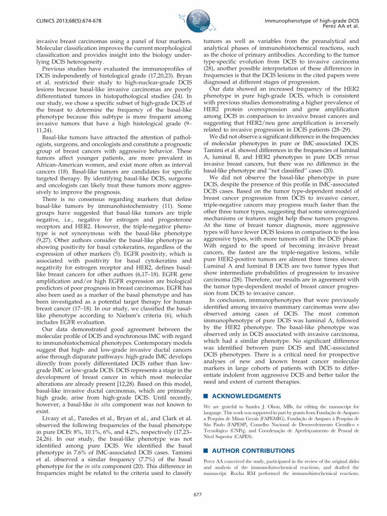
Figure 1 - A) High-grade ductal carcinoma in situ associated with invasive mammary carcinoma positive for estrogen receptor (100x).Arrows indicate the in situ component. B) High-grade ductal carcinoma in situ positive for HER2 (400x). C) High-grade ductal carcinomain situ positive for cytokeratin 5 (100x). D) High-grade ductal carcinoma in situ positive for epidermal growth factor receptor (400x).
CLINICS 2013;68(5):674-678 Immunophenotype of high-grade DCISPerez AA et al.
675

1% of neoplastic cells showed moderate or strong nuclearstaining (15). HER2 overexpression was analyzed accordingto the American Society of Clinical Oncology and College ofAmerican Pathologists (16). Any degree of cytoplasmicstaining for CK5 and any degree of distinct membranousstaining for EGFR were considered positive expression (17).
Immunohistochemical profileThe tumors were divided into five subgroups according
to their immunohistochemical profile: luminal A (ER+/HER2-), luminal B (ER+/HER2+), HER2 (ER-/HER2+),basal-like (ER-/HER2-/EGFR+ and/or CK5+), and ‘‘notclassified’’ (all markers negative) (17,18). The basal-likephenotype was defined according to Nielsen’s criteria (6).
Statistical analysisPearson’s asymptotic and exact chi-square tests were used
to compare proportions. The Mann-Whitney test was usedto compare medians. A p-value,0.05 was consideredstatistically significant. The kappa test was used to assessthe concordance between phenotypes. Kappa values greaterthan 0.80 demonstrated excellent agreement (19). This studywas approved by the Research Ethics Committee of theFederal University of Minas Gerais (protocol 655/08).
& RESULTS
Pure DCIS was detected in 46/131 cases (35% of the total),and 85/131 cases (65% of the total) were DCIS associatedwith invasive carcinoma. Immunohistochemistry was per-formed for 121 cases, including 42 cases of pure DCIS (35%of the total) and 79 cases of DCIS associated with invasivecarcinoma (65% of the total).
The median age at diagnosis was 51 years (standarddeviation ¡14 years) among cases of pure DCIS and 53years (standard deviation ¡19 years) among cases of IMC-associated DCIS (p = 0.913). The median DCIS size was13 mm. There was no significant difference in menopausalstatus (p = 0.779) or median tumor size (p = 0.836) betweenpure and IMC-associated DCIS cases. There was a sig-nificant difference in primary surgical treatment andadjuvant therapy between pure and IMC-associated DCIScases (p,0.05). Cases with DCIS associated with IMC weretreated with more extensive surgery and more oftenreceived adjuvant therapy.
The frequencies of the molecular immunophenotypes ofDCIS are shown in Table 2 and Figure 1. Among samples ofpure DCIS, the luminal A phenotype was the most common(24/42 cases; 57.1%), followed by the HER2 phenotype (07/42 cases; 16.7%), the ‘‘not classified’’ phenotype (06/42cases; 14.3%), and the luminal B phenotype (5/42 cases;11.9%). The basal-like phenotype was not identified amongthe pure DCIS cases. The immunophenotypes of DCISassociated with invasive carcinoma were the following:luminal A (46/79 cases; 58.2%), luminal B (10/79 cases;
12.7%), HER2 (06/79 cases; 7.6%), basal-like (06/79 cases;7.6%), and ‘‘not classified’’ (11/79 cases; 13.9%).
There was no significant difference in frequency betweenimmunophenotypes in pure and IMC-associated DCISsamples (p.0.05). Excellent agreement was observedbetween in situ and invasive components with regard toimmunophenotypes (kappa = 0.867).
& DISCUSSION
Breast cancer comprises a heterogeneous group ofdiseases with regard to presentation, morphology, biologi-cal characteristics, clinical behavior, and response to therapy(6,9,20). In the past 20 years, concomitant with the wide useof screening mammography, the DCIS incidence has risendramatically (21–22). The understanding of the biology andclinical behavior of DCIS is currently limited. Molecularprofiling through gene array studies is likely to have a majorimpact on breast cancer classification and management, andit is important that similar approaches are taken to advancethe understanding of DCIS.
The immunohistochemical staining of paraffin sectionsusing antibody panels has been shown to be a reliablesurrogate for the molecular classification of invasive breastcancers through gene expression profiling studies.Antibodies against estrogen receptor, progesterone receptor,HER2, cytokeratin 5/6, and EGFR have been particularlyuseful for this purpose (4–8). In fact, this approach tomolecular classification (that is, using immunostaining as asurrogate for expression profiling) is arguably the mostpractical approach to phenotyping a large number ofarchived specimens for which fresh tissue is not availablefor expression array analysis.
Recent advances have led to an emerging molecularclassification for invasive breast cancer based on thebiological characteristics of the tumor rather than beinglimited to morphological analysis. Much less attention hasbeen focused on dissecting the biological subtypes of DCIS,the immediate precursor to invasive breast cancer. Therehave been discrepancies in the results of those studies.There have also been discrepancies in the relative frequencyof subtypes between in situ and invasive disease (17,20,23–26).
In our series, we showed that DCIS can be classified intothe five immunophenotypes that have been described for
Table 2 - Immunohistochemical profile of high-gradeDCIS (pure or associated with invasive mammarycarcinoma).
Pure DCIS DCIS + IMC
Phenotype N (%) N (%) p-value*
Luminal A 24 (57.1%) 46 (58.2%) 0.264
Luminal B 05 (11.9%) 10 (12.7%)
HER2 07 (16.7%) 06 (7.6%)
Basal-like 00 (0%) 06 (7.6%)
‘‘Not classified’’ 06 (14.3%) 11 (13.9%)
TOTAL 42 (100%) 79 (100%)
DCIS = ductal carcinoma in situ; IMC = invasive mammary carcinoma
Luminal A: ER+/HER2-; Luminal B: ER+/HER2+; HER2: ER-/HER2+; Basal: ER-/
HER2-/EGFR+ and/or CK5+; ‘‘Not classified’’: ER-/HER2-/EGFR-/CK5-.
p = significance level.* = Exact Pearson’s chi-square test; refers to in situ component (pure or
associated with invasive carcinoma).
Table 1 - Sources and dilutions of the primary antibodies.
Antibody Clone Dilution Manufacturer, Country
ER SP1 Ready to use DAKO, USA
HER2 CB11 1/1000 NovoCastra, UK
CK5 XM26 1/50 NeomarKers, USA
EGFR 31G7 1/25 Zymed, USA
Immunophenotype of high-grade DCISPerez AA et al.
CLINICS 2013;68(5):674-678
676
invasive breast carcinomas using a panel of four markers.Molecular classification improves the current morphologicalclassification and provides insight into the biology under-lying DCIS heterogeneity.
Previous studies have evaluated the immunoprofiles ofDCIS independently of histological grade (17,20,23). Bryanet al. restricted their study to high-nuclear-grade DCISlesions because basal-like invasive carcinomas are poorlydifferentiated tumors in histopathological studies (24). Inour study, we chose a specific subset of high-grade DCIS ofthe breast to determine the frequency of the basal-likephenotype because this subtype is more frequent amonginvasive tumors that have a high histological grade (9–11,24).
Basal-like tumors have attracted the attention of pathol-ogists, surgeons, and oncologists and constitute a prognosticgroup of breast cancers with aggressive behavior. Thesetumors affect younger patients, are more prevalent inAfrican-American women, and exist more often as intervalcancers (18). Basal-like tumors are candidates for specifictargeted therapy. By identifying basal-like DCIS, surgeonsand oncologists can likely treat these tumors more aggres-sively to improve the prognosis.
There is no consensus regarding markers that definebasal-like tumors by immunohistochemistry (11). Somegroups have suggested that basal-like tumors are triplenegative, i.e., negative for estrogen and progesteronereceptors and HER2. However, the triple-negative pheno-type is not synonymous with the basal-like phenotype(9,27). Other authors consider the basal-like phenotype asshowing positivity for basal cytokeratins, regardless of theexpression of other markers (5). EGFR positivity, which isassociated with positivity for basal cytokeratins andnegativity for estrogen receptor and HER2, defines basal-like breast cancers for other authors (6,17–18). EGFR geneamplification and/or high EGFR expression are biologicalpredictors of poor prognosis in breast carcinomas. EGFR hasalso been used as a marker of the basal phenotype and hasbeen investigated as a potential target therapy for humanbreast cancer (17–18). In our study, we classified the basal-like phenotype according to Nielsen’s criteria (6), whichincludes EGFR evaluation.
Our data demonstrated good agreement between themolecular profile of DCIS and synchronous IMC with regardto immunohistochemical phenotypes. Contemporary modelssuggest that high- and low-grade invasive ductal cancersarise through disparate pathways: high-grade IMC developsdirectly from poorly differentiated DCIS rather than low-grade IMC or low-grade DCIS. DCIS represents a stage in thedevelopment of breast cancer in which most molecularalterations are already present [12,28). Based on this model,basal-like invasive ductal carcinomas, which are primarilyhigh grade, arise from high-grade DCIS. Until recently,however, a basal-like in situ component was not known toexist.
Livasy et al., Paredes et al., Bryan et al., and Clark et al.observed the following frequencies of the basal phenotypein pure DCIS: 8%, 10.1%, 6%, and 4.2%, respectively (17,23–24,26). In our study, the basal-like phenotype was notidentified among pure DCIS. We identified the basalphenotype in 7.6% of IMC-associated DCIS cases. Tamimiet al. observed a similar frequency (7.7%) of the basalphenotype for the in situ component (20). This difference infrequencies might be related to the criteria used to classify
tumors as well as variables from the preanalytical andanalytical phases of immunohistochemical reactions, suchas the choice of primary antibodies. According to the tumortype-specific evolution from DCIS to invasive carcinoma(28), another possible interpretation of these differences infrequencies is that the DCIS lesions in the cited papers werediagnosed at different stages of progression.
Our data showed an increased frequency of the HER2phenotype in pure high-grade DCIS, which is consistentwith previous studies demonstrating a higher prevalence ofHER2 protein overexpression and gene amplificationamong DCIS in comparison to invasive breast cancers andsuggesting that HER2/neu gene amplification is inverselyrelated to invasive progression in DCIS patients (28–29).
We did not observe a significant difference in the frequenciesof molecular phenotypes in pure or IMC-associated DCIS.Tamimi et al. showed differences in the frequencies of luminalA, luminal B, and HER2 phenotypes in pure DCIS versusinvasive breast cancers, but there was no difference in thebasal-like phenotype and ‘‘not classified’’ cases (20).
We did not observe the basal-like phenotype in pureDCIS, despite the presence of this profile in IMC-associatedDCIS cases. Based on the tumor type-dependent model ofbreast cancer progression from DCIS to invasive cancer,triple-negative cancers may progress much faster than theother three tumor types, suggesting that some unrecognizedmechanisms or features might help these tumors progress.At the time of breast tumor diagnosis, more aggressivetypes will have fewer DCIS lesions in comparison to the lessaggressive types, with more tumors still in the DCIS phase.With regard to the speed of becoming invasive breastcancers, the fastest are the triple-negative lesions, whilepure HER2-positive tumors are almost three times slower.Luminal A and luminal B DCIS are two tumor types thatshow intermediate probabilities of progression to invasivecarcinoma (28). Therefore, our results are in agreement withthe tumor type-dependent model of breast cancer progres-sion from DCIS to invasive cancer.
In conclusion, immunophenotypes that were previouslyidentified among invasive mammary carcinomas were alsoobserved among cases of DCIS. The most commonimmunophenotype of pure DCIS was luminal A, followedby the HER2 phenotype. The basal-like phenotype wasobserved only in DCIS associated with invasive carcinoma,which had a similar phenotype. No significant differencewas identified between pure DCIS and IMC-associatedDCIS phenotypes. There is a critical need for prospectiveanalyses of new and known breast cancer molecularmarkers in large cohorts of patients with DCIS to differ-entiate indolent from aggressive DCIS and better tailor theneed and extent of current therapies.
& ACKNOWLEDGMENTS
We are grateful to Sandra J. Olson, MBs, for editing the manuscript for
language. This work was supported in part by grants from Fundacao de Amparo
a Pesquisa de Minas Gerais (FAPEMIG), Fundacao de Amparo a Pesquisa de
Sao Paulo (FAPESP), Conselho Nacional de Desenvolvimento Cientıfico e
Tecnologico (CNPq), and Coordenacao de Aperfeicoamento de Pessoal de
Nıvel Superior (CAPES).
& AUTHOR CONTRIBUTIONS
Perez AA conceived the study, participated in the review of the original slides
and analysis of the immunohistochemical reactions, and drafted the
manuscript. Rocha RM performed the immunohistochemical reactions.
CLINICS 2013;68(5):674-678 Immunophenotype of high-grade DCISPerez AA et al.
677
Balabram D performed the statistical analysis. Souza AS separated the original
slides and blocks for immunohistochemistry. Gobbi H participated in the
design and coordination of the study, review of original slides, and analysis of
the immunohistochemical reactions and helped drafting the manuscript. All of
the authors have read and approved the final version of the manuscript.
& REFERENCES
1. Perou CM, Sorlie T, Eisen MB, van de Rijn M, Jeffrey SS, Rees CA, et al.Molecular portraits of human breast tumors. Nature. 2000;406(6797):747-52, http://dx.doi.org/10.1038/35021093.
2. Sorlie T, Perou CM, Tibshirani R, Aas T, Geisler S, Johnsen H, et al. Geneexpression patterns of breast carcinomas distinguish tumor subclasseswith clinical implications. Proc Natl Acad Sci USA. 2001;98(19):10869-74,http://dx.doi.org/10.1073/pnas.191367098.
3. Sorlie T, Tibshirani R, Parker J, Hastie T, Marron JS, Nobel A, et al.Repeated observation of breast tumor subtypes in independent geneexpression data sets. Proc Natl Acad Sci USA. 2003;100(14):8418-23,http://dx.doi.org/10.1073/pnas.0932692100.
4. Rakha EA, Putti TC, Abd El-Rehim DM, Paish C, Green AR, Powe DG,et al. Morphological and immunophenotypic analysis of breast carcino-mas with basal and myoepithelial differentiation. J Pathol. 2006;208(4):495-506, http://dx.doi.org/10.1002/path.1916.
5. Rakha EA, El-Sayed ME, Green AR, Paish EC, Lee AH, Ellis IO. Breastcarcinoma with basal differentiation: a proposal for pathology definitionbased on basal cytokeratin expression. Histopathology. 2007;50(4):434-38,http://dx.doi.org/10.1111/j.1365-2559.2007.02638.x.
6. Nielsen TO, Hsu FD, Jensen K, Cheang M, Karaca G, Hu Z, et al.Immunohistochemical and clinical characterization of the basal-likesubtype of invasive breast carcinoma. Clin Cancer Res. 2004;10(16):5367-74, http://dx.doi.org/10.1158/1078-0432.CCR-04-0220.
7. Livasy CA, Karaca G, Nanda R, Tretiakova MS, Olopade OI, Moore DT,et al. Phenotypic evaluation of the basal-like subtype of invasive breastcarcinoma. Mod Pathol. 2006;19(2):264-71, http://dx.doi.org/10.1038/modpathol.3800528.
8. Matos I, Dufloth R, Alvarenga M, Zeferino LC, Schmitt F. p63,cytokeratin 5, and P-cadherin: three molecular markers to distinguishbasal phenotype in breast carcinomas. Virchows Arch. 2005;447(4):688-94, http://dx.doi.org/10.1007/s00428-005-0010-7.
9. Reis-Filho JS, Tutt AN. Triple negative tumors: a critical review.Histopathology. 2008;52(1):108-18.
10. Silva F, Carvalho S, Milanezi F, Schmitt FC. Basal-like carcinoma of thebreast. Acta Med Port. 2008;21(4):373-78.
11. Fadare O, Tavassoli FA. The phenotypic spectrum of basal-like breastcancers: a critical appraisal. Adv Anat Pathol. 2007;14(5):358-73, http://dx.doi.org/10.1097/PAP.0b013e31814b26fe.
12. Buerger H, Otterbach F, Simon R, Poremba C, Diallo R, Decker T, et al.Comparative genomic hybridization of ductal carcinoma in situ of thebreast – evidence of multiple genetic pathways. J Pathol. 1999;187(4):396-402, http://dx.doi.org/10.1002/(SICI)1096-9896(199903)187:4,396::AID-PATH286.3.0.CO;2-L.
13. Schnitt SJ, Allred C, Britton P, Ellis IO, Lakhani SR, Morrow M, et al.Ductal carcinoma in situ. In: Lakhani SR, Ellis IO, Schnitt SJ, Tan PH, vande Vijver MJ, editors. WHO Classification of Tumours of the Breast.IARC: Lyon; 2012.
14. Scott MA, Lagios MD, Axelsson K, Rogers LW, Anderson TJ, Page DL.Ductal carcinoma in situ of the breast: reproducibility of histological
subtype analysis. Hum Pathol. 1997;28(8):967-73, http://dx.doi.org/10.1016/S0046-8177(97)90013-7.
15. Hammond ME, Hayes DF, Dowsett M, Allred DC, Hagerty KL, Badve S,et al. American Society of Clinical Oncology/College of AmericanPathologists guideline recommendations for immunohistochemical test-ing of estrogen and progesterone receptors in breast cancer. Arch PatholLab Med. 2010;134(7):907-22.
16. Wolff AC, Hammond ME, Schwartz JN, Hagerty KL, Allred DC, Cote RJ,et al. American Society of Clinical Oncology/College of AmericanPathologists guideline recommendations for human epidermal growthfactor receptor 2 testing in breast cancer. J Clin Oncol. 2007;25(1):118-45.
17. Livasy CA, Perou CM, Karaca G, Cowan DW, Maia D, Jackson S, et al.Identification of a basal-like subtype of breast ductal carcinoma in situ.Hum Pathol. 2007;38(2):197-204, http://dx.doi.org/10.1016/j.humpath.2006.08.017.
18. Carey LA, Perou CM, Livasy CA, Dressler LG, Cowan D, Conway K,et al. Race, Breast Cancer Subtypes, and Survival in the Carolina BreastCancer Study. JAMA. 2006;295(21):2492-502, http://dx.doi.org/10.1001/jama.295.21.2492.
19. Landis JR, Koch GG. The measurement of observer agreement forcategorical data. Biometrics. 1977;33(1):159-74, http://dx.doi.org/10.2307/2529310.
20. Tamimi RM, Baer HJ, Marotti J, Galan M, Galaburda L, Fu Y, et al.Comparison of molecular phenotypes of ductal carcinoma in situ andinvasive breast cancer. Breast Cancer Res. 2008;10(4):R67, http://dx.doi.org/10.1186/bcr2128.
21. Burstein HJ, Polyak K, Wong JS, Lester SC, Kaelin CM. Ductal carcinomain situ of the breast. N Engl J Med. 2004;350(14):1430-41.
22. O’Sullivan MJ, Morrow M. Ductal carcinoma in situ – current manage-ment. Surg Clin N Am. 2007;87(2):333-51.
23. Paredes J, Lopes N, Milanezi F, Schmitt FC. P-cadherin and cytokeratin 5:useful adjunct markers to distinguish basal-like ductal carcinomas insitu. Virchows Arch. 2007;450(1):73-80, http://dx.doi.org/10.1007/s00428-006-0334-y.
24. Bryan BB, Schnitt SJ, Collins LC. Ductal carcinoma in situ with basal-likephenotype: a possible precursor to invasive basal-like breast cancer. ModPathol. 2006;19(5):617-21, http://dx.doi.org/10.1038/modpathol.3800570.
25. Dabbs DJ, Chivukula M, Carter G, Bhargava R. Basal phenotype ofductal carcinoma in situ: recognition and immunohistologic profile. ModPathol. 2006;19(11):1506-11.
26. Clark SE, Warwick J, Carpenter R, Bowen RL, Duffy SW, Jones JL.Molecular subtyping of DCIS: heterogeneity of breast cancer reflected inpre-invasive disease. Br J Cancer. 2011;104(1):120-27, http://dx.doi.org/10.1038/sj.bjc.6606021.
27. Carvalho FM, Bacchi LM, Santos PPC, Bacchi CE. Triple-negative breastcarcinomas are a heterogeneous entity that differs between young andold patients. Clinics. 2010;65(10):1033-36, http://dx.doi.org/10.1590/S1807-59322010001000019.
28. Kurbel S. In search of triple-negative DCIS: tumor-type dependent modelof breast cancer progression from DCIS to the invasive cancer. TumorBiol. 2012. Dec 4. [Epub ahead of print].
29. Allred DC, Clark GM, Molina R, Andon AK, Schnitt SJ, Gilchrist KW,et al. Overexpression of HER-2/neu and its relationship with otherprognostic factors change during the progression of in situ to invasivebreast cancer. Hum Pathol. 1992;23(9):974-79, http://dx.doi.org/10.1016/0046-8177(92)90257-4.
Immunophenotype of high-grade DCISPerez AA et al.
CLINICS 2013;68(5):674-678
678