Immunohistochemical Evaluation of Venous Leg Ulcers …€¦ · · 2011-09-12used in several...
10
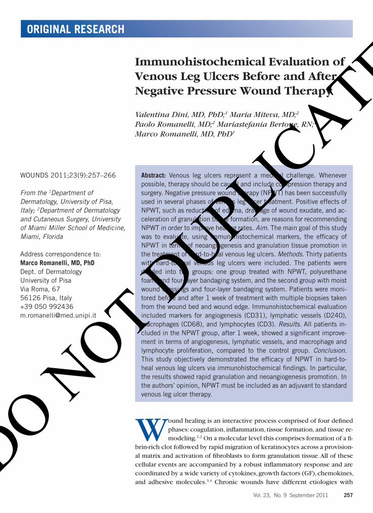
ORIGINAL RESEARCH Vol. 23, No. 9 September 2011 257 Abstract: Venous leg ulcers represent a medical challenge. Whenever possible, therapy should be causal and include compression therapy and surgery. Negative pressure wound therapy (NPWT) has been successfully used in several phases of venous leg ulcer treatment. Positive effects of NPWT, such as reduction of edema, drainage of wound exudate, and ac- celeration of granulation tissue formation, are reasons for recommending NPWT in order to improve healing rates. Aim. The main goal of this study was to evaluate, using immunohistochemical markers, the efficacy of NPWT in terms of neoangiogenesis and granulation tissue promotion in the treatment of hard-to-heal venous leg ulcers. Methods. Thirty patients with hard-to-heal venous leg ulcers were included. The patients were divided into two groups: one group treated with NPWT, polyurethane foam, and four-layer bandaging system, and the second group with moist wound dressings and four-layer bandaging system. Patients were moni- tored before and after 1 week of treatment with multiple biopsies taken from the wound bed and wound edge. Immunohistochemical evaluation included markers for angiogenesis (CD31), lymphatic vessels (D240), macrophages (CD68), and lymphocytes (CD3). Results. All patients in- cluded in the NPWT group, after 1 week, showed a significant improve- ment in terms of angiogenesis, lymphatic vessels, and macrophage and lymphocyte proliferation, compared to the control group. Conclusion. This study objectively demonstrated the efficacy of NPWT in hard-to- heal venous leg ulcers via immunohistochemical findings. In particular, the results showed rapid granulation and neoangiogenesis promotion. In the authors’ opinion, NPWT must be included as an adjuvant to standard venous leg ulcer therapy. WOUNDS 2011;23(9):257–266 From the 1 Department of Dermatology, University of Pisa, Italy; 2 Department of Dermatology and Cutaneous Surgery, University of Miami Miller School of Medicine, Miami, Florida Address correspondence to: Marco Romanelli, MD, PhD Dept. of Dermatology University of Pisa Via Roma, 67 56126 Pisa, Italy +39 050 992436 [email protected] W ound healing is an interactive process comprised of four defined phases: coagulation, inflammation, tissue formation, and tissue re- modeling. 1,2 On a molecular level this comprises formation of a fi- brin-rich clot followed by rapid migration of keratinocytes across a provision- al matrix and activation of fibroblasts to form granulation tissue. All of these cellular events are accompanied by a robust inflammatory response and are coordinated by a wide variety of cytokines, growth factors (GF), chemokines, and adhesive molecules. 3,4 Chronic wounds have different etiologies with Immunohistochemical Evaluation of Venous Leg Ulcers Before and After Negative Pressure Wound Therapy Valentina Dini, MD, PhD; 1 Maria Miteva, MD; 2 Paolo Romanelli, MD; 2 Mariastefania Bertone, RN; 1 Marco Romanelli, MD, PhD 1 DO NOT DUPLICATE
-
Upload
trinhtuyen -
Category
Documents
-
view
217 -
download
1
Transcript of Immunohistochemical Evaluation of Venous Leg Ulcers …€¦ · · 2011-09-12used in several...
ORIGINAL RESEARCH
Vol. 23, No. 9 September 2011 257
Abstract: Venous leg ulcers represent a medical challenge. Whenever possible, therapy should be causal and include compression therapy and surgery. Negative pressure wound therapy (NPWT) has been successfully used in several phases of venous leg ulcer treatment. Positive effects of NPWT, such as reduction of edema, drainage of wound exudate, and ac-celeration of granulation tissue formation, are reasons for recommending NPWT in order to improve healing rates. Aim. The main goal of this study was to evaluate, using immunohistochemical markers, the efficacy of NPWT in terms of neoangiogenesis and granulation tissue promotion in the treatment of hard-to-heal venous leg ulcers. Methods. Thirty patients with hard-to-heal venous leg ulcers were included. The patients were divided into two groups: one group treated with NPWT, polyurethane foam, and four-layer bandaging system, and the second group with moist wound dressings and four-layer bandaging system. Patients were moni-tored before and after 1 week of treatment with multiple biopsies taken from the wound bed and wound edge. Immunohistochemical evaluation included markers for angiogenesis (CD31), lymphatic vessels (D240), macrophages (CD68), and lymphocytes (CD3). Results. All patients in-cluded in the NPWT group, after 1 week, showed a significant improve-ment in terms of angiogenesis, lymphatic vessels, and macrophage and lymphocyte proliferation, compared to the control group. Conclusion. This study objectively demonstrated the efficacy of NPWT in hard-to-heal venous leg ulcers via immunohistochemical findings. In particular, the results showed rapid granulation and neoangiogenesis promotion. In the authors’ opinion, NPWT must be included as an adjuvant to standard venous leg ulcer therapy.
WOUNDS 2011;23(9):257–266
From the 1Department of Dermatology, University of Pisa, Italy; 2Department of Dermatology and Cutaneous Surgery, University of Miami Miller School of Medicine, Miami, Florida
Address correspondence to:Marco Romanelli, MD, PhDDept. of DermatologyUniversity of PisaVia Roma, 6756126 Pisa, Italy+39 050 [email protected]
Wound healing is an interactive process comprised of four defined phases: coagulation, inflammation, tissue formation, and tissue re-modeling.1,2 On a molecular level this comprises formation of a fi-
brin-rich clot followed by rapid migration of keratinocytes across a provision-al matrix and activation of fibroblasts to form granulation tissue. All of these cellular events are accompanied by a robust inflammatory response and are coordinated by a wide variety of cytokines, growth factors (GF), chemokines, and adhesive molecules.3,4 Chronic wounds have different etiologies with
Immunohistochemical Evaluation of Venous Leg Ulcers Before and After Negative Pressure Wound Therapy
Valentina Dini, MD, PhD;1 Maria Miteva, MD;2 Paolo Romanelli, MD;2 Mariastefania Bertone, RN;1 Marco Romanelli, MD, PhD1
Dini.indd 257 9/12/11 9:41 AM
DO NOT D
UPLICATE

Dini et al
258 WOUNDS www.woundsresearch.com
more than 90% falling into three categories: venous leg ulcers, pressure ulcers, and diabetic ulcers.5,6 Previous studies have shown that negative pressure wound ther-apy (NPWT) can be useful in treating chronic wounds, as it has the ability to accelerate the rate of granulation tissue formation, enhances angiogenesis,7–9 and removes excess wound fluid that harbors inhibitory factors and microorganisms.10,11 A recent study showed that NPWT also acts upon lymphatic vessels. NPWT induces initial proliferation of lymphatics, reduces interstitial edema, and improves loco-regional perfusion.12
The aim of this study was to investigate the impact of NPWT (V.A.C. Freedom®, KCI, San Antonio, TX) as an ad-junct to compression therapy for 7 days on the immuno-histochemical expression of a panel of tissue biomarkers in biopsy specimens from non-healing venous leg ulcers assessed before and after treatment.
Materials and MethodsFour immunohistochemical markers (CD3, CD68,
CD31, D2-40) were selected to highlight the changes re-
lated to relevant components of the wound healing pro-cess (ie, local inflammation, angiogenesis, and lymphan-giogenesis). CD3 was chosen as a pan T-cell marker, which is relevant to the healing process. Previous studies have shown that wound healing is impaired in the absence of gamma-delta T cells.13 Furthermore, epidermal T cells from patients with healing wounds are activated and se-crete growth factors. CD68 is a marker for monocytes and macrophages. These cells are important because they in-filtrate the wound after the polymorphonuclear cells and modulate wound angiogenesis by releasing angiogenic factors, such as fibroblast growth factor-2 (FGF-2) and vas-cular endothelial growth factor (VEGF).11 CD31 was cho-sen to highlight angiogenesis: it labels blood vessels and is the most sensitive marker for endothelial cells.14 D2-40 was selected to highlight the lymphatic vessels. It is a monoclonal antibody that recognizes podoplanin, which is highly and constitutively expressed in lymphatic en-dothelial cells.15–17 The authors have recently shown that D2-40 is a useful diagnostic marker in chronic wounds, as it highlights global and architectural differences in the lymphatic network.18
The study was approved by the Institutional Review Board of the Medical University (Pisa, Italy). Patient re-cruitment and all study-related clinical procedures, in-cluding obtaining tissue samples, were performed at the Department of Dermatology, University of Pisa. The histo-pathologic and immunohistochemical analysis (process-ing, staining, and evaluation of the slides) was performed by the Dermatopathology Lab of the Department of Der-matology and Cutaneous Surgery at the University of Mi-ami, Florida.
Thirty patients with the clinical diagnosis of hard-to-heal venous ulcers were included after fulfilling the crite-ria for positive venous Doppler scan, Ankle Brachial Index (ABI) ≥ 0.8, failure to heal > 6 months despite standard treatment, ulcer size ≥ 20 cm2, and no previous treatment with NPWT. The patients were randomly divided into two groups. Group 1 (n = 15) was treated with 125 mmHg continuous NWPT using polyurethane foam (V.A.C. Free-dom) with four-layer bandaging for 1 week. This group included 8 female and 7 male subjects with a mean age of 72 years, ulcer duration of 32 months (average), and mean ulcer size of 89 cm2. The control group (n = 15) received only compression treatment for 1 week with the four layer bandaging system. The control group included 6 female and 9 male patients with a mean age of 65 years, ulcer duration of 47 months (average), and mean ulcer size of 72 cm2.
Keypoints
• The patients were randomly divided into two groups. Group 1 (n = 15) was treated with 125 mmHg con-tinuous NWPT using polyurethane foam (V.A.C. Freedom) with four-layer bandaging for 1 week.
• Four immunohistochemical markers (CD3, CD68, CD31, D2-40) were selected to highlight the chang-es related to relevant components of the wound healing process (ie, local inflammation, angiogen-esis, and lymphangiogenesis).
• The immunohistochemical study also included an instrumental evaluation of the two groups by means of a laser scanning system to assess the speed of granulation tissue formation.
Keypoints
• A recent study showed that NPWT also acts upon lymphatic vessels. NPWT induces initial prolifera-tion of lymphatics, reduces interstitial edema, and improves loco-regional perfusion.
• The aim of the present study was to investigate the impact of NPWT (V.A.C. Freedom®, KCI, San Anto-nio, TX) as an adjunct to compression therapy for 7 days on the immunohistochemical expression of a panel of tissue biomarkers in biopsy specimens from non-healing venous leg ulcers assessed before and after treatment.
Dini.indd 258 9/12/11 9:41 AM
DO NOT D
UPLICATE
Dini et al
Vol. 23, No. 9 September 2011 259
Immunohistochemical analysis. For the purpose of this study, a 4-mm punch biopsy was obtained from the wound bed or the wound edge at the baseline visit (day 0) and after 1 week (day 7) in all patients, accounting for a total of 60 tissue samples. However, only 26 samples (12 samples from 6 patients in the NPWT group and 12 samples from 6 patients in the control group) met the se-lection criteria to enter the immunohistochemical study: 1) site matched specimens from days 0 and 7 obtained from the wound edge (24 samples) or the wound bed (2 samples); 2) sufficient macroscopic tissue (at least 0.3 cm x 0.3 cm). All specimens were fixed in 10% formalin, processed as paraffin-embedded tissue, and stained for hematoxylin and eosin (H&E), CD3, CD68, CD31, and D2-40 (Abcam, Cambridge, MA). At least 4 consecutive sec-tions of all specimens were evaluated in a blinded fashion by two independent dermatopathologists. Edema was graded on the H&E sections as mild (1+) referring to pale papillary and upper dermis, moderate ([2+], pale papil-lary dermis with Gossamer’s stands and diffuse slight sep-aration of the collagen bundles throughout the reticular dermis), and marked or severe ([3+], sub-epidermal cleft/bulla with more pronounced separation of the collagen bundles throughout the reticular dermis).
CD3 and CD68 were assessed by scanning tissue sec-tions under low magnification (x2) to identify the hot spot. Within the hot spot, the number of cells within three adjacent high-power fields (x40) was counted and the mean number for each slide was established. The CD31 stain was assessed in a similar way by counting the number of vessels in a hot spot. In an attempt to elimi-nate false positive results (as histiocytes also stain posi-tive for CD68), a vessel was defined as the presence of three contiguous cells staining for CD31. The number of D2-40 positive lymphatics was calculated per unit area, which was done by counting all the vessels (a lymphatic vessel was defined as at least three contiguous cells stain-ing for D2-40). The width and depth of each section was measured with an ocular micrometer at 2x (Olympus WH10x/22 eye micrometer), and the measurements were expressed in micrometers. The number of lymphatics was calculated per 1000 micrometers/tissue, which is equiva-lent to 1 mm. The depth of each section was defined as the distance from the dermo-epidermal junction to sub-cutaneous fat. If no fat was present in the sections, the depth of the entire specimen was obtained.
Laser scanner measurement. The immunohisto-chemical study also included an instrumental evaluation of the two groups by means of a laser scanning system
to assess the speed of granulation tissue formation. Tech-nical details of the system have been described by the authors in a previous study.19 The laser scanning system is able to calculate the percentage of each area with a corre-sponding color within the wound using region-growing algorithms, which begins with identifying corresponding areas on the image with similar colors and basic tones that the user chooses.
Statistical AnalysisThe statistical analysis was performed by the Division
of Biostatistics, Department of Epidemiology and Public Health, University of Miami Miller School of Medicine. Outcome variables, including the expression of the two inflammatory markers (CD3, CD68), the expression of two vessel markers (CD31, D2-40), and pre- and post-treat-ment mean values, were first compared within control group and within the NPWT group using a paired t test. A subsequent independent t test was used to compare the value changes between the two groups. Edema was analyzed using conditional logistic regression for paired data in two analyses: comparing mild + moderate versus marked, and then mild versus moderate + marked. All sig-nificant results were based on an alpha of 0.05. Differ-ences found between the study groups by using the laser scanner were tested using analysis of variance (ANOVA).
ResultsThe results of the pre- and post-treatment mean values
of each marker in the NPWT group and the control group are shown in Table 1. The value changes for each marker between the two groups are summarized in Table 2.
Inflammatory markers. The mean baseline number of CD3-positive cells in the NPWT-treated wounds was 56.0 (SD ± 24.18) versus 54.89 (SD ± 23.17) post treat-ment. Similar results were obtained in the control group in which the mean number of CD3 cells in the venous ulcers on day 0 was 61.28 (SD ± 15.31) and decreased
Keypoints
• A slight increase in the number of CD31 positive blood vessels was noted in the NPWT group after treatment, whereas no change or even slight de-crease was seen in the number of blood vessels in the control group after compression treatment.
• NPWT treatment appeared to significantly stimulate granulation tissue formation of wound bed com-pared to control group (P < 0.001) at the end of the 1-week evaluation (Table 3).
Dini.indd 259 9/12/11 9:41 AM
DO NOT D
UPLICATE

Dini et al
260 WOUNDS www.woundsresearch.com
slightly to 53.22 (SD ± 16.08) after compression treat-ment. The mean difference in the CD3-positive cells at baseline versus after treatment was -1.17 (SD ± 36.33) in the NPWT group versus -8.06 (SD ± 13.26) in the con-trol group. The T cell population remained stable in the NPWT-treated wounds, whereas in the control group, the T cell number decreased after compression (Figure 1A, B). T cells are very important for the release of growth fac-tors and cytokines and their number should remain high enough during all phases to accelerate the granulation process, as shown in the NPWT-treated samples. However, the results did not reach statistical significance (P = 0.67) due to the small number of samples (6 in each group) and large standard deviations.
CD68 in the NPWT-treated wounds showed a mean baseline value of 52.89 (SD ± 16.98) and a post-treatment
value of 58.28 (SD ± 9.33). In the control group, the mean baseline value for CD68 was 49.17 (SD ± 11.69) and de-creased to 38.11 (SD ± 13.46) after compression. The mean difference between the pre- and post-treatment CD68 values in the NPWT group was 5.39 (SD ± 22.96), as the number of CD68 positive macrophages increased after treatment (Figure 2A, B). In the control group, the mean difference was negative (-11.06, SD ± 15.77), as the number of macrophages was lower in specimens after 7 days of compression. However, the results were not statis-tically significant (P = 0.17).
Vessels. A slight increase in the number of CD31 posi-tive blood vessels was noted in the NPWT group after treatment (53.83, SD ± 16.98 on day 0 and 62.89, SD ± 16.98 on day 7), whereas no change or even slight de-crease was seen in the number of blood vessels in the
Table 1. Within group comparison of all immunohistochemical markers and edema between pre- and post-treatment values in the specimens from the NPWT group and control group.
BiomarkersMean (SD)
PBefore (n=6) After (n=6)
Treatment Inflammatory
CD3 56.06 (24.18) 54.89 (23.17) 0.9404
CD68 52.89 (16.98) 58.28 (9.33) 0.5903
Vessel
CD31 53.83 (14.81) 62.89 (14.18) 0.3935
D2-40 7.67 (2.66) 26.83 (25.93) 0.1204
Edema* 1.17 (0.52) 1.50 (1.05) 0.7711
Control
Inflammatory
CD3 61.28 (15.31) 53.22 (16.08) 0.1969
CD68 49.17 (11.69) 38.11 (13.46) 0.1466
Vessel
CD31 37.50 (6.00) 35.39 (7.03) 0.6067
D2-40 6.67 (4.08) 5.50 (7.12) 0.7186
Edema* 1.83 (0.75) 2.17 (0.98) 0.3632
*Edema is coded as 1+ = Mild, 2+ = Moderate, 3+ = Marked
Table 2. Comparisons of the outcome changes in all immunohistochemical markers and edema between the specimens of the NPWT group and control group.
BiomarkersDifference in Mean (SD)
PControl (n=6) Treatment (n=6)
Inflammatory
CD3 -8.06 (13.26) -1.17 (36.33) 0.6772
CD68 -11.06 (15.77) 5.39 (22.96) 0.1788
Vessel
CD31 -2.11 (9.42) 9.06 (23.76) 0.3098
D2-40 -1.17 (7.49) 19.17 (25.10) 0.1070
Edema* 0.33 (0.82) -0.17 (1.33) 0.4506
*Edema is coded as 1+ = Mild, 2+ = Moderate, 3+ = Marked
Dini.indd 260 9/12/11 9:41 AM
DO NOT D
UPLICATE

Dini et al
Vol. 23, No. 9 September 2011 261
control group after compression treatment (37.5, SD ± 6.00 on day 0 and 35.39 ± 7.03 on day 7, [Figure 3A, B]). Only vessels composed of at least three continuous cells positive for CD31 were counted. This is important because CD31 also stained individual cells in the stroma. However, to avoid false positive results, the individual cells in the
stroma were not counted, as they could be either macrophages or single endothelial cells giving rise to new blood vessels (neo-angio-genesis). Although the mean dif-ference between the number of vessels in the specimens before and after the NPWT treatment was positive in the NPWT group (9.06, SD ± 23.76) and negative in the control group (-2.11, SD ± 9.42), the comparison between the two groups did not reach sta-tistical significance (P = 0.3).
The number of lymphatic ves-sels highlighted by the D2-40 monoclonal antibody increased in the specimens from wounds treated with the NPWT system from 7.67 (SD ± 2.66) on day 0 to 26.83 (SD ± 25.93) on day 7. In the control group, the baseline num-ber of lymphatics in the wound specimens before the compres-sion therapy was similar (6.67, SD ± 4.08). The number remained stable and was slightly lower after the treatment (5.50, SD ± 7.12). Most lymphatic vessels in the specimens after NPWT treatment showed open lumina and occa-sionally displayed open inter-en-dothelial junctions suggesting ac-tive lymph formation (Figure 4A). Conversely, D2-40 staining in the control specimens tended to be less dense, less homogeneous, and varied in thickness. Furthermore, the vessels showed more disor-ganized architecture and more often they showed collapsed lu-mina than in the specimens from the NPWT group (Figure 4B). The
mean difference between the number of lymphatics on day 0 and day 7 in the NPWT-treated group showed a positive mean increase of 19.17 (SD ± 25.10), whereas the mean difference in the control specimens was nega-tive (-1.17, SD ± 7.49). Despite this impressive difference, the results were not statistically significant (P = 0.1) most
Figure 1. A) Immunohistochemical staining for CD3 before and B) after NPWT treatment. The positive cells are highlighted in brown (magnification x10).
Figure 2. A) Immunohistochemical staining for CD68 before and B) after NPWT treatment. The positive cells are highlighted in brown (magnification x10).
Figure 3. A) Immunohistochemical staining for CD31 shows increased number of blood vessels in the specimen after the NPWT treatment; the endothelial cells are highlighted in brown. B) The number of blood vessels in the control specimen after the compression is lower (magnification x4).
A B
A B
A B
Dini.indd 261 9/12/11 9:41 AM
DO NOT D
UPLICATE

Dini et al
262 WOUNDS www.woundsresearch.com
likely due to the small sample of specimens and consider-able SD (up to ± 25.10).
Edema. Edema did not change in specimens before and after treatment in either group (the baseline values of 1.17, SD ± 0.52 (mild edema) in the NPWT group re-
mained stable after 7 days of NPWT therapy: 1.50, SD ± 1.05 (mild edema). In the control group, the baseline value of 1.83 ([SD ± 0.75], mild to moderate edema) showed very little increase to 2.17 ([SD ± 0.98], moderate edema) af-ter 7 days of compression. The mean dif-ference of the pre- and post-treatment values in the NPWT group was negative (0.17, SD ± 1.33), whereas the mean dif-ference in the control group was posi-tive (0.33, SD ± 0.82). Since these values were obtained by subjective evaluation of H&E stained sections and the P value was greater than 0.05 (P = 0.45), the authors concluded that microscopic assessment of edema is not a sensitive enough marker for monitoring healing in chronic wound tissues.
Granulation tissue. NPWT treatment appeared to significantly stimulate granulation tissue formation of wound bed compared to control group (P < 0.001) at the end of the 1-week evaluation (Figure 7). At the first dress-ing change (after 3 days of treatment), the mean percent-
Figure 4. A) Immunohistochemical staining for the D2-40 monoclonal anti-body highlights homogenous staining of dermal lymphatics in the specimen after the NPWT treatment. The lymphatics reveal open lumina and open inter-endothelial junctions suggesting active formation of lymph. B) There is diffuse non-specific background staining; the lymphatic vessels in control specimens after the compression therapy reveal collapsed lumina and dis-torted morphology referred to as a “cork screw” pattern (magnification x10).
Figure 5. Venous leg ulcer before, during, and after 1 week of NPWT.
Figure 6. Venous leg ulcer before and after 1 week of NPWT.
A B
Dini.indd 262 9/12/11 9:41 AM
DO NOT D
UPLICATE

Dini et al
Vol. 23, No. 9 September 2011 263
age of granulation tissue in the NPWT group increased from 14.6% ± 12.8% to 70.5% ± 10.5% and this value re-mained stable at 1 week of treatment (Figures 5, 6).
DiscussionNPWT is a treatment that has benefited both chronic
and acute wounds. This method of exposing a wound to subatmospheric pressure for an extended period to pro-mote debridement and healing was first described by Fleischmann et al20 in 1993. Morykwas et al21 investigated the physiological basis of the NPWT (V.A.C.) therapy and suggested that removal of the interstitial fluid decreases localized edema and increases blood flow, which in turn decreases local bacterial load. Although this may be an im-portant mechanism, we have shown no statistically signif-icant difference in the edema score in tissue specimens before and after the NPWT treatment when compared to control specimens. One explanation is that reduction of edema may be an important mechanism for a selected subset of wounds, as suggested previously.7 Although in the present study minimal fluid was extracted from the NPWT-treated wounds, a clinically dramatic healing re-sponse was achieved. Furthermore, the assessment of edema in H&E stained specimens is subjective and de-pendent upon the dermatopathologist’s skill and experi-ence, which makes it difficult to use as a standard tool to evaluate and monitor chronic wounds.
Saxena et al7 suggested another mechanism for the NPWT-assisted wound improvement, ie, tissue deforma-tion stretches individual cells thereby promoting prolif-eration in the wound microenvironment. Studies have shown that cells that are allowed to stretch tend to divide and proliferate in the presence of soluble mitogens, while retracted cells remain quiescent. Cells that cannot extend assume a more spherical shape (by retraction), are growth arrested, and undergo apoptosis.22,23 Saxena et al7 created
a computer model of a wound and simulated NPWT ap-plication. In this model, they altered the pressure, pore diameter, and pore volume fraction to study the effects of NPWT-induced material deformation. The morphology of deformation in this wound model was compared to his-tological sections of wounds treated with NPWT. The re-sults showed that most elements stretched by the NPWT application caused 5%–20% strain, which is similar to the in vitro strain levels proven to promote cellular prolif-eration. The conclusion was that negative pressure stimu-lates wound healing by promoting cell division, angiogen-esis, and local proliferation of growth factors. Consequent studies have confirmed that the NPWT treated wounds manifest on histologic examination increased neo-vascu-larization as well as increased VEGF.11 We detected rela-tively stable number of CD3 positive cells and CD68 posi-tive cells in the specimens of the NPWT treated wounds whereas these populations were slightly decreased in the control group. Although not statistically significant, these results showed a trend of maintaining a favorable healing environment in the NPWT treated wounds, as both CD3 (T-cells) and CD68 (macrophages) are important for the intercellular communications and release of growth fac-tors and cytokines to promote cellular proliferation. We did not assess the number of polymorphonuclear cells as part of the inflammation, as they are numerous and not easily identifiable in histologic specimens of chronic wounds that show signs of neutrophilic karyorrhexis (nu-clear dust), diffuse fibrin, and necrosis.
In severely ischemic, hypoxic wound conditions, in-creasing oxygen concentrations results in accelerated wound healing with increased blood vessel growth.24,25
In addition, clinical and experimental studies have shown that mechanical stress triggers VEGF secretion26,27 and that NPWT treatment stimulates neovascularisation in traumatic wounds.11 The present results showed a slight increase in the number of CD31 positive blood vessels in the NPWT group after treatment compared to the num-ber of blood vessels in the control group after compres-sion, which showed a slight decrease. However, the mean difference in the pre- and post-treatment values between both groups was not statistically significant. One possible explanation is that neovascularisation is a very dynamic process and the density of blood vessels in specimens from chronic wounds may depend on the time point of tissue sampling. Jacobs et al8 found that NPWT treated wounds showed significantly higher mean vessel density by day 3, which was lost by days 5–7. We have not assessed specimens in the process of treatment but only at base-
Figure 7. Percentage of granulation tissue formation.
%
Dini.indd 263 9/12/11 9:41 AM
DO NOT D
UPLICATE

Dini et al
264 WOUNDS www.woundsresearch.com
line (day 0) and at the end (day 7). Another possible factor that counteracts with the results may be the fact that only well developed, identifiable blood vessels composed of at least three contiguous cells with/without lumina were counted. In most specimens, particularly those obtained from the NPWT-treated wounds, significant background CD31 staining of individual cells was seen. This might be in part due to macrophages that co-express CD31, but more significantly, might have been tiny individual endo-thelial cell islands of neo-angiogenesis in the stroma.
It has been shown that lymphatic function significant-ly decreases with increasing severity of chronic venous insufficiency.28,29 Only one study has looked at the mor-phology of lymphatics at microscopic level by using the D2-40 monoclonal antibody in specimens from chronic venous ulcers versus normal site matched skin tissue from skin neoplasm excisions.18 This study showed that chronic venous ulcers specimens showed more dermal lymphatic vessels per unit area than controls, which dem-onstrated global and architectural differences compared to lymphatics from control specimens. This suggests lym-phatic dysfunction in the pathogenesis of chronic venous ulcers and makes D2-40 a marker of potential interest for monitoring the treatment effect on the wound tissue at the microscopic level.
We showed that the number of lymphatic vessels highlighted by the D2-40 monoclonal antibody increased in the specimens from wounds treated with the NPWT system from day 0 to day 7. In the control group the final number of lymphatics after the compression therapy was similar to the baseline number. Although the mean dif-ference between the pre- and post-treatment values was not statistically significant between the two groups due to the small number of samples and large standard devia-tions, there was a definite trend toward lymhangiogen-esis in the NPWT group versus no change in the control specimens. Furthermore, the morphology of the lymphat-ics displayed homogenous and thick ribbon-like staining, open lumina, and increased inter-endothelial junctions. In the control specimens, the morphology of the lym-phatics showed more collapsed lumina (as a possible sign of lymph overload) and a more heterogenous stain-ing pattern. This suggests that many of the otherwise “suf-ficient” number of lymphatic vessels detected in the con-trol specimens may be dysfunctional or non-functional.
Prior to this study, only one study investigated the effect of the NPWT treatment on the lymph vessels in different types of wounds referring to the global and lo-cal architecture of lymphatics as well as to deviations of
their normal morphology.12 Specimens from 13 patients with chronic venous ulcers who underwent NPWT treat-ment for between 8 and 16 days (mean 15 days) were evaluated. The D2-40 monoclonal antibody was used to highlight the lymphatics. The results showed that there was 58%–62% lymph vessel proliferation by the day 4 post the NPWT application. However, 1%–4% regression of the number of the D2-40 positive vessels was observed by day 8 with further 30%–31% regression by day 12. In-terestingly, those patients with less than 5 risk factors for non-healing wounds (eg, infection, ischemia, trauma, radi-ation, diabetes, smoking) did not demonstrate any lymph vessel regression, while the ones with 5–10 risk factors demonstrated lymph vessel proliferation on day 4 after NPWT treatment followed by lymph vessel regression. This may be a valid explanation as to why in the present study the increase of lymphatics in the NPWT specimens on day 7 was not statistically significant compared to the control specimens on same day: 1) we evaluated the specimens only at the baseline and at the end of the treat-ment; 2) we did not assess and correlate for risk factors.
NPWT treatment seems to induce morphological and quantitative alterations in the lymph vessel network in chronic venous ulcers by stimulating lymphangiogen-esis more efficiently in the first days of the treatment. This proliferation may not be sustained by the end of the treatment (day 7) because of underlying disease and risk factors that impair wound healing and in par-ticular lead to lymphatic regression. The increase in the lymphatics number in the NPWT group correlated with a better clinical outcome in terms of granulation rate and pain relief.
ConclusionNPWT applied on stable venous leg ulcers has a posi-
Keypoints
• Although some of the differences showed greater ab-solute magnitude change, there was no statistically significant change (P > 0.05) due to the small num-ber of samples collected (6 in each group) and the large standard deviations (mainly in the treatment group). Nevertheless, the lymph vessels showed ar-chitectural and morphological improvement in the NPWT group compared to the controls.
• Further large, prospective studies are needed to confirm our immunohistochemical results and to explore in more detail the microscopic features of NPWT’s effect on tissue healing.
Dini.indd 264 9/12/11 9:41 AM
DO NOT D
UPLICATE

Dini et al
Vol. 23, No. 9 September 2011 265
tive impact on the microscopic features in tissue speci-mens of treated wounds versus controls. All biomarkers referring to inflammation, angiogenesis, and lymphangio-genesis showed an absolute positive change in the treat-ment group, except for CD3, which showed negative absolute change in the treatment group and greater nega-tive absolute change in the control group. The other three biomarkers showed negative absolute change in the con-trol group but positive absolute change in the treatment group. Although some of the differences showed greater absolute magnitude change, there was no statistically sig-nificant change (P > 0.05) due to the small number of samples collected (6 in each group) and the large stan-dard deviations (mainly in the treatment group). Never-theless, the lymph vessels showed architectural and mor-phological improvement in the NPWT group compared to the controls. These data suggest another beneficial mechanism of the NPWT system on the wound healing, which refers to increased lymphangiogenesis and im-proved lymph drainage. Further large, prospective studies are needed to confirm our immunohistochemical results and to explore in more detail the microscopic features of NPWT’s effect on tissue healing.
AcknowledgementsThis study was supported by an unrestricted educa-
tional grant from KCI International. This research was based on Dr. Dini’s abstract, which won the 2010 SAWC Spring Young Investigator’s Award.
References1. Gottrup F, Karlsmark T. Leg ulcers: uncommon presenta-
tions. Clin Dermatol. 2005;23(6):601–611.
2. Broughton G, 2nd, Janis JE, Attinger CE. Wound healing:
an overview. Plast Reconstr Surg. 2006;117(7 Suppl):1e–
S–32e-S.
3. Nickoloff BJ, Bonish BK, Marble DJ, et al. Lessons learned
from psoriatic plaques concerning mechanisms of tissue
repair, remodeling, and inflammation. J Investig Dermatol
Symp Proc. 2006;11(1):16–29.
4. Diegelmann RF, Evans MC. Wound healing: an overview
of acute, fibrotic and delayed healing. Front Biosci.
2004;9:283–289.
5. Mustoe TA, O’Shaughnessy K, Kloeters O. Chronic wound
pathogenesis and current treatment strategies: a unifying
hypothesis. Plast Reconstr Surg. 2006;117(7 Suppl):35S–
41S.
6. Mustoe T. Understanding chronic wounds: a unifying hy-
pothesis on their pathogenesis and implications for ther-
apy. Am J Surg. 2004;187(5A):65S–70S.
7. Saxena V, Hwang CW, Huang S, Eichbaum Q, Ingber D,
Orgill DP. Vacuum-assisted closure: microdeformations
of wounds and cell proliferation. Plast Reconstr Surg.
2004;114(5):1086–1096.
8. Jacobs S, Simhaee DA, Marsano A, Fomovsky GM, Niedt G,
Wu JK. Efficacy and mechanisms of vacuum-assisted clo-
sure (VAC) therapy in promoting wound healing: a rodent
model. J Plast Reconstr Aesthet Surg. 2009;62(10):1331–
1338.
9. Loree S, Dompmartin A, Penven K, Harel D, Leroy D. Is
Vacuum Assisted Closure a valid technique for debriding
chronic leg ulcers? J Wound Care. 2004;13(6):249–252.
10. Venturi ML, Attinger CE, Mesbahi AN, Hess CL, Graw KS.
Mechanisms and clinical applications of the vacuum-as-
sisted closure (VAC) Device: a review. Am J Clin Derma-
tol. 2005;6(3):185–194.
11. Labler L, Rancan M, Mica L, Harter L, Mihic-Probst D, Keel
M. Vacuum-assisted closure therapy increases local inter-
leukin-8 and vascular endothelial growth factor levels in
traumatic wounds. J Trauma. 2009;66(3):749–757.
12. Labanaris AP, Polykandriotis E, Horch RE. The effect of vac-
uum-assisted closure on lymph vessels in chronic wounds.
J Plast Reconstr Aesthet Surg. 2009;62(8):1068–1075.
13. Havran WL, Jameson JM. Epidermal T cells and wound
healing. J Immunol. 2010;184(10):5423–5428.
14. Poblet E, Gonzalez-Palacios F, Jimenez FJ. Different immu-
noreactivity of endothelial markers in well and poorly
differentiated areas of angiosarcomas. Virchows Arch.
1996;428(4–5):217–221.
15. Fukunaga M. Expression of D2-40 in lymphatic endothe-
lium of normal tissues and in vascular tumours. Histopa-
thology. 2005;46(4):396–402.
16. Ordonez NG. Podoplanin: a novel diagnostic immunohis-
tochemical marker. Adv Anat Pathol. 2006;13(2):83–88.
17. Gomaa AH, Yaar M, Bhawan J. Cutaneous immunoreactiv-
ity of D2-40 antibody beyond the lymphatics. Am J Der-
matopathol. 2007;29(1):18–21.
18. Fernandez AP, Miteva M, Roberts B, Ricotti C, Rouhani P,
Romanelli P. Histopathologic analysis of dermal lymphatic
alterations in chronic venous insufficiency ulcers using
D2-40. J Am Acad Dermatol. 2011;64(6):1123 e1–12.
19. Bertone MS, Dini V, Romanelli P, Rizzello F, Romanelli M. Ob-
jective analysis of heterologous collagen efficacy in hard
to heal venous leg ulcers. WOUNDS. 2008;20(9):245–249.
20. Fleischmann W, Strecker W, Bombelli M, Kinzl L. [Vacuum
sealing as treatment of soft tissue damage in open frac-
tures]. Unfallchirurg. 1993;96(9):488–492.
21. Argenta LC, Morykwas MJ. Vacuum-assisted closure: a new
Dini.indd 265 9/12/11 9:41 AM
DO NOT D
UPLICATE

Dini et al
266 WOUNDS www.woundsresearch.com
method for wound control and treatment: clinical experi-
ence. Ann Plast Surg. 1997;38(6):563–577.
22. Chen CS, Mrksich M, Huang S, Whitesides GM, Ingber
DE. Geometric control of cell life and death. Science.
1997;276(5317):1425–1428.
23. Mochitate K, Pawelek P, Grinnell F. Stress relaxation of
contracted collagen gels: disruption of actin filament
bundles, release of cell surface fibronectin, and down-
regulation of DNA and protein synthesis. Exp Cell Res.
1991;193(1):198–207.
24. Hopf HW, Gibson JJ, Angeles AP, et al. Hyperoxia and angio-
genesis. Wound Repair Regen. 2005;13(6):558–564.
25. Marx RE, Ehler WJ, Tayapongsak P, Pierce LW. Relationship
of oxygen dose to angiogenesis induction in irradiated
tissue. Am J Surg. 1990;160(5):519–524.
26. Brauchle M, Funk JO, Kind P, Werner S. Ultraviolet B and
H2O2 are potent inducers of vascular endothelial growth
factor expression in cultured keratinocytes. J Biol Chem.
1996;271(36):21793–21797.
27. Bonomo SR, Davidson JD, Yu Y, Xia Y, Lin X, Mustoe TA.
Hyperbaric oxygen as a signal transducer: upregulation
of platelet derived growth factor-beta receptor in the
presence of HBO2 and PDGF. Undersea Hyperb Med.
1998;25(4):211–216.
28. Bollinger A, Pfister G, Hoffmann U, Franzeck UK. Fluores-
cence microlymphography in chronic venous incompe-
tence. Int Angiol. 1989;8(4 Suppl):23–26.
29. Bollinger A. Microlymphatics of human skin. Int J Micro-
circ Clin Exp. 1993;12(1):1–15.
Dini.indd 266 9/12/11 9:41 AM
DO NOT D
UPLICATE