Immunofluorescence of the epizootic ulcerative … definition of EUS, which is ‘the presence of...
8
DISEASES OF AQUATIC ORGANISMS Dis Aquat Org Vol. 55: 77–84, 2003 Published June 20 Aphanomyces invadans (= piscicida) is the pathogen that causes epizootic ulcerative syndrome (EUS) (Hatai et al. 1977, Roberts et al. 1994), and is one of the most economically destructive diseases of fresh- and brack- ish-water fish in the Asia-Pacific region (ACIAR 1998, Chinabut 1998). It is an oomycete protist with filamen- tous hyphae, which infiltrates the muscle tissue of fish. EUS is characterised by large ulcers on the surface of the fish (Viswanath et al. 1997), and causes death by osmotic imbalance (Cruz-Lacierda & Shariff 1994), or through secondary infections that enter through the ulcers (Boonyaratpalin 1989). Histopathologically, Aphanomyces invadans is char- acterised by massive necrosis of muscle around the invading hyphae, which subsequently become en- closed by granulomas (Wada et al. 1994, Viswanath et al. 1997). At present, the most commonly reported method for histopathological examination of EUS is Grocott’s (1955) methenamine silver stain (GMS) for muco-polysaccharides (Wada et al. 1994, Viswanath et al. 1997), although it is unable to distinguish between A. invadans and the large number of opportunistic fun- gal and oomycete pathogens that may take advantage of the ulcers raised by EUS (Lilley & Roberts 1997). Identification of A. invadans is essential to confirm the case definition of EUS, which is ‘the presence of inva- sive Aphanomyces infection and necrotizing ulcerative lesions typically leading to a granulomatous response’ (Roberts et al. 1994). Lilley et al. (1997) developed polyclonal antibodies against Aphanomyces invadans in an attempt to estab- lish a more specific diagnostic test, but found that the antibodies cross-reacted with a wide range of patho- genic and non-pathogenic oomycetes. Cross-reactivity is a frequently reported problem with antibodies raised to oomycetes, and has severely restricted the useful- ness of polyclonal antibodies produced in a number of different studies (Bullis et al. 1990, Peterson et al. 1996). Monoclonal antibodies (MAbs) have been widely used as a diagnostic tool in the study of fish diseases (Adams et al. 1995), and also in the research of oomy- cetes (Hardham et al. 1985, Estrada-Garcia et al. 1989, Beakes et al. 1995). Following the problems of cross- reactivity associated with the polyclonal rabbit anti- Aphanomyces invadans serum encountered by Lilley et al. (1997), the intention of the present study was to develop MAbs that specifically detect A. invadans. Materials and methods. A range of pathogenic and non-pathogenic oomycete isolates, listed in Table 1, was used in the study. They were all maintained on agar media based on glucose and peptone, and were induced to sporulate by washing and placing in fil- tered, autoclaved pond water (Miles et al. 2001). Ger- mination was induced by collecting the propagules © Inter-Research 2003 · www.int-res.com *Corresponding author. Email: [email protected] NOTE Immunofluorescence of the epizootic ulcerative syndrome pathogen, Aphanomyces invadans, using a monoclonal antibody David J. C. Miles, Kim D. Thompson*, James H. Lilley, Alexandra Adams Institute of Aquaculture, University of Stirling, Stirling FK9 4LA, Scotland, UK ABSTRACT: A monoclonal antibody (MAb), designated 3gJC9, was raised against a protein antigen of Aphanomyces invadans, the oomycete pathogen that causes epizootic ulcer- ative syndrome (EUS). The antigen was expressed on the sur- face of hyphae and secreted extracellularly. MAb 3gJC9 did not cross-react with other oomycete or fungal pathogens of fish, although it did react to the crayfish plague pathogen A. astaci. The MAb was used for immunofluorescent staining on histological sections of fish infected with EUS, and was found to be more sensitive than conventional staining methods for detecting A. invadans. It thus has utility in confirming the case definition of EUS. It also revealed very small filamentous structures, the significance of which is unclear, but they may represent an early stage of infection, thus allowing earlier detection of the disease, since they are not detected using conventional staining methods. KEY WORDS: Aphanomyces invadans · Monoclonal antibody · Immunohistochemistry · IFAT · Epizootic ulcerative syndrome · Extracellular product Resale or republication not permitted without written consent of the publisher
Transcript of Immunofluorescence of the epizootic ulcerative … definition of EUS, which is ‘the presence of...

DISEASES OF AQUATIC ORGANISMSDis Aquat Org
Vol. 55: 77–84, 2003 Published June 20
Aphanomyces invadans (= piscicida) is the pathogenthat causes epizootic ulcerative syndrome (EUS) (Hataiet al. 1977, Roberts et al. 1994), and is one of the mosteconomically destructive diseases of fresh- and brack-ish-water fish in the Asia-Pacific region (ACIAR 1998,Chinabut 1998). It is an oomycete protist with filamen-tous hyphae, which infiltrates the muscle tissue of fish.EUS is characterised by large ulcers on the surface ofthe fish (Viswanath et al. 1997), and causes death byosmotic imbalance (Cruz-Lacierda & Shariff 1994), orthrough secondary infections that enter through theulcers (Boonyaratpalin 1989).
Histopathologically, Aphanomyces invadans is char-acterised by massive necrosis of muscle around theinvading hyphae, which subsequently become en-closed by granulomas (Wada et al. 1994, Viswanath etal. 1997). At present, the most commonly reported
method for histopathological examination of EUS isGrocott’s (1955) methenamine silver stain (GMS) formuco-polysaccharides (Wada et al. 1994, Viswanath etal. 1997), although it is unable to distinguish betweenA. invadans and the large number of opportunistic fun-gal and oomycete pathogens that may take advantageof the ulcers raised by EUS (Lilley & Roberts 1997).Identification of A. invadans is essential to confirm thecase definition of EUS, which is ‘the presence of inva-sive Aphanomyces infection and necrotizing ulcerativelesions typically leading to a granulomatous response’(Roberts et al. 1994).
Lilley et al. (1997) developed polyclonal antibodiesagainst Aphanomyces invadans in an attempt to estab-lish a more specific diagnostic test, but found that theantibodies cross-reacted with a wide range of patho-genic and non-pathogenic oomycetes. Cross-reactivityis a frequently reported problem with antibodies raisedto oomycetes, and has severely restricted the useful-ness of polyclonal antibodies produced in a number ofdifferent studies (Bullis et al. 1990, Peterson et al.1996).
Monoclonal antibodies (MAbs) have been widelyused as a diagnostic tool in the study of fish diseases(Adams et al. 1995), and also in the research of oomy-cetes (Hardham et al. 1985, Estrada-Garcia et al. 1989,Beakes et al. 1995). Following the problems of cross-reactivity associated with the polyclonal rabbit anti-Aphanomyces invadans serum encountered by Lilleyet al. (1997), the intention of the present study was todevelop MAbs that specifically detect A. invadans.
Materials and methods. A range of pathogenic andnon-pathogenic oomycete isolates, listed in Table 1,was used in the study. They were all maintained onagar media based on glucose and peptone, and wereinduced to sporulate by washing and placing in fil-tered, autoclaved pond water (Miles et al. 2001). Ger-mination was induced by collecting the propagules
© Inter-Research 2003 · www.int-res.com*Corresponding author. Email: [email protected]
NOTE
Immunofluorescence of the epizootic ulcerative syndromepathogen, Aphanomyces invadans, using a monoclonal antibody
David J. C. Miles, Kim D. Thompson*, James H. Lilley, Alexandra Adams
Institute of Aquaculture, University of Stirling, Stirling FK9 4LA, Scotland, UK
ABSTRACT: A monoclonal antibody (MAb), designated3gJC9, was raised against a protein antigen of Aphanomycesinvadans, the oomycete pathogen that causes epizootic ulcer-ative syndrome (EUS). The antigen was expressed on the sur-face of hyphae and secreted extracellularly. MAb 3gJC9 didnot cross-react with other oomycete or fungal pathogens offish, although it did react to the crayfish plague pathogen A.astaci. The MAb was used for immunofluorescent staining onhistological sections of fish infected with EUS, and was foundto be more sensitive than conventional staining methods fordetecting A. invadans. It thus has utility in confirming thecase definition of EUS. It also revealed very small filamentousstructures, the significance of which is unclear, but they mayrepresent an early stage of infection, thus allowing earlierdetection of the disease, since they are not detected usingconventional staining methods.
KEY WORDS: Aphanomyces invadans · Monoclonal antibody ·Immunohistochemistry · IFAT · Epizootic ulcerative syndrome ·Extracellular product
Resale or republication not permitted without written consent of the publisher
Dis Aquat Org 55: 77–84, 200378
Isol
ate
Isol
ated
fro
mL
ocat
ion
Su
pp
lied
by
Des
crip
tion
Cro
ss-r
eact
ivit
y of
MA
bs
Sp
ecie
s1g
A1G
e2g
M3g
F3g
JA
11e5
F5
E11
C9
PA
8S
trip
ed s
nak
ehea
dN
onth
abu
ri, T
hai
lan
dD
r. J
. H. L
ille
yE
US
pat
hog
en+
++
++
Ap
han
omyc
es i
nva
dan
sC
han
na
stri
ata
B99
CR
eba
carp
Mym
ensi
ng
h, B
ang
lad
esh
Dr.
J. H
. Lil
ley
EU
S p
ath
ogen
++
++
+A
ph
anom
yces
in
vad
ans
Cir
rhin
us
reb
a
T99
G2
Gia
nt
gou
ram
iB
ang
kok
Noi
, Th
aila
nd
Mis
s. V
. Pan
yaw
ach
ira
EU
S p
ath
ogen
++
++
–A
ph
anom
yces
in
vad
ans
Osp
hro
nem
us
gou
ram
y
UM
3A
tlan
tic
men
had
enW
icom
oco
Riv
er, M
aryl
and
, D
r. V
. Bla
zer
UM
pat
hog
en+
++
+–
Ap
han
omyc
es i
nva
dan
sB
revo
orti
a ty
ran
nu
sU
SA
FD
L45
8W
hit
e-cl
awed
cra
yfis
hR
iver
Arr
ow, H
ertf
ord
shir
e,
Dr.
D. J
. Ald
erm
anC
rayf
ish
pla
gu
e p
ath
ogen
––
––
––
––
Ap
han
omyc
es a
stac
iA
ust
rop
otam
obiu
s p
alli
pes
UK
AS
EA
N1
Fis
h p
ond
wat
erK
aset
sart
, Ban
gk
ok,
Dr.
L. G
. Wil
lou
gh
by
Pla
nt
pat
hog
en–
––
––
––
–A
ph
anom
yces
lae
vis
Th
aila
nd
SA
11S
trip
ed s
nak
ehea
dN
onth
abu
ri, T
hai
lan
dD
r. J
. H. L
ille
yN
on-i
nva
sive
wou
nd
+
+–
––
––
–A
ph
anom
yces
sp
.C
han
na
stri
ata
pat
hog
en
84-1
240
(624
27)
Atl
anti
c m
enh
aden
Nor
th C
arol
ina,
US
AD
r. M
. J. D
ykst
raN
on-i
nva
sive
UM
iso
late
– –
– –
– –
– –
– –
Ap
han
omyc
es s
p.
Bre
voor
tia
tyra
nn
us
99E
xtA
ph
Str
iped
sn
akeh
ead
Ban
gk
ok, T
hai
lan
dD
r. J
. H. L
ille
yN
on-i
nva
sive
wou
nd
–
––
––
––
– –
Ap
han
omyc
es s
p.
Ch
ann
a st
riat
ap
ath
ogen
AC
HLY
A99
Cli
mb
ing
per
chB
ang
kok
, Th
aila
nd
Dr.
J. H
. Lil
ley
Non
-in
vasi
ve w
oun
d
– –
– –
– –
– –
– –
Ach
lya
sp.
An
abas
tes
tun
din
eus
pat
hog
en
TF
23S
trip
ed s
nak
ehea
dU
don
Th
ani,
Th
aila
nd
Dr.
L. G
. Wil
lou
gh
by
Non
-in
vasi
ve w
oun
d
– –
– –
––
––
–S
apro
leg
nia
sp
.C
han
na
stri
ata
pat
hog
en
P32
Lak
e w
ater
Lak
e W
ind
erm
ere,
UK
Dr.
L. G
. Wil
lou
gh
by
Sap
rop
hyt
e–
––
– –
– –
– –
Sap
role
gn
ia f
erax
795
(420
60)
Sk
elly
Lak
e U
llsw
ater
, UK
Dr.
L. G
. Wil
lou
gh
by
Sap
rop
hyt
e–
––
––
––
–S
apro
leg
nia
au
stra
lis
Cor
egon
us
lava
ren
tus
TP
41 (
4206
2)B
row
n t
rou
tH
atch
ery,
Lak
e D
r. L
. G. W
illo
ug
hb
yS
apro
leg
nia
sis
pat
hog
en–
––
––
––
–S
apro
leg
nia
par
asit
ica
Sal
mo
tru
tta
Win
der
mer
e, U
K
E3
(361
44)
Lak
e w
ater
Lak
e W
ind
erm
ere,
UK
Dr.
L. G
. Wil
lou
gh
by
Sap
rop
hyt
e+
+–
– –
– –
Sap
role
gn
ia d
icli
na
3501
dU
nk
now
nIn
dia
Pro
f. M
. W. D
ick
Inse
ct p
ath
ogen
– –
– –
––
––
–L
epto
leg
nia
cau
dat
a
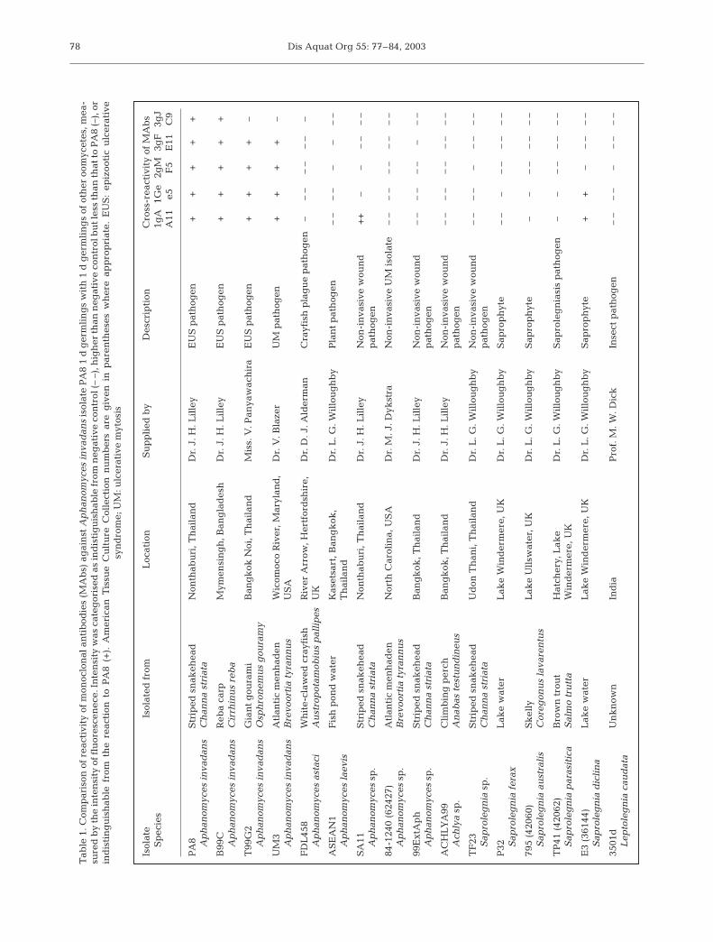
Tab
le 1
. Com
par
ison
of
reac
tivi
ty o
f m
onoc
lon
al a
nti
bod
ies
(MA
bs)
ag
ain
st A
ph
anom
yces
inva
dan
s is
olat
e P
A8
1 d
ger
mli
ng
s w
ith
1 d
ger
mli
ng
sof
oth
er o
omyc
etes
, mea
-su
red
by
the
inte
nsi
ty o
f flu
ores
cen
ece.
Inte
nsi
ty w
as c
ateg
oris
ed a
s in
dis
tig
uis
hab
le fr
om n
egat
ive
con
trol
(– –
), h
igh
er th
an n
egat
ive
con
trol
bu
t les
s th
an th
at to
PA
8 (–
), o
rin
dis
tin
gu
ish
able
fro
m t
he
reac
tion
to
PA
8 (+
). A
mer
ican
Tis
sue
Cu
ltu
re C
olle
ctio
n n
um
ber
s ar
e g
iven
in
par
enth
eses
wh
ere
app
rop
riat
e. E
US
: ep
izoo
tic
ulc
erat
ive
syn
dro
me;
UM
: ulc
erat
ive
myt
osis

Miles et al.: MAbs to Aphanomyces invadans
produced by sporulation, and diluting the pond watercontaining them in an equal volume of glucose-peptone broth media containing 100 mM calcium chlo-ride to stimulate germination (Deacon & Saxena 1998).They were incubated overnight at 24°C and collectedby centrifuging at 3000 × g for 20 min.
The germlings were prepared for experimental useby fixing in formaldehyde as described by Burr (1991),and after a final wash with phosphate buffered salineat pH 7.4 (PBS), the germlings were resuspended inPBS and the concentration of the suspension deter-mined by counting the germlings in a haemocytome-ter.
Soluble antigens were collected after disruption ofthe germlings by shaking with 0.5 mm silicon beads for160 s in a bead shaker (Biospec Products). The concen-tration of the soluble antigen was assessed spectropho-tometrically.
Preparation of monoclonal antibodies: The MAbswere prepared after Adams et al. (1995). Three BALB/cmice received 3 × 100 µl intraperitoneal injections ofsoluble antigen prepared from germlings of Aphano-myces invadans isolate PA8 over a 60 d period. Theantigen was prepared in autoclaved PBS and mixed 1:1with the Titremax gold adjuvant (CytRx Corporation).Mice initially received soluble antigen prepared from asuspension of germlings at 2.8 × 10⁵ ml–1, then 32 dlater they were boosted with soluble antigen obtainedfrom 4.0 × 10⁵ germlings ml–1, and 19 d later with solu-ble antigen obtained from 2.5 × 10⁵ germlings ml–1.The last immunisation was given intravenously 28 dlater, when 100 µl of soluble antigen from 2.5 × 10⁵
germlings ml–1 prepared in PBS was injected into thecaudal vein of the mice. Three days later, the B-cellswere harvested from the spleen of the mice and fusedwith SP2 myeloma cells to make hybridomas.
Hybridomas producing antibodies against Aphano-myces invadans were identified by dot-blot and ELISA(Adams et al. 1995), and positive wells were expandedand recloned 3 times until monoclonal cell lines wereobtained. The isotypes of the MAbs were determinedby ELISA, using isotype-specific secondary antibodies(Sigma Chemical).
Characterisation of MAbs by Western blot: Todetermine the molecular weights of the antigensrecognised by the MAbs, 2 samples of soluble PA8germling extract, and 1 of extracellular products (ECP)from culture, were prepared. All of the protein wasdigested from 1 sample of soluble extract by digestingit with 1 mg ml–1 proteinase K (Sigma) for 60 min at60°C.
The ECP was prepared by filtering a broth culture ofisolate PA8 germlings through grade 541 filter paper(Whatman PLC), then through 0.45 and 0.22 µm sterilefilters (Sartorius). The concentration of the filtrate was
increased by centrifuging at 3000 ×g for 30 min in aVivascience concentrator with a molecular weight cut-off of 5 kDa (Sartorius), which increased the concen-tration of solutes larger than 5 kDa by approximately18 times.
All samples were subjected to electrophoresis, trans-ferred to nitrocellulose paper and analysed by Westernblot after Lilley et al. (1997), with the following modifi-cations: the nitrocellulose membrane was incubated inthe hybridoma supernatant overnight at 22°C, and inthe secondary antibody, 1% v/v biotin-labelled anti-mouse immunoglobulin (Ig) (Diagnostics Scotland)diluted in antibody buffer (1% w/v bovine serum albu-min [Sigma] in PBS) for 90 min at 22°C. This was fol-lowed by a further 90 min incubation, in 1% v/v strep-tavidin peroxidase (Diagnostics Scotland) diluted inantibody buffer. The molecular weight of bands recog-nised by the MAbs were calculated using standardsrun at the same time as the gel.
Staining of fixed germlings by immunofluorescenceantibody technique (IFAT): Initial screening of theMAbs was carried out using IFAT on germlingsobtained from 16 different oomycete isolates affixed tomicroscope slides (Table 1). The intensity and speci-ficity of the staining was recorded. In preliminary stud-ies, antibodies labelled with fluorescin isothiocyanate(FITC) were found to be considerably more effectivefor staining the germlings than those labelled withenzymes.
Microscope slides (Surgipath Europe) were pre-pared for immunoassays by immersing them in 3% v/v3-aminopropyltriethoxysilane (APES) (Sigma) in ace-tone (Fisher Scientific) for 5 min. The slides werewashed in 100% acetone for 5 min followed by a rinsein distilled water for 5 min before air-drying. The slideswere divided into a number of sections with a PAP-pen, and 40 µl suspensions containing 2.5 × 10³
germlings were added to each section. The germlingswere allowed to air-dry onto the slides at 40°C, beforewashing the slides by gentle sluicing with PBS fol-lowed by 3 × 5 min incubations in PBS.
The IFAT was carried out according to Anderson(1990), with modifications: tissue culture supernatantcontaining the MAbs were applied to the samples for4 h at 22°C, after which anti-mouse Ig labelled withFITC (Diagnostics Scotland) was applied at 2% v/v inantibody buffer, and incubated for 2 h at 22°C. Eachslide contained a positive control of germlings of iso-late PA8, and a negative control with germlings of thetest isolate incubated with culture medium rather thantissue culture supernatant. The slides were stored inthe dark at 4°C for no more than 7 d, before viewingunder an Olympus IMT-2 microscope with a reflectedepi-fluorescent attachment and exciter and barrier fil-ters for FITC. The intensity of the staining obtained
79

Dis Aquat Org 55: 77–84, 2003
with the MAbs against the test germlings was com-pared to the controls in order to evaluate each of theMAbs.
Immunohistochemistry by IFAT: After a number ofpreliminary trials, a technique based on a modificationof the method of Lilley et al. (1997) was used to stainAphanomyces invadans in the tissues of EUS-infectedfish. Tissue sections were treated as stated, but the sec-tions were placed onto APES-treated slides. The tissuesections were incubated with neat hybridoma super-natant overnight at 4°C in a humidified chamber andthen with FITC-labelled anti-mouse Ig for 1 h. Thereaction was enhanced with a second cycle (Linsen-mayer et al. 1988), by repeating the application of thegoat serum, MAb supernatant and secondary anti-body, and all wash steps described by Lilley et al.(1997). After a final wash step, 5% v/v methyl green inPBS was added for 5 min to reduce background stain-ing (Lannan et al. 1991). The slides were washed inrunning water for 5 min, mounted with a cover slip andstored in the dark at 4°C for not more than 7 d beforeexamining.
Determining the specificity of the MAbs againstAphanomyces invadans: To confirm the specificity ofthe MAbs, IFAT was performed on tissue sections col-lected from a range of fish tissues infected with variousoomycete and fungal diseases (Table 2). In the case ofthe sections infected with crayfish plague, the effect ofadding a third reaction cycle was also assessed. Anyreaction with hyphae in the sections was recorded.
Comparison of IFAT with GMS: Tissue sampleswere collected from 48 striped snakehead Channa stri-ata that had been experimentally infected with EUS by
immersion challenge with Aphanomyces invadans(Miles 2001). Two sections were cut from each of48 sections and mounted on APES-treated slides. Onesection was stained with GMS, following the protocolof Chinabut & Roberts (1999), and 1 by IFAT. The pres-ence or absence of A. invadans was assessed and com-pared between the 2 sections.
Results and discussion. Five MAbs were identifiedthat reacted with Aphanomyces invadans isolate PA8.All 5 were used in IFAT, and 4 were found to cross-react with several oomycetes other than A. invadans(Table 1). The fifth, an antibody of isotype IgM desig-nated MAb 3gJC9, gave the strongest and cleareststaining of A. invadans germlings and only cross-reacted with 1 other isolate, A. astaci (Table 1). This isa crustacean pathogen, which is taxonomically similar(Hart 1998), but does not infect fish (Lilley & Roberts1997, Oidtmann et al. 1999). The fluorescent stainingwith MAb 3gJC9 was observed over the entire surfaceof all A. invadans germlings stained (Fig. 1), and a sim-ilar reaction was observed with the germlings of A.astaci (Table 1). Since MAb 3gJC9 did not cross-reactwith the germlings screened from other species ofoomycte found in fish, this was the only MAb to beused in the remainder of the study.
In Western blot analysis with MAb 3gJC9, bandswere observed with both undigested germling extractsand ECP, but no reaction was obtained with samples ofgermling extract (isolate PA8) digested with proteinaseK, indicating that the MAb recognised a protein epi-tope present on both Aphanomyces invadans germ-lings and their ECP (Fig 2). A band was recognised at33 kDa in both cases, while further bands were recog-
80
Section reference Host species Pathogen Disease Supplied by
5950020U Striped snakehead Saprolegnia sp. Isolate Injection challenge Dr. J. H. LilleyChanna striata TF29
500-007SKIN Channel catfish Saprolegnia sp. Saprolegniasis Dr. L. Khoo & Dr. A. GrootersIctaurus punctatus
R970036B Atlantic salmon Exophiala sp. Exophialasis Dr. R. CollinsSalmo salar
599-1070-1 Channel catfish Branchiomyces sp. Branchiomycosis Dr. L. Khoo & Dr. A. GrootersIctaurus punctatus
82-1240 Atlantic menhaden Unknown Ulcerative mycosis Dr. E. J. NogaBrevoortia tyrannus
1247/96/II Noble crayfish Aphanomyces astaci Crayfish plague Dr. B. OidtmannAstacus astacus
629/96/c Noble crayfish Aphanomyces astaci Crayfish plague Dr. B. OidtmannAstacus astacus
Table 2. Tissue sections of aquatic organisms with hyphal diseases other than epizootic ulcerative syndrome (EUS) that were used to assess cross-reactivity of monoclonal antibody (MAb) 3gJC9

Miles et al.: MAbs to Aphanomyces invadans
nised at 35 and 38 kDa in the germling extract, and adoublet at 25 kDa and single bands at 23, 24, 46, 72and 102 kDa in the ECP preparation.
When muscle sections of EUS-infected stripedsnakeheads were stained with MAb 3gJC9 in IFAT,strong fluorescence could be seen around the hyphaein infected muscle sections (Fig. 3). Most of the fluores-cence was concentrated in a rim around the hyphae, al-though the surrounding tissue also fluoresced, particu-larly where it was heavily necrotic. Well-developedgranulomata appeared to concentrate the fluorescencemuch closer to the hyphae. No fluorescence was ob-served in undamaged muscle tissue in unin-fected fish, or away from the locus of infec-tion in infected fish (data not shown).Sections treated with hybridoma culturemedium as a negative control had lower lev-els of background fluorescence compared totissue sections infected with the pathogen,although a weak non-specific reaction wasobserved around the cell walls of the hy-phae. This was much weaker and easily dis-tinguishable from sections stained withMAb 3gJC9, and was not strong enough tophotograph clearly, so no figure is includedhere.
The antigen recognised by MAb 3gJC9was located in necrotic and inflamed tissue surrounding Aphanomyces invadanshyphae, suggesting the tissue damage seenin the early stages of A. invadans infectionis due to ECPs secreted by the hyphae.Where granulomatas had formed, the ECPantigen was confined to a small area imme-diately surrounding the hyphae, which
implies that a major function of the granulomatousreaction characteristic of EUS may be to prevent thespread of ECPs.
At the periphery of the infected site, pinpoint areas offluorescence were observed around the hyphae (Fig. 3),often extending into parts of the sections that still con-tained intact muscle fibres. In some cases, developedhyphae were absent or only found in the dermis, andonly the sources of pinpoint fluorescence appeared inthe musculature (Fig. 4). At higher magnifications, thepinpoints appeared filamentous, but no clear pho-tographs were obtained at magnifications above ×40.The shape and immunogenicity of these, and the factthat they were nearly always found near more devel-oped hyphae, suggests that they may be a hitherto un-reported part of the Aphanomyces invadans mycelium.They were not stained by GMS, which is possibly why
81
Fig. 1. Aphanomyces invadans isolate PA8 1 d germlingsstained by immunofluorescence antibody technique (IFAT)with monoclonal antibody (Mab) 3gJC9. Photographed at
1000× magnification; scale bar indicates 10 µm
Fig. 2. Response mono-clonal antibodies (MAbs)3gJC9 in Western blots tolanes: (a) 1 d germling ex-tract; (b) 1 d germling ex-tract digested with pro-teinase K; and (c) extra-cellular products from 1 d
germlings
Fig. 3. Aphanomyces invadans in striped snakehead Channa striata muscletissue stained with 2-cycle immunofluorescence antibody technique (IFAT)with monoclonal antibody (MAb) 3gJC9, showing developed hyphae (H)surrounded by myonecrosis (N) and pinpoint sources of fluorescence (P).
Photographed at 100× magnification; scale bar indicates 10 µm

Dis Aquat Org 55: 77–84, 2003
they have not been reported before, and they werenearly always found in apparently undamaged tissue,or necrotic tissue that had probably only recently be-come infected. In some cases, they were the onlysources of fluorescence that had penetrated beyond thedermis, so they may be involved in the earliest stages ofinfection. If they are indeed an early stage of themycelium, they may produce very little muco-polysac-charide which would be stained by GMS, but be rich inthe ECP that contains the antigen that 3gJC9 recog-nises. They were never found in sections where thehyphae were enclosed by granulomata, which furthersupports such a possibility. However, the purpose of
this study was not to investigate the natureof these filaments, and further investigationis necessary before any firm conclusionscan be drawn as to their form and function.
Many pathogenic oomycetes use en-zymes expressed extracellularly to invadetheir hosts, including Aphanomyces astaci,which uses them to penetrate the chitinouscuticle of crayfish (Söderhäll & Unestam1975). The pathology of A. invadans infec-tions often involves considerable tissuedamage some distance away from the hy-phae (Wada et al. 1994), implying that it se-cretes ECPs that are important to the pro-cess of infection. The nature of the ECPsfrom A. invadans remains obscure, al-though the haemagglutinating and hae-molytic properties associated with themycelium (Kurata et al. 2000) are likely tobe caused by enzymes expressed extracel-lularly.
Of the 48 GMS-stained muscle sectionssampled from striped snakeheads chal-lenged with EUS, invasive hyphae couldbe seen in 23 of them, and were apparentlyabsent in the other 25. Of all the samples,those which stained positive with GMSalso appeared positive with IFAT. In addi-tion, 2 sections where no hyphae had beenobserved following GMS were identifiedas positive by IFAT. This was because ei-ther the hyphae were confined to the der-mis and therefore did not appear invasive,or the hyphae simply were not visible withthe GMS stain. There were a further3 cases in which hyphae could not be seen,but the filamentous structures describedabove were present. Equivalent structureswere not observed in sections stained withGMS (Fig. 4). These results show that theimmunohistochemical stain was as sensi-tive as GMS in detecting invasive
Aphanomyces invadans hyphae, but was also able todetect A. invadans in cases where their presence couldnot be established by GMS.
In sections of fish infected with Saprolegnia spp.,Exophiala sp. and Branchimyces sp., the fluorescenceof the hyphae was no stronger than the background.However, Section 82-1240 from an Atlantic menhadenBrevoortia tyrannus infected with ulcerative mycosisshowed strong fluorescence of the hyphae in a manneridentical to the sections of EUS-infected fish, support-ing the conclusion of Blazer et al. (1999, 2002) andKiryu et al. (2002) that the large-scale fish mortalitiesthat have been reported from the SE USA since 1984
82
Fig. 4. Muscle section of striped snakehead Channa striata sampled 11 d after immersion challenge with Aphanomyces invadans and stained with (a) Grocott’s (1955) methenamine silver stain (GMS) and (b) 2-cycle im-munofluorescence antibody technique (IFAT) using monoclonal antibody(MAb) 3gJC9. Note the hyphae in the dermis (H) and pinpoint sources of fluorescence (P) that are only visible in the section stained by IFAT.
Photographed at 40× magnification; scale bars indicate 50 µm

Miles et al.: MAbs to Aphanomyces invadans
(Noga & Dykstra 1986), dubbed ulcerative mycosis, arein fact caused by Aphanomyces invadans.
Sections of crayfish infected with Aphanomycesastaci did not induce as strong a reaction as sections ofEUS-infected fish, but the hyphae fluoresced stronglyafter a third reaction cycle. Most IFAT and immunohis-tochemistry protocols used in fish diagnostics do notinvolve multiple reaction cycling (Adams et al. 1995),but the use of 2 cycles is not unprecedented, as Lin-senmayer et al. (1988) reported the use of up to 4 cycleswith some MAbs. In the present study, 3 cycles werenecessary to elicit clear and repeatable fluorescencefrom A. astaci in crayfish muscle, although A. astacigermlings appeared to elicit a response as strong asthat for A. invadans. The fact that the 2 species aregenetically more similar to each other than to otherAphanomyces spp. (Hart 1998), and that they are theonly invasive animal pathogens in the genus, impliesthat the ECP antigen detected by MAb 3gJC9 may beinvolved in the pathogenicity of both species. How-ever, the nature of this antigen was not established inthis study, and further research is necessary to estab-lish whether this is indeed the case.
The principal application of MAb 3gJC9 is likely tobe in the confirmation of the presence of Aphanomycesinvadans in infected fish, which must be carried outhistopathologically in order to confirm the case defini-tion of the disease (Roberts et al. 1994). Although theIFAT procedure described here is more time-consum-ing than GMS, and requires an epifluorescence micro-scope, it is more sensitive and detected A. invadans in5 sections where it was not detected by GMS. In par-ticular, the use of IFAT to stain A. invadans hyphae inthe dermis of fish may enable detection of early stagesof the infection. The same hyphae in a section stainedwith GMS would not be identified as A. invadans, asthey do not appear to be invasive and so would be con-sidered to be a saprophyte or opportunist.
Acknowledgements. This study was funded by the Depart-ment for International Development Aquaculture ResearchProgramme and Aquaculture Vaccines Ltd, UK. The authorswould like to extend their thanks to the people who providedoomycete isolates and sections for histopathology, who arelisted in Tables 1 & 2. Further assistance was given by Dr. J.D. Albaladejo and Mrs. E. Lumingkit of the Bureau of Fish-eries and Aquatic Resources, Philippines; and Mrs. H.McEwan and Miss F. Muir of the University of Stirling, Scot-land, UK.
LITERATURE CITED
ACIAR (Australian Centre for International Agriculture)(1998) Reducing fish losses to epizootic ulcerative syn-drome — an ex ante evaluation. Centre for InternationalEconomics, Canberra
Adams A, Thompson KD, Morris D, Farias C, Chen SC (1995)Development and use of monoclonal antibody probes forimmunohistochemistry, ELISA and IFAT to detect bacter-ial and parasitic fish pathogens. Fish Shellfish Immunol 5:537–547
Anderson DP (1990) Fluorescent antibody test. In: Stolen JS,Fletcher TC, Anderson DP, Robertson BS, van MuiswinkelWB (eds) Techniques in fish immunology, Vol I. SOS Pub-lications, Fair Haven, NJ, p 1–8
Beakes GW, Burr AW, Wood SE, Hardham AR (1995) Theapplication of spore surface features in defining taxo-nomic versus ecological groupings in oomycete fungi. CanJ Bot 73:S701–711
Blazer VS, Vogelbein WK, Densmore CL, May EB, Lilley JH,Zwerner DE (1999) Aphanomyces as a cause of ulcerativeskin lesions of menhaden from Chesapeake Bay tribu-taries. J Aquat Anim Health 11:340–349
Blazer VS, Lilley JH, Schill WB, Kiryu Y, Densmore CL,Panyawachira V, Chinabut S (2002) Aphanomycesinvadans in Atlantic menhaden from Chesapeake Baytributaries. J Aquat Anim Health 14:1–10
Boonyaratpalin S (1989) Bacterial pathogens involved in theepizootic ulcerative syndrome of fish in southeast Asia.J Aquat Anim Health 1:272–276
Bullis RA, Noga EJ, Levy MG (1990) Immunological relation-ship of the fish-pathogenic oomycete Saprolegnia parasit-ica to other oomycetes and unrelated fungi. J Aquat AnimHealth 2:223–227
Burr AW (1991) Comparative diplanetic processes of sal-monid-pathogenic and saprophytic isolates of the Sapro-legnia parasitica-diclina complex. PhD thesis, Universityof Newcastle-upon-Tyne
Chinabut S (1998) Epizootic ulcerative syndrome: informationup to 1997. Fish Pathol 33:321–326
Chinabut S, Roberts RJ (1999) Pathology and histopathologyof epizootic ulcerative syndrome (EUS). Aquatic AnimalHealth Research Institute, Bangkok
Cruz-Lacierda ER, Shariff M (1994) The haematologicalchanges in snakehead (Ophiocephalus striatus) affectedby epizootic ulcerative syndrome. In: Chou LM, MunroAD, Lam TJ, Chen TW and 7 others (eds) Proc 3rd Asian Fisheries Forum. Asian Fisheries Society, Manila,p 361–364
Deacon JW, Saxena G (1998) Germination triggers ofzoospore cysts of Aphanomyces eutiches and Phytoph-thora parasitica. Mycol Res 102:33–41
Estrada-Garcia MT, Green JR, Booth JM, White JG, CallowJA (1989) Monoclonal antibodies to cell surface compo-nents of zoospores and cysts of the fungus Pythiumaphanidermatum reveal species-specific antigens. ExpMycol 13:348–355
Grocott RG (1955) A stain for fungi in tissue sections andsmears. Am J Clin Pathol 25:975–979
Hardham AR, Suzaki E, Perkin JL (1985) The detection ofmonoclonal antibodies specific for surface components onzoospores and cysts of Phytophthora cinnamomi. ExpMycol 9:264–268
Hart D (1998) Development of diagnostic systems for thestudy of molecular epidemiology and taxonomy of theAphanomyces fungus associated with EUS of tropical andfreshwater fishes. University of Glasgow VeterinarySchool, Glasgow
Hatai K, Egusa S, Takahashi S, Ooe K (1977) Study on thepathogenic fungus of mycotic granulomatosis. I. Isolationand pathogenicity of fungus from cultured ayu infectedwith the disease. Fish Pathol 12:129–133
Kiryu Y, Shields JD, Vogelbein WK, Zwerner DE, Kator H,
83

Dis Aquat Org 55: 77–84, 2003
Blazer VS (2002) Induction of skin ulcers in Atlantic men-haden by injection and aqueous exposure to the zoosporesof Aphanomyces invadans. J Aquat Anim Health 14:11–24
Kurata O, Kanai H, Hatai K (2000) Hemagglutinating andhaemolytic capacities of Aphanomyces piscicida. FishPathol 35:29–33
Lannan CN, Ewing SA, Fryer JL (1991) A fluorescent anti-body test for detection of the rickettsia causing disease inChilean salmonids. J Aquat Anim Health 3:229–234
Lilley JH, Roberts RJ (1997) Pathogenicity and culture studiescomparing the Aphanomyces involved in epizootic ulcera-tive syndrome (EUS) with other similar fungi. J Fish Dis20:135–144
Lilley JH, Thompson KD, Adams A (1997) Characterization ofAphanomyces invadans by electrophoresis and Westernblot analysis. Dis Aquat Org 30:187–197
Linsenmayer TF, Fitch JM, Schmid TM (1988) Multiple reac-tion cycling: a method of enhancenment of the immuno-chemical signal of monoclonal antibodies. J HistochemCytochem 36:1075–1078
Miles DJC (2001) Studies on host reponses to Aphanomycesinvadans. PhD thesis, University of Stirling
Miles DJC, Polchana J, Lilley JH, Kanchanakhan S, Thomp-son KD, Adams A (2001) Immunostimulation of stripedsnakehead Channa striata against epizootic ulcerative
syndrome. Aquaculture 195:1–19Noga EJ, Dykstra MJ (1986) Ooomycete fungi associated with
ulcerative mycosis in menhaden, Brevoortia tyrannus(Latrobe). J Fish Dis 9:47–53
Oidtmann B, Cerenius L, Schmid I, Hoffmann R, Soderhall K(1999) Crayfish plague epizootics in Germany — classifi-cation of two German isolates of the crayfish plague fungus Aphanomyces astaci by random amplification ofpolymorphic DNA. Dis Aquat Org 35:235–238
Peterson AB, Olson LW, Rosendahl S (1996) Use of polyclonalantibodies to detect oospores of Aphanomyces. Mycol Res100:494–499
Roberts RJ, Campbell B, MacRae IH (1994) Proceedings of theODA regional seminar on epizootic ulcerative syndrome.Aquatic Animal Health Research Institute, Bangkok
Söderhäll K, Unestam T (1975) Properties of extracellular en-zymes from Aphanomyces astaci and their relevance in thepenetration of crayfish cuticle. Physiol Plant 35:140–146
Viswanath TS, Mohan CV, Shankar (1997) Clinical andhistopathological characterization of different types oflesions associated with epizootic ulcerative syndrome(EUS). J Aquac Trop 12:35–42
Wada S, Yuasa K, Rha S, Nakamura K, Hatai K (1994)Histopathology of Aphanomyces infection in dwarfgourami (Colisa lalia). Fish Pathol 29:229–237
84
Editorial responsibility: David Bruno, Aberdeen, Scotland
Submitted: October 3, 2002; Accepted: February 14, 2003Proofs received from author(s): June 2, 2003