Immune Response Carcinoembryonic Antigen Patients...
9
Immune Response to the Carcinoembryonic Antigen in Patients Treated with an Anti-Idiotype Antibody Vaccine Kenneth A. Foon, Mala Chakraborty, William J. John, Amanda Sherratt, Heinz Kohler, and Malaya Bhattacharya-Chatterjee Lucille Parker Markey Cancer Center and the Departments of Medicine and Microbiology and Immunology, University of Kentucky Medical Center, Lexington, Kentucky 40536-0093 Abstract We have generated an IgG1 murine monoclonal anti-idio- type antibody (Ab2) designated 3H1, which mimics a spe- cific epitope on the carcinoembryonic antigen (CEA). Pa- tients with CEA positive tumors are immunologically "tol- erant" to CEA. We used 3H1 as a surrogate for CEA for vaccine therapy of 12 patients with advanced colorectal can- cer. Each of the patients received a minimum of four intra- cutaneous injections of aluminum hydroxide precipitated 3H1 at either 1, 2, or 4 mg dosage per injection. 9 of 12 patients demonstrated anti-anti-idiotypic (Ab3) response to 3H1. All nine patients generated specific anti-CEA anti- body demonstrated by reactivity with radiolabeled purified CEA; some cases were confirmed by immunoprecipitation of purified CEA. We also demonstrated Ab3 stained both autologous and allogeneic colonic tumors. 7 of 12 patients demonstrated idiotype specific T cell proliferative responses and four also showed T cell proliferation to CEA. Toxicity was limited to local reaction with mild fever and chills. All 12 patients eventually progressed after finishing 4-13 dosages. This is the first report demonstrating that a vaccine therapy is capable of breaking "immune tolerance" to CEA in pa- tients with CEA positive tumors. Future studies will focus on treating patients with minimal residual disease. (J. Clin. Invest. 1995. 96:334-342.) Key words: carcinoembryonic antigen (CEA) * anti-idiotype antibody * vaccine therapy. colorectal cancer * tumor immunology Introduction Carcinoembryonic antigen (CEA)' is an 18-kD glycoprotein tumor-associated antigen present on entodermally derived neo- plasms of the gastrointestinal tract as well as other adenocarci- nomas ( 1). CEA is also found in the digestive organs of the human fetus and, thus the name, CEA was derived. Circulating CEA can be detected in the great majority of patients with CEA positive tumors. Specific monoclonal antibodies have been Address correspondence to Kenneth A. Foon, M.D., Lucille Parker Mar- key Cancer Center, 800 Rose Street, Room CC140, Lexington, KY 40536-0093. Phone: 606-257-4500; FAX: 606-323-2074. Receivedfor publication 22 September 1994 and accepted in revised form 14 February 1995. 1. Abbreviation used in this paper: CEA, carcinoembryonic antigen. raised against CEA (2-4) and some have been radiolabeled for diagnostic and clinical studies (5). As with most tumor- associated antigens which are seen as self-antigens by the im- mune system, cancer patients are immunologically "tolerant" to CEA, likely related to its oncofetal origin. However, a limited number of reports from the 1970's suggested that some patients with CEA positive tumors may have minimal humoral and cel- lular immunity to CEA (6-10); these results are controversial. The network hypothesis of Lindemann and Jerne (11, 12) offers an elegant approach to transform epitope structures into idiotypic determinants expressed on the surface of antibodies. According to the network concept, immunization with a given tumor-associated antigen will generate production of antibodies against this tumor-associated antigen, termed Abl; this Abi is then used to generate a series of anti-idiotype antibodies against the Abl, termed Ab2. Some of these Ab2 molecules can effec- tively mimic the three-dimensional structure of the tumor-asso- ciated antigen identified by the Abl. These particular anti-idio- types called Ab2U fit into the paratopes of Abl, and express the internal image of the tumor-associated antigen. The Ab2,/ can induce specific immune responses similar to those induced by the original tumor-associated antigen and can, therefore, be used as surrogate tumor-associated antigens. Immunization with Ab2p can lead to the generation of anti-anti-idiotype antibodies (Ab3) that recognize the corresponding original tumor-associ- ated antigen identified by Abl. Because of this AbI-like reactiv- ity, the Ab3 is also called Ab ' to indicate that it might differ in its other idiotopes from Abl. For several reasons, we consider CEA an excellent tumor- associated antigen for active immunotherapy with anti-idiotype antibody. First of all, CEA is typically present at high levels on the tumor cell surface. CEA is one of the most well-charac- terized antigens, its gene sequence is known and its three dimen- sional structures have been identified (13). CEA is a member of the immunoglobulin supergene family (14) located on chro- mosome 19 which is thought to be involved in cell-cell interac- tions. Since CEA is considered an adhesion molecule (15, 16), it might play an important role in the metastatic process by mediating attachment of tumor cells to normal cells. Thus, ac- tive immunotherapy targeted to CEA might be particularly ben- eficial in preventing metastasis. Highly purified CEA is available from several sources and it can be used conveniently in serologic assays. Inasmuch as some of the epitopes on CEA are shared by normal tissues, immunization with intact CEA molecule might trigger poten- tially harmful autoimmune reactions. Whereas an Ab2P gener- ated against an anti-CEA monoclonal antibody that recognizes a CEA-specific epitope, would be theoretically safer and more effective. Furthermore, Ab2fl expressed in a different molecular environment have been shown to overcome the immunosuppres- sion in the host by stimulating "silent clones," and/or allowing 334 Foon et al. J. Clin. Invest. © The American Society for Clinical Investigation, Inc. 0021-9738/95/07/0334/09 $2.00 Volume 96, July 1995, 334-342
Transcript of Immune Response Carcinoembryonic Antigen Patients...
Immune Response to the Carcinoembryonic Antigen in Patients Treated withan Anti-Idiotype Antibody VaccineKenneth A. Foon, Mala Chakraborty, William J. John, Amanda Sherratt, Heinz Kohler,and Malaya Bhattacharya-ChatterjeeLucille Parker Markey Cancer Center and the Departments of Medicine and Microbiology and Immunology, University of KentuckyMedical Center, Lexington, Kentucky 40536-0093
Abstract
Wehave generated an IgG1 murine monoclonal anti-idio-type antibody (Ab2) designated 3H1, which mimics a spe-cific epitope on the carcinoembryonic antigen (CEA). Pa-tients with CEApositive tumors are immunologically "tol-erant" to CEA. Weused 3H1 as a surrogate for CEA forvaccine therapy of 12 patients with advanced colorectal can-cer. Each of the patients received a minimum of four intra-cutaneous injections of aluminum hydroxide precipitated3H1 at either 1, 2, or 4 mg dosage per injection. 9 of 12patients demonstrated anti-anti-idiotypic (Ab3) responseto 3H1. All nine patients generated specific anti-CEA anti-body demonstrated by reactivity with radiolabeled purifiedCEA; some cases were confirmed by immunoprecipitationof purified CEA. Wealso demonstrated Ab3 stained bothautologous and allogeneic colonic tumors. 7 of 12 patientsdemonstrated idiotype specific T cell proliferative responsesand four also showed T cell proliferation to CEA. Toxicitywas limited to local reaction with mild fever and chills. All 12patients eventually progressed after finishing 4-13 dosages.This is the first report demonstrating that a vaccine therapyis capable of breaking "immune tolerance" to CEA in pa-tients with CEA positive tumors. Future studies will focuson treating patients with minimal residual disease. (J. Clin.Invest. 1995. 96:334-342.) Key words: carcinoembryonicantigen (CEA) * anti-idiotype antibody * vaccine therapy.colorectal cancer * tumor immunology
Introduction
Carcinoembryonic antigen (CEA)' is an 18-kD glycoproteintumor-associated antigen present on entodermally derived neo-plasms of the gastrointestinal tract as well as other adenocarci-nomas ( 1). CEA is also found in the digestive organs of thehuman fetus and, thus the name, CEAwas derived. CirculatingCEA can be detected in the great majority of patients withCEApositive tumors. Specific monoclonal antibodies have been
Address correspondence to Kenneth A. Foon, M.D., Lucille Parker Mar-key Cancer Center, 800 Rose Street, Room CC140, Lexington, KY40536-0093. Phone: 606-257-4500; FAX: 606-323-2074.
Receivedfor publication 22 September 1994 and accepted in revisedform 14 February 1995.
1. Abbreviation used in this paper: CEA, carcinoembryonic antigen.
raised against CEA (2-4) and some have been radiolabeledfor diagnostic and clinical studies (5). As with most tumor-associated antigens which are seen as self-antigens by the im-mune system, cancer patients are immunologically "tolerant"to CEA, likely related to its oncofetal origin. However, a limitednumber of reports from the 1970's suggested that some patientswith CEApositive tumors may have minimal humoral and cel-lular immunity to CEA(6-10); these results are controversial.
The network hypothesis of Lindemann and Jerne (11, 12)offers an elegant approach to transform epitope structures intoidiotypic determinants expressed on the surface of antibodies.According to the network concept, immunization with a giventumor-associated antigen will generate production of antibodiesagainst this tumor-associated antigen, termed Abl; this Abi isthen used to generate a series of anti-idiotype antibodies againstthe Abl, termed Ab2. Some of these Ab2 molecules can effec-tively mimic the three-dimensional structure of the tumor-asso-ciated antigen identified by the Abl. These particular anti-idio-types called Ab2U fit into the paratopes of Abl, and expressthe internal image of the tumor-associated antigen. The Ab2,/can induce specific immune responses similar to those inducedby the original tumor-associated antigen and can, therefore, beused as surrogate tumor-associated antigens. Immunization withAb2p can lead to the generation of anti-anti-idiotype antibodies(Ab3) that recognize the corresponding original tumor-associ-ated antigen identified by Abl. Because of this AbI-like reactiv-ity, the Ab3 is also called Ab ' to indicate that it might differin its other idiotopes from Abl.
For several reasons, we consider CEAan excellent tumor-associated antigen for active immunotherapy with anti-idiotypeantibody. First of all, CEA is typically present at high levelson the tumor cell surface. CEA is one of the most well-charac-terized antigens, its gene sequence is known and its three dimen-sional structures have been identified (13). CEAis a memberof the immunoglobulin supergene family (14) located on chro-mosome 19 which is thought to be involved in cell-cell interac-tions. Since CEA is considered an adhesion molecule (15, 16),it might play an important role in the metastatic process bymediating attachment of tumor cells to normal cells. Thus, ac-tive immunotherapy targeted to CEAmight be particularly ben-eficial in preventing metastasis.
Highly purified CEA is available from several sources andit can be used conveniently in serologic assays. Inasmuch assome of the epitopes on CEA are shared by normal tissues,immunization with intact CEA molecule might trigger poten-tially harmful autoimmune reactions. Whereas an Ab2P gener-ated against an anti-CEA monoclonal antibody that recognizesa CEA-specific epitope, would be theoretically safer and moreeffective. Furthermore, Ab2fl expressed in a different molecularenvironment have been shown to overcome the immunosuppres-sion in the host by stimulating "silent clones," and/or allowing
334 Foon et al.
J. Clin. Invest.© The American Society for Clinical Investigation, Inc.0021-9738/95/07/0334/09 $2.00Volume 96, July 1995, 334-342
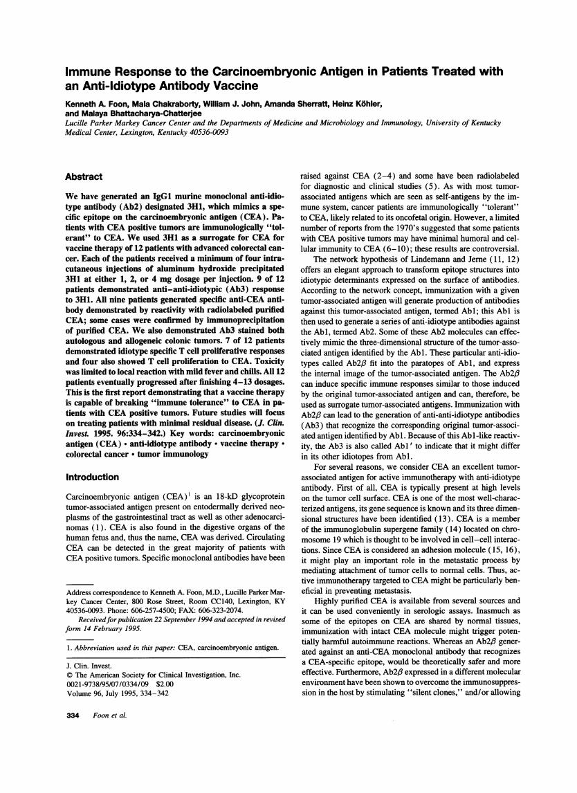
Table L Patient Characteristics
Patient Dosage No. Metastatic Baseline Hurnoral Date offNo. Age/sex (mg) doses disease CEAlevel response Cellular study Why off
1 72/M 4 7 lung 160 + + 3/10/94 progression2 43/F 2 4 liver 110 + + 12/9/93 progression3 46/F 1 4 lung, liver 140 + + 12/2/93 progression4 61/F 2 4 lung, ileum 60 - - 12/11/93 progression5 60/M 1 7 lung, liver 3 + - 5/2/94 progression6 68/M 4 8 lung, liver 81 + - 5/16/94 progression7 47/M 2 4 liver 15 + + 3/17/94 progression8 80/F 1 4 liver 42 - - 3/17/94 progression9 51/M 4 4 liver 210 + + 4/794 progression
10 36/M 1 8 pelvis 1 - - 6/28/94 progression11 70/M 4 13 lung 58 + + 2/20/95 progression12 53/F 2 5 lung, liver 35 + + 6/9/94 progression
T cell help to become active, making the overall immune re-sponse stronger which the nominal antigen (e.g., CEA) is un-able to do (17, 18). Therefore, an appropriate anti-idiotypeantibody would be an excellent candidate to induce anti-tumorimmunity in CEApositive cancer patients.
A number of investigators have generated anti-idiotype anti-bodies in rats, mice, baboons and humans that mimic CEA(19-27). Wehave generated and characterized an anti-idiotypemurine monoclonal antibody to a murine monoclonal antibodydesignated 8019 that identifies a specific epitope on CEA(28).This is a highly restricted CEA epitope that is not found onnormal adult tissues and hematopoietic cells including granulo-cytes. The IgGl anti-idiotype antibody generated to 8019 wasshown to be an internal image by generating anti-anti-idiotypic(Ab3) responses in mice, rabbits (29), and monkeys (30)which recognized CEA. This anti-idiotype antibody was usedto treat the patients reported in this clinical trial.
Methods
Selection of patients. All of the patients had CEA positive advancedcolorectal carcinoma and failed standard therapies (Table I). Baselinestudies included complete physical examination, chest radiography,computer axial tomography examination of the abdomen, serum CEAlevel, routine blood counts, and chemistries. All of the patients had beenoff prior therapy for at least four weeks and staging was repeated at theconclusion of therapy.
Treatment schedule. The patients were treated intracutaneously witheither 1, 2, or 4 mg of aluminum hydroxide precipitated anti-idiotypeantibody every other week for four injections. If the patients were stableat the end of the four injections, they were then continued with injectionson a monthly basis and evaluated every 3 mo. Patients were removedfrom study if they demonstrated growth of their tumor.
Generation of anti-idiotype antibody for the clinical trial. Murinemonoclonal antibody 8019 was used to immunize syngeneic BALB/cmice for the production of anti-idiotype antibody. Immunization ofBALB/c mice, hybridoma fusion and cloning, selection of anti-idiotype(Ab2), and production of ascites in bulk quantities in mice were doneas previously described (31, 32). The Ab2 anti-idiotype 3H1 (IgGl)was purified from ascites by affinity chromatography on protein A-CL Sepharose 4B column. The purity of the isolated immunoglobulin(> 95%) was determined by sodium dodecyl sulfate polyacrylamide gelelectrophoresis (SDS-PAGE) and high pressure liquid chromatographytechniques. Sterility, pyrogenicity, polynucleotides, mycoplasma, and
adventitious virus contamination and retrovirus removal validation testswere done in accordance with the United States Food and Drug Adminis-tration guidelines.
Preparation of final product. To augment the immunogenicity ofanti-idiotype vaccine an adjuvant is typically required. Aluminum hy-droxide has been approved by the United States Food and Drug Adminis-tration for use as an adjuvant in humans. For this clinical trial, anti-idiotype 3H1 was precipitated with aluminum hydroxide. Briefly, 1 mlof 2% Alu-Gel S (Serva Fine Biochem, Inc., Garden City, NY) wasadded to 5-mg aliquots of purified monoclonal anti-idiotype antibody.The volume was then adjusted to 10 ml with D-PBS and the mixtureincubated on a vortex for 1 h at room temperature. The mixture wasthen centrifuged at 2,000 rpm at 250C for 10 min. The amount ofantibody bound in the gel layer was determined by measuring spectro-photometrically the amount of unbound antibody in the supernatant.The Alu-Gel precipitated antibody was stored at 40C until use. Theseprocedures were performed aseptically in a laminar flow hood and thefinal product was sterile and clearly labeled as anti-idiotype 3H1 Alu-Gel and aliquoted into pyrogen-free, sterile glass vials.
The final product was tested for sterility, pyrogenicity and generalsafety in guinea pigs before use. An Investigational NewDrug Applica-tion was approved through the United States Food and Drug Administra-tion (BB-IND 5055).
Assays for humoral immunity. The development of humoral immu-nity induced by immunization with Alu-Gel-precipitated Ab2 was as-sessed by testing sera obtained from patients at different time points. Thesera was initially tested for total human anti-murine-antibody responsesincluding anti-iso/allo/and anti-anti-idiotype antibodies by sandwichradioimmunoassay (33). Briefly, microtiter plates were coated with 3H1and incubated with different dilutions of patients' sera. After washing,the antigen-antibody reaction was tagged using "2I-labeled anti-Id 3H1in a homogeneous sandwich radioimmunoassay. Since 3H1 is injected asintact IgGl, patients are expected to mount human anti-mouse antibodyresponses.
Specific Ab3 response to Ab2. Sera from immunized patients withpositive human anti-mouse antibody responses were tested for the pres-ence of anti-anti-idiotypic antibodies. Sera were pre-incubated with nor-mal murine immunoglobulin to block human antibodies against isotypicand allotypic determinants and then checked for the presence of anti-anti-idiotype (Ab3) by reaction with the immunizing anti-idiotype(3H1) coated onto microtiter plates. Unrelated Ab2 was used as control.After washing, the antigen-antibody reaction was tagged using '25I-labeled anti-idiotype reagent in a homogeneous sandwich radioimmuno-assay as above. Pretreatment, non-immune sera and sera from normaldonors were used as controls in these assays.
Inhibition of the binding between Abl and Ab2 by patients' Ab3
Immune Response to CEA in Patients Treated with an Anti-ldiotype Vaccine 335
antibodies by radioimmunoassay. Pre-immune and hyperimmune patientsera samples were treated with unrelated murine immunoglobulins toremove anti-idiotypic and allotypic reactivities. Serial dilution's of serawere then tested for inhibition in the Abl-Ab2 binding assay. All assayswere performed in triplicate. For direct binding inhibition assay betweenAbI and Ab2, purified Ab2 3H1 was used to coat plates (500 ng/well)and the binding of radiolabeled 8019 (Abl) to Ab2 was tested forinhibition in the presence of different patients' hyperimmune Ab3 seraand Abl. This demonstrated whether Ab3 in patients' sera shared idi-otopes with 8019 (Abl). Also, this inhibition assay between Abl-Ab2binding by Ab3 sera indicated whether Ab3 is a true anti-anti-idiotype.
Detection of anti-CEA antibodies in patients immunized with Ab23HI. This assay was conducted to determine whether some of the Ab3induced in patients by monoclonal murine Ab2 were of the AbI typeand will bind to CEA. Purified CEA was radioiodinated with 1125 bythe Chloramine T method. Radiolabeled CEA (1 X 10⁶ cpm) was re-acted with 0.5 ml of patient's serum pre-adsorbed on protein G-Sepha-rose beads. After reactions, the beads were washed and counted ina gamma-ray spectrophotometer. Pre-immune sera, phosphate-bufferedsaline-bovine serum albumin as well as Ab3 sera obtained from apatient treated with an unrelated murine monoclonal antibody for T celllymphoma were used as controls in these assays.
Purified CEA. Purified CEAwas obtained commercially from Rou-gier Biotech, Montreal, Canada (cat. No. 70015). CEA was isolatedfrom human liver metastasis of colonic tumors by perchloric acid extrac-tion and purified twice by ion-exchange chromatography followed bygel filtration and several steps of HPLC chromatography. The CEAis100% pure, produced a single band at 18 kD by high power liquidchromatography and SDS-PAGEand was immunoprecipitated as a sin-gle band by horse as well as rabbit anti-CEA antibody. The CEAprepa-ration was resolved into two closely migrating bands at 18 and 20 kDby Western blot analysis using murine monoclonal anti-CEA antibody.Werechecked the material by Western blot analysis using monoclonalantibody 8019.
Flow cytometry analysis with Abi and patient's Ab3. CEA-positivecolorectal cancer derived LS174-T cells (1 X 10⁶ per well) and CEA-negative B cell lymphoma, Raji cells (1 X 10⁶ per well) were reactedwith Abl (8019) and patient's immune sera (Ab3) at 1:100 dilution at4°C for 60 min. After washing, the cells were incubated with eithergoat anti-human or goat anti-mouse F(ab' )2 IgG-FITC labeled antibody(Tago) for 30 min at 4°C. They were then washed twice, fixed in 2%paraformaldehyde and analyzed by flow cytometry (FACS®&Star, BectonDickinson).
Purification of anti-anti-idiotypic antibody (Ab3)from hyperimmu-nized patients' sera. 50 ml of hyperimmune serum were passed overan immunoadsorbent column consisting of immunizing anti-idiotypeimmunoglobulin (3H1) coupled to Sepharose 4B. Anti-anti-idiotypicantibodies (Ab3 ) bound to the column were eluted with 0.1 Mglycine-hydrochloric acid buffer (pH 2.4). The eluted antibody was neutralizedwith 3M Tris, dialyzed against PBS, pH 7.2, and then passed over animmunoadsorbent column consisting of allotype matched normal mouseimmunoglobulin coupled to Sepharose 4B to remove anti-isotypic andanti-allotypic reactivities. Antibody that passed through was concen-trated and used as purified Ab3. The isotype of Ab3 was determined byELISA using human anti-isotype specific reagents (Tago).
Epitope analysis of Ab3 by radioimmunoassay inhibition assay. Todetermine whether Ab3 sera compete with Abl for binding to humancolon carcinoma cells, the binding of radioiodinated 8019 to confluentmonolayers of LS174T cells was tested for inhibition in the presencepurified Ab3 and AbI preparations.
Immunoprecipitation of CEA by Abi and Ab3. Purified CEA waslabeled with "2I by The Chloramine T-method and reacted with purifiedAb3 ( 10 gg) or AbI ( 10 jg) or unrelated control Ab3 from lymphomapatient (10 jig) or PBS-BSA control, previously adsorbed on to proteinG-Sepharose beads. After washings, The antigen-antibody coated beadswere analyzed by SDS-PAGEaccording to the method of Laemmli (34)and radioautographed.
Immunoperoxidase staining of tumor sections with Abl and Ab3.
The reactivities of monoclonal Abl and purified Ab3 at 10 Jig/ml solu-tion were compared on surgical specimens of colonic adenocarcinomasby a very sensitive staining method (biotin-Streptavidin reagents; Vec-tor, Burlingame, CA) as described in detail elsewhere (27). All sectionswere counterstained with Meyer's hematoxylin. Pertinent specificitytests were performed, including block of the endogenous peroxidase,omission of the first layer, or substitution of nonimmune homologousserum for the specific antiserum and P3-653 myeloma culture superna-tant as the control.
Assay for T cell proliferative response. Peripheral blood mononu-clear cells were isolated by standard Ficoll-Hypaque density gradientcentrifugation method and S x 105 cells per well were incubated withdifferent concentrations of 3HI-Alu-Gel and control 4DC6-Alu-Gel ( 10ng to 2 jig) in RPMI medium with 5%heat-inactivated fetal calf serumand penicillin and streptomycin. The nonspecific mitogen phytohemag-glutinin-P was used as a positive control at 2 and 1 jg per well. Afterthe cells were incubated for 5 d at 37TC in an atmosphere containing5% carbon dioxide, they were pulsed with [3H]thymidine (1 JiLCi perwell) for 20 h. Data were expressed as mean counts (triplicate wells)per minute of [3H]thymidine incorporation. The Standard Deviation ofthe data was < 10% for each determination.
Peripheral blood mononuclear cells isolated from some selected pa-tients were also incubated with different concentrations of purified CEA(10-250 ng) as per protocol above.
Assay for circulating CEA in serum. CEA was quantified in heat-extracted serum. For this, lml of 0.2 msodium acetate buffer, pH 5.0,was added to 0.5 ml of serum, vortex-mixed, incubated for 15 min at90TC, and centrifuged ( 1,200 g, 10 min). The supernatants were assayedthe same day or stored frozen at -20°C until assay. 100 ml of super-natant was then assayed by the enzyme immunoassay for CEA as de-scribed (35).
Results
Humoral responses to anti-idiotype. The development of hu-moral immunity induced by immunization with Alu-Gel-precip-itated Ab2, 3H1 was assessed by testing sera obtained frompatients before therapy and after each treatment with the vac-cine. Hyperimmune sera (after the fourth injection of 3H1)from nine of twelve patients showed significant levels of totalhuman anti-mouse antibody responses including anti-iso/allo/and anti-anti-idiotypic antibodies against immunizing Ab2,3H1, as determined by homogeneous sandwich radioimmunoas-say (data not shown). Next the sera from these immunizedpatients were checked for their ability to inhibit the binding of
'25I-labeled Abl, 8019 to Ab2 3H1 on the plate by radioimmu-noassay or vice versa (inhibition of radiolabeled Ab2 bindingto AbI on the plate). These reactions were done in the presenceof excess normal murine immunoglobulin to block human anti-bodies against isotypic and allotypic determinants. Fig. 1 dem-onstrates representative data from the first five patients. Serafrom patients 1, 2, 3, and 5, at 1 / 10 dilution, inhibited bindingof iodinated 8019 to 3H1 by 62- 100% and inhibition ofbinding decreased with increasing dilution of the sera. Serafrom patient 4 showed minimal nonspecific inhibition at alldilutions used and pre-immune sera showed no inhibition. Al-though steric hindrance by Ab3 binding can not be excluded inthese assays, the results suggest the presence of true anti-anti-idiotypic antibodies that share idiotypes with Abl. Again, nineout of twelve patients were positive for Ab3 responses by thisassay.
Induction of anti-CEA antibodies by anti-idiotype 3H1.Next, we investigated whether 3H1 could induce an anti-CEAantibody response in immunized patients. For this, the crude
336 Foon et al.

C0 #
=60 e
C
40
0 1 00 200Dilution
Figure 1. Inhibition of Abl (8019) binding to Ab2 (3H1) on the plateby patients' Ab3 sera by radioimmunoassay. Purified 3H1 was used tocoat the plate (500 ng/well) and the binding of radiolabeled 8019(- 90,000 cpm) to 3H1 was tested for inhibition in the presence ofvarious dilutions of Ab3 sera obtained from patients after the fourthimmunization.
sera obtained from patients after the fourth treatment were testedfor the presence of antibody binding to radiolabeled purifiedCEA. Weroutinely used post fourth immunization because thiswas the number of injections all 12 patients received. For pa-tients who received more than four injections, immune re-sponses remained comparable or continued to increase in titer.A pure preparation of CEA was used to reduce the risk ofobtaining false positive results due to nonspecific binding. Asshown in Fig. 2, immunization with 3H1 induced antibodiesthat bound to radiolabeled CEA. Nine of twelve patients devel-oped anti-CEA antibodies measurable by this assay. Patients 4,8, and 10 were anergic for human anti-mouse antibody re-sponse and did not produce antibodies against CEA, while pa-tients 1, 2, 3, 5, and 12 showed high binding, and patients 6,7, 9, and 11 showed binding greater than the background countobtained with PBS-BSA (Sample 13) or pre-immune sera (datanot shown). Sample 14 was the AbI 8019 antibody used as apositive anti-CEA (8019) control.
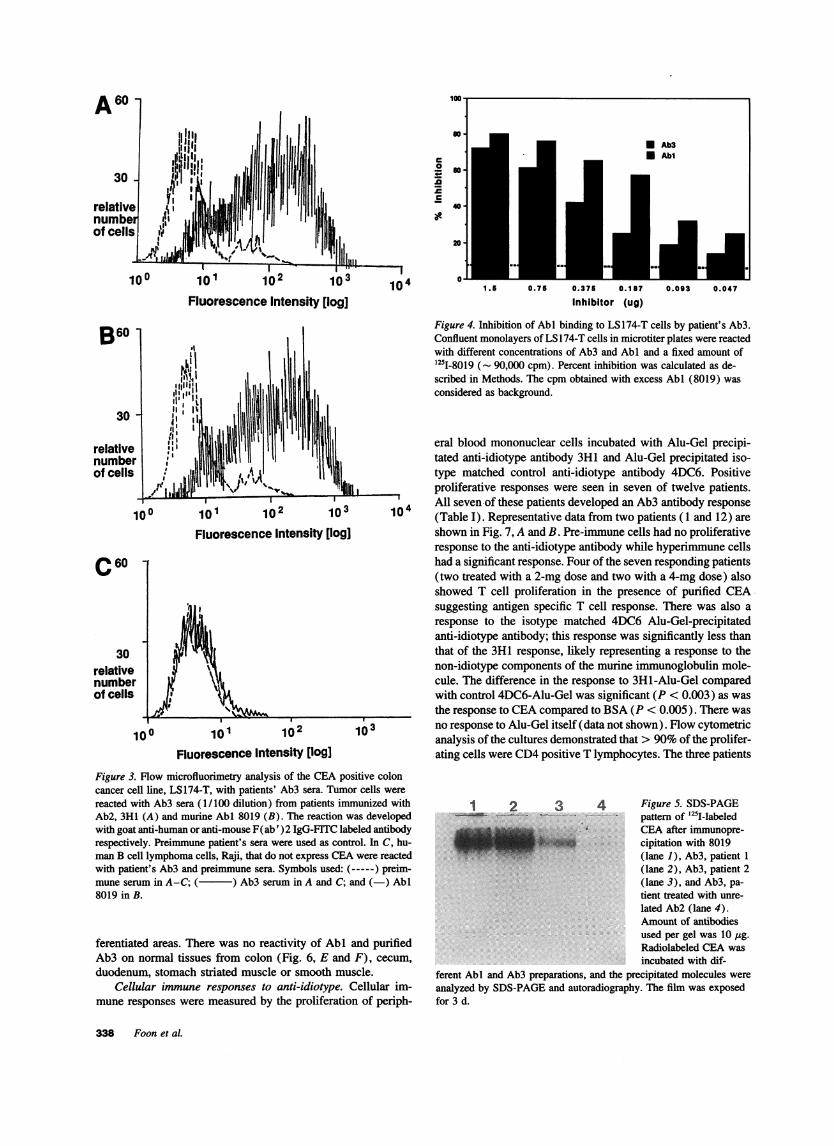
Immuneflow cytometry analysis with Abl and patient's Ab3.To determine the reactivity with cell-surface CEA, culturedCEApositive human colon cancer LS174T cells were tested byimmune flow cytometry. As shown in Fig. 3, crude sera froma representative 3Hl-immunized patient bound to LS174 T cells(A) similar to the binding pattern obtained with 8019 (B) anddid not bind to human B cell lymphoma cells which do notexpress CEA(Fig. 3 C). Similar results were found with all ofthe positive patients.
Competition of Abl and patient's Ab3for binding to LS] 74-T cells. If Ab3 has a similar binding site as Abi, it shouldcompete with AbI for binding to CEA on LS174-T cells. Afixed amount of radiolabeled 8019 was co-incubated with differ-ent concentrations of patientUs purified Ab3 or AbI prepara-tions and LS174-T cells (Fig. 4).
Purified 8019-IgGI (Abi ) inhibited binding by 80% at 0.75.g whereas patient's purified Ab3 (from patient 1) produced60%inhibition at the same concentration. Overall, the inhibition
an
Eoa.
1031 2 3 4 5 6 7 I 9 10 11 12 13 14
Sample Number
Figure 2. Reactivity of patients' Ab3 with purified radiolabeled CEA.Patients' sera (0.5 ml) obtained after the fourth immunization was ad-sorbed on to protein G-Sepharose 4B beads and reacted with (- Ix 106 cpm) radiolabeled CEA. After the reaction, the mixture wascentrifuged, the precipitate was washed and counted in a gamma-counter. Each sample was performed in duplicate and the mean of thecpm bound is shown. Samples 1 through 12 were obtained from patients1 through 12. Sample 13 was PBS-BSA control and 15 was AbI 8019,used at 10 jig concentration. No reactivity (cpm bound) greater thanPBS-BSA was observed with pre-immune sera from these patients.
curves obtained with AbI and Ab3 were very similar at differentdilutions. This indicated that the patient's Ab3 bound to thesame antigenic epitope as Abi and therefore contained antibodymolecules with Abl' properties.
Immunoprecipitation of CEA by AbN and Ab3. It had beenpreviously shown that AbM 8019 specifically immunoprecipi-tated the 18 kD CEAby SDS-PAGEanalysis (29). To confirmthat the Ab3 induced by 3H1 was specific for the CEAmolecule,the iodinated purified CEApreparation was immunoprecipitatedby purified Ab3 preparations obtained from two patients as wellas Abl and analyzed by SDS-PAGE. The results in Fig. 5indicate that both patient's Ab3 (lanes 2 and 3) precipitated thesame 18 kD CEAband as that of murine Abl 8019 (lane 1).There was no cross-reactivity (lane 4) when the iodinated CEAwas reacted with purified Ab3 obtained from a patient treatedwith an unrelated Ab2 (4DC6).. When iodinated CEA, pre-treated with either of the two positive patients' Ab3 prepara-tions, was reacted with 8019, there was no significant immuno-precipitation suggesting that the iodinated preparation was de-pleted of CEA(data not shown).
Immunoreactivity of Abi and patients' Ab3 on colonic tu-mor sections and nonnal tissues. Wecompared the reactivitiesof Abl (8019) with that of patients' purified Ab3 by a sensitiveimmunoperoxidase assay on autologous and allogeneic colontumor specimens surgically removed from patients. The patternof reactivity of patient Ab3 on autologous malignant colon tis-sues was identical to that obtained with allogeneic tumor speci-mens (Fig. 6, A and B, respectively). AbI 8019 showed identi-cal staining patterns (Fig. 6 C), whereas there was no reactivitywith control Ab3 obtained from a patient treated with an unre-lated Ab2 (4DC6) (Fig. 6 D). Reactions with Abl or purifiedAb3 (Fig. 6, A-C) resulted in the staining of both tumor cellsas well as secreted mucinous materials. The staining was apicalin gland-like structures and granular (cytoplasmic) in less dif-
Immune Response to CEA in Patients Treated with an Anti-Idiotype Vaccine 337
c
0
.C
c
Fluorescence Intensity [log]
so
U.
40
* Ab3* Abl
1.5 0.75 0.375 0.187 0.093 0.047
Inhibitor (ug)
Figure 4. Inhibition of AbI binding to LS174-T cells by patient's Ab3.Confluent monolayers of LS 174-T cells in microtiter plates were reactedwith different concentrations of Ab3 and AbN and a fixed amount of'25I-8019 (- 90,000 cpm). Percent inhibition was calculated as de-scribed in Methods. The cpm obtained with excess AbI (8019) wasconsidered as background.
30 -
relativenumberof cells
100
100
101 102 103
Fluorescence Intensity [log]
101 102 103
Fluorescence Intensity [log]
Figure 3. Flow microfluorimetry analysis of the CEApositive coloncancer cell line, LS174-T, with patients' Ab3 sera. Tumor cells werereacted with Ab3 sera (1/100 dilution) from patients immunized withAb2, 3H1 (A) and murine Abl 8019 (B). The reaction was developedwith goat anti-human or anti-mouse F(ab' )2 IgG-FITC labeled antibodyrespectively. Preimmune patient's sera were used as control. In C, hu-man B cell lymphoma cells, Raji, that do not express CEAwere reactedwith patient's Ab3 and preimmune sera. Symbols used: (-----) preim-mune serum in A-C; ( ) Ab3 serum in A and C; and (-) AbI8019 in B.
ferentiated areas. There was no reactivity of Abl and purifiedAb3 on normal tissues from colon (Fig. 6, E and F), cecum,duodenum, stomach striated muscle or smooth muscle.
Cellular immune responses to anti-idiotype. Cellular im-mune responses were measured by the proliferation of periph-
eral blood mononuclear cells incubated with Alu-Gel precipi-tated anti-idiotype antibody 3H1 and Alu-Gel precipitated iso-type matched control anti-idiotype antibody 4DC6. Positiveproliferative responses were seen in seven of twelve patients.
104 All seven of these patients developed an Ab3 antibody response10~ (Table I). Representative data from two patients (1 and 12) are
shown in Fig. 7, A and B. Pre-immune cells had no proliferativeresponse to the anti-idiotype antibody while hyperimmune cellshad a significant response. Four of the seven responding patients(two treated with a 2-mg dose and two with a 4-mg dose) alsoshowed T cell proliferation in the presence of purified CEAsuggesting antigen specific T cell response. There was also aresponse to the isotype matched 4DC6 Alu-Gel-precipitatedanti-idiotype antibody; this response was significantly less thanthat of the 3H1 response, likely representing a response to thenon-idiotype components of the murine immunoglobulin mole-cule. The difference in the response to 3H1-Alu-Gel comparedwith control 4DC6-Alu-Gel was significant (P < 0.003) as wasthe response to CEAcompared to BSA(P < 0.005). There wasno response to Alu-Gel itself (data not shown). Flow cytometricanalysis of the cultures demonstrated that > 90%of the prolifer-ating cells were CD4positive T lymphocytes. The three patients
1 2 3 4 Figure 5. SDS-PAGEpattern of '25I-labeledCEAafter immunopre-cipitation with 8019(lane 1), Ab3, patient 1(lane 2), Ab3, patient 2(lane 3), and Ab3, pa-tient treated with unre-lated Ab2 (lane 4).Amount of antibodiesused per gel was 10 lig.Radiolabeled CEAwasincubated with dif-
ferent AbI and Ab3 preparations, and the precipitated molecules wereanalyzed by SDS-PAGEand autoradiography. The film was exposedfor 3 d.
B60
C60
30relativenumberof cells
338 Foon et al.
104
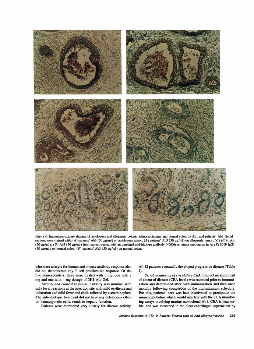
Figure 6. Immunoperoxidase staining of autologous and allogeneic colonic adenocarcinomas and normal colon by Abl and patients' Ab3. Serialsections were stained with; (A) patients' Ab3 (50 .g/ml) on autologous tumor; (B) patients' Ab3 (50 jig/ml) on allogeneic tumor; (C) 8019 IgG,(50 ig/ml); (D) Ab3 (50 jig/ml) from patient treated with an unrelated anti-idiotype antibody (4DC6) on tumor sections as in A; (E) 8019 IgGI(50 ,g/ml) on normal colon; (F) patients' Ab3 (50 jg/ml) on normal colon.
who were anergic for human anti-mouse antibody response alsodid not demonstrate any T cell proliferative response. Of thefive nonresponders, three were treated with 1 mg, one with 2mg and one with 4 mg dosage of 3H1-Alu-Gel.
Toxicity and clinical response. Toxicity was minimal withonly local reactions at the injection site with mild erythema andinduration and mild fever and chills relieved by acetaminophen.The anti-idiotypic treatment did not have any deleterious effecton hematopoietic cells, renal, or hepatic function.
All 12 patients eventually developed progressive disease (TableI).
Serial monitoring of circulating CEA. Indirect measurementof extent of disease (CEA level) was recorded prior to immuni-zation and determined after each immunization and then oncemonthly following completion of the immunization schedule.For this, patients' sera was heat-inactivated to precipitate theimmunoglobulins which would interfere with the CEAmonitor-ing assays involving murine monoclonal Abl. CEAis heat sta-
Patients were monitored very closely for disease activity. ble, and was measured in the clear centrifuged supernatant by
Immune Response to CEA in Patients Treated with an Anti-Idiotype Vaccine 339

A prE post
60000
E dooma.0
20000
0Medium 3H1-AIu-Gel 4DC6-Alu
B 3=0-
mmO- * PaPotd
2nOOO-
E 15000-a.0
10000
0Medium 3H1-Alu-Geil 4Db6-Afu.GeI CEA: BSA
,000
PHA1
Figure 7. T cell proliferation assay with patients' (No. 1, A and No.12, B) peripheral blood mononuclear cells in the presence of 3HI-Alu-Gel, iso-allotype matched control 4DC6-Alu-Gel, purified CEA, purifiedbovine serum albumin (BSA), and phytohemagglutinin. Peripheralblood mononuclear cells were isolated from blood obtained after fourimmunizations and cultured with 100 ng of different antigens and 2 jigof phytohemagglutinin as described in Methods. [3H]Thymidine incor-poration was measured in pre- (solid bars) and post-therapy (hatchedbars) samples. Data are expressed as mean cpm of triplicate wells. TheS.D. of the data was < 10% for each determination.
routine assay. The serial monitoring of CEA correlated withdisease progression and all patients who clinically progressedhad a rise in their serum CEA levels except patients 5 and 10who did not secrete CEA.
Discussion
Wehave demonstrated that nine of twelve patients injected withaluminum hydroxide precipitated anti-idiotype antibody 3H1generated anti-CEA antibody by direct binding to radiolabeledpurified CEA. None of these patients had pre-existing antibodyto CEA. Wealso demonstrated binding to autologous and allo-geneic tumor as well as immunoprecipitation of purified CEAinselected patients. While the three patients who did not generate ahumoral immune response may have been truly anergic, it ispossible that the two who had elevated CEA levels (patients 4and 8) generated small quantities of antibody that was boundto circulating CEA as immune complexes. Indeed, many pa-tients had increasing levels of circulating immune complexesas determined by routine Raji cell assay (data not shown). Also,there is the possibility that some of the circulating anti-CEAantibodies may have bound to patients' tumor cells or were of
low affinity. However, five of the patients still showed highbinding of antibody to radiolabeled CEA, while four othersshowed modest binding. In future studies, we will also stimulatepatients' peripheral blood mononuclear cells in vitro with CEAor Ab2 for the induction of tumor-specific antibody (36).
Seven patients demonstrated idiotype specific T cell prolif-erative responses of primarily CD4 T cells. Four of them alsodemonstrated CEA-specific T cell proliferation in vitro. Wewillalso study these patients' sera for in vitro killing of culturedtumor cells by antibody-dependent cell-mediated cytotoxicity(ADCC) and complement mediated cytotoxicity (CMC)assays. Wedo not have fresh or frozen autologous tumor tissuesfrom these patients to test for in vitro cytotoxic T cell (CTL)induction, but will use HLA-Class 1 matched cultured colontumor cell lines as targets.
To our knowledge, this is the first report in the literaturedescribing the generation of specific and reproducible immunityto CEA. Whether this Ab3 and/or cellular immunity can medi-ate a potential anti-tumor effect remains to be determined. All ofthe patients in this study had prior chemotherapy and advanceddisease; tumor regression was not noted. Future studies willfocus on adjuvant trials where the goal will be elimination ofminimal residual disease.
Another approach to generate active immunity to CEAhasbeen the development of a recombinant vaccinia virus express-ing the human CEAgene (37, 38). This was made possible bythe cloning of the CEA gene (39). These investigators havedemonstrated anti-CEA immune responses in animals but havenot yet reported success in clinical trials. Another novel ap-proach to generate CEA immunity that has not yet entered theclinic, is cDNA immunization by a polynucleotide vaccine(40). Other investigators have generated anti-idiotype antibod-ies that are the internal images of CEA(19-27); clinical resultsare not reported.
It was interesting that our 3H1 anti-idiotype antibody waseffective in eliciting immune responses despite the absence ofa strong adjuvant. Aluminum hydroxide-precipitation, althoughconsidered weakly immunogenic, appeared to be quite adequatein eliciting immune responses. Aggregation of soluble idiotypicdeterminants by aluminum hydroxide precipitation likely helpedto increase its antigenicity. Also, our antibody was a foreignprotein and was injected as an intact immunoglobulin. The Fcportion of the murine immunoglobulin probably served as a"carrier" to help promote the immune responses. It was offurther interest that our anti-idiotype antibody and purified CEAwere able to stimulate an in vitro CD4 T cell proliferativeresponse in treated patients. Webelieve the response observedin some patients against the purified CEAis based on the recog-nition of processed idiotypic peptides which have homology tothe CEAsequence. In preliminary experiments, we have identi-fied a peptide sequence region of CEAwhich has homology toa CDRof the light chain of our 3H1 anti-idiotype vaccine.
A variety of non-CEA anti-idiotype antibodies have beenshown to generate active tumor immunity in animal modelswith both humoral immune responses and delayed hypersensi-tivity (41-49). Perhaps the first suggestion in man that anti-idiotype responses might correlate with clinical responses werein patients treated with a non-CEA AbI antibody for colorectalcancer who developed anti-idiotypic antibodies and improvedclinically (50). Subsequent clinical trials using polyclonal non-CEA goat anti-idiotype (Ab2) vaccines for colorectal cancer(51), and monoclonal anti-idiotypes for malignant melanoma
340 Foon et al.
(52, 53) have demonstrated that anti-idiotype vaccine therapyleads to active immune responses. In a recent study, idiotype-specific immune responses were induced in patients with B celllymphoma against the immunoglobulin idiotype expressed bytheir tumors (54). This latter study differed from ours in thatthe idiotype immunogen was derived from the patients' tumors,coupled to keyhole limpet hemocyanin and mixed with a potentadjuvant. Therefore, while we used a single anti-idiotype anti-body as an antigen surrogate to treat all of our patients, theseinvestigators generated an individualized vaccine for each oftheir patients.
In summary, we have demonstrated specific active immunityto CEAin patients with advanced colorectal cancer treated withan anti-idiotype antibody that "mimics" CEA. In this Phaselb clinical trial, we could only accrue patients who failed con-ventional therapy. All of them had widespread advanced dis-ease. The main purpose of this clinical trial was not to assesstumor response, but to determine the host's immunological re-sponse to the vaccine therapy. Some primary questions havebeen resolved. This anti-idiotype antibody can evoke an Ab3as well as cellular immune response in patients, and any Ab3so derived, behaves as an Abi-like antibody (AbI ' ). The inten-sity of the Ab3 response appeared to correlate positively withanti-CEA antibody (Abl') and T cell proliferative responses.Immune responses appeared independent of the level of circulat-ing CEA. While there are too few patients to compare the 1mg, 2 and 4 mg doses; it is clear that patients were able togenerate immunity at each of these doses. At the completion ofthis study we will statistically compare the different dose levels.Toxicity was restricted to local cutaneous reactions lasting 24-48 h with mild fever and chills and was relieved by acetamino-phen.
Collectively, the immune responses in patients treated withan idiotype vaccine, which induced humoral and cellular re-sponses against an otherwise non-immunogenic tumor antigen,justify follow-up clinical studies in patients with minimal tumorburden, as well as basic immunobiological studies to understandthe mechanisms of the T cell response at the clonal level. Suchstudies may lead to the development of second generation idio-type vaccines consisting of cytokine-antibody fusion proteins(55) and of idiotype derived peptide vaccines (56).
Acknowledgments
This work was supported by National Institutes of Health grant P01-CA 57165.
References
1. Gold, P., and S. 0. Freedman. 1965. Demonstration of tumor specificantigens in human colonic carcinomata by immunological tolerance and absorp-tion techniques. J. Exp. MedL 121:439-462.
2. Primus, F. J., K. D. Newell, A. Blue, and D. M. Goldenberg. 1983. Immuno-logical heterogeneity of carcinoembryonic antigen distinguished by monoclonalantibodies. Cancer Res. 43:686-92.
3. Hansen, H. J., D. M. Goldenberg, E. S. Newman, R. Grebenau, and R. M.Sharkey. 1993. Characterization of second-generation monoclonal antibodiesagainst carcinoembryonic antigen. Cancer. 71:3478-85.
4. Kuroki, M., F. Arakawa, M. Haruno, M. Murakami, M. Wakisaka, H.Higuchi, S. Oikawa, H. Nakazato, and Y. Matsuoka. 1992. Biochemical character-ization of 25 distinct carcinoembryonic antigen (CEA) epitopes recognized by57 monoclonal antibodies and categorized into seven groups in terms of domainstructure of the CEAmolecule. Hybirdoma. 11:391-407.
5. Goldenberg, D. M. 1993. Monoclonal antibodies in cancer detection andtherapy. 1993. Am J. MeiL 94:297-312.
6. Pressman, D., C. T. Ming, and A. L. Grossberg. 1979. Carcinoembryonicantigen-binding immunoglobulin isolated from normal human serum by affinitychromatography. JNCI (J. NatL. Cancer Institue). 62:1367-1371.
7. Kapsopoulou-Dominos, K., and F. A. Anderer. 1979. Circulating carci-noembryonic antigen immune complexes in sera of patients with carcinomata ofthe gastrointestinal tract. Clin. Exp. Immunol. 35:190-195.
8. Kapsopoulou-Dominos, K., and F. A. Anderer. 1979. An approach to theroutine estimation of circulating carcinoembryonic antigen immune complexes inpatients with carcinomata of the gastrointestinal tract. Clin. Exp. Immunol. 37:25-32.
9. Lejtenyi, C. M., S. 0. Freedman, and P. Gold. 1971. Response of lympho-cytes from patients with gastrointestinal cancer to the carcinoembryonic antigenof the human digestive system. Cancer. 28:115.
10. Orefice, S., G. Fossati, E. Pietrojusti, and G. Bonfanti. 1982. Delayedcutaneous hypersensitivity reaction to carcinoembryonic antigen in cancer pa-tients. Tumori. 68:473-475.
11. Lindenmann, J. 1973. Speculations on Ids and homobodies. Ann. Immunol.(Paris). 124:171-184.
12. Jerne, N. K. 1974. Towards a network theory of the immune system. Ann.Immunol. (Paris). 125C:373-389.
13. Paxton, R. J., G. Mooser, H. Pande, T. D. Lee, and J. E. Shivley. 1987.Sequence analysis of carcinoembryonic antigen: Identification of glycosylationsites and hormology with the immunoglobulin super-gene family. Proc. Nad.Acad. Sci. USA. 84:920-924.
14. Thompson, J., and W. Zimmerman. 1988. The carcinoembryonic antigengene family: structure, expression, and evolution. Tumour BioL 9:63-83.
15. Benchimol, S., A. Fuks, S. Jothy, N. Beauchemia, K. Shirota, and C.Stanners. 1989. Carcinoembryonic antigen, a human tumor marker functions asan intercellular adhesion molecule. Cell. 57:327-334.
16. Oikawa, S., C. Inuzuka, M. Kuroki, Y. Matsuoka, G. Kosaki, and H.Nakazato. 1989. Cell adhesion activity of non-specific cross-reacting antigen(NCA) and carcinoembryonic antigen (CEA) expressed on CHOcell surface:homophilic and heterophilic adhesion. Biochem. Biophys. Res. Commun. 164:39-45.
17. Cazenave, P. A. 1977. Idiotypic-anti-idiotypic regulation of antibody syn-thesis in rabbits. Proc. Natl. Acad. Sci. USA. 74:5122-5125.
18. Bona, C., E. Herber-Katz, and W. E. Paul. 1981. Idiotype-anti-idiotyperegulation. L. Immunization with a levan-binding myeloma protein leads to theappearance of auto-anti-(anti-idiotype) antibodies and to the activation of silentclones. J. Exp. Med. 153:951-967.
19. Tsujisaki, M., K. Imai, S. Tokuchi, Y. Hanzawa, T. Ishida, H. Kitagawa,Y. Hinoda, and A. Yachi. 1991. Induction of antigen-specific immune responsewith use of anti-idiotypic monoclonal antibodies to anti-carcinoembryonic antigenantibodies. Cancer Res. 51:2599-2604.
20. Tsujisaki, M., Y. Hinoda, S. Tokuchi, Y. Hanzawa, Y. Arimura, J. Masuya,H. Kitagawa, E. Okochi, T. Shimamura, J. Hamuro, K. Imai, and A. Yachi. 1993.The analysis of internal image-bearing anti-idiotypic monoclonal antibody inrelation to carcinoembryonic antigen. J. Immunol. 150:508-516.
21. Durrant, L. G., G. W. L. Denton, E. Jacobs, M. Mee, R. Moss, E. B.Austin, R. W. Baldwin, J. D. Hardcastle, and R. A. Robins. 1992. An idiotypicreplica of carcinoembryonic antigen inducing cellular and humoral responsesdirected against human colorectal tumors. Int. J. Cancer. 50:811-816.
22. Gaida, F-J, U. Fenger, C. Wagener, and M. Neumaier. 1992. A monoclonalanti-idiotypic antibody bearing the image of an epitope specific to the humancarcinoembryonic antigen. Int. J. Cancer. 51:459-465.
23. de Moraes, J. Z., C. R. W. Carneiro, F. Buchegger, J.-P. Mach, and J. D.Lopes. 1992. Induction of an immune response through the idiotypic networkwith monoclonal anti-idiotype antibodies in the carcinoembryonic antigen system.J. Cell. Biochen. 50:324-335.
24. Austin, E. B., R. A. Robins, R. W. Baldwin, and L. G. Durrant. 1991.Introduction of delayed hypersensitivity to human tumor cells with a humanmonoclonal anti-idiotypic antibody. J. Natl. Cancer Inst. 83:1245-1248.
25. Losman, M. J., K. E. Novick, D. M. Goldenberg, and M. Monestier. 1994.Mimicry of a carcinoembryonic antigen epitope by a rat monoclonal anti-idiotypeantibody. Int. J. Cancer. 56:580-584.
26. Irvine, K., and J. Schlom. 1993. Induction of delayed-type hypersensitivityresponses by monoclonal anti-idiotypic antibodies to tumor cells expressing carci-noembryonic antigen and tumor-associated glycoprotein-72. Cancer Immunol.Immnunother. 36:281-292.
27. Losman, M. J., M. Monestier, H. J. Hansen, and D. M. Goldenberg. 1990.Baboon anti-idiotype antibodies mimic a carcinoembryonic antigen epitope. lnt.J. Cancer. 46:310-314.
28. Koprowski H., Z. Steplewski, K. Mitchell, M. Herlyn, D. Herlyn, and P.Fuhrer. 1979. Colorectal carcinoma antigens detected by hybridoma antibodies.Sornat. Cell Genet. 5:957.
29. Bhattacharya-Chatterjee, M., S. Mukerjee, W. Biddle, K. A. Foon, and H.Kohler. 1990. Murine monoclonal anti-idiotype antibody as a potential networkantigen for human carcinoembryonic antigen. J. Immunol. 145:2758-2765.
30. Chakraborty, M., K. A. Foon, H. Kdhler, and M. Bhattcharya-Chatteijee.1994. Murine monoclonal anti-idiotype antibody induces a specific antibody re-
Immune Response to CEA in Patients Treated with an Anti-Idiotype Vaccine 341

sponse to human carcinoembryonic antigen (CEA) in cynomolgus monkeys. FA-SEB (Fed. Am. Soc. Exp. Biol.) J. A504.
31. Bhattacharya-Chatterjee, M., M. W. Pride, B. K. Seon, and H. Kohler.1987. Idiotype vaccines against human T-cell acute lymphoblastic leukemia(TALL). I. Generation and characterizations of biologically active monoclonalanti-idiotypes. J. Immunol. 5:562-573.
32. Bhattacharya-Chatterjee, M., S. K. Chattedjee, S. Vasile, B. K. Seon, andH. Kohler. 1988. Idiotype vaccines against T-cell leukemia. II. Generation andcharacterization of monoclonal idiotype cascade (Abl, Ab2 and Ab3). J. Immu-nol. 141:1398-1403.
33. Khazaeli, M. B., M. N. Saleh, R. H. Wheeler, W. J. Huster, H. Holden,R. Carrano, and A. F. LoBuglio. 1988. Phase I trial of multiple large dosesof murine monoclonal antibody C017-lA. II. Pharmacokinetics and immuneresponse. J. Natl. Cancer Inst. 80:937-942.
34. Laemmli, U.K. 1970. Cleavage of structural proteins during the assemblyof the head of leactoriophage T4. Nature (Lond.). 227:680-685.
35. Hansen, H. J., G. LaFontaine, E. S. Newman, M. K. Schwartz, A. Malkin,K. Mojzisik, E. W. Martin, and D. M. Goldenberg. 1989. Solving the problem ofantibody interference in commercial "sandwich"-type immunoassays of carci-noembryonic antigen. Clin. Chem. 35(1 ):146-151.
36. DeFreifas, Er., H. Suzuki, D. Herlyn, M. Lubeck, H. Sears, M. Herlyn,and H. Koprowski. 1985. Human antibody induction to the idiotypic and anti-idiotypic determinants of a monoclonal antibody against gastrointestinal carci-noma antigens. Curr. Top. Microbiol. Immunol. 119:75-88.
37. Kantor, J., K. Irvin, S. Abram, H. Kaufman, J. DiPietro, and J. Schlom.1992. Antitumor activity and immune responses induced by a recombinant carci-noembryonic antigen-vaccinia virus vaccine. J. Natl. Cancer Inst. 84:1084-1091.
38. Kantor, J., K. Irvine, S. Abrams, P. Snoy, R. Olsen, J. Greiner, H. Kaufman,D. Eggensperger, and J. Schlom. 1992. Immunogenicity and safety of a recombi-nant vaccinia virus vaccine expressing the carcinoembryonic antigen gene in anon-human primate. Cancer Res. 52:6917-6925.
39. Robbins, F. F., J. A. Kantor, M. Salgaller, P. H. Hand, P. D. Fernsten,and J. Schlom. 1991. Transduction and expression of the human CEAgene in amurine colon carcinoma cell line. Cancer Res. 51:3652-3662.
40. Conroy, R. M., A. F. LoBuglio, J. Kantor, J. Schlom, F. Loechel, S. E.Moore, L. A. Sumerel, D. L. Barlow, S. Abrams, and D. T. Curiel. 1994. Immuneresponse to a carcinoembryonic antigen polynucleotide vaccine. Cancer Res.54:1164-1168.
41. Dunn, P. L., C. A. Johnson, J. M. Styles, S. S. Pease, and C. J. Dean.1987. Vaccination with syngeneic monoclonal anti-idiotype protects against atumor challenge. Immunol. 60:181-186.
42. Lee, V. K., T. G. Harriott, V. K. Kuchroo, W. J. Halliday, I. Hellstrom,and K. E. Hellstrom. 1985. Monoclonal anti-idiotope antibodies related to a murineoncofetal bladder tumor antigen induce specific cell-mediated tumor immunity.Proc. Natl. Acad. Sci. USA. 82:6286-6290.
43. Binz, H., B. Meier, and H. Wigzell. 1982. Induction or elimination oftumor-specific immunity against a chemically induced rat tumor using auto-anti-idiotypic antibodies. Int. J. Cancer. 29:417-423.
44. Kennedy, R. C., G. R. Dreesman, J. S. Butel, and R. E. Landford. 1985.Suppression of in vivo tumor formation induced by simian virus 40-transformedcells in mice receiving anti-idiotypic antibodies. J. Exp. Med. 162:1432-1449.
45. Forstrom, J. W., K. A. Nelson, G. T. Nepom, I. Hellstrom, and K. E.Hellstrom. 1983. Immunization to a syngeneic sarcomas by a monoclonal auto-anti-idiotypic antibody. Nature (Lond.). 303:627-629.
46. Nelson, K. A., E. George, C. Swenson, J. W. Forstrom, and K. E. Hell-strom. 1987. Immunotherapy of murine sarcomas with auto-anti-idiotypic mono-clonal antibodies which bind to tumor-specific T cells. J. Immunol. 139:2110-2117.
47. Kaminski, M. S., K. Kitamura, D. G. Maloney, and R. Levy. 1987. Idiotypevaccination against murine B cell lymphoma. Inhibition of tumor immunity byfree idiotype protein. J. Immunol. 138:1289-1296.
48. Raychaudhuri, S., Y. Saeki, J. J. Chen, H. Iribe, H. Fuji, and H. Kohler.1987. Tumor- specific idiotype vaccines. II. Analysis of the tumor-related networkresponse induced by the tumor and by internal image antigens (Ab2,8). J. Immu-noL 139:271-278.
49. Raychaudhuri, S., Y. Saeki, J. J. Chen, H. Iribe, H. Fuji, and H. Kohler.1987. Tumor- specific idiotype vaccines. III. Induction of T helper cells by anti-idiotype and tumor cells. J. Immunol. 139:2096-2102.
50. Koprowski, H., D. Herlyn, M. Lubeck, E. Defreitas, and H. F. Sears.1984. Human anti- idiotype antibodies in cancer patients: is the modulation ofthe immune response beneficial for the patient? Proc. Natl. Acad. Sci. USA.81:216-219.
51. Heryln, D., M. Wettendorff, E. Schmoll, D. Iliopoulos, I. Schedel, U.Dreikhausen, R. Raab, A. H. Ross, H. Jaksche, M. Scriba, and H. Koprowski.1987. Anti-idiotype immunization of cancer patients: Modulation of the immuneresponse. Proc. Nati. Acad. Sci. USA. 84:8055-8059.
52. Mittleman, A., et al. 1990. Active specific immunotherapy in patientswith melanoma. A clinical trial with mouse anti-idiotypic monoclonal antibodieselicited with syngeneic anti-high-molecular-weight-melanoma-associated antigenmonoclonal antibodies. J. Clin. Invest. 86:2136-2144.
53. Mittleman, A., et al. 1992. Human high molecular weight melanoma-associated antigen (HMW-MAA)mimicry by mouse anti-idiotypic monoclonalantibody MK2-23: induction of humoral anti-HMW-MAA immunity and pro-longation of survival in patients with stage IV melanoma. Proc. Natl. Acad. Sci.USA. 89:466-470.
54. Kwak, L., M. Campbell, B. Czerwinski, S. Hart, R. Miller, and R. Levy.1992. Induction of immune responses in patients with B cell lymphoma againstthe surface-immunoglobulin idiotype expressed by their tumors. N. Engl. J. Med.327:1209-1215.
55. Tao, M-H., and R. Levy. 1993. Idiotype/granulocyte-macrophage colony-stimulating factor fusion protein as a vaccine for B-cell lymphoma. Nature(Lond.). 362:755-758.
56. Williams, W. W., S. D. London, D. B. Weiner, S. Wadsworth, J. A.Berzofsky, F. Robey, D. H. Rubing, and M. I. Greene. 1989. Immune responseto a molecularly defined internal image idiotype. J. Immunol. 142:4392-4400.
342 Foon et al.


![Coagulation disorders in coronavirus infected patients ... · receptor activation, and tissue-factor pathway activation [11–13]. Platelets, upon antigen recognition, become activated](https://static.fdocuments.net/doc/165x107/60867dd3ca258b0c706673ee/coagulation-disorders-in-coronavirus-infected-patients-receptor-activation.jpg)