Immune-Complex Mimics as a Molecular Platform for Adjuvant ... · Immune-Complex Mimics as a...
11
Immune-Complex Mimics as a Molecular Platform for Adjuvant-Free Vaccine Delivery Ilaria Pepponi, Elena Stylianou ¤ , Craig van Dolleweerd, Gil Reynolds Diogo, Matthew J. Paul, Pascal M. W. Drake, Julian K.-C. Ma, Rajko Reljic* Infection and Immunity Research Centre, St. George’s University of London, London, United Kingdom Abstract Protein-based vaccine development faces the difficult challenge of finding robust yet non-toxic adjuvants suitable for humans. Here, using a molecular engineering approach, we have developed a molecular platform for generating self- adjuvanting immunogens that do not depend on exogenous adjuvants for induction of immune responses. These are based on the concept of Immune Complex Mimics (ICM), structures that are formed between an oligomeric antigen and a monoclonal antibody (mAb) to that antigen. In this way, the roles of antigens and antibodies within the structure of immune complexes are reversed, so that a single monoclonal antibody, rather than polyclonal sera or expensive mAb cocktails can be used. We tested this approach in the context of Mycobacterium tuberculosis (MTB) infection by linking the highly immunogenic and potentially protective Ag85B with the oligomeric Acr (alpha crystallin, HspX) antigen. When combined with an anti-Acr monoclonal antibody, the fusion protein formed ICM which bound to C1q component of the complement system and were readily taken up by antigen-presenting cells in vitro. ICM induced a strong Th1/Th2 mixed type antibody response, which was comparable to cholera toxin adjuvanted antigen, but only moderate levels of T cell proliferation and IFN-c secretion. Unfortunately, the systemic administration of ICM did not confer statistically significant protection against intranasal MTB challenge, although a small BCG-boosting effect was observed. We conclude that ICM are capable of inducing strong humoral responses to incorporated antigens and may be a suitable vaccination approach for pathogens other than MTB, where antibody-based immunity may play a more protective role. Citation: Pepponi I, Stylianou E, van Dolleweerd C, Diogo GR, Paul MJ, et al. (2013) Immune-Complex Mimics as a Molecular Platform for Adjuvant-Free Vaccine Delivery. PLoS ONE 8(4): e60855. doi:10.1371/journal.pone.0060855 Editor: Pere-Joan Cardona, Fundacio ´ Institut d’Investigacio ´ en Cie `ncies de la Salut Germans Trias i Pujol. Universitat Auto ` noma de Barcelona. CIBERES, Spain Received December 11, 2012; Accepted March 3, 2013; Published April 23, 2013 Copyright: ß 2013 Pepponi et al. This is an open-access article distributed under the terms of the Creative Commons Attribution License, which permits unrestricted use, distribution, and reproduction in any medium, provided the original author and source are credited. Funding: The work was supported by Dunhill Medical Trust grant R40/0207 (www.dunhillmedical.org.uk). The funders had no role in study design, data collection and analysis, decision to publish, or preparation of the manuscript. Competing Interests: The authors have declared that no competing interests exist. * E-mail: [email protected] ¤ Current address: Jenner Institute, John Radcliffe Hospital, University of Oxford, Oxford, United Kingdom Introduction Selection of safe and effective adjuvants is a major obstacle to protein-based vaccine development. For maximum protection against many viral and bacterial infections, neutralising antibodies and robust cellular effectors are likely to be needed at the portal of entry as well as systemically. However, development of effective subunit vaccines has been greatly impeded by the lack of appropriate adjuvants. A wide range of experimental adjuvants have been evaluated in animal models, including ADP-ribosylating bacterial enterotoxins (cholera toxin and heat labile enterotoxin) and mutant variants such as LTK63 and LTR72, synthetic CpG oligodeoxynucleotides, as well as delivery systems with adjuvanting properties including micro- and nano-particles and immune modulators. Adjuvant development thus remains one of the focal points in TB vaccine research field and a significant number of these molecules are currently undergoing human safety clinical trials (recently reviewed in [1]). However, the only adjuvants currently licensed for human use are aluminum compounds (e.g. Alum), MF59 and monophosphoryl lipid A (MPL), reflecting the inherent difficulties in adjuvant development and licensing. Hence, generating vaccines with built-in adjuvanticity that would not rely on exogenous adjuvants could significantly speed up the process of vaccine development and testing, including the clinical trials stage. Here, we explore the potential of immune complexes as the adjuvant-free vaccines. It has long been known that primary and secondary antibody responses to model antigens can be enhanced by immunization with immune complexes (IC) [2–4]. Similar enhancement has also been demonstrated with viral antigens including Venezuelan equine encephalomyelitis virus vaccine [5], Hepatitis B surface antigen [6], HIV gp120 [7], and simian immunodeficiency virus (SIV) gp120 [8]. Importantly, it is now well established that IC can be cross-presented by antigen-presenting cells (APC) and can stimulate potent MHC class I as well as class II – restricted T cell responses [9,10]. Indeed, antibody-mediated enhancement of SIV Gag antigen processing and cross presentation was recently demonstrated [11]. IC can enhance immune responsiveness through several mechanisms [12], including activation of the complement cascade [13,14] and promoting of the Fc receptor- mediated recognition by APC [6,15]. It has also been suggested that, through binding to Fc receptors and complement receptors, the IC localize on the surface of follicular dendritic cells (FDCs), which play an important role in selection and affinity maturation of B cells, or that the complexes might directly stimulate B cells via their complement receptors [16]. Antibody binding to antigen also PLOS ONE | www.plosone.org 1 April 2013 | Volume 8 | Issue 4 | e60855
Transcript of Immune-Complex Mimics as a Molecular Platform for Adjuvant ... · Immune-Complex Mimics as a...

Immune-Complex Mimics as a Molecular Platform forAdjuvant-Free Vaccine DeliveryIlaria Pepponi, Elena Stylianou¤, Craig van Dolleweerd, Gil Reynolds Diogo, Matthew J. Paul,
Pascal M. W. Drake, Julian K.-C. Ma, Rajko Reljic*
Infection and Immunity Research Centre, St. George’s University of London, London, United Kingdom
Abstract
Protein-based vaccine development faces the difficult challenge of finding robust yet non-toxic adjuvants suitable forhumans. Here, using a molecular engineering approach, we have developed a molecular platform for generating self-adjuvanting immunogens that do not depend on exogenous adjuvants for induction of immune responses. These are basedon the concept of Immune Complex Mimics (ICM), structures that are formed between an oligomeric antigen and amonoclonal antibody (mAb) to that antigen. In this way, the roles of antigens and antibodies within the structure ofimmune complexes are reversed, so that a single monoclonal antibody, rather than polyclonal sera or expensive mAbcocktails can be used. We tested this approach in the context of Mycobacterium tuberculosis (MTB) infection by linking thehighly immunogenic and potentially protective Ag85B with the oligomeric Acr (alpha crystallin, HspX) antigen. Whencombined with an anti-Acr monoclonal antibody, the fusion protein formed ICM which bound to C1q component of thecomplement system and were readily taken up by antigen-presenting cells in vitro. ICM induced a strong Th1/Th2 mixedtype antibody response, which was comparable to cholera toxin adjuvanted antigen, but only moderate levels of T cellproliferation and IFN-c secretion. Unfortunately, the systemic administration of ICM did not confer statistically significantprotection against intranasal MTB challenge, although a small BCG-boosting effect was observed. We conclude that ICM arecapable of inducing strong humoral responses to incorporated antigens and may be a suitable vaccination approach forpathogens other than MTB, where antibody-based immunity may play a more protective role.
Citation: Pepponi I, Stylianou E, van Dolleweerd C, Diogo GR, Paul MJ, et al. (2013) Immune-Complex Mimics as a Molecular Platform for Adjuvant-Free VaccineDelivery. PLoS ONE 8(4): e60855. doi:10.1371/journal.pone.0060855
Editor: Pere-Joan Cardona, Fundacio Institut d’Investigacio en Ciencies de la Salut Germans Trias i Pujol. Universitat Autonoma de Barcelona. CIBERES, Spain
Received December 11, 2012; Accepted March 3, 2013; Published April 23, 2013
Copyright: � 2013 Pepponi et al. This is an open-access article distributed under the terms of the Creative Commons Attribution License, which permitsunrestricted use, distribution, and reproduction in any medium, provided the original author and source are credited.
Funding: The work was supported by Dunhill Medical Trust grant R40/0207 (www.dunhillmedical.org.uk). The funders had no role in study design, datacollection and analysis, decision to publish, or preparation of the manuscript.
Competing Interests: The authors have declared that no competing interests exist.
* E-mail: [email protected]
¤ Current address: Jenner Institute, John Radcliffe Hospital, University of Oxford, Oxford, United Kingdom
Introduction
Selection of safe and effective adjuvants is a major obstacle to
protein-based vaccine development. For maximum protection
against many viral and bacterial infections, neutralising antibodies
and robust cellular effectors are likely to be needed at the portal of
entry as well as systemically. However, development of effective
subunit vaccines has been greatly impeded by the lack of
appropriate adjuvants. A wide range of experimental adjuvants
have been evaluated in animal models, including ADP-ribosylating
bacterial enterotoxins (cholera toxin and heat labile enterotoxin)
and mutant variants such as LTK63 and LTR72, synthetic CpG
oligodeoxynucleotides, as well as delivery systems with adjuvanting
properties including micro- and nano-particles and immune
modulators. Adjuvant development thus remains one of the focal
points in TB vaccine research field and a significant number of
these molecules are currently undergoing human safety clinical
trials (recently reviewed in [1]). However, the only adjuvants
currently licensed for human use are aluminum compounds (e.g.
Alum), MF59 and monophosphoryl lipid A (MPL), reflecting the
inherent difficulties in adjuvant development and licensing. Hence,
generating vaccines with built-in adjuvanticity that would not rely
on exogenous adjuvants could significantly speed up the process of
vaccine development and testing, including the clinical trials stage.
Here, we explore the potential of immune complexes as the
adjuvant-free vaccines.
It has long been known that primary and secondary antibody
responses to model antigens can be enhanced by immunization
with immune complexes (IC) [2–4]. Similar enhancement has also
been demonstrated with viral antigens including Venezuelan
equine encephalomyelitis virus vaccine [5], Hepatitis B surface
antigen [6], HIV gp120 [7], and simian immunodeficiency virus
(SIV) gp120 [8]. Importantly, it is now well established that IC can
be cross-presented by antigen-presenting cells (APC) and can
stimulate potent MHC class I as well as class II – restricted T cell
responses [9,10]. Indeed, antibody-mediated enhancement of SIV
Gag antigen processing and cross presentation was recently
demonstrated [11]. IC can enhance immune responsiveness
through several mechanisms [12], including activation of the
complement cascade [13,14] and promoting of the Fc receptor-
mediated recognition by APC [6,15]. It has also been suggested
that, through binding to Fc receptors and complement receptors,
the IC localize on the surface of follicular dendritic cells (FDCs),
which play an important role in selection and affinity maturation
of B cells, or that the complexes might directly stimulate B cells via
their complement receptors [16]. Antibody binding to antigen also
PLOS ONE | www.plosone.org 1 April 2013 | Volume 8 | Issue 4 | e60855
leads to protection of the antigen from proteolysis intracellularly
[17] and extracellularly [18], and this can lead to modulation of
antigen processing, as well as antigen presentation [19–21]. These
properties of IC raise the prospect of using them for vaccination,
with the significant advantage that an additional adjuvant may not
be required.
However, conventional preparation of IC is not applicable for
vaccine development, as it relies on the use of either polyclonal
antisera or expensive cocktails of monoclonal antibodies (mAbs) to
achieve complexing with a given antigen. Thus, the complexity of
formulation does not lend itself to pharmaceutical development.
To solve this problem, Chargelegue and co-workers [22]
described, for the first time, the production of recombinant IC
in transgenic tobacco plants, by expressing tetanus toxin fragment
C fused to a cognate mAb. The design of the recombinant IC
fusion molecule resulted in the expression of antibody-antigen at
the 1:2 ratio (an antigen molecule was fused to each antibody
heavy chain). Moreover, it was demonstrated in immunisation
studies [22] that serum antibody responses leading to protective
immunity were induced without additional adjuvant. In a similar
fashion, Phoolcharoen et al. transiently expressed Ebola-based
immune complexes in plants by genetically fusing Ebola GP1
glycoprotein subunit to a specific antibody heavy chain. Mice
immunised with Ebola IC developed high titres of Ebola-specific
IgG antibodies [23]. Thus, these studies show that IC might
represent an attractive immunisation strategy if they could be
produced in recombinant form, which is a prerequisite for a large
scale good-manufacturing practice (GMP) production.
Here, we describe a molecular engineering approach for
generating recombinant IC mimics (ICM), by reversing the roles
of the antigen and antibody within the structure of IC. Unlike the
classical immune complexes which typically contain an antigen
and several (i.e. a minimum of 2) polyclonal antibody molecules
bound to different epitopes, the ICM consist of an oligomeric
antigen and multiple copies of a single monoclonal antibody, as
schematically depicted in Fig.1A. The resulting complex could be
expected to be functionally indistinguishable from the true IC
(Fig.1B) but is based on a single mAb rather than a cocktail of
mAbs or polyclonal sera, making a standardised, large-scale
preparation of these molecules feasible. The added advantage of
the ICM approach is that it is possible to include additional
antigens by genetically fusing them to the selected oligomeric
protein. We generated such molecules based on two mycobacterial
antigens and a single mAb, and tested the resulting ICM for
immunogenic potential and protective capacity in the context of
MTB infection.
Materials and Methods
Cloning of Acr, Ag85B and Acr-Ag85B MTB antigensThe Acr coding sequence (plasmid containing the ACR gene
was kindly provided by Dr. Kris Huygen, Institute Pasteur,
Brussels, Belgium) was amplified by PCR using ACR-For: 5’-CA
GGA TCC CAT ATG GCC ACC ACC CTT CCC GTT CAG-
3’ and ACR-Rev: 5’-AA AGC GGA TCC TCA GTT GGT GGA
CCG GAT CTG-3’ primers which contain NdeI and BamHI
recognition sequences, respectively. After digestion with NdeI and
BamHI, the fragment was cloned into pET-15b plasmid (Strata-
gene). Similarly, Ag85B was generated using the primers Ag85B-
For: 59-CAG GAT CCC ATA TGT TCT CTC GTC CTG GTT
TG-39 and Ag85B-Rev: 59-AAA GCG CCT CAA CCT GCT
CCC AAA GAA GA-39, with the pGEM-Ag85B-ESAT6 vector
(kindly provided by Dr. Roger Frutos, Campus International de
Baillarguet, France) as the template; the PCR product was then
cloned into NdeI and BamHI linearised pET15b vector. The vector
contains an IPTG (Isopropyl b-D-1-thiogalactopyranoside)-induc-
ible T7 polymerase promoter and a generic His-tag sequence to
assist purification of recombinant proteins. To generate the Acr-
Ag85B fusion protein, the Acr gene was first amplified by PCR as
before, except the reverse primer was 5’-AA AGC GGA TCC
ACC GCC TGC GGC CGC GTT GGT GGA CCG GAT CTG
AAT GTG CTT-3’. This primer contains the restriction sequence
for BamHI, to allow cloning of Ag85B, and lacks the stop codon to
allow translation of the second TB antigen. The obtained PCR
product was cloned into NdeI/BamHI-cleaved pET-15b vector. To
clone Ag85B in tandem with Acr, the obtained pET-15b:ACR was
digested with BamHI. The Ag85B-coding DNA fragment was
amplified by PCR using the primers Ag85B-For (as above) and
Ag85B-Rev: 5’-AA AGC GGA TCC TCA ACC TGC TCC CAA
AGA AGA-3’, each containing a BamHI site. After cloning of the
Ag85B sequence into the BamHI-linearised and calf intestinal
phosphatase (CIP)-treated pET-15b:ACR plasmid, orientation of
the inserted gene was verified by restriction enzyme analysis and
sequencing; the plasmid was then used to transform BL21 E. coli
cells for expression.
Expression and purification of recombinant proteinsA 1/50 volume of an overnight culture was added to LB broth
supplemented with 50 mg/mL of carbenicillin and 34 mg/mL of
chloramphenicol. Growth was monitored by measuring optical
density at 600 nm. IPTG was added to a final concentration of
0.5 mM when OD600 reached 0.6. The cell culture was then
incubated for 4 h before harvesting by centrifugation (5000 g) for
15 minutes at 4uC. Cell pellet was re-suspended in 24 mL of
phosphate-buffered saline (PBS) pH7.2, containing 0.05 mg/mL
PMSF(phenylmethylsulfonyl fluoride), a protease inhibitor, and
1 mg/mL of lysozyme, and incubated on ice for 30 minutes. In
order to completely disrupt cells, Triton X-100 was added to 1%
(v/v), together with DNase (5 mg/mL), and the solution was
incubated at room temperature for 20–30 minutes. At this stage
the whole cell extract (WCE) was used for the evaluation of protein
expression, or processed for further purification as follows. The
WCE was centrifuged at 20000 g for 30 minutes at 4uC;
supernatant (soluble WCE, sWCE), was removed and the pellet
(insoluble WCE, iWCE), was dissolved in 8 M urea to solubilise
proteins contained within the inclusion bodies. The iWCE
obtained from 1 L culture was dissolved in 40 mL of DIMAC-5
(8 M urea, 5 mM imidazole, 0.5 M NaCl, 20 mM Tris-HCL,
pH 7.9) and filtered through a 0.22 mm filter. The filtered sample
was applied to chelating sepharose Ni-chromatography column
(Pharmacia) equilibrated in DIMAC-5. To eliminate contaminants
and impurities the column was washed with 40 column volumes
(cv) of DIMAC-5 and 20 cv of DIMAC-20 (same as DIMAC-5
except 20 mM imidazole), followed by a 20 cv of 50% v/v 1,2-
hexandiol, to remove LPS. Bound protein was eluted in DIMAC-
100 (with 100 mM imidazole), concentrated and dialysed against
10 mM ammonium carbonate buffer, pH 7.7, containing 0.1 mM
CaCl2.
SDS-PAGE and Western blottingProtein samples were mixed with 1/4 volume of 4xSDS-loading
buffer, boiled for 3 minutes and loaded onto a 4–12% gradient
polyacrylamide gel (Tris-glycine; BioRad). Following separation of
proteins, gels were either stained with Coomassie Blue or subjected
to Western blot analysis. This was performed by transferring
proteins to a nitrocellulose membrane using a semi-dry transfer
system (HoeferTM TE70, Amersham Biosciences), followed by
blocking of blots in 5% (w/v) non-fat dried milk in PBS at 4uC and
Immune-Complex Based Vaccination
PLOS ONE | www.plosone.org 2 April 2013 | Volume 8 | Issue 4 | e60855

incubated at room temperature for 2 h with appropriate primary
and secondary antibodies diluted in PBS-dried milk 5% (w/v). The
primary antibody for Acr was TBG65 mAb and the secondary
antibody was a goat anti-mouse IgG1 horseradish peroxidase-
conjugated antibody (1:1000; Jackson Immuno Research); for
Ag85B, a rabbit polyclonal anti-Ag85B antibody (1:1000) was
Figure 1. Schematic representation of immune complex mimics (ICM) based on Acr-Ag85B fusion protein and an anti-Acr mAb (A)and the classical immune complexes (IC) based on Ag85B antigen of MTB and polyclonal Abs (B). For ICM, the fusion protein is depictedas a trimer, which is one of the predominant molecular forms for Acr in solution. Each mAb molecule must bind to a different monomer unit of Acr(A); in contrast, polyclonal Abs can bind to the same Ag85B molecule (B).doi:10.1371/journal.pone.0060855.g001
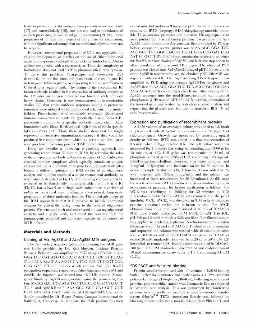
Figure 2. Expression, purification and chemical crosslinking of recombinant proteins. A) Coomassie Blue staining of purified, His-taggedproteins separated by 12% SDS-PAGE; 1. Acr-Ag85B (50 kDa), 2. Acr (20 kDa) and 3. Ag85B (32 kDa). B) Western blot analysis using antigen-specificantibodies; 1. Acr, 2. Acr-Ag85B and 3, Ag85B (1 and 2 probed with anti-Acr mAb TBG65; 3 probed with rabbit anti-Ag85B serum). C) Chemicalcrosslinking of Acr; shown is a Coomassie-stained (1, and 2) or Western blot (3 and 4) analysed sample with crosslinker (1 and 3) or without crosslinker(2 and 4). Letters indicate various molecular forms based on expected size (M-monomer, D-dimer, Tr-trimer, Te-tetramer, H-hexamer, O-oligomers). D)Chemical crosslinking of Acr-Ag85B fusion protein; shown is a sample with (1 and 3) or without (2 and 4) crosslinker. Letters indicate variousmolecular forms as for Acr (C). E) Chemical crosslinking of Ag85B (internal control); Coomassie and Western blot analysis of a sample with (1 and 3) orwithout (2 and 4) crosslinker.doi:10.1371/journal.pone.0060855.g002
Immune-Complex Based Vaccination
PLOS ONE | www.plosone.org 3 April 2013 | Volume 8 | Issue 4 | e60855

used, followed by a donkey anti-rabbit monoclonal antibody
conjugated to horseradish peroxidase (1:1000; Jackson Immuno
Research). In some instances, the fusion protein was detected by a
mouse monoclonal anti-polyHistidine antibody (SIGMA) conju-
gated to horseradish peroxidase (1:1000). After washing of blots
with PBS, they were developed using enzymatic chemilumines-
cence (ECL-Enhanced Chemiluminescence, Amersham Biosci-
ences).
Cross-linking of polymeric proteins50 mg of protein in PBS (Acr, Ag85B-Acr or Ag85B as the
negative control, all at 1 mg/ml) was crosslinked by addition of
5 mM Disuccinimidyl suberate (DSS, Pierce). Following addition
of the crosslinker, the samples were incubated for 30 minutes at
room temperature, before the reaction was quenched by addition
of SDS-PAGE loading buffer and 100 mM DTT (dithiothreitol).
Crosslinked protein was then analysed by SDS-PAGE and
Western blotting.
TBG65 mAb purificationMouse TBG65 hybridoma cell line (generated by Prof. Juraj
Ivanyi, King’s College London, [24]) was grown in RPMI
complete medium (Invitrogen). A CELLine CL-1000 (Integra
Biosciences) flask, specifically designed for high-yield antibody
production, was inoculated with 2.56107 cells undergoing
exponential growth. Cells were incubated in a tissue culture
incubator (37uC, 5% CO2, 90% relative humidity) and partially
harvested every 3–4 days over a period of 1 month. Antibody
purification was performed by affinity chromatography on an
Affigel-15 (Bio-Rad) column with immobilised Acr. After washing
the column with 30 cv of PBS, antibody was eluted in 0.1 M
glycine pH 2.5 and the fractions neutralised by addition of 1/10
volumes of 1 M Tris (uncorrected pH). Finally, protein was
concentrated to 5 mg/ml and dialysed against PBS.
Preparation of ICMsThe purified TBG65mAb and the Acr-Ag85B fusion protein
were combined in PBS pH 7.2 at a molar ratio ranging from 1:1 to
1:20. The total protein concentration in the solution was kept
below 1 mg/ml, to avoid formation of large aggregates that could
precipitate. After 1 h incubation at room temperature the ICMs
were placed on ice and subsequently kept at 4uC, prior to
application. ICMs were tested for their functionality by a C1q
binding ELISA and in a cell-binding assay.
Complement C1q ELISA96 well plates (Nunc Maxisorp ImmunoTM plates) were
incubated with 10 mg/mL of C1q (Calbiochem) component of
the complement system, at 37uC for 2 h. Plates were blocked with
PBS-2.5% BSA (4uC overnight) and then serial dilutions of ICM
were added in a final volume of 50 mL/well and incubated at 37uCfor 2 h. The plates were washed five times in distilled water, and
the secondary antibody (goat anti-mouse IgG HRP-conjugated
antiserum; The Binding Site) was added in PBS-2.5% BSA and
again incubated at 37uC for 2 h. Detection was performed using
TMB (3,3’, 5,5"-tetramethylbenzidine) substrate (Sigma) and the
absorbance was measured at 450 nm.
Binding of ICMs to antigen-presenting cells16106 spleen-derived APC, prepared as described by Pal et al
[25], were re-suspended in 100 mL of PBS containing 5% BSA.
ICMs (10 mg/mL) were added and cells incubated on ice for 2 h.
Unbound protein was removed by repeated (3x) addition of the
binding buffer and recovery of cells by centrifugation. Secondary
anti-mouse IgG-FITC antibody (Sigma) was added at a concen-
tration of 1 mg/mL and cells incubated for a further 1 h on ice.
Cells were then washed 3 times in binding buffer as before, re-
suspended in 1 mL of binding buffer and analysed for green
fluorescence in a Becton-Dickinson flow cytometer. Ten thousand
cells were counted and results expressed as percentage of cells
positive for FITC staining.
Immunisation of miceSix to eight weeks old female Balb/c mice (Harlan, UK) were
used in all immunisation experiments. The mice were kept under
defined environmental conditions (12:12 h light:dark cycle, 19–
21uC, 55% relative humidity, pathogen free). Mice were
immunised subcutaneously (s.c) with 50 mg of protein administered
at the base of the tail, followed by a boost immunisation three
weeks later. All administrations were standardised for the amount
of Ag85B in the vaccine formulation. The 1:20 antibody:antigen
molar ratio in ICMs was used in all experiments, following initial
testing of various ratios in a pilot experiment. In some
experiments, mice were first immunised with 56105 BCG (s.c)
followed by two s.c boosts with 50 mg ICMs at weeks six and eight.
In all experiments, PBS or BCG (Pasteur strain, grown in 7H9
Middlebrook medium) immunised mice were used for comparison
and mice immunised with Ag85B on its own (negative control) or
together with cholera toxin (CT fragment b) adjuvant (positive
control) were used to determine immune responses induced by
ICMs.
Humoral responseSpecific humoral responses in sera of mice immunised with
ICMs were tested by ELISA. At various time points during
immunisation, blood was collected from the tail vein or by cardiac
puncture (end of protocol) and fractionated to obtain sera. 96 well
plates (Nunc Maxisorp ImmunoTM plates) were coated with 10
mg/mL antigen solution (either Acr or Ag85B) in PBS for 2 h at
37uC. The plates were then washed with distilled water and
blocked overnight with a solution of 2.5% BSA in PBS at 4uC.
Serial dilutions of sera were added and plates incubated for 2 h at
37uC. After washing, peroxidase-conjugated detection antibodies
(sheep anti-mouse, The Binding Site) diluted 1:1000 in PBS-2.5%
BSA were added, and incubated as before. Total antigen-specific
IgG or IgG1and IgG2a subtypes were analysed. Following a
further washing step, the binding was detected by adding 100 mL
of TMB substrate (Sigma) to each well and the reaction was
stopped with 50 mL/well of 2 M sulfuric acid. The absorbance at
450 nm was read on an ELISA plate reader (Sunrise Tecan).
T cell proliferation and IFN-c detectionSpleens were aseptically extracted from mice and kept on ice.
The spleens were cut in small pieces and squeezed through a 70
mm cell strainer (BD FalconTM) in complete RPMI medium
(Sigma) supplemented with 10% fetal bovine serum (FBS, Sigma),
100 U/mL penicillin and 0.1 mg/mL streptomycin (Sigma). The
released cells were spun for 5 minutes at 190 g in a ROTINA 48R
centrifuge (Hettich Zentrifugen) and pelleted. Supernatant was
discarded and cells were re-suspended in 3 mL of red blood cell
lysing buffer (Sigma) and incubated for 2 minutes at room
temperature to eliminate the red blood cells. Complete RPMI
medium was added to the cell suspension and the cells were spun
again as above, followed by two additional washing steps. Cells
were then counted and seeded into 96 well flat bottom plates (BD
FalconTM) at a density of 36105 cells/well, in 200 mL medium.
Three different stimuli were used: 5 mg/mL of concanavalin A
Immune-Complex Based Vaccination
PLOS ONE | www.plosone.org 4 April 2013 | Volume 8 | Issue 4 | e60855

(Sigma), 10 mg/mL of Ag85B and 10 mg/mL of Acr. Cells were
incubated at 37uC for 48 h and a sample of medium taken for
IFN-c detection, using mouse IFN-c kit, R&D systems, USA. The
cultures were then pulsed with 1 mCi/well of 3[H]-thymidine (GE
Healthcare) and incubated for a further 24 h before harvest. At
harvest, cells were transferred onto a glass fiber filter (Wallac) using
a TOMTEC harvester and fixed with a scintillator sheet
(MeltiLexTM A, Perkin Elmer), prior to determining counts per
minute (CPM) in a liquid scintillator counter (Wallac).
Challenge of mice with H37Rv MTBAll pathogenic work was conducted in a containment level 3
laboratory in a Class 1 microbiological safety cabinet. Two weeks
after the last immunisation (week 5 for homologous immunisation,
week 10 for BCG-prime, ICM-boost), mice were lightly anaes-
thetized and challenged intranasally with 70,000 CFU (colony-
forming units) of MTB laboratory strain H37Rv (kindly provided
by P. Butcher at St George’s, and grown in 7H9 Middlebrook
medium). Four weeks later, mice were culled and lungs recovered.
Organs were homogenised in 5 mL of distilled water using a
Stomacher-80 biomaster (Seward) and serial dilutions plated out
on Middlebrook 7H11 mycobacteriological plates. CFUs were
counted three weeks later and the results expressed as logarithmic
values using GraphPad software.
Statistical analysisResults were analysed by ANOVA followed by Tukey’s (using
GraphPad PRISM software) or in some instances by Student’s t-
test, for a single group-to-group comparison (i.e. BCG vs
BCG+ICM). In all instances, p,0.05 was considered a statistically
significant difference.
Ethics StatementAnimal work was conducted at the St George’s University of
London (SGUL) Biological Research Facility which is a designated
establishment for animal research. The work in this study was
approved by the SGUL Ethical Research Committee, as part of
the process of obtaining the UK Home Office animal project
licence. Due care was taken at all times to minimise suffering of
animals during the experimentation. Experimental end-points
always preceded the onset of any visible symptoms of TB disease.
Results
Expression and molecular analysis of Acr, Ag85B and Acr-Ag85B
The recombinant proteins were expressed and purified from
BL21 inclusion bodies, and analysed by SDS-PAGE and
Coomassie staining (Fig.2A). Acr-Ag85B (lane 1) migrated as a
50 kDa protein band corresponding to the expected size of the
fusion protein. Acr preparation (lane 2) contained a dominant
20 kDa protein band (slightly larger than expected size of 18 kDa,
presumably due to presence of the His-tag) and two minor
contaminants, one of which (the 40 kDa band) corresponds to Acr
dimer. Ag85B (lane 3) appeared as a dominant 32 kDa protein
band, with some lower molecular weight contaminants also
present, but not recognised by anti-Ag85B antibodies (see further
below). To confirm the identity of the purified proteins, Western
blot analysis was performed, using anti-Acr and anti-Ag85B
antibodies (Fig.2B). The results show that all three dominant
protein bands were recognised with corresponding specific
antibodies, and that the Acr preparation (lane 1) also contained
an additional immune-reactive 40–42 kDa protein band, possibly
non-dissociated dimer.
To verify the oligomeric status of Acr and Acr-Ag85B, chemical
crosslinking using a short-length crosslinker (favouring intra-
rather than inter-molecular linking) was performed and the
proteins analysed by SDS-PAGE, followed by Coomassie staining
or Western blotting. As can be seen in Fig.2C, Acr readily formed
dimers, trimers, tetramers, hexamers and higher oligomers. The
fusion protein of Acr and Ag85B also formed higher molecular
forms, ranging in size from 100 kDa (dimer) to above 250 kDa,
which were not well separated on the top of the 4–12% gel
(Fig.2D). As expected, Ag85B on its own failed to form higher
molecular forms in the presence of the crosslinker, as demonstrat-
ed by the lack of definable molecular bands, despite the
monomeric band appearing slightly diminished on Coomassie
staining (Fig.2E).
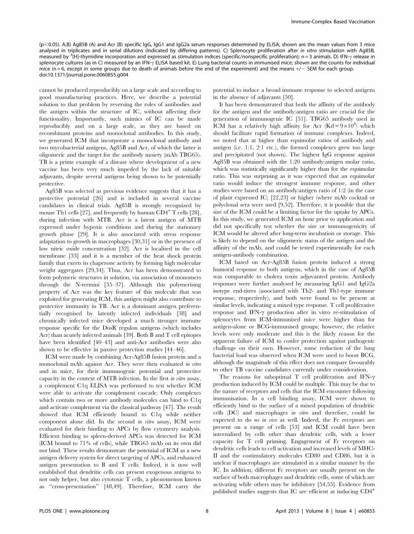
Preparation and functional properties of ICMICM were prepared by combining the anti-Acr IgG1 mAb
TBG65 and the Acr-Ag85B fusion protein at two different molar
ratios (i.e. 1:1 and 1:20) in PBS buffer. To test the functional
properties of ICM, we conducted two assays, namely complement
C1q binding by ELISA and binding to the surface of antigen-
presenting cells (APC) by flow cytometry. In the first assay, the
ICMs were shown to bind the complement C1q component in a
dose dependent manner while neither component alone showed
any binding (Fig. 3A). For testing the ability of ICM to bind to
APC, we generated mouse spleen derived APC as described
previously [25]. Their phenotype was analysed by surface
molecule staining and they were found to be positive for CD16/
32 (93%), CD11b (98%), CD11c (65%) and MHC II (51%) (not
shown), consistent with myeloid dendritic cells and macrophages.
Following incubation with ICM, 71% of the APC were stained
positive (Fig3B), while the cells incubated with the antibody alone
showed only background staining.
ICM-induced IgG responses in miceFollowing immunisation of mice, we analysed the IgG immune
responses to both Ag85B and Acr in sera by antigen-specific
ELISA. Initially, we tested different ratios of antibody/antigen
within the ICM (i.e. 1:1 or 1:20) and found that the lower ratios
induced a stronger humoral response (Fig.3C); hence the 1:20
ratio was used in all subsequent experiments. Mice immunised
with PBS, BCG or Ag85B alone showed no significant IgG
response to either Ag85B or Acr (Fig.4A,B; final antibody titres
shown only). In contrast, mice immunised with ICM, showed an
Ag85B-specific IgG response comparable to that induced by
formulating Ag85B with cholera toxin (CT). The end-point titre
increased from 1:100 (post-prime), to 8100 (after first boost), to
72900 (after second boost). A similar pattern was also observed for
IgG responses to Acr. When analysing IgG subtype responses,
both IgG1 and IgG2a were found to be present and with similar
end-point titres, indicating a mixed type Th1/Th2 antibody
response. When compared to the CT-adjuvanted group, ICM
induced somewhat weaker IgG1 but higher IgG2a responses,
consistent with the CT being a strong Th2 response promoting
adjuvant.
T cell response and IFN-c productionAt the end of the immunisation protocol, mice were sacrificed
and the spleen cells recovered. Cell cultures were stimulated with
Ag85B, medium alone or with 5 mg/ml Concanavalin A (not
shown), as an internal control for proliferation. After 48 h, cell
culture supernatant was analysed for IFN-c, while the remaining
culture was pulsed with [3H]-thymidine and analysed for
radioactive incorporation 24 h later. Cell proliferation was
Immune-Complex Based Vaccination
PLOS ONE | www.plosone.org 5 April 2013 | Volume 8 | Issue 4 | e60855

expressed as stimulation index, obtained by dividing antigen-
specific with medium-induced thymidine incorporation. As shown
in Fig.4C, PBS and BCG immunised groups showed no splenocyte
proliferation. Cells from animals immunised with Ag85B alone
showed some antigen-specific proliferation and ICM induced a
further proliferative response, although this was not statistically
significantly different from Ag85B alone. However, the ICM-
induced proliferation was statistically significantly higher than in
the PBS group (p = 0.0037, Student’s t test). On the other hand,
cells from Ag85B+CT immunised animals showed twice the level
of cell proliferation induced by ICM. A similar trend was observed
for the IFN-c response in culture supernatants (Fig.4D), except
that the BCG-immunised animals showed the highest level of IFN-
c production.
Bacterial load in MTB-infected miceFour weeks after pathogenic challenge with MTB H37Rv strain
(9 weeks from the start of immunisation), mice were culled and the
bacterial load in their lungs determined by the standard CFU
assay. As shown in Fig.4E, the groups were significantly different
(p = 0.0018, one way ANOVA) but the only groups that showed a
statistically significant reduction in bacterial load when compared
to PBS-immunised mice, were BCG and Ag85B+CT (p,0.05,
Tukey’s test). ICM-immunised animals showed a similar bacterial
number to the negative control.
BCG-boosting potential of ICMSince ICM failed to confer protection against MTB in
immunised mice, they were subsequently tested as a boost vaccine
to BCG. Mice were immunised with BCG (week 1) and boosted
twice with ICM (weeks 6 and 8). Their humoral and cellular
immune responses were analysed as before (week 10) and also the
bacterial load in their lungs compared to BCG alone or the
negative control (week 14). As shown in Fig.5, the ICM-boosted,
BCG-primed group showed an enhanced IgG antibody response
to Ag85B (5A) and Acr (5B), and a small though not statistically
significant increase in T-cell proliferation (5C). A statistically
significant (p,0.05) increase in IFN-c production in response to
Ag85B was measured (5D), although the proliferative response and
cytokine secretion in all cultures was affected by reduced cell
viability in this particular experiment. The bacterial load in the
ICM-boosted animals, when compared to BCG-alone, was
significantly lower (p = 0.037, Student’s t test), but the magnitude
of the effect (a 5-fold reduction) was relatively small (Fig.5E,
representative of 2 similar experiments). Note also that the
increased protection by BCG in this, compared to the experiment
in Fig.4 may have been due to extension of immunisation time
from 10 to 14 weeks.
Discussion
This is a proof-of-principle study in which we investigated the
immunogenic potential of recombinant immune complex mimics
(ICM) as a novel molecular platform for adjuvant-free vaccine
delivery. IC have many immunogenic properties that could be
utilised in vaccine design. Unfortunately, the key obstacle for
translating the potential of IC into a vaccine modality is that they
Figure 3. Functional evaluation of ICM in vitro and in vivo. A)Complement C1q binding ELISA; ICM were used at the 1:20 antibody-antigen ratio and the neat sample contained 5 mg/m total protein (ICM)or the equivalent amount for individual components; each barrepresents mean value from triplicate assays and the patterns indicateserial dilutions. B) Analysis of binding of ICM to spleen-derived APCs byflow cytometry; shown are the proportions of cells (out of 10,000
counted) that bound either mAb alone or ICM. C) Serum anti-Ag85B IgGresponses from mice immunised with an equimolar (1:1) or a low (1:20)antibody-antigen ratio, twice at the base of the tail, at 3-week intervals.Mice were culled 3 weeks after the final immunisation. Shown are themean values and corresponding serial dilutions from a pilot experiment(n = 3 mice).doi:10.1371/journal.pone.0060855.g003
Immune-Complex Based Vaccination
PLOS ONE | www.plosone.org 6 April 2013 | Volume 8 | Issue 4 | e60855

Figure 4. Immune responses and MTB bacterial counts in mice immunised with ICM. Mice were immunised with 50 mg ICM (1:20 antibodyantigen ratio) or with Ag85B alone (30 mg), Ag85B+CT, BCG and PBS; two weeks after the final immunisation mice were either culled and their tissues(blood and spleens) used for immunological evaluation, or challenged i.n. with 70,000 MTB H37Rv. * Indicates statistically significant difference
Immune-Complex Based Vaccination
PLOS ONE | www.plosone.org 7 April 2013 | Volume 8 | Issue 4 | e60855
cannot be produced reproducibly on a large scale and according to
good manufacturing practices. Here, we describe a potential
solution to that problem by reversing the roles of antibodies and
the antigen within the structure of IC, without affecting their
functionality. Importantly, such mimics of IC can be made
reproducibly and on a large scale, as they are based on
recombinant proteins and monoclonal antibodies. In this study,
we generated ICM that incorporate a monoclonal antibody and
two mycobacterial antigens, Ag85B and Acr, of which the latter is
oligomeric and the target for the antibody moiety (mAb TBG65).
TB is a prime example of a disease where development of a new
vaccine has been very much impeded by the lack of suitable
adjuvants, despite several antigens being shown to be potentially
protective.
Ag85B was selected as previous evidence suggests that it has a
protective potential [26] and is included in several vaccine
candidates in clinical trials. Ag85B is strongly recognized by
mouse Th1 cells [27], and frequently by human CD4+ T cells [28],
during infection with MTB. Acr is a latent antigen of MTB
expressed under hypoxic conditions and during the stationary
growth phase [29]. It is also associated with stress response
adaptation to growth in macrophages [30,31] or in the presence of
low nitric oxide concentration [32]. Acr is localised in the cell
membrane [33] and it is a member of the heat shock protein
family that exerts its chaperone activity by forming high molecular
weight aggregates [29,34]. Thus, Acr has been demonstrated to
form polymeric structures in solution, via association of monomers
through the N-termini [35–37]. Although this polymerising
property of Acr was the key feature of this molecule that was
exploited for generating ICM, this antigen might also contribute to
protective immunity in TB. Acr is a dominant antigen preferen-
tially recognised by latently infected individuals [38] and
chronically infected mice developed a much stronger immune
response specific for the DosR regulon antigens (which includes
Acr) than acutely infected animals [39]. Both B and T cell epitopes
have been identified [40–43] and anti-Acr antibodies were also
shown to be effective in passive protection studies [44–46].
ICM were made by combining Acr-Ag85B fusion protein and a
monoclonal mAb against Acr. They were then evaluated in vitro
and in mice, for their immunogenic potential and protective
capacity in the context of MTB infection. In the first in vitro assay,
a complement C1q ELISA was performed to test whether ICM
were able to activate the complement cascade. Only complexes
which contain two or more antibody molecules can bind to C1q
and activate complement via the classical pathway [47]. The result
showed that ICM efficiently bound to C1q while neither
component alone did. In the second in vitro assay, ICM were
evaluated for their binding to APCs by flow cytometry analysis.
Efficient binding to spleen-derived APCs was detected for ICM
(ICM bound to 71% of cells), while TBG65 mAb on its own did
not bind. These results demonstrate the potential of ICM as a new
antigen delivery system for direct targeting of APCs, and enhanced
antigen presentation to B and T cells. Indeed, it is now well
established that dendritic cells can present exogenous antigens to
not only helper, but also cytotoxic T cells, a phenomenon known
as ‘‘cross-presentation’’ [48,49]. Therefore, ICM carry the
potential to induce a broad immune response to selected antigens
in the absence of adjuvants [50].
It has been demonstrated that both the affinity of the antibody
for the antigen and the antibody:antigen ratio are crucial for the
generation of immunogenic IC [51]. TBG65 antibody used in
ICM has a relatively high affinity for Acr (Kd = 96108) which
should facilitate rapid formation of immune complexes. Indeed,
we noted that at higher than equimolar ratios of antibody and
antigen (i.e. 1:1, 2:1 etc.), the formed complexes grew too large
and precipitated (not shown). The highest IgG response against
Ag85B was obtained with the 1:20 antibody:antigen molar ratio,
which was statistically significantly higher than for the equimolar
ratio. This was surprising as it was expected that an equimolar
ratio would induce the strongest immune response, and other
studies were based on an antibody:antigen ratio of 1:2 (in the case
of plant expressed IC; [22,23] or higher (where mAb cocktail or
polyclonal sera were used [9,52]. Therefore, it is possible that the
size of the ICM could be a limiting factor for the uptake by APCs.
In this study, we generated ICM an hour prior to application and
did not specifically test whether the size or immunogenicity of
ICM would be altered after long-term incubation or storage. This
is likely to depend on the oligomeric status of the antigen and the
affinity of the mAb, and could be tested experimentally for each
antigen-antibody combination.
ICM based on Acr-Ag85B fusion protein induced a strong
humoral response to both antigens, which in the case of Ag85B
was comparable to cholera toxin adjuvanted protein. Antibody
responses were further analysed by measuring IgG1 and IgG2a
isotype end-titres (associated with Th2- and Th1-type immune
response, respectively), and both were found to be present at
similar levels, indicating a mixed type response. T cell proliferative
response and IFN-c production after in vitro re-stimulation of
splenocytes from ICM-immunised mice were higher than for
antigen-alone or BCG-immunised groups; however, the relative
levels were only moderate and this is the likely reason for the
apparent failure of ICM to confer protection against pathogenic
challenge on their own. However, some reduction of the lung
bacterial load was observed when ICM were used to boost BCG,
although the magnitude of this effect does not compare favourably
to other TB vaccine candidates currently under consideration.
The reasons for suboptimal T cell proliferation and IFN-cproduction induced by ICM could be multiple. This may be due to
the nature of receptors and cells that the ICM encounter following
immunisation. In a cell binding assay, ICM were shown to
efficiently bind to the surface of a mixed population of dendritic
cells (DC) and macrophages in vitro and therefore, could be
expected to do so in vivo as well. Indeed, the Fc receptors are
present on a range of cells [53] and ICM could have been
internalised by cells other than dendritic cells, with a lesser
capacity for T cell priming. Engagement of Fc receptors on
dendritic cells leads to cell activation and increased levels of MHC-
II and the costimulatory molecules CD80 and CD86, but it is
unclear if macrophages are stimulated in a similar manner by the
IC. In addition, different Fc receptors are usually present on the
surface of both macrophages and dendritic cells, some of which are
activating while others may be inhibitory [54,55]. Evidence from
published studies suggests that IC are efficient at inducing CD4+
(p,0.05). A,B) Ag85B (A) and Acr (B) specific IgG, IgG1 and IgG2a serum responses determined by ELISA; shown are the mean values from 3 miceanalysed in triplicates and in serial dilutions (indicated by differing patterns). C) Splenocyte proliferation after in vitro stimulation with Ag85B,measured by 3[H]-thymidine incorporation and expressed as stimulation indices (specific/nonspecific proliferation); n = 3 animals. D) IFN-c release insplenocyte cultures (as in C) measured by an IFN-c ELISA based kit. E) Lung bacterial counts in immunised mice; shown are the counts for individualmice (n = 6, except in some groups due to death of animals before the end of the experiment) and the means +/2 SEM for each group.doi:10.1371/journal.pone.0060855.g004
Immune-Complex Based Vaccination
PLOS ONE | www.plosone.org 8 April 2013 | Volume 8 | Issue 4 | e60855
Figure 5. Immune responses and MTB bacterial counts in mice immunised with BCG and boosted with ICM. Mice were immunised s.c.with BCG and twice boosted with ICM six and eight weeks later; 2 weeks after the final boost, mice were either culled and their tissues (blood andspleens) used for immunological evaluation, or challenged i.n. with 70,000 H37Rv. * Indicates statistically significant difference (p,0.05). A,B) Ag85B
Immune-Complex Based Vaccination
PLOS ONE | www.plosone.org 9 April 2013 | Volume 8 | Issue 4 | e60855
and CD8+ T cells and that they enhance antigen processing
[9,52,56], but that this could be largely dependent on the size of
IC and the identity of the Fc receptors they target [57]. If IC are
too large, uptake could occur by phagocytic rather than endocytic
(i.e. Fc-receptor mediated) mechanisms, and that could determine
the efficiency of antigen presentation and the nature of the
immune response induced. Only DC but not macrophages can
present antigens in the context of MHC I after IC uptake, and
considering that CD8+ T cell responses may be important for
immunity against intracellular bacterial pathogens such as MTB
[58,59], a selective ICM targeting to these cells may be critically
important to induce such immune responses.
However, this study does provide conclusive evidence that it is
possible to make self-adjuvanting immunogens that can induce
strong antibody responses. Given the versatility of the ICM
delivery platform, it would be of interest to further test this
approach against pathogens which require predominantly humor-
al rather than cellular immune responses for protection. Indeed,
the vast majority of the currently licensed vaccines rely on
antibodies as the key component of the protective immune
response [60]. In fact, to our knowledge, BCG and Herpes zoster
are the only vaccines that rely almost exclusively on cellular rather
than humoral immune responses for protection. Thus, the
advantage of ICM over other vaccine approaches is that they
can induce antibody responses without the use of adjuvants, live
vectors or attenuated or inactivated organisms, making their use
safe and simplifying the licensing process. The ICM approach is
generic and could be easily adapted for various pathogens in two
possible ways; 1. Using oligomeric antigens from those pathogens,
on their own or fused to additional protective antigens, and a mAb
against the oligomeric antigen; 2. using Acr antigen and the anti-
Acr mAb as a generic platform, to which antigens from other
pathogens could be linked. In both instances, the antibody would
have to be fully ‘humanised’ to be applicable in human subjects.
On the other hand, Acr, like most heat shock proteins, is a
molecular adjuvant in its own right, which is an added advantage
of the latter approach. Examples of infections where the ICM
could be tested may include HIV, rabies and Influenza, where
rapid interception of viruses at the port of entry by pre-existing
antibodies appears to be the only realistic way of preventing
infection.
Author Contributions
Conceived and designed the experiments: RR JM. Performed the
experiments: IP ES CD GD MP RR. Analyzed the data: IP GD MP PD
RR. Contributed reagents/materials/analysis tools: RR CD JM PD.
Wrote the paper: IP RR JM.
References
1. Moreno-Mendieta SA, Rocha-Zavaleta L, Rodriguez-Sanoja R (2010) Adju-
vants in tuberculosis vaccine development. FEMS Immunol Med Microbiol 58:
75–84.
2. Laissue J, Cottier H, Hess MW, Stoner RD (1971) Early and enhanced germinal
center formation and antibody responses in mice after primary stimulation with
antigen-isologous antibody complexes as compared with antigen alone.
J Immunol 107: 822–831.
3. Osato K (1972) Antigen-antibody complexes in the immune response. I. Analysis
of the effectiveness of complexes on the primary antibody response. Immunology
23: 545–557.
4. Terres G, Morrison SL, Habicht GS, Stoner RD (1972) Appearance of an early
"primed state" in mice following the concomitant injections of antigen and
specific antiserum. J Immunol 108: 1473–1481.
5. Houston WE, Kremer RJ, Crabbs CL, Spertzel RO (1977) Inactivated
Venezuelan equine encephalomyelitis virus vaccine complexed with specific
antibody: enhanced primary immune response and altered pattern of antibody
class elicited. J Infect Dis 135: 600–610.
6. Celis E, Chang TW (1984) Antibodies to hepatitis B surface antigen potentiate
the response of human T lymphocyte clones to the same antigen. Science 224:
297–299.
7. Abdel-Motal U, Wang S, Lu S, Wigglesworth K, Galili U (2006) Increased
immunogenicity of human immunodeficiency virus gp120 engineered to express
Galalpha1-3Galbeta1-4GlcNAc-R epitopes. J Virol 80: 6943–6951.
8. Polyanskaya N, Bergmeier LA, Sharpe SA, Cook N, Leech S, et al. (2001)
Mucosal exposure to subinfectious doses of SIV primes gut-associated antibody-
secreting cells and T cells: lack of enhancement by nonneutralizing antibody.
Virology 279: 527–538.
9. Regnault A, Lankar D, Lacabanne V, Rodriguez A, Thery C, et al. (1999)
Fcgamma receptor-mediated induction of dendritic cell maturation and major
histocompatibility complex class I-restricted antigen presentation after immune
complex internalization. J Exp Med 189: 371–380.
10. Schuurhuis DH, Ioan-Facsinay A, Nagelkerken B, van Schip JJ, Sedlik C, et al.
(2002) Antigen-antibody immune complexes empower dendritic cells to
efficiently prime specific CD8+ CTL responses in vivo. J Immunol 168: 2240–
2246.
11. Villinger F, Mayne AE, Bostik P, Mori K, Jensen PE, et al. (2003) Evidence for
antibody-mediated enhancement of simian immunodeficiency virus (SIV) Gag
antigen processing and cross presentation in SIV-infected rhesus macaques.
J Virol 77: 10–24.
12. Marusic-Galesic S, Marusic M, Pokric B (1992) Cellular immune response to the
antigen administered as an immune complex in vivo. Immunology 75: 325–329.
13. Klaus GG (1978) The generation of memory cells. II. Generation of B memory
cells with preformed antigen-antibody complexes. Immunology 34: 643–652.
14. Wiersma EJ, Coulie PG, Heyman B (1989) Dual immunoregulatory effects of
monoclonal IgG-antibodies: suppression and enhancement of the antibody
response. Scand J Immunol 29: 439–448.
15. Schalke BC, Klinkert WE, Wekerle H, Dwyer DS (1985) Enhanced activation of
a T cell line specific for acetylcholine receptor (AChR) by using anti-AChR
monoclonal antibodies plus receptors. J Immunol 134: 3643–3648.
16. Heyman B (1990) The immune complex: possible ways of regulating the
antibody response. Immunol Today 11: 310–313.
17. Manca F, Fenoglio D, Kunkl A, Cambiaggi C, Sasso M, et al. (1988) Differential
activation of T cell clones stimulated by macrophages exposed to antigen
complexed with monoclonal antibodies. A possible influence of paratope
specificity on the mode of antigen processing. J Immunol 140: 2893–2898.
18. Jemmerson R, Paterson Y (1986) Mapping epitopes on a protein antigen by the
proteolysis of antigen-antibody complexes. Science 232: 1001–1004.
19. Antoniou AN, Watts C (2002) Antibody modulation of antigen presentation:
positive and negative effects on presentation of the tetanus toxin antigen via the
murine B cell isoform of FcgammaRII. Eur J Immunol 32: 530–540.
20. Eisenberg RJ, Long D, Pereira L, Hampar B, Zweig M, et al. (1982) Effect of
monoclonal antibodies on limited proteolysis of native glycoprotein gD of herpes
simplex virus type 1. J Virol 41: 478–488.
21. Simitsek PD, Campbell DG, Lanzavecchia A, Fairweather N, Watts C (1995)
Modulation of antigen processing by bound antibodies can boost or suppress
class II major histocompatibility complex presentation of different T cell
determinants. J Exp Med 181: 1957–1963.
22. Chargelegue D, Drake PM, Obregon P, Prada A, Fairweather N, et al. (2005)
Highly immunogenic and protective recombinant vaccine candidate expressed
in transgenic plants. Infect Immun 73: 5915–5922.
23. Phoolcharoen W, Bhoo SH, Lai H, Ma J, Arntzen CJ, et al. (2011) Expression of
an immunogenic Ebola immune complex in Nicotiana benthamiana. Plant
Biotechnol J 9: 807–816.
24. Falero-Diaz G, Challacombe S, Rahman D, Mistry M, Douce G, et al. (2000)
Transmission of IgA and IgG monoclonal antibodies to mucosal fluids following
intranasal or parenteral delivery. Int Arch Allergy Immunol 122: 143–150.
(A) and Acr (B) specific IgG, IgG1 and IgG2a serum responses determined by ELISA; shown are the mean values from 3 mice analysed in triplicates andin serial dilutions (indicated by differing patterns). C) Splenocyte proliferation after in vitro stimulation with Ag85B, measured by 3[H]-thymidineincorporation and expressed as stimulation indices (specific/nonspecific proliferation); n = 3 animals. D) IFN-c release in splenocyte cultures (as in C)measured by an IFN-c ELISA based kit. E) Lung bacterial counts in immunised mice; shown are the counts for individual mice (n = 6) and the means+/2 SEM for each group.doi:10.1371/journal.pone.0060855.g005
Immune-Complex Based Vaccination
PLOS ONE | www.plosone.org 10 April 2013 | Volume 8 | Issue 4 | e60855

25. Pal R, Marwaha S, Pepponi I, Mann JF, Paul MJ, et al. (2010) Generation of
self-renewing immature dendritic cells from mouse spleen that can take upmycobacteria and present antigens to T cells. APMIS 118: 729–738.
26. Boesen H, Jensen BN, Wilcke T, Andersen P (1995) Human T-cell responses to
secreted antigen fractions of Mycobacterium tuberculosis. Infect Immun 63:1491–1497.
27. Andersen P, Andersen AB, Sorensen AL, Nagai S (1995) Recall of long-livedimmunity to Mycobacterium tuberculosis infection in mice. J Immunol 154:
3359–3372.
28. Mustafa AS, Amoudy HA, Wiker HG, Abal AT, Ravn P, et al. (1998)Comparison of antigen-specific T-cell responses of tuberculosis patients using
complex or single antigens of Mycobacterium tuberculosis. Scand J Immunol 48:535–543.
29. Yuan Y, Crane DD, Simpson RM, Zhu YQ, Hickey MJ, et al. (1998) The 16-kDa alpha-crystallin (Acr) protein of Mycobacterium tuberculosis is required for
growth in macrophages. Proc Natl Acad Sci U S A 95: 9578–9583.
30. Chang Z, Primm TP, Jakana J, Lee IH, Serysheva I, et al. (1996)Mycobacterium tuberculosis 16-kDa antigen (Hsp16.3) functions as an
oligomeric structure in vitro to suppress thermal aggregation. J Biol Chem271: 7218–7223.
31. Raja A, Uma Devi KR, Ramalingam B, Brennan PJ (2002) Immunoglobulin G,
A, and M responses in serum and circulating immune complexes elicited by the16-kilodalton antigen of Mycobacterium tuberculosis. Clin Diagn Lab Immunol
9: 308–312.32. Cunningham AF, Spreadbury CL (1998) Mycobacterial stationary phase
induced by low oxygen tension: cell wall thickening and localization of the 16-kilodalton alpha-crystallin homolog. J Bacteriol 180: 801–808.
33. Yuan Y, Crane DD, Barry CE, 3rd (1996) Stationary phase-associated protein
expression in Mycobacterium tuberculosis: function of the mycobacterial alpha-crystallin homolog. J Bacteriol 178: 4484–4492.
34. Jackett PS, Bothamley GH, Batra HV, Mistry A, Young DB, et al. (1988)Specificity of antibodies to immunodominant mycobacterial antigens in
pulmonary tuberculosis. J Clin Microbiol 26: 2313–2318.
35. Berengian AR, Parfenova M, McHaourab HS (1999) Site-directed spin labelingstudy of subunit interactions in the alpha-crystallin domain of small heat-shock
proteins. Comparison of the oligomer symmetry in alphaA-crystallin, HSP 27,and HSP 16.3. J Biol Chem 274: 6305–6314.
36. Kennaway CK, Benesch JL, Gohlke U, Wang L, Robinson CV, et al. (2005)Dodecameric structure of the small heat shock protein Acr1 from Mycobacte-
rium tuberculosis. J Biol Chem 280: 33419–33425.
37. Taylor RG, Walker DC, McInnes RR (1993) E. coli host strains significantlyaffect the quality of small scale plasmid DNA preparations used for sequencing.
Nucleic Acids Res 21: 1677–1678.38. Roupie V, Romano M, Zhang L, Korf H, Lin MY, et al. (2007) Immunogenicity
of eight dormancy regulon-encoded proteins of Mycobacterium tuberculosis in
DNA-vaccinated and tuberculosis-infected mice. Infect Immun 75: 941–949.39. Li Q, Yu H, Zhang Y, Wang B, Jiang W, et al. (2011) Immunogenicity and
protective efficacy of a fusion protein vaccine consisting of antigen Ag85B andHspX against Mycobacterium tuberculosis infection in mice. Scand J Immunol
73: 568–576.40. Caccamo N, Meraviglia S, La Mendola C, Bosze S, Hudecz F, et al. (2004)
Characterization of HLA-DR- and TCR-binding residues of an immunodomi-
nant and genetically permissive peptide of the 16-kDa protein of Mycobacteriumtuberculosis. Eur J Immunol 34: 2220–2229.
41. Demissie A, Leyten EM, Abebe M, Wassie L, Aseffa A, et al. (2006) Recognitionof stage-specific mycobacterial antigens differentiates between acute and latent
infections with Mycobacterium tuberculosis. Clin Vaccine Immunol 13: 179–186.
42. Leyten EM, Lin MY, Franken KL, Friggen AH, Prins C, et al. (2006) Human T-
cell responses to 25 novel antigens encoded by genes of the dormancy regulon ofMycobacterium tuberculosis. Microbes Infect 8: 2052–2060.
43. Wilkinson RJ, Wilkinson KA, De Smet KA, Haslov K, Pasvol G, et al. (1998)
Human T- and B-cell reactivity to the 16kDa alpha-crystallin protein ofMycobacterium tuberculosis. Scand J Immunol 48: 403–409.
44. Reljic R, Clark SO, Williams A, Falero-Diaz G, Singh M, et al. (2006) Intranasal
IFNgamma extends passive IgA antibody protection of mice against Mycobac-terium tuberculosis lung infection. Clin Exp Immunol 143: 467–473.
45. Reljic R, Williams A, Ivanyi J (2006) Mucosal immunotherapy of tuberculosis: is
there a value in passive IgA? Tuberculosis (Edinb) 86: 179–190.
46. Williams A, Reljic R, Naylor I, Clark SO, Falero-Diaz G, et al. (2004) Passive
protection with immunoglobulin A antibodies against tuberculous early infection
of the lungs. Immunology 111: 328–333.
47. Reid KB (1986) Activation and control of the complement system. Essays
Biochem 22: 27–68.
48. Heath WR, Carbone FR (2001) Cross-presentation, dendritic cells, toleranceand immunity. Annu Rev Immunol 19: 47–64.
49. Matheoud D, Perie L, Hoeffel G, Vimeux L, Parent I, et al. (2010) Cross-
presentation by dendritic cells from live cells induces protective immuneresponses in vivo. Blood 115: 4412–4420.
50. Celis E, Zurawski VR, Jr, Chang TW (1984) Regulation of T-cell function by
antibodies: enhancement of the response of human T-cell clones to hepatitis Bsurface antigen by antigen-specific monoclonal antibodies. Proc Natl Acad
Sci U S A 81: 6846–6850.
51. Wen YM (2009) Antigen-antibody immunogenic complex: promising novelvaccines for microbial persistent infections. Expert Opin Biol Ther 9: 285–291.
52. den Haan JM, Bevan MJ (2002) Constitutive versus activation-dependent cross-
presentation of immune complexes by CD8(+) and CD8(2) dendritic cells invivo. J Exp Med 196: 817–827.
53. Hogarth PM, Pietersz GA (2012) Fc receptor-targeted therapies for the
treatment of inflammation, cancer and beyond. Nat Rev Drug Discov 11:311–331.
54. Cady CT, Powell MS, Harbeck RJ, Giclas PC, Murphy JR, et al. (2010) IgG
antibodies produced during subcutaneous allergen immunotherapy mediateinhibition of basophil activation via a mechanism involving both FcgammaRIIA
and FcgammaRIIB. Immunol Lett 130: 57–65.
55. Pinheiro da Silva F, Aloulou M, Skurnik D, Benhamou M, Andremont A, et al.
(2007) CD16 promotes Escherichia coli sepsis through an FcR gamma inhibitory
pathway that prevents phagocytosis and facilitates inflammation. Nat Med 13:
1368–1374.
56. Guyre PM, Graziano RF, Goldstein J, Wallace PK, Morganelli PM, et al. (1997)
Increased potency of Fc-receptor-targeted antigens. Cancer Immunol Immun-
other 45: 146–148.
57. Bachmann MF, Jennings GT (2010) Vaccine delivery: a matter of size,
geometry, kinetics and molecular patterns. Nat Rev Immunol 10: 787–796.
58. Jacobsen M, Detjen AK, Mueller H, Gutschmidt A, Leitner S, et al. (2007)
Clonal expansion of CD8+ effector T cells in childhood tuberculosis. J Immunol
179: 1331–1339.
59. Winau F, Weber S, Sad S, de Diego J, Hoops SL, et al. (2006) Apoptotic vesicles
crossprime CD8 T cells and protect against tuberculosis. Immunity 24: 105–117.
60. Plotkin SA (2010) Correlates of protection induced by vaccination. Clin VaccineImmunol 17: 1055–1065.
Immune-Complex Based Vaccination
PLOS ONE | www.plosone.org 11 April 2013 | Volume 8 | Issue 4 | e60855