Imaging of Cystic Fibrosis Lung Disease and Clinical ... · Imaging of Cystic Fibrosis Lung Disease...
12
Imaging of Cystic Fibrosis Lung Disease and Clinical Interpretation Bildgebung der Lunge bei Mukoviszidose und klinische Interpretation Authors M. O. Wielpütz 1, 2, 3 , M. Eichinger 1, 2, 3 , J. Biederer 1, 2, 4 , S. Wege 5 , M. Stahl 2, 6 , O. Sommerburg 2, 6 , M. A. Mall 2, 6, 7 , H. U. Kauczor 1, 2, 3 , M. Puderbach 1, 2, 3, 8 Affiliations Affiliation addresses are listed at the end of the article. Key words ● ▶ thorax ● ▶ CT-quantitative ● ▶ tracheobronchial tree ● ▶ MR-functional imaging ● ▶ fibrosis, cystic received 8.12.2015 accepted 8.3.2016 Bibliography DOI http://dx.doi.org/ 10.1055/s-0042-104936 Published online: 13.4.2016 Fortschr Röntgenstr 2016; 188: 834–845 © Georg Thieme Verlag KG Stuttgart · New York · ISSN 1438-9029 Correspondence Dr. Mark Oliver Wielpütz Diagnostic and Interventional Radiology, University Hospital of Heidelberg Im Neuenheimer Feld 110 69120 Heidelberg Germany Tel.: ++ 49/62 21/56 64 10 Fax: ++ 49/62 21/56 57 30 [email protected] heidelberg.de Review 834 Wielpütz MO et al. Imaging of Cystic … Fortschr Röntgenstr 2016; 188: 834–845 Zusammenfassung ▼ Die progressive Lungenerkrankung bestimmt Morbidität und Mortalität der autosomal-rezessiv vererbten Mukoviszidose (Cystische Fibrose, CF). Die Implementierung der CF in das Neuge- borenen-Screening erlaubt eine Diagnosestellung häufig bereits in einem präsymptomatischen Stadium. Verbesserungen der Therapie haben zu- dem eine stetig zunehmende Lebenserwartung ermöglicht, sodass die Mehrzahl der Patienten heute erwachsen ist. Da bildgebende Verfahren detaillierte Informationen über den regionalen Krankheitsverlauf bieten, werden heute Kontrol- len in regelmäßigen Abständen empfohlen. Rönt- genaufnahmen des Thorax, die Computertomo- grafie (CT) und die Magnetresonanztomografie (MRT) stehen zur Verfügung – jedes Verfahren mit spezifischen Stärken und Schwächen, sodass die Wahl des Verfahrens an die individuelle kli- nische Situation des Patienten angepasst werden kann. Die CT bietet die höchste Detailauflösung und kann mittels Software nachverarbeitet wer- den, welche Atemwegsveränderungen quantita- tiv erfassen kann und potenziell eine objektivere Schweregradeinteilung ermöglicht. Die CT hat daher die Röntgenaufnahme an spezialisierten Zentren weitgehend abgelöst. Entsprechend ist die Strahlenexposition der CF-Erkrankten anges- tiegen, die altersbedingt besonders sensibel für ionisierende Strahlen sind und während ihres Le- bens eine relevante Dosis akkumulieren können. Die MRT als alternatives strahlungsfreies Schnitt- bildverfahren stellt die typischen morphologi- schen Veränderungen der CF mit vergleichbarer klinischer Information bei etwas geringerer De- tailauflösung dar. Mehr als jedes andere Verfah- ren ermöglicht die MRT eine Beurteilung der re- gionalen Lungenfunktion, wobei sich die zeitlich hoch aufgelöste Perfusions-MRT als praktikabel erwiesen hat. Abstract ▼ Progressive lung disease in cystic fibrosis (CF) is the life-limiting factor of this autosomal recessive genetic disorder. Increasing implementation of CF newborn screening allows for a diagnosis even in pre-symptomatic stages. Improvements in therapy have led to a significant improvement in survival, the majority now being of adult age. Imaging pro- vides detailed information on the regional distri- bution of CF lung disease, hence longitudinal ima- ging is recommended for disease monitoring in the clinical routine. Chest X-ray (CXR), computed tomography (CT) and magnetic resonance imaging (MRI) are now available as routine modalities, each with individual strengths and drawbacks, which need to be considered when choosing the optimal modality adapted to the clinical situation of the pa- tient. CT stands out with the highest morphological detail and has often been a substitute for CXR for regular severity monitoring at specialized centers. Multidetector CT data can be post-processed with dedicated software for a detailed measurement of airway dimensions and bronchiectasis and poten- tially a more objective and precise grading of dis- ease severity. However, changing to CT was inse- parably accompanied by an increase in radiation exposure of CF patients, a young population with high sensitivity to ionizing radiation and lifetime accumulation of dose. MRI as a cross-sectional imaging modality free of ionizing radiation can de- pict morphological hallmarks of CF lung disease at lower spatial resolution but excels with compre- hensive functional lung imaging, with time-re- solved perfusion imaging currently being most va- luable. Key Points: ▶ Hallmarks are bronchiectasis, mucus plugging, air trapping, perfusion abnormalities, and em- physema. ▶ Imaging is more sensitive to disease progres- sion than lung function testing. This document was downloaded for personal use only. Unauthorized distribution is strictly prohibited.
Transcript of Imaging of Cystic Fibrosis Lung Disease and Clinical ... · Imaging of Cystic Fibrosis Lung Disease...
Imaging of Cystic Fibrosis Lung Disease and ClinicalInterpretationBildgebung der Lunge bei Mukoviszidose und klinischeInterpretation
Authors M. O. Wielpütz1, 2, 3, M. Eichinger1, 2, 3, J. Biederer1, 2, 4, S. Wege5, M. Stahl2, 6, O. Sommerburg2, 6, M. A. Mall2, 6, 7,H. U. Kauczor1, 2, 3, M. Puderbach1, 2, 3, 8
Affiliations Affiliation addresses are listed at the end of the article.
Key words
●▶ thorax●▶ CT-quantitative●▶ tracheobronchial tree●▶ MR-functional imaging●▶ fibrosis, cystic
received 8.12.2015accepted 8.3.2016
BibliographyDOI http://dx.doi.org/10.1055/s-0042-104936Published online: 13.4.2016Fortschr Röntgenstr 2016; 188:834–845 © Georg ThiemeVerlag KG Stuttgart · New York ·ISSN 1438-9029
CorrespondenceDr. Mark Oliver WielpützDiagnostic and InterventionalRadiology, University Hospital ofHeidelbergIm Neuenheimer Feld 11069120 HeidelbergGermanyTel.: ++ 49/6221/5664 10Fax: ++ 49/6221/56 [email protected]
Review834
Wielpütz MO et al. Imaging of Cystic… Fortschr Röntgenstr 2016; 188: 834–845
Zusammenfassung▼Die progressive Lungenerkrankung bestimmtMorbidität undMortalität der autosomal-rezessivvererbten Mukoviszidose (Cystische Fibrose, CF).Die Implementierung der CF in das Neuge-borenen-Screening erlaubt eine Diagnosestellunghäufig bereits in einem präsymptomatischenStadium. Verbesserungen der Therapie haben zu-dem eine stetig zunehmende Lebenserwartungermöglicht, sodass die Mehrzahl der Patientenheute erwachsen ist. Da bildgebende Verfahrendetaillierte Informationen über den regionalenKrankheitsverlauf bieten, werden heute Kontrol-len in regelmäßigen Abständen empfohlen. Rönt-genaufnahmen des Thorax, die Computertomo-grafie (CT) und die Magnetresonanztomografie(MRT) stehen zur Verfügung – jedes Verfahrenmit spezifischen Stärken und Schwächen, sodassdie Wahl des Verfahrens an die individuelle kli-nische Situation des Patienten angepasst werdenkann. Die CT bietet die höchste Detailauflösungund kann mittels Software nachverarbeitet wer-den, welche Atemwegsveränderungen quantita-tiv erfassen kann und potenziell eine objektivereSchweregradeinteilung ermöglicht. Die CT hatdaher die Röntgenaufnahme an spezialisiertenZentren weitgehend abgelöst. Entsprechend istdie Strahlenexposition der CF-Erkrankten anges-tiegen, die altersbedingt besonders sensibel fürionisierende Strahlen sind und während ihres Le-bens eine relevante Dosis akkumulieren können.Die MRT als alternatives strahlungsfreies Schnitt-bildverfahren stellt die typischen morphologi-schen Veränderungen der CF mit vergleichbarerklinischer Information bei etwas geringerer De-tailauflösung dar. Mehr als jedes andere Verfah-ren ermöglicht die MRT eine Beurteilung der re-gionalen Lungenfunktion, wobei sich die zeitlichhoch aufgelöste Perfusions-MRT als praktikabelerwiesen hat.
Abstract▼Progressive lung disease in cystic fibrosis (CF) isthe life-limiting factor of this autosomal recessivegenetic disorder. Increasing implementation of CFnewborn screening allows for a diagnosis even inpre-symptomatic stages. Improvements in therapyhave led to a significant improvement in survival,the majority now being of adult age. Imaging pro-vides detailed information on the regional distri-bution of CF lung disease, hence longitudinal ima-ging is recommended for disease monitoring inthe clinical routine. Chest X-ray (CXR), computedtomography (CT) and magnetic resonance imaging(MRI) are now available as routine modalities, eachwith individual strengths and drawbacks, whichneed to be considered when choosing the optimalmodality adapted to the clinical situation of the pa-tient. CTstands out with the highest morphologicaldetail and has often been a substitute for CXR forregular severity monitoring at specialized centers.Multidetector CT data can be post-processed withdedicated software for a detailed measurement ofairway dimensions and bronchiectasis and poten-tially a more objective and precise grading of dis-ease severity. However, changing to CT was inse-parably accompanied by an increase in radiationexposure of CF patients, a young population withhigh sensitivity to ionizing radiation and lifetimeaccumulation of dose. MRI as a cross-sectionalimaging modality free of ionizing radiation can de-pict morphological hallmarks of CF lung disease atlower spatial resolution but excels with compre-hensive functional lung imaging, with time-re-solved perfusion imaging currently being most va-luable.Key Points:
▶Hallmarks are bronchiectasis, mucus plugging,air trapping, perfusion abnormalities, and em-physema.
▶ Imaging is more sensitive to disease progres-sion than lung function testing.
Thi
s do
cum
ent w
as d
ownl
oade
d fo
r pe
rson
al u
se o
nly.
Una
utho
rized
dis
trib
utio
n is
str
ictly
pro
hibi
ted.
Introduction▼Cystic fibrosis (CF) remains the most common lethal hereditarydisease among white populations. Progressive lung disease de-termines more than 90% of morbidity and mortality, but im-provements in diagnostics and therapy have given rise to pro-longed survival of CF patients in the past, averaging around 40years [1]. Implementation of screening programs in specializedcenters in Germany and other Western countries has led to ear-lier diagnosis [2], thus enabling treatment in a pre-symptomaticstage. Pulmonary function testing underestimates the earlystages of CF lung disease and has limited predictive value in pul-monary exacerbations [3, 4]. Imaging provides regional informa-tion on the distribution and severity of the different componentsof CF lung disease. The hallmarks of the CF lung are bronchiecta-sis as one early sign of lung damage, airway wall thickening, con-solidations and atelectasis, as well as emphysema in advancedstages of lung disease. Mucus plugging as well as air trappingand perfusion impairment are linked to basic pathophysiologyand are potentially reversible under therapy. Bronchiectatic de-struction of lung lobes, dilatation of bronchial arteries and pul-monary hemorrhage are sequelae, which may require invasivetreatment and ultimately, lung transplantation. Originally, chestX-ray was employed to depict morphological changes in the CFlung [5]. It has often been replaced by computed tomography(CT) at specialized centers, because of its higher sensitivity forearly and subtle changes in the CF lung [6–8]. However, the useof CT for short-term follow-up in infants and preschool childrenas well as lifelong longitudinal monitoring are accompanied byan accumulation of radiation dose [9, 10]. Most recently, magnet-ic resonance imaging (MRI) has emerged as a radiation-free tech-nique for assessing the CF lung [11, 12]. Besides morphologicalinformation comparable to CT, MRI can depict several compo-nents of lung function, i. e. respiratory movements, ventilationand perfusion. CXR, CT and MRI each have intensively studied in-dividual strengths and drawbacks. Based on the experience atour center, we intend to give an overview of the presentation ofCF lung disease in the different imaging techniques, their currentstatus regarding their application in the clinical routine, and toprovide the reader with a rationale to decide on the appropriatemodality tailored to the individual clinical question. Profoundknowledge of the Fleischner Society’s terminology for airway dis-ease is pivotal [13, 14]. A look at future MRI applications is givento conclude this review.
Technical aspects and requirements▼Chest X-Ray (CXR)A posterior-anterior as well as a lateral view is recommended inadolescents and adults. A study employing systematic scoringcould show that the lateral view does not contain relevant addi-tional information and may be omitted in young children [15].
Computed Tomography (CT)German and international guidelines on CT protocols for CF aremissing, and many different acquisition techniques for differentage groups have been discussed in the past decade. Some authorshave suggested using limited slice sampling to restrict radiationexposure [8, 16]. Non-contrast-enhanced multidetector CT withfull volume coverage and reconstructed overlapping slice thick-nesses of preferably 1.5mm or less has the highest sensitivity formorphological changes and should be given preference over in-cremental high-resolution CT [17, 18]. These datasets not only al-low for exact comparison of follow-up exams, multiplanar refor-mats and maximum intensity projections (MIP) for betteridentification of airway changes, but also enable dedicated post-processing with advanced software tools [17, 19]. Age-adaptedlow-dose acquisitions with an effective radiation dose of lessthan 2mSv even in adults are sufficient for the evaluation of mor-phological changes including ground glass opacities and mosaicperfusion [20]. A combined protocol of end-inspiratory withend-expiratory scans is generally recommended to enhance thesensitivity for small airway obstruction [21, 22], and both acqui-sitions may be performed with similar exposure settings, but ad-ded radiation dose (●▶ Table 1). At the same time, all technical po-tential available for dose reduction must be exploited, such asreduction of overbeaming, automatic tube current modulation,iterative reconstruction, etc. [23, 24].●▶ Table 1 seeks to summar-ize the most important protocol components for CT.In young children unable to cooperate, CT scanning may requiresedation. High-end CT scanners provide a high-pitch mode thatdelivers nearly artifact-free images even in free-breathing chil-dren without the need for sedation (●▶ Table 1). To acquire pairedinspiratory and expiratory scans in uncooperative children,usually anesthesia, intubation and controlled ventilation are nec-essary [25]. However, this would rather be applied for researchthan for clinical imaging.The i. v. application of iodinated contrast agents in CF is restrictedto specific situations, such as pulmonary emergencies includingpulmonary arterial embolism and hemorrhage. In advanced CFlung disease, hypertrophy of bronchial arteries frequently occursand can be identified by CT and MR angiography alike. Currently,CT angiography is recommended to identify and delineate the
Kernaussagen:
▶Bildgebende Zeichen der Mukoviszidose sind Bronchiektasen,Mukoidimpaktionen, Air-Trapping, Perfusionsstörungen undEmphysem.
▶Die Bildgebung ist sensitiver als die Lungenfunktionsprüfungfür die Beurteilung der Krankheitsprogression.
▶Die CT hat die höchste morphologische Auflösung, jedoch be-gleitet von bedeutsamer Strahlenexposition.
▶Die MRT zeigt vergleichbare morphologische Details, ihreStärke sind zusätzliche funktionelle Informationen.
▶Die MRT stellt reversible Veränderungen wie Mukoidimpak-tionen und Perfusionsstörungen sensitiv dar.
▶CT provides the highest morphological detail but is associatedwith radiation exposure.
▶MRI shows comparable sensitivity for morphology but excelswith additional functional information.
▶MRI sensitively depicts reversible abnormalities such as mu-cus plugging and perfusion abnormalities.
Citation Format:
▶Wielpütz MO, Eichinger M, Biederer J et al. Imaging of CysticFibrosis Lung Disease and Clinical Interpretation. FortschrRöntgenstr 2016; 188: 834–845
Wielpütz MO et al. Imaging of Cystic… Fortschr Röntgenstr 2016; 188: 834–845
Review 835
Thi
s do
cum
ent w
as d
ownl
oade
d fo
r pe
rson
al u
se o
nly.
Una
utho
rized
dis
trib
utio
n is
str
ictly
pro
hibi
ted.

course of dilated bronchial arteries when embolization proce-dures are planned [26] (●▶ Table 1).
Magnetic Resonance Imaging (MRI)Lung proton MRI sequences with dedicated protocols are nowreadily provided by all large vendors [27]. Depending on patientsize and ability to breath-hold, it is useful to prepare three sepa-rate protocols (●▶ Fig. 1,●▶ Table 2) [11, 28, 29]. Each should startwith balanced steady-state free precession (bSSFP) sequences.Acquired in free-breathing, a negative distance factor (-50% slicethickness) provides an overview of respiratory movements. Air-way changes are assessed using spoiled gradient echo sequences(GRE). In children unable to breath-hold, a T1-weighted fast spinecho (FSE) sequence and averaging may be used. Mucus pluggingwithin the large airways is sensitively depicted by T2-weightedsequences, for example a half-Fourier single shot fast spin echo
acquisition. A four-dimensional dynamic contrast-enhanced per-fusion study (spoiled GRE) at high temporal resolution (1.5 s perlung volume with 20–30 consecutive acquisitions) with intrave-nous application of gadolinium-based contrast by a power injec-tor is recommended [27]. The common side effects of i. v. contrastinjection, dose as well as national prescription regulations needto be considered with respect to patient age. For a quick reviewof these large datasets, perfusion maps with subtraction of thepre-contrast series from the series with the highest parenchymalenhancement are very helpful. Multi-phasic MR angiography athigh spatial resolution can be added for the confident identifica-tion of dilated bronchial arteries, for which the perfusion studymay serve to determine circulatory time (contrast volume maybe split into doses of 20–50% for perfusion imaging and 50–80% for angiography). In case of incorrect timing of contrast bo-lus or image deterioration due to coughing or patient movement,
Table 1 Overview of CT acquisi-tion parameters.
Tab. 1 Überblick über dieCT-Akquisitionsparameter.
0–5 years 6–18 years ≥18 years
detector lines ≥ 16 ≥ 16 ≥ 16
acquisition volumetric volumetric volumetric
tube potential (kV) 80 – 100 80 – 100 120
effective tubecurrent (mAs)
≤ bodyweight (kg) + 5 [69] ≤ bodyweight (kg) + 5 [69] “low-dose”1
automatic currentmodulation
yes yes yes
reconstruction kernel sharp, medium soft sharp, medium soft sharp, medium soft
iterative reconstruction yes yes yes
reconstructed slicethickness
≤ 1.5mm ≤ 1.5mm ≤ 1.5mm
reconstructionincrement
≥ 25 % overlap ≥ 25 % overlap ≥ 25 % overlap
high-pitch mode fixation, no sedation yes if dyspnoeic
sedation if no high-pitch mode no no
expiratory scan study conditions, intubationrequired [25]
yes yes
1 An exact definition of low-dose is currently missing. Typical effective mAs is 20–70mAs, adapted to bodyweight.Niedrigdosis („low-dose“) ist bislang nicht exakt definiert. Typische effektive mAs zwischen 20–70mAs nach Körpergewicht.
Fig. 1 MRI protocol options. Three separate basic MRI protocols should bekept ready to use, optimized to the patient’s ability to breath-hold andcomply with the procedure. cor = coronary plane, tra = transverse plane,sag = sagittal plane, bSFFP =balanced steady-state free-precession se-quence; 50% slice overlap should be used. FSE = fast spin echo sequence;for T1-weighted acquisitions averaging 3–4x should be used to compen-sate for breathing artifacts; for T2-weighted acquisitions a half-fourier sin-gle shot technique or rotating phase encoding should be used. nav = navi-gator techniques. 3D GRE= three-dimensional gradient echo sequence;echo-sharing should be used for perfusion imaging. ce= contrast-en-hanced.
Abb.1 Optionen für MRT-Protokolle. Es ist empfehlenswert drei separateMRT-Protokolle bereitzuhalten, die an die individuelle Fähigkeit des Patien-ten zur Kooperation und zum Atemanhalt angepasst sind. cor = koronareSchicht, tra = transversale Schicht, sag = sagittale Schicht, bSFFP = BalancedSteady-State Free-Precession-Sequenz; 50% Schichtüberlappung sollte ge-wählt werden. FSE = Fast-Spin-Echo-Sequenz; für T1-gewichtete Akquisitio-nen sollten 3–4 Mittelungen als Kompensation für Atembewegungen ge-wählt werden; für T2-gewichtete Akquisitionen sollte eine Half-FourierSingle-Shot-Technik oder rotierende Phasenkodierung gewählt werden.nav =Navigatortechnik. 3D-GRE =dreidimensionale Gradientenechose-quenz; Echo-Sharing sollte für die Perfusionsmessung verwendet werden.ce= kontrastmittelverstärkt.
Wielpütz MO et al. Imaging of Cystic… Fortschr Röntgenstr 2016; 188: 834–845
Review836
Thi
s do
cum
ent w
as d
ownl
oade
d fo
r pe
rson
al u
se o
nly.
Una
utho
rized
dis
trib
utio
n is
str
ictly
pro
hibi
ted.
recirculating vessel contrast is still sufficient to acquire addition-al T1-weighted images with reasonable angiographic quality. Theoverall room time for this imaging protocol approximates 30min.The standard protocol (●▶ Fig. 1) may be further extended to thespecific needs, e. g. by adding further functional studies and car-diac sequences [30]. Moreover, ultra-short echo time (UTE) se-quences as introduced recently offer a potentially high parenchy-mal signal and may produce CT-like images of the CF lung, buttheir added value compared to the established CF MRI protocolshas not yet been assessed [31].Routine sedation is usually necessary in preschool children (< 6years). For propofol an incidence of up to 42% for atelectasismay mask or even simulate relevant pathology [11, 32]. Chloralhydrate or phenobarbital have been reported to produce less at-electasis [33], and chloral hydrate, administered rectally or orallyunder monitoring by a pediatrician, has been used at our institu-tion as the preferred medication with satisfactory results for thepast 10 years [11].
Morphological changes of the CF lung▼AirwaysCharacteristic airway abnormalities in CF are mucus plugging to-gether with inflammatory airway wall thickening and progres-sive bronchiectasis (●▶ Fig. 2–4) that usually appear in heteroge-neous combinations of different severity [25, 34]. Recent CT andMRI studies in infants and young children with CF also demon-strated high variability and regional heterogeneity of early le-sions throughout the lung without predilection for a specific re-gion that, especially in early disease, cannot be captured by globalmeasures, such as spirometry, due to functional compensation bystructurally normal areas [8, 11, 18]. Bronchiectasis is consideredone of the earliest irreversible structural abnormalities detected
by morphologic imaging even in asymptomatic infants identifiedby newborn screening, and also correlates with disease severityand exacerbation rate [6, 25]. Bronchiectasis may appear as su-perimposed line shadows and ring shadows on CXR, dependingon the course of the airway in relation to the image plane(●▶ Fig. 3, 4) [13, 35]. Affection of the small airways, which areusually not visualized by CXR, may lead to visibility of groupedmottled shadows. CXR has the lowest sensitivity for early chang-es in the CF lung, whereas CT is considered the reference stand-ard because of its high isotropic resolution. Multiplanar refor-mats help to identify central to peripheral bronchiectasis.However, even in MDCT, the visualization of small airways is pre-cluded by the system-inherent resolution of 200–300µm [17]. Ifsmall airways (by convention smaller than 1mm in diameter) areaffected by wall thickening, mucus plugging or bronchiectasis(usually a combination of all three), they may increase in sizeover the resolution threshold and become visible as centrilobularnodules, often grouped with a tree-in-bud appearance. In moreadvanced disease, sacculations, or cystic bronchiectasis, may beobserved, which ultimately may lead to the destruction of awhole lung lobe.As expected from the higher spatial resolution, MDCT is superiorto MRI in the depiction of small peripheral airways. However, theaforementioned pathological changes of the CF lung representhigh signal components against the black background of healthylung tissue (“plus pathologies”). This facilitates detection and re-sults in a comparably high sensitivity of MRI for most pathologiesas with CT. Recent CT studies reported bronchiectasis in approx.30% at the age of 3months, and progression to approx. 60% at theage of 3 years [8, 25]. The aforementioned pathological changesof the CF lung represent high signal components against the blackbackground of healthy lung tissue (“plus pathologies”). This facil-itates detection and results in a comparably high sensitivity forMRI as with CT for most pathologies [29]. MRI detected a similar
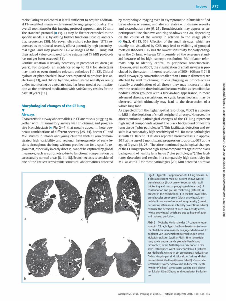
Fig. 2 Typical CT appearance of CF lung disease. a,b This adolescent male CF patient shows typicalbronchiectasis (black arrow) together with wallthickening and mucus plugging (white arrow). Aconsolidation and pleural thickening (asterisk) ispresent in the middle lobe. c In the left lower lobe,bronchoceles are present (black arrowhead), em-bedded in an area of reduced lung density (mosaicperfusion). dMinimum intensity projections (MinIP)enhance the detection of such low-density areas,(white arrowhead) which are due to hyperinflationand reduced perfusion.
Abb.2 Typische Merkmale der CF-Lungenerkran-kung im CT. a, b Typische Bronchiektasen (schwar-zer Pfeil) bei einemmännlichen Jugendlichen mit CFbegleitet von Bronchialwandverdickungen sowieMukoidimpaktion (weißer Pfeil). Eine Konsolidie-rung sowie angrenzende pleurale Verdickung(Sternchen) ist im Mittellappen erkennbar. c Derlinke Unterlappen weist Bronchozelen auf (schwar-zer Pfeilkopf), welche in ein Lungenareal reduzierterDichte eingelagert sind (Mosaikperfusion). d Mini-mum-Intensitäts-Projektionen (MinIP) können dieSichtbarkeit solcher Areale mit reduzierter Dichte(weißer Pfeilkopf) verbessern, welche die Folge ei-ner lokalen Überblähung und reduzierter Perfusionsind.
Wielpütz MO et al. Imaging of Cystic… Fortschr Röntgenstr 2016; 188: 834–845
Review 837
Thi
s do
cum
ent w
as d
ownl
oade
d fo
r pe
rson
al u
se o
nly.
Una
utho
rized
dis
trib
utio
n is
str
ictly
pro
hibi
ted.
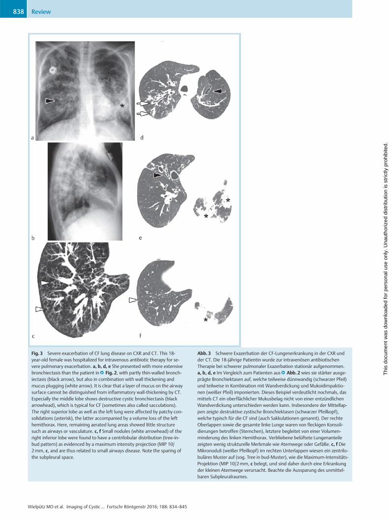
Fig. 3 Severe exacerbation of CF lung disease on CXR and CT. This 18-year-old female was hospitalized for intravenous antibiotic therapy for se-vere pulmonary exacerbation. a, b, d, e She presented with more extensivebronchiectasis than the patient in●▶ Fig. 2, with partly thin-walled bronch-iectasis (black arrow), but also in combination with wall thickening andmucus plugging (white arrow). It is clear that a layer of mucus on the airwaysurface cannot be distinguished from inflammatory wall-thickening by CT.Especially the middle lobe shows destructive cystic bronchiectasis (blackarrowhead), which is typical for CF (sometimes also called sacculations).The right superior lobe as well as the left lung were affected by patchy con-solidations (asterisk), the latter accompanied by a volume loss of the lefthemithorax. Here, remaining aerated lung areas showed little structuresuch as airways or vasculature. c, f Small nodules (white arrowhead) of theright inferior lobe were found to have a centrilobular distribution (tree-in-bud pattern) as evidenced by a maximum intensity projection (MIP 10/2mm, c, and are thus related to small airways disease. Note the sparing ofthe subpleural space.
Abb.3 Schwere Exazerbation der CF-Lungenerkrankung in der CXR undder CT. Die 18-jährige Patientin wurde zur intravenösen antibiotischenTherapie bei schwerer pulmonaler Exazerbation stationär aufgenommen.a, b, d, e Im Vergleich zum Patienten aus●▶ Abb.2 wies sie stärker ausge-prägte Bronchiektasen auf, welche teilweise dünnwandig (schwarzer Pfeil)und teilweise in Kombination mit Wandverdickung und Mukoidimpaktio-nen (weißer Pfeil) imponierten. Dieses Beispiel verdeutlicht nochmals, dasmittels CT ein oberflächlicher Mukusbelag nicht von einer entzündlichenWandverdickung unterschieden werden kann. Insbesondere der Mittellap-pen zeigte destruktive zystische Bronchiektasen (schwarzer Pfeilkopf),welche typisch für die CF sind (auch Sakkulationen genannt). Der rechteOberlappen sowie die gesamte linke Lunge waren von fleckigen Konsoli-dierungen betroffen (Sternchen), letztere begleitet von einer Volumen-minderung des linken Hemithorax. Verbliebene belüftete Lungenanteilezeigten wenig strukturelle Merkmale wie Atemwege oder Gefäße. c, f DieMikronoduli (weißer Pfeilkopf) im rechten Unterlappen wiesen ein zentrilo-buläres Muster auf (sog. Tree in bud-Muster), wie die Maximum-Intensitäts-Projektion (MIP 10/2mm, c belegt, und sind daher durch eine Erkrankungder kleinen Atemwege verursacht. Beachte die Aussparung des unmittel-baren Subpleuralraumes.
Wielpütz MO et al. Imaging of Cystic… Fortschr Röntgenstr 2016; 188: 834–845
Review838
Thi
s do
cum
ent w
as d
ownl
oade
d fo
r pe
rson
al u
se o
nly.
Una
utho
rized
dis
trib
utio
n is
str
ictly
pro
hibi
ted.
overall prevalence of approx. 90% in patients aged 0 to 6 years(mean age: 3.1 years) [11]. In a direct comparison, MRI showed ahigh correlation with CT-diagnosed structural abnormalities in aCF population aged 7–42 years (mean age: 16.7 years) [29]. Evenkey features such as the tree-in-bud pattern could be observed.
Mucus plugging is linked to the basic ion-transport defect andconstitutes the second most frequent morphological abnormal-ity (●▶ Fig. 2–4) [11, 36]. Whereas mucus plugging received lit-tle attention in recent CT studies, MRI detected a high overallprevalence of mucus plugging of approx. 63% of cases, making
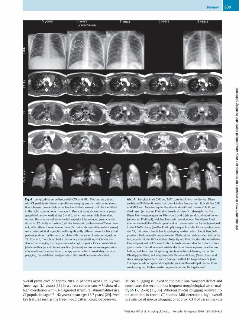
Fig. 4 Longitudinal surveillance with CXR and MRI. This female patientwith CF participates in our surveillance imaging program with annual rou-tine follow-up. Irreversible bronchiectasis (black arrow) could be identifiedin the right superior lobe from age 5. These airways showed mucus plug-ging (black arrowhead) at age 5 and 6, which was reversible thereafter.Around this area as well as in the left superior lobe reduced parenchymalsignal on T2 (white arrowhead) similar to mosaic perfusion on CTwas pres-ent, with different severity over time. Perfusion abnormalities (white arrow)were detected at all ages, but with significantly different severity. Note thatperfusion abnormalities also correlate with the areas of reduced signal onT2. At age 6, the subject had a pulmonary exacerbation, which was evi-denced on imaging by the presence of a right superior lobe consolidation(circle) with adjacent pleural reaction (asterisk) and more severe perfusionabnormalities. One year later (therapy was enacted immediately), mucusplugging, consolidation and perfusion abnormalities were alleviated.
Abb.4 Longitudinale CXR und MRT zum Krankheitsmonitoring. Dieseweibliche CF-Patientin nimmt an dem lokalen Programmmit jährlicher CXRund MRT zum Monitoring der Krankheitsaktivität teil. Irreversible Bron-chiektasen (schwarzer Pfeil) sind bereits ab dem 5. Lebensjahr sichtbar.Diese Atemwege zeigten im Alter von 5 und 6 Jahren Mukoidimpaktionen(schwarzer Pfeilkopf), welches hiernach reversibel war. Um dieses Arealebenso wie im linken Oberlappen fand sich ein reduziertes Parenchymsignalin der T2-Wichtung (weißer Pfeilkopf), vergleichbar der Mosaikperfusion inder CT, mit unterschiedlicher Ausprägung zu den unterschiedlichen Zeit-punkten. Perfusionsstörungen (weißer Pfeil) zeigten sich zu allen Zeitpunk-ten, jedoch mit deutlich variabler Ausprägung. Beachte, dass das reduzierteParenchymsignal in T2-gewichteten Aufnahmen mit den Perfusionsstörun-gen korreliert. Im Alter von 6 erlebte die Patientin eine pulmonale Exazer-bation, welche in der Bildgebung durch eine Konsolidierung im rechtenOberlappen (Kreis) mit angrenzender Pleuraverdickung (Sternchen), undstark ausgeprägten Perfusionsstörungen auffiel. Im folgenden Jahr (eineTherapie wurde umgehend eingeleitet) waren Mukoidimpaktionen, Kon-solidierung und Perfusionsstörungen wieder deutlich gebessert.
Wielpütz MO et al. Imaging of Cystic… Fortschr Röntgenstr 2016; 188: 834–845
Review 839
Thi
s do
cum
ent w
as d
ownl
oade
d fo
r pe
rson
al u
se o
nly.
Una
utho
rized
dis
trib
utio
n is
str
ictly
pro
hibi
ted.

it the secondmost frequent morphological abnormality in clini-cally stable infants and preschool children with CF (mean age:3.1 years) [11]. Neither CT nor CXR can distinguish mucus onthe airway surface from inflammatory wall thickening of largerairways (●▶ Fig. 2, 3). The possibility for different tissue contrastsin combination with contrast enhancement is a clear advantageof MRI. Wall thickening due to edema will lead to high signalintensity on T2-weighted images, reflecting active inflamma-tion (●▶ Fig. 4). Contrast enhancement of the airway wall on T1-weighted sequences is also a marker of inflammation, whereasintraluminal fluid will show a low signal. Importantly, mucusplugging may become a useful outcome measure in early CFlung disease as a potentially reversible abnormality [11, 12, 36].
ParenchymaConsolidations are typical signs of infection and are found in pul-monary exacerbations in CF. In many cases, an atelectasis withreduced volume and displacement of the pulmonary fissures oc-curs in exacerbation, unlike typical lobar pneumonia in other-wise healthy patients [37]. CXR usually has the lowest sensitivity,while CT andMRI perform equally well (●▶ Fig. 2–5) [38]. OnMRI,consolidations stand out brightly on T2-weighted sequences(●▶ Fig. 4). In case of a destroyed segment or lobe, bronchiectasisembedded in persistent consolidation and volume loss are evi-dent. In a group of 10 patients with pulmonary exacerbations(age range: 0–6 years, mean age: 3.7 years), consolidations onMRI were more frequent than in a comparable group in a clinical-ly stable situation [11]. Moreover, they were alleviated under an-
Fig. 5 Functional CT and MRI. This figure refers to the same patient as
●▶ Fig. 3a, b. Coronary reconstructions of the acquisition in end-inspirationassist in depicting the course of cystic bronchiectasis (black arrow). Mucusplugging (black arrowhead) was present especially in the right inferior andpatchy consolidations (asterisk) in the left inferior lobe. Mosaic perfusionmay be suspected in both lungs. c, d End-expiratory acquisitions assist inidentifying areas of air-trapping as a cause of mosaic perfusion by increas-ing the density of normal lung tissue able to exhale normally. Areas of air-trapping do not significantly increase in density in expiration and show areduction of vascularity. Please note the volume loss and limited diaphrag-matic movement of the left lung as indexed by the black line. e, f PerfusionMRI revealed an altogether inhomogeneous lung perfusion (compare ex-amples in●▶ Fig. 4) as well as areas of complete perfusion loss (white arrow)nicely matching air-trapping on expiratory CT.
Abb. 5 Funktionelle CT und MRT. Selbe Patientin wie in●▶ Abb. 3a, b. Kor-onore Rekonstruktionen der Akquisition in End-Inspiration sind hilfreich, umden Verlauf der zystischen Bronchiektasen (schwarzer Pfeil) nachzuvollzie-hen. Mokoidimpaktionen (schwarzer Pfeilkopf) fanden sich vor allem imrechten und fleckige Konsolidierungen (Sternchen) im linken Unterlappen.Eine Mosaikperfusion ist angedeutet beidseits erkennbar. c, d End-exspira-torische Aufnahmen dienen dem Nachweis von Air-Trapping als Ursache derMosaikperfusion durch Zunahme der Dichte von Lungengewebe aus dem inAusatmung die Luft normal entweichen kann. Lungengewebe mit Air-Trap-ping nimmt dagegen in Exspiration nicht an Dichte zu und weißt reduzierteGefäßkaliber auf. Beachte die Volumenreduktion und verminderte Zwerch-fellbeweglichkeit der linken Lunge, markiert durch die schwarze Hilfslinie.e, f In der Perfusions-MRT zeigte sich eine insgesamt inhomogene Perfusion(vergleiche Beispiele in●▶ Abb. 4) sowie flächige Perfusionsausfälle (weißerPfeil), welche sehr gut mit dem Air-Trapping korrelierten.
Wielpütz MO et al. Imaging of Cystic… Fortschr Röntgenstr 2016; 188: 834–845
Review840
Thi
s do
cum
ent w
as d
ownl
oade
d fo
r pe
rson
al u
se o
nly.
Una
utho
rized
dis
trib
utio
n is
str
ictly
pro
hibi
ted.

tibiotic therapy, making it a potentially reversible abnormality.However, mucus plugging and perfusion abnormalities seemedto play a greater role in exacerbation and were more responsiveto treatment than consolidations [11]. Peripheral consolidationsmay lead to pleural thickening and enhancement of the adjacentpleura (●▶ Fig. 2, 4) [11, 39]. Recent work using quantitative CThas confirmed earlier histopathological descriptions that adoles-cent and adult CF patients develop emphysema (age range: 7–66years, median age: 20.1 years) [40, 41]. This is also supported by amouse model showing that emphysema formation in advancedCF is pathophysiologically linked to emphysema in COPD [42–44].
Functional imaging – air trapping and lung perfusion▼Small airway obstruction prevents air from being exhaled fromlung volumes the size of a lobule to whole lobes. These have a re-duced alveolar oxygen level and may be hyperinflated. The phys-iological effect called hypoxic pulmonary vasoconstriction (HPV,formerly “Euler-Lilljestrand-Reflex”) leads to reduced perfusionto such lung areas in order to prevent intrapulmonary shunting.In airway diseases such as CF, airway obstruction frequently oc-curs and thus leads to a redistribution of the pulmonary bloodvolume. On inspiratory CTscans, the reduced capillary blood con-tent may be detected by a reduced parenchymal density inHounsfield Units (HU). It is often surrounded by and sharply de-lineated against normal lung and shows reduced vessel numbersand calibers also. Such an appearance was termed mosaic perfu-sion [13, 14, 45]. Its visual perception may be enhanced by end-expiratory CT acquisitions: During normal expiration, lung vol-ume as well as the amount of air per voxel decreases, thus leadingto an increase of its density value on CT. As compared to normal,lung areas with small-airway obstruction do not significantlychange volume or increase in density on expiratory acquisitions.Thus, the density difference between areas of airway obstructionand normal lung is expanded, increasing the sensitivity of CT fordetection. If a mosaic of different densities is seen on expiratoryCT, it is generally termed “air trapping” [13, 14]. Using expiratoryCT, air trapping has been described in approx. 70% of newborns,infants and preschool children with CF (age range: 0–5 years)(●▶ Fig. 2, 5) [8, 18, 25].Similar to mosaic perfusion, areas of lower signal intensity mayalso be visible on T2-weighted as well as post-contrast T1-weighted sequences withMRI due to the higher parenchymal sig-nal of normal lung, but the sensitivity may be lower than with CT(●▶ Fig. 4). The effect of HPV implies that imaging of lung perfu-sion approximates lung ventilation [46]. Thus, perfusion MRIshould – in theory – identify identical areas of pathology as airtrapping on expiratory CT. However, data on direct comparisonis missing. Typical patchy or wedge-shaped perfusion defects oc-cur on MRI and it was shown that these areas of hypoperfusioncorrelate with the degree of parenchymal changes in pediatric(age range: 0–6 years, mean age: 3.1 years) and adolescent (agerange: 11–19 years, median age: 16 years) CF patients (●▶ Fig. 4,5) [11, 47]. Abnormal perfusion on MRI was already detected inthe first year of life, with an overall prevalence of 85% in pre-school children, comparable to the aforementioned prevalenceof air trapping [11]. Maybe more importantly, perfusion altera-tions occurred even without detectable parenchymal changes[11]. This suggests that air trapping and perfusion abnormalitiesmay be the earliest signs of disease detectable in the CF lung,
even before morphological changes become visible. Air trap-ping/perfusion abnormalities may reflect reversible disease andhold the possibility for therapy monitoring [11], but may becomefixed in advanced CF with extensive parenchymal damage.
Scoring and quantitative imaging▼To quantify disease severity and facilitate patient follow-up andmonitoring of therapeutic effects in CF, visual scoring systemshave been developed for CXR (e. g. Chrispin-Norman Score, Bras-field Score, Wisconsin Score) [15, 35, 48, 49], CT (e. g. BhallaScore, Helbich Score, Brody Score) [4, 50, 51], and more recently,MRI (Eichinger Score) [39]. These scoring systems are necessarybecause the described changes in the CF lung show a heteroge-neous distribution within one patient and between different pa-tients, and may intra-individually show a different course overtime. Thus, the scores encompass structural changes (CXR, CT,MRI) as well as functional changes (air trapping on CT, perfusionabnormalities on MRI), and assign a numeric score to lung re-gions or lobes depending on the severity of the individual pathol-ogy. A previous study reported that the correlation of lung func-tion parameters with CT was higher than with CXR, indicatingthat CT provides a more precise grading than CXR [52]. Most im-portantly, CT scoring proved to be superior over pulmonary func-tion testing in detecting subtle disease progression [6], and hasalready been used to detect therapy response [7]. A more ad-vanced approach uses a grid overlay on selected CT slices and al-lows a reader to assign a pathology to each lung-containingsquare, leading to semi-automatic scoring [53]. Still, automaticobjective quantification of image information remains desirable.There is high potential in the direct quantification of airwaychanges by generation- and lobe-based quantitative post-proces-sing of non-enhanced thin-slice CT datasets (●▶ Fig. 6). Putativeimaging biomarkers such as wall thickness or airway diameter,air trapping, and emphysema may be derived [19, 40], but ahigh amount of automation is necessary to avoid any user inter-action and bias [19].Dedicated software tools for the quantification of MRI perfusionbased on the indicator dilution theory are already available,which can at least perform the initial step of segmenting thelung from the 4D perfusion dataset [54, 55]. Four parametershave been developed to reflect the characteristics of pulmonaryhemodynamics: pulmonary blood flow (PBF), blood volume(PBV), mean transit time (MTT), and time-to-peak (TTP) [56].Using a modification of these parameters it could be shown thatperfusion in the CF lung may not only be reduced by peak quan-tity but also delayed [57]. It has been speculated that delayed per-fusion may reflect increased bronchial arterial supply in ad-vanced lung disease. Because these receive blood from thesystemic circulation, increased flow will result in a left-to-leftshunt, which is still of uncertain clinical significance.
Advanced ventilation and perfusion imaging with MRI▼An option for direct visualization of lung ventilation is the imagingof nuclei other than 1H, namely 3He and 129Xe [58]. By this ap-proach, MRI after inhalation of the noble gas will display ventilatedairspace only. Hyperpolarized 3He-MRI depicted a high number ofventilation defects in CF patients compared to healthy volunteers,which correlated with a decrease in lung function [59, 60], but
Wielpütz MO et al. Imaging of Cystic… Fortschr Röntgenstr 2016; 188: 834–845
Review 841
Thi
s do
cum
ent w
as d
ownl
oade
d fo
r pe
rson
al u
se o
nly.
Una
utho
rized
dis
trib
utio
n is
str
ictly
pro
hibi
ted.

showed poor correlation with chest X-ray scoring [61]. Ventilationdefects are present even in CF patients with normal lung functiontesting and may change after airway clearance treatment [62]. So-phisticated technical prerequisites and the price for noble gas iso-topes make this promising research tool expensive and rather un-likely to be introduced into routine patient care.A promising technique is the direct regional quantification of T1relaxation times. As a physical parameter, it is thought to providean objective parameter for the characterization of pulmonary tis-sue independent of scanner type or observer [63]. Preliminaryresults obtained in patients indicate that T1 relaxation time issignificantly shorter in lungs affected by emphysema or cysticfibrosis [64]. Furthermore, T1 mapping can be combined withoxygen-enhanced MRI, which exploits the paramagnetic effectof molecular oxygen (O2) for the indirect assessment of lung ven-tilation. The slope of T1 decrease at different oxygen levels cor-related with perfusion abnormalities [65].Another newly developed technique relies on the periodical sig-nal changes of free-breathing bSSFP sequences at high temporal
resolution induced by respiration and pulsatory blood inflow[66]. A mathematical Fourier decomposition separates these dif-ferent frequency peaks and allows for the calculation of ventila-tion and perfusion maps. Preliminary results from patients withCF (age range: 0–30 years, median age: 4.1 years) show a goodagreement with contrast-enhanced perfusion imaging [54](●▶ Fig. 7).
Summary and outlook▼Although many authors advocate regular imaging studies at spe-cialized CF centers, data on the actual impact of imaging findingson treatment decisions and patient survival is lacking. Therefore,German and international guidelines usually do not specify atwhat age surveillance imaging of the CF lung should be started,or even which modality should be employed [67]. Chest CT is su-perior to CXR due to higher sensitivity for morphological changesin the CF lung, but routine surveillance CT acquisitions have sub-
Fig. 6 Quantitative CT post-processing. a The initial step of automatic air-way analysis is the segmentation of the whole airway tree from the CT da-taset. Bronchiectasis can nicely be seen as buddings at the end of an airwaybranch on the 3D volume rendering. b A centerline is then calculated foreach airway segment, which represents the long axis of each airway. c Sub-sequently, secondary reconstructions running perpendicular to the airwayaxis (centerline) are produced, which show an axial view for each airwaysegment. d On these, the inner (green line) and outer (red line) airway wallmay be detected andmeasured by sophisticated algorithms. The yellow linemarks the points of maximum wall attenuation. Images by YACTA, pro-gramming by Oliver Weinheimer, Heidelberg.
Abb.6 Quantitative CT-Nachverarbeitung. a Als erster Schritt einer auto-matischen Atemwegsanalyse erfolgt die Segmentierung des gesamtenAtemwegsbaums aus einem CT-Datensatz. Bronchiektasen lassen sichleicht als Knospungen an den Enden der Atemwege in der 3D gerendertenRekonstruktion erkennen. b Hiernach wird eine Mittellinie (Centerline) be-rechnet, die der Längsachse eines jeden Atemwegs folgt. c Sekundäre Re-konstruktionen senkrecht zur Atemwegsachse (Centerline) werden erstellt,die nun eine axiale Sicht des jeweiligen Atemwegssegments erlauben. d Aufdiesen kann nun die innere (grüne Linie) und äußere (rote Linie) Begren-zung der Atemwegswand mittels spezieller Rechenalgorithmen detektiertwerden. Die gelbe Linie zeigt die Punkte der höchsten Dichte der Atem-wegswand. Bilder erstellt mit YACTA, programmiert von Oliver Weinhei-mer, Heidelberg.
Wielpütz MO et al. Imaging of Cystic… Fortschr Röntgenstr 2016; 188: 834–845
Review842
Thi
s do
cum
ent w
as d
ownl
oade
d fo
r pe
rson
al u
se o
nly.
Una
utho
rized
dis
trib
utio
n is
str
ictly
pro
hibi
ted.

sequently led to an increase in radiation exposure to CF patients,whichmay even rise further with earlier diagnosis and prolongedsurvival [10]. A remaining role for CXR could be imaging at an-nual follow-up together with MRI as a cross-sectional modalityfor use of CXR as a reference when it is repeated at interim pre-sentations between annual follow-up, for example in the caseof exacerbation. Recently, chest MRI has entered clinical routinepractice in CF [12]. Thus, radiologists and clinicians now can optfor the optimal modality adapted to the clinical context of theirCF patients (●▶ Table 2). The risk of sedation in preschool childrenand allergies against MRI contrast material must be weighedagainst the risk from radiation exposure [9, 10, 68]. Importantly,to use MRI in CF as a routine surveillance tool is not limited to the
depiction of structural information as with CT just using a radia-tion-free method. MRI’s capability for combined morphologicaland functional imaging at sufficient spatial and high temporal re-solution to obtain information on regional lung function shouldbe taken into account as well. To appreciate its advantages overCT, a perfusion study, which is available on most state-of-the-artMRI scanners already, should be included in the MRI protocol(●▶ Fig. 1).
Affiliations1 Department of Diagnostic and Interventional Radiology, Subdivision ofPulmonary Imaging, University Hospital of Heidelberg, Heidelberg, Germany
2 Translational Lung Research Center Heidelberg (TLRC), Member of theGerman Lung Research Center (DZL), Heidelberg, Germany
3 Department of Diagnostic and Interventional Radiology with Nuclear Medi-cine, Thoraxklinik at the University Hospital of Heidelberg, Heidelberg,Germany
4 Radiologie Darmstadt, Groß-Gerau Community Hospital, Groß-Gerau,Germany
5 Department of Pulmonology and Respiratory Medicine, Cystic FibrosisCenter, Thoraxklinik at the University Hospital of Heidelberg, Heidelberg,Germany
6 Division of Pediatric Pulmonology & Allergy and Cystic Fibrosis Center,Department of Pediatrics, University of Heidelberg, Heidelberg, Germany
7 Department of Translational Pulmonology, University Hospital Heidelberg,Heidelberg, Germany
8 Department of Diagnostic and Interventional Radiology, Hufeland Hospital,Bad Langensalza, Germany
Conflicts of interest/Support statement: This study was suppor-ted by grants from the Bundesministerium für Bildung und For-schung (BMBF) to the German Center for Lung Research (DZL)(82DZL00401, 82DZL00402, 82DZL00404). Supported by a finan-
Table 2 Suggested imaging scheme according to experience in Heidelbergfor life-long imaging surveillance of CF patients starting at birth.
Tab. 2 Heidelberger Schema als Vorschlag zum longitudinalen bildgeben-den Monitoring der CF ab-Geburt.
CXR CT MRI
diagnosis, screening < 1 year X X (no CM)
diagnosis ≥ 1 year X X
annual follow-up < 18 years X X
annual follow-up ≥ 18 years X X
clinical exacerbation X (X)
emergency, hemorrhage X (CM)
CM=contrast material.CM=Kontrastmittel.
Fig. 7 Non-contrast enhanced combined ventila-tion and perfusion imaging with MRI. a, b Apartfrom areas with reduced parenchymal signal on T2-weighted imaging (white arrowhead) this school-age female with CF in stable clinical conditionshowed few airway abnormalities. Contrast-en-hanced perfusion MRI revealed areas of reducedperfusion (white arrow, b), comparable to theaforesaid areas with reduced T2-signal. c, d Fourier-decomposition MRI detected nicely matching areasof reduced ventilation (black arrow, c) and perfu-sion (white arrow, d) without the need for contrastmaterial injection.
Abb.7 Kontrastmittelfreie kombinierte Bildge-bung von Ventilation und Perfusion mittels MRT.a, b Neben flächigen Signalminderungen des Pa-renchyms in der T2-Wichtung (weißer Pfeilkopf)zeigte diese Patientin im Schulalter mit stabiler CFkaum Atemwegsveränderungen. Die kontrastmit-telverstärkte Perfusions-MRT deckte deutlicheAreale mit reduzierter Perfusion auf (weißer Pfeil,b), vergleichbar zu den vorgenannten Arealen mitT2-Signalminderung. c, d Die Fourier-Dekomposi-tions-MRT erlaubte die Detektion von hierzu gutkorrelierenden Arealen mit reduzierter Ventilation(schwarzer Pfeil, c) und Perfusion (weißer Pfeil, d)ohne die Notwendigkeit einer Kontrastmittelinjek-tion.
Wielpütz MO et al. Imaging of Cystic… Fortschr Röntgenstr 2016; 188: 834–845
Review 843
Thi
s do
cum
ent w
as d
ownl
oade
d fo
r pe
rson
al u
se o
nly.
Una
utho
rized
dis
trib
utio
n is
str
ictly
pro
hibi
ted.
cial grant from the Christiane Herzog Stiftung, Stuttgart, Germa-ny, and theMukoviszidose e.V. (S02/09), Bonn, the German CysticFibrosis Association. Technical support provided by SiemensHealthcare, Germany.
References01 Stern M, Wiedemann B, Wenzlaff P. From registry to quality manage-
ment: the German Cystic Fibrosis Quality Assessment project 1995 –
2006. Eur Respir J 2008; 31: 29–3502 Sommerburg O, Hammermann J, Lindner M et al. Five years of experi-
ence with biochemical cystic fibrosis newborn screening based onIRT/PAP in Germany. Pediatr Pulmonol 2015; 50: 655–664
03 Kerem E, Reisman J, Corey M et al. Prediction of mortality in patientswith cystic fibrosis. N Engl J Med 1992; 326: 1187–1191
04 Brody AS, Sucharew H, Campbell JD et al. Computed tomography corre-lates with pulmonary exacerbations in children with cystic fibrosis.Am J Respir Crit Care Med 2005; 172: 1128–1132
05 Terheggen-Lagro S, Truijens N, van Poppel N et al. Correlation of six dif-ferent cystic fibrosis chest radiograph scoring systems with clinicalparameters. Pediatr Pulmonol 2003; 35: 441–445
06 de Jong PA, Lindblad A, Rubin L et al. Progression of lung disease oncomputed tomography and pulmonary function tests in children andadults with cystic fibrosis. Thorax 2006; 61: 80–85
07 Davis SD, Fordham LA, Brody AS et al. Computed tomography reflectslower airway inflammation and tracks changes in early cystic fibrosis.Am J Respir Crit Care Med 2007; 175: 943–950
08 Sly PD, Brennan S, Gangell C et al. Lung disease at diagnosis in infantswith cystic fibrosis detected by newborn screening. Am J Respir CritCare Med 2009; 180: 146–152
09 Donadieu J, Roudier C, Saguintaah M et al. Estimation of the radiationdose from thoracic CT scans in a cystic fibrosis population. Chest2007; 132: 1233–1238
10 O'Connell OJ, McWilliams S, McGarrigle A et al. Radiologic imaging incystic fibrosis: cumulative effective dose and changing trends over 2decades. Chest 2012; 141: 1575–1583
11 Wielpütz MO, Puderbach M, Kopp-Schneider A et al. Magnetic Reso-nance Imaging Detects Changes in Structure and Perfusion, and Re-sponse to Therapy in Early Cystic Fibrosis Lung Disease. Am J RespirCrit Care Med 2014; 189: 956–965
12 Wielpütz MO,Mall MA. Imaging modalities in cystic fibrosis: emergingrole of MRI. Curr Opin Pulm Med 2015; 21: 609–616
13 Hansell DM, Bankier AA,MacMahon H et al. Fleischner Society: glossaryof terms for thoracic imaging. Radiology 2008; 246: 697–722
14 Wormanns D, Hamer OW. Glossar thoraxradiologischer Begriffeentsprechend der Terminologie der Fleischner Society. FortschrRöntgenstr 2015; 187: 638–661
15 Benden C,Wallis C, Owens CM et al. The Chrispin-Norman score in cys-tic fibrosis: doing away with the lateral view. Eur Respir J 2005; 26:894–897
16 O'Connor OJ, Vandeleur M, McGarrigle AM et al. Development of low-dose protocols for thin-section CT assessment of cystic fibrosis in pe-diatric patients. Radiology 2010; 257: 820–829
17 Kauczor HU,Wielpütz MO, OwsijewitschM et al. Computed tomograph-ic imaging of the airways in COPD and asthma. J Thorac Imaging 2011;26: 290–300
18 Mott LS, Park J, Gangell CL et al. Distribution of early structural lungchanges due to cystic fibrosis detected with chest computed tomog-raphy. J Pediatr 2013; 163: 243–248 e241–243
19 Wielpütz MO, Eichinger M, Weinheimer O et al. Automatic airway anal-ysis onmultidetector computed tomography in cystic fibrosis: correla-tion with pulmonary function testing. J Thorac Imaging 2013; 28:104–113
20 Bankier AA, Schaefer-Prokop C,DeMaertelaer V et al. Air trapping: com-parison of standard-dose and simulated low-dose thin-section CTtechniques. Radiology 2007; 242: 898–906
21 Hansell DM. Small airways diseases: detection and insights with com-puted tomography. Eur Respir J 2001; 17: 1294–1313
22 Eichinger M, Heussel CP, Kauczor HU et al. Computed tomography andmagnetic resonance imaging in cystic fibrosis lung disease. J Magn Re-son Imaging 2010; 32: 1370–1378
23 Kubo T, Lin PJ, Stiller W et al. Radiation dose reduction in chest CT: a re-view. Am J Roentgenol 2008; 190: 335–343
24 Stiller W. Grundlagen der Mehrzeilendetektor-Computertomografie.Der Radiologe 2011; 51: 1061–1078
25 Sly PD, Gangell CL, Chen L et al. Risk factors for bronchiectasis in chil-dren with cystic fibrosis. N Engl J Med 2013; 368: 1963–1970
26 Hayes D Jr, Winkler MA, Kirkby S et al. Preprocedural planning withprospectively triggered multidetector row CT angiography prior tobronchial artery embolization in cystic fibrosis patients with massivehemoptysis. Lung 2012; 190: 221–225
27 Biederer J, Beer M, Hirsch W et al.MRI of the lung (2/3). Why ... when ...how? Insights Imaging 2012; 3: 355–371
28 Puderbach M, Eichinger M, Gahr J et al. Proton MRI appearance of cysticfibrosis: comparison to CT. Eur Radiol 2007; 17: 716–724
29 Puderbach M, Eichinger M, Haeselbarth J et al. Assessment of morpho-logical MRI for pulmonary changes in cystic fibrosis (CF) patients:comparison to thin-section CT and chest x-ray. Invest Radiol 2007;42: 715–725
30 Biederer J, Heussel CP, Puderbach M et al. Functional magnetic resonanceimaging of the lung. Semin Respir Crit Care Med 2014; 35: 74–82
31 Dournes G, Grodzki D,Macey J et al. Quiet Submillimeter MR Imaging ofthe Lung Is Feasible with a PETRA Sequence at 1.5 T. Radiology 2015;276: 258–265
32 Lutterbey G,Wattjes MP, Doerr D et al. Atelectasis in children undergo-ing either propofol infusion or positive pressure ventilation anesthesiafor magnetic resonance imaging. Paediatr Anaesth 2007; 17: 121–125
33 Blitman NM, Lee HK, Jain VR et al. Pulmonary atelectasis in children an-esthetized for cardiothoracic MR: evaluation of risk factors. J ComputAssist Tomogr 2007; 31: 789–794
34 Loeve M, van Hal PT, Robinson P et al. The spectrum of structural ab-normalities on CT scans from patients with CF with severe advancedlung disease. Thorax 2009; 64: 876–882
35 Chrispin AR, Norman AP. The systematic evaluation of the chest radio-graph in cystic fibrosis. Pediatr Radiol 1974; 2: 101–105
36 Mall MA, Hartl D. CFTR: cystic fibrosis and beyond. Eur Respir J 2014;44: 1042–1054
37 Rosenfeld M, Ratjen F, Brumback L et al. Inhaled hypertonic saline in in-fants and children younger than 6 years with cystic fibrosis: the ISISrandomized controlled trial. JAMA 2012; 307: 2269–2277
38 Eibel R,Herzog P,Dietrich O et al. Pulmonary abnormalities in immuno-compromised patients: comparative detection with parallel acquisi-tion MR imaging and thin-section helical CT. Radiology 2006; 241:880–891
39 Eichinger M, Optazaite DE, Kopp-Schneider A et al. Morphologic andfunctional scoring of cystic fibrosis lung disease usingMRI. Eur J Radiol2012; 81: 1321–1329
40 Wielpütz MO,Weinheimer O, Eichinger M et al. Pulmonary emphysemain cystic fibrosis detected by densitometry on chest multidetectorcomputed tomography. PLoS One 2013; 8: e73142
41 Mets OM, Roothaan SM, Bronsveld I et al. Emphysema Is Common inLungs of Cystic Fibrosis Lung Transplantation Patients: A Histopatholo-gical and Computed Tomography Study. PLoS One 2015; 10: e0128062
42 Mall M, Grubb BR,Harkema JR et al. Increased airway epithelial Na+ ab-sorption produces cystic fibrosis-like lung disease in mice. Nat Med2004; 10: 487–493
43 Mall MA, Harkema JR, Trojanek JB et al. Development of chronic bron-chitis and emphysema in β-epithelial Na+ channel-overexpressingmice. Am J Respir Crit Care Med 2008; 177: 730–742
44 Wielpuetz MO, Eichinger M, Zhou Z et al. In vivo monitoring of cystic fi-brosis-like lung disease in mice by volumetric computed tomography.Eur Respir J 2011; 38: 1060–1070
45 Stern EJ,Müller NL, Swensen SJ et al. CT mosaic pattern of lung attenua-tion: etiologies and terminology. Journal of thoracic imaging 1995; 10:294–297
46 Hopkins SR, Wielpütz MO, Kauczor HU. Imaging lung perfusion. J ApplPhysiol 2012; 113: 328–339
47 Eichinger M, Puderbach M, Fink C et al. Contrast-enhanced 3D MRI oflung perfusion in children with cystic fibrosis–initial results. Eur Radi-ol 2006; 16: 2147–2152
48 Brasfield D, Hicks G, Soong S et al. The chest roentgenogram in cysticfibrosis: a new scoring system. Pediatrics 1979; 63: 24–29
49 Weatherly MR, Palmer CG, Peters ME et al. Wisconsin cystic fibrosischest radiograph scoring system. Pediatrics 1993; 91: 488–495
50 Bhalla M, Turcios N, Aponte V et al. Cystic fibrosis: scoring system withthin-section CT. Radiology 1991; 179: 783–788
51 Helbich TH, Heinz-Peer G, Eichler I et al. Cystic fibrosis: CT assessment oflung involvement in children and adults. Radiology 1999; 213: 537–544
Wielpütz MO et al. Imaging of Cystic… Fortschr Röntgenstr 2016; 188: 834–845
Review844
Thi
s do
cum
ent w
as d
ownl
oade
d fo
r pe
rson
al u
se o
nly.
Una
utho
rized
dis
trib
utio
n is
str
ictly
pro
hibi
ted.

52 Demirkazik FB, Ariyurek OM, Ozcelik U et al. High resolution CT in chil-dren with cystic fibrosis: correlation with pulmonary functions andradiographic scores. Eur J Radiol 2001; 37: 54–59
53 Rosenow T, Oudraad MC, Murray CP. PRAGMA-CF. et al. A QuantitativeStructural Lung Disease Computed Tomography Outcome in YoungChildren with Cystic Fibrosis. Am J Respir Crit Care Med 2015; 191:1158–1165
54 Bauman G, Puderbach M, Heimann T et al. Validation of Fourier decom-position MRI with dynamic contrast-enhanced MRI using visual andautomated scoring of pulmonary perfusion in young cystic fibrosis pa-tients. Eur J Radiol 2013; 82: 2371–2377
55 Kohlmann P, Strehlow J, Jobst B et al. Automatic lung segmentationmethod for MRI-based lung perfusion studies of patients with chronicobstructive pulmonary disease. Int J Comput Assist Radiol Surg 2014;10: 15
56 Ohno Y, Hatabu H, Murase K et al. Quantitative assessment of regionalpulmonary perfusion in the entire lung using three-dimensional ultra-fast dynamic contrast-enhanced magnetic resonance imaging: Preli-minary experience in 40 subjects. J Magn Reson Imaging 2004; 20:353–365
57 Risse F, Eichinger M, Kauczor HU et al. Improved visualization of de-layed perfusion in lung MRI. Eur J Radiol 2011; 77: 105–110
58 van Beek EJ,Wild JM, Kauczor HU et al. Functional MRI of the lung usinghyperpolarized 3-helium gas. J Magn Reson Imaging 2004; 20: 540–554
59 Donnelly LF, MacFall JR, McAdams HP et al. Cystic fibrosis: combinedhyperpolarized 3He-enhanced and conventional proton MR imagingin the lung–preliminary observations. Radiology 1999; 212: 885–889
60 Mentore K, Froh DK, de Lange EE et al. Hyperpolarized HHe 3MRI of thelung in cystic fibrosis: assessment at baseline and after bronchodilatorand airway clearance treatment. Acad Radiol 2005; 12: 1423–1429
61 van Beek EJ, Hill C, Woodhouse N et al. Assessment of lung disease inchildrenwith cystic fibrosis using hyperpolarized 3-HeliumMRI: com-parison with Shwachman score, Chrispin-Norman score and spirome-try. Eur Radiol 2007; 17: 1018–1024
62 Bannier E, Cieslar K, Mosbah K et al. Hyperpolarized 3He MR for sensi-tive imaging of ventilation function and treatment efficiency in youngcystic fibrosis patients with normal lung function. Radiology 2010;255: 225–232
63 Triphan SM, Jobst BJ, Breuer FA et al. Echo time dependence of observedT in the human lung. J Magn Reson Imaging 2015; DOI: 10.1002/jmri.24840
64 Stadler A, Jakob PM, Griswold M et al. T1 mapping of the entire lungparenchyma: Influence of respiratory phase and correlation to lungfunction test results in patients with diffuse lung disease. Magn ResonMed 2008; 59: 96–101
65 Jakob PM, Wang T, Schultz G et al. Assessment of human pulmonaryfunction using oxygen-enhanced T(1) imaging in patients with cysticfibrosis. Magn Reson Med 2004; 51: 1009–1016
66 Bauman G, Scholz A, Rivoire J et al. Lung ventilation- and perfusion-weighted Fourier decomposition magnetic resonance imaging: in vivovalidation with hyperpolarized 3He and dynamic contrast-enhancedMRI. Magn Reson Med 2013; 69: 229–237
67 Müller FM, Bend J, Rietschel E et al. S3-Leitlinie „Lungenerkrankung beiMukoviszidose “, Modul 1: Diagnostik und Therapie nach dem erstenNachweis von Pseudomonas aeruginosa; 2013, http://www.awmf.org/uploads/tx_szleitlinien/026-022l_S3_Lungenerkrankung_bei_Mukoviszidose_Modul_1_2013-06_01.pdf
68 Kuo W, Ciet P, Tiddens HA et al. Monitoring cystic fibrosis lung diseaseby computed tomography. Radiation risk in perspective. Am J RespirCrit Care Med 2014; 189: 1328–1336
69 Stöver B, Rogalla P. CT-Untersuchungen bei Kindern. Der Radiologe2008; 48: 243–248
Wielpütz MO et al. Imaging of Cystic… Fortschr Röntgenstr 2016; 188: 834–845
Review 845
Thi
s do
cum
ent w
as d
ownl
oade
d fo
r pe
rson
al u
se o
nly.
Una
utho
rized
dis
trib
utio
n is
str
ictly
pro
hibi
ted.