Imaging Findings of the Developing Temporal Bone in Fetal ...
11
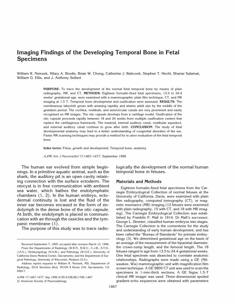
Imaging Findings of the Developing Temporal Bone in Fetal Specimens William R. Nemzek, Hilary A. Brodie, Brian W. Chong, Catherine J. Babcook, Stephen T. Hecht, Shariar Salamat, William G. Ellis, and J. Anthony Seibert PURPOSE: To trace the development of the normal fetal temporal bone by means of plain radiography, MR, and CT. METHODS: Eighteen formalin-fixed fetal specimens, 13.5 to 24.4 weeks’ gestational age, were examined with a mammographic plain film technique, CT, and MR imaging at 1.5 T. Temporal bone development and ossification were assessed. RESULTS: The membranous labyrinth grows with amazing rapidity and attains adult size by the middle of the gestation period. The cochlea, vestibule, and semicircular canals are very prominent and easily recognized on MR images. The otic capsule develops from a cartilage model. Ossification of the otic capsule proceeds rapidly between 18 and 24 weeks from multiple ossification centers that replace the cartilaginous framework. The mastoid, internal auditory canal, vestibular aqueduct, and external auditory canal continue to grow after birth. CONCLUSION: The study of fetal developmental anatomy may lead to a better understanding of congenital disorders of the ear. Faster MR scanning techniques may provide a method for in utero evaluation of the fetal temporal bone. Index terms: Fetus, growth and development; Temporal bone, anatomy AJNR Am J Neuroradiol 17:1467–1477, September 1996 The human ear evolved from simple begin- nings. In a primitive aquatic animal, such as the shark, the auditory pit is an open cavity retain- ing connection with the surface ectoderm. The otocyst is in free communication with ambient sea water, which bathes the endolymphatic chambers (1, 2). In the human embryo, ecto- dermal continuity is lost and the fluid of the inner ear becomes encased in the form of en- dolymph in the dense bone of the otic capsule. At birth, the endolymph is placed in communi- cation with air through the ossicles and the tym- panic membrane (1). The purpose of this study was to trace radio- logically the development of the normal human temporal bone in fetuses. Materials and Methods Eighteen formalin-fixed fetal specimens from the Car- negie Embryological Collection of normal fetuses at the University of California, Davis, were examined with plain film radiography, computed tomography (CT), or mag- netic resonance (MR) imaging (12 fetuses were examined with plain radiography, 15 with CT, and 18 with MR imag- ing). The Carnegie Embryological Collection was estab- lished by Franklin P. Mall in 1914. Dr Mall’s successor, George L. Streeter, classified human embryos into stages. The Carnegie Collection is the cornerstone for the study and understanding of early human development, and has been called the “Bureau of Standards” for primate embry- ology (3). We determined gestational age on the basis of an average of the measurement of the biparietal diameter, the crown-rump length, and the femoral length. The 18 fetuses ranged in age from 13.5 to 24.4 gestational weeks. One fetal specimen was dissected to correlate anatomic relationships. Radiographs were made using a GE (Mil- waukee, Wis) mammographic unit with magnification film screen technique. A GE 9800 CT unit was used to scan the specimens in 1-mm-thick sections. A GE Signa 1.5-T clinical MR imager was used. Three-dimensional spoiled gradient-echo sequences were obtained with parameters Received September 7, 1995; accepted after revision March 12, 1996. From the Departments of Radiology (W.R.N., B.W.C., C.J.B., S.T.H., J.A.S.), Otolaryngology (H.A.B.), and Pathology (W.G.E.), University of California Davis Medical Center, Sacramento; and the Department of Sur- gical Pathology, University of Wisconsin, Madison (S.S.). Address reprint requests to William R. Nemzek, MD, Department of Radiology, 2516 Stockton Blvd, TICON II Room 216, Sacramento, CA 95817. AJNR 17:1467–1477, Sep 1996 0195-6108/96/1708 –1467 q American Society of Neuroradiology 1467
Transcript of Imaging Findings of the Developing Temporal Bone in Fetal ...
Imaging Findings of the Developing Temporal Bone in FetalSpecimens
William R. Nemzek, Hilary A. Brodie, Brian W. Chong, Catherine J. Babcook, Stephen T. Hecht, Shariar Salamat,William G. Ellis, and J. Anthony Seibert
PURPOSE: To trace the development of the normal fetal temporal bone by means of plainradiography, MR, and CT. METHODS: Eighteen formalin-fixed fetal specimens, 13.5 to 24.4weeks’ gestational age, were examined with a mammographic plain film technique, CT, and MRimaging at 1.5 T. Temporal bone development and ossification were assessed. RESULTS: Themembranous labyrinth grows with amazing rapidity and attains adult size by the middle of thegestation period. The cochlea, vestibule, and semicircular canals are very prominent and easilyrecognized on MR images. The otic capsule develops from a cartilage model. Ossification of theotic capsule proceeds rapidly between 18 and 24 weeks from multiple ossification centers thatreplace the cartilaginous framework. The mastoid, internal auditory canal, vestibular aqueduct,and external auditory canal continue to grow after birth. CONCLUSION: The study of fetaldevelopmental anatomy may lead to a better understanding of congenital disorders of the ear.Faster MR scanning techniques may provide a method for in utero evaluation of the fetal temporalbone.
Index terms: Fetus, growth and development; Temporal bone, anatomy
AJNR Am J Neuroradiol 17:1467–1477, September 1996
67
The human ear evolved from simple begin-nings. In a primitive aquatic animal, such as theshark, the auditory pit is an open cavity retain-ing connection with the surface ectoderm. Theotocyst is in free communication with ambientsea water, which bathes the endolymphaticchambers (1, 2). In the human embryo, ecto-dermal continuity is lost and the fluid of theinner ear becomes encased in the form of en-dolymph in the dense bone of the otic capsule.At birth, the endolymph is placed in communi-cation with air through the ossicles and the tym-panic membrane (1).The purpose of this study was to trace radio-
Received September 7, 1995; accepted after revision March 12, 1996.From the Departments of Radiology (W.R.N., B.W.C., C.J.B., S.T.H.,
J.A.S.), Otolaryngology (H.A.B.), and Pathology (W.G.E.), University ofCalifornia Davis Medical Center, Sacramento; and the Department of Sur-gical Pathology, University of Wisconsin, Madison (S.S.).
Address reprint requests to William R. Nemzek, MD, Department ofRadiology, 2516 Stockton Blvd, TICON II Room 216, Sacramento, CA95817.
AJNR 17:1467–1477, Sep 1996 0195-6108/96/1708–1467
q American Society of Neuroradiology
14
logically the development of the normal humantemporal bone in fetuses.
Materials and MethodsEighteen formalin-fixed fetal specimens from the Car-
negie Embryological Collection of normal fetuses at theUniversity of California, Davis, were examined with plainfilm radiography, computed tomography (CT), or mag-netic resonance (MR) imaging (12 fetuses were examinedwith plain radiography, 15 with CT, and 18 with MR imag-ing). The Carnegie Embryological Collection was estab-lished by Franklin P. Mall in 1914. Dr Mall’s successor,George L. Streeter, classified human embryos into stages.The Carnegie Collection is the cornerstone for the studyand understanding of early human development, and hasbeen called the “Bureau of Standards” for primate embry-ology (3). We determined gestational age on the basis ofan average of the measurement of the biparietal diameter,the crown-rump length, and the femoral length. The 18fetuses ranged in age from 13.5 to 24.4 gestational weeks.One fetal specimen was dissected to correlate anatomicrelationships. Radiographs were made using a GE (Mil-waukee, Wis) mammographic unit with magnification filmscreen technique. A GE 9800 CT unit was used to scan thespecimens in 1-mm-thick sections. A GE Signa 1.5-Tclinical MR imager was used. Three-dimensional spoiledgradient-echo sequences were obtained with parameters

TABLE 1: Measurement of basal turn of the cochlea and lateral semicircular canal in fetuses and adults
Fetuses Adults
Age, wkBasalTurn,mm
LateralSemicircularCanal, mm
Age, yBasalTurn,mm
LateralSemicircularCanal, mm
1. 13.5 4.8 . . . 1. 18 8.5 5.02. 14.2 5.0 . . . 2. 24 9.0 5.53. 14.4 7.5 . . . 3. 30 8.5 5.54. 15 6.5 . . . 4. 42 9.0 5.05. 15.5 4.8 . . . 5. 42 8.5 6.56. 16.2 8.5 . . . 6. 46 8.5 5.57. 17.4 9.0 4.3 7. 70 9.0 5.38. 18.4 8.5 . . . 8. 76 9.5 5.59. 18.5 8.5 . . .
10. 19.3 8.5 4.311. 19.5 9.0 5.012. 19.5 9.5 . . .
13. 20.5 8.5 5.014. 21 8.3 5.015. 21.4 8.5 5.516. 22.3 8.5 4.817. 23.3 8.5 5.018. 24.4 9.0 5.3
1468 NEMZEK AJNR: 17, September 1996
of 40/8/4 (repetition time/echo time/excitations) and aflip angle of 458, an imaging matrix of 512 3 256, and acontiguous section thickness of 0.7 mm. Spin-echo T1-weighted sequences were obtained with parameters of500/90/3, a matrix of 512 3 384, and 3.0-mm-thick sec-tions with a 0.5-mm section gap. Fast spin-echo T2-weighted sequences were obtained with parameters of4000–5000/90–96/3–4, an echo train length of 12, a ma-trix of 512 3 384, and 3.0-mm-thick sections with a0.5-mm section gap.
The length of the basal turn of the cochlea and thediameter of the lateral semicircular canal were measuredon CT scans and MR images obtained of the fetal speci-mens and of eight adult subjects who were referred withnonotologic disorders. The length of the basal turn of thecochlea was considered to be the longest diameter thatcould be obtained through the first turn of the cochlea.
Results
The average length of the basal turn of theadult cochlea was 8.8 mm and the averagemaximum diameter of the adult lateral semicir-cular canal was 5.5 mm. The measurements ofthe fetal specimens and normal adults are givenin Table 1. The horizontal semicircular canalswere not measured in all fetuses, because thisstructure could not be defined in all specimens.Note that the otic capsules of the fetus haveachieved adult dimensions at approximately 21weeks’ gestational age.The fast spin-echo T2-weighted sequences
produced the best quality images, successfully
differentiating bright signal intensity of en-dolymph and perilymph from the intermediatesignal of unossified cartilage and the low signalof bone. In one adult, a fast spin-echo T2-weighted image (Fig 1A) showed high signal
Abbreviations for Figures 1 through 12
C cochleaCA cochlear aqueductCC common crusELD endolymphatic ductELS endolymphatic sacL labyrinthine segment of facial
nerve canalLSCC lateral semicircular canalPSCC posterior semicircular canalSIG sigmoid sinusSSCC superior semicircular canalSS subarcuate spaceTR tympanic ringV vestibuleVA vestibular aqueducti incusiac internal auditory canalis interscalar septumm malleusmo modioluspp pyramidal processst stapes

Fig 1. Comparison of adult and early fetal otic capsule structures on T2-weighted fast spin-echo MR images.A, In the adult, there is high signal intensity of endolymph and perilymph in membranous cochlea, and vestibule and semicircular
canals are surrounded by signal void of dense bony otic capsule.B, Sagittal image of fetus, gestational age 14 weeks 4 days. Basal turn of cochlea is the first structure that can be readily identified.C, Fetus, gestational age 15 weeks 5 days. Fluid is seen in cochlea and vestibule. Basal turn of cochlea is two thirds of adult size. The
upper turns of the membranous cochlea are now visible (arrow) as a single space. Note unossified cartilage is of intermediate signalintensity.
Fig 2. Fetus, gestational age 17 weeks 4 days.A, CT scan shows ossification in squamous portion (black arrow) and zygomatic
process of the temporal bone (white arrow). Note very early ossification of the malleus andincus.
B, T2-weighted fast spin-echo MR image shows vestibule and lateral semicircular canalbefore ossification.
Fig 3. CT scan of fetus, gestational age18 weeks 4 days, shows earliest ossificationof otic capsule. Note discontinuous calcifi-cation around the apical turn of the cochlea(small arrows). Note interscalar septumand progressive ossification of ossicles.
AJNR: 17, September 1996 TEMPORAL BONE 1469
intensity of fluid in the tiny membranous laby-rinth surrounded by the signal void of theheavily ossified petrous pyramid and the densebone of the otic capsule.The basal turn of the cochlea was the first
structure that was seen consistently in our ma-terial, and is easily identified on T2-weightedMR images. It was well seen in a fetus of 14weeks 4 days’ gestational age (Fig 1B). A fastspin-echo T2-weighted axial MR image of a fe-
tus of 15 weeks 5 days shows fluid in the co-chlea and vestibule (Fig 1C). These structuresare nearly two thirds of adult size. Because os-sification had not yet occurred, the otic capsuleis depicted as a structure of intermediate signalintensity relative to cartilage. Note the large sizeof the otic capsule as compared with the re-mainder of the fetal skull.A CT scan of a fetus of 17 weeks 4 days (Fig
2A) reveals ossification in the squamous and

Fig 4. Fetus, gestational age 19weeks 3 days.
A, CT scan shows that cochlea andvestibule are reaching full adult size.There is now a continuous shell of bonesurrounding the cochlea.
B, CT scan shows short internal audi-tory canal. The canal for the cochlearnerve (arrowhead) passes directly ante-riorly. The labyrinthine segment of the fa-cial nerve canal is present. The commoncrus is partially encircled by bone.
T2-weighted fast spin-echo axial (C)and sagittal (D) MR images show ossifi-cation in cochlea and vestibule as de-creased signal intensity. Endolymphaticduct is parallel to common crus.
1470 NEMZEK AJNR: 17, September 1996
zygomatic processes of the temporal bone.Note minimal ossification in the malleus andincus. The corresponding fast spin-echo T2-weighted MR image (Fig 2B) shows the co-chlea, vestibule, and lateral semicircular canal,which are not yet ossified.The earliest ossification of the otic capsule in
this series is seen in a fetus of 18 weeks 4 days’gestational age (Fig 3). Discontinuous seg-ments of calcification surround the apical turnof the cochlea. The layer of ossification is thick-est at the basal turn and is beginning to sur-round the vestibule. The semicircular canalswere not seen on CT scans. Ossification of theossicles was also proceeding. The interscalarseptum is identified; this is the spiral bony par-tition between the turns of the cochlea, extend-ing from the otic capsule, which serves to an-chor the modiolus.At 19 weeks 3 days, the vestibule and co-
chlea have nearly grown to adult size and theossification of the cochlea is seen as a morecontinuous shell of peripheral calcification on
CT scans (Fig 4A). The otic capsule is seen asa structure of low signal intensity on the fastspin-echo T2-weighted MR image (Fig 4C).Also note ossification of the malleus and incuson both the CT and MR studies. In Figure 4Bthere is a short internal auditory canal, and anopening for the cochlear nerve is coursing di-rectly anteriorly. The common crus is sur-rounded by a partial ring of ossification. Thelateral semicircular canal is not yet visible byCT. The labyrinthine segment of the facial nervecanal is well developed. The endolymphaticduct can be seen at the level of the commoncrus (Fig 4D).A fetus of 18 weeks 5 days (Fig 5) shows
more advanced ossification of the otic capsuledespite a younger gestational age.At 20 weeks 5 days (Fig 6) the tympanic ring
is identified on both CT and MR studies.At 21 weeks 4 days (Fig 7) the cochlea and
lateral semicircular canal have reached adultsize. There is further ossification of the otic cap-sule. The interscalar septum is denser. There is
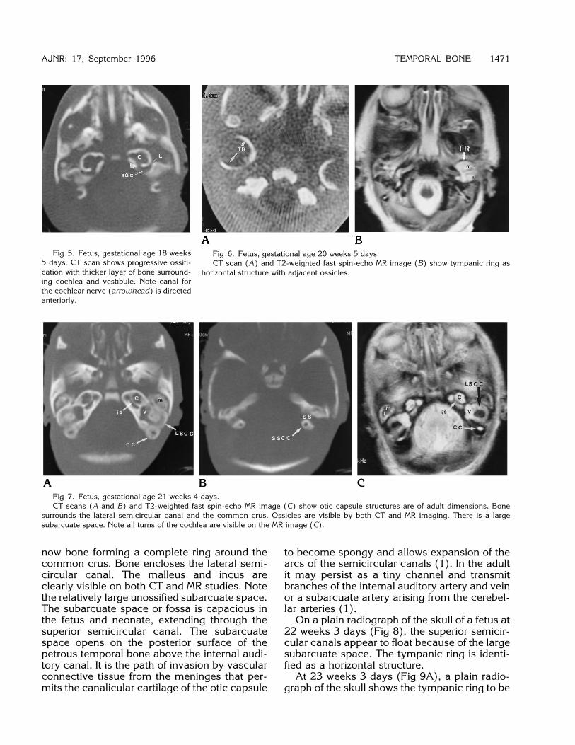
Fig 5. Fetus, gestational age 18 weeks5 days. CT scan shows progressive ossifi-cation with thicker layer of bone surround-ing cochlea and vestibule. Note canal forthe cochlear nerve (arrowhead) is directedanteriorly.
Fig 6. Fetus, gestational age 20 weeks 5 days.CT scan (A) and T2-weighted fast spin-echo MR image (B) show tympanic ring as
horizontal structure with adjacent ossicles.
Fig 7. Fetus, gestational age 21 weeks 4 days.CT scans (A and B) and T2-weighted fast spin-echo MR image (C) show otic capsule structures are of adult dimensions. Bone
surrounds the lateral semicircular canal and the common crus. Ossicles are visible by both CT and MR imaging. There is a largesubarcuate space. Note all turns of the cochlea are visible on the MR image (C).
AJNR: 17, September 1996 TEMPORAL BONE 1471
now bone forming a complete ring around thecommon crus. Bone encloses the lateral semi-circular canal. The malleus and incus areclearly visible on both CT and MR studies. Notethe relatively large unossified subarcuate space.The subarcuate space or fossa is capacious inthe fetus and neonate, extending through thesuperior semicircular canal. The subarcuatespace opens on the posterior surface of thepetrous temporal bone above the internal audi-tory canal. It is the path of invasion by vascularconnective tissue from the meninges that per-mits the canalicular cartilage of the otic capsule
to become spongy and allows expansion of thearcs of the semicircular canals (1). In the adultit may persist as a tiny channel and transmitbranches of the internal auditory artery and veinor a subarcuate artery arising from the cerebel-lar arteries (1).On a plain radiograph of the skull of a fetus at
22 weeks 3 days (Fig 8), the superior semicir-cular canals appear to float because of the largesubarcuate space. The tympanic ring is identi-fied as a horizontal structure.At 23 weeks 3 days (Fig 9A), a plain radio-
graph of the skull shows the tympanic ring to be

horizontal on the anteroposterior projection (Fig9B). The anterior aspect of the tympanic ring isseen on CT scans. The cochlear aqueduct andvestibular aqueduct are identified. The malleusand incus are seen, and the stapes (Fig 9C) isnoted in the region of the oval window. Thesubarcuate space remains prominent (Fig 9D).At 24 weeks 4 days (Fig 10), the subarcuate
space is decreasing in size owing to progressiveossification. The otic capsule and modiolus aremore dense. The ossicles are of adult size andare ossified. The internal auditory canal is di-minutive relative to the structures of the oticcapsule. The labyrinthine segment of the facialnerve canal is quite distinct and well developed.On MR images the endolymphatic duct and theendolymphatic sac and vestibular aqueduct areseen. Also visible are the pyramidal process,the facial nerve recess, and the sinus tympanion the posterior wall of the tympanic cavity.
Fig 8. Fetus, gestational age 22 weeks 3 days. Lateral plainradiograph of the skull shows a large subarcuate space beneaththe superior semicircular canal. The tympanic ring is an ossifiedhorizontal structure that is separated from the squamous temporalbone.
Fig 9. Fetus, gestational age 23 weeks3 days.
A, Anteroposterior plain radiograph ofthe skull shows the horizontal tympanicring is isolated from the rest of the tempo-ral bone.
B–D, On CT scans, note the tympanicring and vestibular and cochlear aque-ducts. The stapes is now identified in theoval window. There is a large subarcuatespace.
1472 NEMZEK AJNR: 17, September 1996
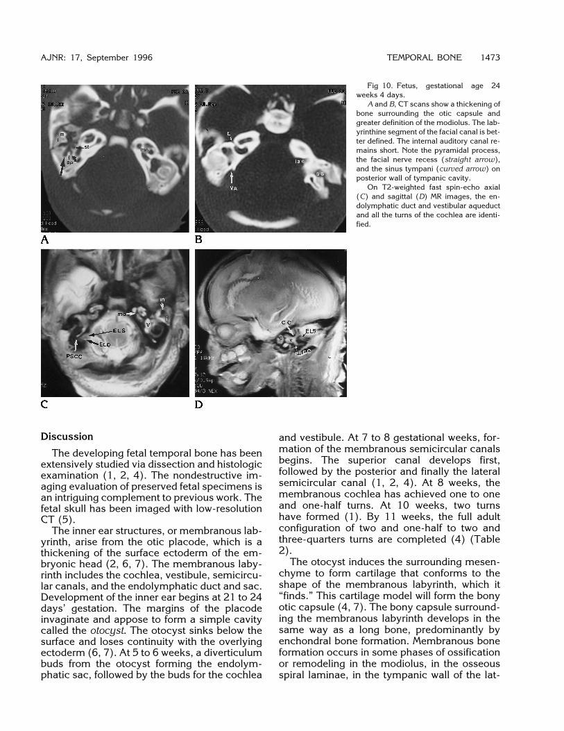
Fig 10. Fetus, gestational age 24weeks 4 days.
A and B, CT scans show a thickening ofbone surrounding the otic capsule andgreater definition of the modiolus. The lab-yrinthine segment of the facial canal is bet-ter defined. The internal auditory canal re-mains short. Note the pyramidal process,the facial nerve recess (straight arrow),and the sinus tympani (curved arrow) onposterior wall of tympanic cavity.
On T2-weighted fast spin-echo axial(C) and sagittal (D) MR images, the en-dolymphatic duct and vestibular aqueductand all the turns of the cochlea are identi-fied.
AJNR: 17, September 1996 TEMPORAL BONE 1473
Discussion
The developing fetal temporal bone has beenextensively studied via dissection and histologicexamination (1, 2, 4). The nondestructive im-aging evaluation of preserved fetal specimens isan intriguing complement to previous work. Thefetal skull has been imaged with low-resolutionCT (5).The inner ear structures, or membranous lab-
yrinth, arise from the otic placode, which is athickening of the surface ectoderm of the em-bryonic head (2, 6, 7). The membranous laby-rinth includes the cochlea, vestibule, semicircu-lar canals, and the endolymphatic duct and sac.Development of the inner ear begins at 21 to 24days’ gestation. The margins of the placodeinvaginate and appose to form a simple cavitycalled the otocyst. The otocyst sinks below thesurface and loses continuity with the overlyingectoderm (6, 7). At 5 to 6 weeks, a diverticulumbuds from the otocyst forming the endolym-phatic sac, followed by the buds for the cochlea
and vestibule. At 7 to 8 gestational weeks, for-mation of the membranous semicircular canalsbegins. The superior canal develops first,followed by the posterior and finally the lateralsemicircular canal (1, 2, 4). At 8 weeks, themembranous cochlea has achieved one to oneand one-half turns. At 10 weeks, two turnshave formed (1). By 11 weeks, the full adultconfiguration of two and one-half to two andthree-quarters turns are completed (4) (Table2).The otocyst induces the surrounding mesen-
chyme to form cartilage that conforms to theshape of the membranous labyrinth, which it“finds.” This cartilage model will form the bonyotic capsule (4, 7). The bony capsule surround-ing the membranous labyrinth develops in thesame way as a long bone, predominantly byenchondral bone formation. Membranous boneformation occurs in some phases of ossificationor remodeling in the modiolus, in the osseousspiral laminae, in the tympanic wall of the lat-

TABLE 2: Milestones in temporal bone development and corresponding imaging findings
Gestational Age, wk Developmental Milestone Imaging Finding
3 Otic placode . . .
4 Otocyst . . .
5–6 Budding of diverticular for membranous labyrinth(first the endolymphatic sac then cochlea andvestibule)
. . .
8 Cochlea, one to one and one-half turns; formationof semicircular canals begins (first the superior,then the posterior, and then the lateral); oticcapsule cartilagenous model
. . .
10 Cochlea, two turns . . .
11 Cochlea, two to two and three-quarters turns;tympanic ring semicircle of bone
. . .
14 . . . Basal turn of cochlea is first seen on MR15 Ossicles reach adult size (ossification begins in
incus, then malleus, and finally stapes)MR shows basal and upper turns ofcochlea and vestibule
16 Otic membranous labyrinth nearing adult size;ossification begins in cochlea
CT shows squamous and zygomaticprocess with minimal ossification ofmalleus and incus
17 . . . CT shows tympanic ring; MR showscochlea, vestibule, and lateralsemicircular canal
18 . . . CT shows cochlea, vestibule, andinterscalar septum; posterior andlateral semicircular canals are partiallyossified; short internal auditory canal ispresent with well-developedlabyrinthine facial nerve canal
19 Tympanic ring forms nine tenths of a circle CT shows confluent ossification ofcochlea and vestibule; MR showscommon crus and endolymphatic duct
20 Ossicles are of adult configuration; superiorsemicircular canal is full size; endolymphaticsac is small, parallel to common crus
CT shows well-ossified tympanic ring andincus; MR shows tympanic ring
21 Cochlea and posterior semicircular canal are fullsize; modiolus ossification begins
Plain radiography shows large subarcuatespace; CT shows common cruscompletely surrounded by bone; lateralsemicircular canal is well seen
22 Lateral semicircular canal is full size . . .
23 . . . CT shows cochlear and vestibularaqueduct
24 . . . CT shows well-developed middle ear,including facial recess, pyramidaleminence, modiolus, and vestibularaqueduct
At birth: Tympanic bone is a flat ring that will form bony external auditory canal. External auditory canal continues growth till age 10. Theendolymphatic duct, endolymphatic sac, and vestibular aqueduct are small at birth and continue to grow till age 3. The mastoid is beginningpneumatization; it will be well developed at 3 years. The styloid process develops after birth.
1474 NEMZEK AJNR: 17, September 1996
eral semicircular canal, and in the external os-tium of the vestibular aqueduct (1). Unlike along bone, such as the femur, which will con-tinue to grow into the late teens, the otic capsuleand ossicles grow with amazing rapidity toreach adult size by about 21 weeks’ gestation(1). Ossification proceeds rapidly between 16and 22 weeks. Cartilage is not changed to boneuntil each particular segment of the membra-nous inner ear has reached its adult size (1, 8).
The inner or endosteal layer of bone surround-ing the otic capsule does not change throughoutlife after initial ossification at midgestation (Fig11). This inner layer of bone completely encir-cles the labyrinth except in the region just an-terior to the oval window. Here, at the fissulaante fenestram, cartilage may persist in theadult temporal bone. This is a site of predilec-tion for otosclerosis (1, 9, 10). New bone maybe produced in the inner layer of bone in re-

AJNR: 17, September 1996 TEMPORAL BONE 1475
sponse to infection or trauma, which will oblit-erate the lumen of the labyrinth (1).Growth of the phylogenetically older vestibu-
lar portion of the membranous labyrinth occursbefore that of the canalicular portion (11). Os-sification occurs later in the canalicular divisionof the otic capsule (1), as is noted in our study.The lateral semicircular canal is the most com-monly malformed structure of the inner ear,perhaps because it is the last to develop (6).Isolated defects of the lateral semicircular canalare common, but if the superior semicircularcanal is maldeveloped, then the posterior andthe lateral semicircular canals are also abnor-mal (6). Again, cartilage is not changed intobone until the inner ear structure has reachedmaximum adult dimensions (1, 4, 8). Ossifica-tion begins in the region of the superior semi-circular canal, followed by the posterior andthen the lateral semicircular canals (4).In our study, the T2-weighted MR images
showed high signal intensity of fluid in the mem-branous labyrinth, which, at 14 weeks, was thefirst recognizable fetal temporal bone structure.Ossification was later identified by CT, first inthe squamous portion of the temporal bone andlater in the otic capsule. Ossification may pro-ceed more rapidly in some fetuses (Figs 4 and5), but the pattern follows a well-defined se-quence (12).The ossicles develop from the cartilage of the
first and second branchial arches (Meckel and
Fig 11. Fetus, gestational age 26 weeks. Histologic sectionshows the cochlear aqueduct at the basilar turn of the cochlea.There are three layers of bone: a thin layer of endosteal bonesurrounding the cochlea (1), a middle layer of intrachondrial bone(2), and an outer layer of periosteal bone (3). (Photograph cour-tesy of Fred Linthicum, MD, House Ear Institute, Los Angeles,Calif.)
Reichert cartilage) (1, 7). Between 15 and 20weeks, the ossicles attain adult size and config-uration. Ossification proceeds by enchondralbone formation. Ossification begins first in theincus, followed by the malleus and stapes (2,8). Ossicular bone, like that of the otic capsule,changes little during life and also demonstratespoor capacity for repair (2).The tympanic ring is a semicircular structure
formed from membranous bone (1). Ossifica-tion begins as two parallel plates oriented in ahorizontal position (13). The ring will eventuallyclose and join inferiorly, but the superior portionremains open, like an annulet. The tympanicring has two functions: it provides the scaffold-ing for the attachment of the tympanic mem-brane and it forms the bony and cartilaginousexternal auditory canal. The tympanic ring re-mains horizontally oriented at birth, which ac-counts for the near horizontal position of thetympanic membrane in the neonate. The tym-panic membrane continues to grow until about3 years of age and gradually assumes the morevertical orientation seen in the adult. The defin-itive form of the external auditory canal ispresent at 1 year of age, but adult size is notattained until 10 years (13).Even though the ossicles and labyrinth are
full size in midgestation, the internal auditorycanal is relatively short, and the vestibular aq-ueduct is small. MR imaging showed the en-dolymphatic duct in two of our specimens. Thevestibular aqueduct and endolymphatic sacwere seen posterior to the vestibule (Figs 9Cand 10C). Note that the endolymphatic duct is arelatively small structure and the endolym-phatic sac is a paddle-shaped structure lyingalmost parallel to the common crus in a dis-sected fetus of 18 weeks (Fig 12A). The en-dolymphatic duct is diminutive, but the en-dolymphatic sac is almost the same size as thesigmoid sinus in a fetus of 15 weeks’ gestation(Fig 12B and C). During the latter half of fetallife, the endolymphatic duct continues to grow,and it changes from a straight to an inferiorcourse bending downward at a 308 to 608 angle(1). The endolymphatic sac also continues toenlarge. The vestibular aqueduct and internalauditory canal continue to grow after birth. By 3years of age the vestibular aqueduct hasreached adult size (7, 14–16). The vestibularaqueduct and internal auditory canal grow syn-chronously in parallel with the periaqueductalair cells (14).
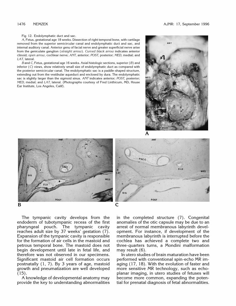
Fig 12. Endolymphatic duct and sac.A, Fetus, gestational age 18 weeks. Dissection of right temporal bone, with cartilage
removed from the superior semicircular canal and endolymphatic duct and sac, andinternal auditory canal. Anterior genu of facial nerve and greater superficial nerve arisefrom the geniculate ganglion (straight arrow). Curved black arrow indicates anteriorclinoid; open arrow, cochlear nerve; ANT, anterior; POST, posterior; MED, medial; andLAT, lateral.
B and C, Fetus, gestational age 16 weeks. Axial histologic sections, superior (B) andinferior (C) views, show relatively small size of endolymphatic duct as compared withthe posterior semicircular canal. The endolymphatic sac is a paddle-shaped structure,extending out from the vestibular aqueduct and enclosed by dura. The endolymphaticsac is slightly larger than the sigmoid sinus. ANT indicates anterior; POST, posterior;MED, medial; and LAT, lateral. (Photographs courtesy of Fred Linthicum, MD, HouseEar Institute, Los Angeles, Calif).
1476 NEMZEK AJNR: 17, September 1996
The tympanic cavity develops from theendoderm of tubotympanic recess of the firstpharyngeal pouch. The tympanic cavityreaches adult size by 37 weeks’ gestation (7).Expansion of the tympanic cavity is responsiblefor the formation of air cells in the mastoid andpetrous temporal bone. The mastoid does notbegin development until late in fetal life, andtherefore was not observed in our specimens.Significant mastoid air cell formation occurspostnatally (1, 7). By 3 years of age, mastoidgrowth and pneumatization are well developed(15).A knowledge of developmental anatomy may
provide the key to understanding abnormalities
in the completed structure (7). Congenitalanomalies of the otic capsule may be due to anarrest of normal membranous labyrinth devel-opment. For instance, if development of themembranous labyrinth is interrupted before thecochlea has achieved a complete two andthree-quarters turns, a Mondini malformationmay result (6).In utero studies of brain maturation have been
performed with conventional spin-echo MR im-aging (17, 18). With the evolution of faster andmore sensitive MR technology, such as echo-planar imaging, in utero studies of fetuses willbecome more common, expanding the poten-tial for prenatal diagnosis of fetal abnormalities.
AJNR: 17, September 1996 TEMPORAL BONE 1477
Conclusion
This work details fetal development of thetemporal bone with the use of high-resolutionCT, MR imaging, and plain radiography. Wehave shown that the otic capsule and ossiclesdevelop by enchondral bone formation throughan intermediate cartilage model. By the middleof gestation these structures have reached adultsize. Ossification begins in the cochlea, followedby the semicircular canals. Apposition of a shellof ossification proceeds rapidly between 18 and24 weeks’ gestation. There is very little subse-quent remodeling after birth. The internal audi-tory canal, vestibular aqueduct, mastoid, and ex-ternal auditory canal continue to grow after birth.The study of fetal developmental anatomy
may lead to a better understanding of congen-ital ear disorders.
AcknowledgmentsWe thank Professor Ronan O’Rahilly and the Carnegie
Laboratories of Embryology for supplying the normal fetalspecimens; Dan Kroeker, Glen Davis, and Charles Burnsfor technical assistance; and Kathy Sommers for invalu-able help in preparing the manuscript.
References1. Donaldson JA, Duckert LG, Lambert PM, Rubel EW. Surgical
Anatomy of the Temporal Bone. New York, NY: Raven Press; 19922. Schuknecht HF, Gulya AJ. Anatomy of the Temporal Bone with
Surgical Implications. Philadelphia, Pa: Lea & Febiger; 19863. O’Rahilly R. Early human development and the chief sources of
information on staged human embryos. Eur J Obstet GynecolReprod Biol 1979;9:273–280
4. Bast TH, Anson BJ. The Temporal Bone and the Ear. Springfield,Ill: Charles C Thomas; 1949
5. Virapongse C, Shapiro R, Sarwar M, Bhimani S, Crelin ES. Com-puted tomography in the study of the development of the skullbase, 1: normal morphology. J Comput Assist Tomogr 1985;9:85–94
6. Jackler RK, Luxford WM, House WF. Congenital malformation ofthe inner ear: a classification based on embryogenesis. Laryngo-scope 1987;97(suppl 40):2–14
7. Sperber GH. Craniofacial Embryology. 4th ed. London, England:Wright, Butterworth Scientific; 1989
8. Glasscock ME, Shambaugh GE. Surgery of the Ear. 4th ed. Phil-adelphia, Pa: Saunders; 1990
9. Swartz JD, Harnsberger HR. Imaging of the Temporal Bone. 2nded. New York, NY: Thieme; 1992:241
10. Rovsing H. Otosclerosis: fenestral and cochlear. Radiol Clin NorthAm 1974;12:505–515
11. Bagger-Sjoback D. Embryology of the human endolymphaticduct and sac. ORL J Otorhinolaryngol Relat Spec 1991;53:61–67
12. Bach-Peterson S, Kjaer I. Ossification of lateral components in theprenatal cranial base. J Craniofac Genet Dev Biol 1993;13:76–82
13. Anson BJ, Bast TH, Richany SF. The fetal and early postnataldevelopment of the tympanic ring and related structures in man.Ann Otol Rhinol Laryngol 1955;64:802–823
14. Fujita S, Sando I. Postnatal development of the vestibular aque-duct in the relation to the internal auditory canal: computer-aidedthree-dimensional reconstruction and measurement study. AnnOtol Rhinol Laryngol 1994;103:719–722
15. Kodama A, Sando I. Postnatal development of the vestibularaqueduct and endolymphatic sac. Ann Otol Rhinol LaryngolSuppl 1982;91:3–12
16. Kodama A, Sando I. Dimensional anatomy of the vestibular aq-ueduct and the endolymphatic sac (rugose portion) in humantemporal bones: statistical analysis of 79 bones. Ann Otol RhinolLaryngol Suppl 1982;91:13–20
17. Girard N, Raybaud C, Poncet M. In vivo MR study of brain matu-ration in normal fetuses. AJNR Am J Neuroradiol 1995;16:407–413
18. Yuh WTC, Nguyen HD, Fisher DJ, et al. MR of fetal central ner-vous system abnormalities. AJNR Am J Neuroradiol 1994;15:459–464