Imaging and Treatment of Sacral Insufficiency …REVIEW ARTICLE Imaging and Treatment of Sacral...
10
REVIEW ARTICLE Imaging and Treatment of Sacral Insufficiency Fractures E.M. Lyders C.T. Whitlow M.D. Baker P.P. Morris SUMMARY: SIFs are a common, though often unsuspected, cause of low back pain in the elderly. Although numerous radiographic modalities can be used to diagnose SIFs, bone scintigraphy and MR imaging are the most sensitive. Conservative management involves various combinations of bed rest, rehabilitation, and analgesics. More recently, sacroplasty has emerged as an alternative therapy for the treatment of SIFs, with prospective studies and case reports suggesting that it is a safe and effective therapy. This article reviews the imaging appearance of SIFs and discusses treatment options with a focus on sacroplasty. ABBREVIATIONS: FEA finite-element analysis; MDP methylene diphosphonate; PMMA polymethylmethacrylate; SIF sacral insufficiency fracture; VAS visual analog pain scale. S IFs are a common cause of debilitating back pain in the elderly. Since they were first described as a clinical entity in 1982 by Lourie, 1 the awareness of this entity among health care professionals has increased. However, there is often a delay in diagnosis because clinical symptoms are frequently vague and nonspecific and can mimic a variety of pathologic processes in a predominantly elderly population, including radiculopathy and metastatic disease. Bone scintigraphy and MR imaging are the most sensitive studies to detect SIFs, though findings have been described in a wide variety of radiologic modalities. The standard of care for the treatment of SIFs has been conserva- tive management, with variable courses of bed rest, rehabilita- tion, and analgesics prescribed. 2-4 More recently, sacroplasty, a minimally invasive procedure akin to vertebroplasty in the thoracolumbar spine, has been advocated as an alternative to conservative therapy. Retrospective case series and prospec- tive studies suggest that sacroplasty is a safe and effective pro- cedure, providing early symptomatic relief in patients with SIFs. 5-8 This article discusses the imaging findings and treat- ment of SIFs. SIFs commonly affect elderly women with osteoporosis, 9,10 though other reported risk factors include pelvic radiation, steroid-induced osteopenia, rheumatoid arthritis, multiple myeloma, Paget disease, renal osteodystrophy, and hyperpara- thyroidism. 2,9,11 Of these, osteoporosis is the most prevalent, and almost all patients with SIFs will demonstrate severe os- teopenia on dual x-ray absorptiometry, even if other risk fac- tors are present. 2 Prior pelvic radiation is another well- established risk factor for the development of SIFs, with a reported prevalence of 21%–34%. 12,13 The incidence may even be as high as 89%, as suggested by a prospective study of patients undergoing pelvic radiation for cervical cancer. 14 Almost all patients with SIFs are older than 55 years of age, with a mean age between 70 and 75 years in most stud- ies. 4,6,15,16 The true incidence of SIFs is unknown but has been reported to be between 1% and 5% in at-risk patient popula- tions. 17-19 Antecedent trauma is not identified in two-thirds of patients 3,4 and, when present, is usually minor. 16 Patients with SIFs most commonly present with diffuse low back pain, which may radiate to the buttock, hip, or groin. 3,15,20 Patients may have some tenderness to palpation in the lower back and sacral region, though this is not a consis- tent finding. 15 Neurologic symptoms related to SIFs are un- usual, though may be seen in 5%– 6% of patients, most com- monly manifesting as a sacral radiculopathy. 2 However, a case of cauda equina syndrome related to SIFs has been reported. 21 SIFs can occasionally be confused with metastatic disease, both clinically and on imaging studies, 22 resulting in unneces- sary work-up and biopsy. 9-10 This is frequently a confounding factor in elderly patient populations, many of whom have a known primary malignancy or are being evaluated for an oc- cult tumor. 16,22,23 In fact, approximately 45% of patients with SIFs have a history of malignancy. 2 Insufficiency fractures are a subtype of stress fracture that results from normal stress applied to abnormal bone that has lost its elastic resistance. Bone insufficiency is often the result of osteoporosis or other metabolic bone disease, though osse- ous metastatic disease and marrow replacement processes can also cause insufficiency fractures. SIFs most commonly in- volve the sacral ala, lateral to the neural foramina and medial to the sacroiliac joints (zone 1) (Fig 1). 24 Fractures may be unilateral or bilateral and are reported with relatively equal frequency in the literature. 2,9 There may also be a horizontal component to the fracture through the sacral bodies. This unique fracture pattern may be related to axial loading and weight-bearing transmitted through the spine, resulting in sa- cral alar strain. 25,26 Additionally, osteoporosis causes asym- metric loss of bony trabeculae in the sacral ala compared with the vertebral bodies, placing the lateral aspect of the sacrum at increased risk of insufficiency. 27 Of note, there is a high incidence of concomitant pelvic insufficiency fractures, and radiologists should be aware of this association. SIFs are most frequently associated with in- sufficiency fractures of the pubic rami and parasymphyseal region, with a reported coincidence of 88%. 9 Associated in- sufficiency fractures have also been described in the superior acetabulum and iliac wing. 28,29 It has been suggested that the From the Division of Radiological Sciences, Department of Radiology, Wake Forest University School of Medicine, Winston-Salem, North Carolina. Please address correspondence to P. Pearse Morris, MB, BCh, Division of Radiological Sciences, Department of Radiology, 2nd Floor, Meads Hall, Wake Forest University School of Medicine, Winston-Salem, NC 27157; e-mail: [email protected] Indicates open access to non-subscribers at www.ajnr.org DOI 10.3174/ajnr.A1666 REVIEW ARTICLE AJNR Am J Neuroradiol 31:201–10 Feb 2010 www.ajnr.org 201
Transcript of Imaging and Treatment of Sacral Insufficiency …REVIEW ARTICLE Imaging and Treatment of Sacral...
REVIEW ARTICLE
Imaging and Treatment of Sacral InsufficiencyFractures
E.M. LydersC.T. Whitlow
M.D. BakerP.P. Morris
SUMMARY: SIFs are a common, though often unsuspected, cause of low back pain in the elderly.Although numerous radiographic modalities can be used to diagnose SIFs, bone scintigraphy and MRimaging are the most sensitive. Conservative management involves various combinations of bed rest,rehabilitation, and analgesics. More recently, sacroplasty has emerged as an alternative therapy for thetreatment of SIFs, with prospective studies and case reports suggesting that it is a safe and effectivetherapy. This article reviews the imaging appearance of SIFs and discusses treatment options with afocus on sacroplasty.
ABBREVIATIONS: FEA � finite-element analysis; MDP � methylene diphosphonate; PMMA �polymethylmethacrylate; SIF � sacral insufficiency fracture; VAS � visual analog pain scale.
SIFs are a common cause of debilitating back pain in theelderly. Since they were first described as a clinical entity in
1982 by Lourie,1 the awareness of this entity among health careprofessionals has increased. However, there is often a delay indiagnosis because clinical symptoms are frequently vague andnonspecific and can mimic a variety of pathologic processes ina predominantly elderly population, including radiculopathyand metastatic disease. Bone scintigraphy and MR imaging arethe most sensitive studies to detect SIFs, though findings havebeen described in a wide variety of radiologic modalities. Thestandard of care for the treatment of SIFs has been conserva-tive management, with variable courses of bed rest, rehabilita-tion, and analgesics prescribed.2-4 More recently, sacroplasty,a minimally invasive procedure akin to vertebroplasty in thethoracolumbar spine, has been advocated as an alternative toconservative therapy. Retrospective case series and prospec-tive studies suggest that sacroplasty is a safe and effective pro-cedure, providing early symptomatic relief in patients withSIFs.5-8 This article discusses the imaging findings and treat-ment of SIFs.
SIFs commonly affect elderly women with osteoporosis,9,10
though other reported risk factors include pelvic radiation,steroid-induced osteopenia, rheumatoid arthritis, multiplemyeloma, Paget disease, renal osteodystrophy, and hyperpara-thyroidism.2,9,11 Of these, osteoporosis is the most prevalent,and almost all patients with SIFs will demonstrate severe os-teopenia on dual x-ray absorptiometry, even if other risk fac-tors are present.2 Prior pelvic radiation is another well-established risk factor for the development of SIFs, with areported prevalence of 21%–34%.12,13 The incidence mayeven be as high as 89%, as suggested by a prospective study ofpatients undergoing pelvic radiation for cervical cancer.14
Almost all patients with SIFs are older than 55 years ofage, with a mean age between 70 and 75 years in most stud-ies.4,6,15,16 The true incidence of SIFs is unknown but has been
reported to be between 1% and 5% in at-risk patient popula-tions.17-19 Antecedent trauma is not identified in two-thirds ofpatients3,4 and, when present, is usually minor.16
Patients with SIFs most commonly present with diffuselow back pain, which may radiate to the buttock, hip, orgroin.3,15,20 Patients may have some tenderness to palpation inthe lower back and sacral region, though this is not a consis-tent finding.15 Neurologic symptoms related to SIFs are un-usual, though may be seen in 5%– 6% of patients, most com-monly manifesting as a sacral radiculopathy.2 However, a caseof cauda equina syndrome related to SIFs has been reported.21
SIFs can occasionally be confused with metastatic disease,both clinically and on imaging studies,22 resulting in unneces-sary work-up and biopsy.9-10 This is frequently a confoundingfactor in elderly patient populations, many of whom have aknown primary malignancy or are being evaluated for an oc-cult tumor.16,22,23 In fact, approximately 45% of patients withSIFs have a history of malignancy.2
Insufficiency fractures are a subtype of stress fracture thatresults from normal stress applied to abnormal bone that haslost its elastic resistance. Bone insufficiency is often the resultof osteoporosis or other metabolic bone disease, though osse-ous metastatic disease and marrow replacement processes canalso cause insufficiency fractures. SIFs most commonly in-volve the sacral ala, lateral to the neural foramina and medialto the sacroiliac joints (zone 1) (Fig 1).24 Fractures may beunilateral or bilateral and are reported with relatively equalfrequency in the literature.2,9 There may also be a horizontalcomponent to the fracture through the sacral bodies. Thisunique fracture pattern may be related to axial loading andweight-bearing transmitted through the spine, resulting in sa-cral alar strain.25,26 Additionally, osteoporosis causes asym-metric loss of bony trabeculae in the sacral ala compared withthe vertebral bodies, placing the lateral aspect of the sacrum atincreased risk of insufficiency.27
Of note, there is a high incidence of concomitant pelvicinsufficiency fractures, and radiologists should be aware ofthis association. SIFs are most frequently associated with in-sufficiency fractures of the pubic rami and parasymphysealregion, with a reported coincidence of 88%.9 Associated in-sufficiency fractures have also been described in the superioracetabulum and iliac wing.28,29 It has been suggested that the
From the Division of Radiological Sciences, Department of Radiology, Wake ForestUniversity School of Medicine, Winston-Salem, North Carolina.
Please address correspondence to P. Pearse Morris, MB, BCh, Division of RadiologicalSciences, Department of Radiology, 2nd Floor, Meads Hall, Wake Forest University Schoolof Medicine, Winston-Salem, NC 27157; e-mail: [email protected]
Indicates open access to non-subscribers at www.ajnr.org
DOI 10.3174/ajnr.A1666
REVIEWA
RTICLE
AJNR Am J Neuroradiol 31:201–10 � Feb 2010 � www.ajnr.org 201
sacrum is the initial site of failure and that this results in in-creased stress on the rest of the bony pelvis.9
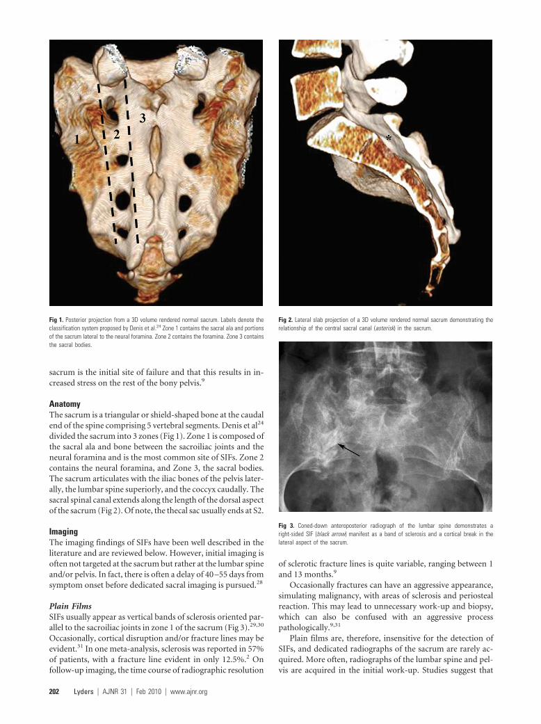
AnatomyThe sacrum is a triangular or shield-shaped bone at the caudalend of the spine comprising 5 vertebral segments. Denis et al24
divided the sacrum into 3 zones (Fig 1). Zone 1 is composed ofthe sacral ala and bone between the sacroiliac joints and theneural foramina and is the most common site of SIFs. Zone 2contains the neural foramina, and Zone 3, the sacral bodies.The sacrum articulates with the iliac bones of the pelvis later-ally, the lumbar spine superiorly, and the coccyx caudally. Thesacral spinal canal extends along the length of the dorsal aspectof the sacrum (Fig 2). Of note, the thecal sac usually ends at S2.
ImagingThe imaging findings of SIFs have been well described in theliterature and are reviewed below. However, initial imaging isoften not targeted at the sacrum but rather at the lumbar spineand/or pelvis. In fact, there is often a delay of 40 –55 days fromsymptom onset before dedicated sacral imaging is pursued.28
Plain FilmsSIFs usually appear as vertical bands of sclerosis oriented par-allel to the sacroiliac joints in zone 1 of the sacrum (Fig 3).29,30
Occasionally, cortical disruption and/or fracture lines may beevident.31 In one meta-analysis, sclerosis was reported in 57%of patients, with a fracture line evident in only 12.5%.2 Onfollow-up imaging, the time course of radiographic resolution
of sclerotic fracture lines is quite variable, ranging between 1and 13 months.9
Occasionally fractures can have an aggressive appearance,simulating malignancy, with areas of sclerosis and periostealreaction. This may lead to unnecessary work-up and biopsy,which can also be confused with an aggressive processpathologically.9,31
Plain films are, therefore, insensitive for the detection ofSIFs, and dedicated radiographs of the sacrum are rarely ac-quired. More often, radiographs of the lumbar spine and pel-vis are acquired in the initial work-up. Studies suggest that
Fig 1. Posterior projection from a 3D volume rendered normal sacrum. Labels denote theclassification system proposed by Denis et al.24 Zone 1 contains the sacral ala and portionsof the sacrum lateral to the neural foramina. Zone 2 contains the foramina. Zone 3 containsthe sacral bodies.
Fig 2. Lateral slab projection of a 3D volume rendered normal sacrum demonstrating therelationship of the central sacral canal (asterisk) in the sacrum.
Fig 3. Coned-down anteroposterior radiograph of the lumbar spine demonstrates aright-sided SIF (black arrow) manifest as a band of sclerosis and a cortical break in thelateral aspect of the sacrum.
202 Lyders � AJNR 31 � Feb 2010 � www.ajnr.org
only 20%–38% of SIFs and pelvic ring fractures are identifiedon plain films,2,4 and these are often overlooked prospectively.Even when evaluated retrospectively, less than 50% of SIFsdiagnosed on bone scintigraphy were evident on plainfilms.32,33 SIFs may not be evident in a large percentage ofpatients because of a high prevalence of osteopenia and obscu-ration by overlying bowel gas (Fig 4).31
Nuclear MedicineBone scintigraphy with technetium Tc99m�labeled MDP isone of the most sensitive examinations for the detection ofSIFs, and the imaging findings have been well documented.2,33
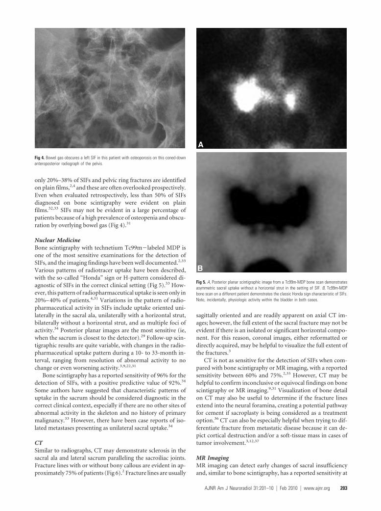
Various patterns of radiotracer uptake have been described,with the so-called “Honda” sign or H-pattern considered di-agnostic of SIFs in the correct clinical setting (Fig 5).33 How-ever, this pattern of radiopharmaceutical uptake is seen only in20%– 40% of patients.4,31 Variations in the pattern of radio-pharmaceutical activity in SIFs include uptake oriented uni-laterally in the sacral ala, unilaterally with a horizontal strut,bilaterally without a horizontal strut, and as multiple foci ofactivity.34 Posterior planar images are the most sensitive (ie,when the sacrum is closest to the detector).29 Follow-up scin-tigraphic results are quite variable, with changes in the radio-pharmaceutical uptake pattern during a 10- to 33-month in-terval, ranging from resolution of abnormal activity to nochange or even worsening activity.3,9,22,31
Bone scintigraphy has a reported sensitivity of 96% for thedetection of SIFs, with a positive predictive value of 92%.34
Some authors have suggested that characteristic patterns ofuptake in the sacrum should be considered diagnostic in thecorrect clinical context, especially if there are no other sites ofabnormal activity in the skeleton and no history of primarymalignancy.33 However, there have been case reports of iso-lated metastases presenting as unilateral sacral uptake.34
CTSimilar to radiographs, CT may demonstrate sclerosis in thesacral ala and lateral sacrum paralleling the sacroiliac joints.Fracture lines with or without bony callous are evident in ap-proximately 75% of patients (Fig 6).2 Fracture lines are usually
sagittally oriented and are readily apparent on axial CT im-ages; however, the full extent of the sacral fracture may not beevident if there is an isolated or significant horizontal compo-nent. For this reason, coronal images, either reformatted ordirectly acquired, may be helpful to visualize the full extent ofthe fractures.3
CT is not as sensitive for the detection of SIFs when com-pared with bone scintigraphy or MR imaging, with a reportedsensitivity between 60% and 75%.2,35 However, CT may behelpful to confirm inconclusive or equivocal findings on bonescintigraphy or MR imaging.9,31 Visualization of bone detailon CT may also be useful to determine if the fracture linesextend into the neural foramina, creating a potential pathwayfor cement if sacroplasty is being considered as a treatmentoption.36 CT can also be especially helpful when trying to dif-ferentiate fracture from metastatic disease because it can de-pict cortical destruction and/or a soft-tissue mass in cases oftumor involvement.3,12,37
MR ImagingMR imaging can detect early changes of sacral insufficiencyand, similar to bone scintigraphy, has a reported sensitivity at
Fig 4. Bowel gas obscures a left SIF in this patient with osteoporosis on this coned-downanteroposterior radiograph of the pelvis.
Fig 5. A, Posterior planar scintigraphic image from a Tc99m-MDP bone scan demonstratesasymmetric sacral uptake without a horizontal strut in the setting of SIF. B, Tc99m-MDPbone scan on a different patient demonstrates the classic Honda sign characteristic of SIFs.Note, incidentally, physiologic activity within the bladder in both cases.
AJNR Am J Neuroradiol 31:201–10 � Feb 2010 � www.ajnr.org 203
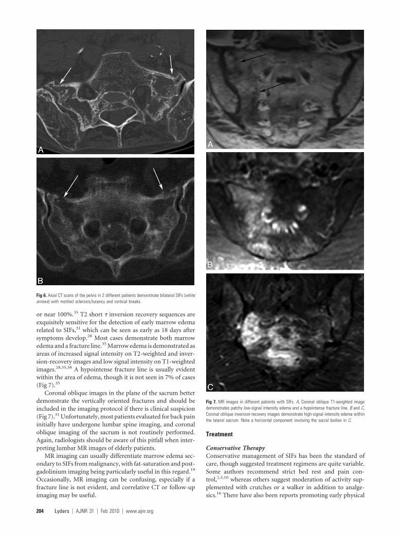
or near 100%.35 T2 short � inversion recovery sequences areexquisitely sensitive for the detection of early marrow edemarelated to SIFs,31 which can be seen as early as 18 days aftersymptoms develop.28 Most cases demonstrate both marrowedema and a fracture line.35 Marrow edema is demonstrated asareas of increased signal intensity on T2-weighted and inver-sion-recovery images and low signal intensity on T1-weightedimages.28,35,38 A hypointense fracture line is usually evidentwithin the area of edema, though it is not seen in 7% of cases(Fig 7).35
Coronal oblique images in the plane of the sacrum betterdemonstrate the vertically oriented fractures and should beincluded in the imaging protocol if there is clinical suspicion(Fig 7).31 Unfortunately, most patients evaluated for back paininitially have undergone lumbar spine imaging, and coronaloblique imaging of the sacrum is not routinely performed.Again, radiologists should be aware of this pitfall when inter-preting lumbar MR images of elderly patients.
MR imaging can usually differentiate marrow edema sec-ondary to SIFs from malignancy, with fat-saturation and post-gadolinium imaging being particularly useful in this regard.19
Occasionally, MR imaging can be confusing, especially if afracture line is not evident, and correlative CT or follow-upimaging may be useful.
Treatment
Conservative TherapyConservative management of SIFs has been the standard ofcare, though suggested treatment regimens are quite variable.Some authors recommend strict bed rest and pain con-trol,2,3,10 whereas others suggest moderation of activity sup-plemented with crutches or a walker in addition to analge-sics.16 There have also been reports promoting early physical
Fig 6. Axial CT scans of the pelvis in 2 different patients demonstrate bilateral SIFs (whitearrows) with mottled sclerosis/lucency and cortical breaks.
Fig 7. MR images in different patients with SIFs. A, Coronal oblique T1-weighted imagedemonstrates patchy low-signal intensity edema and a hypointense fracture line. B and C,Coronal oblique inversion-recovery images demonstrate high-signal-intensity edema withinthe lateral sacrum. Note a horizontal component involving the sacral bodies in C.
204 Lyders � AJNR 31 � Feb 2010 � www.ajnr.org
rehabilitation.20,23 Although most patients improve symp-tomatically following conservative therapy, the time coursecan be prolonged and quite variable. Symptoms resolve in
most patients by 12 months but can vary between 6 and 15months.2,3,15,20,23
However, not all patients improve with conservative ther-apy, and prolonged bed rest is associated with significant mor-bidity, especially in the elderly. Of primary concern is throm-boembolic disease, with a reported incidence of deep venousthrombosis following pelvic fracture between 29% and 61%,and a 2%–12% incidence of pulmonary embolism.23 Otheradverse sequelae associated with prolonged bed rest includeloss of muscle mass, cardiac dysfunction, pneumonia, decub-itus ulceration, and bone demineralization.23 In fact, half ofpatients with pelvic insufficiency fractures will not return totheir prior functional level, and there is a reported 14.3% over-all mortality.39
SacroplastySacroplasty has emerged as a minimally invasive alternative toconservative therapy for SIFs. Similar to vertebroplasty in thethoracolumbar spine, it involves injection of PMMA cementinto the fractured sacrum under imaging guidance.5,6,40-42 Thegoal of sacroplasty is to provide early symptomatic relief, al-lowing more rapid mobilization. This would limit the need forsignificant narcotic analgesics, and lessen the risks associatedwith prolonged bed rest. Several retrospective and prospectivecase series suggest that sacroplasty can safely and effectivelyprovide early symptomatic relief,5-7,41 though these findingshave not been verified in controlled prospective randomizedtrials.
Injection of cement into the sacrum was first used for thetherapy of painful bony metastases43,44 and was first describedin sacral osteoporotic insufficiency fractures by Garant in2002.40 Sacroplasty has increasingly been used for both theseindications, though it has not yet achieved widespread accep-tance, unlike vertebroplasty, probably due to the lack of vali-dating controlled studies or because of unique technical con-siderations related to sacral anatomy.
The technical aspects of sacroplasty vary significantly be-tween operators, including the technique of imaging guid-ance, as well as the needle approach. There are 2 basic needleapproaches described in the literature: a posterior ap-
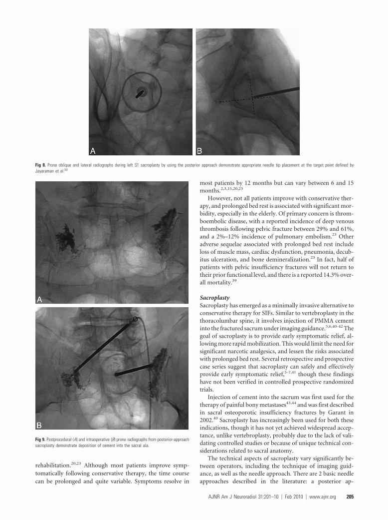
Fig 8. Prone oblique and lateral radiographs during left S1 sacroplasty by using the posterior approach demonstrate appropriate needle tip placement at the target point defined byJayaraman et al.50
Fig 9. Postprocedural (A) and intraoperative (B) prone radiographs from posterior-approachsacroplasty demonstrate deposition of cement into the sacral ala.
AJNR Am J Neuroradiol 31:201–10 � Feb 2010 � www.ajnr.org 205
proach5,40-42,45 and the long-axis approach.36,46,47 A midlineapproach has also been used by some physicians to treat thehorizontal or sacral body (zone 3) component of the fracture(F.R. Hellinger, unpublished data, 2009). Although the exacttechnique of these different approaches is beyond the scope ofthis article, a basic overview of the methodology and advan-tages of each is described.
Posterior ApproachThe posterior or dorsal approach is similar to that in used invertebroplasty, and many authors probably use this approach,given their familiarity with vertebroplasty. The patient isplaced prone on the procedure table. Following the adminis-tration of a local anesthetic, an 11- or 13-gauge bone biopsyneedle is percutaneously inserted through the posterior sacralcortex into the sacral ala and lateral aspect of the sacrum (zone1) in a plane parallel to the sacroiliac joint (Figs 8 and 9).5,40
Often, 2–3 needles are inserted on a single side to ensure ade-quate dispersal of cement throughout the sacrum. On a fluo-roscopic anteroposterior projection, the image intensifier islined up parallel to the L5-S1 disk space and the ipsilateralsacroiliac joint.48 On the lateral fluoroscopic view, the ventralcortical margin can be difficult to discern because of theunique shape of the sacrum with its concave ventral sur-face.5 Fluoroscopic landmarks have been confirmed withanatomic dissection and identify a well-defined triangularbreach zone.49 Recently, a lateral target point for fluoroscopi-cally guided sacroplasty has been described at the intersectionof lines drawn through the corners of the S1 vertebral body(Fig 8).50 In our experience, acquiring a preoperative CT scanof the pelvis with multiplanar reformatted imaging is helpfulto delineate the complex sacral anatomy for each patient.
One of the concerns with fluoroscopically guided sacro-plasty is inadvertent transgression of the ventral sacral cortexby the procedure needle. Venography through the procedureneedle has been suggested as a method to confirm needle tipplacement,40 though other authors have found limited successby using this technique.5 CT guidance allows accurate needleplacement and allows the physician to remain confident thatthe needle tip remains intramedullary. Both CT5,47,51 and CTfluoroscopically41 guided sacroplasties have been described.However, observation of cement injection under fluoroscopy,either by using a mobile C-arm or transfer of the patient to afluoroscopy suite, is recommended because it is difficult tomonitor cement extrusion on CT.41 This is especially true inthe craniocaudal direction, which is the preferred direction ofcement migration given the commonly vertically oriented sa-cral fractures.
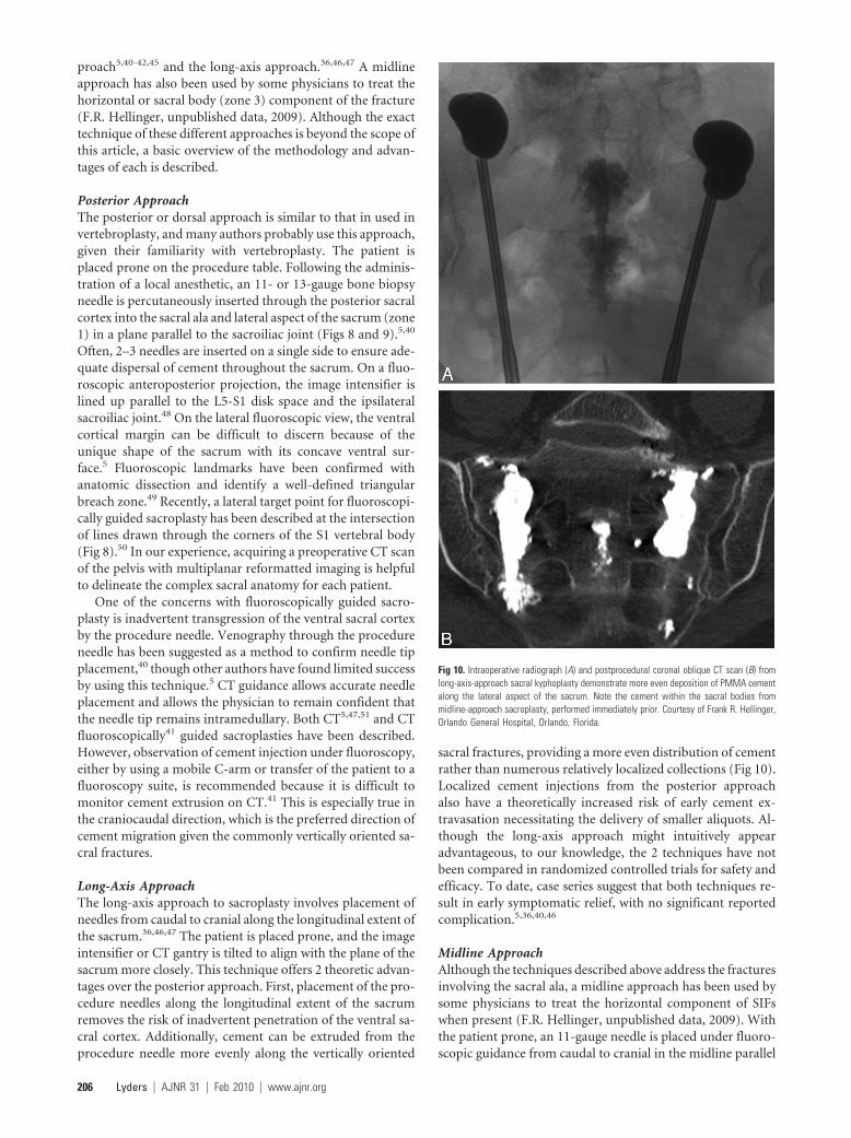
Long-Axis ApproachThe long-axis approach to sacroplasty involves placement ofneedles from caudal to cranial along the longitudinal extent ofthe sacrum.36,46,47 The patient is placed prone, and the imageintensifier or CT gantry is tilted to align with the plane of thesacrum more closely. This technique offers 2 theoretic advan-tages over the posterior approach. First, placement of the pro-cedure needles along the longitudinal extent of the sacrumremoves the risk of inadvertent penetration of the ventral sa-cral cortex. Additionally, cement can be extruded from theprocedure needle more evenly along the vertically oriented
sacral fractures, providing a more even distribution of cementrather than numerous relatively localized collections (Fig 10).Localized cement injections from the posterior approachalso have a theoretically increased risk of early cement ex-travasation necessitating the delivery of smaller aliquots. Al-though the long-axis approach might intuitively appearadvantageous, to our knowledge, the 2 techniques have notbeen compared in randomized controlled trials for safety andefficacy. To date, case series suggest that both techniques re-sult in early symptomatic relief, with no significant reportedcomplication.5,36,40,46
Midline ApproachAlthough the techniques described above address the fracturesinvolving the sacral ala, a midline approach has been used bysome physicians to treat the horizontal component of SIFswhen present (F.R. Hellinger, unpublished data, 2009). Withthe patient prone, an 11-gauge needle is placed under fluoro-scopic guidance from caudal to cranial in the midline parallel
Fig 10. Intraoperative radiograph (A) and postprocedural coronal oblique CT scan (B) fromlong-axis-approach sacral kyphoplasty demonstrate more even deposition of PMMA cementalong the lateral aspect of the sacrum. Note the cement within the sacral bodies frommidline-approach sacroplasty, performed immediately prior. Courtesy of Frank R. Hellinger,Orlando General Hospital, Orlando, Florida.
206 Lyders � AJNR 31 � Feb 2010 � www.ajnr.org
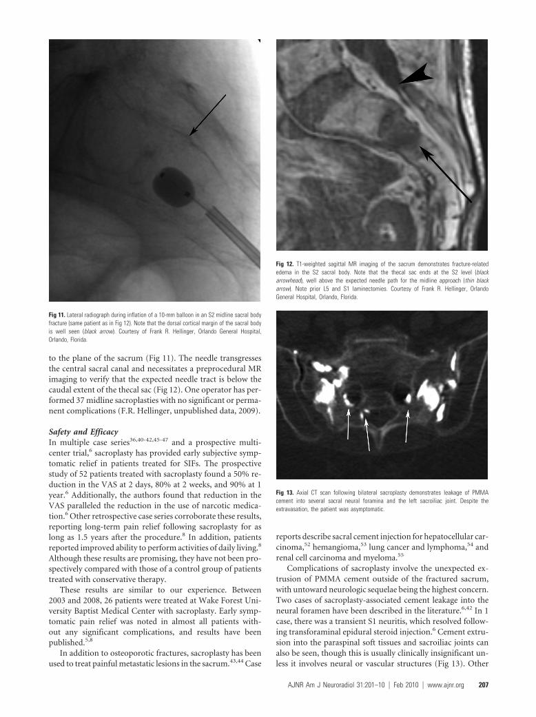
to the plane of the sacrum (Fig 11). The needle transgressesthe central sacral canal and necessitates a preprocedural MRimaging to verify that the expected needle tract is below thecaudal extent of the thecal sac (Fig 12). One operator has per-formed 37 midline sacroplasties with no significant or perma-nent complications (F.R. Hellinger, unpublished data, 2009).
Safety and EfficacyIn multiple case series36,40-42,45-47 and a prospective multi-center trial,6 sacroplasty has provided early subjective symp-tomatic relief in patients treated for SIFs. The prospectivestudy of 52 patients treated with sacroplasty found a 50% re-duction in the VAS at 2 days, 80% at 2 weeks, and 90% at 1year.6 Additionally, the authors found that reduction in theVAS paralleled the reduction in the use of narcotic medica-tion.6 Other retrospective case series corroborate these results,reporting long-term pain relief following sacroplasty for aslong as 1.5 years after the procedure.8 In addition, patientsreported improved ability to perform activities of daily living.8
Although these results are promising, they have not been pro-spectively compared with those of a control group of patientstreated with conservative therapy.
These results are similar to our experience. Between2003 and 2008, 26 patients were treated at Wake Forest Uni-versity Baptist Medical Center with sacroplasty. Early symp-tomatic pain relief was noted in almost all patients with-out any significant complications, and results have beenpublished.5,8
In addition to osteoporotic fractures, sacroplasty has beenused to treat painful metastatic lesions in the sacrum.43,44 Case
reports describe sacral cement injection for hepatocellular car-cinoma,52 hemangioma,53 lung cancer and lymphoma,54 andrenal cell carcinoma and myeloma.55
Complications of sacroplasty involve the unexpected ex-trusion of PMMA cement outside of the fractured sacrum,with untoward neurologic sequelae being the highest concern.Two cases of sacroplasty-associated cement leakage into theneural foramen have been described in the literature.6,42 In 1case, there was a transient S1 neuritis, which resolved follow-ing transforaminal epidural steroid injection.6 Cement extru-sion into the paraspinal soft tissues and sacroiliac joints canalso be seen, though this is usually clinically insignificant un-less it involves neural or vascular structures (Fig 13). Other
Fig 11. Lateral radiograph during inflation of a 10-mm balloon in an S2 midline sacral bodyfracture (same patient as in Fig 12). Note that the dorsal cortical margin of the sacral bodyis well seen (black arrow). Courtesy of Frank R. Hellinger, Orlando General Hospital,Orlando, Florida.
Fig 12. T1-weighted sagittal MR imaging of the sacrum demonstrates fracture-relatededema in the S2 sacral body. Note that the thecal sac ends at the S2 level (blackarrowhead), well above the expected needle path for the midline approach (thin blackarrow). Note prior L5 and S1 laminectomies. Courtesy of Frank R. Hellinger, OrlandoGeneral Hospital, Orlando, Florida.
Fig 13. Axial CT scan following bilateral sacroplasty demonstrates leakage of PMMAcement into several sacral neural foramina and the left sacroiliac joint. Despite theextravasation, the patient was asymptomatic.
AJNR Am J Neuroradiol 31:201–10 � Feb 2010 � www.ajnr.org 207
theoretic complications of sacroplasty include extrusion of ce-ment into the sacral spinal canal with associated neurologiccompromise, infection, or venous emboli; however, to ourknowledge, these have yet to be reported in the literature.
One technical consideration unique to sacroplasty involvesthe lack of resistance during cement extrusion. Unlike verte-bral bodies during vertebroplasty, the capacious medullarycavity of the sacrum offers little resistance or feedback to theoperator during cement extrusion.5,56 Close fluoroscopicmonitoring is necessary to ensure that cement does not leakinto the neural foramina, presacral space, or sacroiliac joint.
Other Treatment OptionsSacroplasty with balloon augmentation, or sacral kyphoplasty,has been described in 2 case reports suggesting the feasibility of
this method.53,56 The sacral kyphoplasties were performed forosteoporotic fractures56 and a sacral hemangioma.53 Sacralkyphoplasty is technically similar to sacroplasty, and severalballoon systems are commercially available. One suggested ad-vantage of sacral kyphoplasty is the creation of a compactedbony layer outside the balloon, which could theoreticallylower the likelihood of cement extravasation.56 One studyevaluating balloon sacroplasty in cadaveric sacral modelsdemonstrated more controlled localized deposition ofcement.49
Fluoroscopic placement of transiliosacral screws has alsobeen reported in case series as an alternative method of sacralstabilization in patients with SIF.57 A similar procedure usingcomputer assistance for screw placement has also beenreported.58
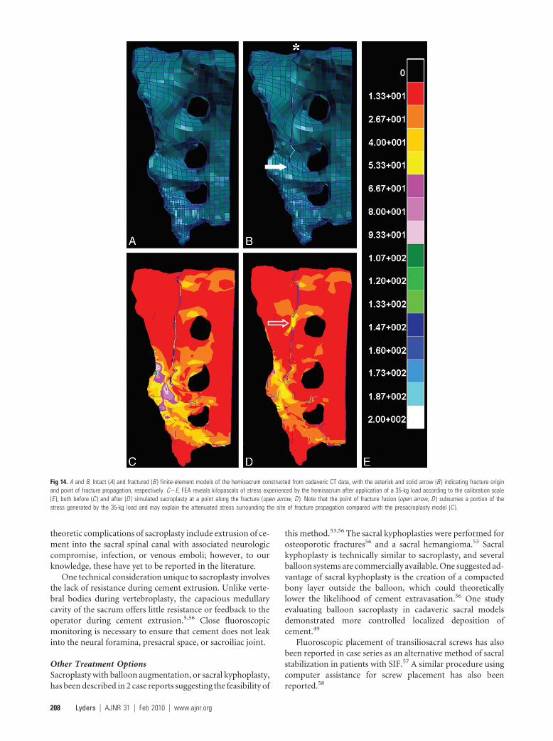
Fig 14. A and B, Intact (A) and fractured (B ) finite-element models of the hemisacrum constructed from cadaveric CT data, with the asterisk and solid arrow (B ) indicating fracture originand point of fracture propagation, respectively. C�E, FEA reveals kilopascals of stress experienced by the hemisacrum after application of a 35-kg load according to the calibration scale(E ), both before (C ) and after (D ) simulated sacroplasty at a point along the fracture (open arrow, D ). Note that the point of fracture fusion (open arrow, D ) subsumes a portion of thestress generated by the 35-kg load and may explain the attenuated stress surrounding the site of fracture propagation compared with the presacroplasty model (C ).
208 Lyders � AJNR 31 � Feb 2010 � www.ajnr.org
BiomechanicsDespite the subjective symptomatic relief experienced by pa-tients, the exact mechanism of pain relief has not been fullyelucidated. It has been suggested that excessive sheer strain onthe sacral ala in insufficient bone results in fracture, with painthe result of micromotion at the fracture site.7 Biomechanicalevaluation of cadaveric sacral models following sacroplastyhas not shown any significant restoration of strength or stiff-ness following sacroplasty.16 Furthermore, the amount andlocation of PMMA cement used in sacroplasty has no bearingon the strength or stiffness restoration in cadaveric sacralmodels.59 However, these models assess the biomechanicalproperties of the entire sacrum and do not assess local biome-chanical properties, especially near the cement-bone interface.
It has been suggested that sacral fracture stabilization andattenuation of fracture micromotion by the PMMA cementmay underlie reports of sacroplasty-associated pain relief. FEAis an engineering method used to evaluate the mechanicalproperties of bone in computer-generated 3D geometric mod-els of osseous structures. FEA has demonstrated an 83% de-crease in maximal principal stress at the point of sacral fracturepropagation in cadaveric sacral models after sacroplasty. FEAalso demonstrated significantly diminished fracture gap mi-cromotion after sacroplasty (Fig 14).48 Local compressive,tensile, and shear strains are also reduced 40%– 60% followingcement injection in nonfractured models, as documented byFEA.60 These biomechanical features may explain subjectivepain relief following sacroplasty.
Sacroplasty has been criticized for disrupting normal osse-ous healing following sacral fracture.61,62 Tsiridis62 noted thatmost patients with SIF recover completely with conservativetherapy, albeit along an often prolonged and variable timecourse. Ehara61 suggested that sacroplasty may be more indi-cated in patients with delayed union or prolonged severe pain.
ConclusionsSacral fractures are a common yet underdiagnosed cause oflow back pain, predominantly in elderly women with osteo-porosis. Plain radiographic, scintigraphic, CT, and MR imag-ing findings have been well described in the literature. Bonescintigraphy and MR imaging are the most sensitive examina-tions and can be diagnostic, especially in the correct clinicalsetting. CT can be a helpful adjunct in cases in which scintig-raphy and MR imaging are inconclusive.
Sacroplasty has recently emerged as a minimally invasivealternative to conservative treatment for SIFs. Prospectivestudies and case reports suggest that it is a safe and effectivetherapy, resulting in early symptomatic relief in patients withthese fractures. However, randomized controlled trials com-paring sacroplasty and conservative therapy are necessary tovalidate this technique.
The optimal technique for performing sacroplasty remainsuncertain because various combinations of imaging guidancehave been used and several different needle approaches havebeen described. Additionally, sacral kyphoplasty has been de-scribed. At this point in time, the exact technique used is de-pendent on the experience and comfort of the physicianbecause all reported techniques have similar subjective painrelief and no significant reported complications. Still, studies
comparing the safety and efficacy of the various techniques arenecessary.
AcknowledgmentsWe thank Frank R. Hellinger, MD, PhD, Orlando GeneralHospital, Orlando, Florida, for technical advice regardingmidline sacroplasty.
References1. Lourie H. Spontaneous osteoporotic fracture of the sacrum: an unrecognized
syndrome in the elderly. JAMA 1982;248:715–172. Gotis-Graham I, McGuigan L, Diamond T, et al. Sacral insufficiency fractures
in the elderly. J Bone Joint Surg BR 1994;76:882– 863. Peh WC, Khong PL, Ho WY, et al. Sacral insufficiency fractures: spectrum of
radiological features. Clin Imaging 1995;19:92–1014. Finiels H, Finiels PJ, Jacquot JM, et al. Fractures of the sacrum caused by bone
insufficiency: meta-analysis of 508 cases [in French]. Presse Med 1997;26:1568 –73
5. Pommersheim W, Huang-Hellinger F, Baker M, et al. Sacroplasty: a treatmentfor sacral insufficiency fractures. AJNR Am J Neuroradiol 2003;24:1003– 07
6. Frey ME, DePalma MJ, Cifu DX, et al. Percutaneous sacroplasty for osteopo-rotic sacral insufficiency fractures: a prospective, multicenter, observationalpilot study. Spine J 2008;8:367–73. Epub 2007 Jul 20
7. Frey ME, DePalma MJ, Cifu DX, et al. Efficacy and safety of percutaneoussacroplasty for painful osteoporotic sacral insufficiency fractures: a prospec-tive, multicenter trial. Spine J 2007;32:1635– 40
8. Whitlow CT, Mussat-Whitlow BJ, Mattern CWT, et al. Sacroplasty versusvertebroplasty: comparable clinical outcomes for the treatment of fracture-related pain. AJNR Am J Neuroradiol 2007;28:1266 –70
9. De Smet AA, Neff JR. Pubic and sacral insufficiency fractures: clinical courseand radiologic findings. AJR Am J Roentgenol 1985;145:601– 06
10. Wild A, Jaeger M, Haak H, et al. Sacral insufficiency fracture: an unsuspectedcause of low back pain in elderly women. Arch Orthop Trauma Surg2002;122:58 – 60
11. Dasgupta B, Shah N, Brown H, et al. Sacral insufficiency fractures: an unsus-pected cause of low back pain. Br J Rheumatol 1998;37:789 –93
12. Peh WC, Khong PL, Sham JS, et al. Sacral and pubic insufficiency fracturesafter irradiation of gynaecological malignancies. Clin Oncol (R Coll Radiol)1995;7:117–22
13. Abe H, Nakamura M, Takahashi S, et al. Radiation-induced insufficiency frac-tures of the pelvis: evaluation with 99m Tc-methylene diphosphonate scin-tigraphy. AJR Am J Roentgenol 1992;158:599 – 602
14. Blomlie V, Rofstad E, Talle K, et al. Incidence of radiation-induced insuffi-ciency fractures of the female pelvis: evaluation with MR imaging. AJR Am JRoentgenol 1996;167:1205–10
15. Rawlings CE 3rd, Wilkins RH, Martinez S, et al. Osteoporotic sacral fractures:a clinical study. Neurosurgery 1988;22:72–76
16. Newhouse KE, El-Khoury GY, Buckwalter JA. Occult sacral fractures in os-teopenic patients. J Bone Joint Surg Am 1992;74:1472–77
17. Weber M, Hasler P, Gerber H. Insufficiency fractures of the sacrum: 20 casesand review of the literature. Spine 1993;18:2507–12
18. West SG, Troutner JL, Baker MR, et al. Sacral insufficiency fractures in rheu-matoid arthritis. Spine 1994;19:2117–21
19. Featherstone T. Magnetic resonance imaging in the diagnosis of sacral stressfracture. Br J Sports Med 1999;33:276 –77
20. Lin J, Lachmann E, Nagler W. Sacral insufficiency fractures: a report of twocases and a review of the literature. J Womens Health Gend Based Med2001;10:699 –705
21. Muthukumar T, Butt SH, Cassar-Pullicino VN, et al. Cauda equina syndromepresentation of sacral insufficiency fractures. Skeletal Radiol 2007;36:309 –13
22. Lundin B, Bjorkholm E, Lundell M, et al. Insufficiency fractures of the sacrumafter radiotherapy for gynaecological malignancy. Acta Oncol 1990;29:211–15
23. Babayev M, Lachmann E, Nagler W. The controversy surrounding sacral in-sufficiency fractures: to ambulate or not to ambulate. Am J Phys Med Rehab2000;79:404 – 09
24. Denis F, Davis S, Comfort T. Sacral fractures: an important problem. ClinOrthop Relat Res 1988;227:67– 81
25. Leroux JL, Denat B, Thomas E, et al. Sacral insufficiency fractures presentingas acute low-back pain: biomechanical aspects. Spine 1993;18:2502– 06
26. Kayanja M, Tsai E, Yamashita T, et al. The biomechanics of insufficiency frac-tures and augmentation of the sacrum. Spine J 2006;6(suppl):96
27. Peretz AM, Hipp JA, Heggeness MH. The internal bony architecture of thesacrum. Spine 1998;23:971–74
28. Grangier C, Garcia J, Howarth NR, et al. Role of MRI in the diagnosis of insuf-ficiency fractures of the sacrum and acetabular roof. Skeletal Radiol1997;26:517–24
AJNR Am J Neuroradiol 31:201–10 � Feb 2010 � www.ajnr.org 209
29. Peh WC, Khong PL, Yin Y, et al. Imaging of pelvic insufficiency fractures.Radiographics 1996;16:335– 48
30. Cooper KL, Beabout JW, Swee RG. Insufficiency fractures of the sacrum.Radiology 1985;156:15–20
31. Blake SP, Connors AM. Sacral insufficiency fracture. Br J Radiol 2004;77:891–96
32. Schneider R, Yacovone J, Ghelman B. Unsuspected sacral fractures: detectionby radionuclide bone scanning. AJR Am J Roentgenol 1985;144:337– 41
33. Ries T. Detection of osteoporotic sacral fractures with radionuclides. Radiol-ogy 1983;146:783– 85
34. Fujii M, Abe K, Hayashi K, et al. Honda sign and variants in patients suspectedof having a sacral insufficiency fracture. Clin Nucl Med 2005;30:165– 69
35. Cabarrus MC, Ambekar A, Lu Y, et al. MRI and CT of insufficiency fractures ofthe pelvis and the proximal femur. AJR Am J Roentgenol 2008;191:995–1001
36. Smith DK, Dix JE. Percutaneous sacroplasty: long-axis injection technique.AJR Am J Roentgenol 2006;186:1252–55
37. Gacetta DJ, Yandow DR. Computed tomography of spontaneous osteoporoticsacral fractures. J Comput Assist Tomogr 1984;8:1190 –91
38. Brahme SK, Cervilla V, Vint V, et al. Magnetic resonance appearance of sacralinsufficiency fractures. Skelet Radiol 1990;19:489 –93
39. Taillandier J, Langue F, Alemanni M, et al. Mortality and functional outcomesof pelvic insufficiency fractures in older patients. Joint Bone Spine 2003;70:287– 89
40. Garant M. Sacroplasty: a new treatment for sacral insufficiency fracture. J VascInterv Radiol 2002;13:1265– 67
41. Butler CL, Given CA, Michel SJ, et al. Percutaneous sacroplasty for the treat-ment of sacral insufficiency fractures. AJR Am J Roentgenol 2005;184:1956 –59
42. Heron J, Connell DA, James SL. CT-guided sacroplasty for the treatment ofsacral insufficiency fractures. Clin Radiol 2007;62:1094 –100
43. Dehdashti AR, Martin JB, Jean B, et al. PMMA cementoplasty in symptomaticmetastatic lesions of the S1 vertebral body. Cadiovasc Intervent Radiol2000;23:235–37
44. Marcy PY, Palussiere J, Magne N, et al. Percutaneous cementoplasty for pelvicbony metastases. Support Care Cancer 2000;8:500 – 03
45. Layton KF, Thielen KR, Wald JT. Percutaneous sacroplasty using CT fluoros-copy. AJNR Am J Neuroradiol 2006;27:356 –58
46. Binaghi S, Guntern D, Schnyder P, et al. A new, easy, fast, and safe method forCT-guided sacroplasty. Eur Radiol 2006;16:2875–78
47. Strub WM, Hoffman M, Ernst RJ, et al. Sacroplasty by CT and fluoroscopicguidance: is the procedure right for your patient? AJNR Am J Neuroradiol2007;28:38 – 41
48. Whitlow CT, Yazdani SK, Reedy ML, et al. Investigating sacroplasty: technicalconsiderations and finite element analysis of polymethylmethacrylate infu-sion into cadaveric sacrum. AJNR Am J Neuroradiol 2007;28:1036 – 41
49. Betts A. Sacral vertebral augmentation: confirmation of fluoroscopic land-marks by open dissection. Pain Physician 2008;11:57– 65
50. Jayaraman MV, Chang H, Ahn SA. An easily identifiable anatomic landmarkfor fluoroscopically guided sacroplasty: anatomic description and validationwith treatment in 13 patients. AJNR Am J Neuroradiol 2009;30:1070 –73. Epub2009 Feb 4
51. Brook AL, Mirsky DM, Bello JA. Computerized tomography guidedsacroplasty: a practical treatment for sacral insufficiency fracture—a casereport. Spine 2005;30:450 –54
52. Uemura A, Matsusako M, Numaguchi Y, et al. Percutaneous sacroplasty forhemorrhagic metastases from hepatocellular carcinoma. AJNR Am J Neuro-radiol 2005;26:493–95
53. Atalay B, Caner H, Yilmaz C, et al. Sacral kyphoplasty for relieving pain causedby sacral hemangioma. Spinal Cord 2006;44:196 –99
54. Zhang J, Wu C, Gu Y, et al. Percutaneous sacroplasty for sacral metastatictumors under fluoroscopic guidance only. Korean J Radiol 2008;9:572–76
55. Wee B, Shimal A, Stirling AJ, et al. CT-guided sacroplasty in advanced sacraldestruction secondary to tumour infiltration. Clin Radiol 2008;63:906 –12.Epub 2008 May 14
56. Deen H, Nottmeier EW. Balloon kyphoplasty for treatment of sacral insuffi-ciency fractures. Neruosurg Focus 2005;18:1–5
57. Scuibba DM, Wolinsky JP, Than KD, et al. CT fluoroscopically guided percu-taneous placement of transiliosacral rod for sacral insufficiency fracture:case report and technique. AJNR Am J Neuroradiol 2007;28:1451–54
58. Tjardes T, Paffrath T, Baethis H, et al. Computer-assisted percutaneousplacement of augmented iliosacral screws. Spine 2008;33:1497–500
59. Richards AM, Mears SC, Knight TA, et al. Biomechanical analysis of sacro-plasty: does volume or location of cement matter? AJNR Am J Neuroradiol2009;30:315–17. Epub 2008 Nov 6
60. Anderson DE, Cotton JR. Mechanical analysis of percutaneous sacroplastyusing CT image based finite-element models. Med Eng Phys 2007;29:316 –25.Epub 2006 May 24
61. Ehara S. Percutaneous sacroplasty for osteoporotic insufficiency fractures.AJR Am J Roentgenol 2006;186:580, author reply 580 – 81
62. Tsiridis E. Treatment of sacral insufficiency fractures. AJR Am J Roentgenol2006;186:E21, author reply E21
210 Lyders � AJNR 31 � Feb 2010 � www.ajnr.org