ImageCLEF 2020: Deep Learning for Tuberculosis in Chest CT ...
10
ImageCLEF 2020: Deep Learning for Tuberculosis in Chest CT Image Analysis based on multi-axis projections Tetsuya Asakawa 1 and Masaki Aono 2 1 Department of Computer Science and Engineering, Toyohashi University of Technology, Aichi, Japan [email protected] 2 [email protected] Abstract. ImageCLEF 2020 Tuberculosis Task is an example of the challenging research problem in the field of CT image analysis. The pur- pose of this research is to make accurate estimates for the three labels (affected, pleurisy, caverns) for each of the lungs. We describe the tu- berculosis task and approach for chest CT image analysis, then perform multi-label CT image analysis using the task dataset. We propose fine- tuning deep neural network model that uses inputs from multiple CNN features. In addition, this paper presents two approaches for applying mask data to the extracted 2D image data and for extracting a set of 2D projection images along multi-axis based on the 3D chest CT data. Our submissions on the task test dataset reached a mean AUC value of about 75% and a minimum AUC value of about 69% Keywords: Computed Tomography, Tuberculosis, Deep Learning, Multi- label classification. 1 Introduction With the spread of various virus (such as Tuberculosis, Coronavirus, and In- fluenza), medical researchers perform to give the necessary treatment for viruses in recent years. However, there is nothing to identify the disease early. Early diagnosis needed to give the necessary treatment, develop specific medicine, and prevent the death of patients. Therefore, several researchers have invested their efforts in recent years, especially within the medical image analysis community. In fact, a task dedicated to the tuberculosis had been adopted as part of the ImageCLEF evaluation campaign in its editions of the four last years [1][2][3][4]. In ImageCLEF 2020 the main task [5], “ImageCLEFmed Tuberculosis” is con- sidered to be CT Report (CTR). In the task, the problem consists of generating an automatic report that includes the following information in binary form (0 Copyright c ⃝ 2020 for this paper by its authors. Use permitted under Creative Com- mons License Attribution 4.0 International (CC BY 4.0). CLEF 2020, 22-25 Septem- ber 2020, Thessaloniki, Greece.
Transcript of ImageCLEF 2020: Deep Learning for Tuberculosis in Chest CT ...
ImageCLEF 2020: Deep Learning forTuberculosis in Chest CT Image Analysis based
on multi-axis projections
Tetsuya Asakawa1 and Masaki Aono2
1 Department of Computer Science and Engineering, Toyohashi University ofTechnology, Aichi, Japan
[email protected] [email protected]
Abstract. ImageCLEF 2020 Tuberculosis Task is an example of thechallenging research problem in the field of CT image analysis. The pur-pose of this research is to make accurate estimates for the three labels(affected, pleurisy, caverns) for each of the lungs. We describe the tu-berculosis task and approach for chest CT image analysis, then performmulti-label CT image analysis using the task dataset. We propose fine-tuning deep neural network model that uses inputs from multiple CNNfeatures. In addition, this paper presents two approaches for applyingmask data to the extracted 2D image data and for extracting a set of2D projection images along multi-axis based on the 3D chest CT data.Our submissions on the task test dataset reached a mean AUC value ofabout 75% and a minimum AUC value of about 69%
Keywords: Computed Tomography, Tuberculosis, Deep Learning, Multi-label classification.
1 Introduction
With the spread of various virus (such as Tuberculosis, Coronavirus, and In-fluenza), medical researchers perform to give the necessary treatment for virusesin recent years. However, there is nothing to identify the disease early. Earlydiagnosis needed to give the necessary treatment, develop specific medicine, andprevent the death of patients. Therefore, several researchers have invested theirefforts in recent years, especially within the medical image analysis community.In fact, a task dedicated to the tuberculosis had been adopted as part of theImageCLEF evaluation campaign in its editions of the four last years [1][2][3][4].In ImageCLEF 2020 the main task [5], “ImageCLEFmed Tuberculosis” is con-sidered to be CT Report (CTR). In the task, the problem consists of generatingan automatic report that includes the following information in binary form (0
Copyright c⃝ 2020 for this paper by its authors. Use permitted under Creative Com-mons License Attribution 4.0 International (CC BY 4.0). CLEF 2020, 22-25 Septem-ber 2020, Thessaloniki, Greece.
or 1): Left Lung Affected, Right Lung Affected, Caverns Left, Caverns Right,Pleurisy Left, Pleurisy Right. The purpose of this research is to automaticallyanalyze the 3D CT images of TB patients to detect semantic information for thetype of Tuberculosis.
In this paper, we also employ a new fine-tuning neural network model whichuses features coming from pre-trained CNN models as input. In addition, existingdeep learning MODELS had weak classifications, therefore we propose a newfully connected 2 layers. The new contributions of this paper is to propose a novelfeature building techniques, which incorporates features from two CNN modelsto predict Tuberculosis from images, unlike most recent research only concernedwith adopting single CNN features. In the following, we first describe the taskswhich were conducted in Section 2 followed by dataset of ImageCLEF2020, InSection 3, we introduce masking the dataset, experimental settings, and featureused in this research . In Section 4, we describe experiments we have carried out.In Section 5 we conclude this paper .
2 Dataset of ImageCLEF 2020
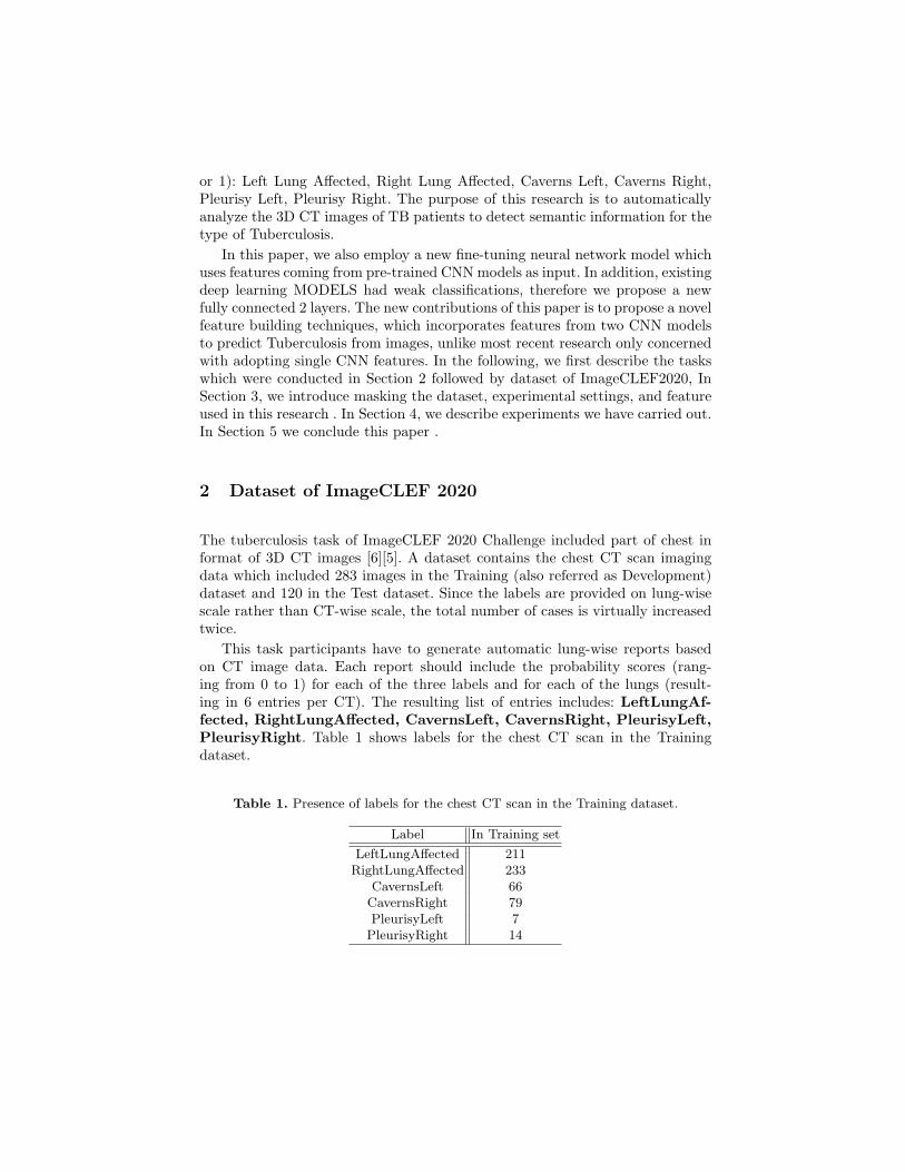
The tuberculosis task of ImageCLEF 2020 Challenge included part of chest informat of 3D CT images [6][5]. A dataset contains the chest CT scan imagingdata which included 283 images in the Training (also referred as Development)dataset and 120 in the Test dataset. Since the labels are provided on lung-wisescale rather than CT-wise scale, the total number of cases is virtually increasedtwice.
This task participants have to generate automatic lung-wise reports basedon CT image data. Each report should include the probability scores (rang-ing from 0 to 1) for each of the three labels and for each of the lungs (result-ing in 6 entries per CT). The resulting list of entries includes: LeftLungAf-fected, RightLungAffected, CavernsLeft, CavernsRight, PleurisyLeft,PleurisyRight. Table 1 shows labels for the chest CT scan in the Trainingdataset.
Table 1. Presence of labels for the chest CT scan in the Training dataset.
Label In Training set
LeftLungAffected 211RightLungAffected 233
CavernsLeft 66CavernsRight 79PleurisyLeft 7PleurisyRight 14
3 Proposed Method
We propose a multi-label analysis system to predict Tuberculosis from CT scanimages. The first step is the input data pre-processing. After pre-processing inputdata, we will describe our deep neural network model that enables the multi-label outputs, given CT scan images. In addition, we add an optional step tothe first step. We use a CT scan movie not CT scan images. We will detail ourproposed system in the following section.
3.1 Input Data Pre-processing
First, we remind the reader that in train and test data, 3D CT scans are providedin compressed Nifti format. We decompressed the files and extracted the slicesof x-axis, y-axis, and z-axis from the three dimensions of the 3D image shown inFig. 1. For each dimension for each Nifti image, we obtained a number of slicesranging according to the dimension: 512 images for the X and Y dimensions,and from 110 to 250 images for the Z dimension.
After extracting slices along x-axis, y-axis, and z-axis, we propose to filterthe slices of each patient using mask data [7][8]. We extract a filtering CT scanimage, as shown in Fig. 2. Indeed, we can notice that many slices contain relevantinformation including bone, space, fat, and skin except for the lungs that couldhelp to classify the samples. This is why we added a step to the filter and selecteda number of slices per patient.
3.2 Proposed deep neural network model
To solve our multi-label problem, we propose new combined neural networkmodels which allow inputs coming from End-to-end (CNN) features.
Training and Validation sets The training dataset consists of 108 891, 77468, 31 497 images extracted from the filtered CT image for x, y and z axisrespectively.
We have divided the train data into training and validation data with 8:2ratio in random. CNN features were extracted using pre-trained CNN-basedneural networks, including VGG16, ResNet50, NasNet-Large and EfficientNetB07. In order to deal with the above feature, we propose a deep neural networkarchitecture where we allow multiple inputs and a multi-hot vector output.
Our system incorporates CNN features, which can be extracted using deepconvolutional neural networks pre-trained on the ImageNet [9] such as VGG16[10], ResNet50[11], NasNet-Large [12] and EfficientNet B07[13]. Because of thelack of dataset in visual sentiment analysis, we adopt transfer learning for thefeature extraction to prevent over fitting. We decreased the dimensions of fully-connected layers used in CNN models. In addition, we reduced the vector to 2048dimensions. This was introduced with the expectation of reducing the numberof parameters and unifying the dimensions.
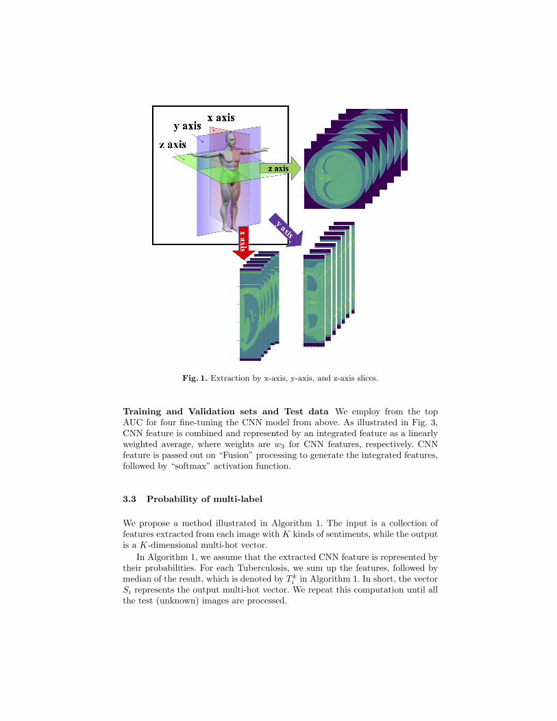
Fig. 1. Extraction by x-axis, y-axis, and z-axis slices.
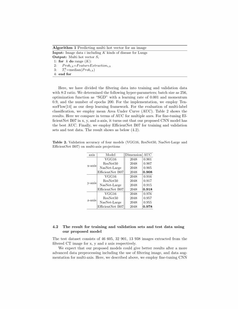
Training and Validation sets and Test data We employ from the topAUC for four fine-tuning the CNN model from above. As illustrated in Fig. 3,CNN feature is combined and represented by an integrated feature as a linearlyweighted average, where weights are w3 for CNN features, respectively. CNNfeature is passed out on “Fusion” processing to generate the integrated features,followed by “softmax” activation function.
3.3 Probability of multi-label
We propose a method illustrated in Algorithm 1. The input is a collection offeatures extracted from each image with K kinds of sentiments, while the outputis a K-dimensional multi-hot vector.
In Algorithm 1, we assume that the extracted CNN feature is represented bytheir probabilities. For each Tuberculosis, we sum up the features, followed bymedian of the result, which is denoted by T k
i in Algorithm 1. In short, the vectorSi represents the output multi-hot vector. We repeat this computation until allthe test (unknown) images are processed.
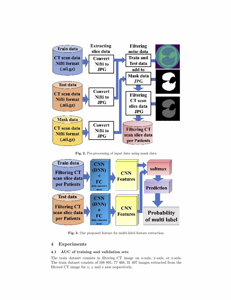
Fig. 2. Pre-processing of input data using mask data.
Fig. 3. Our proposed feature for multi-label feature extraction.
4 Experiments
4.1 AUC of training and validation sets
The train dataset consists in filtering CT image on x-axis, y-axis, or z-axis.The train dataset consists of 108 891, 77 468, 31 497 images extracted from thefiltered CT image for x, y and z axis respectively.
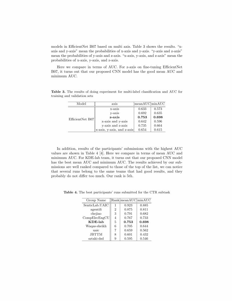
Algorithm 1 Predicting multi hot vector for an image
Input: Image data i including K kinds of disease for LungsOutput: Multi hot vector Si
1: for k do range (K):2: Probi,k=FeatureExtractioni,k
3: T ki =median(Probi,k)
4: end for
Here, we have divided the filtering data into training and validation datawith 8:2 ratio. We determined the following hyper-parameters; batch size as 256,optimization function as “SGD” with a learning rate of 0.001 and momentum0.9, and the number of epochs 200. For the implementation, we employ Ten-sorFlow[14] as our deep learning framework. For the evaluation of multi-labelclassification, we employ mean Area Under Curve (AUC). Table 2 shows theresults. Here we compare in terms of AUC for multiple axes. For fine-tuning Ef-ficientNet B07 in x, y, and z-axis, it turns out that our proposed CNN model hasthe best AUC. Finally, we employ EfficientNet B07 for training and validationsets and test data. The result shows as below (4.2).
Table 2. Validation accuracy of four models (VGG16, ResNet50, NasNet-Large andEfficientNet B07) on multi-axis projections
axis Model Dimension AUC
VGG16 2048 0.901
x-axisResNet50 2048 0.907
NasNet-Large 2048 0.905EfficientNet B07 2048 0.908
VGG16 2048 0.916
y-axisResNet50 2048 0.917
NasNet-Large 2048 0.915EfficientNet B07 2048 0.918
VGG16 2048 0.976
z-axisResNet50 2048 0.957
NasNet-Large 2048 0.955EfficientNet B07 2048 0.978
4.2 The result for training and validation sets and test data usingour proposed model
The test dataset consists of 46 605, 32 901, 13 938 images extracted from thefiltered CT image for x, y and z axis respectively.
We expect that our proposed models could give better results after a moreadvanced data preprocessing including the use of filtering image, and data aug-mentation for multi-axis. Here, we described above, we employ fine-tuning CNN
models in EfficientNet B07 based on multi axis. Table 3 shows the results. “x-axis and y-axis” mean the probabilities of x-axis and y-axis. “y-axis and z-axis”mean the probabilities of y-axis and z-axis. “x-axis, y-axis, and z-axis” mean theprobabilities of x-axis, y-axis, and z-axis.
Here we compare in terms of AUC. For z-axis on fine-tuning EfficientNetB07, it turns out that our proposed CNN model has the good mean AUC andminimum AUC.
Table 3. The results of doing experiment for multi-label classification and AUC fortraining and validation sets
Model axis meanAUC minAUC
x-axis 0.633 0.573y-axis 0.692 0.635
EfficientNet B07z-axis 0.753 0.698
x-axis and y-axis 0.642 0.596y-axis and z-axis 0.735 0.664
x-axis, y-axis, and z-axis 0.654 0.615
In addition, results of the participants’ submissions with the highest AUCvalues are shown in Table 4 [4]. Here we compare in terms of mean AUC andminimum AUC. For KDE-lab team, it turns out that our proposed CNN modelhas the best mean AUC and minimum AUC. The results achieved by our sub-missions are well ranked compared to those of the top of the list, we can noticethat several runs belong to the same teams that had good results, and theyprobably do not differ too much. Our rank is 5th.
Table 4. The best participants’ runs submitted for the CTR subtask
Group Name Rank meanAUC minAUC
SenticLab.UAIC 1 0.923 0.885agentili 2 0.875 0.811chejiao 3 0.791 0.682
CompElecEngCU 4 0.767 0.733KDE-lab 5 0.753 0.698
Waqas-sheikh 6 0.705 0.644uaic 7 0.659 0.562
JBTTM 8 0.601 0.432sztaki-dsd 9 0.595 0.546
5 Conclusions
In this research, we proposed a model for predicting each of the three labelsand for each of the lungs as a multi-label problem from chest CT images. Weperformed Chest CT Image analysis where we proposed a combined deep neuralnetwork model which enabled inputs to come from CNN features. In multi-label Chest CT Image analysis, we also introduced a threshold-based multi-labelprediction algorithm. Specifically, after training our deep neural network, wecould predict the existence of a disease for given unknown CT scan images.Experimental results demonstrate that all our proposed models outperform theindividual pre-trained CNN model in terms of mean AUC and minimum AUC.
In this research, we proposed a model for Tuberculosis CT Image analysiswhich accurately estimates multi-label problems from given images. The multi-label problems are evoking multiple different types of Tuberculosis findings si-multaneously.
In the future, given an arbitrary CT or X-ray image might be included theoptimal weights for the neural networks. Moreover, we hope our proposed modelcan encourage further research on the early detection of several viruses or un-known diseases. We also expect that our proposed model will be widely used inthe field of medical computing.
Acknowledgment
A part of this research was carried out with the support of the Grant-in-Aid forScientific Research (B) (issue number 17H01746), and Grant for Education andResearch in Toyohashi University of Technology.
References
1. Yashin Dicente Cid, Alexander Kalinovsky, Vitali Liauchuk, Vassili Kovalev, , andHenning Muller. Overview of ImageCLEFtuberculosis 2017 - predicting tubercu-losis type and drug resistances. In CLEF2017 Working Notes, CEUR WorkshopProceedings, Dublin, Ireland, September 11-14 2017. CEUR-WS.org <http://ceur-ws.org>.
2. Bogdan Ionescu, Henning Muller, Mauricio Villegas, Alba Garcıa Seco de Herrera,Carsten Eickhoff, Vincent Andrearczyk, Yashin Dicente Cid, Vitali Liauchuk, Vas-sili Kovalev, Sadid A. Hasan, Yuan Ling, Oladimeji Farri, Joey Liu, Matthew Lun-gren, Duc-Tien Dang-Nguyen, Luca Piras, Michael Riegler, Liting Zhou, MathiasLux, and Cathal Gurrin. Overview of ImageCLEF 2018: Challenges, datasets andevaluation. In Experimental IR Meets Multilinguality, Multimodality, and Interac-tion, Proceedings of the Ninth International Conference of the CLEF Association(CLEF 2018), Avignon, France, September 10-14 2018. LNCS Lecture Notes inComputer Science, Springer.
3. Bogdan Ionescu, Henning Muller, Renaud Peteri, Yashin Dicente Cid, Vitali Li-auchuk, Vassili Kovalev, Dzmitri Klimuk, Aleh Tarasau, Asma Ben Abacha, Sa-did A. Hasan, Vivek Datla, Joey Liu, Dina Demner-Fushman, Duc-Tien Dang-Nguyen, Luca Piras, Michael Riegler, Minh-Triet Tran, Mathias Lux, Cathal Gur-rin, Obioma Pelka, Christoph M. Friedrich, Alba Garcıa Seco de Herrera, Narciso
Garcia, Ergina Kavallieratou, Carlos Roberto del Blanco, Carlos Cuevas Rodrıguez,Nikos Vasillopoulos, Konstantinos Karampidis, Jon Chamberlain, Adrian Clark,and Antonio Campello. ImageCLEF 2019: Multimedia Retrieval in Medicine, Lifel-ogging, Security and Nature: Multimedia Retrieval in Medicine, Lifelogging, Secu-rity and Nature. In Experimental IR Meets Multilinguality, Multimodality, andInteraction, volume 2380 of Proceedings of the 10th International Conference ofthe CLEF Association (CLEF 2019), Lugano, Switzerland, September 9-12 2019.LNCS Lecture Notes in Computer Science, Springer.
4. Obioma Pelka, Christoph M Friedrich, Alba Garcıa Seco de Herrera, and Hen-ning Muller. Medical image understanding: Overview of the ImageCLEFmed 2020concept prediction task. In CLEF2020 Working Notes, Workshop Proceedings,Thessaloniki, Greece, September 22-25 2020. CEUR-WS.org.
5. Bogdan Ionescu, Henning Muller, Renaud Peteri, Asma Ben Abacha, VivekDatla, Sadid A. Hasan, Dina Demner-Fushman, Serge Kozlovski, Vitali Liauchuk,Yashin Dicente Cid, Vassili Kovalev, Obioma Pelka, Christoph M. Friedrich, AlbaGarcıa Seco de Herrera, Van-Tu Ninh, Tu-Khiem Le, Liting Zhou, Luca Piras,Michael Riegler, Pal Halvorsen, Minh-Triet Tran, Mathias Lux, Cathal Gurrin,Duc-Tien Dang-Nguyen, Jon Chamberlain, Adrian Clark, Antonio Campello, Dim-itri Fichou, Raul Berari, Paul Brie, Mihai Dogariu, Liviu Daniel Stefan, and Mi-hai Gabriel Constantin. Overview of the ImageCLEF 2020: Multimedia Retrievalin Medical, Lifelogging, Nature, and Internet Applications. In Experimental IRMeets Multilinguality, Multimodality, and Interaction, volume 12260 of Proceed-ings of the 11th International Conference of the CLEF Association (CLEF 2020),Thessaloniki, Greece, September 22-25 2020. LNCS Lecture Notes in ComputerScience, Springer.
6. Serge Kozlovski, Vitali Liauchuk, Yashin Dicente Cid, Aleh Tarasau, Vassili Ko-valev, and Henning Muller. Overview of ImageCLEFtuberculosis 2020 - auto-matic CT-based report generation. In CLEF2020 Working Notes, CEUR Work-shop Proceedings, Thessaloniki, Greece, September 22-25 2020. CEUR-WS.org<http://ceur-ws.org>.
7. Yashin Dicente Cid, Oscar Alfonso Jimenez del Toro, Adrien Depeursinge, andHenning Muller. Efficient and fully automatic segmentation of the lungs in ctvolumes. In Orcun Goksel, Oscar Alfonso Jimenez del Toro, Antonio Foncubierta-Rodrıguez, and Henning Muller, editors, Proceedings of the VISCERAL AnatomyGrand Challenge at the 2015 IEEE ISBI, CEUR Workshop Proceedings, pages31–35. CEUR-WS, May 2015.
8. Vitali Liauchuk and Vassili Kovalev. Imageclef 2017: Supervoxels and co-occurrence for tuberculosis CT image classification. In Linda Cappellato, NicolaFerro, Lorraine Goeuriot, and Thomas Mandl, editors, Working Notes of CLEF2017 - Conference and Labs of the Evaluation Forum, Dublin, Ireland, September11-14, 2017, volume 1866 of CEUR Workshop Proceedings. CEUR-WS.org, 2017.
9. Olga Russakovsky, Jia Deng, Hao Su, Jonathan Krause, Sanjeev Satheesh, SeanMa, Zhiheng Huang, Andrej Karpathy, Aditya Khosla, Michael Bernstein, Alexan-der C. Berg, and Li Fei-Fei. ImageNet Large Scale Visual Recognition Challenge.International Journal of Computer Vision (IJCV), 115(3):211–252, 2015.
10. Karen Simonyan and Andrew Zisserman. Very deep convolutional networks forlarge-scale image recognition, 2014.
11. Kaiming He, Xiangyu Zhang, Shaoqing Ren, and Jian Sun. Deep residual learningfor image recognition. 2016 IEEE Conference on Computer Vision and PatternRecognition (CVPR), Jun 2016.
12. Barret Zoph, Vijay Vasudevan, Jonathon Shlens, and Quoc V. Le. Learning trans-ferable architectures for scalable image recognition. 2018 IEEE/CVF Conferenceon Computer Vision and Pattern Recognition, Jun 2018.
13. Mingxing Tan and Quoc V. Le. Efficientnet: Rethinking model scaling for convo-lutional neural networks. ICML 2019, 05 2019.
14. Google. Tensorflow. https://github.com/tensorflow.