Early Versus Delayed Cholecystectomy for Acute Calculous Cholecystitis
Volume 6 • Issue 2 1000710J Clin Case RepISSN: 2165-7920 JCCR, an open access journal
Open AccessCase Report
Iida et al., J Clin Case Rep 2016, 6:2 DOI: 10.4172/2165-7920.1000710
Journal of Clinical Case ReportsJour
nal o
f Clinical Case Reports
ISSN: 2165-7920
A Case of Xanthogranulomatous Cholecystitis with High CA19-9 Levels that Normalized Post-CholecystectomyTomoya Iida1*, Takeya Adachi1, Suguru Nakagaki1, Takashi Yabana1, Akira Goto1, Yoshihiro Kondo1, Takashi Kawamata2, Takuji Ota2, Yoshito Watanabe2, Hayato Echizenya2, Hiroshi Gondo2 and Kiyoshi Kasai3
1Department of Gastroenterology, Otaru City General Hospital, Japan2Department of Surgery, Otaru City General Hospital, Japan3Department of Diagnostic Pathology, Otaru City General Hospital, Japan
*Corresponding author: Tomoya Iida, Department of Gastroenterology, Otaru City General Hospital, Japan, Tel: +81-134-25-1211; Fax: +81-134-32-6424; E-mail:[email protected]
Received January 10, 2016; Accepted February 14, 2016; Published February 18, 2016
Citation: Iida T, Adachi T, Nakagaki S, Yabana T, Goto A, et al. (2016) A Case of Xanthogranulomatous Cholecystitis with High CA19-9 Levels that Normalized Post-Cholecystectomy. J Clin Case Rep 6: 710. doi:10.4172/2165-7920.1000710
Copyright: © 2016 Iida T, et al. This is an open-access article distributed under the terms of the Creative Commons Attribution License, which permits unrestricted use, distribution, and reproduction in any medium, provided the original author and source are credited.
AbstractThe patient was an 81-year-old male. His blood tests revealed a mild hepatic dysfunction and an abnormally high
Carbohydrate antigen 19-9 (2,830 U/ml). Ultrasonography, contrast-enhanced computed tomography and magnetic resonance cholangiopancreatography were carried out, and showed that the gallbladder was filled with microcalculi, that the gallbladder was enlarged, and that the gallbladder wall had thickened; however, no calculi were found in the common bile duct, and positron emission tomography was performed for the detection of malignancies but the findings were poor; therefore, the condition was diagnosed as calculous cholecystitis, and cholecystectomy was performed. The pathological findings indicated a xanthogranulomatous cholecystitis, and the levels of Carbohydrate antigen 19-9 returned to normal immediately after surgery. The immunostaining of Carbohydrate antigen 19-9 showed that epithelial mucosa of the gallbladder, cytoplasm of multinucleated foreign-body giant cells, and infiltrating macrophages were positive, and suggested that the abnormally high levels of Carbohydrate antigen 19-9 may have been due to xanthogranulomatous cholecystitis. In some cases, Carbohydrate antigen 19-9 levels can be high in benign diseases such as cholangitis and pancreatitis, but markedly high levels are rare. Only two cases of xanthogranulomatous cholecystitis have been reported to have shown abnormally high Carbohydrate antigen 19-9 that returned to normal after cholecystectomy. We report our experience along with a discussion based on theliterature.
Keywords: Carbohydrate antigen 19-9; Cholecystectomy;Xanthogranulomatous cholecystitis; Gallbladder; Cholecystitis
IntroductionThe levels of carbohydrate antigen 19-9 (CA19-9) are often
high in biliary tract cancers and pancreatic cancers. In some cases, CA19-9 levels can also be high in benign diseases such as cholangitis and pancreatitis, but markedly high levels are rare. In this study, we experienced a case in which findings of malignancies from various imaging tests were preoperatively unlikely despite abnormally high levels of CA19-9, calculous cholecystitis was suspected and cholecystectomy was performed, and as a result, pathological findings led to the diagnosis of Xanthogranulomatous Cholecystitis (XGC) and the levels of CA19-9 returned to normal immediately after surgery. Only two cases of XGC have been reported to have shown abnormally high CA19-9 that returned to normal after cholecystectomy; [1,2] and in this communication, we report our experience along with a discussion based on the literature.
Case ReportThe patient was an 81-year-old male who was receiving outpatient
treatment in our hospital for hypertension and Buerger’s disease, for each, he was taken amlodipine and warfarin, he developed an abdominal pain.
Physical findings on admission: stature: 167 cm, body weight: 77.9 kg. Body temperature: 37.0°C. The palpebral conjunctiva showed no anemia, and the bulbar conjunctiva showed no icterus. The abdomen was flat and soft, and showed slight tenderness from the epigastric region to the right hypochondrium; however, there were no symptoms of peritoneal irritation.
Laboratory findings on admission: Blood tests revealed elevated levels of hepatobiliary enzymes; asparate aminotransferase (AST), Alanine Aminotransferase (ALT), Alkaline Phosphatase (ALP), Lactate Dehydrogenase (LDH), and γ-glutamyl transpeptidase (γ-GTP) were
61 IU/l, 224 IU/l, 206 IU/l, 220 IU/l, 670 IU/l, respectively. However, the increase in the levels of white blood cell (WBC) and C-reactive protein (CRP) remained mild. The level of carcinoembryonic antigen (CEA) was normal (1.8 ng/ml) whereas CA19-9 was markedly elevated (2,830 U/ml).
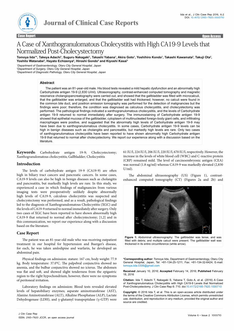
The abdominal ultrasonography (US) (Figure 1), contrast-enhanced computed tomography (CT) (Figures 2a and 2b) and
Figure 1: Abdominal ultrasonography: The gallbladder was tense, and was filled with debris; and multiple calculi were present. The gallbladder wall was thickened in its entire circumference (white arrow).

Citation: Iida T, Adachi T, Nakagaki S, Yabana T, Goto A, et al. (2016) A Case of Xanthogranulomatous Cholecystitis with High CA19-9 Levels that Normalized Post-Cholecystectomy. J Clin Case Rep 6: 710. doi:10.4172/2165-7920.1000710
Page 2 of 4
Volume 6 • Issue 2 • 1000710J Clin Case RepISSN: 2165-7920 JCCR, an open access journal
magnetic resonance cholangiopancreatography (MRCP) (Figures 3a-3c) revealed that the gallbladder was filled with microcalculi, that the gallbladder was enlarged, and that the gallbladder wall had thickened; however, there were no calculi in the common bile duct. The endoscopic ultrasonography (EUS) (Figure 4) showed that the condition was complicated with cholangitis, but there were no apparent calculi in the common bile duct. Therefore, calculous cholecystitis was suspected, and the patient was admitted to our hospital in May 2015.
Conservative treatment was carried out through fasting and by using antibiotics; and as a result, the abdominal findings, the inflammatory reaction that was found in blood tests, as well as the elevation of the levels of hepatobiliary enzymes, tended to improve. However, the levels of CA19-9 increased even further and reached 4,639 U/ml at hospital day 7 after admission; therefore, in consideration of the possibility of a combined presence of gallbladder cancer and
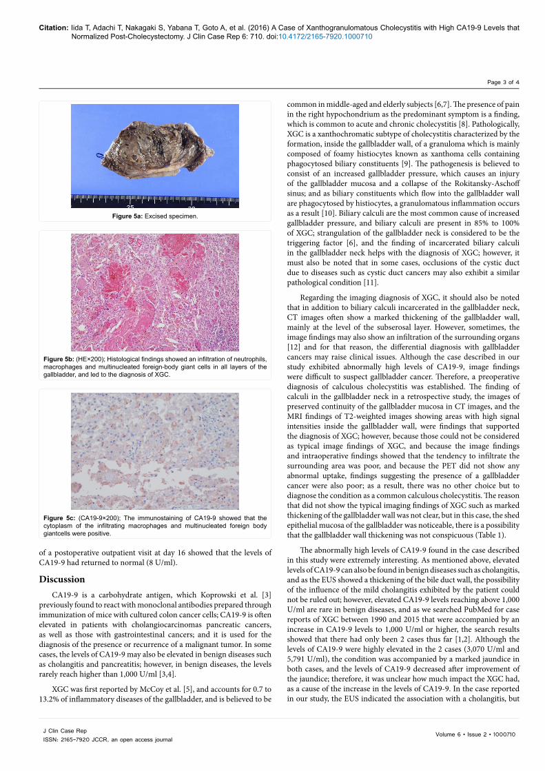
other malignant tumors, upper and lower gastrointestinal endoscopy and Positron Emission Tomography (PET) were carried out, but there were no abnormal findings. The condition was diagnosed as calculous cholecystitis; cholecystectomy was performed at hospital day 15; and histological findings showed the degeneration and remarkable peeling of epithelial mucosa, the infiltration of neutrophils, macrophages and multinucleated foreign-body giant cells in all layers of the gallbladder, and led to the diagnosis of XGC; in addition, the immunostaining of CA19-9 showed that epithelial mucosa, shed mucosa, cytoplasm of the infiltrating macrophages, and multinucleated foreign body giant cells were positive (Figures 5a-5c). The patient was discharged postoperatively at hospital day 7; and blood tests conducted at the time
Figure 2a: Abdominal-pelvic contrast-enhanced CT: The gallbladder was distended; the gallbladder wall was thickened, and showed a slightly delayed enhancement (white arrow head).
Figure 2b: An enhancement of the gallbladder bed was found (white arrow).
Figure 3a: A large number of microcalculi were found in the neck of the gallbladder (white arrow).
Figure 3b: The gallbladder wall showed a high signal intensity in T2 (white arrow).
Figure 3c: There were no calculi in the common bile duct.
Figure 4: Endoscopic ultrasound (EUS): The area of the bifurcation of the cystic duct (CD) was identifiable, but detailed evaluation of the condition inside the gallbladder was impossible. The common bile duct (CBD) wall was mildly thickened, and suggested the combined presence of a cholangitis, but there were no calculi in the CBD. PV: Portal vein.
Citation: Iida T, Adachi T, Nakagaki S, Yabana T, Goto A, et al. (2016) A Case of Xanthogranulomatous Cholecystitis with High CA19-9 Levels that Normalized Post-Cholecystectomy. J Clin Case Rep 6: 710. doi:10.4172/2165-7920.1000710
Page 3 of 4
Volume 6 • Issue 2 • 1000710J Clin Case RepISSN: 2165-7920 JCCR, an open access journal
of a postoperative outpatient visit at day 16 showed that the levels of CA19-9 had returned to normal (8 U/ml).
DiscussionCA19-9 is a carbohydrate antigen, which Koprowski et al. [3]
previously found to react with monoclonal antibodies prepared through immunization of mice with cultured colon cancer cells; CA19-9 is often elevated in patients with cholangiocarcinomas pancreatic cancers, as well as those with gastrointestinal cancers; and it is used for the diagnosis of the presence or recurrence of a malignant tumor. In some cases, the levels of CA19-9 may also be elevated in benign diseases such as cholangitis and pancreatitis; however, in benign diseases, the levels rarely reach higher than 1,000 U/ml [3,4].
XGC was first reported by McCoy et al. [5], and accounts for 0.7 to 13.2% of inflammatory diseases of the gallbladder, and is believed to be
common in middle-aged and elderly subjects [6,7]. The presence of pain in the right hypochondrium as the predominant symptom is a finding, which is common to acute and chronic cholecystitis [8]. Pathologically, XGC is a xanthochromatic subtype of cholecystitis characterized by the formation, inside the gallbladder wall, of a granuloma which is mainly composed of foamy histiocytes known as xanthoma cells containing phagocytosed biliary constituents [9]. The pathogenesis is believed to consist of an increased gallbladder pressure, which causes an injury of the gallbladder mucosa and a collapse of the Rokitansky-Aschoff sinus; and as biliary constituents which flow into the gallbladder wall are phagocytosed by histiocytes, a granulomatous inflammation occurs as a result [10]. Biliary calculi are the most common cause of increased gallbladder pressure, and biliary calculi are present in 85% to 100% of XGC; strangulation of the gallbladder neck is considered to be the triggering factor [6], and the finding of incarcerated biliary calculi in the gallbladder neck helps with the diagnosis of XGC; however, it must also be noted that in some cases, occlusions of the cystic duct due to diseases such as cystic duct cancers may also exhibit a similar pathological condition [11].
Regarding the imaging diagnosis of XGC, it should also be noted that in addition to biliary calculi incarcerated in the gallbladder neck, CT images often show a marked thickening of the gallbladder wall, mainly at the level of the subserosal layer. However, sometimes, the image findings may also show an infiltration of the surrounding organs [12] and for that reason, the differential diagnosis with gallbladder cancers may raise clinical issues. Although the case described in our study exhibited abnormally high levels of CA19-9, image findings were difficult to suspect gallbladder cancer. Therefore, a preoperative diagnosis of calculous cholecystitis was established. The finding of calculi in the gallbladder neck in a retrospective study, the images of preserved continuity of the gallbladder mucosa in CT images, and the MRI findings of T2-weighted images showing areas with high signal intensities inside the gallbladder wall, were findings that supported the diagnosis of XGC; however, because those could not be considered as typical image findings of XGC, and because the image findings and intraoperative findings showed that the tendency to infiltrate the surrounding area was poor, and because the PET did not show any abnormal uptake, findings suggesting the presence of a gallbladder cancer were also poor; as a result, there was no other choice but to diagnose the condition as a common calculous cholecystitis. The reason that did not show the typical imaging findings of XGC such as marked thickening of the gallbladder wall was not clear, but in this case, the shed epithelial mucosa of the gallbladder was noticeable, there is a possibility that the gallbladder wall thickening was not conspicuous (Table 1).
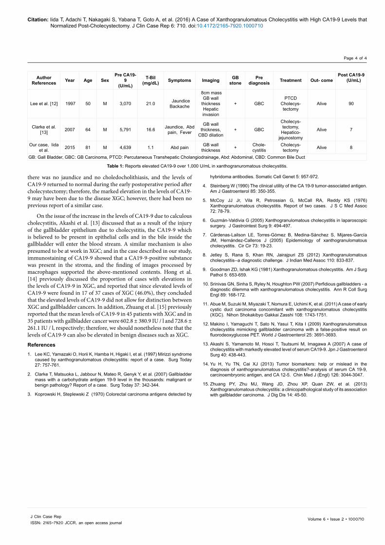
The abnormally high levels of CA19-9 found in the case described in this study were extremely interesting. As mentioned above, elevated levels of CA19-9 can also be found in benign diseases such as cholangitis, and as the EUS showed a thickening of the bile duct wall, the possibility of the influence of the mild cholangitis exhibited by the patient could not be ruled out; however, elevated CA19-9 levels reaching above 1,000 U/ml are rare in benign diseases, and as we searched PubMed for case reports of XGC between 1990 and 2015 that were accompanied by an increase in CA19-9 levels to 1,000 U/ml or higher, the search results showed that there had only been 2 cases thus far [1,2]. Although the levels of CA19-9 were highly elevated in the 2 cases (3,070 U/ml and 5,791 U/ml), the condition was accompanied by a marked jaundice in both cases, and the levels of CA19-9 decreased after improvement of the jaundice; therefore, it was unclear how much impact the XGC had, as a cause of the increase in the levels of CA19-9. In the case reported in our study, the EUS indicated the association with a cholangitis, but
Figure 5a: Excised specimen.
Figure 5b: (HE×200); Histological findings showed an infiltration of neutrophils, macrophages and multinucleated foreign-body giant cells in all layers of the gallbladder, and led to the diagnosis of XGC.
Figure 5c: (CA19-9×200); The immunostaining of CA19-9 showed that the cytoplasm of the infiltrating macrophages and multinucleated foreign body giantcells were positive.
Citation: Iida T, Adachi T, Nakagaki S, Yabana T, Goto A, et al. (2016) A Case of Xanthogranulomatous Cholecystitis with High CA19-9 Levels that Normalized Post-Cholecystectomy. J Clin Case Rep 6: 710. doi:10.4172/2165-7920.1000710
Page 4 of 4
Volume 6 • Issue 2 • 1000710J Clin Case RepISSN: 2165-7920 JCCR, an open access journal
there was no jaundice and no choledocholithiasis, and the levels of CA19-9 returned to normal during the early postoperative period after cholecystectomy; therefore, the marked elevation in the levels of CA19-9 may have been due to the disease XGC; however, there had been no previous report of a similar case.
On the issue of the increase in the levels of CA19-9 due to calculous cholecystitis, Akashi et al. [13] discussed that as a result of the injury of the gallbladder epithelium due to cholecystitis, the CA19-9 which is believed to be present in epithelial cells and in the bile inside the gallbladder will enter the blood stream. A similar mechanism is also presumed to be at work in XGC; and in the case described in our study, immunostaining of CA19-9 showed that a CA19-9-positive substance was present in the stroma, and the finding of images processed by macrophages supported the above-mentioned contents. Hong et al. [14] previously discussed the proportion of cases with elevations inthe levels of CA19-9 in XGC, and reported that since elevated levels ofCA19-9 were found in 17 of 37 cases of XGC (46.0%), they concludedthat the elevated levels of CA19-9 did not allow for distinction between XGC and gallbladder cancers. In addition, Zhuang et al. [15] previously reported that the mean levels of CA19-9 in 45 patients with XGC and in 35 patients with gallbladder cancer were 602.8 ± 380.9 IU / l and 728.6 ± 261.1 IU / l, respectively; therefore, we should nonetheless note that the levels of CA19-9 can also be elevated in benign diseases such as XGC.
References
1. Lee KC, Yamazaki O, Horii K, Hamba H, Higaki I, et al. (1997) Mirizzi syndrome caused by xanthogranulomatous cholecystitis: report of a case. Surg Today27: 757-761.
2. Clarke T, Matsuoka L, Jabbour N, Mateo R, Genyk Y, et al. (2007) Gallbladdermass with a carbohydrate antigen 19-9 level in the thousands: malignant orbenign pathology? Report of a case. Surg Today 37: 342-344.
3. Koprowski H, Steplewski Z (1970) Colorectal carcinoma antigens detected by
hybridoma antibodies. Somatic Cell Genet 5: 957-972.
4. Steinberg W (1990) The clinical utility of the CA 19-9 tumor-associated antigen. Am J Gastroenterol 85: 350-355.
5. McCoy JJ Jr, Vila R, Petrossian G, McCall RA, Reddy KS (1976)Xanthogranulomatous cholecystitis. Report of two cases. J S C Med Assoc72: 78-79.
6. Guzmán-Valdivia G (2005) Xanthogranulomatous cholecystitis in laparoscopicsurgery. J Gastrointest Surg 9: 494-497.
7. Cárdenas-Lailson LE, Torres-Gómez B, Medina-Sánchez S, Mijares-GarcíaJM, Hernández-Calleros J (2005) Epidemiology of xanthogranulomatouscholecystitis. Cir Cir 73: 19-23.
8. Jetley S, Rana S, Khan RN, Jairajpuri ZS (2012) Xanthogranulomatouscholecystitis--a diagnostic challenge. J Indian Med Assoc 110: 833-837.
9. Goodman ZD, Ishak KG (1981) Xanthogranulomatous cholecystitis. Am J Surg Pathol 5: 653-659.
10. Srinivas GN, Sinha S, Ryley N, Houghton PW (2007) Perfidious gallbladders - a diagnostic dilemma with xanthogranulomatous cholecystitis. Ann R Coll SurgEngl 89: 168-172.
11. Abue M, Suzuki M, Miyazaki T, Nomura E, Uchimi K, et al. (2011) A case of early cystic duct carcinoma concomitant with xanthogranulomatous cholecystitis(XGC). Nihon Shokakibyo Gakkai Zasshi 108: 1743-1751.
12. Makino I, Yamaguchi T, Sato N, Yasui T, Kita I (2009) Xanthogranulomatouscholecystitis mimicking gallbladder carcinoma with a false-positive result onfluorodeoxyglucose PET. World J Gastroenterol 25: 3691-3693.
13. Akashi S, Yamamoto M, Hosoi T, Tsutsumi M, Imagawa A (2007) A case ofcholecystitis with markedly elevated level of serum CA19-9. Jpn J Gastroenterol Surg 40: 438-443.
14. Yu H, Yu TN, Cai XJ (2013) Tumor biomarkers: help or mislead in thediagnosis of xanthogranulomatous cholecystitis?-analysis of serum CA 19-9,carcinoembryonic antigen, and CA 12-5. Chin Med J (Engl) 126: 3044-3047.
15. Zhuang PY, Zhu MJ, Wang JD, Zhou XP, Quan ZW, et al. (2013)Xanthogranulomatous cholecystitis: a clinicopathological study of its association with gallbladder carcinoma. J Dig Dis 14: 45-50.
Author References Year Age Sex
Pre CA19-9
(U/mL)
T-Bil(mg/dL) Symptoms Imaging GB
stonePre
diagnosis Treatment Out- comePost CA19-9
(U/mL)
Lee et al. [12] 1997 50 M 3,070 21.0 JaundiceBackache
8cm massGB wall
thickness Hepatic invasion
+ GBCPTCD
Cholecys- tectomy
Alive 90
Clarke et al. [13] 2007 64 M 5,791 16.6 Jaundice, Abd
pain, Fever
GB wall thickness,
CBD dilation+ GBC
Cholecys- tectomy,
Hepatico- jejunostomy
Alive 7
Our case, Iida et al. 2015 81 M 4,639 1.1 Abd pain GB wall
thickness + Chole- cystitis
Cholecys- tectomy Alive 8
GB: Gall Bladder, GBC: GB Carcinoma, PTCD: Percutaneous Transhepatic Cholangiodrainage, Abd: Abdominal, CBD: Common Bile Duct
Table 1: Reports elevated CA19-9 over 1,000 U/mL in xanthogranuromatous cholecystitis.