IGF1 dependence of dietary energy balance effects on murine MET1 ...
30
IGF1 dependence of dietary energy balance effects on murine MET1 mammary tumor progression, epithelial-to-mesenchymal transition and chemokine expression Nikki A. Ford 1 , Nomeli P. Nunez 1 , Valerie B. Holcomb 1 , and Stephen D. Hursting 1,2 1 Department of Nutritional Sciences, University of Texas, Austin, Texas 78722, USA 2 Department of Molecular Carcinogenesis, University of Texas MD Anderson Cancer Center, Science Park, Smithville, TX 78957, USA Corresponding Author: Stephen D. Hursting, PhD, MPH; [email protected], (512)495-3021 (phone), (512)471-5844 (fax). Address: Dell Pediatric Research Institute, 1400 Barbara Jordan Blvd. DPRI 2.834, Austin, TX 78722 Short Title: IGF1, energy balance, and mammary cancer Keywords: energy balance, epithelial-to-mesenchymal transition, IGF1, chemokine, calorie restriction Page 1 of 30 Accepted Preprint first posted on 14 November 2012 as Manuscript ERC-12-0329 Copyright © 2012 by the Society for Endocrinology.
Transcript of IGF1 dependence of dietary energy balance effects on murine MET1 ...
IGF1 dependence of dietary energy balance effects on murine MET1
mammary tumor progression, epithelial-to-mesenchymal transition and
chemokine expression
Nikki A. Ford1, Nomeli P. Nunez
1, Valerie B. Holcomb
1, and Stephen D. Hursting
1,2
1Department of Nutritional Sciences, University of Texas, Austin, Texas 78722, USA
2Department of Molecular Carcinogenesis, University of Texas MD Anderson Cancer Center,
Science Park, Smithville, TX 78957, USA
Corresponding Author: Stephen D. Hursting, PhD, MPH; [email protected],
(512)495-3021 (phone), (512)471-5844 (fax). Address: Dell Pediatric Research Institute, 1400
Barbara Jordan Blvd. DPRI 2.834, Austin, TX 78722
Short Title: IGF1, energy balance, and mammary cancer
Keywords: energy balance, epithelial-to-mesenchymal transition, IGF1, chemokine, calorie
restriction
Page 1 of 30 Accepted Preprint first posted on 14 November 2012 as Manuscript ERC-12-0329
Copyright © 2012 by the Society for Endocrinology.
ABSTRACT
Luminal breast tumors with little or no estrogen receptor-α expression confer poor prognosis. Using the
Met1 murine model of luminal breast cancer, we characterized the insulin-like growth factor 1 (IGF1)-
dependency of diet-induced obesity (DIO) and calorie restriction (CR) effects on tumor growth, growth
factor signaling, epithelial-to-mesenchymal transition (EMT) and chemokine expression. Liver-specific
IGF1-deficient (LID) and littermate control (LC) mice were administered control, DIO or 30% CR diets
for 3 months before orthotopic injection of Met1 cells. Tumors grew for 1 month, were excised and
assessed for Akt pathway activation and mRNA expression of chemokine and EMT constituents. LID
mice, regardless of diet, displayed reduced Met1 tumor growth and downregulated Akt, EMT and
chemokine pathways. CR, relative to control, reduced serum IGF1 and Met1 tumor growth in LC (but
not LID) mice. DIO, relative to control, increased Met1 tumor growth and chemokine expression in LID
mice, and had no effect on serum IGF1 or pAkt or cyclin D1 expression in either genotype. Thus,
circulating IGF1 (in association with Akt, EMT and chemokines) regulated Met1 tumor growth. While
the anticancer effects of CR were largely IGF1-dependent, the procancer effects of DIO manifested only
when circulating IGF1 levels were low. Thus, in a murine model of luminal breast cancer, IGF1 and its
downstream signaling pathway, EMT and chemokines present possible mechanistic regulatory targets.
Transplanted MMTV-1 Wnt1 mammary tumor growth was also reduced in LID mice, relative to LC
mice, suggesting the IGF1 effects on mammary tumor growth are not limited to Met1 tumors.
Page 2 of 30

INTRODUCTION
The prevalence of obesity, an established risk and progression factor for estrogen receptor
(ER)α-positive luminal A breast cancer, has dramatically increased in the United States and
many other parts of the world over the past 25 years (Bianchini, et al. 2002; Wu, et al. 2003).
Little is known, however, about the effects of obesity or energy balance modulation on
progression of luminal breast tumors with little or no ERα expression (referred to hereafter as
ERαlow
), which relative to luminal A tumors confer a worse prognosis, typically respond poorly
to tamoxifen treatment and display activation of growth factor signaling pathways (Loi, et al.
2009). The identification of mechanistic targets and intervention strategies for preventing or
controlling ERαlow
luminal breast cancers are urgently needed due to the lack of targeted
therapies for these aggressive breast cancers (Uray and Brown 2011).
Calorie restriction (CR) is a low calorie diet regimen that prevents obesity and decreases
progression of mammary and many other tumor types in various animal models (Hursting and
Berger 2010; Hursting, et al. 2010; Nunez, et al. 2008). The effects of dietary energy balance
alterations, such as CR and diet-induced obesity (DIO), on ERαlow
luminal breast cancer are
poorly characterized, at least in part due to a paucity of relevant animal models of this intrinsic
subtype of breast cancer. Comparative oncogenomic studies of human and mouse mammary
tumors show that tumors from the polyoma middle-T antigen transgenic mice cluster closely
with human luminal B breast tumors and share many of the pathological and molecular
hallmarks of human luminal B tumors (Fluck and Schaffhausen 2009; Herschkowitz, et al. 2007;
Zhu, et al. 2011). Furthermore, the Met1 cell line, derived from a spontaneous tumor from a
polyoma middle-T antigen transgenic mouse, progressively loses ER and progesterone receptor
(PR) positivity with tumor progression, therefore providing a rapid and relevant model for
Page 3 of 30
studying ERαlow
luminal mammary tumor progression when orthotopically transplanted into
syngeneic mice (Bonuccelli, et al. 2009; Borowsky, et al. 2005; Lin, et al. 2003; Namba, et al.
2006).
Findings from models of luminal A, basal-like, and Her2-positive rodent mammary
tumor models (Dunlap, et al. 2012; Nickerson, et al. 1999; Wu et al. 2003), as well as from a
broad spectrum of colon, prostate, pancreatic, skin and other epithelial cancer models (Lashinger,
et al. 2011; Moore, et al. 2008b; Olivo-Marston, et al. 2009), indicate that circulating levels of
insulin-like growth factor 1 (IGF1) are central to the dietary energy balance-cancer link. Limited
evidence suggests plausible relationships between serum IGF1 levels, tumor Akt signaling,
tumor expression of epithelial-to-mesenchymal transition (EMT) and chemokine genes, and
mammary tumor progression (Graham, et al. 2008; Kim, et al. 2007; Lorenzatti, et al. 2011;
Sivakumar, et al. 2009; Walsh and Damjanovski 2011). Alterations in systemic IGF1 can
modulate cellular signaling through receptor tyrosine kinases and induce changes in downstream
proliferation and survival regulators such as Akt and the mammalian target of rapamycin
(mTOR) (Moore, et al. 2008a). Akt and mTOR signaling pathway components are frequently
altered in human cancers (Fresno Vara, et al. 2004) and are upregulated by DIO and
downregulated by CR in mammary and other tumors (De Angel, et al. 2012; Moore et al. 2008a).
Akt activation can induce EMT (Nath, et al. 2008), while Akt inhibitors can suppress EMT
(Hong, et al. 2009). Characteristics of EMT, an orchestrated program in which cell-cell and cell-
extracellular matrix interactions are altered to enhance tumor progression and initiate local
invasion and metastasis (Thiery 2003), include the progressive loss of epithelial markers, such as
E-cadherin, and gain of mesenchymal markers, such as snail, slug, vimentin, N-cadherin, twist,
transforming growth factor B1 (Tgfb1), and matrix metalloproteinases (Mmps) (Rucklidge, et al.
Page 4 of 30
1994). Increased body adiposity (Calabro and Yeh 2007; Subbaramaiah, et al. 2011) and
tumoral Akt activation (Balkwill 1998, 2004; Roca, et al. 2008) are also associated with
increased tumoral chemokine expression. Chemokines, which are a subfamily of secreted
cytokines from adipocytes, macrophages and other cells that stimulate directed chemotaxis in
nearby responsive cells, increase inflammation, tumor progression, and invasion in multiple
tumor types (Balkwill 2004; Kulbe, et al. 2004; Szlosarek and Balkwill 2004).
Modulation of dietary fat content has been demonstrated to be effective in regulating ER
negative breast cancers (Chlebowski, et al. 2006), and IGF1 inhibitors are efficacious against
triple negative breast cancer cells (Litzenburger, et al. 2011). Unfortunately, the effects of energy
balance and/or IGF1 modulation on ERαlow
luminal mammary tumor progression are poorly
understood. The purpose of this study was to test the hypothesis that DIO enhances, and CR
suppresses, ERαlow
luminal mammary tumor progression, at least in part, through IGF1-
dependent pathways. To test this hypothesis, we compared the effects of DIO and CR on
progression of orthotopically transplanted Met1 tumors, systemic IGF1 (and other serum
hormones), tumoral Akt activation, and EMT and chemokine gene expression in mice without
genetic reduction of circulating IGF-1.
MATERIALS AND METHODS
Mice
All procedures involving animals were approved and monitored by the National Cancer Institute
Animal Care and Use Committee or the University of Texas Institutional Animal Care and Use
Committee. All diets were purchased from Research Diets, Inc., New Brunswick, NJ. Liver-
specific IGF-1-deficient (loxP+/+ Cre+/-; LID) mice (Yakar, et al. 1999), backcrossed to FVB/N
Page 5 of 30
mice for >10 generations, were originally obtained from Dr. Derek LeRoith (NIDDK, Bethesda,
MD), and maintained in Dr. Hursting’s breeding colony at the NCI-Frederick National
Laboratory (Frederick, MD) or his colony at the University of Texas at Austin (for the Wnt1
study). For these studies, female LID mice and floxed IGF-1 littermate control (loxP+/+Cre-/-;
LC) mice were obtained at 3 months of age and were placed on a control diet (modified AIN-
76A, #D12450B) for 1 week following receipt. Mice were individually housed with free access
to water and exposed to a 12 h light/dark cycle.
Diet treatments and Met1 tumor transplantation, monitoring and sample collection
Female LID and LC mice were randomized (n = 10 per group) to receive: i) control diet fed ad
libitum (#D12450B); ii) a 30% CR regimen (#D0302702); or iii) a DIO regimen fed ad libitum
(#D12492). The CR diet was balanced so that all essential nutrients would be consumed at the
control level when dietary intake was restricted by 30% relative to controls. Food intake was
measured biweekly and body weights were recorded weekly. Mice consumed the experimental
diets for 3 months prior to syngeneic orthotopic transplantation of 1x10⁵ Met1 mammary tumor
cells into the fourth mammary fat pad. The Met1 cell line (a gift from Dr. William Mueller) was
originally derived from mammary carcinomas in FVB/N-Tg(MMTV-PyVmT) mice (Borowsky
et al. 2005), and were maintained in complete media (low glucose Dulbecco modified Eagle
medium, supplemented with 10% fetal bovine serum, penicillin-streptomycin and glutamine) at
37ºC in a humidified 5% CO2 incubator. In preparation for transplantation, cells were washed
with PBS, trypsinized, and viable cells were quantified by trypan blue exclusion using a
hemacytometer (Fisher Scientific, Waltham, MA).
Page 6 of 30
Mice continued to consume the experimental diets for another month after transplantation
and before euthanasia. Tumors were measured in three dimensions using electronic calipers
twice weekly by one technician, and tumor volume (4/3 π lwh) was calculated. At the end of the
treatment period, mice were fasted for 12 hours and then anesthetized using isoflurane. Blood
was collected by cardiac puncture and the mice were then killed by cervical dislocation. Blood
was allowed to coagulate at room temperature for 30 minutes and then centrifuged at 10000 x g
for 5 minutes to obtain serum samples that were stored at -80ºC until analyzed for hormones and
adipokines. Ex vivo caliper measurements of mammary tumors was taken (by the same
technician) before tumor tissue was snap frozen in liquid nitrogen and stored at -80ºC until
analyzed for protein and/or gene expression.
Serum hormones and adipokines
Serum leptin, insulin, and adiponectin concentrations (n = 10 per group) from Met1 tumor
bearing mice were measured using a Luminex-based LINCOplex bead array assay (Millipore,
Billerica, MA) on a BioRad BioPlex multianalyte detection system (BioRad, Inc., Hercules, CA).
Serum IGF1 (n = 10 per group) was measured using an ELISA (R&D Systems, Minneapolis,
MN) on a Synergy 2 plate reader (BioTek, Winooski, VT).
Phosphorylated protein expression analyses
Met1 mammary tumor tissue was homogenized and lysed in a radioimmunoprecipitation assay
buffer (Sigma-Adrich, St. Louis, MO) with a protease inhibitor cocktail tablet (Roche Applied
Sciences, Indianapolis, IN) and phosphatase inhibitor cocktails I and II (Sigma-Aldrich).
Expression of phosphorylated (p) Akt (Ser473), pErk (Thr185/Tyr187), and pStat3 (Tyr705)
proteins were quantified on tumor lysates (20 µg) from a random sample of 6 mice per diet-
Page 7 of 30

genotype group, using Luminex-based Milliplex Map bead array assays (Millipore) on a BioRad
BioPlex multianalyte detection system following manufacturer’s instructions.
Gene expression analyses
RNA was extracted from Met1 mammary tumor tissue from random samplings of each diet-
genotype group (n = 6 mice/sampling) using an RNeasy Mini Kit (Qiagen, Gaithersburg, MD)
per manufacturer’s instructions. Complimentary DNA was synthesized from extracted RNA
using the high capacity cDNA reverse transcription kit (Applied Biosystems, Foster City, CA)
with the addition of an RNase inhibitor (Ambion, Austin, TX) per manufacturer’s directions.
Gene expression for cyclin D1, a panel of EMT markers, and a panel of chemokines was
measured by real-time quantitative reverse transcriptase polymerase chain reaction (qRT-PCR)
using the Taqman gene expression assay with Taqman universal PCR mastermix (Applied
Biosystems) on an EPgradient S Realplex 4 thermocycler (Eppendorf, Hauppauge, NY). Gene
expression is reported for LID mice relative to LC mice. In addition, an inflammatory gene
superarray analysis was performed in duplicate for 176 inflammation-related genes on a random
subsampling of extracted tumor RNA from 3 LID DIO mice and 3 LID control diet-fed mice,
using an ABI 7900HT FAST qPCR thermocycler (Applied Biosystems). Gene expression is
reported relative to LID control.
MMTV-Wnt1 tumor transplantation, monitoring and sample collection
Female LID and LC mice (same as described above; n=10/group) were obtained at 3
months of age from Dr. Hursting’s colony at the University of Texas at Austin. Mice were
individually housed with free access to water and exposed to a 12 h light/dark cycle and allowed
Page 8 of 30

to acclimate for 1 week prior to syngeneic orthotopic transplantation of Wnt1 mammary tumor
brei, as previously described (Nunez, et al., 2008). In brief, a suspension of MMTV-Wnt1
mammary tumor cells was derived (Varticovski, et al. 2007) from 6 spontaneously developed
mammary carcinomas in MMTV-Wnt1 FVB/NJ (002934; Jackson Laboratory, Bar Harbor, ME,
http://www.jax.org). MMTV-Wnt1 FVB/NJ mice were euthanized with CO2, and tumors were
collected aseptically using blunt dissection, trimmed of extraneous tissues, mechanically
dissociated by mincing and passage through a 40-micron mesh sterile screen, and suspended in
serum-free RPMI 1640 (Quality Biological, Gaithersburg, MD). Cells were further dissociated
by serial passage through a syringe with 18- gauge needles. The cell suspension was washed
twice and resuspended in serum-free RPMI 1640 medium, and viable cell counts were
determined by Hemocytometer counting, following 0.4% trypan blue staining. Cells were
resuspended at 2x10⁶ cells per ml in 10% DMSO cell freezing medium and cryopreserved using
stepped rate freezing. For orthotopic implantation, 1x10⁵ cells were implanted subcutaneously in
50 mL serum-free RPMI 1640 medium into the 4th
mammary fat pad of recipient LID and LC
mice fed control diet (modified AIN-76A, #D12450B). Mice continued to consume the control
diet for another month after transplantation and before euthanasia. Tumors were measured in
three dimensions using electronic calipers twice weekly by one technician, and tumor volume
(4/3 π lwh) was calculated. At the end of the 4 week period after tumor transplantation, mice
were fasted for 12 hours and then anesthetized using isoflurane. Blood was collected by cardiac
puncture and the mice were then killed by cervical dislocation. Blood was allowed to coagulate
at room temperature for 30 minutes and then centrifuged at 10000 x g for 5 minutes to obtain
serum samples that were stored at -80ºC until analyzed for hormones and adipokines. Ex vivo
caliper measurements of mammary tumors were recorded.
Page 9 of 30
Statistics
Summarized data are expressed as means ± standard deviation, and analyses were performed
using SAS 9.2 (Cary, NC). Molecular analysis could not be performed on all LID tumors due to
the size of the tumor at excision; therefore samples were randomly selected from all other groups
for analysis. Met1 tumor growth was analyzed by repeated measures ANOVA. Body weight,
feed intake, serum adipokines, tumor volume, protein expression and mRNA gene expression
were analyzed by two-way analysis of variance (ANOVA) followed by Tukey’s post hoc test.
When significant interactions were detected, analysis of simple effects was completed.
Differences in relative gene expression using the super array were considered biologically
significant when a minimum of a two-fold expression difference occurred, and P ≤ 0.05 was
achieved. A priori questions addressing the effects of diet on each genotype independently were
analyzed by one-way ANOVA followed by Tukey’s post hoc analysis. Final MMTV-Wnt1
tumor volume was assessed by student’s t-test. Differences were considered statistically
significant at P ≤ 0.05.
RESULTS
Body weight, energy intake and serum hormone levels from Met1 tumor bearing mice
Final body weights and serum hormone levels after 4 weeks of tumor growth (4 months of diet
treatment) and average weekly energy intake throughout the study were analyzed for effects of
genotype and/or diet (Table 1). LID mice weighed less than LC mice on DIO or control diet
(P<0.01), and LID and LC mice showed the same rank order in weight in response to the 3
dietary interventions (CR < control < DIO; P<0.01 within each genotype). By design, total
Page 10 of 30
caloric intake also significantly varied by dietary group (P<0.01) in the same rank order as
weight, and genotype had no effect.
Circulating IGF1 levels were lower in LID mice relative to LC mice, regardless of diet
(P<0.01). In both LID and LC mice, CR reduced IGF1 levels (P <0.01), but DIO had no
significant effect, relative to control. CR and DIO were significantly different from each other in
both LID and LC mice (P<0.01 in both).
Serum concentrations of insulin, leptin and adiponectin were elevated (P<0.01 each) in
LID mice as compared to LC mice (Table 1). Relative to control, CR decreased and DIO
increased insulin and leptin levels in LC mice; in LID mice, CR decreased (P<0.01), but DIO
had no significant effect on leptin, and neither diet affected insulin levels. While no diet effect on
adiponectin was observed in LC mice, CR and DIO (relative to control) both decreased
adiponectin in LID mice (P=0.02). Lastly, relative to control, CR reduced, and DIO increased,
the ratio of leptin to adiponectin in both genotypes (P<0.01 for both).
Met1 Mammary tumor volume
Met1 tumor volume at 3 weeks and study completion was significantly lower in LID mice
relative to LC mice, irrespective of diet (P<0.01) (Fig. 1). Regarding diet effects, in LC mice,
relative to control, CR significantly reduced (P=0.01), but DIO had no effect, on final mammary
tumor volume. In LID mice, DIO increased final tumor volume (P<0.01), but CR had no
significant effect, relative to control. After only 4 weeks of tumor growth, macroscopic Met1
metastases were undetectable in lung or liver tissue.
Page 11 of 30
Cell signaling proteins in Met1 tumor tissue
Phosphorylated tumoral levels of Akt (Fig. 2A) and other signaling molecules in response to
dietary energy balance interventions were compared between LID and LC mice. Tumors from
LID mice had significantly reduced expression of pAkt relative to tumors from LC mice
(P<0.05). No significant diet effect was found for either genotype (LC, P=0.68; LID, P=0.14).
Expression of pErk(Thr185/Tyr187) and pSTAT3(Tyr705) in tumor tissue was not modulated by
diet or genotype (data not shown).
Cyclin D1, EMT and cytokine gene expression in Met1 tumor tissue
The mRNA expression of cyclin D1 and several EMT-related genes were significantly altered in
LID mice as compared to LC mice. Specifically, relative mRNA expression of cyclin D1 (Fig.
2B), a key downstream target of p-Akt, mirrored the relative p-Akt protein expression (Fig. 2A)
in terms of diet and genotype effects. For EMT genes (Fig. 3), relative to expression in LC
tumors, the expression of Mmp2, which is responsible for matrix degradation, was
downregulated in LID tumors, as were Stat3, N-cadherin, fibronectin, vimentin, snail and twist
(each P<0.01). In contrast, the epithelial marker, E-cadherin was upregulated in LID mouse
tumors (P=0.01). There were no differences between LID and LC mice in mRNA expression of
the EMT-related genes Mmp9 (Fig. 3), Tgfb1 (Fig. 3), slug, Zeb1, Foxc2, or Pou5f1 (data not
shown).
The dietary regimens significantly impacted the expression of EMT markers in mammary
tumor tissue from LC, but not LID mice, with the exception of E-cadherin (Fig. 3). In LC mice
(but not LID mice), and relative to control, DIO upregulated the expression of Mmp9, twist,
Tgfb1 and vimentin (P<0.03 for each). The expression of the epithelial marker, E-cadherin, was
Page 12 of 30
downregulated by DIO, compared with control, in both LC and LID mice (each P<0.01). In LID
mice (but not LC mice) and relative to control, CR upregulated E-cadherin expression (P<0.01).
CR had no significant effects on other EMT genes in either LC or LID mice. Neither DIO nor
CR altered the mRNA expression of the EMT-related genes snail (Fig. 3), N-cadherin (Fig. 3),
slug, Zeb1, Foxc2, or Pou5f1 (data not shown) in LC or LID mice.
In mammary tissue from LID mice, inflammatory marker mRNA superarray analysis
identified 17 significantly upregulated genes in response to DIO relative to control diet (Table 2).
All changes in gene expression were confirmed by qRT-PCR. The majority (10/17) of the
modulated genes were chemokine ligands (n=9) or receptor (n=1), and the expression of these 10
genes was further evaluated in tumors from LC and LID mice in response to the dietary regimens
(Fig. 4). The expression of Ccl1, Ccl3, Ccl4, Ccl5, Ccl6, Ccl17, Cxcl2, Cxcl9 and Ccr5, but not
Cxcl5, was significantly reduced in tumors from LID mice relative to LC mice (P<0.03 for
each). In LID mice, DIO, compared to control, increased the tumoral expression of each of these
genes, and for several of them, to levels near that in LC mice. Tumoral chemokine expression
was not significantly affected by DIO or CR in LC mice, or by CR in LID mice.
MMTV-Wnt1 mammary tumor
The effects of genetically reduced IGF1 levels were also assessed in a model of basal-like ER-α
low breast cancer. As observed in the Met1 study, in LID mice relative to LC mice serum IGF-1
was 70% lower, while insulin and leptin levels were significantly elevated (data not shown).
After 4 weeks of tumor growth, Wnt1 tumors in LID mice on control diet were 57% smaller than
tumors in LC mice on control diet (P<0.01) (Fig. 5). By comparison, Met1 tumors were 95%
smaller in LID mice on control diet relative to LC mice on control diet (P<0.01) (Fig. 1).
Page 13 of 30
DISCUSSION
In this study, we characterized the effects of dietary energy balance interventions (CR
and DIO) on Met1 mammary tumor growth, growth factor signaling and expression of EMT and
chemokine genes in LID and LC mice. We found that i) IGF1 is a critical component
modulating Met1 tumor growth, Akt activation, EMT, and chemokine gene expression,
regardless of diet; ii) in conditions of low circulating IGF-1, the effects of CR on tumor growth
are repressed ; and iii) DIO increases tumor growth only when circulating IGF1 levels are low
and potentially by elevating the expression of chemokines. Our results implicate components of
the IGF1/Akt, EMT and chemokine pathways as primary targets for reducing ERαlow
luminal
mammary tumor growth. In addition, the impact of reduced IGF1 levels on tumor growth was
similar in transplanted MMTV-Wnt1 mammary tumors (a model of ΕR−α low basal-like breast
cancer), suggesting these findings may have relevance beyond the Met1 mammary tumor model.
Circulating IGF1 levels affect tumor growth in a variety of animal models. Reduced
circulating levels of IGF1 in LID mice, relative to LC mice, suppress development of
carcinogen-induced skin and colon tumors (Moore et al. 2008b; Olivo-Marston et al. 2009) and
growth of transplanted colon and pancreatic adenocarcinomas (Lashinger et al. 2011; Wu, et al.
2002). Reduced circulating levels of IGF1 also decrease development of mammary tumors
induced either by exposure to the carcinogen 7,12-dimethybenz (a)anthracene (DMBA) or by
crossing LID mice with C3(1)/SV40 large T-antigen transgenic mice, a model of basal-like
triple-negative breast cancer (Herschkowitz et al. 2007; Wu et al. 2003; Zhu et al. 2011). Here
we further demonstrate that reduced circulating levels of IGF1 also decrease tumor growth in
orthotopically transplanted ERαlow
Met1 luminal-type mammary tumors and Wnt1 basal-like
mammary tumors. The current study is the first, to our knowledge, to show that reduction in
Page 14 of 30
circulating IGF1 levels (either genetically or by CR), despite elevated circulating levels of
insulin, decreases ERαlow
luminal mammary tumor growth. We also demonstrate in the Met1
ERαlow
luminal B breast cancer model that, despite enhanced serum insulin levels, reduced
circulating levels of IGF1 decrease Akt signaling (pAkt and cyclin D1) and EMT and chemokine
gene expression, all of which regulate tumor growth, progression and/or metastatic potential in
other cancer models (Ahmad, et al. 1999; Akekawatchai, et al. 2005; Durfort, et al. 2012; Mira,
et al. 2001; Mira, et al. 1999; Singh, et al. 2009; Wu, et al. 2010).
Pharmacological inhibitors of the IGF1 pathway effectively diminish breast tumor growth
in several preclinical models of breast cancer (Gualberto and Pollak 2009; Haluska, et al. 2006;
Maloney, et al. 2003; Sabbatini, et al. 2009; Sun, et al. 2011). Unfortunately, clinical application
has been limited by safety concerns (Gualberto and Pollak 2009) and efficacy due to high
phosphorylation of IGF1/Insulin receptors in all breast cancer subtypes (Law, et al. 2008).
Consequently, identifying effective targeted or complimentary interventions with fewer side
effects could prove valuable. Our findings suggest that CR (in LC mice) partially mimics liver
IGF1 deficiency on Met1 tumor growth, systemic IGF1 levels, tumoral Akt pathway signaling,
and EMT and chemokine gene expression. In addition, the inhibitory effects of CR on tumor
growth are repressed in LID mice, which provide further evidence that the anticancer effects of
CR (at least in this model) are largely IGF1 pathway-dependent. Thus, lifestyle interventions
such as low-calorie diets, either alone or in combination with low-dose pharmacological
approaches, may represent safe and effective strategies for controlling luminal B breast cancer,
including suppressing ERαlow
mammary tumor progression.
Our results further indicate that factors in addition to components of the classical IGF1
signaling pathway may also represent targets for controlling luminal B tumor progression. We
Page 15 of 30
found that tumors from LID mice, relative to LC mice, have enhanced EMT (i.e., decreased
MMP2, stat3, twist, snail, fibronectin, vimentin, and N-cadherin, and increased E-cadherin, gene
expression), and increased expression of several chemokines (i.e., Ccl1, Ccl3, Ccl4, Ccl5, Ccl6,
Ccl17, Cxcl, Cxcl9 and Ccr5), in addition to decreased expression of phosphorylated Akt and its
downstream target cyclin D1. Limited evidence suggests that activation of the IGF1 pathway
promotes invasiveness through upregulation of EMT in MCF-7 breast cancer cells, MCF10A
immortalized mammary epithelial cells and ARCaPM prostate cancer cells and that, in contrast,
reduced activation of the IGF1 pathway attenuates invasion and EMT of MDA-MB-231 breast
cancer cells (Graham et al. 2008; Kim et al. 2007; Lorenzatti et al. 2011; Walsh and
Damjanovski 2011). While elevated IGF1 has been shown to increase the expression of
inflammatory cytokines such as Il1β, Il6 and Tnfα (Wu et al. 2010), modulation of chemokine
expression (other than Ccl2 - Ccl5 in muscle tissue (Pelosi, et al. 2007)) by IGF1 in mammary
tumors has not, to our knowledge, previously been demonstrated.
Our finding that Met1 tumor growth is insensitive to DIO, relative to control diet, in LC
mice is consistent with previous findings in MMTV-neu transgenic mice (a model of an
ER negative, Her2-overexpressing luminal breast cancer), in which spontaneous tumor
development and serum IGF1 levels were not altered by a high fat, DIO diet (Cleary, et al.
2004). These findings in two different luminal models of breast cancer are in contrast to
previous reports in mouse or rat models of other mammary tumor subtypes (or with undefined
subtypes) that show DIO enhances mammary tumor growth (Dogan, et al. 2007; Dunlap et al.
2012; Gu, et al. 2011; Kim, et al. 2011; Nunez et al. 2008). For example, MMTV-Wnt1
mammary tumors display a molecular profile similar to many mouse and human basal-like
mammary tumors, and their growth is highly responsive to changes in circulating IGF1 and is
Page 16 of 30
enhanced in DIO mice, and reduced in CR mice, relative to controls (Dunlap et al. 2012; Nunez
et al. 2008). In that model, MMTV-Wnt1 tumor cells were injected into syngeneic C57BL/6
mice, which generally gain more weight and have higher IGF1 levels on the DIO diet than FVB
mice, the background strain of the Met1 cells and MMTV-Wnt-1 brei used in the present
experiments, as well as the MMTV-neu mice used by Cleary and colleagues (Cleary et al. 2004).
Thus, additional studies are needed to fully discern whether the lack of effect of DIO on Met1
tumor growth relates to the biology of luminal-type versus basal-like tumors, or to the muted
response of the FVB host mice to DIO. We observed a modest enhancing effect of DIO
(associated with increased tumoral expression of several chemokine genes) on Met1 tumor
growth, in association with increased chemokine gene expression, when IGF1 levels were
genetically reduced. To our knowledge, no previous studies have suggested an interrelationship
between DIO, IGF1 and chemokine gene expression in mammary cancer.
Two recent in vitro studies utilizing sera from ob/ob mice and media from adipocyte
cultures (Kushiro, et al. 2011; Kushiro and Nunez 2011), as well as a recent report of an in vivo
study using MMTV-Wnt1 cells (Dunlap et al. 2012), suggest that increased adiposity promotes
EMT. This is consistent with our observation that several EMT markers are increased in tumors
from LC mice in response to DIO, including Mmp9, Twist, vimentin, and Tgfb, while E-
cadherin is decreased. Elevated markers of EMT are associated with enhanced tumor
aggressiveness (Rucklidge et al. 1994; Thiery 2003). For example, upregulation of Mmp9
expression in primary mammary epithelial cells results in elevated tumor proliferation rates,
morphological changes and tissue architecture remodeling (Lee, et al. 2000). Additionally,
increased proliferation of human breast cancer cells (MDA-231) is associated with increased
expression of Tgfβ (Tobin, et al. 2002), while increased angiogenesis in MCF7 mammary tumor
Page 17 of 30
cells is associated with increased expression of Twist (Mironchik, et al. 2005). A limitation of
this study is the lack of macroscopic metastasis to lung or liver tissue which may be attributed to
the short duration of tumor growth (4 weeks). Future studies are needed to further establish the
links between energy balance, IGF1 and EMT in ERαlow
luminal mammary tumor progression
and metastasis. Because tumors from LID mice display increased E-cadherin expression and
decreased expression of several other EMT-related genes regardless of diet, a novel association
between IGF1 and EMT in a model of ERαlow
luminal B breast cancer is suggested that warrants
further investigation.
In conclusion, in a murine model of ERαlow
luminal B breast cancer, circulating IGF1, in
association with tumoral Akt, EMT and chemokine signaling, regulates tumor growth. Also
while the anticancer effects of CR, at least in part, are IGF1-dependent, the procancer effects of
DIO associated with enhanced chemokine expression manifest only when circulating IGF1 levels
are low. Thus, components of the IGF1/Akt, EMT and chemokine pathways represent possible
mechanistic targets for inhibiting ERαlow
luminal breast cancer.
Page 18 of 30
DECLARATION OF INTEREST
The authors have no conflicts of interest to report.
FUNDING
This work was supported by the Breast Cancer Research Foundation (UTA09-001068; Hursting),
the NIEHS Center for Research on Environmental Disease (17183/98015573; Hursting), and an
American Institute for Cancer Research Postdoctoral Fellowship (Ford).
AUTHOR CONTRIBUTIONS
NAF, NPN and SDH contributed to study design and interpretation of data and wrote the
manuscript. NAF also conducted the wet lab analyses for the Met1 study. VBH conducted the
MMTV-Wnt1 study.
ACKNOWLEDGMENTS
We thank Dr. Susan N. Perkins for assistance with study design, Mark A. Smith for assistance
with data analysis, and Faye Geigerman for assistance with serum hormone and tumor
phosphorylated protein analysis.
Page 19 of 30

REFERENCES
Ahmad S, Singh N & Glazer RI 1999 Role of AKT1 in 17beta-estradiol- and insulin-like growth factor I
(IGF-I)-dependent proliferation and prevention of apoptosis in MCF-7 breast carcinoma cells. Biochem
Pharmacol 58 425-430.
Akekawatchai C, Holland JD, Kochetkova M, Wallace JC & McColl SR 2005 Transactivation of CXCR4
by the insulin-like growth factor-1 receptor (IGF-1R) in human MDA-MB-231 breast cancer epithelial
cells. J Biol Chem 280 39701-39708.
Balkwill F 1998 The molecular and cellular biology of the chemokines. J Viral Hepat 5 1-14.
Balkwill F 2004 Cancer and the chemokine network. Nat Rev Cancer 4 540-550.
Bianchini F, Kaaks R & Vainio H 2002 Overweight, obesity, and cancer risk. Lancet Oncol 3 565-574.
Bonuccelli G, Casimiro MC, Sotgia F, Wang C, Liu M, Katiyar S, Zhou J, Dew E, Capozza F, Daumer
KM, et al. 2009 Caveolin-1 (P132L), a common breast cancer mutation, confers mammary cell
invasiveness and defines a novel stem cell/metastasis-associated gene signature. Am J Pathol 174 1650-
1662.
Borowsky AD, Namba R, Young LJ, Hunter KW, Hodgson JG, Tepper CG, McGoldrick ET, Muller WJ,
Cardiff RD & Gregg JP 2005 Syngeneic mouse mammary carcinoma cell lines: two closely related cell
lines with divergent metastatic behavior. Clin Exp Metastasis 22 47-59.
Calabro P & Yeh ET 2007 Obesity, inflammation, and vascular disease: the role of the adipose tissue as
an endocrine organ. Subcell Biochem 42 63-91.
Chlebowski RT, Blackburn GL, Thomson CA, Nixon DW, Shapiro A, Hoy MK, Goodman MT, Giuliano
AE, Karanja N, McAndrew P, et al. 2006 Dietary fat reduction and breast cancer outcome: interim
efficacy results from the Women's Intervention Nutrition Study. J Natl Cancer Inst 98 1767-1776.
Cleary MP, Grande JP, Juneja SC & Maihle NJ 2004 Diet-induced obesity and mammary tumor
development in MMTV-neu female mice. Nutr Cancer 50 174-180.
De Angel RE, Conti CJ, Wheatley KE, Brenner AJ, Otto G, Degraffenried LA & Hursting SD 2012 The
enhancing effects of obesity on mammary tumor growth and Akt/mTOR pathway activation persist after
weight loss and are reversed by RAD001. Mol Carcinog . doi: 10.1002/mc.21878.
Dogan S, Hu X, Zhang Y, Maihle NJ, Grande JP & Cleary MP 2007 Effects of high-fat diet and/or body
weight on mammary tumor leptin and apoptosis signaling pathways in MMTV-TGF-alpha mice. Breast
Cancer Res 9 R91.
Dunlap SM, Chiao LJ, Nogueira L, Usary J, Perou CM, Varticovski L & Hursting SD 2012 Dietary
Energy Balance Modulates Epithelial-to-Mesenchymal Transition and Tumor Progression in Murine
Claudin-Low and Basal-like Mammary Tumor Models. Cancer Prev Res (Phila) 5 930-942.
Durfort T, Tkach M, Meschaninova MI, Rivas MA, Elizalde PV, Venyaminova AG, Schillaci R &
Francois JC 2012 Small Interfering RNA Targeted to IGF-IR Delays Tumor Growth and Induces
Proinflammatory Cytokines in a Mouse Breast Cancer Model. PLoS One 7 e29213.
Fluck MM & Schaffhausen BS 2009 Lessons in signaling and tumorigenesis from polyomavirus middle T
antigen. Microbiol Mol Biol Rev 73 542-563.
Fresno Vara JA, Casado E, de Castro J, Cejas P, Belda-Iniesta C & Gonzalez-Baron M 2004 PI3K/Akt
signalling pathway and cancer. Cancer Treat Rev 30 193-204.
Graham TR, Zhau HE, Odero-Marah VA, Osunkoya AO, Kimbro KS, Tighiouart M, Liu T, Simons JW
& O'Regan RM 2008 Insulin-like growth factor-I-dependent up-regulation of ZEB1 drives epithelial-to-
mesenchymal transition in human prostate cancer cells. Cancer Res 68 2479-2488.
Page 20 of 30
Gu JW, Young E, Patterson SG, Makey KL, Wells J, Huang M, Tucker KB & Miele L 2011
Postmenopausal obesity promotes tumor angiogenesis and breast cancer progression in mice. Cancer Biol
Ther 11 910-917.
Gualberto A & Pollak M 2009 Emerging role of insulin-like growth factor receptor inhibitors in
oncology: early clinical trial results and future directions. Oncogene 28 3009-3021.
Haluska P, Carboni JM, Loegering DA, Lee FY, Wittman M, Saulnier MG, Frennesson DB, Kalli KR,
Conover CA, Attar RM, et al. 2006 In vitro and in vivo antitumor effects of the dual insulin-like growth
factor-I/insulin receptor inhibitor, BMS-554417. Cancer Res 66 362-371.
Herschkowitz JI, Simin K, Weigman VJ, Mikaelian I, Usary J, Hu Z, Rasmussen KE, Jones LP, Assefnia
S, Chandrasekharan S, et al. 2007 Identification of conserved gene expression features between murine
mammary carcinoma models and human breast tumors. Genome Biol 8 R76.
Hong SH, Briggs J, Newman R, Hoffman K, Mendoza A, LeRoith D, Helman L, Yakar S & Khanna C
2009 Murine osteosarcoma primary tumour growth and metastatic progression is maintained after marked
suppression of serum insulin-like growth factor I. Int J Cancer 124 2042-2049.
Hursting SD & Berger NA 2010 Energy balance, host-related factors, and cancer progression. J Clin
Oncol 28 4058-4065.
Hursting SD, Smith SM, Lashinger LM, Harvey AE & Perkins SN 2010 Calories and carcinogenesis:
lessons learned from 30 years of calorie restriction research. Carcinogenesis 31 83-89.
Kim EJ, Choi MR, Park H, Kim M, Hong JE, Lee JY, Chun HS, Lee KW & Yoon Park JH 2011 Dietary
fat increases solid tumor growth and metastasis of 4T1 murine mammary carcinoma cells and mortality in
obesity-resistant BALB/c mice. Breast Cancer Res 13 R78.
Kim HJ, Litzenburger BC, Cui X, Delgado DA, Grabiner BC, Lin X, Lewis MT, Gottardis MM, Wong
TW, Attar RM, et al. 2007 Constitutively active type I insulin-like growth factor receptor causes
transformation and xenograft growth of immortalized mammary epithelial cells and is accompanied by an
epithelial-to-mesenchymal transition mediated by NF-kappaB and snail. Mol Cell Biol 27 3165-3175.
Kulbe H, Levinson NR, Balkwill F & Wilson JL 2004 The chemokine network in cancer--much more
than directing cell movement. Int J Dev Biol 48 489-496.
Kushiro K, Chu RA, Verma A & Nunez NP 2011 Adipocytes Promote B16BL6 Melanoma Cell Invasion
and the Epithelial-to-Mesenchymal Transition. Cancer Microenviron 5 73-82.
Kushiro K & Nunez NP 2011 Ob/ob serum promotes a mesenchymal cell phenotype in B16BL6
melanoma cells. Clin Exp Metastasis 28 877-886.
Lashinger LM, Malone LM, McArthur MJ, Goldberg JA, Daniels EA, Pavone A, Colby JK, Smith NC,
Perkins SN, Fischer SM, et al. 2011 Genetic reduction of insulin-like growth factor-1 mimics the
anticancer effects of calorie restriction on cyclooxygenase-2-driven pancreatic neoplasia. Cancer Prev
Res (Phila) 4 1030-1040.
Law JH, Habibi G, Hu K, Masoudi H, Wang MY, Stratford AL, Park E, Gee JM, Finlay P, Jones HE, et
al. 2008 Phosphorylated insulin-like growth factor-i/insulin receptor is present in all breast cancer
subtypes and is related to poor survival. Cancer Res 68 10238-10246.
Lee PP, Hwang JJ, Murphy G & Ip MM 2000 Functional significance of MMP-9 in tumor necrosis
factor-induced proliferation and branching morphogenesis of mammary epithelial cells. Endocrinology
141 3764-3773.
Lin EY, Jones JG, Li P, Zhu L, Whitney KD, Muller WJ & Pollard JW 2003 Progression to malignancy
in the polyoma middle T oncoprotein mouse breast cancer model provides a reliable model for human
diseases. Am J Pathol 163 2113-2126.
Litzenburger BC, Creighton CJ, Tsimelzon A, Chan BT, Hilsenbeck SG, Wang T, Carboni JM, Gottardis
MM, Huang F, Chang JC, et al. 2011 High IGF-IR activity in triple-negative breast cancer cell lines and
tumorgrafts correlates with sensitivity to anti-IGF-IR therapy. Clin Cancer Res 17 2314-2327.
Page 21 of 30
Loi S, Sotiriou C, Haibe-Kains B, Lallemand F, Conus NM, Piccart MJ, Speed TP & McArthur GA 2009
Gene expression profiling identifies activated growth factor signaling in poor prognosis (Luminal-B)
estrogen receptor positive breast cancer. BMC Med Genomics 2 37.
Lorenzatti G, Huang W, Pal A, Cabanillas AM & Kleer CG 2011 CCN6 (WISP3) decreases ZEB1-
mediated EMT and invasion by attenuation of IGF-1 receptor signaling in breast cancer. J Cell Sci 124
1752-1758.
Maloney EK, McLaughlin JL, Dagdigian NE, Garrett LM, Connors KM, Zhou XM, Blattler WA,
Chittenden T & Singh R 2003 An anti-insulin-like growth factor I receptor antibody that is a potent
inhibitor of cancer cell proliferation. Cancer Res 63 5073-5083.
Mira E, Lacalle RA, Gonzalez MA, Gomez-Mouton C, Abad JL, Bernad A, Martinez AC & Manes S
2001 A role for chemokine receptor transactivation in growth factor signaling. EMBO Rep 2 151-156.
Mira E, Manes S, Lacalle RA, Marquez G & Martinez AC 1999 Insulin-like growth factor I-triggered cell
migration and invasion are mediated by matrix metalloproteinase-9. Endocrinology 140 1657-1664.
Mironchik Y, Winnard PT, Jr., Vesuna F, Kato Y, Wildes F, Pathak AP, Kominsky S, Artemov D,
Bhujwalla Z, Van Diest P, et al. 2005 Twist overexpression induces in vivo angiogenesis and correlates
with chromosomal instability in breast cancer. Cancer Res 65 10801-10809.
Moore T, Beltran L, Carbajal S, Strom S, Traag J, Hursting SD & DiGiovanni J 2008a Dietary energy
balance modulates signaling through the Akt/mammalian target of rapamycin pathways in multiple
epithelial tissues. Cancer Prev Res (Phila) 1 65-76.
Moore T, Carbajal S, Beltran L, Perkins SN, Yakar S, Leroith D, Hursting SD & Digiovanni J 2008b
Reduced susceptibility to two-stage skin carcinogenesis in mice with low circulating insulin-like growth
factor I levels. Cancer Res 68 3680-3688.
Namba R, Young LJ, Abbey CK, Kim L, Damonte P, Borowsky AD, Qi J, Tepper CG, MacLeod CL,
Cardiff RD, et al. 2006 Rapamycin inhibits growth of premalignant and malignant mammary lesions in a
mouse model of ductal carcinoma in situ. Clin Cancer Res 12 2613-2621.
Nath AK, Brown RM, Michaud M, Sierra-Honigmann MR, Snyder M & Madri JA 2008 Leptin affects
endocardial cushion formation by modulating EMT and migration via Akt signaling cascades. J Cell Biol
181 367-380.
Nickerson T, Zhang J & Pollak M 1999 Regression of DMBA-induced breast carcinoma following
ovariectomy is associated with increased expression of genes encoding insulin-like growth factor binding
proteins. Int J Oncol 14 987-990.
Nunez NP, Perkins SN, Smith NC, Berrigan D, Berendes DM, Varticovski L, Barrett JC & Hursting SD
2008 Obesity accelerates mouse mammary tumor growth in the absence of ovarian hormones. Nutr
Cancer 60 534-541.
Olivo-Marston SE, Hursting SD, Lavigne J, Perkins SN, Maarouf RS, Yakar S & Harris CC 2009 Genetic
reduction of circulating insulin-like growth factor-1 inhibits azoxymethane-induced colon tumorigenesis
in mice. Mol Carcinog 48 1071-1076.
Pelosi L, Giacinti C, Nardis C, Borsellino G, Rizzuto E, Nicoletti C, Wannenes F, Battistini L, Rosenthal
N, Molinaro M, et al. 2007 Local expression of IGF-1 accelerates muscle regeneration by rapidly
modulating inflammatory cytokines and chemokines. FASEB J 21 1393-1402.
Roca H, Varsos Z & Pienta KJ 2008 CCL2 protects prostate cancer PC3 cells from autophagic death via
phosphatidylinositol 3-kinase/AKT-dependent survivin up-regulation. J Biol Chem 283 25057-25073.
Rucklidge GJ, Edvardsen K & Bock E 1994 Cell-adhesion molecules and metalloproteinases: a linked
role in tumour cell invasiveness. Biochem Soc Trans 22 63-68.
Sabbatini P, Rowand JL, Groy A, Korenchuk S, Liu Q, Atkins C, Dumble M, Yang J, Anderson K,
Wilson BJ, et al. 2009 Antitumor activity of GSK1904529A, a small-molecule inhibitor of the insulin-like
growth factor-I receptor tyrosine kinase. Clin Cancer Res 15 3058-3067.
Page 22 of 30
Singh A, Greninger P, Rhodes D, Koopman L, Violette S, Bardeesy N & Settleman J 2009 A gene
expression signature associated with "K-Ras addiction" reveals regulators of EMT and tumor cell
survival. Cancer Cell 15 489-500.
Sivakumar R, Koga H, Selvendiran K, Maeyama M, Ueno T & Sata M 2009 Autocrine loop for IGF-I
receptor signaling in SLUG-mediated epithelial-mesenchymal transition. Int J Oncol 34 329-338.
Subbaramaiah K, Howe LR, Bhardwaj P, Du B, Gravaghi C, Yantiss RK, Zhou XK, Blaho VA, Hla T,
Yang P, et al. 2011 Obesity is associated with inflammation and elevated aromatase expression in the
mouse mammary gland. Cancer Prev Res (Phila) 4 329-346.
Sun Z, Shushanov S, LeRoith D & Wood TL 2011 Decreased IGF Type 1 Receptor Signaling in
Mammary Epithelium during Pregnancy Leads to Reduced Proliferation, Alveolar Differentiation, and
Expression of Insulin Receptor Substrate (IRS)-1 and IRS-2. Endocrinology 152 3233-3245.
Szlosarek P & Balkwill F 2004 The inflammatory cytokine network of epithelial cancer: therapeutic
implications. Novartis Found Symp 256 227-237.
Thiery JP 2003 Epithelial-mesenchymal transitions in development and pathologies. Curr Opin Cell Biol
15 740-746.
Tobin SW, Douville K, Benbow U, Brinckerhoff CE, Memoli VA & Arrick BA 2002 Consequences of
altered TGF-beta expression and responsiveness in breast cancer: evidence for autocrine and paracrine
effects. Oncogene 21 108-118.
Uray IP & Brown PH 2011 Chemoprevention of hormone receptor-negative breast cancer: new
approaches needed. Recent Results Cancer Res 188 147-162.
Varticovski L, Hollingshead MG, Robles AI, Wu X, Cherry J, Munroe DJ, Lukes L, Anver MR, Carter
JP, Borgel SD, et al. 2007 Accelerated preclinical testing using transplanted tumors from genetically
engineered mouse breast cancer models. Clin Cancer Res 13 2168-2177.
Walsh LA & Damjanovski S 2011 IGF-1 increases invasive potential of MCF 7 breast cancer cells and
induces activation of latent TGF-beta1 resulting in epithelial to mesenchymal transition. Cell Commun
Signal 9 10.
Wu Y, Brodt P, Sun H, Mejia W, Novosyadlyy R, Nunez N, Chen X, Mendoza A, Hong SH, Khanna C,
et al. 2010 Insulin-like growth factor-I regulates the liver microenvironment in obese mice and promotes
liver metastasis. Cancer Res 70 57-67.
Wu Y, Cui K, Miyoshi K, Hennighausen L, Green JE, Setser J, LeRoith D & Yakar S 2003 Reduced
circulating insulin-like growth factor I levels delay the onset of chemically and genetically induced
mammary tumors. Cancer Res 63 4384-4388.
Wu Y, Yakar S, Zhao L, Hennighausen L & LeRoith D 2002 Circulating insulin-like growth factor-I
levels regulate colon cancer growth and metastasis. Cancer Res 62 1030-1035.
Yakar S, Liu JL, Stannard B, Butler A, Accili D, Sauer B & LeRoith D 1999 Normal growth and
development in the absence of hepatic insulin-like growth factor I. Proc Natl Acad Sci U S A 96 7324-
7329.
Zhu M, Yi M, Kim CH, Deng C, Li Y, Medina D, Stephens RM & Green JE 2011 Integrated miRNA and
mRNA expression profiling of mouse mammary tumor models identifies miRNA signatures associated
with mammary tumor lineage. Genome Biol 12 R77.
Page 23 of 30
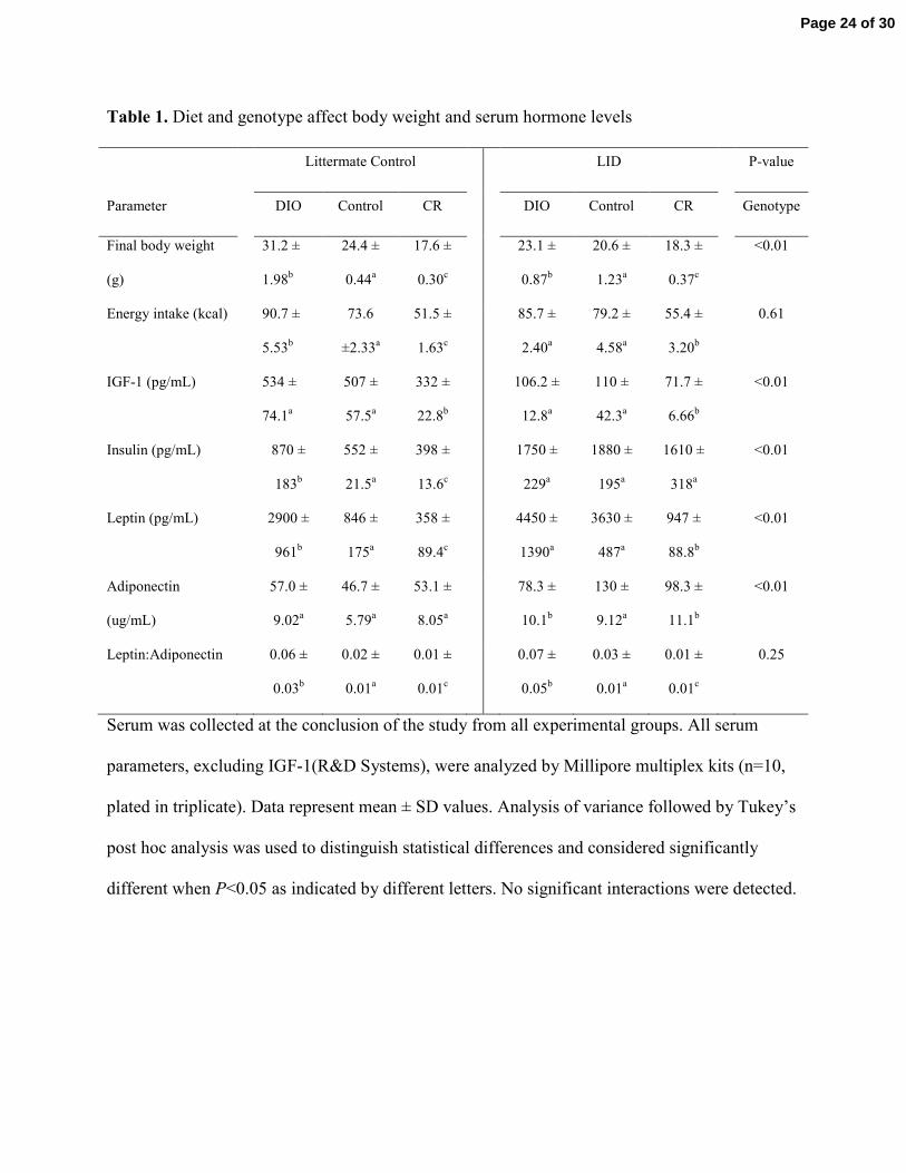
Table 1. Diet and genotype affect body weight and serum hormone levels
Littermate Control LID P-value
Parameter DIO Control CR DIO Control CR Genotype
Final body weight
(g)
31.2 ±
1.98b
24.4 ±
0.44a
17.6 ±
0.30c
23.1 ±
0.87b
20.6 ±
1.23a
18.3 ±
0.37c
<0.01
Energy intake (kcal) 90.7 ±
5.53b
73.6
±2.33a
51.5 ±
1.63c
85.7 ±
2.40a
79.2 ±
4.58a
55.4 ±
3.20b
0.61
IGF-1 (pg/mL) 534 ±
74.1a
507 ±
57.5a
332 ±
22.8b
106.2 ±
12.8a
110 ±
42.3a
71.7 ±
6.66b
<0.01
Insulin (pg/mL) 870 ±
183b
552 ±
21.5a
398 ±
13.6c
1750 ±
229a
1880 ±
195a
1610 ±
318a
<0.01
Leptin (pg/mL) 2900 ±
961b
846 ±
175a
358 ±
89.4c
4450 ±
1390a
3630 ±
487a
947 ±
88.8b
<0.01
Adiponectin
(ug/mL)
57.0 ±
9.02a
46.7 ±
5.79a
53.1 ±
8.05a
78.3 ±
10.1b
130 ±
9.12a
98.3 ±
11.1b
<0.01
Leptin:Adiponectin 0.06 ±
0.03b
0.02 ±
0.01a
0.01 ±
0.01c
0.07 ±
0.05b
0.03 ±
0.01a
0.01 ±
0.01c
0.25
Serum was collected at the conclusion of the study from all experimental groups. All serum
parameters, excluding IGF-1(R&D Systems), were analyzed by Millipore multiplex kits (n=10,
plated in triplicate). Data represent mean ± SD values. Analysis of variance followed by Tukey’s
post hoc analysis was used to distinguish statistical differences and considered significantly
different when P<0.05 as indicated by different letters. No significant interactions were detected.
Page 24 of 30
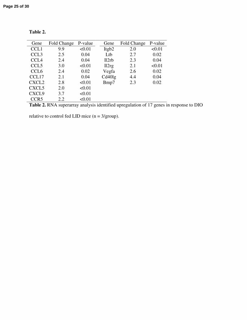
Table 2.
Gene Fold Change P-value Gene Fold Change P-value
CCL1 9.9 <0.01 Itgb2 2.0 <0.01
CCL3 2.5 0.04 Ltb 2.7 0.02
CCL4 2.4 0.04 Il2rb 2.3 0.04
CCL5 3.0 <0.01 Il2rg 2.1 <0.01
CCL6 2.4 0.02 Vegfa 2.6 0.02
CCL17 2.1 0.04 Cd40lg 4.4 0.04
CXCL2 2.8 <0.01 Bmp7 2.3 0.02
CXCL5 2.0 <0.01
CXCL9 3.7 <0.01
CCR5 2.2 <0.01
Table 2. RNA superarray analysis identified upregulation of 17 genes in response to DIO
relative to control fed LID mice (n = 3/group).
Page 25 of 30
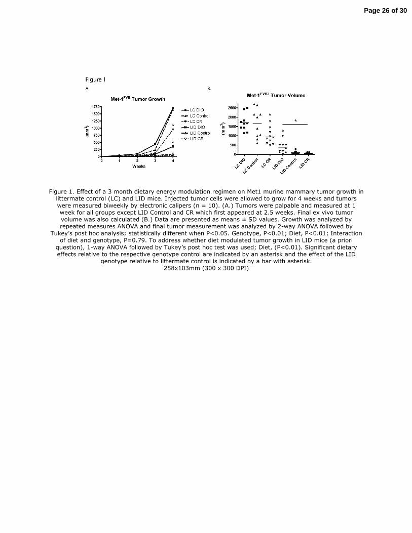
Figure 1. Effect of a 3 month dietary energy modulation regimen on Met1 murine mammary tumor growth in littermate control (LC) and LID mice. Injected tumor cells were allowed to grow for 4 weeks and tumors were measured biweekly by electronic calipers (n = 10). (A.) Tumors were palpable and measured at 1 week for all groups except LID Control and CR which first appeared at 2.5 weeks. Final ex vivo tumor volume was also calculated (B.) Data are presented as means ± SD values. Growth was analyzed by repeated measures ANOVA and final tumor measurement was analyzed by 2-way ANOVA followed by
Tukey’s post hoc analysis; statistically different when P<0.05. Genotype, P<0.01; Diet, P<0.01; Interaction of diet and genotype, P=0.79. To address whether diet modulated tumor growth in LID mice (a priori
question), 1-way ANOVA followed by Tukey’s post hoc test was used; Diet, (P<0.01). Significant dietary effects relative to the respective genotype control are indicated by an asterisk and the effect of the LID
genotype relative to littermate control is indicated by a bar with asterisk. 258x103mm (300 x 300 DPI)
Page 26 of 30
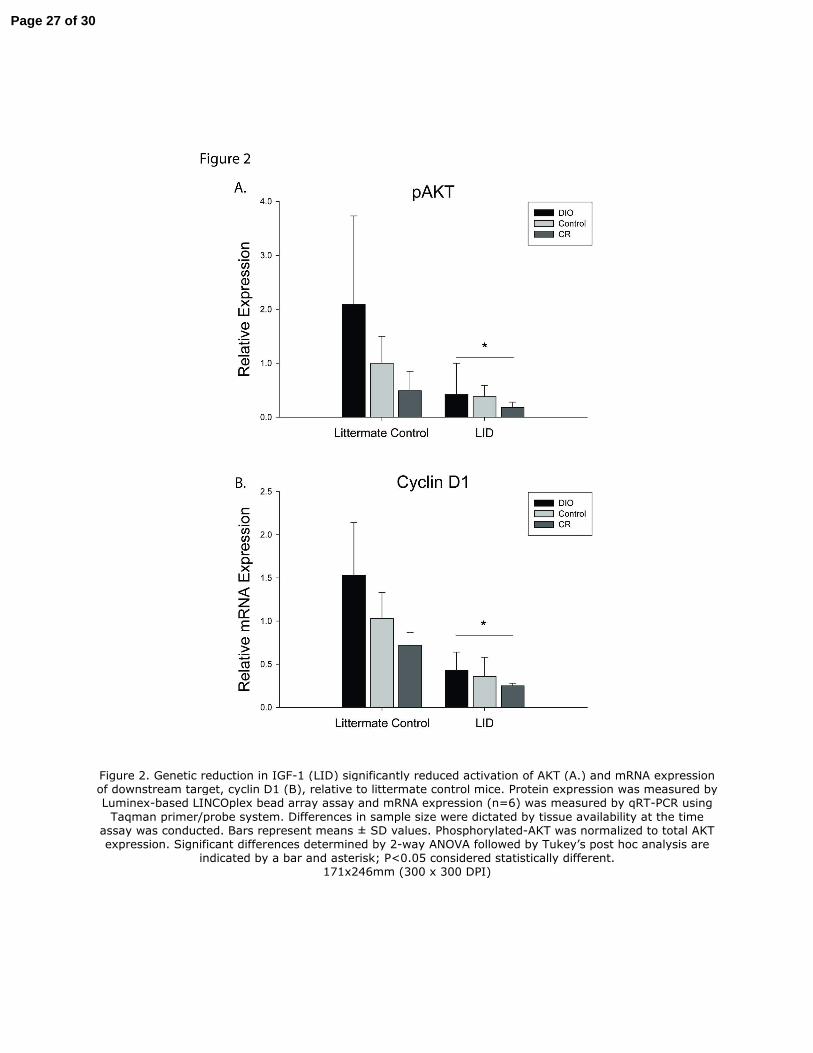
Figure 2. Genetic reduction in IGF-1 (LID) significantly reduced activation of AKT (A.) and mRNA expression of downstream target, cyclin D1 (B), relative to littermate control mice. Protein expression was measured by Luminex-based LINCOplex bead array assay and mRNA expression (n=6) was measured by qRT-PCR using
Taqman primer/probe system. Differences in sample size were dictated by tissue availability at the time assay was conducted. Bars represent means ± SD values. Phosphorylated-AKT was normalized to total AKT expression. Significant differences determined by 2-way ANOVA followed by Tukey’s post hoc analysis are
indicated by a bar and asterisk; P<0.05 considered statistically different. 171x246mm (300 x 300 DPI)
Page 27 of 30
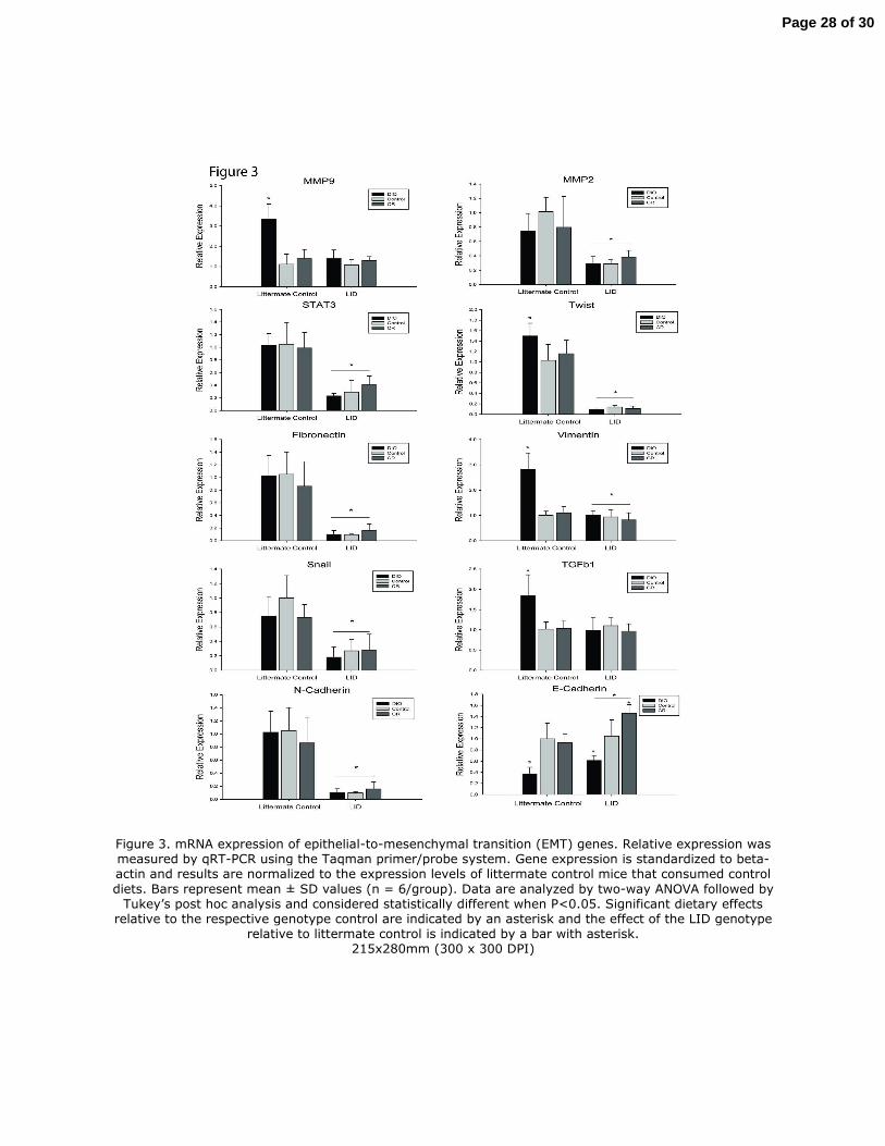
Figure 3. mRNA expression of epithelial-to-mesenchymal transition (EMT) genes. Relative expression was measured by qRT-PCR using the Taqman primer/probe system. Gene expression is standardized to beta-actin and results are normalized to the expression levels of littermate control mice that consumed control
diets. Bars represent mean ± SD values (n = 6/group). Data are analyzed by two-way ANOVA followed by Tukey’s post hoc analysis and considered statistically different when P<0.05. Significant dietary effects
relative to the respective genotype control are indicated by an asterisk and the effect of the LID genotype relative to littermate control is indicated by a bar with asterisk.
215x280mm (300 x 300 DPI)
Page 28 of 30
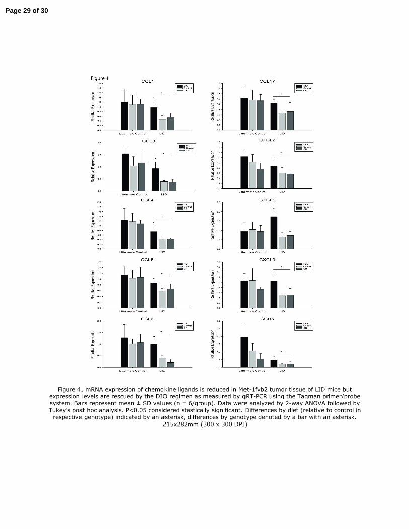
Figure 4. mRNA expression of chemokine ligands is reduced in Met-1fvb2 tumor tissue of LID mice but expression levels are rescued by the DIO regimen as measured by qRT-PCR using the Taqman primer/probe system. Bars represent mean ± SD values (n = 6/group). Data were analyzed by 2-way ANOVA followed by
Tukey’s post hoc analysis. P<0.05 considered stastically significant. Differences by diet (relative to control in respective genotype) indicated by an asterisk, differences by genotype denoted by a bar with an asterisk.
215x282mm (300 x 300 DPI)
Page 29 of 30
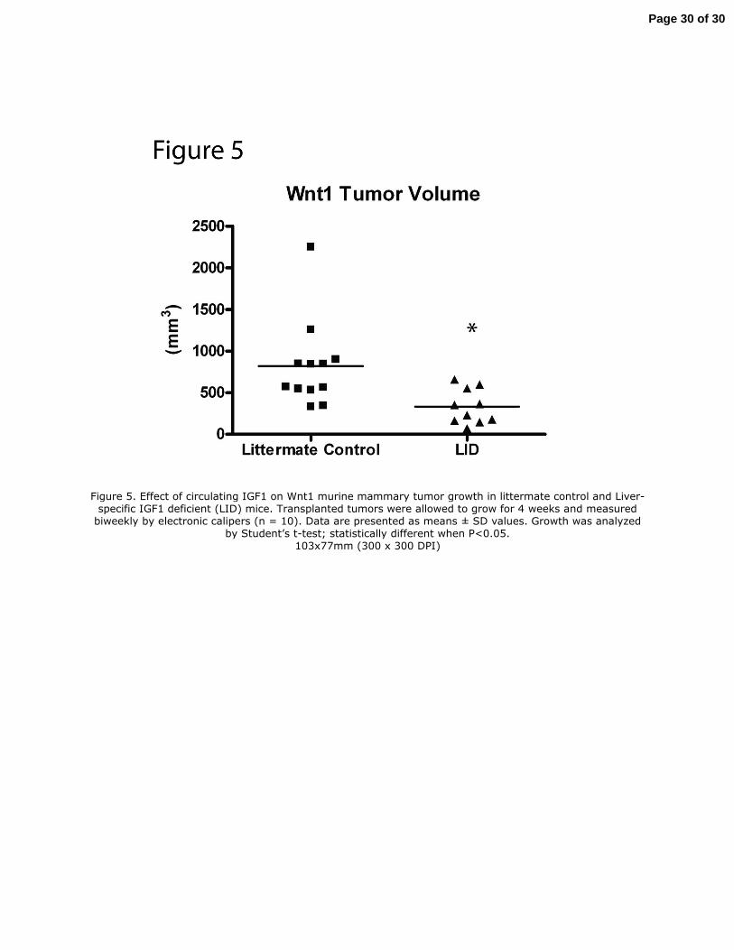
Figure 5. Effect of circulating IGF1 on Wnt1 murine mammary tumor growth in littermate control and Liver-specific IGF1 deficient (LID) mice. Transplanted tumors were allowed to grow for 4 weeks and measured biweekly by electronic calipers (n = 10). Data are presented as means ± SD values. Growth was analyzed
by Student’s t-test; statistically different when P<0.05. 103x77mm (300 x 300 DPI)
Page 30 of 30