Idiopathic Sudden Sensorineural Hearing Loss in Sweden...
61
Linköping Studies in Health Science Thesis No. 101 Idiopathic Sudden Sensorineural Hearing Loss in Sweden Diagnostic Protocol and Treatment in Relation to Outcome Ramesh Nosrati-Zarenoe __________________________________________________ Department of Clinical and Experimental Medicin Division of Oto-Rhino-Laryngology Faculty of Health Science, Linköping University SE-58185 Linköping, Sweden Linköping 2009
Transcript of Idiopathic Sudden Sensorineural Hearing Loss in Sweden...

Linköping Studies in Health Science Thesis No. 101
Idiopathic Sudden Sensorineural Hearing Loss in Sweden Diagnostic Protocol and Treatment in Relation to Outcome
Ramesh Nosrati-Zarenoe
__________________________________________________
Department of Clinical and Experimental Medicin
Division of Oto-Rhino-Laryngology Faculty of Health Science, Linköping University
SE-58185 Linköping, Sweden
Linköping 2009

Cover illustration: Nassir Mashkouri
Copyright © by Ramesh Nosrati-Zarenoe ISBN 978-91-7393-600-2
ISSN 1100-6013
Printed in Linköping, Sweden by Unitryck, 2009

To my father Who will always be with me in spirit


CONTENTS ABSTRACT ......................................................................................................... 7
LIST OF ORIGINAL PAPERS......................................................................... 9
ABBREVIATIONS ........................................................................................... 11
INTRODUCTION............................................................................................. 13
BACKGROUND................................................................................................ 15
The auditory system ....................................................................................... 15
The outer ear................................................................................................................ 15
The middle ear ............................................................................................................. 15
The inner ear................................................................................................................ 16
The central auditory system ......................................................................................... 17
Hearing impairment........................................................................................ 17
Etiologic hypotheses for ISSNHL.................................................................. 18
Infectious theory........................................................................................................... 18
Immunologic theory ..................................................................................................... 18
Membrane breaks......................................................................................................... 19
Vascular theory ............................................................................................................ 20
AIMS................................................................................................................... 21
Study I .......................................................................................................................... 21
Study II ......................................................................................................................... 21
METHODS......................................................................................................... 23
Swedish national database for SSNHL .......................................................... 23
Assessment of hearing loss and hearing recovery.......................................... 23
Categorization of laboratory tests................................................................... 25
MATERIAL ....................................................................................................... 27
Study I .......................................................................................................................... 27
Study II ......................................................................................................................... 27
STATISTICAL METHODS ............................................................................ 29
ETHICAL CONSIDERATIONS..................................................................... 30
RESULTS........................................................................................................... 31
Prognostic factors ........................................................................................... 31
Study I .......................................................................................................................... 31

Study II ......................................................................................................................... 31
Radiological examination............................................................................... 33
Study I .......................................................................................................................... 33
Study II ......................................................................................................................... 33
Hematological examination............................................................................ 33
Study I .......................................................................................................................... 33
Study II ......................................................................................................................... 33
Therapy........................................................................................................... 34
Study I .......................................................................................................................... 34
Study II ......................................................................................................................... 34
Audiometry..................................................................................................... 35
Study I .......................................................................................................................... 35
Study II ......................................................................................................................... 35
DISCUSSION..................................................................................................... 37
Prognostic factors ........................................................................................... 37
Radiological examination............................................................................... 37
Laboratory examinations................................................................................ 38
Treatment........................................................................................................ 39
Audiometry..................................................................................................... 40
Suggestions for further research..................................................................... 41
CONCLUSIONS................................................................................................ 43
ACKNOWLEDGEMENTS.............................................................................. 45
APPENDIX ........................................................................................................ 47
REFERENCES .................................................................................................. 49
SVENSK SAMMANFATTNING .................................................................... 57

Abstract 7
ABSTRACT
Idiopathic Sudden Sensorineural Hearing Loss (ISSNHL) is a rapid loss of hearing caused by damage to the cochlea (inner ear) or auditory nerve. Spontaneous recovery has been seen in 32% - 81%. The incidence of the ISSNHL has been estimated to be between 5 and 20 per 100,000 per year. Different theories (infections, vascular catastrophes, immunologic damage or intracochlear membrane break) about the etiology have resulted in different treatment policies. The effect of therapy is difficult to evaluate for a single physician who sees just a few patients annually.
The aim of the present thesis was to analyze the management and treatment of ISSNHL patients in Sweden with regard to outcome.
A national database was developed for Sweden with half of all ENT clinics in Sweden participating by submitting a questionnaire for each patient with SSNHL. The questionnaire covered the patient’s background, current disorder, past and family history of different diseases, examinations and treatment. Audiograms at the onset of SSNHL and after three months were requested.
All results were analyzed using ordinal logistic regression looking for interactions with hearing recovery and remaining hearing loss as dependent variables. Independent of treatment or no therapy heredity for hearing loss (I, II), older age (I, II) and presence of vertigo (II) was significantly associated with negative outcome. 40% of all patients had an MRI or CT, where 3 – 4% had acoustic neuroma. 24% of patients with ISSNHL who had hematological tests taken had one or more pathological findings. Blood screening varied from simple routine tests to a complete analysis with such tests as HSP70, Anti-Neutrophilic Cytoplasmic Antibodies (ANCA) and Borrelia tests. There was no association between any of these laboratory tests and either hearing improvement or remaining hearing loss evaluating the tests separately (I, II) or after categorization in comparison with those who had normal laboratory findings (II). Patients with hearing loss in the mid-frequency region had significantly better odds for hearing improvement compared to the other three frequency regions (low, high and “flat loss”). Almost 60% of patients with ISSNHL were medically treated, of which nearly 90% got corticosteroids. The medication had no association with either hearing improvement or remaining hearing loss. However, patients who were prescribed rest or sick leave had higher odds for hearing improvement regardless of other treatment. Those patients who did not receive any treatment at all also came significantly later to the ENT clinics than those treated medically and consequently had worse prognosis.
Conclusion: There is no standard program for management or treatment of ISSNHL in Sweden. The diagnostic protocol varies. MRI is an underused resource to get specific diagnoses for the condition especially acoustic neuromas. Regardless of pathological findings, treatment is mainly limited to corticosteroids or no medication with no difference in outcome. A randomized placebo controlled study is necessary to evaluate whether there is an effect of corticosteroids on ISSNHL.


List of original papers 9
LIST OF ORIGINAL PAPERS
This thesis is based on the following papers, which will be referred to in the text by their roman numerals:
I. Nosrati-Zarenoe R, Arlinger S, Hultcrantz E. Idiopathic sudden sensorineural hearing loss: results drawn from the Swedish national database. Acta Otolaryngol. 2007 Nov;127(11):1168-75.
II. Nosrati-Zarenoe R, Hansson M, Hultcrantz E. Assessment of different diagnostic approaches to idiopathic sudden sensorineural hearing loss and their influence on treatment and outcome. Submitted to Acta Otolaryngologica.
Reprints were made with the kind permission of copyright holder.


Abbreviations 11
ABBREVIATIONS
ABR Auditory brainstem responses
AICA Anterior inferior cerebellar arteries
ANA Anti-nuclear antibodies
ANCA Anti-neutrophilic cytoplasmic antibodies
CI Confidence intervals
CRP C-reactive protein
CSF Cerebrospinal fluid
CT Computed tomography
dB Decibel
ENT Ear, nose and throat
Hb Hemoglobin
HDL High density lipoprotein
HSP-70 Heat-shock protein 70
IgG antibodies Immunoglobulin G antibodies
IgM antibodies Immunoglobulin M antibodies
ISSNHL Idiopathic Sudden Sensorineural Hearing Loss
kHz kiloHertz
LDL Low density lipoprotein
MRI Magnetic resonance imaging
OAE Otoacoustic emission
OR Odds ratio
PTA Pure tone average
SD Standard deviation
SLE Systemic lupus erythematosus
SSNHL Sudden Sensorineural Hearing Loss


Introduction 13
INTRODUCTION
Idiopathic Sudden Sensorineural Hearing Loss (ISSNHL) involves a rapid loss of hearing that is caused by damage to the cochlea (inner ear) or auditory nerve. This hearing loss can be accompanied by tinnitus and/or vertigo.
A standard definition of ISSNHL does not exist, nor is there a standard method for reporting recovery. However, agreement has been reached with regard to the requirement for a 30 dB or more hearing loss in at least three contiguous frequencies1-5. The definition of “sudden” can vary from 24 hour to 72 hours in different studies 2, 6, 7.
A standard method for audiological assessment with respect to the configuration of hearing loss and hearing recovery does not exist. Hearing recovery has been reported in different categories, such as “complete recovery”, “good recovery” and “fair recovery”, but there has been no agreement with regard to the actual degree of improvement that is indicated by each of these categories. The configuration of hearing loss has often been mixed with the degree of hearing loss8, 9.
Regardless of treatment, spontaneous recovery can occur within a few hours to few days after onset. Complete and partial recovery is often combined when reporting spontaneous recovery, which is seen in 32% - 81% of the cases2, 6, 10, 11. A larger recovery rate has been reported for patients with hearing loss in the low frequencies compared to those with loss in the high frequencies10.
The incidence of ISSNHL is difficult to estimate due to the rate of spontaneous recovery, which results in not every patient seeking help. In 1944, De Kleyn reported an increase of ISSNHL between the years 1936 and 1942 in Amsterdam12. Since 1958, the overall incidence has been reported to be 5 – 20 per 100,000 per year13. Recently, there have been two epidemiological studies that have shown that the prevalence of patients seeking help has increased. One study showed an increase from 3.9 to 27.5 per 100,000 persons annually in Japan14, and another study in Germany showed a prevalence of 160 per 100,000 persons per year in the city of Dresden15.
Since the etiology of ISSNHL still remains unknown, a standardized treatment does not exist. Different theories with regard to its etiology have resulted in different treatment policies over the years in Sweden and in many other countries16-19. These treatments have included anti-stress treatment with bed rest and blockage of the ganglion stellatum in the 1950’s20, treatment with dextran 40 and other hyperosmolar hemodiluting drugs in the 1970’s21 and corticosteroid treatment since the 1980’s2.
Due to its low incidence, its rate of spontaneous recovery and its unknown etiology, the effect of therapy is difficult to evaluate for a single physician who sees just a few

14 Introduction
patients annually. By compiling the data from a large number of patients, it will be possible to identify prognostic factors and the impact of the various treatments.
The purpose of the present thesis was to analyze the management and treatment of ISSNHL patients in Sweden with regard to their outcome.

Background 15
BACKGROUND The auditory system
The auditory system is described as the peripheral and central auditory system. The peripheral auditory system is comprised of three components, which are the outer, middle and inner ear. The central auditory system includes the auditory pathways and auditory cortex22.
Fig. 1. The peripheral auditory system
The outer ear
The outer ear consists of the pinna/auricle and the ear canal/external auditory meatus. The pinna/auricle collects and directs sound waves that are traveling through the air into the ear canal/external auditory meatus, which, in turn, transmits the sound waves to the tympanic membrane (eardrum). The ear canal forms a resonance channel that amplifies the sound pressure up to 15 – 25 dB in 2 – 5 kHz.
The middle ear
The middle ear is an air-filled cavity that includes the tympanic membrane (eardrum) and the ossicular chain. The ossicular chain consists of three interconnected bones, which are the malleus (hammer), incus (anvil) and stapes (stirrup). The malleus is

16 Background
attached to the tympanic membrane, and the footplate of the stapes inserts into the oval window of the inner ear. The incus is located between the malleus and the stapes. Thus, the movements of the tympanic membrane (eardrum) will set the malleus, incus and stapes into motion. Attached to the ossicular chain are the stapedius and tensor tympani muscles. The sound is not amplified evenly across the ossicular chain, and contractions of these muscles protect the inner ear by reducing the intensity of sound transmission to the inner ear.
The inner ear
The inner ear consists of two separately functional systems. These include the sensory organ of hearing, which is called the cochlea, and the organ of balance, which is the vestibular system.
The cochlea is spiral-shaped and contains fluid-filled chambers. The two outer chambers, which are the scala vestibuli and scala tympani, are a part of the bony labyrinth and are filled with perilymph. The middle chamber, which is the scala media and is also called the cochlear duct, is filled with endolymph. Both perilymph and endolymph are clear solutions that contain electrolytes and proteins, and they are chemically quite different from each other. The perilymph is rich in sodium salts, whereas the endolymph is rich in potassium salts. The cochlear duct separates the two chambers from each other by the Reissner's membrane and the basilar membrane, on which the organ of Corti lies. The third partition consists of the stria vascularis, which is a rich bed of capillaries and secretory cells that are responsible for the production of endolymph. The scala vestibuli ends at the oval window, where the footplate of the stapes sits, and the scala tympani ends at the round window. A vibration coming from the stapes into the inner ear via the oval window moves the perilymph in the scala vestibule, which, in turn, vibrates the endolymph in the scala media, the perilymph in the scala tympani, the basilar membrane and the hair bundles of the hair cells in the organ of Corti.
The organ of Corti consists of approximately 3500 inner hair cells and 15,000 outer hair cells. The hair cells lie on the basilar membrane and are covered by the tectorial membrane. The basal parts of the organ of Corti are responsible for detecting the highest frequencies that we can perceive, and the frequencies that can be
Fig. 2. Cross section of Cochlea.

Background 17
detected gradually decrease as they move towards the apical parts. The inner hair cells transform the sound vibrations in the fluids of the cochlea into electrical signals. The outer hair cells amplify the low-level sounds by mechanically enhancing the motion of the tectorial membrane in order to increase the stimulation of the inner hair cells. They also add to the frequency resolution.
Both the outer and inner hair cells are associated with afferent and efferent neurons. The afferent neurons, which carry information to the brain, contact mainly the inner hair cells, and the efferent neurons, which carry information back to the hair cells, connect mostly to the outer hair cells.
The labyrinth artery is an end artery and is the sole blood vessel that supplies the inner ear. The artery divides into the cochlear artery and the anterior vestibular artery. The cochlear artery further divides into the main cochlear artery and the vestibulocochlear artery. The main cochlear artery lies below the organ of Corti and has more capillaries in the apical portion of the cochlea then in the basal part. The distance between the vas spirale and the spiral border near the inner hair cells is shorter in the apical part then in the basal part23.
The central auditory system
A vibration that comes from the middle ear is transmuted into a neural signal in the cochlea. The neural signal transfers from the hair cells to the spiral ganglion, which is found in the spiral bony structure located centrally in the cochlea (modiolus). Axons from the ganglion cells are bundled together to form the auditory portion of the eighth cranial nerve. The auditory nerve carries the signal into the brainstem and synapses in the cochlear nucleus. From the cochlear nucleus, auditory information is split into at least two streams, which include the ventral cochlear nucleus and the dorsal cochlear nucleus. The ventral cochlear nuclear cells project to a collection of nuclei called the superior olive, which helps to determine the direction of the sound. The dorsal cochlear nucleus analyzes the quality of sound. Both streams of information proceed to the sensory thalamus and, from there, to the auditory cortex that is located in the temporal lobes. Hearing impairment
Hearing loss can be classified into four categories24:
− Conductive hearing loss is caused by difficulties in the transmission of sound into the inner ear. A diagnosis can be made via observation of an air-bone gap on audiometry, which indicates that hearing is better when sound is transmitted in such a way that it bypasses the middle ear ossicular chain. The air-bone gap should be more than 10 dB.

18 Background
− Sensorineural hearing loss occurs without an air-bone gap, since air conduction is equal to bone conduction. A diagnosis is made through audiometry. Patients with cochlear damage have no Otoacoustic Emissions-testing (OAE), and those with auditory nerve damage fail the Brainstem Auditory Evoked Responses-testing (ABR).
− Mixed hearing loss is a combination of sensorineural hearing loss and conductive hearing loss.
− Central hearing loss is caused by damage to the central pathways. The diagnosis can not be made by pure tone audiometry, since that is often normal in affected individuals. A patient with central hearing loss usually has poor scores on their speech reception threshold or word recognition scores.
Etiologic hypotheses for ISSNHL
A cause for SSNHL can be found in 10% of all cases25. Hallberg (1956) stated that ”Sudden unilateral or bilateral impairment of hearing is a symptom, not a disease.”26.
Infectious theory
Herpes zoster that causes sensorineural hearing loss was first reported by Hunt, in 190727. SSNHL has also been reported to be caused by known infectious diseases, of both bacterial and viral origins, such as by Borrelia and Syphilis28, 29, the mumps30, 31 and rubella32, 33.
Both an acute and latent viral attack may damage the inner ear and cause hearing loss34. Upper respiratory infection35, as an example of an acute viral infection, and members of the herpes virus family36, as an example of latent infections, have been proposed to cause SSNHL. Virus infection can damage the organ of Corti, the ganglion cells, the nervous fibers, the tectorial membrane and the stria vascularis37.
Two antiviral drugs, acyclovir and valacyclovir, are used to treat SSNHL as a result of this theory4, 38. However, the four published randomized, placebo-controlled clinical studies that used acyclovir and valacyclovir did not show any benefit for the treatment of ISSNHL3-5, 39.
Immunologic theory
McCabe (1979) was the first to suggest autoimmunity as a cause of SSNHL, based upon clinical data, pathological findings in autoimmune tests and positive response to steroid therapy40. Hearing loss may be a consequence of local autoimmune processes

Background 19
within the inner ear41, 42 or of systemic autoimmune diseases, such as Cogan’s syndrome43, Wegener’s granulomatosis44 and systemic lupus erythematosus (SLE)45.
Although the inner ear was initially considered as an immunologically privileged organ, it is capable of producing a strong immune response. The presence of antibodies against antigens in the inner ear and the formation of immune complexes in the stria vascularis, endolymphatic sac and ducts support the immunologic theory46-48. Autoimmunity can cause damage to the cellular components of the organ of Corti and affect the stria vascularis and spiral ligament. Autoimmunity can also cause dysfunction of the endothelial cells and fibrocytes II, which leads to the impaired diffusion of K+ through marginal cells to the endolymph fluid. This, in turn, can affect the supporting cells of the organ of Corti, which precedes a late effect upon the hair cells49 The existence of glucocorticoid receptors in the stria vascularis and supporting cells also suggests their role as immune targets in the inner ear50.
Treatment of ISSNHL with corticosteroids was the result of this immune theory. Since this therapy was first used in the 1980s, there have been many studies on the impact of corticosteroids on SSNHL2, 10, 51-54. However, the only two randomized, double-blinded, placebo-controlled studies (19802, 200110) with low power showed contradictory results.
Membrane breaks
Rupture of the oval or round window can cause loss of the perilymph and result in pressure alteration between the chambers that contain perilymph and endolymph55, 56. There have been studies on the temporal bones that support this theory57, 58. The membrane break does not happen spontaneously, but rather occurs after a sudden pressure alteration in the middle ear that can be caused by head injuries, barotraumas or intense physical exercise26, 59. The fact that many of these patients report hearing loss upon awaking6 and that not all individuals with high intracranial and intra-abdominal pressure, such as women in childbirth or weight lifters, experience SSNHL argues against this theory. In addition, not all of the temporal bones studies found evidence of active or healed ruptures of the oval or round window, basilar membrane or Reissner’s membrane34, 37, 60.
Surgery with the intention to repair oval or round window perilymph fistulae combined with strict bed rest has been used in cases of ISSNHL with a history of recent trauma or barotrauma61, 62.

20 Background
Vascular theory
In 1949, Rasmussen suggested vascular occlusion or ischemia as a mechanism for ISSNHL63. Vascular or hematological diseases that are associated with SSNHL, such as Buerger’s disease64, leukemia65 and sickle cell anemia66 have also been reported.
The labyrinth artery is an end artery that solely carries red blood cells and oxygen into the inner ear23. Tissue injury that results from oxygen deprivation and ischemia can occur in the cochlea within 60 seconds6. A total but temporary blood circulation blockage can cause damage to the hair cells, the ganglion cells and the spiral ligament and can also cause neuronal loss and an alteration of the tectorial membrane after 30 minutes. This damage is irreversible even after the blood flow is restored67. The labyrinth artery is extremely vulnerable to blood pressure oscillation and abnormalities in blood flow68. Blood flow has an inverse relationship with blood viscosity69. Low blood flow causes anoxia due to hyperviscosity, which results in cochlear hypofunction and the inability to maintain cochlear metabolism70. A correlation between ISSNHL and blood viscosity has been shown71-73.
Different treatments regimes for ISSNHL, such as fibrinogen apheresis74, Rheopheresis75, dextran infusion76, hyperbaric oxygen77 and pentoxifylline78, have been developed in response to this vascular theory. There is evidence both to support and refute such treatment76-80.

Aims 21
AIMS
Study I
− To analyze which variables, such as background data, concomitant disease, audiogram shape and laboratory tests, can best predict the outcome of ISSNHL.
− To investigate the treatment policy of ISSNHL in Sweden.
− To evaluate the effects of treatments on outcomes.
Study II
− To explore the different diagnostic test batteries for ISSNHL that are currently used in Sweden.
− To evaluate if and how positive diagnostic findings result in treatment modifications.
− To investigate whether such treatment modifications influence the outcome of ISSNHL.


Methods 23
METHODS Swedish national database for SSNHL
The two publications (I, II) in this thesis are based on data from a national database in Sweden for sudden sensorineural hearing loss. The database began gathering data from patients with SSNHL in winter 2002 – 2003. Approximately half of all ENT clinics in Sweden have been contributing data to the database.
To build the database, a questionnaire was developed that covered the patient’s past medical history, potential precipitating events that preceded the SSNHL, traumas, family history of different diseases, especially hearing loss, the current disease, the diagnostic protocol including laboratory, radiological and further audiological examinations and all other treatments. The time course of the hearing loss’s onset and associated symptoms, such as tinnitus and vertigo, were also requested. The questionnaire included the results of an ENT examination. Information on radiological investigations (MRI or CT), laboratory work-ups and the use of BRA or a vestibular work-up were requested. This information request was phrased in general terms so as not to influence the doctor's own diagnostic practices on their decision-making process. The questions with regard to treatment included the use of corticosteroids, antiviral therapy, rheological treatment or “other” drugs, and prescription of rest or surgery of a suspected fistula. In the case that pathological test results were performed, the complete lab-sheet for that patient was requested. See appendix.
For each patient, after informed consent, a questionnaire was completed by the otorhinolaryngologist, and two audiograms, one of which was taken during the first visit to the ENT clinic as a result of the symptoms of SSNHL and the other which was taken after three months, were requested. A copy of previous pure tone audiogram was requested in those cases where the patient was known to have a prior diagnosed hearing loss before the onset of SSNHL. Assessment of hearing loss and hearing recovery
Sudden Sensorineural Hearing Loss was defined as a hearing loss of at least three contiguous frequencies between 0.125 kHz to 6 kHz, with a mean of 30 dB or more and that occurred within 24 hours.
The hearing loss was characterized by a comparison of the audiogram taken at the first visit after onset of SSNHL to an audiogram that was taken not more than two years before the acute hearing loss. If no previous audiogram was available, hearing was compared to the non-affected ear in its present state.

24 Methods
Four frequency regions were created to describe the hearing loss:
Low frequency region:
1. Pure-tone average (PTA) of low frequencies (125, 250, 500 Hz) > PTA of mid frequencies (1000, 1500, 2000 Hz) and high frequencies (3000, 4000, 6000 Hz) by at least 10 dB.
2. Hearing loss in the low and mid frequencies – PTA of low frequencies > PTA of mid frequencies with a difference less than 10 dB.
Mid frequency region:
1. PTA of mid frequencies > PTA of low- and high frequencies by at least 10dB.
2. Hearing loss in the low and mid frequencies – PTA of mid frequencies > PTA of low frequencies with a difference less than 10 dB.
High frequency region:
1. PTA of high frequencies > PTA of low- and mid frequencies by at least 10dB.
2. Hearing loss in the mid and high frequencies – PTA of high frequencies > PTA of mid frequencies with a difference less than 10 dB.
Flat loss:
The differences between the PTA for all three frequency regions were less than 10 dB.
The audiogram taken at the first visit and the audiogram obtained three months after the onset of SSNHL were compared with respect to the PTA characterizing the loss to determine the degree of hearing recovery and remaining hearing loss (table I).

Methods 25
Table I. Hearing improvement and remaining hearing loss after recovery.
Improvement
Large improvement >30 dBModerate improvement 10 – 30 dB
No improvement ± 10 dB
Hearing loss after recovery
No remaining hearing loss Difference between initial audiogram and audiogram at the follow-up < 10 dB
Partial recovery The difference ≥10 dB and
the improvement ≥10 dB
No regress The difference ≥10 dB and the improvement < 10 dB
Categorization of laboratory tests
In study II, the pathological results of laboratory tests were categorized by one or more pathological values prior to analysis:
− “Arteriosclerosis associated variables”: LDL-cholesterol/HDL-cholesterol ratio >3, Total cholesterol >5 mmol/L and C-reactive protein (CRP) >3 mg/L in patients with or without earlier known cardiovascular disease.
− “Inflammation/infection”: CRP >10 mg /L, Erythrocyte sedimentation rate >20 mm, Leukocyte count >10 x 109 ml/L, Hemoglobin count (Hb) <120 g/L, Thrombocyte count >150 x 109 ml/L and Borrelia tests “positive” (IgG antibodies and IgM antibodies) in patients with or without ongoing clinical infection.
− “Autoimmune variables”: HSP-70, Cardiolipin, Antiphospholipid, Anti-Neutrophilic Cytoplasmic Antibodies (ANCA) and Antinuclear antibodies (ANA) “positive”.

Material 27
MATERIAL
Study I
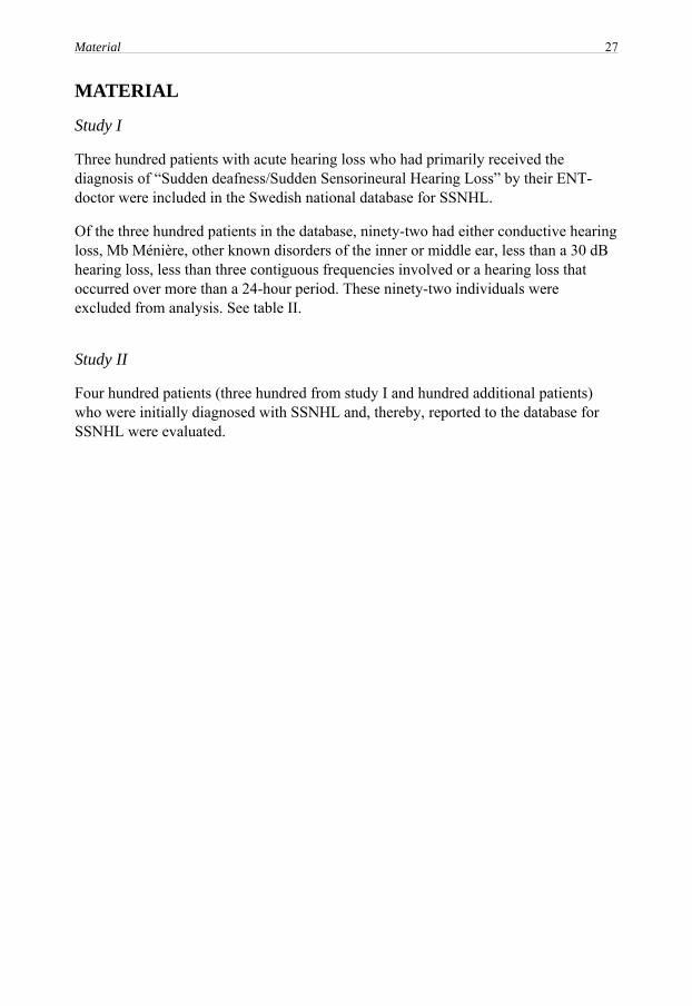
Three hundred patients with acute hearing loss who had primarily received the diagnosis of “Sudden deafness/Sudden Sensorineural Hearing Loss” by their ENT-doctor were included in the Swedish national database for SSNHL.
Of the three hundred patients in the database, ninety-two had either conductive hearing loss, Mb Ménière, other known disorders of the inner or middle ear, less than a 30 dB hearing loss, less than three contiguous frequencies involved or a hearing loss that occurred over more than a 24-hour period. These ninety-two individuals were excluded from analysis. See table II.
Study II
Four hundred patients (three hundred from study I and hundred additional patients) who were initially diagnosed with SSNHL and, thereby, reported to the database for SSNHL were evaluated.

28 Material
Table II. Final diagnosis after examination for patients initially reported as SSNHL (n=92)
Diagnoses Number
Acoustic neuromas 5Trauma acoustic trauma 2 head trauma 2 trauma after water irrigation of the ear 2 barotrauma 3Subdural hematoma 1
Myeloma 1
Mb Ménière 5
Hydrops 1
Progressive hearing loss 1
Bleeding in Pons (infarct at the root entry zone) 1
Transient ischemic attacks 1
Coronary disease 3
Did not fulfill the criteria for SSNHL Hearing loss less than 30dB 44 Hearing loss did not occur within 24 hours 20

Statistical methods 29
STATISTICAL METHODS
Statistical analysis was performed using Minitab software, version 13.32 for Windows, for paper I and version 15 for Windows, for paper II.
Descriptive statistics were used in both papers to show the characteristics of the subjects. The data was expressed as the number of cases and percentage. Parametric data was expressed as mean ± standard deviation (SD).
Ordinal/Ordered logistic regression was used for all analyses with regard to hearing recovery. This method performs a logistic regression on an ordinal response variable (categorical variables that have three or more possible levels with a natural ordering) with the help of both continuous and categorical predictors.
The estimated probability for hearing improvement and no remaining hearing loss in relation to the prognostic factor, the frequency regions (low, mid, high and “flat loss”) and the frequency regions (low, mid and high) in relation to the number of days from the onset of ISSNHL was expressed as an odds ratio (OR) and 95% confidence intervals (CI).
Seasonal variance and gender differences with regard to age distribution, the presence of tinnitus and/or vertigo, the comparison between treatment options and laboratory tests, the interval between the onset of hearing loss and the first visit to the ENT clinic and the pure-tone average between different frequency regions were performed using χ2-test. The level of significance was set at p<0.05.

30 Ethical considerations
ETHICAL CONSIDERATIONS
The Medical Research Ethic Committee of Linkoping’s University, Sweden (registrations number 02-337) approved the database. Participants were given oral information about the database by their local ENT doctors. A written informed consent was not requested, as there was no intervention that was performed and as the participant’s identity (security number) was not reported in the database.
The data were handled confidentially. No more than the participant’s birth date and gender were provided on the questionnaires, audiograms and requested lab-sheets.

Results 31
RESULTS
None of the patients were bilaterally affected. For further descriptive data, see table III. Prognostic factors
All variables in the questionnaire were analyzed using ordinal logistic regression in order to look for interactions with hearing recovery and remaining hearing loss as dependent variables. Independent of treatment or no therapy, the following factors was significantly associated with outcome:
Study I
“Heredity for hearing loss” was associated with a significantly lower odds for improvement, with an odds ratio of 3.02 (95% CI 1.34 – 6.80 p=0.008), but was not associated with the remaining hearing loss.
An older age was related to a reduced chance of both improvement of hearing in dB (OR 1.05, 95% CI 1.03 – 1.08, p=0.000) and the remaining hearing loss (OR 1.03, 95% CI 1.01 – 1.05, p=0.008).
Study II
“Heredity for hearing loss” was significantly associated with a lower odds for hearing improvement (OR 2.08, 95% CI 1.10 – 3.94, p=0.02). There was no significant association between “Heredity for hearing loss” and the remaining hearing loss after recovery.
An older age was related to a reduced chance of hearing improvement, with an odds ratio of 1.03 (95% CI 1.01 – 1.05, p=0.000), and was connected to an increased level of remaining hearing loss (OR 0.98, 95% CI 0.96 – 0.99, p=0.001).

32 Results
Table III. Profiles of the ISSNHL patients.
Variables Study I (n=208)Number (%)
Study II (n=300)Number (%)
Gender Female 98 (47) 146 (49) Male 110 (53) 154 (51)
Age Mean ± SD (years) 56.1 ± 16 57.4 ± 16 Range (years) 15 - 87 8 - 87
Affected ear Left 96 (46) 141 (47) Right 112 (54) 159 (53)
Prevalence of associated symptoms Tinnitus 107 (51.4) 149 (49.7) Vertigo With nystagmus 1 (0.48) 1 (0.33) Without nystagmus 7 (3.4) 11 (3.7) No info. about nystagmus 1 (0.48) 1 (0.33) Tinnitus and vertigo With nystagmus 10 (4.8) 16 (5.3) Without nystagmus 38 (18.3) 49 (16.3) No info. about nystagmus 7 (3.4) 11 (3.7)
No info about tinnitus or vertigo 1 (0.48) 1 (0.33) No associated symptoms 36 (17.3) 61 (20.3)
Interval between onset of hearing loss and first visit at the ENT clinics Mean ± SD (days) 16.4 ± 31.5 18.3 ± 42.6 Range (days) 0 - 212 0 - 498

Results 33
Radiological examination
Study I
Seventy-six (37%) of the two hundred and eight patients with ISSNHL had an MRI or CT, and four had pathological findings.
No significant association was found between pathological MRI-findings and either hearing recovery or remaining hearing loss for the patients with ISSNHL.
Study II
One hundred and fifty-eight (40%) of the four hundred patients with SSNHL had an MRI or CT, and twenty-two had pathological findings. Out of the twenty-two with pathological findings, ten patients had accidental findings that were not connected to the hearing tracts and thereby were defined as ISSNHL. Out of remaining twelve patients: five had acoustic neuroma, one had subdural hematoma, one had a pons infarction and five had different vascular abnormalities that might have had a possible connection to SSNHL. The five individuals who had different vascular abnormalities had all a low frequency hearing loss.
No significant association was found between pathological MRI-findings and either hearing recovery or remaining hearing loss for the three hundred patients with ISSNHL. Hematological examination
Study I
One hundred and forty-two (68%) of the two hundred and eight patients with ISSNHL had hematological tests taken, and thirty-three (23%) of those had one or more pathological findings.
There was no association between any of the laboratory tests and either hearing improvement or remaining hearing loss, even when the tests were evaluated separately.
Study II
Two hundred and fifty-eight (65%) of the four hundred total patients with SSNHL had hematological tests taken. Of the three hundred patients who were classified as having ISSNHL, one hundred ninety six had one or more laboratory tests taken, and forty-seven (24%) of these had one or more pathological findings.

34 Results
There was no association between any of the laboratory tests and either hearing improvement or remaining hearing loss, even when the tests were evaluated separately or after the tests were compared to those who had normal laboratory findings. Therapy
Study I
One hundred and five (50%) out of two hundred and eight patients were treated with corticosteroids, and ninety (44%) received no medical treatment. Of the remaining eleven patients, three received antiviral therapy, and five received antibiotics. The main corticosteroid used was Prednisolone (80%). The dosage, duration of treatment and tapering schedule varied from 80 mg to 25 mg per day for five days to four weeks. No significant difference in outcome was seen that depended upon the dosage given.
There were no significant differences in outcome between the different treatment groups and those with no medical treatment.
Study II
One hundred and eighty-two (61%) of three hundred patients with ISSNHL were treated medically. Of these, one hundred and sixty-six patients were given corticosteroids either as a single treatment or in combination with antiviral therapy, antibiotics or blood flow promoting therapy (acetylsalicylic acid). The main corticosteroid used was Prednisolone (84%). The dosage, duration of treatment and tapering schedule varied from 80 mg to 25 mg per day for five days to four weeks. Seventy-seven of three hundred patients with ISSNHL (26%) were prescribed “to rest” or “to stay home on sick leave” for up to four weeks, and this was the only treatment for twenty-two of these patients.
Patients who had been prescribed rest or been on sick leave during the first period of the disease had higher odds for hearing improvement (OR 0.46, 95% CI 0.27 – 0.79, p=0.005), regardless of any other treatment.
The medical treatment had no significant association with either hearing improvement or remaining hearing loss.
Ninety-six patients did not receive any treatment at all. Those patients came significantly later to the ENT clinics after the onset of SSNHL than those who were treated medically (p=0.001).
For patients who came on corresponding days to the ENT clinic, there was no difference in hearing outcome between the ones who were medically treated or not.

Results 35
Audiometry
Study I
42% of the patients had their hearing loss in the low frequency region, 26% in the mid frequency region, 30% in the high frequency region and 3% had a “flat loss”.
Patients with mid frequency hearing loss had a significantly higher chance for improvement when compared to those with low (p=0.000) or high frequency hearing loss (p=0.000), if they visited an ENT clinic the same day as the onset of ISSNHL. The probability for improvement of the hearing loss decreased with number of days to ENT visit regardless of the frequency region. This probability significantly decreased more rapidly for patients with mid or high frequency losses compared to those with low frequency losses (p=0.002 resp. p=0.05).
Total or partial recovery for patients with mid frequency hearing loss was significantly more likely when compared to those with low (p=0.002) or high frequency hearing loss (p=0.014), if they visited an ENT clinic the same day as the onset of ISSNHL. The probability for no remaining hearing loss decreased with number of days before the first visit regardless of the frequency region. For patients with mid or high frequency hearing loss, the probability significantly decreased more rapidly when compared to those who were affected in the low frequency region (p=0.007 resp. p=0.038).
Study II
40% of the patients had their hearing loss in the low frequency region, 28% in the mid frequency region, 23% in the high frequency region and 9% had a “flat loss”.
Patients with hearing loss in the mid frequency region had significantly higher odds for hearing improvement when compared to those in the groups with a hearing loss in the low frequency region (OR 2.43, 95% CI 1.38 – 4.27, p=0.002), the high frequency region (OR 3.83, 95% CI 1.99 – 7.37, p=0.000) or a “flat loss” (OR 2.75, 95% CI 1.16 – 6.50, p= 0.021). The odds for a residual hearing loss was lower for patients with hearing loss in the mid frequency region when compared to those with a hearing loss in the low frequency region (OR 0.45, 95% CI 0.26 – 0.78, p=0.005), the high frequency region (OR 0.31, 95% CI 0.16 – 0.58, p=0.000) or a “flat loss” (OR 0.43, 95% CI 0.18 – 1.01, p= 0.05).


Discussion 37
DISCUSSION
Sudden sensorineural hearing loss is one of the most mysterious and controversial unsolved entities in otolaryngology. The lack of a standard definition for SSNHL, the lack of a standard method for audiological assessment with regard to the configuration of the hearing loss and hearing recovery, the low incidence rate and the fact that spontaneous recovery happens in up to 80% of cases make any evaluation of treatment impossible for those ENT doctors who only see a few patients a year.
The criteria for SSNHL and for the assessment of hearing recovery were developed by the Sudden Deafness Research Committee of the Ministry of Health and Welfare in Japan (1973 and 1981, respectively). However, no specific instruction with regard to how sudden the onset of hearing loss should be, the degree of hearing loss or the affected frequencies were actually provided. Although many authors have selected a 30 dB hearing loss in three contiguous frequencies for their studies1-5, the onset of hearing loss differs from 24 to 72 hours2, 6, 7. In the present studies (I, II), hearing loss over a period of 24 hours was selected in order to stress the concept of “sudden” hearing loss and to avoid including patients with Mb Ménière or endolymphatic hydrops, where the hearing loss usually develops within a few days. Prognostic factors
Prognostic factors for ISSNHL have been studied by many authors. The findings from this database demonstrate that the presence of vertigo (I) and a higher age (I, II) at onset are related to a reduced probability for recovery, which is also in accordance with earlier studies2, 76, 81, 82. The presence of tinnitus has been considered to be a positive prognostic factor for hearing recovery by Cvorović et al 200882, but, in the present studies (I, II), no such connection was found. However, “heredity for hearing loss” or a close relative who was affected by hearing loss as a prognostic factor for SSNHL does not seem to have been previously discussed. In the present studies (I, II), heredity was clearly related to a decreased probability of improvement. This might suggest that in some cases the ISSNHL is the first sign of a hereditary, progressive hearing loss. Radiological examination
The discovery of only 1.6% to 3.2% acoustic neuromas in the present studies (I resp. II) was a low number when compared to other studies on SSNHL83-85. This difference could be due to the fact that only 38% of the patients had had an MRI or CT.
In study II, the hearing loss for the patients with acoustic neuroma was experienced in all frequency regions, and hearing improvements were also seen irrespective of

38 Discussion
treatment. It is interesting that the patient with a “flat loss” had a large hearing improvement even though rest was the only treatment that had been prescribed. Otherwise, it might be expected that an acoustic neuroma would react to corticosteroids, thereby improving hearing when the tumor diminished or ceased pressing upon the blood vessels that feed the cochlea, as had been discussed in earlier works86.
Since hearing improvement was possible for almost all of the patients with tumors prior to their final diagnosis, a radiological examination of all patients with ISSNHL would be valuable in order to identify treatable acoustic neuromas.
In study II, different vascular abnormalities were found by MRI in as many patients as were found to have acoustic neuromas. All of them had a low frequency hearing loss, which could theoretically be due to an occlusion of a blood vessel that feeds that part of the cochlea. Unfortunately, these potential occlusions can not presently be radiologically visualized. However, none of these patients had been given any specific treatment that was associated with their vascular abnormality, with the exception of the individual with myeloma and occlusion of one of the AICAs.
The MRI examination is most often performed several weeks after the first visit to the ENT clinic, which makes it difficult for the physician to take the findings into consideration while deciding the acute treatment for the hearing loss. If the SSNHL of some patients is due to vascular accidents, the CT or MRI must be performed as part of the initial examination in order for these tests to be of any value for the patients. This situation is quite similar to cases in which the best treatment of stroke and heart infarction occurs when patients are examined as soon as possible. Laboratory examinations
An acute hearing loss can be a symptom that can be caused by a multitude of known diseases within the vascular system or by different traumas and tumors within the hearing tracts43, 44, 64, 65, 87. In most cases of SSNHL, it will not be possible to arrive at a specific diagnosis. However, the assessments of these possible mechanisms should still be performed in order to find the approximately 10% of cases for which one can arrive at an identifiable, and hopefully treatable, diagnosis88.
When a test battery is used as part of a blood laboratory examination, the results are not always easy to evaluate. Pathological tests may not necessarily be specific for the patient’s hearing disorder, and, even if the tests are specific, often at least two tests within weeks of each other are needed in order to see the rising titers of, for example, Borrelia antibodies, in order to determine whether a possible infection is ongoing or has already past.
Discussion 39
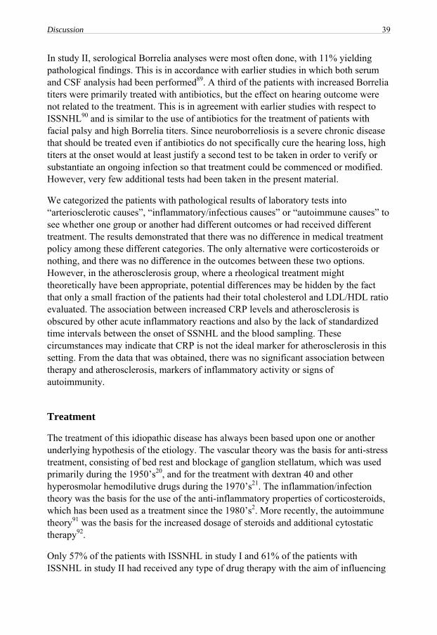
In study II, serological Borrelia analyses were most often done, with 11% yielding pathological findings. This is in accordance with earlier studies in which both serum and CSF analysis had been performed89. A third of the patients with increased Borrelia titers were primarily treated with antibiotics, but the effect on hearing outcome were not related to the treatment. This is in agreement with earlier studies with respect to ISSNHL90 and is similar to the use of antibiotics for the treatment of patients with facial palsy and high Borrelia titers. Since neuroborreliosis is a severe chronic disease that should be treated even if antibiotics do not specifically cure the hearing loss, high titers at the onset would at least justify a second test to be taken in order to verify or substantiate an ongoing infection so that treatment could be commenced or modified. However, very few additional tests had been taken in the present material.
We categorized the patients with pathological results of laboratory tests into “arteriosclerotic causes”, “inflammatory/infectious causes” or “autoimmune causes” to see whether one group or another had different outcomes or had received different treatment. The results demonstrated that there was no difference in medical treatment policy among these different categories. The only alternative were corticosteroids or nothing, and there was no difference in the outcomes between these two options. However, in the atherosclerosis group, where a rheological treatment might theoretically have been appropriate, potential differences may be hidden by the fact that only a small fraction of the patients had their total cholesterol and LDL/HDL ratio evaluated. The association between increased CRP levels and atherosclerosis is obscured by other acute inflammatory reactions and also by the lack of standardized time intervals between the onset of SSNHL and the blood sampling. These circumstances may indicate that CRP is not the ideal marker for atherosclerosis in this setting. From the data that was obtained, there was no significant association between therapy and atherosclerosis, markers of inflammatory activity or signs of autoimmunity. Treatment
The treatment of this idiopathic disease has always been based upon one or another underlying hypothesis of the etiology. The vascular theory was the basis for anti-stress treatment, consisting of bed rest and blockage of ganglion stellatum, which was used primarily during the 1950’s20, and for the treatment with dextran 40 and other hyperosmolar hemodilutive drugs during the 1970’s21. The inflammation/infection theory was the basis for the use of the anti-inflammatory properties of corticosteroids, which has been used as a treatment since the 1980’s2. More recently, the autoimmune theory91 was the basis for the increased dosage of steroids and additional cytostatic therapy92.
Only 57% of the patients with ISSNHL in study I and 61% of the patients with ISSNHL in study II had received any type of drug therapy with the aim of influencing

40 Discussion
the outcome. Eighty-nine percent (study I) and ninety-one percent (study II) of those who were medically treated received corticosteroids, even if the underlying etiology was unknown. The outcome, that the individuals who had received corticosteroids had the same odds for recovery as those who did not receive any drugs is in accordance with the conclusions from the latest Cochrane report in 200693. The only randomized, double-blinded, placebo-controlled study where a positive effect of corticosteroids was proposed actually had too few patients for such a conclusion to be drawn2.
Corticosteroids did not influence outcome in any patients with positive autoimmune signs in our study (II), although an effect on those might have been expected94. One potential explanation could be that too few patients had had those tests taken.
A recent study by Aoki et al reported that a very high dosage of corticosteroids seems to give a significantly better recovery rate, with 56% recovering two months after treatment95. However, this study did not use a control group without medical treatment.
Surprisingly enough, the 26% of patients who had been prescribed to rest had better odds for recovery (II). Earlier, bed rest and sick leave were standard treatment for ISSNHL patients, but this has nowadays become very uncommon. No research seems to have been performed with regard to that specific treatment modality. It is quite possible that all individuals who are afflicted by ISSNHL would experience some gain from rest, especially those individuals who, in their questionnaire, had reported recent stress before the onset of the disease. Audiometry
Comparing results from studies based on audiometric pattern is difficult since different classifications are used. One hypothesis about variation in recovery is that different types of hair cells are involved: If only the outer hair cells are damaged, a central adaptation might be possible so the remaining undamaged outer hair cells can reorganize the cortical pattern and in that way restore hearing96. If however, the inner hair cells are involved, it is more difficult to visualize reorganization as their number in the cochlea is so limited and they are strictly tonotropically arranged both in the cochlea and all along the central pathways. The weakness of this theoretical approach regarding the site of damage and the possibility of recovery is that it does not fit with the clinical observation that patients with all types of audiograms seem to have a chance to a spontaneous recovery of varying degrees. Also that the hearing improvement can come about as quickly as the onset of the hearing loss. A more slowly developing recovery over a couple of months would better fit the theory regarding reorganization96.
The results that patients with an hearing loss in the mid frequency region had the best recovery rate has been reported earlier in several studies2, 6, 76 and can be explained on

Discussion 41
the basis of a vascular theory for ISSNHL: In the cases when two arteries are supplying the cochlea, which occurs in about 50% of the cases97 and the main internal auditory artery is occluded distally to the branching of the anterior vestibular artery, one would theoretically expect a hearing loss in the lower and mid frequency region of the cochlea which has a chance of recovery through collaterals from the second main artery98. The same reasoning can be adapted to the five patients in the present investigation with vascular aberrations that were seen on MRI, who all had low frequency hearing loss and none or moderate improvement. They may have had the same occlusion of the main auditory artery, but with no collateral supply from a second artery98. Suggestions for further research
An agreement for a standard definition for SSNHL with standards for assessment of hearing loss and of report of hearing recovery are necessary for comparison of studies in future research.
ISSNHL may not be a single disease but a symptom of multiple disorders. If so, a comprehensive test battery is needed to identify underlying diseases for adequate treatment.


Conclusions 43
CONCLUSIONS
The results from the Swedish national database for SSNHL demonstrate that:
− MRI is an underused resource to get specific diagnoses for the condition both with respect to acoustic neuromas and to vascular abnormalities.
− Regardless of diagnostic protocol, treatment is mainly limited to corticosteroids or to no medical treatment with no difference in outcome.
− Hearing loss in the mid frequency region has the best odds for recovery regardless of the treatment chosen.
− Patients who are prescribed “rest” recover better than those without “rest”.
A randomized double-blind placebo controlled study with high power is necessary to evaluate whether or not there is an effect of corticosteroids in the dosage presently used in Sweden.


Acknowledgements 45
ACKNOWLEDGEMENTS
A number of people have contributed, inspired and helped me in preparing this thesis. I wish to express my sincere gratitude to all of you. I especially want to thank:
Professor Elisabeth Hultcrantz, my main supervisor, for sharing her vast knowledge in “Sudden Deafness”, guidance in the scientific world, and above all for providing ideas, excellent teaching, enthusiasm and encouragement. Thank you for always having time to give advice and support wherever you have been at the moment.
Professor emeritus Stig Arlinger, my co-supervisor, for sharing his endless knowledge in audiology, skillful editing and answering my questions so quickly.
Magnus Hansson, my co-author, at the department of Clinical Chemistry of Karolinska University Hospital, for valuable comments and help with clarification of laboratory tests.
Magnus Johansson, for help with system development of the database.
All staff at the ENT clinics and Audiological clinics in the hospitals of Bollnäs, Bäckefors, Enköping, Esklistuna, Fagersta, Gällivare, Gävle, Helsingborg, Huddinge, Hudiksvall, Jönköping, Kalmar, Karlskrona, Katrineholm, Kristianstad, Köping, Lindesberg, Linköping, Lund, Lundby, Malmö, Mora, Möndal, Norrköping, Sala, Skövde, Stockholm, Strömstad, Sundsvall, Södertälje, Trollhättan, Uddevalla, Umeå, Uppsala, Visby, Värnamo, Västerås, Ängelholm, Örebro, Örnsköldsvik and Östersund, for contributing to the database.
Ed Paulette, for linguistic guidance and careful editing.
Olle Eriksson and Karl Wahlin, at the department of Computer and Information Science, division of Statistics, for help with statistical counseling and for having patience and always answering my “odd” questions.
Docent Torbjörn Ledin, at the department of Clinical and Experimental Medicine, division of Otorhinolaryngology, for clarifying statistical terms for me and for valuable advice and support.
Docent Dag Hydén, at the ENT clinic of Linköping University Hospital, for valuable comments on the papers.
Anette Lagergren, at the Audiological clinic of Linköping University Hospital, for support and good help in providing me time for my scientific work.

46 Acknowledgements
Gunilla Hydén, Torsten Pettersson, Joakim Blomgren and Lars Andersson, at the ENT clinic, the Audiological clinic of Linköping University Hospital and the division of Speech, Language Pathology of Linköping University, for always patiently answering my computer related questions.
My colleagues, at the Audiological clinic of Linköping University Hospital, Motala Hospital, the division of Technical Audiology and the division of Speech, Language Pathology of Linköping University, for providing a nice and friendly working environment and for the very entertaining coffee break discussions.
My friends Pia Granath, for letting me chill out in her office between the hard work and for patiently listening to me babbling about anything possible. Linda Lindström and Elisabeth Ericsson, for all help and support. Dinners with you have been perfect breaks from work and have helped me to think of other things than Sudden Deafness.
My mother Pari, my late father Qasem and my brothers Ramin and Ramtin for love, support and for believing in me.
Finally, I have many reasons to be grateful to my beloved husband Reza and our wonderful children Alma and Arvid. Reza, for your love, support and patience. You are everything I have ever wished for with your kindness, concern and selflessness. Alma (3,5 years old) and Arvid (1,5 year old), for being who you are and bringing me down to the earth again when I feel lost in the mist.
Financial support has gratefully been received from the Country Council of South East Sweden, Linköping University, StingerFonden, Stiftelsen Tysta Skolan and Gunnar Arnbrinks Stiftelse.

Appendix 47
APPENDIX
Questionnaire (translation from the Swedish original)


References 49
REFERENCES
1. Whitaker, S., Idiopathic sudden hearing loss, Am J Otol, 1980, 1(3):180-183.
2. Wilson, W. R., Byl, F. M. and Laird, N., The efficacy of steroids in the treatment of idiopathic sudden hearing loss. A double-blind clinical study, Arch Otolaryngol, 1980, 106(12):772-776.
3. Stokroos, R. J., Albers, F. W. J. and Tenvergert, E. M., Antiviral treatment of Idiopathic Sudden Sensorineural Hearing Loss: A prospective, randomized, double-blind clinical trial, Acta Otolaryngol, 1998, 118(4):488-495.
4. Tucci, D. L., Farmer Jr, J. C., Kitch, R. D. and Witsell, D. L., Treatment of sudden sensorineural hearing loss with systemic steroids and valacyclovir, Otol Neurotol, 2002, 23(3):301-308.
5. Westerlaken, B. O., Stokroos, R. J., Wit, H. P., Dhooge, I. J. M. and Albers, F. W. J., Treatment of idiopathic sudden sensorineural hearing loss with antiviral therapy: A prospective, randomized, double-blind clinical trial, Ann Otol Rhinol Laryngol, 2003, 112(11):993-1000.
6. Mattox, D. E. and Simmons, F. B., Natural history of sudden sensorineural hearing loss, Ann Otol Rhinol Laryngol, 1977, 86(4 Pt 1):463-480.
7. Tran Ba Huy, P., Idiopathic sudden sensorineural hearing loss: Reasons for defeat, conditions for victory, Otorhinolaryngol Nova, 1999, 9(5):171-177.
8. Moskowitz, D., Lee, K. J. and Smith, H. W., Steroid use in idiopathic sudden sensorineural hearing loss, Laryngoscope, 1984, 94(5 I):664-666.
9. Nakashima, T. and Yanagita, N., Outcome of sudden deafness with and without vertigo, Laryngoscope, 1993, 103(10):1145-1149.
10. Cinamon, U., Bendet, E. and Kronenberg, J., Steroids, carbogen or placebo for sudden hearing loss: a prospective double-blind study, Eur Arch Otorhinolaryngol, 2001, 258(9):477-480.
11. Weinaug, P., [Spontaneous remission in sudden deafness], Hno, 1984, 32(8):346-351.
12. De Kleyn, A., Sudden complete or partial loss of function of the octavus-system in apparently normal persons, Acta Otolaryngol, 1944, 32(5):407-429.
13. Van Caneghem, D., [Sudden deafness.], Acta Otorhinolaryngol Belg, 1958, 12(1):5-17.

50 References
14. Teranishi, M., Katayama, N., Uchida, Y., Tominaga, M. and Nakashima, T., Thirty-year trends in sudden deafness from four nationwide epidemiological surveys in Japan, Acta Otolaryngol, 2007, 127(12):1259-1265.
15. Klemm, E., Deutscher, A. and Mosges, R., [A Present Investigation of the Epidemiology in Idiopathic Sudden Sensorineural Hearing Loss.], Laryngorhinootologie, 2009.
16. Goodhill, V., Sudden deafness and round window rupture, Laryngoscope, 1971, 81(9):1462-1474.
17. Kellerhals, B., Acoustic trauma and cochlear microcirculation. An experimental and clinical study on pathogenesis and treatment of inner ear lesions after acute noise exposure, Adv Otorhinolaryngol, 1972, 18:91-168.
18. Wilson, W. R., Veltri, R. W., Laird, N. and Sprinkle, P. M., Viral and epidemiologic studies of idiopathic sudden hearing loss, Otolaryngol Head Neck Surg, 1983, 91(6):653-658.
19. Campbell, K. C. and Klemens, J. J., Sudden hearing loss and autoimmune inner ear disease, J Am Acad Audiol, 2000, 11(7):361-367.
20. Wilmot, T. J., Sudden perceptive deafness in young people, J Laryngol Otol, 1959, 73:466-468.
21. Kellerhals, B., Hippert, F. and Pfaltz, C. R., Treatment of acute acoustic trauma with low molecular weight dextran, Pract Otorhinolaryngol (Basel), 1971, 33(4):260-264.
22. Pickles, J. O., An introduction to the physiology of hearing, 2nd ed. Academic press, London;, 1988.
23. Kirikae, I., Nomura, Y. and Hiraide, F., The capillary in the human cochlea, Acta Otolaryngol, 1969, 67(1):1-8.
24. Roeser, R. J., Valente, M. and Hosford-Dunn, H., Audiology diagnosis, 1st ed.Thieme Medical Publishers, New York;, 2000.
25. Mattox, D. E. and Lyles, C. A., Idiopathic sudden sensorineural hearing loss, Am J Otol, 1989, 10(3):242-247.
26. Hallberg, O. E., Sudden deafness of obscure origin, Laryngoscope, 1956, 66(10):1237-1267.
27. Hunt, J. R., On herpetic inflammations of the geniculate ganglion: A new syndrome and its complications, J Nerv Ment Dis, 1907, 34:73-96.

References 51
28. Peltomaa, M., Pyykko, I., Seppala, I., Viitanen, L. and Viljanen, M., Lyme borreliosis, an etiological factor in sensorineural hearing loss?, Eur Arch Otorhinolaryngol, 2000, 257(6):317-322.
29. Finizia, C., Jönsson, R. and Hanner, P., Serum and cerebrospinal fluid pathology in patients with sudden sensorineural hearing loss, Acta Otolaryngol, 2001, 121(7):823-830.
30. Vuori, M., Lahikainen, E. A. and Peltonen, T., Perceptive deafness in connection with mumps. A study of 298 servicemen suffering from mumps, Acta Otolaryngol, 1962, 55:231-236.
31. Okamoto, M., Shitara, T., Nakayama, M., Takamiya, H., Nishiyama, K., Ono, Y. and Sano, H., Sudden deafness accompanied by asymptomatic mumps, Acta Otolaryngol Suppl, 1994, 514:45-48.
32. Veltri, R. W., Wilson, W. R., Sprinkle, P. M., Rodman, S. M. and Kavesh, D. A., The implication of viruses in idiopathic sudden hearing loss: primary infection or reactivation of latent viruses?, Otolaryngol Head Neck Surg, 1981, 89(1):137-141.
33. Kobayashi, H., Suzuki, A. and Nomura, Y., Unilateral hearing loss following rubella infection in an adult, Acta Otolaryngol Suppl, 1994, 514:49-51.
34. Vasama, J. P. and Linthicum, F. H., Idiopathic sudden sensorineural hearing loss: Temporal bone histopathologic study, Ann Otol Rhinol Laryngol, 2000, 109(6):527-532.
35. Van Dishoeck, H. A. E. and Bierman, T. A., Sudden perceptive deafness and viral infection, Ann Otol Rhinol Laryngol, 1957, 66:963-980.
36. Koide, J., Yanagita, N., Hondo, R. and Kurata, T., Serological and clinical study of herpes simplex virus infection in patients with sudden deafness, Acta Otolaryngol Suppl, 1988, 456:21-26.
37. Schuknecht, H. F. and Donovan, E. D., The pathology of idiopathic sudden sensorineural hearing loss, Arch Otorhinolaryngol, 1986, 243(1):1-15.
38. Hirai, T., Kimura, S. and Fujii, M., Anti-viral Treatment for Three Cases of Sudden Onset Total Deafnesses, Pract Otorhinolaryngol, 2003, 96(12):1045-1048.
39. Uri, N., Doweck, I., Cohen-Kerem, R. and Greenberg, E., Acyclovir in the treatment of idiopathic sudden sensorineural hearing loss, Otolaryngol Head Neck Surg, 2003, 128(4):544-549.
40. McCabe, B. F., Autoimmune sensorineural hearing loss, Ann Otol Rhinol Laryngol, 1979, 88(5 Pt 1):585-589.

52 References
41. Woolf, N. K. and Harris, J. P., Cochlear pathophysiology associated with inner ear immune responses, Acta Otolaryngol, 1986, 102(5-6):353-364.
42. Ichimiya, I., Kurono, Y., Hirano, T. and Mogi, G., Changes in immunostaining of inner ears after antigen challenge into the scala tympani, Laryngoscope, 1998, 108(4 Pt 1):585-591.
43. García Berrocal, J. R., Vargas, J. A., Vaquero, M., Cajal, S. R. Y. and Ramírez-Camacho, R. A., Cogan's syndrome: An oculoaudiovestibular disease, Postgrad Med J, 1999, 75(883):262-264.
44. Kempf, H. G., Ear involvement in Wegener's granulomatosis, Clin Otolaryngol Allied Sci, 1989, 14(5):451-456.
45. Bowman, C. A., Linthicum, F. H., Jr., Nelson, R. A., Mikami, K. and Quismorio, F., Sensorineural hearing loss associated with systemic lupus erythematosus, Otolaryngol Head Neck Surg, 1986, 94(2):197-204.
46. Cole, R. R. and Jahrsdoerfer, R. A., Sudden hearing loss: an update, Am J Otol, 1988, 9(3):211-215.
47. Cadoni, G., Marinelli, L., De Santis, A., Romito, A., Manna, R. and Ottaviani, F., Sudden hearing loss in a patient hepatitis C virus (HCV) positive on therapy with alpha-interferon: a possible autoimmune-microvascular pathogenesis, J Laryngol Otol, 1998, 112(10):962-963.
48. Yoshida, Y., Yamauchi, S., Shinkawa, A., Horiuchi, M. and Sakai, M., Immunological and virological study of sudden deafness, Auris Nasus Larynx, 1996, 23:63-68.
49. Ramírez Camacho, R., García Berrocal, J. R., Trinidad, A., Martín Marero, A. and Buján, J., Cochlear cytotoxic activity of cisplatin in experimentation animals. A study using scanning electron microscopy, Actividad citotóxica coclear del cisplatino en animales de experimentación. Un estudio con microscopía electrónica de barrido., 2002, 53(8):538-542.
50. Shimazaki, T., Ichimiya, I., Suzuki, M. and Mogi, G., Localization of glucocorticoid receptors in the murine inner ear, Ann Otol Rhinol Laryngol, 2002, 111(12 Pt 1):1133-1138.
51. Kanzaki, J., Taiji, H. and Ogawa, K., Evaluation of hearing recovery and efficacy of steroid treatment in sudden deafness, Acta Otolaryngol Suppl, 1988, 106(456):31-36.
52. Russolo, M. and Bianchi, M., The treatment of sudden hearing loss, La terapia della sordita improvvisa idiopatica, 1997, 17(5):319-324.
References 53
53. Kurokawa, T. and Yamaura, K., Four cases of idiopathic sudden sensorineural hearing loss, Pract Otolog, 1998, 91(3):221-225.
54. Jeyakumar, A., Francis, D. and Doerr, T., Treatment of idiopathic sudden sensorineural hearing loss, Acta Otolaryngol, 2006, 126(7):708-713.
55. Simmons, F. B., Theory of membrane breaks in sudden hearing loss, Arch Otolaryngol, 1968, 88(1):41-48.
56. Simmons, F. B., The double membrane break syndrome in sudden hearing loss, Laryngoscope, 1979, 89(1):59-66.
57. Gussen, R., Sudden hearing loss associated with cochlear membrane rupture. Two human temporal bone reports, Arch Otolaryngol, 1981, 107(10):598-600.
58. Gussen, R., Sudden deafness associated with bilateral Reissner's membrane ruptures, Am J Otolaryngol, 1983, 4(1):27-32.
59. Yoon, T. H., Paparella, M. M., Schachern, P. A. and Alleva, M., Histopathology of sudden hearing loss, Laryngoscope, 1990, 100(7):707-715.
60. Merchant, S. N., Adams, J. C. and Nadol Jr, J. B., Pathology and pathophysiology of idiopathic sudden sensorineural hearing loss, Otol Neurotol, 2005, 26(2):151-160.
61. Singleton, G. T., Nolan Post, K., Karlan, M. S. and Bock, D. G., Perilymph fistulas; Diagnostic criteria and therapy, Ann Otol Rhinol Laryngol, 1978, 87(6 I):797-803.
62. Karhuketo, T. S. and Puhakka, H. J., Endoscope-guided round window fistula repair, Otol Neurotol, 2001, 22(6):869-873.
63. Rasmussen, H., Sudden deafness, Acta Otolaryngol, 1949, 37:65-70.
64. Kirikae, I., Nomura, Y., Shitara, T. and Kobayashi, T., Sudden deafness due to Buerger's disease, Arch Otolaryngol, 1962, 75:502-505.
65. Zechner, G. and Altmann, F., The temporal bone in leukemia. Histological studies, Ann Otol Rhinol Laryngol, 1969, 78(2):375-387.
66. Odetoyinbo, O. and Adekile, A., Sensorineural hearing loss in children with sickle cell anemia, Ann Otol Rhinol Laryngol, 1987, 96(3 Pt 1):258-260.
67. Perlman, H. B., Kimura, R. and Fernandez, C., Experiments on temporary obstruction of the internal auditory artery, Laryngoscope, 1959, 69(6):591-613.

54 References
68. Hultcrantz, E., Linder, J. and Angelborg, C., Sympathetic effects on cochlear blood flow at different blood pressure levels, Inserm, 1977, 68:271-278.
69. Dormandy, J. A., Influence of blood viscosity on blood flow and the effect of low molecular weight dextran, Br Med J, 1971, 4(789):716-719.
70. Scheibe, F., Haupt, H. and Baumgartl, H., Effects of experimental cochlear thrombosis on oxygenation and auditory function of the inner ear, Eur Arch Otorhinolaryngol, 1997, 254(2):91-94.
71. Maass, B., [Blood supply of the internal ear. Anatomico-functional considerations], Hno, 1982, 30(10):355-364.
72. Klemm, E., Altmann, E. and Lange, O., [Rheologic problems of microcirculation and consequences of drug therapy for sudden deafness], Laryngol Rhinol Otol (Stuttg), 1983, 62(2):62-64.
73. Ohinata, Y., Makimoto, K., Kawakami, M., Haginomori, S., Araki, M. and Takahashi, H., Blood viscosity and plasma viscosity in patients with sudden deafness, Acta Otolaryngol, 1994, 114(6):601-607.
74. Ullrich, H., Kleinjung, T., Steffens, T., Jacob, P., Schmitz, G. and Strutz, J., Improved treatment of sudden hearing loss by specific fibrinogen aphaeresis, J Clin Apher, 2004, 19(2):71-78.
75. Valbonesi, M., Mora, F., Mora, R. and Carlier, P., Rheopheresis for sudden hearing loss (SHL), Int J Artif Organs, 2004, 27(9):806-809.
76. Hultcrantz, E., Stenquist, M. and Lyttkens, L., Sudden deafness: a retrospective evaluation of dextran therapy, ORL J Otorhinolaryngol Relat Spec, 1994, 56(3):137-142.
77. Topuz, E., Yigit, O., Cinar, U. and Seven, H., Should hyperbaric oxygen be added to treatment in idiopathic sudden sensorineural hearing loss?, Eur Arch Otorhinolaryngol, 2004, 261(7):393-396.
78. De Oliveira Penido, N., Lisboa Ramos, H. V., Barros, F. A., Mendonca Cruz, O. L. and Toledo, R. N., Clinical, etiological and progression factors of hearing in sudden deafness, Braz J Otorhinolaryngol., 2005, 71(5):633-638.
79. Probst, R., Tschopp, K., Ludin, E., Kellerhals, B., Podvinec, M. and Pfaltz, C. R., A randomized, double-blind, placebo-controlled study of dextran/pentoxifylline medication in acute acoustic trauma and sudden hearing loss, Acta Otolaryngol, 1992, 112(3):435-443.

References 55
80. Kronenberg, J., Almagor, M., Bendet, E. and Kushnir, D., Vasoactive therapy versus placebo in the treatment of sudden hearing loss: A double-blind clinical study, Laryngoscope, 1992, 102(1):65-68.
81. Narozny, W., Kuczkowski, J., Kot, J., Stankiewicz, C., Sicko, Z. and Mikaszewski, B., Prognostic factors in sudden sensorineural hearing loss: our experience and a review of the literature, Ann Otol Rhinol Laryngol, 2006, 115(7):553-558.
82. Cvorovic, L., Deric, D., Probst, R. and Hegemann, S., Prognostic model for predicting hearing recovery in idiopathic sudden sensorineural hearing loss, Otol Neurotol, 2008, 29(4):464-469.
83. Berg, H. M., Cohen, N. L., Hammerschlag, P. E. and Waltzman, S. B., Acoustic neuroma presenting as sudden hearing loss with recovery, Otolaryngol Head Neck Surg, 1986, 94(1):15-22.
84. Yanagihara, N. and Asai, M., Sudden hearing loss induced by acoustic neuroma: significance of small tumors, Laryngoscope, 1993, 103(3):308-311.
85. Chaimoff, M., Nageris, B. I., Sulkes, J., Spitzer, T. and Kalmanowitz, M., Sudden hearing loss as a presenting symptom of acoustic neuroma, Am J Otolaryngol - Head and Neck Medicine and Surgery, 1999, 20(3):157-160.
86. Aronzon, A., Ruckenstein, M. J. and Bigelow, D. C., The efficacy of corticosteroids in restoring hearing in patients undergoing conservative management of acoustic neuromas, Otol Neurotol, 2003, 24(3):465-468.
87. Kosugi, E. M., Tangerina, R. P., Dib, G. C., Ramos, H. V. L. and Penido, N. O., Vestibular schwannoma presenting as sudden hearing loss, Schwanoma vestibular como causa de surdez súbita, 2004, 70(6):795-799.
88. Fetterman, B. L., Saunders, J. E. and Luxford, W. M., Prognosis and treatment of sudden sensorineural hearing loss, Am J Otol, 1996, 17(4):529-536.
89. Hyden, D., Roberg, M. and Odkvist, L., Borreliosis as a cause of sudden deafness and vestibular neuritis in Sweden, Acta Otolaryngol Suppl, 1995, 520 Pt 2:320-322.
90. Lorenzi, M. C., Bittar, R. S., Pedalini, M. E., Zerati, F., Yoshinari, N. H. and Bento, R. F., Sudden deafness and Lyme disease, Laryngoscope, 2003, 113(2):312-315.
91. Veldman, J. E., Hanada, T. and Meeuwsen, F., Diagnostic and therapeutic dilemmas in rapidly progressive sensorineural hearing loss and sudden deafness. A reappraisal of immune reactivity in inner ear disorders, Acta Otolaryngol, 1993, 113(3):303-306.

56 References
92. McClelland, L., Powell, R. J. and Birchall, J., Role of ciclosporin in steroid-responsive sudden sensorineural hearing loss, Acta Otolaryngol, 2005, 125(12):1356-1360.
93. Wei, B. P., Mubiru, S. and O'Leary, S., Steroids for idiopathic sudden sensorineural hearing loss, Cochrane Database Syst Rev, 2006(1):CD003998.
94. Zeitoun, H., Beckman, J. G., Arts, H. A., Lansford, C. D., Lee, D. S., El-Kashlan, H. K., Telian, S. A., et al., Corticosteroid response and supporting cell antibody in autoimmune hearing loss, Arch Otolaryngol Head Neck Surg, 2005, 131(8):665-672.
95. Aoki, D., Takegoshi, H. and Kikuchi, S., Evaluation of super-high-dose steroid therapy for sudden sensorineural hearing loss, Otolaryngol Head Neck Surg, 2006, 134(5):783-787.
96. Eggermont, J. J., The role of sound in adult and developmental auditory cortical plasticity, Ear Hear, 2008, 29(6):819-829.
97. Mazzoni, A., Internal auditory artery supply to the petrous bone, Ann Otol Rhinol Laryngol, 1972, 81(1):13-21.
98. Hultcrantz, E., Sudden deafness - A critical evaluation of pathogenesis and 'cure', Otorhinolaryngol Nova, 1999, 9(5):178-189.

Svensk sammanfattning 57
SVENSK SAMMANFATTNING
Idiopatisk Plötslig Sensorineural hörselnedsättning i Sverige Diagnostiskt protokoll och behandling i relation till resultat
Bakgrund
Idiopatisk Plötslig Sensorineural Hörselnedsättning (IPSH) är en av de gåtfulla sjukdomar vilka drabbar innerörat och nerverna som passerar den inre hörselkanalen. Oavsett behandling återkommer hörseln fullständigt hos ungefär en tredjedel av patienterna och delvis hos en tredjedel, medan resten får kvarstående betydande hörselnedsättning ofta i kombination med tinnitus eller obehag av ökad ljudkänslighet. Sjukdomen har tidigare beräknats drabba 5 – 20 per 100 000 invånare årligen men incidensen är osäker, då det är svårt att veta hur många som insjuknat pga. högt spontant tillfrisknande innan något läkarbesök har skett.
Orsaken till sjukdomen är okänd. Detta har lett till att patienter både i Sverige och globalt genom åren har fått olika behandlingar beroende på vilken hypotes om sjukdomens orsak läkarna har stött sig på:
Infektionsteorin: Redan 1907 rapporterade Hunt att Herpes zoster kan orsaka plötslig hörselnedsättning. Andra virala och bakteriella sjukdomar såsom Borrelia, syfilis, påssjuka och rubella (Röda Hund) är också kända orsaker till plötslig hörselnedsättning. Om man antar att alla fall av IPSH har denna genes så använder man antivirala läkemedel, (acyklovir och valacyclovir). De fyra hitintills publicerade randomiserade, placebo-kontrollerade kliniska studier med acyklovir och valacyclovir visar emellertid inte någon signifikant effekt av den behandlingen.
Immunologisk teori: McCabe (1979) var först med att föreslå autoimmunitet som orsak till plötslig hörselnedsättning. Han baserade sin hypotes på kliniska data, patologiska fynd vid autoimmunologiska tester och förbättrad hörsel vid kortikosteroid terapi. Hörselnedsättningen kan vara en följd av lokala autoimmuna processer som bryter ned enbart vävnaden i innerörat eller en systemisk autoimmun sjukdom som t.ex. Cogans syndrom, Wegeners sjukdom och systemisk lupus erythematosus (SLE). Behandling av IPSH med kortikosteroider är baserat på denna immunologiska teori. Sedan man började behandla IPSH med kortikosteroider på 1980-talet har det gjorts många studier om deras effekt, men de enda två som varit randomiserade dubbelblinda placebokontrollerade från 1980 och 2001 har visat motsägande resultat och haft låg ”power”.
Fistelteorin: Rupturer antingen inom snäckan med jonrubbningar som följd och orsak till hörselnedsättning, eller av membranen i runda eller ovala fönstret med ett resulterande perilymfaläckage som symptomorsak är en teori om IPSH. Rupturen anses inte ske spontant utan uppträder efter plötslig tryckändring i mellanörat i samband med huvudskador, barotrauma eller intensiv fysisk aktivitet. Kirurgi med

58 Svensk sammanfattning
tätning av misstänkta hål i runda fönstermembranet i kombination med ordination av strikt sängläge är en behandlingsmetod, men inte heller här är man ense om nyttan av denna terapi.
Blodkärlteorin: Ett minskat blodflöde i den ändartär som försörjer örat skulle kunna orsaka plötslig hörselnedsättning. Sjukdomar i blodkärl eller hematologiska åkommor förknippade med plötslig hörselnedsättning har också rapporterats såsom vid Buergers sjukdom, leukemi och sickelcellsanemi. Vid IPSH har olika behandlingar såsom extrakorporeal defibrinogering, Rheomacrodex® och andra hyperosmolara blodförtunningsmedel använts baserat på denna teori. Det finns vetenskapligt stöd både för och mot sådan terapi.
På grund av den låga incidensen, det spontana tillfrisknandet i vissa fall och möjligheter av mer än en orsak till sjukdomen är effekten av behandling av IPSH alltid svår att bedöma på en enskild klinik där man endast ser ett mindre antal patienter per år. Genom att samla data från ett stort antal patienter finns möjlighet att identifiera prognostiska faktorer och effekt av behandling. Syfte
Syftet med denna avhandling är att redovisa vilken utredning och behandling patienter med IPSH får i Sverige och hur resultaten är vad gäller hörseltillfrisknande.
Studie I
− Att kartlägga vilka variabler vid insjuknandet i IPSH som har prognostisk värde såsom ålder, tinnitus, yrsel, tidigare sjukdomar och hörselkurvornas utseende.
− Att kartlägga behandlingspolicyn för IPSH i Sverige.
− Att utvärdera behandlingseffekten med avseende på hörseltillfrisknande.
Studie II
− Att undersöka olika diagnostiska testbatterier för IPSH som för närvarande används i Sverige.
− Att utvärdera om och hur patologiska diagnostiska fynd leder till behandlingsmodifikationer.
− Att undersöka om sådana behandlingsmodifikationer påverkar tillfrisknandet.
Svensk sammanfattning 59
Metod
Den nationella databasen om plötslig sensorineural hörselnedsättning startades sent 2002 i Linköping.
Ett frågeformulär hade tagits fram som fylldes i av den först konsulterade öronläkaren. Frågorna berörde patientens tidigare sjukdomar, trauma, tinnitus, yrsel, ärftlighet för olika sjukdomar särskilt hörselnedsättning samt viken utredning och behandling som planerades/gavs av den aktuella plötsliga hörselnedsättningen. Två audiogram, ett taget vid insjuknandet och ett vid kontroll efter tre månader skickades tillsammans med frågeformuläret till databasen. Se appendix.
För bedömning av hörselnedsättningen jämfördes det initiala audiogrammet med ett audiogram taget innan den akuta hörselnedsättningen. För att kategorisera nedsättningarna definierades fyra frekvensområden: basområdet: skillnad i hörtröskelmedelvärde vid 125-250-500 Hz, mellanregistret vid 1000-1500-2000 Hz, diskantområdet vid 3000-4000-6000 Hz och ”flat loss” om skillnaderna i försämring mellan de tre frekvensbanden var mindre än 10 dB.
I studie II kategoriserades patologiska fynd på följande sätt:
− “Arteriosklerosassocierade variabler”: LDL-kolesterol/HDL-kolesterol >3, Totalkolesterol >5 mmol/L och C-reaktivt protein (CRP) >3 mg/L hos patienter med eller utan tidigare hjärt-kärl sjukdomar.
− “Inflammation/infektionassocierade variabler”: CRP >10 mg/L, ”Sänka” >20 mm, Vita blodkroppar >10 x 109ml/L, Hemoglobinhalt (Hb) <120 g/L, Trombocytantal >150 x 109ml/L och Borrelia test “positiv” (IgG antikroppar och IgM antikroppar) hos patienter med eller utan klinisk infektion.
− “Autoimmuna variabler”: Förekomst av patologiska värden av HSP-70, Kardiolipin, Antifosfolipid, Antineutrofila cytoplasmiska antikroppar (ANCA) och Antinukleära antikroppar (ANA) “positiv”.
Material
Studie I
300 patienter med plötslig hörselnedsättning som initialt hade diagnostiserats som ”Plötslig dövhet/plötslig sensorineural hörselnedsättning” av sina lokala öronläkare och som inkluderats i den svenska nationella databasen. Av dessa 300 patienter hade 92 antingen ledningshinder, Mb Ménière, eller andra kända inneröre- eller

60 Svensk sammanfattning
mellanöresjukdomar, eller mindre än 30dB hörselförlust, färre än tre närliggande frekvenser drabbade eller insjuknat under mer är 24 timmar. Se tabell II.
Studie II
400 patienter (300 från studie I samt ytterligare 100) vilka initialt hade diagnostiserats som ”Plötslig dövhet/plötslig sensorineural hörselnedsättning” och därför rapporterats till databasen. Resultat och diskussion
Alla variabler i frågeformuläret analyserades med ordinal logistisk regression med avsikten att hitta samband med tillfrisknande.
Ärftlighet för hörselnedsättning (I, II), ålder (I, II) och yrsel vid insjuknande (II) var signifikant relaterat till sämre chans för tillfrisknande. Detta stämmer väl överens med resultat från tidigare studier.
37% av patienterna med IPSH i studie I och 40% av alla inkluderade patienterna i databasen (studie II) hade genomgått MR eller CT. Fyra patienter i studie I hade patologiska fynd. Fem av tjugotvå patienter med patologiska fynd i studie II hade akustikusneurinom och fem hade olika kärlförändringar som möjligen kunde kopplas till IPSH. Dessa patienters behandling hade inte varit annorlunda än övrigas. Väl att märka hade många även hunnit få en hörselförbättring innan MR/CT undersökningen, som hade gjorts flera veckor efter insjuknandet. Därför hade patologiska fynd inte påverkat val av initial behandling.
68% av patienterna med IPSH i studie I och 65% av patienterna med IPSH i studie II hade hematologiska prover tagna. 23% (I) och 24% (II) hade en eller flera patologiska fynd. Det fanns ingen korrelation mellan något av fynden av laboratorietesterna och tillfrisknande i jämförelse med dem som hade normala laboratorieresultat oavsett om man utvärderade testerna separat eller efter kategorisering. Patienterna med patologiska fynd av laboratorietester hade kategoriserats i "arteriosklerosassocierade variabler", " inflammation/infektionassocierade variabler " eller "autoimmuna variabler" för att se huruvida dessa grupper hade fått olika behandlingar och om detta påverkat resultatet. Resultaten visade ingen skillnad av den medicinska behandlingen mellan dessa olika kategorier. Det enda alternativet var kortikosteroider eller ingenting och det förelåg ingen skillnad i tillfrisknande dem emellan.
57% av patienterna med IPSH i studie I och 61% av patienterna med IPSH i studie II hade fått någon form av behandling. 89% (II) och 91% (II) av de som behandlats medikamentellt hade fått kortikosteroider, vanligen i form av prednisolon. Kortikosteroidens dos och behandlingsschema kunde variera från 25 mg till 80 mg/dag

Svensk sammanfattning 61
från en vecka upp till en månad i nedtrappande doser. Man kunde inte se något samband mellan den medicinska behandlingen och tillfrisknandet. Att de som fick kortikosteroider hade samma odds för tillfrisknande som de som inte fick något alls är helt i enlighet med slutsatserna från den senaste Cochrane rapporten om IPSH från 2006.
26% av tre hundra patienter med IPSH i studie II hade ordinerats sängläge eller hade sjukskrivits från en vecka till en månad. Tjugotvå av dessa patienter har enbart fått denna terapi. Dessa patienter, som hade ordinerats sängläge eller sjukskrivits hade signifikant högre chans till tillfrisknande än de som inte hade ordinerats vila. Förr var sängläge eller sjukskrivning standardbehandling för IPSH patienter, men detta har nuförtiden blivit mycket ovanligt. Ingen forskning verkar ha gjorts med hänsyn till denna specifika behandlingsmetod.
Patienter med hörselförlust i mellanregistret hade signifikant bättre chans till tillfrisknande (I, II). Detta har kunnat visas i en del tidigare studier och kan förklaras utifrån blodkärlsteorin: I de fall där det finns två artärer till cochlean, vilket förekommer i cirka 50% av fallen, och det ena är blockerat, kan man teoretiskt räkna med en hörselnedsättning i de lägre och mellersta frekvenserna med chans till tillfrisknande eftersom det då finns anastomoser. Slutsatser
− MRT är en underutnyttjad resurs för att få specifika diagnoser vad gäller både akustiskneurom och blodskärlförändringar som orsak till hörselnedsättningen och borde göras akut för att komma till nytta för patienten.
− Hörselnedsättning i mellanregistret har störst chans för tillfrisknande oavsett vilken behandling som getts.
− Patienter som ordinerats vila återhämtar sig bättre än de som inte vilat.
− Oavsett olika patologiska fynd i utredningen är behandlingen av IPSH i Sverige främst begränsad till kortikosteroider eller ingen medicinsk terapi med ingen skillnad i tillfrisknande.
− För att kunna utvärdera behandlingseffekten av kortikosteroider i den dos som används i Sverige idag är en randomiserad dubbelblind placebo kontrollerad studie med hög ”power” nödvändig.


![George Kampessis, Nicholas Maroudias, Papacharalampous ... steroids for sudden... · Over 60 years since its first report by De Kleyn [1], sudden sensorineural hearing loss (SSNHL)](https://static.fdocuments.net/doc/165x107/5c79ad9809d3f2bd0e8b9610/george-kampessis-nicholas-maroudias-papacharalampous-steroids-for-sudden.jpg)