Identification and Characterization of Glycoproteins on the ... · Identification and...
11
Identification and Characterization of Glycoproteins on the Spore Surface of Clostridium difficile Philippa C. R. Strong, Kelly M. Fulton, Annie Aubry, Simon Foote, Susan M. Twine, Susan M. Logan Vaccine Program, Human Health Therapeutics Portfolio, National Research Council of Canada, Ottawa, Ontario, Canada In this study, we identify a major spore surface protein, BclA, and provide evidence that this protein is glycosylated. Following extraction of the spore surface, solubilized proteins were separated by one-dimensional PAGE and stained with glycostain to reveal a reactive high-molecular-mass region of approximately 600 kDa. Tandem mass spectrometry analysis of in-gel digests showed this band to contain peptides corresponding to a putative exosporangial glycoprotein (BclA3) and identified a number of glycopeptides modified with multiple N-acetyl hexosamine moieties and, in some cases, capped with novel glycans. In addition, we demonstrate that the glycosyltransferase gene sgtA (gene CD3350 in strain 630 and CDR3194 in strain R20291), which is lo- cated immediately upstream of the bclA3 homolog, is involved in the glycosylation of the spore surface, and is cotranscribed with bclA3. The presence of anti--O-GlcNAc-reactive material was demonstrated on the surface of spores by immunofluorescence and in surface extracts by Western blotting, although each strain produced a distinct pattern of reactivity. Reactivity of the spore surface with the anti--O-GlcNAc antibody was abolished in the 630 and R20291 glycosyltransferase mutant strains, while com- plementation with a wild-type copy of the gene restored the -O-GlcNAc reactivity. Phenotypic testing of R20291 glycosyltrans- ferase mutant spores revealed no significant change in sensitivity to ethanol or lysozyme. However, a change in the resistance to heat of R20291 glycosyltransferase mutant spores compared to R20291 spores was observed, as was the ability to adhere to and be internalized by macrophages. C lostridium difficile is a Gram-positive, spore-forming anaer- obe and is the major cause of antibiotic-associated diarrhea (1). The incidence of C. difficile infection has been rapidly increas- ing in North America and Europe in recent years, and this increase in infections has been associated with higher rates of morbidity and mortality (2). Recent estimates of the incidence of C. difficile- associated diarrhea (CDAD) in the United States indicate as many as 500,000 cases per year with up to 20,000 deaths (1, 3). The primary focus of much of the research has been on two toxins, TcdA and TcdB, that are produced by C. difficile and cause tissue damage and a severe inflammatory response, which can lead in the more serious cases to potentially lethal pseudomembranous coli- tis. While toxin activity is recognized as the major virulence factor associated with CDAD, other aspects of C. difficile virulence are less well understood. Spore production in C. difficile is an integral part of the infec- tious process. This recalcitrant, dormant form of C. difficile can survive indefinitely outside the host and is known to persist in the hospital environment (4). It has been demonstrated in mice that antibiotic treatment suppresses the diversity of the gut micro- biome and promotes the production of these highly infectious spores, which are then disseminated into the environment (so- called supershedder state) (5). As such, more recently there has been increased attention on the process of spore formation in C. difficile as well as studies of spore structure and biochemical com- position (6–12). To date, the major focus of the studies on spore structure has been to identify spore coat proteins and demonstrate enzymatic activity. Pretreatment of spores either by enzymatic digestion or sonication was utilized in these studies to remove the exosporangial layer prior to analysis. In contrast to spores of C. difficile, the spores of another im- portant toxin-producing, Gram-positive pathogen, Bacillus an- thracis, have been extensively characterized. These spores have been shown to be enclosed by an exosporangial layer which is composed of a number of different proteins (13, 14) and which includes an outermost hair-like nap layer. The filaments of the nap layer are primarily composed of a highly immunogenic collagen- like protein, BclA (15–17), as well as a second exosporangial pro- tein, BclB (18). Both BclA and BclB have been well characterized and shown to be glycosylated with an O-linked pentasaccharide that contains the novel terminal sugar 2-O-methyl-4-(3-hydroxy- 3-methylbutamido)-4,6-dideoxy-D-glucopyranose, which is also referred to as anthrose (16). The utility of this structure as a target for sensitive spore detection systems (19) or as a target for thera- peutic intervention against anthrax has been explored as well (16, 20, 21). Although considerable progress has been made recently in the analysis of spore coat proteins from C. difficile, the identification and characterization of both the exosporangial and glycan-con- taining components is less well advanced. In this study, we dem- onstrate by mass spectrometry (MS) analysis that the BclA3 ho- molog from C. difficile is a surface-associated glycoprotein modified with a novel oligosaccharide. In addition, we identify a glycosyltransferase gene which is involved in the biosynthesis of surface-associated glycan components. Received 23 January 2014 Accepted 4 May 2014 Published ahead of print 9 May 2014 Address correspondence to Susan M. Logan, [email protected]. P.C.R.S. and K.M.F. contributed equally to this work. Supplemental material for this article may be found at http://dx.doi.org/10.1128 /JB.01469-14. Copyright © 2014, American Society for Microbiology. All Rights Reserved. doi:10.1128/JB.01469-14 July 2014 Volume 196 Number 14 Journal of Bacteriology p. 2627–2637 jb.asm.org 2627 on August 8, 2016 by Australian National Univ. http://jb.asm.org/ Downloaded from
Transcript of Identification and Characterization of Glycoproteins on the ... · Identification and...

Identification and Characterization of Glycoproteins on the SporeSurface of Clostridium difficile
Philippa C. R. Strong, Kelly M. Fulton, Annie Aubry, Simon Foote, Susan M. Twine, Susan M. Logan
Vaccine Program, Human Health Therapeutics Portfolio, National Research Council of Canada, Ottawa, Ontario, Canada
In this study, we identify a major spore surface protein, BclA, and provide evidence that this protein is glycosylated. Followingextraction of the spore surface, solubilized proteins were separated by one-dimensional PAGE and stained with glycostain toreveal a reactive high-molecular-mass region of approximately 600 kDa. Tandem mass spectrometry analysis of in-gel digestsshowed this band to contain peptides corresponding to a putative exosporangial glycoprotein (BclA3) and identified a number ofglycopeptides modified with multiple N-acetyl hexosamine moieties and, in some cases, capped with novel glycans. In addition,we demonstrate that the glycosyltransferase gene sgtA (gene CD3350 in strain 630 and CDR3194 in strain R20291), which is lo-cated immediately upstream of the bclA3 homolog, is involved in the glycosylation of the spore surface, and is cotranscribed withbclA3. The presence of anti-�-O-GlcNAc-reactive material was demonstrated on the surface of spores by immunofluorescenceand in surface extracts by Western blotting, although each strain produced a distinct pattern of reactivity. Reactivity of the sporesurface with the anti-�-O-GlcNAc antibody was abolished in the 630 and R20291 glycosyltransferase mutant strains, while com-plementation with a wild-type copy of the gene restored the �-O-GlcNAc reactivity. Phenotypic testing of R20291 glycosyltrans-ferase mutant spores revealed no significant change in sensitivity to ethanol or lysozyme. However, a change in the resistance toheat of R20291 glycosyltransferase mutant spores compared to R20291 spores was observed, as was the ability to adhere to andbe internalized by macrophages.
Clostridium difficile is a Gram-positive, spore-forming anaer-obe and is the major cause of antibiotic-associated diarrhea
(1). The incidence of C. difficile infection has been rapidly increas-ing in North America and Europe in recent years, and this increasein infections has been associated with higher rates of morbidityand mortality (2). Recent estimates of the incidence of C. difficile-associated diarrhea (CDAD) in the United States indicate as manyas 500,000 cases per year with up to 20,000 deaths (1, 3). Theprimary focus of much of the research has been on two toxins,TcdA and TcdB, that are produced by C. difficile and cause tissuedamage and a severe inflammatory response, which can lead in themore serious cases to potentially lethal pseudomembranous coli-tis. While toxin activity is recognized as the major virulence factorassociated with CDAD, other aspects of C. difficile virulence areless well understood.
Spore production in C. difficile is an integral part of the infec-tious process. This recalcitrant, dormant form of C. difficile cansurvive indefinitely outside the host and is known to persist in thehospital environment (4). It has been demonstrated in mice thatantibiotic treatment suppresses the diversity of the gut micro-biome and promotes the production of these highly infectiousspores, which are then disseminated into the environment (so-called supershedder state) (5). As such, more recently there hasbeen increased attention on the process of spore formation in C.difficile as well as studies of spore structure and biochemical com-position (6–12). To date, the major focus of the studies on sporestructure has been to identify spore coat proteins and demonstrateenzymatic activity. Pretreatment of spores either by enzymaticdigestion or sonication was utilized in these studies to remove theexosporangial layer prior to analysis.
In contrast to spores of C. difficile, the spores of another im-portant toxin-producing, Gram-positive pathogen, Bacillus an-thracis, have been extensively characterized. These spores havebeen shown to be enclosed by an exosporangial layer which is
composed of a number of different proteins (13, 14) and whichincludes an outermost hair-like nap layer. The filaments of the naplayer are primarily composed of a highly immunogenic collagen-like protein, BclA (15–17), as well as a second exosporangial pro-tein, BclB (18). Both BclA and BclB have been well characterizedand shown to be glycosylated with an O-linked pentasaccharidethat contains the novel terminal sugar 2-O-methyl-4-(3-hydroxy-3-methylbutamido)-4,6-dideoxy-D-glucopyranose, which is alsoreferred to as anthrose (16). The utility of this structure as a targetfor sensitive spore detection systems (19) or as a target for thera-peutic intervention against anthrax has been explored as well (16,20, 21).
Although considerable progress has been made recently in theanalysis of spore coat proteins from C. difficile, the identificationand characterization of both the exosporangial and glycan-con-taining components is less well advanced. In this study, we dem-onstrate by mass spectrometry (MS) analysis that the BclA3 ho-molog from C. difficile is a surface-associated glycoproteinmodified with a novel oligosaccharide. In addition, we identify aglycosyltransferase gene which is involved in the biosynthesis ofsurface-associated glycan components.
Received 23 January 2014 Accepted 4 May 2014
Published ahead of print 9 May 2014
Address correspondence to Susan M. Logan, [email protected].
P.C.R.S. and K.M.F. contributed equally to this work.
Supplemental material for this article may be found at http://dx.doi.org/10.1128/JB.01469-14.
Copyright © 2014, American Society for Microbiology. All Rights Reserved.
doi:10.1128/JB.01469-14
July 2014 Volume 196 Number 14 Journal of Bacteriology p. 2627–2637 jb.asm.org 2627
on August 8, 2016 by A
ustralian National U
niv.http://jb.asm
.org/D
ownloaded from

MATERIALS AND METHODSBacterial strains and growth conditions. C. difficile strains used in thisstudy are listed in Table 1. Initial experiments were carried out usingstrains 630�erm and R20291. Comparisons to other C. difficile strainsfrom a variety of ribotypes (QCD-32g58, BI-6, CD20, CF5, and M68)revealed R20291 to be the more representative strain. R20291 is also amore clinically relevant strain and a better sporeformer than strain 630.For these reasons, later experiments, particularly the biological assays,were focused on R20291 spores. All strains were routinely grown underanaerobic conditions on brain heart infusion agar medium (BD, Sparks,MD) supplemented with 5 g/liter yeast extract, 1.2 g/liter NaCl, 0.5 g litercysteine HCl, 5 mg/liter hemin, 1 mg/liter vitamin K, and 1 mg/liter res-azurin (BHIS). Erythromycin (2.5 �g/ml) and thiamphenicol (15 �g/ml)were added as required for the growth of mutant and complementedmutant strains.
MS analysis of spores. Spores were harvested from BHIS agar platesinto phosphate-buffered saline (PBS) following 7 days of incubation un-der anaerobic conditions, heat treated at 56°C for 15 min, collected bycentrifugation (500 � g for 30 min), and washed once in PBS. Sporenumbers (CFU/ml) were determined by serial dilution and plating onBHI containing 0.1% sodium taurocholate (BHI-ST; Sigma-Aldrich,Oakville, Canada). Approximately 5 � 109 spores were resuspended in200 �l of extraction buffer (2.4 ml 1 M Tris, pH 6.8, 0.8 g ASB-14, 4 ml100% glycerol, 1% dithiothreitol [DTT], 3.8 ml double-distilled H2O)and were left for 30 min at room temperature. Spores were removed bycentrifugation, and soluble material was collected for analysis.
Protein-containing endospore surface extractions were separated us-ing 3 to 8% NuPAGE Novex Tris-acetate minigels by following the man-ufacturer’s instructions (Invitrogen, Life Technologies). High-molecular-mass HiMark protein standards (31 to 500 kDa) were used as markers.The gel was stained using Emerald-Q glycostain per the manufacturer’sinstructions (Invitrogen, Life Technologies) and subsequently with non-fixing silver stain (22). Protein bands were excised, reduced for 1 h with 10mM DTT at 56°C, and alkylated for 1 h with 55 mM iodoacetamide in thedark (23) prior to digestion with trypsin as described previously (24) orwith proteinase K. Proteinase K digests were carried out using 100 �g/mlof enzyme in 50 mM ammonium bicarbonate for 15 to 40 h. The resultingpeptides were analyzed by nanoliquid chromatography coupled to tan-dem MS (nLC-MS/MS) using electrospray ionization (ESI) as the ionsource, as recently described (24). Briefly, peptides were analyzed bynanoflow reversed-phase liquid chromatography (RPLC) coupled to MSusing ESI (nanoRPLC-ESI-MS) using a nanoAcquity UltraPerformanceLC (UPLC) system coupled to a quadrupole-time-of-flight (Q-TOF) Ul-tima hybrid mass spectrometer (Waters, Milford, MA). The peptides werefirst loaded onto a 180-�m-inner-diameter (ID), 20-mm by 5-�m sym-
metry C18 trap column (Waters, Milford, MA) and then eluted into a100-�m-ID, 10-cm by 1.7-�m BEH130 C18 column (Waters, Milford,MA) using a linear gradient from 1% to 45% solvent B (acetonitrile plus0.1% formic acid) for 18 min, 45% to 85% solvent B for 3 min, and 85% to1% solvent B for 1 min. Solvent A was 0.1% formic acid in high-perfor-mance liquid chromatography (HPLC)-grade water. The peak list files ofMS/MS spectra from tryptic digests were searched against the NCBI da-tabase using the MASCOT search engine (version 2.3.0; Matrix Science,London, United Kingdom). A mass tolerance for precursor ions of 0.8 Dawas used for precursor and fragment ions. Ion scores of 30 and aboveindicated identity. In addition, all spectral matches were verified manu-ally. Unmatched MS/MS spectra and all MS/MS spectra from proteinase Kdigests were examined manually to determine the sequences of peptide y-and b-type ions.
Construction of CD3350 and CDR3194 insertional mutants andcomplemented mutants. The target site was identified for the CD3350gene from C. difficile 630 using the TargeTron gene knockout system(Sigma-Aldrich), and it was used to design a 45-bp retargeting sequencefor the gene. A derivative of plasmid pMTL007C-E2 carrying the retarget-ing sequence was obtained from DNA2.0 (Menlo Park, CA) and used togenerate mutants in strains 630�erm and R20291 according to Heap et al.(25, 26). A minimum of two Erm-resistant transconjugants for each strainwere checked by PCR using the ErmRAM primers to verify splicing of thegroup I intron following integration and also using flanking primers forthe CD3350 gene to verify the disruption of CD3350 and CDR3194 genesby the erm cassette.
Each of the CD3350 and CDR3194 glycosyltransferase mutant strainswere complemented with a wild-type copy of the C. difficile CD3350 geneusing plasmid pRPF185 (27). The entire coding sequence of the gene,including the Shine-Dalgarno sequence, was cloned under the control ofthe inducible Ptet promoter. Plasmids were transferred to �CD3350 and�CDR3194 mutant strains via conjugation, and gene expression was in-duced by plating onto BHIS agar containing anhydrous tetracycline at 500ng/ml after growth to mid- to late-log phase in BHIS broth.
Western blotting. Spore samples were harvested at 72 h, resuspendedto 1 � 107 spores/100 �l in 1� Laemmli loading buffer, and heated to95°C for 5 min. Spore extracts were separated on 3 to 8% NuPAGE NovexTris-acetate minigels and blotted onto polyvinylidene difluoride (PVDF).The membrane was probed with a 1:5,000 dilution of anti-�-O-GlcNAc(Covance, Montreal, Canada) in PBS– 0.1% Tween 20 (PBS-T). Reactivitywas detected with anti-mouse IgM horseradish peroxidase (HRP) conju-gate (Sigma-Aldrich, Oakville, Canada) secondary antibody at a 1:10,000dilution in PBS-T. Blots were imaged with an ECL Prime Western blottingdetection kit (GE Healthcare, Baie D’Urfe, QC, Canada) according to themanufacturer’s instructions, followed by exposure to X-ray film.
Transcription of CD3350 and BclA3 genes by RT-PCR. To determineif the CD3350 and bclA3 genes are cotranscribed, reverse transcriptasePCR (RT-PCR) was performed using primers designed to amplify acrossthe intergenic region between the two genes from C. difficile 630. RNAtemplate was extracted from broth-grown cells (4 h) using a TRIzol ex-traction procedure (28). All RNA samples were treated with RNase-freeDNase (Thermo Scientific) to remove contaminating DNA. RNA wasquantified and 30 ng was used for each RT-PCR using a Sensi-Script RTkit (Qiagen) and PCR amplification using TopTaq Master. In addition,PCR amplifications were performed with the same primers using genomicDNA to verify amplicon size and specificity of primer pairs. Control PCRsof RNA without reverse transcriptase confirmed the absence of contami-nating DNA in samples.
Spore production for biological testing. For production of maturespores, plates were incubated for 7 days in an anaerobic incubator (DonWhitely Scientific, United Kingdom) on BHI at 37°C. Spores were har-vested from agar and heat treated at 60°C for 20 min. To purify spores,samples were washed 10� in H2O and the number of spores (CFU/ml)determined by serial dilution and plating on BHI-ST.
TABLE 1 C. difficile strains used in this study
Strain Characteristic(s) Reference and/or source
630�erm Ribotype O12 N. Minton, University ofNottingham (44)
R20291 Ribotype O27 B. Wren, LSHTM (45)630�CD3350 630�erm CD3350::erm This studyR20291�CDR3194 R20291 CDR3194::erm This study630�CD3350p3350 630�erm �CD3350
pRPF185-CD3350This study
R20291�CDR3194p3350 R20291 �CDR3194pRPF185-CD3350
This study
QCD-32g58 Ribotype 027 A. Dascal, JewishGeneral Hospital,Montreal (46)
BI-6 Ribotype 0176 B. Wren, LSHTMCD20 Ribotype 023 B. Wren, LSHTMCF5 Ribotype 017 B. Wren, LSHTM (47)M68 Ribotype 017 B. Wren, LSHTM (47)
Strong et al.
2628 jb.asm.org Journal of Bacteriology
on August 8, 2016 by A
ustralian National U
niv.http://jb.asm
.org/D
ownloaded from
Immunofluorescence. Spores at 1 � 108/ml were air dried and heatfixed onto glass coverslips (VWR). The spores were blocked with 5%milk-PBS for 30 min at room temperature and then incubated with a1:100 dilution in PBS of �-O-GlcNAc monoclonal antibody (Covance)for 45 min at room temperature. Coverslips were washed with PBS-T andthen incubated with a 1:100 dilution in PBS of anti-mouse IgG plus IgMfluorescein isothiocyanate (FITC) conjugate (Caltag, Burlingame, CA) for45 min at room temperature in the dark. Coverslips were washed withPBS-T and then mounted onto slides with Vectashield plus 4=,6-di-amidino-2-phenylindole (DAPI) (Vector Laboratories, Burlingame, CA).Slides were examined with an Axioplan 200M (Zeiss) with multiple fieldsof view observed. The experiment was performed in duplicate on at leastthree independent occasions. For quantification of GlcNAc reactivity tospores, slides were prepared as stated above using spores that had beengrown for 7 days and washed with H2O, and then they were examined bymicroscopy. Using an Axioplan 200M microscope (Zeiss), at least 8 fieldsof view were examined per slide, with three replicate slides per sample. Atleast 100 spores per slide were counted for anti-�-O-GlcNAc binding tophase-bright spores. This was performed on at least three occasions toenumerate the percentage of fully mature spores that could be bound withanti-�-O-GlcNAc.
Spore heat resistance assay. Heat resistance of C. difficile spores wasdetermined as previously described (7). Briefly, spores of R20291 andCDR3194 mutant strains were resuspended in 5 ml of PBS at 1 � 106/ml,with starting inoculum numbers confirmed by serial dilution and platingon BHI-ST, and then incubated anaerobically for 24 h at 37°C. One-mlaliquots of spores were heated to 80°C for 20 min in a water bath, plated onBHI-ST, and incubated anaerobically for 24 h at 37°C to determine thenumber of CFU/ml. Percent survival was determined by comparing pre-and postheat treatment CFU/ml. The experiment was performed in trip-licate on at least three independent occasions.
Spore lysozyme resistance assay. Spores of R20291 and CDR3194mutant strains were diluted to 1 � 106/ml in 5 ml PBS with startinginoculum numbers confirmed by serial dilution as described above. Ly-sozyme was added to a final concentration of 250 �g/ml, and 1-ml sam-ples were incubated for 1 h at 37°C. The number of CFU/ml was deter-mined by serial dilution and plating on BHI-ST. Percent survival wasdetermined by comparing pre- and postlysozyme treatment CFU/ml. Theexperiment was performed in triplicate on three independent occasions.
Spore ethanol resistance assay. Spores of R20291 and CDR3194 mu-tant strains were diluted to 1 � 106/ml in 5 ml 70% ethanol. The timepoint at which the spore count was 0 CFU/ml was confirmed by serialdilution and plating on BHI-ST as described above. One-ml aliquots wereincubated at room temperature for 20 min, plated on BHI-ST, and incu-bated anaerobically for 24 h at 37°C to determine the number of CFU/ml.Percent survival was determined by comparing pre- and postethanoltreatment CFU/ml. Experiments were performed in triplicate on threeindependent occasions.
Macrophage assay. J774A.1 macrophages were cultured at 5 � 105
cells/well on coverslips in a 24-well plate in 1 ml RPMI media supple-mented with 10% fetal bovine serum (FBS) (R10) under 5% CO2 at 37°Cfor 24 h. Spores were diluted to 5 � 106/ml (multiplicity of infection[MOI], 10:1) in R10, and CFU/ml was calculated by dilution series andplating on BHI-ST. J774A.1 cells were washed with PBS, and then 1-mlspores were added to four replicate wells. The plate was incubated for 30min at 37°C and 5% CO2, and then wells were washed with PBS beforecells were fixed with 250 �l 4% formaldehyde for 15 min at room temper-ature. Cells were washed with PBS and then incubated with a rabbit an-ti-C. difficile polyclonal antisera (CD3) at a 1:100 dilution in PBS for 45min at room temperature. Coverslips were washed with PBS and thenincubated with anti-rabbit Alexa fluor 488 (Invitrogen) at a 1:1,000 dilu-tion in PBS for 45 min at room temperature. Coverslips were washed withPBS, and then cells were permeabilized with 0.1% Triton-PBS for 15 minat room temperature. Coverslips were washed with PBS and then incu-bated for 45 min at room temperature in a 1:100 dilution of CD3 antisera
in PBS. Coverslips were washed in PBS and then incubated in a 1:1,000dilution of anti-rabbit Alexa Fluor 594 (Invitrogen) for 45 min at roomtemperature. Coverslips were washed with PBS and then mounted ontoslides with Vectashield plus DAPI (Vector Laboratories) and sealed withnail polish. Slides were examined with an Axioplan 200M microscope(Zeiss). Three coverslips per strain, with 50 J774A.1 cells per coverslip,were counted utilizing z stack images to gain a three-dimensional (3D)representation of the cell. Adhesion and internalization were quantifiedby counting adhered (red/green) and internalized (red) spores and calcu-lating percent adhered or internalized per J774A.1 cell based on knownMOIs. The assay was performed in triplicate on three independent occa-sions.
Statistical analysis. Student’s t test with Welch’s correction was usedfor pairwise comparisons.
RESULTSBioinformatic identification of BclA and BclB homologs instrains of Clostridium difficile. The Gram-positive spore-form-ing bacterium Bacillus anthracis elaborates two glycosylated sporesurface proteins, denoted BclA (BAS1130; GenBank accessionnumber YP_027402 in strain Sterne) and BclB (BAS2281;YP_028542 in strain Sterne), for Bacillus collagen-like protein(Fig. 1a). Homologs of BclA have also been found within the ge-nome sequences of Bacillus cereus and Bacillus thuringiensis (29).BLAST searches of the BclA and BclB sequences against genomesequences of three C. difficile strains revealed Bcl protein ho-mologs. C. difficile 630, the first strain to have a completed genomesequence, had three ORFs with homology to BclA and BclB. Thesewere found in different regions of the C. difficile 630 genome, asshown in Fig. 1b. Percent homology and expect (E) values of sig-nificance are shown in Table 2. The open reading frame (ORF)CD3349 was annotated BclA3 (YP_001089866) despite showinggreater homology to BclB; therefore, we refer to this protein asBclA3 to remain consistent with the genome annotation. Figure1b also shows C. difficile 630 CD3349 (BclA3; YP_001089866) tobe located 66 bp downstream of a putative glycosyltransferase(CD3350; YP_001089867). The glycosyltransferase gene has highhomology to a B. anthracis glycosyltransferase (BAS 1131;YP_027403) that is likely responsible for the transfer of carbohy-drate components to exosporangial proteins in this species. Theproximity of the genes and high homology with genes of knownfunction within B. anthracis provided early suggestions that C.difficile exosporangial proteins are glycosylated.
We next searched the genomes of C. difficile strains QCD-32g58 and R20291. Within the strain QCD-32g58 genome, only asingle ORF had significant homology to the Bcl proteins;CdifQ_040500019311 (WP_009891815) showed 62% homologyto BclA and 50% homology to BclB of B. anthracis. A BLASTsearch of the glycosyltransferase gene BAS 1131 against the QCD-32g58 genome sequence showed a homolog, a putative glycosyl-transferase (CdifQ_040500019316; WP_009891817), 78 bp up-stream of the putative exosporium glycoprotein gene (CdifQ_040500019311). The genome of strain R20291 contained two bclgene homologs, with the translated product of CDR20291_3090(BclA2; YP_003219565) showing 64% homology to B. anthracisBclB and that of CDR20291_3193 (BclA3; YP_003219669) show-ing 56% homology to B. anthracis BclA. In addition, CDR20291_3194 (YP_003219670), which lies 78 bp downstream of BclA3,showed 46% identity to the B. anthracis glycosyltransferase (Ta-ble 2).
In all three strains, the Bcl protein homologs have only short,
C. difficile Spore Glycosylation Patterns
July 2014 Volume 196 Number 14 jb.asm.org 2629
on August 8, 2016 by A
ustralian National U
niv.http://jb.asm
.org/D
ownloaded from
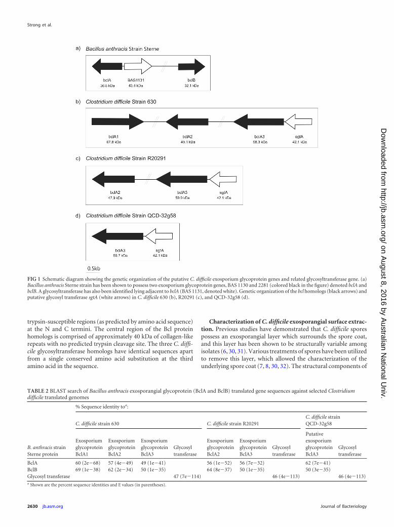
trypsin-susceptible regions (as predicted by amino acid sequence)at the N and C termini. The central region of the Bcl proteinhomologs is comprised of approximately 40 kDa of collagen-likerepeats with no predicted trypsin cleavage site. The three C. diffi-cile glycosyltransferase homologs have identical sequences apartfrom a single conserved amino acid substitution at the thirdamino acid in the sequence.
Characterization of C. difficile exosporangial surface extrac-tion. Previous studies have demonstrated that C. difficile sporespossess an exosporangial layer which surrounds the spore coat,and this layer has been shown to be structurally variable amongisolates (6, 30, 31). Various treatments of spores have been utilizedto remove this layer, which allowed the characterization of theunderlying spore coat (7, 8, 30, 32). The structural components of
FIG 1 Schematic diagram showing the genetic organization of the putative C. difficile exosporium glycoprotein genes and related glycosyltransferase gene. (a)Bacillus anthracis Sterne strain has been shown to possess two exosporium glycoprotein genes, BAS 1130 and 2281 (colored black in the figure) denoted bclA andbclB. A glycosyltransferase has also been identified lying adjacent to bclA (BAS 1131, denoted white). Genetic organization of the bcl homologs (black arrows) andputative glycosyl transferase sgtA (white arrows) in C. difficile 630 (b), R20291 (c), and QCD-32g58 (d).
TABLE 2 BLAST search of Bacillus anthracis exosporangial glycoprotein (BclA and BclB) translated gene sequences against selected Clostridiumdifficile translated genomes
B. anthracis strainSterne protein
% Sequence identity toa:
C. difficile strain 630 C. difficile strain R20291C. difficile strainQCD-32g58
ExosporiumglycoproteinBclA1
ExosporiumglycoproteinBclA2
ExosporiumglycoproteinBclA3
Glycosyltransferase
ExosporiumglycoproteinBclA2
ExosporiumglycoproteinBclA3
Glycosyltransferase
PutativeexosporiumglycoproteinBclA3
Glycosyltransferase
BclA 60 (2e�68) 57 (4e�49) 49 (1e�41) 56 (1e�52) 56 (7e�32) 62 (7e�41)BclB 69 (1e�38) 62 (2e�34) 50 (1e�35) 64 (8e�37) 50 (1e�35) 50 (3e�35)Glycosyl transferase 47 (7e�114) 46 (4e�113) 46 (4e�113)a Shown are the percent sequence identities and E values (in parentheses).
Strong et al.
2630 jb.asm.org Journal of Bacteriology
on August 8, 2016 by A
ustralian National U
niv.http://jb.asm
.org/D
ownloaded from

the exosporangial layer have yet to be characterized; thus, in thisstudy we focused on identifying and characterizing spore surface-associated protein components. Using a detergent-based extrac-tion, we removed surface-associated components from sporepreparations which had not been extensively water washed ortreated with enzymes/sonication to facilitate retention of surfacestructures. Endospores of strains 630, QCD-32g58, and R20291were incubated in detergent solutions to extract the spore surfaceproteins, and then intact spores were removed by centrifugation.The protein-containing supernatants were resolved by a 3 to 8%
NuPAGE gradient gel. The high-molecular-mass region of the gelshows diffuse banding patterns reactive with both silver stain(Fig. 2a) and glycostain (Fig. 2b), suggesting high-molecular-masscomplexes containing glycoproteins. Proteinase K digestion ofspores had only a marginal effect on the migration of this material,suggesting a more complex composition. A pattern of staining inthis region distinct from that of strain 630 was obtained forR20291 and QCD32g58 spore extracts. Glycostaining revealed areactive high-molecular-mass band in R20291 and QCD32g58 ex-tracts only.
Initially, extraction of all gel bands of molecular masses of�160 kDa was performed, and each band was digested with eithertrypsin or proteinase K and analyzed by nLC-MS/MS. No BclAprotein identification was made from these analyses. Subse-quently, the high-molecular-mass region (�160 kDa) of each lanewas excised in bands and digested with trypsin or proteinase K.The trypsin digests for all three strains did not result in any proteinidentifications by nLC-MS/MS. Analysis of the MS/MS spectrafrom proteinase K digests, however, yielded several spore surfaceprotein identifications, as indicated by the numbered annotationsin Fig. 2a, lane 3, and summarized in Table 3 for R20291 and Fig.S1 in the supplemental material for QCD-32g58.
De novo sequencing of the peptide MS/MS spectra from theproteinase K digests of gel bands 1 to 3 from surface extract ofR20291 spores revealed a number of peptides that correspondedto the putative exosporium glycoprotein BclA3 (CDR20291_3193). Further inspection of the MS/MS spectra showed peptides
FIG 2 NuPAGE gel analysis of C. difficile endospore surface protein extracts.(a) Silver-stained 3 to 8% NuPAGE; (b) Pro-Emerald Q glycostain 3 to 8%NuPAGE. Lane 1, HiMark-prestained molecular mass marker; lane 2,630�erm spore surface extract; lane 3, R20291 spore surface extract; lane 4,QCD-32g58 spore surface extract. Arrows indicate regions of the gel that wereexcised, enzymatically digested, and analyzed by nLC-MS/MS.
TABLE 3 nLC-MS/MS analysis of glycoreactive peptidesa
Gel band Accession no. (designation)Protein (molecular mass,kDa) Peptide sequence
Glycan mass, Da (monosaccharideneutral losses, Da)
1 YP_003219669 (CDR20291_3193) Exosporium glycoproteinBclA3 (59.5)
308AGLIGPTGATGV319 609 (203-203-203)145TGPTGATGADGITGP159 983 (203-203-203-374)344VGPTGATGA352b 406 (203-203)189GLIGPTGATGTPGA202 406 (203-203)278TGATGLIGPTGATGA292 1,186 (203-203-203-203-374)278TGATGLIGPTGATGA292 1,241 (203-203-203-203-429)212TGIGITGPTGATGA225c 1,184 (203-203-203-203-372)212TGIGITGPTGA222d 1,095 (203-203-203-486)309GLIGPTGATGVTGA322 1,071 (203-203-203-462)299TGVTGATGAAGLIGP313 609 (203-203-203)
2 YP_003219669 (CDR20291_3193) Exosporium glycoproteinBclA3 (59.5)
278TGATGLIGPTGATGA202 1,187 (203-203-203-203-375)191IGPTGATGTPGATGPTGA208 1,661 (203-203-203-222-203-203-424)424TGPTGATGPTGADGL438 609 (203-203-203)
3 YP_003219669 (CDR20291_3193) Exosporium glycoproteinBclA3 (59.5)
119GVTGPTGPTGPTGATGA135 609 (203-203-203)169GVTGPTGPTGATGV182 609 (203-203-203)344VGPTGATGATGADGL358 609 (203-203-203)359VGPTGPTGATGV370 609 (203-203-203)191IGPTGATGTPGATGPTGA208 609 (203-203-203)311IGPTGATGVTGADGA325 609 (203-203-203)439VGPTGATGATGL450 609 (203-203-203)391VGPTGATGATGADGV405 609 (203-203-203)359VGPTGPTGATGV370 203656ATASGLSLVNTVA668
a High-molecular-mass gel bands of R20291 spore surface extracts were digested with proteinase K, and the numbering of gel bands refers to that of Fig. 2. MS/MS spectra were denovo sequenced, and the identified peptides and observed glycan moieties are indicated.b This sequence was also found at amino acids 391 to 399, 439 to 447, and 487 to 495.c This sequence was also found at amino acids 259 to 272.d This sequence was also found at amino acids 259 to 269.
C. difficile Spore Glycosylation Patterns
July 2014 Volume 196 Number 14 jb.asm.org 2631
on August 8, 2016 by A
ustralian National U
niv.http://jb.asm
.org/D
ownloaded from

with ions that did not correspond to peptide y- or b-type ions butwere characteristic of carbohydrate-associated fragment ions. Forexample, from tandem mass spectrometry analyses of band 1 (Fig.2a), which migrated to a molecular mass of greater than 600 kDa,
the MS/MS spectrum of the putative glycoprotein peptideAGLIGPTGATGV modified with three N-acetyl hexosamine(HexNAc) moieties is shown in Fig. 3A. The glycan modificationwas observed as sequential neutral losses of 203 Da from the gly-
FIG 3 Mass spectrometry analysis of peptides from proteinase K digestion of C. difficile R20291 endospore surface extracts. (a) nLC-MS/MS spectrum of thedoubly protonated glycopeptide ion at m/z 811.8. Peptide type y and b ions were visible and gave the peptide sequence AGLIGPTGATGV, a peptide from theBclA3 protein. The spectrum was dominated in the high-m/z region by sequential neutral losses of 203 Da, with the unmodified peptide ion observed at m/z1,013.5. Combined with the observed intense glycan oxonium ion at m/z 204 and neutral losses of water to give glycan-related ions at m/z 186 and 168, thisspectrum suggested the peptide was modified with a chain of 3 HexNAc moieties. (b) nLC-MS/MS spectrum of a doubly protonated glycopeptides ion at m/z1,129. Peptide type y and b ions corresponded to a sequence of TGPTGATGADGITGP, corresponding to the BclA3 protein. The high-m/z region of the spectrumwas dominated by sequential neutral losses of 374 Da, 203 Da, 203 Da, and 203 Da. An intense oxonium ion was observed at m/z 375, and a very weak oxoniumion was observed at m/z 204 (not indicated). Glycan-related fragment ions were observed at m/z 300 and 272. (c) Peptide sequence coverage map of BclA3 proteinhomolog from spore surface protein extraction (band 1). Boldface and underlining indicate peptides modified with glycan moieties. Two of the peptides shownto be modified with glycan are shown in boldface gray text to indicate that the amino acid sequence appears in the BclA3 protein more than once. Dottedunderlining indicates a glycopeptide sequence that is common to both BclA3 and BclA2 proteins.
Strong et al.
2632 jb.asm.org Journal of Bacteriology
on August 8, 2016 by A
ustralian National U
niv.http://jb.asm
.org/D
ownloaded from

copeptide precursor ion in the high-m/z region of the MS/MSspectrum. In addition, an intense glycan oxonium ion was ob-served at m/z 204 which was common to all of the identified gly-copeptides. In addition, more complex glycosylation patternswere observed in some cases, with intense ions observed in glyco-peptide spectra that did not correspond to HexNAc residues orpeptide type y or b fragment ions (for example, oxonium ionscorresponding to masses of 486 Da, 372 Da, and 374 Da). Figure3b shows an MS/MS spectrum of the BclA peptide TGPTGATGADGITGP modified with three HexNAc moieties and an addi-tional mass of 374 Da. The sequential neutral losses suggest thatHexNAc is the linking sugar. This glycan neutral mass was alsolinked to a putative glycan oxonium ion at m/z 375. Other intenseions were also observed in the low-m/z region of this MS/MS spec-trum, including putative glycan fragment ions at m/z 300 and 272.The absence of potential N-linked glycosylation sites suggestedthe glycans are O-linked through threonine residues within eachpeptide. Observed sequential neutral losses of 203 Da in the high-m/z region of the spectrum and the presence of an intense ioncorresponding to the unmodified form of the peptide suggest theglycan is composed of oligosaccharide chains attached to a single-amino-acid residue in each identified glycopeptide.
Table 3 shows the complete list of surface protein peptides andglycopeptides identified from each of the annotated bands indi-cated in Fig. 2a. The unknown glycans varied in observed massfrom 281 to 486 Da, and it is possible that these are modifiedHexNAc moieties. Tandem mass spectrometry analysis of protei-nase K digests of bands 2 and 3 showed BclA3 glycopeptides mod-ified with chains of HexNAc moieties, predominantly in trimers.In these cases, only peptides modified with HexNAc moieties wereobserved at detectable levels.
All of the identified glycopeptides reside within the central col-lagen-like repeating domain of the BclA3 protein; however, sam-ple limitations prohibited the identification of the precise sites ofmodification within the respective peptides or further character-ization of the glycan moieties. The central collagen-like repeatdomains of the putative exosporial proteins contained somenonunique regions, which resulted in the identification of multi-ple glycopeptides with amino acid sequences that repeat withinthe protein sequence (for example, TGIGITGPTGA occurs inBclA3 at residues 212 to 222 and 259 to 269). One of the identifiedpeptides, which also happens to be repeated in BclA3 four times, isalso common to both BclA3 and BclA2 (VGPTGATGA). Thenonspecific cleavage by proteinase K produced a number of gly-copeptides with overlapping sequences. Furthermore, multipleglycopeptides were identified that possessed identical peptide se-quences but different glycans.
As indicated above, strains R20291 and QCD-32g58 showedsimilar protein staining patterns for both silver stain and glyco-stains, and nLC-MS/MS analysis also showed that the Bcl proteinof QCD-32g58 is similarly glycosylated predominantly with Hex-NAc moieties (see Fig. S1 in the supplemental material). nLC-MS/MS analysis of the gel digests from strain 630, which showedsignificantly different staining patterns in the high-molecular-mass region of the gel compared to the other two strains, did notyield any protein or glycoprotein identifications.
Anti-�-O-GlcNAc reactivity of C. difficile spores. As the mostabundant glycan modification observed in the MS analysis ofspore surface-extracted material was shown to have a mass corre-sponding to an N acetyl-hexosamine moiety, we next examined
the ability of spores to bind to an O-linked N-acetylglucosamine(�-O-GlcNAc) antibody. A monoclonal antibody (MAb) whichrecognizes O-GlcNAc in a �-O-glycosidic linkage to both threo-nine and serine was utilized in immunofluorescence experimentswith intact spores from a number of C. difficile clinical isolates(Fig. 4a). This antibody had been used previously to demonstratethe presence of �-O-GlcNAc attached to serine and threonineresidues of Listeria monocytogenes flagellin (33).
When the �-O GlcNAc antibody was used in immunofluores-cence reactions with spores of R20291 and 630�erm, both sporepreparations reacted strongly with the antibody. Interestingly,distinct patterns of reactivity with the spore surface were observedby this immunofluorescence method for each strain (Fig. 4a).With R20291 spores, anti-�-O-GlcNAc was uniformly reactiveover the entire spore surface, while with strain 630�erm, anti-�-O-GlcNAc reactivity was restricted to the poles of the spores withonly limited labeling of the central surface (Fig. 4a, arrows). Veg-etative cells of both strains showed no reactivity with anti-�-O-GlcNAc (see Fig. S2 in the supplemental material). To confirm theconservation of �-O-GlcNAc on the surface of multiple C. difficilestrains, a range of spores from different ribotypes and geographiclocations were also tested for anti-�-O-GlcNAc binding. The re-activity pattern observed for R20291 spores was found to be con-served in all strains examined, with the only exception beingspores of 630�erm (Fig. 4a). Any unstained cells in the imageswere either immature spores or cell debris from the washing pro-cess. DAPI binding was observed only with vegetative cells andimmature phase-dark spores; phase-bright spores were consid-ered mature.
RT-PCR of CD3350-bclA3 gene locus. As indicated in Fig. 1,CD3350 (B. anthracis exosporangial glycosyltransferase gene ho-molog) and bclA3 lie immediately adjacent to each other and areorientated in the same direction on the chromosome in both 630and R20291 strains. The two genes are separated by only a shortintergenic region, suggesting they form a single transcriptionalunit. Primers which amplified across this intergenic region wereused to determine if the genes were cotranscribed. RNA samplesextracted from C. difficile 630 cells were subjected to reverse tran-scription, and an amplification product of 257 bp linking CD3350and bclA3 was obtained, confirming cotranscription of these twogenes (see Fig. S3 in the supplemental material). PCRs using thesame primers and total RNA that had not undergone a reversetranscriptase reaction did not yield any amplification product,demonstrating that the RNA was free of contaminating DNA.
Mutagenesis of CD3350 and CDR3194 and spore character-ization. We next generated insertionally inactivated glycosyl-transferase mutants in strains 630�erm and R20291 (�CD3350and �CDR3194) by using ClosTron technology as previously de-scribed (26). Insertion of the TargeTron Erm resistance markerwas confirmed by PCR using primers flanking the gene of interestand with primers specific to the TargeTron Erm resistance marker(data not shown). Vegetative cell growth of both �CD3350 and�CDR3194 was unchanged compared to that of their respectiveparent strains, and motility was unaffected (data not shown). Im-munofluorescence of spores with anti-�-O-GlcNAc antibody re-vealed a complete loss of reactivity for both �CD3350 and�CDR3194 compared to the respective parent strains (Fig. 4b).The percentage of wild-type spores compared to mutant phase-bright spores reacting with anti-�-O-GlcNAc antibody was quan-
C. difficile Spore Glycosylation Patterns
July 2014 Volume 196 Number 14 jb.asm.org 2633
on August 8, 2016 by A
ustralian National U
niv.http://jb.asm
.org/D
ownloaded from

tified microscopically and shown to be 80 to 95% compared to lessthan 1% for the respective mutants (Fig. 4c).
Both �CDR3194 and �CD3350 strains were complementedwith wild-type copies of CD3350 using pRPF185 (27), as evi-denced by both Western blotting and immunofluorescence stud-ies (Fig. 5; also see Fig. S4 in the supplemental material). As can beseen in Fig. 5, lanes 2 and 5, a positive reaction was observed inWestern blotting with spore surface extracts from both R20291and 630�erm spores, respectively. Spore extracts of R20291 dis-played reactivity with the region of gel corresponding to band 4(approximately 400 kDa) from MS analysis. In addition, a secondstrongly reactive band migrating at a molecular mass of approxi-mately 170 kDa on 3 to 8% NuPAGE gel was observed, althoughwe were unable to identify peptides from a proteinase K digestionof this region of the gel by MS analysis. For strain 630�erm, no
reactivity was observed in the corresponding higher-molecular-mass region of the gel, and a series of three distinct reactive bandswas observed at approximately 170 kDa. All reactivity was lost inCD3350 and CDR3194 mutant strains, while the strain-specificpattern of reactivity was restored upon complementation (Fig. 5;also see Fig. S4). On the basis of these results, we now propose thatthis gene be renamed sgtA (spore glycosyl transferase).
Characterization of �sgtA spore surface extract. In parallelwith the spore surface protein extracts of the wild-type strain,spore surface extracts of the R20291�sgtA mutant strain were alsoprepared and analyzed by 3 to 8% NuPAGE Tris-acetate gels toresolve high-molecular-mass material. The protein-stained gelshows a diffuse area of staining at 460 kDa and greater; however,the distinct 600-kDa band was not observed. Similarly, glyco-staining of the same gel showed no detectable reactivity at 600
FIG 4 Immunofluorescence of anti-�-O-GlcNAc binding to spores. (a) Wild-type spores of C. difficile strains from a range of ribotypes and geographicallocations. For 630�erm, GlcNAc binding at poles is marked with arrows. (b) sgtA mutant spores of strains R20291 and 630�erm. First row, merged image ofphase contrast, DAPI, and FITC; 2nd row, DAPI channel only; 3rd row, FITC channel only; 4th row, phase-contrast image only. GlcNAc was visualized withmouse anti-�-O-GlcNAc and anti-mouse IgM-FITC conjugate. (c) Percentage of spores reacting with anti-�-O-GlcNAc after 7 days of growth. At least 100spores were counted in triplicate on three independent occasions for anti-�-O-GlcNAc binding to the surface, as analyzed by immunofluorescence microscopy.
Strong et al.
2634 jb.asm.org Journal of Bacteriology
on August 8, 2016 by A
ustralian National U
niv.http://jb.asm
.org/D
ownloaded from

kDa (see Fig. S5 in the supplemental material). In contrast to MSstudies of gel bands of R20291 spore surface extracts, which iden-tified peptides/glycopeptides in proteinase K digests, the equiva-lent region of the NuPAGE gel of the spore surface protein extrac-tion of �sgtA did not yield any peptide or glycopeptideidentifications. In addition, to date, our analyses of lower-molec-ular-mass protein bands from spore surface extracts have shownno evidence of unglycosylated BclA3.
Resistance of R20291�sgtA spores. As the more clinically rel-evant strain and as shown by immunofluorescence to be a morerepresentative strain of C. difficile spore morphology, phenotypicassays were undertaken on R20291 wild-type spores and �sgtAspores. The heat resistance of spores was examined as previouslydescribed (7). When incubated at 80°C for 20 min, �sgtA sporesshowed significantly lower survival rates than the parent R20291spores (Fig. 6). The susceptibility of spores to 70% ethanol and 250�g/ml lysozyme was also examined, but no significant differencewas observed between wild-type and �sgtA spores (see Fig. S6 inthe supplemental material).
Role of sgtA in adherence and internalization of macrophagecells. To gain insight into a possible biological role for the SgtAglycosyltransferase, we next investigated the ability of spores toadhere to and be internalized by the J774A.1 macrophage cell line
(ATCC TIB-106). Spores were counted based on association withJ774A.1 cells and counted as adhered if green/red and internalizedif red. Spores not associated with cells were ignored, as were anyremaining vegetative cells based on rod shape. As can be seen inFig. 7, it is clear that adherence and internalization of J774A.1macrophage cells by C. difficile R20291 spores was affected follow-ing inactivation of the sgtA gene, with significantly greater num-bers of �sgtA spores being internalized compared to the wild type.
DISCUSSION
This study presents, to our knowledge, the first characterization ofglycoproteins from C. difficile spores and provides direct evidencedemonstrating that BclA3 is a glycoprotein which is glycosylatedwith chains of �-O-linked GlcNAc as well as with additional gly-cans of novel masses. Bioinformatic analysis of clostridial ge-nomes revealed the presence of Bacillus anthracis exosporangialbclA and bclB gene homologs. In addition, immediately upstreamof the B. anthracis bclB gene homologs in all C. difficile strains,and shown in this study to be cotranscribed with bclA3, lies ahomolog of the neighboring B. anthracis glycosyltransferase gene(BAS1131). While the predicted molecular mass of BclA3 basedon the translated amino acid sequence is 57 kDa, identificationof BclA3 was made only in higher-molecular-mass material foundto be migrating in NuPAGE gels at masses corresponding to �460kDa and which stained in a diffuse pattern by silver staining. Inaddition, reactivity with glycostain in this region of the gel wasobserved, suggesting that the higher-molecular-mass materialalso contained carbohydrate. After repeated attempts to identifythe composition of the high-molecular-mass material using tryp-sin digestion of selected regions of the gel, we were finally success-ful in obtaining peptide identifications following extensive protei-nase K digestion of the gel bands. This nLC-MS/MS analysisidentified BclA3 peptides and also provided the first evidence thatthis protein is a glycoprotein. In a fashion similar to that of B.anthracis, it appears that the BclA3 protein of C. difficile is glyco-sylated with predominantly novel tri- or pentasaccharide oligo-saccharides composed of chains of N-acetyl hexosamine sugars,which may be capped with novel glycan moieties. Due to samplelimitations and capping glycan heterogeneity, we were unable to
FIG 5 Restoration of anti-GlcNAc reactivity through complementation. Westernblot of cultures grown on plates for 72 h and run on 3 to 8% Nu-PAGE gel.Complemented strains were induced with 500 ng anhydrotetracycline. Lane 1,HiMark (Invitrogen); lane 2, R20291; lane 3, R20291�CDR3194; lane 4,R20291�3194p3350; lane 5, 630�erm; lane 6, 630�3350; lane 7, 630�3350p3350.
FIG 6 Resistance of R20291 wild-type and �sgtA spores to 80°C for 20 min.Spores were incubated for 20 min in a water bath at 80°C, and then the number ofCFU/ml was determined. Percent survival was calculated by comparing inocula topostheat treatment samples. P � 0.0001 by t test with Welch’s correction.
FIG 7 Adherence and invasion of J774A.1 macrophage cells. Shown are thepercentages of spores adhering to or internalized into J774A.1 macrophagesafter 30 min of incubation at 37°C under 5% CO2. Percentages were calculatedbased on known MOIs and final adhered (green/red) or internalized (red)spores. Fifty J774A.1 cells were counted in triplicate on three independentoccasions. Statistical analysis was performed with a t test with Welch’s correc-tion (*, P � 0.05; ***, P � 0.0001).
C. difficile Spore Glycosylation Patterns
July 2014 Volume 196 Number 14 jb.asm.org 2635
on August 8, 2016 by A
ustralian National U
niv.http://jb.asm
.org/D
ownloaded from

determine the precise molecular structure, although it is clearfrom glycan component neutral masses that the structural com-position of the C. difficile BclA3 glycan is quite distinct from thatpreviously reported for B. anthracis (16). Gel migration character-istics do suggest that C. difficile BclA3 monomers from R20291and QCD-32g58 form some sort of stable, higher-molecular-masscomplex, which is resistant to denaturation by heating and deter-gents. In contrast, spore surface extracts of C. difficile 630 appearto form distinct, lower-molecular-mass complexes which do notappear to contain any BclA3 protein. In addition, spores of 630produced a distinct pattern of �-O-linked GlcNAc reactivity byimmunofluorescence studies, and the significance of these distinctdifferences in spore structure between isolates remains to be de-termined. However, �-O-linked GlcNAc reactivity of spores froma number of clinical isolates demonstrates, for the first time, theconserved nature of this posttranslational modification on C. dif-ficile spores. Insertional inactivation of the glycosyltransferasegene, sgtA (CD3350 and CDR3194), provided direct evidence for arole of the glycosyltransferase enzyme in this spore surface �-O-linked GlcNAc reactivity as well as in the production of glycosy-lated BclA3. While previous work examining sporulation-relatedgene expression demonstrated that the bclA3 gene and the adja-cent glycosyltransferase gene (sgtA) were activated by the sporu-lation sigma factor, k (34, 35), this is the first study to link the sgtAgene to a specific spore glycan-associated function.
A role for surface-associated bacterial glycans in host interac-tions is well documented for many bacterial species (36–40). Inthe current study, we show that glycans on the spore surface im-part resistance of spores to heat treatment as well as appear to playa role in macrophage interactions. In contrast to a previous study,where removal of the exosporangial layer by sonication did notaffect binding and internalization of C. difficile spores by Raw264.7 peritoneal macrophage cells (31, 41), in the current studythe loss of the ability to glycosylate surface-associated proteinsdoes appear to affect uptake by macrophages. It should be noted,however, that distinct strains of C. difficile and different macro-phage cell lines were used in each study, which may explain thedifferent results. It is clear, however, that C. difficile spores dointeract and are internalized by macrophage cells and that the roleof surface-expressed glycans now can be more fully explored.While an ability of C. difficile spores to avoid uptake by macro-phages might be advantageous during infection, further studieswill be required to define the precise role of glycan components inmacrophage interactions. It should be noted that exosporiumstructures of B. anthracis appear to mask epitopes recognized bymacrophages which are involved in induction of cytokines (42).
During the preparation of the manuscript, a detailed charac-terization of BclA1 from C. difficile 630 spores was reported (43). Itappears that the BclA1 protein forms high-molecular-mass com-plexes in extracts of 630 spores in a similar fashion. It is significantthat the genomes of strains R20291 and QCD-32g58, which wereexamined in the current study, encode a truncated bclA1 gene,which may explain why this protein was never identified in ex-tracts. In our analyses of the high-molecular-mass complexes of C.difficile 630 spores, we did not identify any BclA1 peptides and/orglycopeptides, suggesting it forms a distinct complex. Both thecurrent study and the recent BclA1 study also present evidence ofadditional glycoreactive bands in spore extracts, which will likelybe important in our comprehensive understanding of the glyco-biology of C. difficile spores.
While known to be recalcitrant to proteolytic digestion andstructural characterization, the spores of Gram-positive bacterialpathogens have gained considerable attention in recent years. Theinitial demonstration that the exosporangial proteins of Bacillusanthracis were glycoproteins which carried a pentasaccharide gly-can containing the novel sugar, anthrose was the first example ofglycoproteins as a part of spore biology. This study has demon-strated that spores of a second important Gram-positive patho-gen, C. difficile, also carry novel glycoproteins on surface-associ-ated structures. Defining the role of these structures in theinfectious process, as well as exploiting their potential in thera-peutic and diagnostic applications, now can be further pursued.This work adds another example to the constantly expanding dataset of protein glycosylation systems in bacterial pathogens.
ACKNOWLEDGMENTS
We thank K. Siklenka, S. O’Hara, and M. David for preliminary gel and massspectrometry data analyses. We thank R. Fagan and N. Fairweather for provisionof pRPF185 plasmid, A. Dascal for provision of C. difficile QCD-32g58, B.Wren for C. difficile strains R20291, BI-6, CD20, CF5, and M68, and N.Minton for C. difficile 630�erm and the ClosTron mutagenesis system.
This work was funded by the National Research Council Canada.
REFERENCES1. Rupnik M, Wilcox MH, Gerding DN. 2009. Clostridium difficile infec-
tion: new developments in epidemiology and pathogenesis. Nat. Rev. Mi-crobiol. 7:526 –536. http://dx.doi.org/10.1038/nrmicro2164.
2. Freeman J, Bauer MP, Baines SD, Corver J, Fawley WN, Goorhuis B,Kuijper EJ, Wilcox MH. 2010. The changing epidemiology of Clostrid-ium difficile infections. Clin. Microbiol. Rev. 23:529 –549. http://dx.doi.org/10.1128/CMR.00082-09.
3. Ananthakrishnan AN. 2011. Clostridium difficile infection: epidemiol-ogy, risk factors and management. Nat. Rev. Gastroenterol. Hepatol.8:17–26. http://dx.doi.org/10.1038/nrgastro.2010.190.
4. Gerding DN, Muto CA, Owens RC, Jr. 2008. Measures to control andprevent Clostridium difficile infection. Clin. Infect. Dis. 46(Suppl 1):S43–S49. http://dx.doi.org/10.1086/521861.
5. Lawley TD, Clare S, Walker AW, Goulding D, Stabler RA, Croucher N,Mastroeni P, Scott P, Raisen C, Mottram L, Fairweather NF, Wren BW,Parkhill J, Dougan G. 2009. Antibiotic treatment of clostridium difficilecarrier mice triggers a supershedder state, spore-mediated transmission,and severe disease in immunocompromised hosts. Infect. Immun. 77:3661–3669. http://dx.doi.org/10.1128/IAI.00558-09.
6. Lawley TD, Croucher NJ, Yu L, Clare S, Sebaihia M, Goulding D, PickardDJ, Parkhill J, Choudhary J, Dougan G. 2009. Proteomic and genomiccharacterization of highly infectious Clostridium difficile 630 spores. J. Bac-teriol. 191:5377–5386. http://dx.doi.org/10.1128/JB.00597-09.
7. Permpoonpattana P, Phetcharaburanin J, Mikelsone A, Dembek M,Tan S, Brisson MC, La Ragione R, Brisson AR, Fairweather N, HongHA, Cutting SM. 2013. Functional characterization of Clostridium diffi-cile spore coat proteins. J. Bacteriol. 195:1492–1503. http://dx.doi.org/10.1128/JB.02104-12.
8. Permpoonpattana P, Tolls EH, Nadem R, Tan S, Brisson A, CuttingSM. 2011. Surface layers of Clostridium difficile endospores. J. Bacteriol.193:6461– 6470. http://dx.doi.org/10.1128/JB.05182-11.
9. Paredes-Sabja D, Bond C, Carman RJ, Setlow P, Sarker MR. 2008.Germination of spores of Clostridium difficile strains, including isolatesfrom a hospital outbreak of Clostridium difficile-associated disease(CDAD). Microbiology 154:2241–2250. http://dx.doi.org/10.1099/mic.0.2008/016592-0.
10. Burns DA, Heap JT, Minton NP. 2010. SleC is essential for germinationof Clostridium difficile spores in nutrient-rich medium supplementedwith the bile salt taurocholate. J. Bacteriol. 192:657– 664. http://dx.doi.org/10.1128/JB.01209-09.
11. Burns DA, Heeg D, Cartman ST, Minton NP. 2011. Reconsidering thesporulation characteristics of hypervirulent Clostridium difficile BI/NAP1/027. PLoS One 6:e24894. http://dx.doi.org/10.1371/journal.pone.0024894.
Strong et al.
2636 jb.asm.org Journal of Bacteriology
on August 8, 2016 by A
ustralian National U
niv.http://jb.asm
.org/D
ownloaded from

12. Abhyankar W, Hossain AH, Djajasaputra A, Permpoonpattana P, TerBeek A, Dekker HL, Cutting SM, Brul S, de Koning LJ, de Koster CG.2013. In pursuit of protein targets: proteomic characterization of bacterialspore outer layers. J. Proteome Res. 12:4507– 4521. http://dx.doi.org/10.1021/pr4005629.
13. Charlton S, Moir AJ, Baillie L, Moir A. 1999. Characterization of theexosporium of Bacillus cereus. J. Appl. Microbiol. 87:241–245. http://dx.doi.org/10.1046/j.1365-2672.1999.00878.x.
14. Redmond C, Baillie LW, Hibbs S, Moir AJ, Moir A. 2004. Identificationof proteins in the exosporium of Bacillus anthracis. Microbiology 150:355–363. http://dx.doi.org/10.1099/mic.0.26681-0.
15. Sylvestre P, Couture-Tosi E, Mock M. 2002. A collagen-like surface glycoproteinis a structural component of the Bacillus anthracis exosporium. Mol. Microbiol.45:169–178. http://dx.doi.org/10.1046/j.1365-2958.2000.03000.x.
16. Daubenspeck JM, Zeng H, Chen P, Dong S, Steichen CT, Krishna NR,Pritchard DG, Turnbough CL, Jr. 2004. Novel oligosaccharide sidechains of the collagen-like region of BclA, the major glycoprotein of theBacillus anthracis exosporium. J. Biol. Chem. 279:30945–30953. http://dx.doi.org/10.1074/jbc.M401613200.
17. Steichen C, Chen P, Kearney JF, Turnbough CL, Jr. 2003. Identificationof the immunodominant protein and other proteins of the Bacillus an-thracis exosporium. J. Bacteriol. 185:1903–1910. http://dx.doi.org/10.1128/JB.185.6.1903-1910.2003.
18. Waller LN, Stump MJ, Fox KF, Harley WM, Fox A, Stewart GC,Shahgholi M. 2005. Identification of a second collagen-like glycoproteinproduced by Bacillus anthracis and demonstration of associated spore-specific sugars. J. Bacteriol. 187:4592– 4597. http://dx.doi.org/10.1128/JB.187.13.4592-4597.2005.
19. Tamborrini M, Holzer M, Seeberger PH, Schurch N, Pluschke G. 2010.Anthrax spore detection by a Luminex assay based on monoclonal anti-bodies that recognize anthrose-containing oligosaccharides. Clin. VaccineImmunol. 17:1446 –1451. http://dx.doi.org/10.1128/CVI.00205-10.
20. Dong S, McPherson SA, Tan L, Chesnokova ON, Turnbough CL, Jr,Pritchard DG. 2008. Anthrose biosynthetic operon of Bacillus anthracis.J. Bacteriol. 190:2350 –2359. http://dx.doi.org/10.1128/JB.01899-07.
21. Mehta AS, Saile E, Zhong W, Buskas T, Carlson R, Kannenberg E, Reed Y,Quinn CP, Boons GJ. 2006. Synthesis and antigenic analysis of the BclAglycoprotein oligosaccharide from the Bacillus anthracis exosporium. Chem-istry 12:9136–9149. http://dx.doi.org/10.1002/chem.200601245.
22. Blum H, Beier H, Gross HJ. 1987. Improved silver staining of plantproteins, RNA and DNA in polyacrylamide gels. Electrophoresis 8:93–99.http://dx.doi.org/10.1002/elps.1150080203.
23. Gharahdaghi F, Weinberg CR, Meagher DA, Imai BS, Mische SM. 1999.Mass spectrometric identification of proteins from silver-stained poly-acrylamide gel: a method for the removal of silver ions to enhance sensi-tivity. Electrophoresis 20:601– 605.
24. Fulton KM, Zhao X, Petit MD, Kilmury SL, Wolfraim LA, House RV,Sjostedt A, Twine SM. 2011. Immunoproteomic analysis of the humanantibody response to natural tularemia infection with type A or type Bstrains or LVS vaccination. Int. J. Med. Microbiol. 301:591– 601. http://dx.doi.org/10.1016/j.ijmm.2011.07.002.
25. Heap JT, Cartman ST, Kuehne SA, Cooksley C, Minton NP. 2010.ClosTron-targeted mutagenesis. Methods Mol. Biol. 646:165–182. http://dx.doi.org/10.1007/978-1-60327-365-7_11.
26. Heap JT, Kuehne SA, Ehsaan M, Cartman ST, Cooksley CM, Scott JC,Minton NP. 2010. The ClosTron: mutagenesis in Clostridium refined andstreamlined. J. Microbiol. Methods 80:49 –55. http://dx.doi.org/10.1016/j.mimet.2009.10.018.
27. Fagan RP, Fairweather NF. 2011. Clostridium difficile has two paralleland essential Sec secretion systems. J. Biol. Chem. 286:27483–27493. http://dx.doi.org/10.1074/jbc.M111.263889.
28. Aubry A, Hussack G, Chen W, KuoLee R, Twine SM, Fulton KM, FooteS, Carrillo CD, Tanha J, Logan SM. 2012. Modulation of toxin produc-tion by the flagellar regulon in Clostridium difficile. Infect. Immun. 80:3521–3532. http://dx.doi.org/10.1128/IAI.00224-12.
29. Todd SJ, Moir AJ, Johnson MJ, Moir A. 2003. Genes of Bacillus cereusand Bacillus anthracis encoding proteins of the exosporium. J. Bacteriol.185:3373–3378. http://dx.doi.org/10.1128/JB.185.11.3373-3378.2003.
30. Escobar-Cortes K, Barra-Carrasco J, Paredes-Sabja D. 2013. Proteasesand sonication specifically remove the exosporium layer of spores of Clos-tridium difficile strain 630. J. Microbiol. Methods 93:25–31. http://dx.doi.org/10.1016/j.mimet.2013.01.016.
31. Paredes-Sabja D, Cofre-Araneda G, Brito-Silva C, Pizarro-Guajardo M,
Sarker MR. 2012. Clostridium difficile spore-macrophage interactions:spore survival. PLoS One 7:e43635. http://dx.doi.org/10.1371/journal.pone.0043635.
32. Barra-Carrasco J, Olguin-Araneda V, Plaza-Garrido A, Miranda-CardenasC, Cofre-Araneda G, Pizarro-Guajardo M, Sarker MR, Paredes-Sabja D.2013. The Clostridium difficile exosporium cysteine (CdeC)-rich protein isrequired for exosporium morphogenesis and coat assembly. J. Bacteriol. 195:3863–3875. http://dx.doi.org/10.1128/JB.00369-13.
33. Schirm M, Kalmokoff M, Aubry A, Thibault P, Sandoz M, Logan SM.2004. Flagellin from Listeria monocytogenes is glycosylated with beta-O-linked N-acetylglucosamine. J. Bacteriol. 186:6721– 6727. http://dx.doi.org/10.1128/JB.186.20.6721-6727.2004.
34. Fimlaid KA, Bond JP, Schutz KC, Putnam EE, Leung JM, Lawley TD,Shen A. 2013. Global analysis of the sporulation pathway of Clostridiumdifficile. PLoS Genet. 9:e1003660. http://dx.doi.org/10.1371/journal.pgen.1003660.
35. Saujet L, Pereira FC, Serrano M, Soutourina O, Monot M, ShelyakinPV, Gelfand MS, Dupuy B, Henriques AO, Martin-Verstraete I. 2013.Genome-wide analysis of cell type-specific gene transcription duringspore formation in Clostridium difficile. PLoS Genet. 9:e1003756. http://dx.doi.org/10.1371/journal.pgen.1003756.
36. Szymanski CM, Burr DH, Guerry P. 2002. Campylobacter protein gly-cosylation affects host cell interactions. Infect. Immun. 70:2242–2244.http://dx.doi.org/10.1128/IAI.70.4.2242-2244.2002.
37. Scott AE, Twine SM, Fulton KM, Titball RW, Essex-Lopresti AE, AtkinsTP, Prior JL. 2011. Flagellar glycosylation in Burkholderia pseudomalleiand Burkholderia thailandensis. J. Bacteriol. 193:3577–3587. http://dx.doi.org/10.1128/JB.01385-10.
38. Fletcher CM, Coyne MJ, Villa OF, Chatzidaki-Livanis M, Comstock LE.2009. A general O-glycosylation system important to the physiology of amajor human intestinal symbiont. Cell 137:321–331. http://dx.doi.org/10.1016/j.cell.2009.02.041.
39. Lindenthal C, Elsinghorst EA. 1999. Identification of a glycoprotein produced byenterotoxigenic Escherichia coli. Infect. Immun. 67:4084–4091.
40. Benz I, Schmidt MA. 2001. Glycosylation with heptose residues me-diated by the aah gene product is essential for adherence of the AIDA-Iadhesin. Mol. Microbiol. 40:1403–1413. http://dx.doi.org/10.1046/j.1365-2958.2001.02487.x.
41. Paredes-Sabja D, Sarker MR. 2012. Adherence of Clostridium difficilespores to Caco-2 cells in culture. J. Med. Microbiol. 61:1208 –1218. http://dx.doi.org/10.1099/jmm.0.043687-0.
42. Basu S, Kang TJ, Chen WH, Fenton MJ, Baillie L, Hibbs S, Cross AS. 2007.Role of Bacillus anthracis spore structures in macrophage cytokine responses. In-fect. Immun. 75:2351–2358. http://dx.doi.org/10.1128/IAI.01982-06.
43. Pizarro-Guajardo M, Olguin-Araneda V, Barra-Carrasco J, Brito-SilvaC, Sarker MR, Paredes-Sabja D. 2013. Characterization of the collagen-like exosporium protein, BclA1, of Clostridium difficile spores. Anaerobe25:18 –30. http://dx.doi.org/10.1016/j.anaerobe.2013.11.003.
44. Hussain HA, Roberts AP, Mullany P. 2005. Generation of an erythro-mycin-sensitive derivative of Clostridium difficile strain 630 (630Del-taerm) and demonstration that the conjugative transposon Tn916DeltaEenters the genome of this strain at multiple sites. J. Med. Microbiol. 54:137–141. http://dx.doi.org/10.1099/jmm.0.45790-0.
45. Stabler RA, He M, Dawson L, Martin M, Valiente E, Corton C, LawleyTD, Sebaihia M, Quail MA, Rose G, Gerding DN, Gibert M, PopoffMR, Parkhill J, Dougan G, Wren BW. 2009. Comparative genome andphenotypic analysis of Clostridium difficile 027 strains provides insightinto the evolution of a hypervirulent bacterium. Genome Biol. 10:R102.http://dx.doi.org/10.1186/gb-2009-10-9-r102.
46. Forgetta V, Oughton MT, Marquis P, Brukner I, Blanchette R, Haub K,Magrini V, Mardis ER, Gerding DN, Loo VG, Miller MA, Mulvey MR,Rupnik M, Dascal A, Dewar K. 2011. Fourteen-genome comparisonidentifies DNA markers for severe-disease-associated strains of Clostrid-ium difficile. J. Clin. Microbiol. 49:2230 –2238. http://dx.doi.org/10.1128/JCM.00391-11.
47. He M, Sebaihia M, Lawley TD, Stabler RA, Dawson LF, Martin MJ,Holt KE, Seth-Smith HM, Quail MA, Rance R, Brooks K, Churcher C,Harris D, Bentley SD, Burrows C, Clark L, Corton C, Murray V, RoseG, Thurston S, van Tonder A, Walker D, Wren BW, Dougan G, ParkhillJ. 2010. Evolutionary dynamics of Clostridium difficile over short andlong time scales. Proc. Natl. Acad. Sci. U. S. A. 107:7527–7532. http://dx.doi.org/10.1073/pnas.0914322107.
C. difficile Spore Glycosylation Patterns
July 2014 Volume 196 Number 14 jb.asm.org 2637
on August 8, 2016 by A
ustralian National U
niv.http://jb.asm
.org/D
ownloaded from


![Identification and Characterization of Arabidopsis …...Identification and Characterization of Arabidopsis Seed Coat Mucilage Proteins1[OPEN] Allen Yi-Lun Tsai2, Tadashi Kunieda3,](https://static.fdocuments.net/doc/165x107/5e93c006afc9c34a843ac831/identiication-and-characterization-of-arabidopsis-identiication-and-characterization.jpg)