Identification Regulation 1,25-Dihydroxyvitamin D3Receptor ...€¦ · Identification andRegulation...
13
Identification and Regulation of 1,25-Dihydroxyvitamin D3 Receptor Activity and Biosynthesis of 1 ,25-Dihydroxyvitamin D3 Studies in Cultured Bovine Aortic Endothelial Cells and Human Dermal Capillaries Jurgen Merke,* Petra Milde,* Sabina Lewicka,l Ulrike Hugel,* Gunter Klaus,* David J. Mangelsdorf,"1 Mark R. Haussler,11 Ernst W. Rauterberg,t and Eberhard Ritz* Departments of*Internal Medicine, *Immunology, and §Pharmacology, University ofHeidelberg, Federal Republic of Germany; and I'Department of Biochemistry, University ofArizona, Tucson, Arizona 85724 Abstract Because 1,25-dihydroxyvitamin D3 (1,25(OH)2D3) has been shown to play roles in both proliferation and differentiation of novel target cells, the potential expression of 1,25(OH)2D3 receptor (VDR) activity was investigated in cultured bovine aortic endothelial cells (BAEC). Receptor binding assays per- formed on nuclear extracts of BAEC revealed a single class of specific, high-affinity VDR that displayed a 4.5-fold increase in maximal ligand binding (Nm.x) in rapidly proliferating BAEC compared with confluent, density-arrested cells. When confluent BAEC were incubated with activators of protein ki- nase C (PKC), N., increased 2.5-fold within 6-24 h and this upregulation was prevented by sphingosine, an inhibitor of PKC, as well as by actinomycin D or cycloheximide. Immuno- histochemical visualization using a specific MAb disclosed nuclear localized VDR in venular and capillary endothelial cells of human skin biopsies, documenting the expression of VDR, in vivo, and validating the BAEC model. Finally, addi- tional experiments indicated that BAEC formed the 1,25(OH)2D3 hormonal metabolite from 25(OH)D3 substrate, in vitro, and growth curves of BAEC maintained in the pres- ence of 10-8 M 1,25(OH)2D3 showed a 36% decrease in satura- tion density. These data provide evidence for the presence of a vitamin D microendocrine system in endothelial cells, consist- ing of the VDR and a la-hydroxylase enzyme capable of pro- ducing 1,25(OH)2D3. That both components of this system are coordinately regulated, and that BAEC respond to the 1,25(OH)2D3 hormone by modulating growth kinetics, sug- gests the existence of a vitamin D autocrine loop in endothe- lium that may play a role in the development and/or functions of this pathophysiologically significant cell population. Introduction It is well established that 1,25-dihydroxyvitamin D3 (1,25(OH)2D3)' is a crucial hormone in Ca2' homeostasis (1). This paper was presented in part at the Seventh Annual Meeting of the American Society for Bone and Mineral Research (15-18 June 1986; J. Bone Miner. Res. 3:[Suppl. 1] Abstr. 398) and at the Endocrine Society 70th Annual Meeting (8-11 June 1988; Endocrinology. 122[Suppl. 1]:137. [Abstr. 466]). Address reprint requests to Dr. Jurgen Merke, Department of In- ternal Medicine, University of Heidelberg, Bergheimer Strasse 58, D-6900 Heidelberg, Federal Republic of Germany. Receivedfor publication 2 December 1988. 1. Abbreviations used in this paper: BAEC, bovine aortic endothelial cells; DiC8, sn- 1,2-dioctanoylglycerol; MAb 9A7y2b, MAb against the Most, if not all, of the biological actions of 1,25(OH)2D3 are believed to be mediated by a high-affinity nuclear receptor (VDR) for the vitamin D hormone (2). Recent studies of cul- tured human promyelocytic leukemia cells (3), cultured human macrophages (4, 5), skin cells (6, 7) and other cells (8-12) have disclosed several new biological functions of 1,25(OH)2D3. These studies indicate that 1,25(OH)2D3 plays potential roles in cell proliferation/differentiation (13) and also is biosynthesized in several of its peripheral target cells (4-6) in addition to the traditional renal site of formation. Because endothelial cells are a dynamic tissue with sponta- neous or injury-dependent cell renewal and expression of spe- cific cell functions at the blood/vessel-wall interface, these cells were examined to determine whether they are potential targets for 1,25(OH)2D3. Initially, the possible presence of specific binding sites for 1,25(OH)2D3 was probed using cultured bo- vine aortic endothelial cells (BAEC) as a model. When recep- tors for 1,25(OH)2D3 were observed, the following hypotheses were tested: (a) that the growth state of BAEC may be asso- ciated with changes in VDR activity; (b) that BAEC differen- tiation induced by activators of protein kinase C (PKC) (14-17) may be associated with VDR regulation; (c) that growth parameters of BAEC may be altered in response to 1,25(OH)2D3; (d) that the receptor may be expressed in vivo in endothelial cells in venules and capillaries of human skin; and (e) that BAEC may possess la-hydroxylase activity to form the sterol hormone ligand for the receptor. These studies describe the results obtained in examining these possibilities and the data support the conclusion that endothelial cells are a target site for both 1,25(OH)2D3 production and receptor-me- diated action. Methods Materials 1,25(OH)2[26,27-methyl-3H]cholecalciferol (158 Ci/mmol) was ob- tained from Amersham Buchler (Braunschweig, FRG); unlabeled 1,25(OH)2D3 and 25(OH)D3 were gifts from Dr. Calcanis (Hoffinann- La Roche, Grenzach, FRG); lIa-(OH)D3 and 24R,25(OH)2D3 (repimer of 24,25(OH)2D3) were obtained from the Duphar Company (Am- sterdam, The Netherlands); '4C-methylated ovalbumin (20 ACi/mg protein), '4C-methylated globulin (25 1ACi/mg protein), [3H]L amino acids (40 Ci/mmol), [3H]uridine (25 Ci/mmol), and [3H]thymidine (40 Ci/mmol) were purchased from New England Nuclear (Dreieich, FRG); hydroxyapatite, dithiothreitol, Triton X- 100, sphingosine, 12-O-tetradecanoyl- I 3-acetate (TPA), a-4-phorbol-12,1 3-didecanoate (a-PDD), actinomycin D, cycloheximide, and BSA were obtained from Sigma Chemical Co. (Munich, FRG); DME, Dulbecco's PBS, fetal bovine serum, trypsin-EDTA, streptomycin, and penicillin were 1,25(OH)2D3 receptor; a-PDD, a-4-phorbol-12,13-didecanoate; PKC, protein kinase C; RP reverse phase; TPA, 12-O-tetradecanoylphorbol- 13-acetate; VDR, 1 ,25(OH)2D3 receptor. 1,25-Dihydroxyvitamin D3 Receptor in Endothelial Cells 1903 J. Clin. Invest. © The American Society for Clinical Investigation, Inc. 0021-9738/89/06/1903/13 $2.00 Volume 83, June 1989, 1903-1915
Transcript of Identification Regulation 1,25-Dihydroxyvitamin D3Receptor ...€¦ · Identification andRegulation...

Identification and Regulation of 1,25-Dihydroxyvitamin D3 Receptor Activityand Biosynthesis of 1 ,25-Dihydroxyvitamin D3Studies in Cultured Bovine Aortic Endothelial Cells and Human Dermal Capillaries
Jurgen Merke,* Petra Milde,* Sabina Lewicka,l Ulrike Hugel,* Gunter Klaus,* David J. Mangelsdorf,"1 Mark R. Haussler,11Ernst W. Rauterberg,t and Eberhard Ritz*Departments of *Internal Medicine, *Immunology, and §Pharmacology, University of Heidelberg, Federal Republic of Germany; andI'Department of Biochemistry, University ofArizona, Tucson, Arizona 85724
AbstractBecause 1,25-dihydroxyvitamin D3 (1,25(OH)2D3) has beenshown to play roles in both proliferation and differentiation ofnovel target cells, the potential expression of 1,25(OH)2D3receptor (VDR) activity was investigated in cultured bovineaortic endothelial cells (BAEC). Receptor binding assays per-formed on nuclear extracts of BAECrevealed a single class ofspecific, high-affinity VDRthat displayed a 4.5-fold increasein maximal ligand binding (Nm.x) in rapidly proliferatingBAECcompared with confluent, density-arrested cells. Whenconfluent BAECwere incubated with activators of protein ki-nase C (PKC), N., increased 2.5-fold within 6-24 h and thisupregulation was prevented by sphingosine, an inhibitor ofPKC, as well as by actinomycin D or cycloheximide. Immuno-histochemical visualization using a specific MAb disclosednuclear localized VDR in venular and capillary endothelialcells of human skin biopsies, documenting the expression ofVDR, in vivo, and validating the BAECmodel. Finally, addi-tional experiments indicated that BAEC formed the1,25(OH)2D3 hormonal metabolite from 25(OH)D3 substrate,in vitro, and growth curves of BAECmaintained in the pres-ence of 10-8 M1,25(OH)2D3 showed a 36%decrease in satura-tion density. These data provide evidence for the presence of avitamin D microendocrine system in endothelial cells, consist-ing of the VDRand a la-hydroxylase enzyme capable of pro-ducing 1,25(OH)2D3. That both components of this system arecoordinately regulated, and that BAEC respond to the1,25(OH)2D3 hormone by modulating growth kinetics, sug-gests the existence of a vitamin D autocrine loop in endothe-lium that may play a role in the development and/or functionsof this pathophysiologically significant cell population.
IntroductionIt is well established that 1,25-dihydroxyvitamin D3(1,25(OH)2D3)' is a crucial hormone in Ca2' homeostasis (1).
This paper was presented in part at the Seventh Annual Meeting of theAmerican Society for Bone and Mineral Research (15-18 June 1986;J. Bone Miner. Res. 3:[Suppl. 1] Abstr. 398) and at the EndocrineSociety 70th Annual Meeting (8-11 June 1988; Endocrinology.122[Suppl. 1]:137. [Abstr. 466]).
Address reprint requests to Dr. Jurgen Merke, Department of In-ternal Medicine, University of Heidelberg, Bergheimer Strasse 58,D-6900 Heidelberg, Federal Republic of Germany.
Receivedfor publication 2 December 1988.
1. Abbreviations used in this paper: BAEC, bovine aortic endothelialcells; DiC8, sn- 1,2-dioctanoylglycerol; MAb9A7y2b, MAbagainst the
Most, if not all, of the biological actions of 1,25(OH)2D3 arebelieved to be mediated by a high-affinity nuclear receptor(VDR) for the vitamin D hormone (2). Recent studies of cul-tured human promyelocytic leukemia cells (3), culturedhuman macrophages (4, 5), skin cells (6, 7) and other cells(8-12) have disclosed several new biological functions of1,25(OH)2D3. These studies indicate that 1,25(OH)2D3 playspotential roles in cell proliferation/differentiation (13) and alsois biosynthesized in several of its peripheral target cells (4-6) inaddition to the traditional renal site of formation.
Because endothelial cells are a dynamic tissue with sponta-neous or injury-dependent cell renewal and expression of spe-cific cell functions at the blood/vessel-wall interface, these cellswere examined to determine whether they are potential targetsfor 1,25(OH)2D3. Initially, the possible presence of specificbinding sites for 1,25(OH)2D3 was probed using cultured bo-vine aortic endothelial cells (BAEC) as a model. Whenrecep-tors for 1,25(OH)2D3 were observed, the following hypotheseswere tested: (a) that the growth state of BAECmay be asso-ciated with changes in VDRactivity; (b) that BAECdifferen-tiation induced by activators of protein kinase C (PKC)(14-17) may be associated with VDR regulation; (c) thatgrowth parameters of BAECmay be altered in response to1,25(OH)2D3; (d) that the receptor may be expressed in vivoin endothelial cells in venules and capillaries of human skin;and (e) that BAEC may possess la-hydroxylase activity toform the sterol hormone ligand for the receptor. These studiesdescribe the results obtained in examining these possibilitiesand the data support the conclusion that endothelial cells are atarget site for both 1,25(OH)2D3 production and receptor-me-diated action.
Methods
Materials1,25(OH)2[26,27-methyl-3H]cholecalciferol (158 Ci/mmol) was ob-tained from Amersham Buchler (Braunschweig, FRG); unlabeled1,25(OH)2D3 and 25(OH)D3 were gifts from Dr. Calcanis (Hoffinann-La Roche, Grenzach, FRG); lIa-(OH)D3 and 24R,25(OH)2D3 (repimerof 24,25(OH)2D3) were obtained from the Duphar Company (Am-sterdam, The Netherlands); '4C-methylated ovalbumin (20 ACi/mgprotein), '4C-methylated globulin (25 1ACi/mg protein), [3H]L aminoacids (40 Ci/mmol), [3H]uridine (25 Ci/mmol), and [3H]thymidine (40Ci/mmol) were purchased from New England Nuclear (Dreieich,FRG); hydroxyapatite, dithiothreitol, Triton X- 100, sphingosine,12-O-tetradecanoyl- I 3-acetate (TPA), a-4-phorbol- 12,1 3-didecanoate(a-PDD), actinomycin D, cycloheximide, and BSA were obtainedfrom Sigma Chemical Co. (Munich, FRG); DME, Dulbecco's PBS,fetal bovine serum, trypsin-EDTA, streptomycin, and penicillin were
1,25(OH)2D3 receptor; a-PDD, a-4-phorbol-12,13-didecanoate; PKC,protein kinase C; RPreverse phase; TPA, 12-O-tetradecanoylphorbol-13-acetate; VDR, 1 ,25(OH)2D3 receptor.
1,25-Dihydroxyvitamin D3 Receptor in Endothelial Cells 1903
J. Clin. Invest.© The American Society for Clinical Investigation, Inc.0021-9738/89/06/1903/13 $2.00Volume 83, June 1989, 1903-1915

obtained from Gibco Laboratories (Grand Island, NY). sn- 1,2-Dioctanoylglycerol (DiC8) was a gift from Dr. W. Sorg (GermanCancer Research Center, Heidelberg, FRG).
Cell cultureBAECwere a gift from Dr. C. M. Gajdusek, (University of Washingtonat Seattle, Department of Pathology) and were characterized as de-scribed by Schwartz (18). Cells were cultured in 35- and 100-mmplastic dishes (Falcon Labware, Oxnard, CA) in DMEcontaining 4.5g/liter glucose and supplemented with 100 U/liter penicillin, 100ug/ml streptomycin, and 10% fetal bovine serum (FBS) unless other-wise noted. Media were changed twice each week unless otherwisenoted. Density-arrested cells attained a saturation density of 1.2-1.5X I07 cells/100-mm dish 5 d after reaching confluence. Cells werecounted using a Rosenberg chamber and used for experiments betweenpassages 3-12. More than 95% of the cells excluded trypan blue underall experimental conditions.
Incubation conditionsWhen VDRactivity was to be determined in confluent, density-ar-rested cells, cells were maintained in medium containing 10% FBS for5 d after reaching confluence. Before addition of TPA or DiC8, themedium was removed and replaced with medium containing 0.5%FBS. TPA dissolved in acetone was added (10 gl) to give a final con-centration of 10-8 MTPA and 0.01% acetone (or acetone alone ascontrol). For studies with DiC8, 10 ml of medium from the dish wasremoved and supplemented with DiC8 dissolved in a concentratedsolution of 10 ul acetone. The mixture was sonicated for 30 s on ice atlow power output using a sonicator (Bandelin Sonorex RK 100; WestBerlin, FRG). The medium was warmed to 370C and added back tothe cultures. Control cultures were treated identically but in the ab-sence of DiC8. Sphingosine was added 30 min before the addition ofTPA by dissolving it in a concentrated solution of 50 Ml PBS to give afinal concentration of sphingosine of 10-8 M(or PBS as control).
AssaysIncorporation of [3H]thymidine into DNA, [3H]uridine into RNA,and [3H]L amino acids into protein were determined as radioactivity inTCA-precipitable material after incubation of cultures with 2 MCi/ml[3H]thymidine, 2 MCi/ml [3Hluridine, or 5 MCi/ml [3H]L amino acids,respectively, for 60 min. 1,25(OH)2D3 was measured by RIA as de-scribed elsewhere ( 19).
Assay of VDRactivityNuclear preparation and extraction. Cell suspensions (1.5 X I07 cells/ml) obtained by trypsinization were homogenized in TEDbuffer, i.e.,10 mMTris HCI, 1.5 mMEDTA, 2 mMDTT, 10 mMsodium mo-lybdate, pH 7.4. Crude fractions and nuclear extracts were prepared asdescribed elsewhere (7, 10) and used for sucrose density-gradient anal-ysis, binding studies, or DNAaffinity chromatography.
KTEDextracts. Cell extracts were prepared as described elsewhere(7, 10, 20). In brief, cell suspensions (1.5 X 10⁷ cells/ml) were homoge-nized in 0.4 MKTEDbuffer (TED buffer supplemented with 0.4 MKC1). A purified fraction was prepared by centrifugation at 205,000 gfor 30 min using a rotor (model Ti-50; Beckman Instruments, Fuller-ton, CA). The supernatant was used for binding studies.
Sucrose density-gradient centrifugation. Sucrose density-gradientanalysis was performed as follows. 200 Ml samples of nuclear extractwere incubated with 1,25(OH)2[PHID3 alone or together with a 200-fold molar excess of 1,25(OH)2D3, 25(OH)D3, or 24R,25(OH)2D3 for2 h. Subsequently, samples were layered on top of preequilibrated (for2 h at 4°C) gradients and centrifuged at 255,000 g for 21 h (SW 60rotor; Beckman Instruments). Seven-drop fractions were collected.Sedimentation coefficients were calculated using ['4C]ovalbumin (3.7S) and ['4C]y-globulin (7.3 S) as standards.
Scatchard analysis. Saturation analyses according to Scatchard
were carried out as described previously (7, 10). IOO-yd aliquots (pro-tein concentration 0.5-1.0 mg/ml) were incubated for 16 h at 40C withincreasing concentrations (0.1-5.0 nM) of 1,25(OH)2[3H]D3 alone orin the presence of a 100-fold molar excess of unlabeled 1,25(OH)2D3.Bound 1,25(OH)2[3H]D3 was determined using the hydroxyapatiteassay (7).
DNAcellulose chromatography. DNAcellulose was prepared frompolymerized calf thymus DNA(type I, Sigma Chemical Co., St. Louis,MO) and cellulose (CG-l 1; Whatman Instruments, Clifton, NJ) asdescribed earlier (7, 10) and formed into small columns (2 ml). Nuclearextracts were incubated with 1,25(OH)2[3H]D3 in the absence and inthe presence of a 200-fold molar excess of unlabeled 1,25(OH)2D3 andapplied to the preequilibrated (2 h, 40C, TEDbuffer) DNAcellulosecolumns. The columns were then washed with 3-column vol of TEDand eluted with a linear KCI gradient (0.1-0.8 M).
Immunocytochemical visualization of VDRin vascular endothelialcells ofhuman skin. A VDR-specific rat MAb9A7y2b that was raisedagainst chicken intestinal VDR, and has been shown previously toreact with high affinity to several mammalian forms of VDR(21, 22),was used in all experiments. The epitope recognized by this antibody is100% conserved between the avian and mammalian forms of the re-ceptor. This MAbis specific for VDRand does not cross-react with theglucocorticoid receptor or the estrogen receptor (21, 22). To examinewhether VDR is expressed in nuclei of endothelial cells of humandermis, immunostaining was performed using the labeled avidin-bio-tin technique that has been successfully applied to VDRimmunocy-tochemistry by Milde et al. (23). Skin biopsies (medial aspect of thigh)of human volunteers were studied. Cryostat sections were fixed andincubated with 9A7'y2b MAbat a final dilution of 1:1,000 and thedetailed procedures employed were that of Milde et al. (23). In addi-tion, double labeling experiments were performed to further character-ize and localize the cells stained with MAb9A7'y2b. Cryostat sectionswere sequentially incubated with MAb9A7'y2b and with mouse MAbagainst collagen type IV. The binding of the latter was visualized bymouse-specific fluorescein isothiocyanate-labeled antibodies. Con-trols of immunocytochemical staining included the following: (a) cryo-stat sections of chicken intestine stained by MAb9A7y2b as a positivecontrol; (b) human skin biopsy sections incubated with rat MAbof thesame subclass as MAb9A7y2b but directed against an unrelated anti-gen, or against the estrogen receptor, or a polyclonal rat IgG at similarfinal concentrations to that used for MAb9A7'y2b as negative controls.
Metabolism of25(OH)D3. The metabolism of 25(OH)D3 was stud-ied in BAECin the log growth phase (1.4 X 10⁶ cells/100-mm dish) orin growth inhibited confluent cells (1.2 X 10' cells/100-mm dish) sup-plemented with 2.0% FBS. Cells were incubated with 108 Munlabeled25(OH)D3 or 10-8 M25(OH)[3H]D3 (153 Ci/mmol) for 8-12 h ac-cording to the methods of Reichel et al. (4). At the end of the incuba-tion, cells and medium were harvested and vitamin Dmetabolites wereextracted into 20 ml acetonitrile and stored in liquid N2. After additionof 5 ml 0.4 MK2HPO4(pH 10.6), the extract was applied to a C18Sep-Pak cartridge (Waters Associates, Millipore Corp., Milford, MA)equilibrated with 5 ml methanol and 5 ml H20. Vitamin D3 metabo-lites were subsequently eluted with acetonitrile. Samples were driedunder N2 and vitamin D3 metabolites were separated by reverse-phase(RP) HPLCon ultrasphere ODSC-18 columns (10 mmX 250 mm,10-,gm spheres; (Beckman Instruments, Inc., Munich, FRG) in aceto-nitrile/methanol/H20 (70:10:20; vol/vol/vol) at a flow rate of 1 ml/min. The RP-HPLC instrument was equipped with a pump (model8700), an autosampler (model 8780) a variable wave length detector(model 8480 UV; all obtained from Spectra Physics, San Jose, CA),and a fraction collector (model Frac-100; Pharmacia Fine Chemicals,Uppsala, Sweden). 0.5 ml fractions were collected and either analyzedfor radioactivity or for presence of 1,25(OH)2D3 by RIA. Parallel vita-min D3 metabolite samples were analyzed by straight-phase HPLCusing P 102 columns (10 mmX 250 mm, 5 um spheres) (LichrosorbHibar, Merck, Darmstadt, FRG) in a hexane/isopropanol solvent sys-tem (9:1; vol/vol) at a flow rate of 4 ml/min. 2-ml fractions wereanalyzed for radioactivity or by RIA. 4
1904 Merke et al.

Results
Evidence for the presence of 1,25(OH)2D3 VDR in culturedBAEC. To determine if a specific VDRexists in culturedBAEC, cells were maintained in the logarithmic phase ofgrowth (see below) and nuclear extracts were prepared as de-scribed elsewhere (7, 10, 20). Extracts were subjected to su-crose density centrifugation and fractions were assayed forbinding of radiolabeled 1,25(OH)2D3 in the absence or pres-ence of a 200-fold excess of unlabeled hormone.1,25(OH)2[3H]D3 bound to a macromolecule sedimenting inthe range of 3.2-3.5 S, consistent with the known sedimenta-tion coefficient of mammalian VDR(Fig. 1). Excess unlabeled1,25(OH)2D3 competed with radiolabeled 1,25(OH)2D3 forbinding to the receptor, whereas unlabeled 25(OH)D3 did notcompete and 24R,25(OH)2D3 competed only slightly. To fur-ther characterize the VDRmacromolecule, nuclear extracts ofBAECwere labeled with 1,25(OH)2[3H]D3 and subjected toDNAcellulose chromatography as previously described (7).The VDRcomplex bound to DNAcellulose and eluted at 0.22MKCl (Fig. 2), a characteristic feature of the steroid hormonereceptor complex (3, 7, 9). Thus, BAECpossess a protein withthe biochemical properties of the VDR.
Rapidly proliferating BAECexpress increased VDRactiv-ity. To investigate whether expression of VDRdepends on thegrowth state of the cells, BAECwere maintained either in thelogarithmic phase of growth or as nongrowing confluent cellsat saturation density (see below). The binding characteristics ofradiolabeled 1,25(OH)2D3 to its receptor in growing and non-growing cells were then compared. In each of three indepen-dent experiments, maximal binding (Nm.) of 1,25(OH)2D3 toits receptor was 4.5-fold higher in rapidly proliferating BAECcompared with their confluent, density-arrested counterparts(Fig. 3). In contrast, the apparent affinity of the hormone forits receptor (Kd) remained unchanged (Fig. 3).
Activators of PKCcause upregulation of VDR. Activatorsof PKChave previously been shown to stimulate the expres-sion of several biological activities in endothelial cells (14, 15,
3000-
c0
v2
E
on
0.m
In
IU'
on:.
2000
1000
0-
lnM (3H 1-1,25(OH)2D3
(0.22M KCI)
-0.8
£_
0.6
-0.4
-0.2
-0
fraction number
Figure 2. Elution of 1,25(OH)2[3H]D3-labeled receptor from a DNAcellulose column. Nuclear fractions of 107 BAECin logarithmicgrowth phase (1.4 X 10⁵/cm2) were preincubated with 1 nM1,25(OH)2[3H]D3 alone, or with 200-fold molar excess of unlabeled1,25(OH)2D3, and applied to a 1.5 X 6 cm column of DNAcellu-lose. After extensive washing, the column was eluted with a lineargradient of 10 vol of 0.1-0.8 MKCl.
24) and to have profound effects on endothelial cell functions(16). VDRactivity was therefore examined to determine if it isaltered in BAECstimulated by activators of PKC. Incubationof confluent BAECwith 10-8 MTPA resulted in a 2.5-foldincrease in VDRnumber without a change in the Kd value(Fig. 4 A). This stimulatory effect of TPA became apparent
,rgLob(73S) +ov(3.75)
Figure 1. Sucrose density-gradient analysis of1,25(OH)2[3H]D3 bindingto the nuclear fraction ofBAEC. A nuclear extractfrom I07 cells in logarith-mic growth phase (1.4X 10⁵/cm2) was prepared inKTEDbuffer (0.5 mgpro-tein/ml) and incubated for2 h at 4°C with 1 nM1,25(OH)2[3H]D3 alone oralong with 200-fold molarexcess of the unlabeled vi-tamin D metabolites as in-dicated in the figure. Sedi-mentation was performedin linear 5-20% sucrosedensity gradients by ultra-
t centrifugation (255,000 g, for 21 h at 4°C). Arrows in-
dicate ['4C]ovalbumin (3.7-0 S) and ['4C]bovine--y-
globulin (7.3 S).
-6
CT
-
0
9
LI
Ir!l
KO _ 6.1 xl0tM
,/ N,, 63.9 fmo/nmig pot.
006\ (r =89) (growing)
to \ KD= S X 10W°M0M Nm,< 14 fumsnropt.
(r=0.87) (confluent)
0 30 utrritdm i
0 1 2Free [3H-12510H12D3
3(nM]
Figure 3. Influence of BAECcell growth on nuclear1,25(OH)2D3 receptor expres-sion. Growing (1.4 X 10⁵ cells/cm2; *) and confluent (6.1X 10⁵ cells/cm2; o) cells wereused. Nuclear extracts (0.6 mgprotein/ml) were incubatedwith increasing concentrations(0.1-3 nM) of1,25(OH)2[3H]D3 in the pres-ence or absence of a 100-foldmolar excess of unlabeled1,25(OH)2D3 for 16 h at 4°C.Bound and free 1,25(OH)2D3were separated with hydroxy-apatite. The saturation curve(bottom) shows specific bind-ing with a plateau occurring at
2-3 nM. The equilibrium dissociation constants, Kd, were calculatedfrom the slope of the regression lines, as depicted in the Scatchardplots (top); Nma,, was determined by extrapolation of the lines to theabscissa.
1,25-Dihydroxyvitamin D3 Receptor in Endothelial Cells 1905
300-
200-
100-
U0p
.EC
-V
I
U,
I2
OJ5 10 20
fraction number
-
B_-. control
0-0 TPA (108M)
&-6 40 PDD (10-m)IL.(a
0-0
control
Di C8 (8x10 EM)
Di C8 + sphingosineTPA (104M)
e6'c:
.-UI)
N
x
Free (3H -1,25 (OH)2 D3 (nM]0 1 2 3 5
Free C3H1-1.25(OH)2D3 (nMJ
controlN,,,= 36 fmol/mg prot.K0: 5.6 x 1&0M
o-o TPA (104M)N,.. 93 fmolrng prot.
KD0 4.7 x 10¹⁰M
h-^ 4ofPDD (10-8M)Nmx= 37 fmol/mg prot.
KD=4.1 x1CF° M
a_ controlNm.G= 30 frfdA/mg prot.
K0 = 3.8 x 10_1°M
V-- Di C8
Nm.,= 65 fmol/mg prot.
KD= 5.7 x 1o-'M
Di C8+ sphingosineNrm,,= 30 fmol/mg prot.
KD = 3.0 x 1WM
0-o TPA (10-1M)Nm:,: 69 fmol/mg prot.
KD = 4.1 x 10-,M
Figure 4. Effects of TPA, 4a-PDD, DiC8, and sphingosine on specific binding of 1,25(OH)2[3H]D3 in KTEDextracts of confluent BAEC. In A,B, and C, the lower illustration is the saturation curve and the upper graph is the Scatchard analysis of the data. N.m. and Kd are listed below ineach case. (A) BAECwere first grown to confluence in 100-mm plates containing 30 ml of DMEwith 10% FBS. On day five, density-depen-dent, growth-inhibited cells (1.5 X 10⁷ cells/100-mm dish) were fed with medium containing 0.5% FBS and divided into three groups: solventcontrol (30 jtl acetone), 10-8 MTPA, or 10-8 M4 a-PDD. Incubation was continued for 24 h. (B) BAECwere grown as described above anddivided into four groups: control (30 ,l acetone), 10-8 MTPA, 8 X 10⁻⁸ MDiC8, or 8 X 10⁻⁸ MDiC8 in the presence of 5 X 10⁻⁶ Msphingo-sine. Incubation was for 24 h. (C) BAECwere grown as described above and four groups were examined: control (30 g1 acetone), 1o-8 MTPA,5 X 10-6 Msphingosine, or 10-8 MTPA plus 5 X 10⁻⁶ Msphingosine. Incubation was for 24 h.
within 6 h and increased over the next 18 h (data not shown).To assess the effects of TPA on the growth rate of BAEC, therate of [3H]thymidine incorporation into DNAwas measured.Confluent, density-arrested BAEChad low but significantrates of DNAsynthesis when compared with rapidly prolifer-ating cells, presumably reflecting a low cell turnover in con-
fluent cultures (data not shown). However, this basal low rateof DNAsynthesis was further suppressed by 55% in the pres-ence of TPA. This negative effect of TPA on DNAsynthesis
was detectable within 2 h (data not shown) and indicates thatTPA does not enhance VDRlevels by stimulating BAECpro-liferation. Conversely, and as has been observed by others (16),TPA treatment of BAECresulted in extensive cell sprouting.a-PPD, a phorbol ester that is unable to stimulate PKC inseveral biological systems (25), did not induce upregulation ofVDRin BAEC(Fig. 4 A). a-PDD was also unable to inducemorphological changes in BAEC. To investigate further theinvolvement of PKC in the upregulation of VDR, confluent
1906 Merke et al.
A
LL~
,do
100 -
00-
6'
IQ
.,lI
C
02LO
80-
60 -
40
20
0
control
sphingosine (5x1O6 M)
TPA (10-M)
TPA+ sphingosine
1 2 3 5
Free L'HJ-125(OH)2D3 (nM]
_- controlN,,.,, 28.4 fmol/mg prot.
KD = 2.2 x 10&'Mm- sphingosine (5xlo-6M)
Nmxz 32.5 fmol/mg prot.
KD = 2.7x 101O0M
oo TPA (10-8M)Nmax, 69 fmdl/mg prot.
KD = 4.1 x 10"'Mo-a TPA + sphingosine
Nm,,, 29.9 tmol/mg prot.KD - 2.1 x 1WIM
Figure 4 (Continued)
BAECwere incubated with DiC8, another potent activator ofPKC (25, 26). Treatment of BAECwith DiC8 resulted in an
upregulation of VDRactivity similar to that elicited by TPA(Fig. 4 B). Sphingosine, a recently described inhibitor of PKC(27), prevented both TPA-dependent (Fig. 4 C) and DiC8-de-pendent (Fig. 4 B) upregulation of VDRactivity.
PKC-dependent upregulation of VDRrequires transcriptionand translation. To determine whether PKC-dependerit up-regulation of VDRactivity required transcriptional activity,confluent, density-arrested BAECwere incubated with 2,gg/ml actinomycin D. Whereas TPA by itself had no signifi-cant effect on total RNAsynthesis, 2 ug/ml actinomycin Dblocked the rate of uridine incorporation into RNAby > 85%without compromising the viability of the cells within 24 h(data not shown). Actinomycin D prevented TPA-dependent
Table I. Actinomycin D and Cycloheximide PreventTPA-dependent Upregulation of VDRin Growth-inhibited,Density-arrested Endothelial Cells*
VDR(fmol/mg protein)
Experiment A Experiment B
Control 30 Control 30+ TPA (10-8M) 73 + TPA (10-8M) 96+ Actinomycin D (2 sg/ml) 31 + Cycloheximide (5 Atg/ml) 28+ TPA (10-8 M) 37 + TPA (10-8 M) 35
+ Actinomycin D + cycloheximide(2Xgcml) (5mdgml)
1.2 X 10⁷ cells/ 100-mm dish.
induction of VDRactivity (Table I). Similarly, TPA had no
significant effect on the overall rates of amino acid incorpora-tion into protein. 5 gg/ml cycloheximide blocked the rate oflabeled amino acid incorporation into protein by > 85%whileit did not compromise the viability of the cells within 24 h(data not shown). Cycloheximide completely prevented TPA-dependent upregulation of VDR(Table I).
1,25(OH)2D3 affects the growth of BAEC. To study theeffects of 1,25(OH)2D3 on BAECgrowth, cell number and therate of [3H]thymidine incorporation into DNAwere moni-tored in the presence and absence of 10-8 M 1,25(OH)2D3.BAECtreated with 1,25(OH)2D3 attained a significantly lowersaturation density compared with cells maintained in the ab-sence of 1,25(OH)2D3 (Fig. 5). This effect of the hormone was
accompanied by a significant decrease in the rate of [3H]-thymidine incorporation into DNA (Table II). The data inTable II also show that the relative potency of the variousvitamin D metabolites in inhibiting thymidine incorporationparallels their affinities for the VDRas previously shown (7)and illustrated in part by the competition results in Fig. 1.Therefore 1,25(OH)2D3 appears to influence the growth ofBAECvia its binding to the high-affinity nuclear VDR.
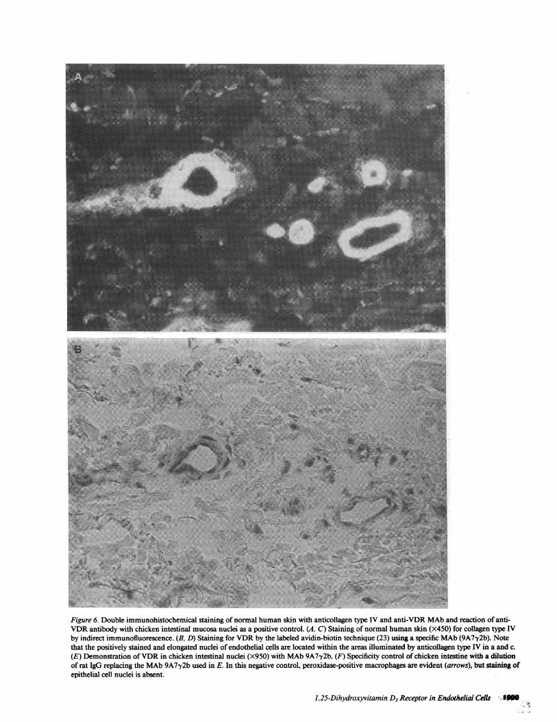
Immunohistochemical evidence of in vivo expression ofVDR in endothelial cells of venules and capillaries of humanskin. The potential in vivo expression of VDRin endothelialcells was studied by examining human tissue, namely dermalvessels in sections of skin biopsies. The reactivity of the nucleiin endothelial cells of dermal blood vessels with antireceptorMAb(MAb 9A7y2b) is demonstrated in Fig. 6 B and D. Dou-ble-staining experiments were carried out with mouse anticol-lagen type IV MAbto visualize the basement membrane of thevessels in the same section. (Fig. 6, A and C). These studiesshowed that immunoreactive VDRis present in the nuclei ofcells within the basement membrane; the topographical rela-tionship to the basement membrane identifies these cells as
endothelial cells. To ascertain the specificity of these findings,several control experiments were performed. Whenapplied tochicken intestinal tissue as a positive control, the anti-VDRantibody decorated the nuclei of the absorptive epithelial cellsthat are known to localize radiolabeled 1,25(OH)2D3 (Fig. 6E). The high sensitivity of this immunocytochemical tech-nique was demonstrated by the observation that MAb9A7,y2breacted to chick intestinal nuclei at dilutions as low as
1.64,000. Under the blocking conditions used, nonspecificbinding of the streptavidin enzyme complex was minimal.Peroxidase-positive macrophages (arrows) in intestine could
1,25-Dihydroxyvitamin D3 Receptor in Endothelial Cells 1907
C
LL
0M 80
60t 60
I.Il 40~
I 2010C=1
es 0 i

*-* control
0-o 1OaIM 1,25(OH)4C6
1 2 3 4 5 6 7 8 days
cell growth curve
Figure 5. Action of 1,25(OH)2D3 on growth parameters of BAEC.Cells were plated at a density of 8.8 X 04 cells per 35-mm dish andcultured in DMEwith 10% FBS; medium was changed daily.1,25(OH)2D3 at a final concentration of 10-8 Mor ethanol (0.02%)as solvent control were added. The data represent the mean of threedishes±SD.
be distinguished easily from other nuclear stained cells becauseof their heavy cytoplasmic staining. Also, chick intestinal nu-
clei were not stained by polyclonal rat IgG (Fig. 6 F). Further-more, rat MAbagainst the estrogen receptor showed no reac-
tion with epithelial cells of chick intestine, whereas heavystaining was observed in smooth muscle cell nuclei of humanuterus (data not shown). Whena separate rat MAbagainst an
unrelated antigen or polyclonal rat IgG was used, no reactivitywith endothelial cells nuclei was observed (data not shown).
BAECsynthesize J,25(OH)2D3from 25(OH)D3. To testthe ability of BAECto form the active hormone, BAECwereexamined under two growth conditions, i.e., confluent, den-sity-arrested and growing (log phase) cells. Parallel cultureswere incubated in the presence of radiolabeled 25(0H)D3 ac-
cording to the procedure of Reichel et al. (4). Transformationof I0-6-I0- " M25(0H)[PHJD3 was examined in BAECin thelog growth phase (1.4 X 10⁶ cells/ 100-mm dish) or in growthinhibited confluent cells (1.2 X 10' cells/l00-mm dish) in cul-ture medium supplemented with 2%FBS. No transformation
Table II. Dose Response of ['H] Thymidine Incorporationinto BAECin Log Phase of Growth*
Vitamin Dmetabolite added [3H]Thymidine incorporation
% of solvent control±SD
Solvent control 100±6.9*10-"1 M1,25(OH)2D3 100±5.810-10 M1,25(OH)2D3 87±1.010-9 M1,25(OH)2D3 78±7.810-8 M1,25(OH)2D3 33±0.210-8 Mvitamin D3 90±7.010-8 M25(OH)D3 86±3.110-8 M24R,25(OH)2D3 81±1.3l0-8M la(OH)D3 71±1.3
Cells were plated at a density of 1.2 X 10⁵ cells/35-mm dish andgrowth in DMEwith 10% FBS. On day 2, the medium was changedand 1,25(OH)2D3 (final concentration I0-I-10i - M), other metabo-lites of vitamin D3, or solvent (0.02% ethanol) were added. 16 h afterthe medium change, 2.5 ,Ci [3H]thymidine/dish were added. Thedata represent the mean of three dishes.* 100% is [3H]thymidine incorporation in control dishes (range33,900 to 35,600 cpm/dish).
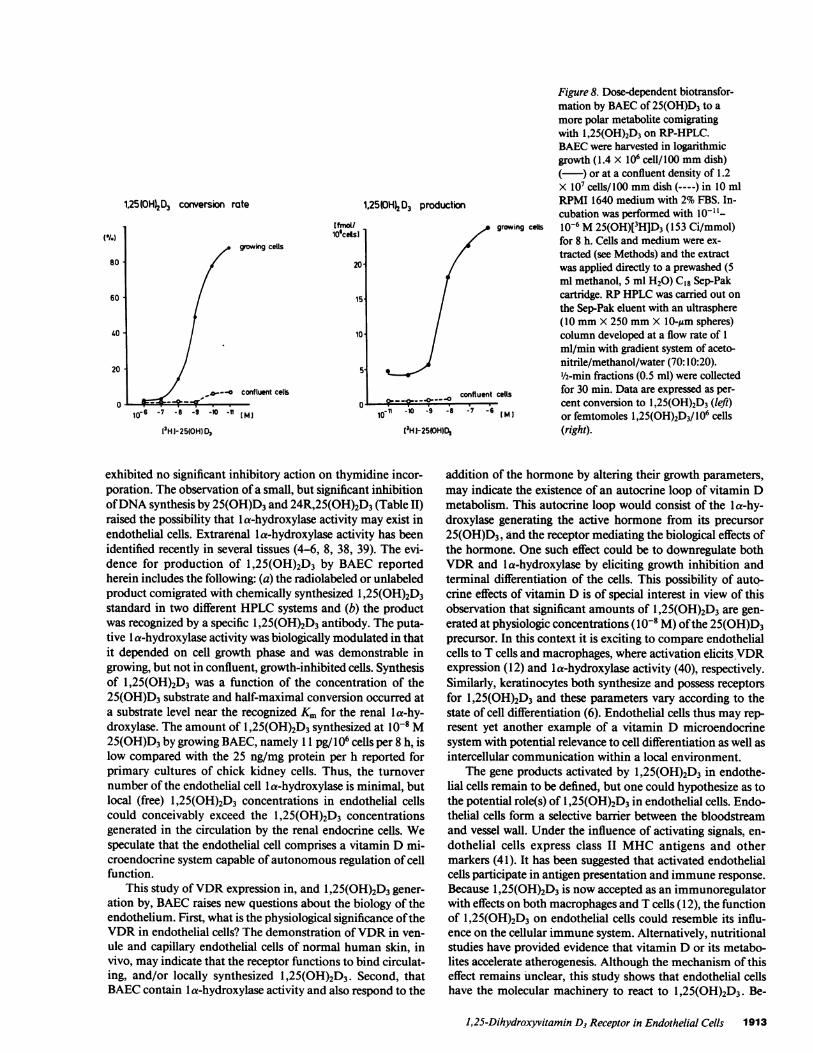
of 25(OH)[3H]D3 was observed after incubation in mediaalone (in the absence of cells) for 8 h. When growing BAECwere incubated with 25(OH)[3H]D3 for 8-12 h, there was con-version of 25(OH)[3H]D3 to a metabolite that comigrated withunlabeled or radioactive 1,25(OH)2D3 in both straight-phase(Fig. 7 A) or RP-HPLC(Fig. 7 B) systems. RIA of 1,25(OH)2D3applied to aliquots of the HPLCfractions confirmed the pres-ence of 1,25(OH)2D3. As depicted in Fig. 8, the metabolism of25(OH)D3 to 1,25(OH)2D3 was substrate concentration de-pendent, with half-maximal conversion occurring between10-' and I0-' Msubstrate. It is noteworthy that there was littleproduction of 1,25(OH)2D3 from 25(OH)D3 by confluent cells(Fig. 8), suggesting that the capacity to form 1,25(OH)2D3 isgreatly enhanced in rapidly growing cells as is the 1,25(OH)2D3receptor number.
Discussion
The findings of this study can be summarized as follows: (a)BAECexpress a VDR-like macromolecule that is indistin-guishable from the classical VDRoriginally described inchicken intestinal mucosa and subsequently demonstrated in anumber of mammalian cells (2); (b) maintenance of BAECatlow cell density to induce cell growth upregulates the expres-sion of VDR; (c) density-arrested BAECcan be stimulated toexpress VDRby activators of PKC; (d) BAECrespond to theaddition of exogenous 1,25(OH)2D3 hormone by a decrease intheir growth rate and saturation density; (e) VDRis expressedin both capillary and venule endothelial cells of normalhuman skin, in vivo; (f) rapidly proliferating BAEC, but notdensity-arrested BAEC, metabolize 25(OH)D3 to a sterol withchromatographic and immunoreactive properties identical tothose of 1,25(0H)2D3. These results represent the first demon-stration of a steroid hormone receptor in endothelial cells andreveal two potential triggers for the upregulation of the VDR,namely increased proliferation and activation of PKC. A fur-
1908 Merke et al.
2500-
2000-
o
1500-x
10
0 10 Owl
500-
: ...'!
,Jom
Figure 6. Double immunohistochemical staining of normal human skin with anticollagen type IV and anti-VDR MAband reaction of anti-VDRantibody with chicken intestinal mucosa nuclei as a positive control. (A, C) Staining of normal human skin (X450) for collagen type IVby indirect immunofluorescence. (B, D) Staining for VDRby the labeled avidin-biotin technique (23) using a specific MAb(9A7y2b). Notethat the positively stained and elongated nuclei of endothelial cells are located within the areas illuminated by anticollagen type IV in a and c.(E) Demonstration of VDRin chicken intestinal nuclei (X950) with MAb9A7y2b. (F) Specificity control of chicken intestine with a dilutionof rat IgG replacing the MAb9A7'y2b used in E. In this negative control, peroxidase-positive macrophages are evident (arrows), but staining ofepithelial cell nuclei is absent.
1,25-Dihydroxyvitamin D3 Receptor in Endothelial Cells I-MM

: .y A
i . .. ...:
-4 w
Figure 6 (Continued)
ther aspect of the results is that rapidly proliferating BAECboth synthesize 1,25(OH)2D3 and decrease their growth rate inresponse to the hormone, suggesting that a function of
1,25(OH)2D3 in endothelial cells could be to modulate prolif-eration via an autocrine feedback mechanism.
Several lines of evidence document the presence of specific
1910 Merke et al.

:,':s wts 1$A
i- 4onr - o _ Al.. ,a:' .vfs', J#,.2 ' ; VIA
. rX
01A
'T r.'. .S"i. a :t .w
~~~~44
.,Z; ., en. ,-.s,4 ~~~~~~~j
:, ~* N ; .NW
Figure 6 (Continued)
VDR in cultured BAEC. For instance, radiolabeled1,25(OH)2D3 specifically binds to the nuclear fraction ofBAECand exhibits the characteristic VDRsedimentation co-efficient of 3.2-3.5 S (Fig. 1). Moreover, the nuclear receptorinteracts with DNAcellulose and the active component can beeluted at a concentration of 0.22 MKCl (Fig. 2). Scatchardanalyses indicate the presence of a single receptor class with aKd of 5 X 10⁻¹⁰ M(Fig. 3). These biochemical properties of theVDR-like activity in BAECare consistent with the presence ofa molecule with two binding domains, one for DNAand an-other for the hormone. Thus, this activity in BAECis distinct
from the previously described serum vitamin D binding pro-tein (28) but shares all characteristics of the intracellular VDRof chicken intestinal mucosa (2, 22) and other tissues (3, 7, 10,11, 12, 29). It is therefore concluded that BAECpossess thespecific VDR.
Having identified the VDRin BAEC, it was next sought todetermine whether expression of the receptor can be modu-lated. One of the characteristics of endothelial cells is theirability to form a confluent, density-arrested monolayer. Anumber of metabolic activities of endothelial cells are' alteredwhen this monolayer is disrupted and cell growth is induced by
1,25-Dihydroxyvitamin D3 Receptor in Endothelial Cells 1911
,., x i..I
A;il
Pfs,.
..
000OW

4
1000
2000}
000-
-)2
x
-. SO
A
I I
rJ L-I-
' i
1500j
250-I.0.0'
B
6 10 20 30Minutes
Figure 7. Metabolism of 25(OH)D3 to I,25(0H)2D3 by BAEC.BAECwere grown to a density of 1.4 X 10⁶ cells/100 mmdish in 10ml RPMI 1640 medium with 2%FBS. Incubation of the cells wascarried out with IO-' M25(OH)[3H]D3 (153 Ci/mmol) or unlabeled25(OH)D3 for 8-12 h. Cells and medium were extracted (seeMethods) and the extract was applied directly to a prewashed (5 mlmethanol, 5 ml H20) C18 Sep-Pak cartridge and the vitamin Dme-tabolites eluted with acetonitrile. The metabolites then were resolvedon two HPLCsystems. (A) Straight-phase HPLCwas performedusing a Lichrosorb Hibar P 102 column (10 mmX 250 mmX 5-Mmspheres) at a flow rate of 4 ml/min using an isocratic system of hex-ane-isoproanol (9:1). ½/2-min fractions (2 ml) were collected and frac-tions were counted ( ) and aliquots (10 1l) evaluated byI,25(0H)2D3 RIA to detect radioinert hormone (-----). (B) RP-HPLCwas performed using an ultrasphere column (10 mmX 250mmX 10-Mm spheres), eluted at a flow rate of 1 ml/min with a gra-dient system of acetonitrile/methanol/water (70:10:20 vol/vol/vol).½/2-min fractions (0.5 ml) were collected for 30 min. Counting andRIA were as in A.
seeding the cells at low cell density (1 8). In these experiments,nuclear extracts prepared from rapidly growing BAECshoweda major increase in the Nm. value for VDR, but no change in
Kd when compared with their nongrowing, density-arrestedcounterparts. These results indicate that rapid growth ofBAECis associated with upregulation of the number of func-tional VDRmolecules rather than with a change in the affinityof the hormone for its receptor. Increased VDRexpression hasbeen observed previously in rapidly proliferating culturedmouse bone cells (30) and osteogenic sarcoma cell lines treatedwith glucocorticoids (31) and retinoic acid (32).
Activation of PKCby TPA has been shown to have pro-found effects on BAEC. Endothelial cells treated with TPAdisplay a more differentiated phenotype (33), including theformation of fenestrae (16) and capillary-like structures (15),synthesis of plasminogen activator and plasminogen activatorinactivator (24), as well as several other morphological alter-ations (14). Therefore, TPA and the short chain diglyceride,DiC8 (26), were used to activate PKCin BAEC. In density-ar-rested endothelial cells, VDRis dose-dependently increased bythe two activators of PKC. Furthermore, VDRinduction wasblocked by the PKC inhibitor sphingosine. The finding ofPKC-dependent VDRupregulation is analogous to previousobservations of increased glucocorticoid receptor activity inmyeloid leukemia cells treated with TPA (34). In that study,increased glucocorticoid receptor expression paralleled induc-tion of differentiation of the leukemic cells into mature macro-phages. It is of interest that the activators of PKCevokechanges in VDRexpression similar to those caused by en-hanced proliferation (compare Figs. 3 and 4). The mechanismsunderlying this upregulation remain to be dstermined, but theresults of experiments employing actinomycin D and cyclo-heximide (Table I) indicate that both transcriptional andtranslational activity are required. However, that TPA inhibitsthe rate of DNAsynthesis in BAECsuggests that the intracel-lular pathways responsible for PKC activation of VDRex-pression are distinct, in part, from those occurring whenBAECare induced to grow rapidly.
Previous studies by Stumpf et al. (35, 36) using autoradio-graphic localization of 1,25(OH)2[3H]D3 in nuclei, in vivo,revealed the presence of VDRin a variety of cells and tissues.However, no specific evidence was obtained by this procedurefor the presence of VDRin endothelial cells. The sensitivity ofthe autoradiographic localization technique maybe lower thanthat used in this study. To examine whether the VDRis ex-pressed in endothelial cells in vivo, venules, arterioles, andcapillaries of normal skin were probed using high affinity MAb9A772b (22, 37), coupled with the labeled acidin-biotinmethod that amplifies the immunohistochemical signal (23).VDRcould be demonstrated in the nuclei of endothelial cellsof human dermal vessels (Fig. 6). By double immune staining,using anticollagen type IV antibodies that delineate the capil-lary basement membrane, endothelial cells could be distin-guished from vascular smooth muscle cells, which also expressVDRactivity (1 1). Therefore, the immunocytochemical datasupport indirectly the biochemical observation of high-affinitybinding of 1,25(OH)2D3 to VDRin cultured BAEC, and ex-tend the finding by documenting that immunoreactive VDRispresent in the nuclei of normal human endothelial cells, invivo.
1,25(OH)2D3 inhibits cell proliferation and induces differ-entiation in a variety of cell systems (2, 3, 6, 7, 9, 12) and, inthis investigation, 1 ,25(OH)2D3 reduced thymidine incorpora-tion and saturation density of BAEC(Table II and Fig. 5). Theeffect was specific, because the parent vitamin D3 compound
1912 Merke et al.
I
1.25(0H)2D3 caxwersion rate
Itmo/lo'cels
growing cells
20
15
10
5-
confluent cells
l'H1-25(0H) %
1.25(0H)2 D3 productton
O.---O confluent10-" -9 -8
(3H1-25(OH)D,
Figure 8. Dose-dependent biotransfor-mation by BAECof 25(OH)D3 to amore polar metabolite comigratingwith 1,25(OH)2D3 on RP-HPLC.BAECwere harvested in logarithmicgrowth (1.4 X 10⁶ cell/100 mmdish)
) or at a confluent density of 1.2X I0' cells/100 mmdish (----) in 10 mlRPMI 1640 medium with 2%FBS. In-cubation was performed with I I-
growing cells 10-6 M25(OH)[3H]D3 (153 Ci/mmol)for 8 h. Cells and medium were ex-tracted (see Methods) and the extractwas applied directly to a prewashed (5ml methanol, 5 ml H20) C18 Sep-Pakcartridge. RPHPLCwas carried out onthe Sep-Pak eluent with an ultrasphere(10 mmX 250 mmX 10-,gm spheres)column developed at a flow rate of 1ml/min with gradient system of aceto-nitrile/methanol/water (70:10:20).½/2-min fractions (0.5 ml) were collected
cells for 30 min. Data are expressed as per-cent conversion to 1,25(OH)2D3 (left)
lMl or femtomoles I,25(0H)2D3/106 cells(right).
exhibited no significant inhibitory action on thymidine incor-poration. The observation of a small, but significant inhibitionof DNAsynthesis by 25(OH)D3 and 24R,25(OH)2D3 (Table II)raised the possibility that la-hydroxylase activity may exist inendothelial cells. Extrarenal la-hydroxylase activity has beenidentified recently in several tissues (4-6, 8, 38, 39). The evi-dence for production of 1,25(OH)2D3 by BAEC reportedherein includes the following: (a) the radiolabeled or unlabeledproduct comigrated with chemically synthesized 1,25(OH)2D3standard in two different HPLCsystems and (b) the productwas recognized by a specific 1,25(OH)2D3 antibody. The puta-tive la-hydroxylase activity was biologically modulated in thatit depended on cell growth phase and was demonstrable ingrowing, but not in confluent, growth-inhibited cells. Synthesisof 1,25(OH)2D3 was a function of the concentration of the25(OH)D3 substrate and half-maximal conversion occurred ata substrate level near the recognized Km for the renal a-hy-droxylase. The amount of I,25(OH)2D3 synthesized at 10-8 M25(OH)D3 by growing BAEC, namely 11 pg/106 cells per 8 h, islow compared with the 25 ng/mg protein per h reported forprimary cultures of chick kidney cells. Thus, the turnovernumber of the endothelial cell la-hydroxylase is minimal, butlocal (free) 1,25(OH)2D3 concentrations in endothelial cellscould conceivably exceed the 1,25(OH)2D3 concentrationsgenerated in the circulation by the renal endocrine cells. Wespeculate that the endothelial cell comprises a vitamin D mi-croendocrine system capable of autonomous regulation of cellfunction.
This study of VDRexpression in, and 1,25(OH)2D3 gener-
ation by, BAECraises new questions about the biology of theendothelium. First, what is the physiological significance of theVDRin endothelial cells? The demonstration of VDRin ven-ule and capillary endothelial cells of normal human skin, invivo, may indicate that the receptor functions to bind circulat-ing, and/or locally synthesized 1,25(OH)2D3. Second, thatBAECcontain I a-hydroxylase activity and also respond to the
addition of the hormone by altering their growth parameters,may indicate the existence of an autocrine loop of vitamin Dmetabolism. This autocrine loop would consist of the la-hy-droxylase generating the active hormone from its precursor25(OH)D3, and the receptor mediating the biological effects ofthe hormone. One such effect could be to downregulate bothVDRand la-hydroxylase by eliciting growth inhibition andterminal differentiation of the cells. This possibility of auto-crine effects of vitamin D is of special interest in view of thisobservation that significant amounts of 1,25(OH)2D3 are gen-erated at physiologic concentrations (1O-8 M) of the 25(OH)D3precursor. In this context it is exciting to compare endothelialcells to T cells and macrophages, where activation elicits VDRexpression (12) and la-hydroxylase activity (40), respectively.Similarly, keratinocytes both synthesize and possess receptorsfor 1,25(OH)2D3 and these parameters vary according to thestate of cell differentiation (6). Endothelial cells thus may rep-resent yet another example of a vitamin D microendocrinesystem with potential relevance to cell differentiation as well asintercellular communication within a local environment.
The gene products activated by 1,25(OH)2D3 in endothe-lial cells remain to be defined, but one could hypothesize as tothe potential role(s) of 1,25(OH)2D3 in endothelial cells. Endo-thelial cells form a selective barrier between the bloodstreamand vessel wall. Under the influence of activating signals, en-
dothelial cells express class II MHCantigens and othermarkers (41). It has been suggested that activated endothelialcells participate in antigen presentation and immune response.Because 1,25(OH)2D3 is now accepted as an immunoregulatorwith effects on both macrophages and T cells (12), the functionof 1,25(OH)2D3 on endothelial cells could resemble its influ-ence on the cellular immune system. Alternatively, nutritionalstudies have provided evidence that vitamin D or its metabo-lites accelerate atherogenesis. Although the mechanism of thiseffect remains unclear, this study shows that endothelial cellshave the molecular machinery to react to 1,25(OH)2D3. Be-
1,25-Dihydroxyvitamin D3 Receptor in Endothelial Cells 1913
(SI.)
80
60
40 -
20
cause injury to the endothelial lining is a key facet of thecurrent theory of the pathogenesis of atherosclerosis, a nega-tive or positive influence of 1,25(OH)2D3 in the reendothelial-ization process could be of great significance to determiningthe incidence of atherosclerotic plaque formation. Finally, ab-normalities in the levels of calcium regulating hormones suchas l,25(QH)2D3 have been recognized in human and experi-mental hypertension (42, 43). Therefore, interactions of1,25(OH)2D3 with endothelial cells as well as with vascularsmooth muscle cells in the control of vascular tone (44-46)represent potentially exciting areas of investigation that couldprovide a link between the 1,25(OH)2D3 hormone and thepathophysiology of coronary artery disease.
Acknowledgments
This research was carried out with the support of DeutscheForschungsgemeinschaft (Me 632/3-3) and National Institutes ofHealth grants to M. R. Haussler.
References
1. Norman, A. W. 1979. Vitamin D, the Calcium HomeostaticSteroid Hormone. W. J. Darby, editor. Academic Press, Inc., NewYork.
2. Haussler, M. R. 1986. Vitamin Dreceptors: nature and function.Annu. Rev. Nutr. 6:527-562.
3. Mangelsdorf, D. J., H. P. Koeffler, C. A. Donaldson, J. W. Pike,and M. R. Haussler. 1984. 1,25-Dihydroxyvitamin D3-induced differ-entiation in a human promyelocytic leukemia cell line (HL-60): re-ceptor-mediated maturation to macrophage-like cells. J. Cell Biol.98:391-398.
4. Reichel, H., H. P. Koeffler, J. Bishop, and A. W. Norman. 1987.25(OH)D3 metabolism by lipopolysaccharide-stimulated normalhuman macrophages. J. tlin. Endocrinol. & Metab. 64:1-7.
5. Adams, J. S., 0. P. Sharma, M. A. Gacad, and F. R. Singer. 1983.Metabolism of 25(OH)D3 by cultured pulmonary alveolar macro-phages in sarcoidosis. J. Clin. Invest. 72:1856-1861.
6. Pillai, S., D. D. Bikle, and P. M. Elias. 1988. 1,25-Dihydroxyvi-tamin D production and receptor binding in human keratinocytesvaries with differentiationt. J. Bid. Chem. 263:5390-5395.
7. Merke, J., D. Schwittay, G. Furstenberger, M. Gross, F. Marks,and E. Ritz. 1985. Demonstration and characterization of1,25(OH)2D3 receptors in basal cells of neonatal and adult mice. CalcifTissue Int. 37:257-267.
8. Reichel, H., H. P. Koeffler, A. Tobler, and A. W. Norman. 1987.1,25(OH)2D3 inhibits y-interferon synthesis by normal human periph-eral blood lymphocytes. Proc. Nati. Acad. Sci. USA. 84:3385-3389.
9. Haussler, C. A., S. L. Marion, J. W. Pike, and M. R. Haussler.1986. 1,25-Dihydroxyvitamin D3 inhibits the clonogenic growth oftransformed cells via its receptor. Biochem. Biophys. Res. Commun.139:136-143.
10. Merke, J., G. Klaus, U. Hugel, R. Waldherr, and E. Ritz. 1986.No 1,25(OH)2D3 receptors in osteoclasts of the Ca'+-deficient chickdespite demonstrable receptors on circulating monocytes. J. Clin. In-vest. 77:312-317.
11. Merke, J., W. Hofmann, D. Goldschmidt, and E. Ritz. 1987.Demonstration of 1,25(OH)2D3 receptors and action in vascularsmooth muscle cells in vitro. Calcif Tissue Int. 41:112-114.
12. Provvedini, D. M., C. D. Tsoukas, L. J. Deftos, and S. C.Manolagas. 1983. 1,25-Dihydroxyvitamin D3 receptors in human leu-kocytes. Science (Wash. DC). 221:1181-1183.
13. Suda, T., E. Abe, C. Miyaura, H. Tanaka, Y. Shiina, T. Haya-shi, H. Nagasawa, K. Chida, H. Hashiba, M. Fukushima, Y. Nishii,and T. Kuroki. 1985. Modulation of cell differentiation and tumorpromotion by 1,25-dihydroxyvitamin D3. In Vitamin D: Chemical,
Biochemical and Clinical Update. A. W. Norman, K. Schaefer, H.-G.Grigoleit, and D. V. Herrath, editors. Walter de Gruyter, Berlin,187-196.
14. Antonov, A. S., M. E. Lukashev, Y. A. Romanow, V. A. Tka-chuk, V. S. Repin, and V. N. Smirnow. 1986. Morphological alter-ations in endothelial cells from human aorta and umbilical vein in-duced by forskolin and phorbol 12-myristate 13-acetate: A synergisticaction of adenylate cyclase and protein kinase C activators. Proc. Natl.Acad. Sci. USA. 83:9704-9708.
15. Montesano, R., and L. Orci. 1985. Tumor promoting phorbolesters induce angiogenesis in vitro. Cell. 42:469-477.
16. Lombardi, T., R. Montesano, M. B. Furie, S. C. Silverstein, andL. Orci. 1986. Endothelial diphragmed fenestrae: in vitro modulationby phorbol myristate acetate. J. Cell Biol. 102:1965-1970.
17. Nishizuka Y. 1986. Studies and prospectives of protein kinaseC. Science (Wash. DC). 233:305-330.
18. Schwartz, S. 1978. Selection and characterization of bovineaortic endothelial cells. In Vitro (Rockville). 14:966-984.
19. Bouillon, R., P. De Moor, E. G. Baggiolini, and M. R. Usko-kovic. 1980. A radioimmunoassay for 1,25-dihydroxycholecalciferol.Clin. Chem. 26:562-567.
20. Merke, J., U. Hugel, A. Zlotkowski, A. Szabo, J. Bommer, G.Mall, and E. Ritz. 1987. Diminished parathyroid 1,25(OH)2D3 recep-tors in experimental uremia. Kidney Int. 32:350-353.
21. Pike, J. W., C. A. Donaldson, S. L. Marion, and M. R. Haussler.1982. Development of hybridomas secreting monoclonal antibodies tothe chick intestinal la,25-dihydroxyvitamin D3 receptor. Proc. Natl.Acad Sci. USA. 79:7719-7723.
22. Pike, J. W., S. L. Marion, C. A. Donaldson, and M. R. Haussler.1983. Serum and monoclonal antibodies against the chick intestinalreceptor for 1,25-dihydroxyvitamin D3. J. Biol. Chem. 258:1289-1296.
23. Clemens, T. L., K. P. Girrett, X.-Y. Zhou, J. W. Pike, M. R.Haussler, and D. W. Dempster. 1988. Immunocytochemical localiza-tion of the 1,25-dihydroxyvitamin D3 receptor in target cells. Endocri-nology. 122:1224-1230.
24. Gross, J. L., D. Moscatelli, E. A. Jaffe, and D. B. Rifkin. 1984.Plasminogen activator and collagen production by cultured capillaryendothelial cells. J. Cell Biol. 95:974-981.
25. Goerig, M., A. J. R. Habenicht, R. Heitz, W. Zeh, H. Katus, B.Kommerell, R. Ziegler, and J. A. Glomset. 1987. sn-1,2-Diacylgly-cerols and phorbol diesters stimulate thromboxane synthesis by denovo synthesis of prostaglandin H synthase in human promyelocyticleukemia cells. J. Clin. Invest. 79:903-91 1.
26. Ebeling, J. G., G. R. Vandenbark, L. J. Kuhn, B. R. Ganong,R. M. Bell, and J. E. Niedel. 1985. Diacylglycerols mimic phorboldiester induction of leukemic cell differentiation. Proc. Natl. Acad. Sci.USA. 82:815-819.
27. Merrill, A. H., A. M. Sereni, V. L. Stevens, Y. A. Hannan,R. M. Bell, and J. M. Kinkade, Jr. 1986. Inhibition of phorbol ester-dependent differentiation of human promyelocytic leukemic (HL-60)cells by sphingosine and other long chain bases. J. Biol. Chem.261:12610-12615.
28. Kream, B. E., H. F. DeLuca, D. M. Moriarity, N. C. Keiidrick,and J. G. Ghazarian. 1979. Origin of 25-hydroxyvitamin D3 bindingprotein from tissue cytosol preparations. Arch. Biochem. Biophys.192:318-323.
29. Merke, J., U. Hugel, and E. Ritz. 1985. Nuclear testicular1,25-dihydroxyvitamin D3 receptors in Sertoli cells and seminiferoustubules of adult rodents. Biochem. Biophys. Res. Commun. 127:303-309.
30. Chen, T. I., and D. Feldman. 1981. Regulation of 1,25(OH)2D3receptors in cultured mouse bone cells. Correlation of receptor con-centration with the rate of cell division. J. Biol. Chem. 256:5561-5566.
31. Manolagas, S. C., J. Abare, and L. J. Deftos. 1984. Glucocorti-coids increase the 1,25(OH)2D3 receptor concentration in rat osteo-genic sarcoma cells. Calcif Tissue Int. 36:153-157.
32. Petkovich, P. M., J. N. M. Heersche, D. 0. Tinker, and G.
1914 Merke et al.
Jones. 1984. Retinoic acid stimulates 1,25-dihydroxyvitamin D3 bind-ing in rat osteosarcoma cells. J. Biol. Chem. 259:8274-8280.
33. Doktrow, S. R., and J. Folkman. 1987. Protein kinase C acti-vators suppress stimulation of capillary endothelial cell growth by an-giogenic endothelial mitorpens. J. Cell Biol. 104:679-687.
34. Hirai, H., T. Murakami, A. Urabe, and F. Takaku. 1985. In-creased glucocorticoid receptor concentration in macrophage differ-entiation of myeloid leukemia cells with TPA. Cancer Res. 45:2456-2461.
35. Stumpf, W. E., M. Sar, F. A. Reid, Y. Tanaka, and H. F.DeLuca. 1979. Target cells for 1,25(OH)2D3 in intestinal tract, stom-ach, kidney, skin, pituitary and parathyroid. Science (Wash. DC).206:1189-1190.
36. Stumpf, W. E., S. A. Clark, S. Madhabananda, and H. F.DeLuca. 1984. Topographical and developmental studies on targetsites of 1,25(OH)2D3 in skin. Cell Tissue Res. 238:489-496.
37. McDonnell, D. P., D. J. Mangelsdorf, J. W. Pike, M. R.Haussler, and B. W. O'Malley. 1987. Molecular cloning of comple-mentary DNAencoding the avian receptor for vitamin D. Science(Wash. DC). 235:1214-1217.
38. Whitsett, J. A., M. Ho, R. C. Tsang, E. J. Norman, and K. G.Adams. 1981. Synthesis of 1,25-dihydroxyvitamin D3 by human pla-centa in vitro. J. Clin. Endocrinol. & Metab. 53:484-488.
39. Howard, G. A., R. T. Turner, D. J. Sherrard, and D. J. Baylink.1981. Humanbone cells in culture metabolize 25-hydroxyvitamin D3
to 1,25-dihydroxyvitamin D3 and 24,25-dihydroxyvitamin D. J. Biol.Chem. 256:7738-7740.
40. Henry, H. L. 1979. Regulation of the hydroxylation of 25-hy-droxyvitamin D3 in vitro and in primary cultures of chick kidney cells.J. Biol. Chem. 254:2722-2729.
41. Vedder, N. B., and J. M. Harlan. 1988. Increased surface ex-pression of CD1lb/CD18 (Mac-i) is not required for stimulated neu-trophil adherence to cultured endothelium. J. Clin. Invest. 81:676-682.
42. Young, E. W., R. D. Bukoski, and D. A. McCarron. 1988.Calcium metabolism in experimental hypertension. Proc. Soc. Exp.Biol. Med. 187:123-141.
43. Merke, J., P. A. Lucas, A. Szabo, G. Cournot-Witmer, G. Mall,R. Bouillon, T. Drfieke, J. Mann, and E. Ritz. 1989. Hyperparathy-roidism and abnormal calcitriol metabolism in the spontaneously hy-pertensive rat. Hypertension (Dallas). In press.
44. Weishaar, R. E., and R. U. Simpson. 1987. Involvement ofvitamin D3 with cardiovascular function. II. Direct and indirect effects.Endocrinol. Metab. 16:E675-E683.
45. Pawlowski, N. A., E. I. Abraham, S. Pontier, W. A. Scott, andZ. A. Cohn. 1985. Human monocyte-endothelial cell interaction invitro. Proc. Natt. Acad. Sci. USA. 82:8208-8212.
46. Yanagisawa, M., H. Kurihara, S. Kimura, Y. Tomobe, M.Kobayashi, Y. Mitsui, Y. Yazaki, K. Goto, and T. Masaki. 1988. Anovel potent vasoconstrictor peptide produced by Vascular endothelialcells. Nature (Lond.). 332:411-415.
1,25-Dihydroxyvitamin D3 Receptor in Endothelial Cells 1915