IDENTIFICATION, PATHOGENICITY, AND … · 147 AbstrAct Dieback of mamey sapote (Pouteria sapota)...
15
147 ABSTRACT Dieback of mamey sapote (Pouteria sapota) grafts is the most important disease during vegetative propagation in commercial nurseries. In 2008, Lasiodiplodia theobromae identified by cultural, morphological and molecular characterization was associated with this disease in 97 % of necrotic rootstocks and scions samples from a nursery in Guerrero, México. The fungus was inoculated onto the binding site of El Mexicano grafted plants. Dieback symptoms and gradual drying with cracking of the bark from the apex to the base of the scion were observed 30 d after inoculation. Abundant mycelia and pycnidia were found on inoculated scions and graft union areas, in the periderm, and on leaf scars. Transverse sections of inoculated scions tissues showed pycnidia. Parenchyma cells of cortex and phloem collapsed and died. L. theobromae mycelia and red inclusions, probably of phenolic nature, were observed in both xylem vessels and pith parenchyma cells. To our knowledge, this is the first study that provides pathogenicity and histopathological data about a disease caused by L. theobromae during the grafting process. Key words: dieback, nursery, vegetative propagation, Pouteria sapota, Lasiodiplodia theobromae. INTRODUCTION M amey sapote [Pouteria sapota (Jacq.) H. E. Moore and Stearn] is a tropical fruit tree native to México and Central America (Popenoe, 1974). In 2009, the cultivated area RESUMEN La muerte descendente de injertos de zapote mamey (Pouteria sapota) es la enfermedad más importante durante la propagación vegetativa en viveros comerciales. En 2008, Lasiodiplodia theobromae identificado por cultivo, morfo- lógica y molecularmente, se asoció con esta enfermedad en 97 % de las muestras de portainjertos y varetas necrosadas en un vivero de Guerrero, México. El hongo se inoculó en el sitio de unión de plantas injertadas de la selección El Mexicano. Síntomas de muerte descendente con deshidra- tación gradual y agrietamiento de la corteza que inició en el ápice y desplazó hacia la base de la vareta, se observaron 30 d después de la inoculación. Abundante micelio y picni- dios se encontraron en varetas, zonas de unión del injerto, peridermis, y en cicatrices foliares. Secciones transversales de tejidos de varetas inoculadas mostraron abundantes pic- nidos. Las células parenquimatosas de la corteza y floema colapsaron y necrosaron. Micelio de L. theobromae e inclu- siones rojas, de probable naturaleza fenólica, se observaron en vasos del xilema y en células parenquimatosas de la mé- dula. Para nuestro conocimiento, este es el primer estudio que provee datos de patogenicidad e histopatología acerca de una enfermedad causada por L. theobromae durante el proceso de injerto. Palabras clave: muerte descendente, vivero, propagación vegeta- tiva, Pouteria sapota, Lasiodiplodia theobromae. INTRODUCCIóN E l zapote mamey [Pouteria sapota (Jacq.) H. E. Moore y Stearn] es un frutal tropical na- tivo de México y América Central (Popenoe, IDENTIFICATION, PATHOGENICITY, AND HISTOPATHOLOGY OF Lasiodiplodia theobromae ON MAMEY SAPOTE GRAFTS IN GUERRERO, MÉXICO IDENTIFICACIÓN, PATOGENICIDAD E HISTOPATOLOGÍA DE Lasiodiplodia theobromae EN INJERTOS DE ZAPOTE MAMEY EN GUERRERO, MÉXICO Juan M. Tovar-Pedraza 1* , José A. Mora-Aguilera 1 , Cristian Nava-Díaz 1 , Daniel Téliz-Ortiz 1 , Guadalupe Valdovinos-Ponce 1 , Ángel Villegas-Monter 2 , Javier Hernández-Morales 1 1 Fitopatología, 2 Fruticultura, Campus Montecillo, Colegio de Postgraduados. Carretera Méxi- co-Texcoco. Km. 36.5, Montecillo, Estado de México. ([email protected]). * Author for correspondence v Autor responsable. Received: February, 2011. Approved: January, 2012. Published as ARTICLE in Agrociencia 46: 147-161. 2012.
Transcript of IDENTIFICATION, PATHOGENICITY, AND … · 147 AbstrAct Dieback of mamey sapote (Pouteria sapota)...

147
AbstrAct
Dieback of mamey sapote (Pouteria sapota) grafts is the most important disease during vegetative propagation in commercial nurseries. In 2008, Lasiodiplodia theobromae identified by cultural, morphological and molecular characterization was associated with this disease in 97 % of necrotic rootstocks and scions samples from a nursery in Guerrero, México. The fungus was inoculated onto the binding site of El Mexicano grafted plants. Dieback symptoms and gradual drying with cracking of the bark from the apex to the base of the scion were observed 30 d after inoculation. Abundant mycelia and pycnidia were found on inoculated scions and graft union areas, in the periderm, and on leaf scars. Transverse sections of inoculated scions tissues showed pycnidia. Parenchyma cells of cortex and phloem collapsed and died. L. theobromae mycelia and red inclusions, probably of phenolic nature, were observed in both xylem vessels and pith parenchyma cells. To our knowledge, this is the first study that provides pathogenicity and histopathological data about a disease caused by L. theobromae during the grafting process.
Key words: dieback, nursery, vegetative propagation, Pouteria sapota, Lasiodiplodia theobromae.
IntroductIon
Mamey sapote [Pouteria sapota (Jacq.) H. E. Moore and Stearn] is a tropical fruit tree native to México and Central America
(Popenoe, 1974). In 2009, the cultivated area
resumen
La muerte descendente de injertos de zapote mamey (Pouteria sapota) es la enfermedad más importante durante la propagación vegetativa en viveros comerciales. En 2008, Lasiodiplodia theobromae identificado por cultivo, morfo-lógica y molecularmente, se asoció con esta enfermedad en 97 % de las muestras de portainjertos y varetas necrosadas en un vivero de Guerrero, México. El hongo se inoculó en el sitio de unión de plantas injertadas de la selección El Mexicano. Síntomas de muerte descendente con deshidra-tación gradual y agrietamiento de la corteza que inició en el ápice y desplazó hacia la base de la vareta, se observaron 30 d después de la inoculación. Abundante micelio y picni-dios se encontraron en varetas, zonas de unión del injerto, peridermis, y en cicatrices foliares. Secciones transversales de tejidos de varetas inoculadas mostraron abundantes pic-nidos. Las células parenquimatosas de la corteza y floema colapsaron y necrosaron. Micelio de L. theobromae e inclu-siones rojas, de probable naturaleza fenólica, se observaron en vasos del xilema y en células parenquimatosas de la mé-dula. Para nuestro conocimiento, este es el primer estudio que provee datos de patogenicidad e histopatología acerca de una enfermedad causada por L. theobromae durante el proceso de injerto.
Palabras clave: muerte descendente, vivero, propagación vegeta-tiva, Pouteria sapota, Lasiodiplodia theobromae.
IntroduccIón
El zapote mamey [Pouteria sapota (Jacq.) H. E. Moore y Stearn] es un frutal tropical na-tivo de México y América Central (Popenoe,
IDENTIFICATION, PATHOGENICITY, AND HISTOPATHOLOGY OF Lasiodiplodia theobromae ON MAMEY SAPOTE GRAFTS
IN GUERRERO, MÉXICO
IDENTIFICACIÓN, PATOGENICIDAD E HISTOPATOLOGÍA DE Lasiodiplodia theobromae EN INJERTOS DE ZAPOTE MAMEY
EN GUERRERO, MÉXICO
Juan M. Tovar-Pedraza1*, José A. Mora-Aguilera1, Cristian Nava-Díaz1, Daniel Téliz-Ortiz1, Guadalupe Valdovinos-Ponce1, Ángel Villegas-Monter2, Javier Hernández-Morales1
1Fitopatología, 2Fruticultura, Campus Montecillo, Colegio de Postgraduados. Carretera Méxi-co-Texcoco. Km. 36.5, Montecillo, Estado de México. ([email protected]).
* Author for correspondence v Autor responsable.Received: February, 2011. Approved: January, 2012.Published as ARTICLE in Agrociencia 46: 147-161. 2012.

148
AGROCIENCIA, 16 de febrero - 31 de marzo, 2012
VOLUMEN 46, NÚMERO 2
with this plant in México was 1524 ha, distributed mainly in the states of Guerrero, Yucatán, Chiapas, Michoacán, Puebla, Oaxaca, Morelos and Veracruz (SIAP, 2010). Mamey sapote trees can be propagated either by sexual or asexual means (Villegas and Mora, 2008). However, members of Sapotaceae are considered difficult to graft, and mamey sapote is particularly complicated (Ogden et al., 1986). Besides the difficulty in propagating this fruit, the success of grafting is limited by the presence of diseases. Information about mamey sapote diseases is rather scarce: 1) leaf spot by Phyllosticta sp. and Phyllachora sp., anthracnose by Colletotrichum gloeosporioides, and root rot caused by Pythium sp., in Florida, USA (Farr et al., 1989); 2) Phyllosticta sapotae induces leaf spot in Cuba and the Bahamas; 3) Uredo sapotae causes curly leaves in El Salvador (Azurdía, 2006); 4) Botryosphaeria sp. and Hypoxylon sp. were associated to dieback syndrome, bark splitting and stem canker, in Guatemala (Álvarez, 1997); 5) in México, there is vegetative and floral proliferation induced by Uredo baruensis (Pereyda et al., 2008), dieback by Lasiodiplodia theobromae (Vázquez et al., 2009), floral necrosis by Alternaria alternata, Pestalotiopsis paeoniicola and Penicillium olsonii (Mora et al., 2008) and fruit rot by L. theobromae and P. paeoniicola (Bautista et al., 2002; Gómez et al., 2009). Death of scions is a severe disease during propagation by grafting of mamey sapote in Alpoyeca, state of Guerrero, México. Lasiodiplodia theobromae (Pat.) Griff. & Maubl. has been associated with symptoms of dieback and necrosis at the binding site of grafting on cashew (Anacardium occidentale L.) propagative material (Freire et al., 2002), as well as in guava (Psidium guajava L.) (Cardoso et al., 2002), citrus (Davis et al., 1987), and grapevine (Aroca et al., 2008). However, there are not experimental bases to determine its role as the causal agent of death of scions of mamey sapote. Based on this information, the aims of this study were to determine the etiology and anatomic abnormalities associated with dieback of grafted scions of mamey sapote.
mAterIAls And methods
Study site and sampling
Alpoyeca, Guerrero, México, is located at 17° 40’ N and 98° 31’ W, at an altitude of 960 m, with an average temperature
1974). En 2009, la superficie total cultivada con este frutal en México fue 1524 ha, distribuidas principalmente en los estados de Guerrero, Yuca-tán, Chiapas, Michoacán, Puebla, Oaxaca, More-los y Veracruz (SIAP, 2010). Las plantas de zapo-te mamey se pueden reproducir de manera sexual o asexual (Villegas y Mora, 2008). Sin embargo, los miembros de la familia Sapotaceae son consi-derados difíciles de injertar, y el zapote mamey es particularmente complicado (Ogden et al., 1986). Además de la dificultad para propagar este frutal, el éxito del injerto está limitado por la presencia de enfermedades. La información sobre las enfermedades del za-pote mamey es escasa: 1) en Florida, EE.UU., se han reportado manchas foliares por Phyllosticta sp. y Phyllachora sp., antracnosis por Colletotrichum gloeosporioides, y pudrición radical por Pythium sp. (Farr et al., 1989); 2) en Cuba y las Bahamas, Phyllosticta sapotae induce manchas foliares; 3) en El Salvador, Uredo sapotae causa enrollamiento de las hojas (Azurdia, 2006); 4) en Guatemala se asoció a Botryosphaeria sp. e Hypoxylon sp. con el síndro-me de muerte descendente, rajadura de corteza y cancro del tallo (Álvarez, 1997); 5) en México está la agalla o proliferación vegetativa y floral inducida por Uredo baruensis (Pereyda et al., 2008), muerte descendente por Lasiodiplodia theobromae (Vásquez et al., 2009), necrosis floral por Alternaria alternata, Pestalotiopsis paeoniicola y Penicillium olsonii (Mora et al., 2008), y pudrición de frutos por L. theobromae y P. paeoniicola (Bautista et al., 2002; Gómez et al., 2009). La muerte de varetas es una enfermedad severa durante la propagación por injerto del zapote ma-mey en Alpoyeca, Guerrero, México. Lasiodiplodia theobromae se ha asociado con síntomas de muerte descendente y necrosis en sitio de unión de injertos en material propagativo de marañón (Anacardium occidentale L.) (Friere et al., 2002), guayaba (Psidium guajava L.) (Cardoso et al., 2002), cítricos (Davis et al., 1987) y vid (Aroca et al., 2008). Sin embar-go, no hay bases experimentales para determinar su función como agente causal de la muerte de varetas de zapote mamey. Con base en esta información, los objetivos de este estudio fueron determinar la etiología y alteraciones anatómicas asociadas con la muerte descendente de varetas injertadas de zapote mamey.
IDENTIFICATION, PATHOGENICITY AND HISTOPATHOLOGY OF Lasiodiplodia theobromae ON MAMEY SAPOTE GRAFTS IN GUERRERO, MÉXICO
149TOVAR-PEDRAZA et al.
of 25.5 °C and an annual rainfall of 780 mm. In March 2008, a commercial mamey sapote nursery was sampled and 10 asymptomatic grafted plants and 20 grafted plants with scion dieback symptoms and dead tissue in the graft union were collected for isolation purposes. In order to know the fungal diversity in asymptomatic scions, field sampling was carried out in three commercial mamey sapote orchards, from which 30 scions with latent apical buds were collected.
Isolation
Tissue samples of 5 mm3 were cut from the graft union area including necrotic tissues. Samples were disinfested by immersion in a 3 % sodium hypochlorite solution for 4 min, washed in sterile distilled water for 3 min and placed in petri dishes with potato-dextrose-agar culture medium (PDA). The dishes were incubated 48 h under continuous black light at 302 °C. Most frequent fungal colonies were purified by monosporic cultures and transferred to new petri dishes with PDA.
Pathogenicity test
In October 2008, pathogenicity of L. theobromae was verified in grafted plants of El Mexicano selection in a nursery in Alpoyeca. The inoculum concentration used was 190 mycelial colony-forming units mL1. In the inoculation test 15 scions were used per treatment: T1, scions were washed by hand using a natural fiber and water, immersed 15 min in mancozeb (1 mL L1), and 100 L of the inoculum were placed on the upper part of the grafting site; T2, scions were washed by hand using a natural fiber and water, immersed 15 min in mancozeb and 100 L of sterile distilled water were placed on the upper part of the grafting site; T3, scions did not have any prophylactic measures and the grafted plants were not disinfested or inoculated. The 45 scions were grafted by using the side veneer technique described by Villegas and Mora (2008). All grafts were covered with clear plastic bags to avoid dehydration. Grafted plants were maintained under 75-80 % shade and continuous irrigation. Fungal infection signs and symptoms were registered 30 d after inoculation (dai). The fungus was re-isolated from the infected tissues; the colony and its reproductive structures were compared with the colony originally inoculated. Incidence of diseased grafts with natural infection was calculated with the following equation: Ii ni Ni=∑ / ; where Ii incidence of diseased scions at the moment i; ni number of diseased scions at the moment i; Ni total population of grafted scions.
mAterIAles y métodos
Sitio de estudio y muestreo
Alpoyeca, Guerrero, México, se localiza a 17° 40’ N y 98° 31’ O, a una altitud de 960 m, con temperatura promedio de 25.5 °C y precipitación anual de 780 mm. En marzo del 2008 se muestreó un vivero comercial de zapote mamey con el propósito de obtener aislamientos, se recolectaron 10 plantas injertadas asintomáticas y 20 plantas injertadas con síntomas de muerte descendente de varetas y necrosis en la unión del injerto. Para conocer la diversidad de especies de hongos en varetas asintomáticas, se realizó un muestreo de campo en tres huertos comerciales de zapote mamey, y se recolectaron 30 varetas rectas con la yema apical cerrada y latente.
Aislamiento
Muestras de tejidos de 5 mm3 se cortaron de la zona de unión del injerto incluyendo tejidos necróticos. Las muestras se desinfestaron por inmersión en una solución de hipoclorito de sodio al 3 % por 4 min, se lavaron por 3 min en agua desti-lada estéril, y se colocaron en cajas petri con medio de cultivo papa-dextrosa-agar (PDA). Las cajas se incubaron 48 h bajo luz negra continua a 302 °C. Las colonias fungosas predo-minantes se purificaron mediante cultivos monospóricos y se transfirieron a nuevas cajas petri con PDA.
Prueba de patogenicidad
En octubre de 2008 se verificó la patogenicidad de L. theobromae en plantas injertadas de la selección El Mexica-no en un vivero de Alpoyeca. La concentración de inóculo usada fue 190 unidades formadoras de colonias mL1. En la prueba de inoculación se usaron 15 varetas por tratamien-to: T1, las varetas se lavaron manualmente usando una fibra natural y agua, inmersas 15 min en mancozeb (1mL L1), y 100 L de inóculo se colocaron en la parte superior de la unión del injerto; T2, las varetas se lavaron manualmente con una fibra natural y agua, inmersas 15 min en mancozeb y 100 L de agua destilada estéril se colocaron en la parte superior de la unión del injerto; T3, las varetas no tuvieron medidas profilácticas y las plantas injertadas no se desinfestaron ni inocularon. Las 45 varetas se injertaron usando la técnica de enchapado lateral descrita por Villegas y Mora (2008). Todos los injertos se cubrieron con bolsas plásticas transparentes para evitar la deshidratación, y las plantas se mantuvieron bajo 75-80 % sombra e irrigación continua. Los signos y síntomas de la infección fungosa se registraron 30 d después de la inoculación

150
AGROCIENCIA, 16 de febrero - 31 de marzo, 2012
VOLUMEN 46, NÚMERO 2
Morphological characterization
In vitro
Characteristics of fungal colonies grown on PDA under continuous black light at 302 °C were recorded. Type of mycelial growth, pigmentation and formation of reproductive structures were registered every 24 h.
In vivo
Signs and symptoms observed on natural and experimentally infected scions were evaluated using a stereoscopic microscope (Nikon Eclipse E400, USA). Longitudinal sections of pycnidia were made by hand and morphological characteristics of 10 pycnidia, 10 germinative tubes, 10 conidiophores, and 100 conidia (50 immature and 50 mature) were observed with a compound microscope (Nikon SMZ800, USA). Identification of the genus was made according to taxonomic keys by Sutton (1980) and Barnett and Hunter (2006), and of the species, those by Burgess et al. (2006).
Scanning electron microscopy (SEM)
Longitudinal and transversal pycnidia sections were fixed in 3 % glutaraldehyde and washed in 0.1M, Sorensen’s phosphate buffer. The specimens were dehydrated through a graded ethyl alcohol series, dried in a critical-point dryer (Sandri-780A, USA) with CO2, coated with gold (Ion Sputter JFC-1100, JEOL, Japan) and examined by a scanning electron microscope (JEOL JSM- 6390, Japan) at the Electron Microscope Unit of Colegio de Postgraduados.
Molecular characterization
DNA of the re-isolated fungus was extracted from monosporic cultures according to the protocol described by Ahrens and Seemüller (1992). Isolated DNA quality was evaluated by electrophoresis on a 1 % agarose gel (Agarose Ultra Pure, Invitrogen) and quantified in a Perkin Elmer spectrophotometer (Lambda BIO 10, USA). Amplification of the ITS1 and ITS2 regions of the ribosomal genes (rRNA) was done by polymerase chain reaction (PCR) using the ITS4 and ITS5 primers (White et al., 1990). The amplification and visualization of final products were done according to the protocol by Ahrens and Seemüller (1992), with the modifications in the PCR reactions suggested by Vásquez et al. (2009). The amplified product was purified by using the Wizard kit (Promega) protocol and sequenced with the Genetic Analyzer model 3100, Applied
(ddi). El hongo se reaisló de los tejidos infectados; la colonia y sus estructuras reproductivas se compararon con las caracterís-ticas de la colonia inoculada originalmente. La incidencia de injertos enfermos con infección natural se calculó con la siguiente ecuación: Ii ni Ni=∑ / ; donde: Ii incidencia de varetas enfermas en el momento i; ni número de varetas enfermas en el momento i; Ni población total de varetas injertadas.
Caracterización morfológica
In vitro
Se registraron las características de las colonias fungosas cre-ciendo en PDA y bajo luz negra continua a 302 °C. El tipo de crecimiento micelial, pigmentación y formación de estructuras de reproducción, se registró cada 24 h.
In vivo
Los síntomas y signos observados en varetas infectadas na-tural y experimentalmente se evaluaron con un microscopio es-tereoscópico (Nikon Eclipse E400, EE.UU.). Cortes transver-sales de picnidos se realizaron manualmente y las características morfológicas de 10 picnidios, 10 tubos germinativos, 10 coni-dióforos y de 100 conidios (50 inmaduros y 50 maduros), se observaron con un microscopio compuesto (Nikon SMZ800, EE.UU.). La identificación del género se realizó con las claves taxonómicas de Sutton (1980) y Barnett y Hunter (2006), y para especie se usaron las de Burgess et al. (2006).
Microscopía electrónica de barrido (MEB)
Cortes longitudinales y transversales de picnidios se fijaron en glutaraldehído al 3 % y lavaron en amortiguador de fosfatos Sorensen’s al 0.1 M. Las muestras se deshidrataron en una se-rie gradual de etanol, se secaron con CO2 en un desecador de punto crítico (Sandri-780A, EE.UU.), se cubrieron con oro en una ionizadora (Ion Sputter JFC-1100, JEOL, Japón) y se examinaron con un microscopio electrónico de barrido (JEOL JSM-6390, Japón) en la Unidad de Microscopía Electrónica del Colegio de Postgraduados.
Identificación molecular
El ADN del hongo re-aislado se extrajo a partir de un cultivo monospórico según la técnica descrita por Ahrens y Seemüller (1992). La calidad del ADN obtenido se evaluó por electroforesis en gel de agarosa al 1 % (Agarose Ultra

IDENTIFICATION, PATHOGENICITY AND HISTOPATHOLOGY OF Lasiodiplodia theobromae ON MAMEY SAPOTE GRAFTS IN GUERRERO, MÉXICO
151TOVAR-PEDRAZA et al.
Biosystem. The sequence generate from this study was deposited in GenBank (NCBI, 2011) under accession number JQ245975.
Histopathology
Sample preparation for light microscopy
Transverse sections 10 mm long and 10-15 mm thick were cut off from mamey sapote grafts from T1, T2 and T3. The sections were fixed in FAA (absolute ethanol, glacial acetic acid, formaldehyde and distilled water), incubated 24 h at room temperature (202 °C), washed with tap water for 20 min and infiltrated in an automatic tissue processor (Tissue-Tek II, Model 4640-B, Japan) as described below. Dehydration was carried out gradually in ethyl alcohol solutions. The samples passed through a mixture of absolute ethanol-xylene (1:1) and three changes in pure xylene and after the last change in xylene, samples were placed in Paraplast (SIGMA) for 48 h. Tissue sections were floated 1 min in a water bath at 65 °C with 3.0 g of grenetin, and mounted on glass slides. The technique of differential staining safranin-fast green was performed as described by Johansen (1940) and Curtis (1986). The sections were dewaxed in three changes of xylene (3 min each) and hydrated in a graded series of ethyl alcohol. Samples were stained with 1 % Safranin (Technical Chemistry) in 50 % ethyl alcohol for 4 h. Then, sections were dehydrated in a graded series of ethyl alcohol at 50, 70 and 96 % (3 min each) and 3-4 drops of 1 % fast green (Technical Chemistry) were added in 96 % ethyl alcohol for 30 s. Excess dye was decanted, the sections were washed in 96 % ethyl alcohol, dehydrated 3 min in absolute alcohol, and passed through three changes of xylene for 3 min each. Finally, sections were mounted in resin and examined by a compound microscope (Nikon SMZ800, USA).
results And dIscussIon
Symptoms on plants in nursery
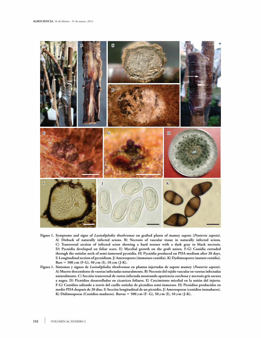
Necrosis appeared in recently grafted plants. Wilting and death of apical bud followed by dieback, gradual drying and cracking of the bark from the apex and moving down toward the base of the scion (Figure 1A), developed 22 d after grafting. The union of the graft presented longitudinal extended necrosis (Figure 1B). The vascular tissue of diseased scions showed hard texture (mummification of xylem and cortex) with a dark gray to black necrosis (Figure 1C). Thirty days after grafting, abundant light gray mycelial growth was observed covering most of the scion and the graft union (Figure 1E).
Pure, Invitrogen) y se cuantificó en un espectrofotómetro Perkin Elmer (Lambda BIO 10, EE.UU.). La amplifica-ción de las regiones internas ITS1 e ITS2 de los genes riboso-males (rRNA) se realizó mediante la reacción en cadena de la polimerasa (PCR) usando la combinación de los iniciadores universales ITS4 e ITS5 (White et al., 1990). La amplifica-ción y visualización de los productos finales se realizó según el protocolo de Ahrens y Seemüller (1992), con las modifica-ciones en las reacciones de PCR sugeridas por Vásquez et al. (2009). El producto amplificado se purificó con el kit Wizard (Promega) y secuenció con el Genetic Analizer modelo 3100, Applied Biosystem. La secuencia generada en este estudio se depositó en el GenBank (NCBI, 2011) con numero de acceso JQ245975.
Histopatología
Preparación de muestras para microscopía de luz
Se cortaron secciones transversales de 10 mm longitud10-15 mm de ancho de injertos de T1, T2 y T3. Las secciones se fijaron en FAA (etanol absoluto, ácido acético glacial, formaldehído y agua destilada), se incubaron 24 h a temperatura ambiente (202 °C), se lavaron con agua por 20 min e infiltraron en un procesador automático de tejidos (Tissue-Tek II, modelo 4640-B, Japón) como se describe a continuación. La deshidratación se realizó gradualmente en soluciones de alcohol etílico. Las muestras pasaron por una mezcla de etanol absoluto-xileno (1:1) y tres cambios en xile-no puro y después del último cambio en xileno, las muestras se colocaron 48 h en Paraplast (SIGMA). Las secciones flo-taron 1 min se colocaron en un baño a 65 °C con 3.0 g de grenetina y se montaron en portaobjetos de vidrio. La técnica de tinción diferencial safranina-verde rápido se realizó según lo descrito por Johansen (1940) y Curtis (1986). Las seccio-nes se desparafinaron en tres cambios de xileno (3 min cada uno) e hidrataron en una serie gradual de alcohol etílico. Las muestras se tiñeron con Safranina (Technical Chemistry) al 1 % en alcohol etílico al 50 % por 4 h. Luego, las seccio-nes se deshidrataron en una serie gradual de alcohol etílico al 50, 70 y 96 % (3 min cada uno) y se agregaron de 3-4 gotas de colorante verde rápido (Technical Chemistry) al 1 % en alcohol etílico al 96 % por 30 s. El exceso de co-lorante se decantó, las secciones se lavaron en alcohol etíli-co al 96 %, se deshidrataron 3 min en alcohol absoluto y pasaron por tres cambios de xileno (3 min cada uno). Fi-nalmente, las secciones se montaron en resina y examina-ron con un microscopio compuesto (Nikon SMZ800, EE.UU.).
152
AGROCIENCIA, 16 de febrero - 31 de marzo, 2012
VOLUMEN 46, NÚMERO 2
Figure 1. Symptoms and signs of Lasiodiplodia theobromae on grafted plants of mamey sapote (Pouteria sapota). A) Dieback of naturally infected scions. B) Necrosis of vascular tissue in naturally infected scions. C) Transversal section of infected scion showing a hard texture with a dark gray to black necrosis. D) Pycnidia developed on foliar scars. E) Mycelial growth on the graft union. F-G) Conidia extruded
through the ostiolar neck of semi-inmersed pycnidia. H) Pycnidia produced on PDA medium after 20 days. I) Longitudinal section of pycnidium. J) Amerospores (immature conidia). K) Dydimospores (mature conidia). Bars 500 m (F-G), 50 m (I), 10 m (J-K).Figura 1. Síntomas y signos de Lasiodiplodia theobromae en plantas injertadas de zapote mamey (Pouteria sapota). A) Muerte descendente de varetas infectadas naturalmente. B) Necrosis del tejido vascular en varetas infectadas
naturalmente. C) Sección transversal de vareta infectada mostrando apariencia corchosa y necrosis gris oscura a negra. D) Picnidios desarrollados en cicatrices foliares. E) Crecimiento micelial en la unión del injerto.
F-G) Conidios saliendo a través del cuello ostiolar de picnidios semi-inmersos. H) Picnidios producidos en medio PDA después de 20 días. I) Sección longitudinal de un picnidio. J) Amerosporas (conidios inmaduros). K) Didimosporas (Conidios maduros). Barras 500 m (F- G), 50 m (I), 10 m (J-K).
A B C
D
H
E
F G
I J K
IDENTIFICATION, PATHOGENICITY AND HISTOPATHOLOGY OF Lasiodiplodia theobromae ON MAMEY SAPOTE GRAFTS IN GUERRERO, MÉXICO
153TOVAR-PEDRAZA et al.
Isolates
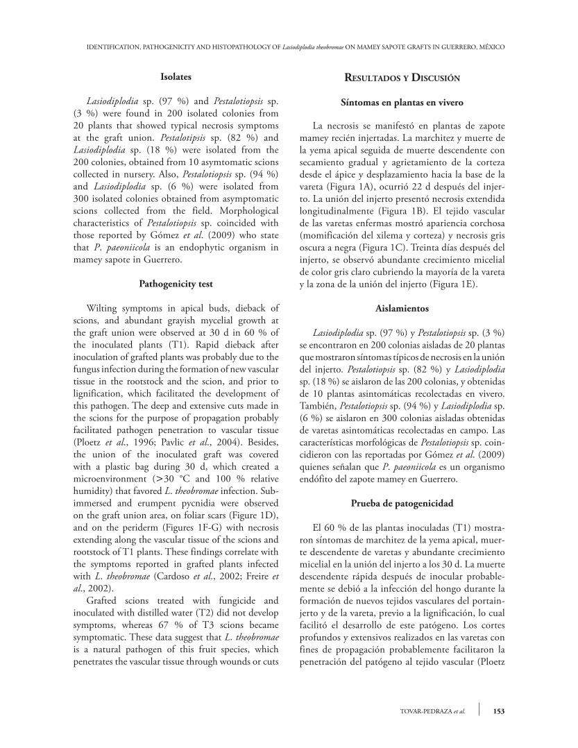
Lasiodiplodia sp. (97 %) and Pestalotiopsis sp. (3 %) were found in 200 isolated colonies from 20 plants that showed typical necrosis symptoms at the graft union. Pestalotipsis sp. (82 %) and Lasiodiplodia sp. (18 %) were isolated from the 200 colonies, obtained from 10 asymtomatic scions collected in nursery. Also, Pestalotiopsis sp. (94 %) and Lasiodiplodia sp. (6 %) were isolated from 300 isolated colonies obtained from asymptomatic scions collected from the field. Morphological characteristics of Pestalotiopsis sp. coincided with those reported by Gómez et al. (2009) who state that P. paeoniicola is an endophytic organism in mamey sapote in Guerrero.
Pathogenicity test
Wilting symptoms in apical buds, dieback of scions, and abundant grayish mycelial growth at the graft union were observed at 30 d in 60 % of the inoculated plants (T1). Rapid dieback after inoculation of grafted plants was probably due to the fungus infection during the formation of new vascular tissue in the rootstock and the scion, and prior to lignification, which facilitated the development of this pathogen. The deep and extensive cuts made in the scions for the purpose of propagation probably facilitated pathogen penetration to vascular tissue (Ploetz et al., 1996; Pavlic et al., 2004). Besides, the union of the inoculated graft was covered with a plastic bag during 30 d, which created a microenvironment (30 °C and 100 % relative humidity) that favored L. theobromae infection. Sub-immersed and erumpent pycnidia were observed on the graft union area, on foliar scars (Figure 1D), and on the periderm (Figures 1F-G) with necrosis extending along the vascular tissue of the scions and rootstock of T1 plants. These findings correlate with the symptoms reported in grafted plants infected with L. theobromae (Cardoso et al., 2002; Freire et al., 2002). Grafted scions treated with fungicide and inoculated with distilled water (T2) did not develop symptoms, whereas 67 % of T3 scions became symptomatic. These data suggest that L. theobromae is a natural pathogen of this fruit species, which penetrates the vascular tissue through wounds or cuts
resultAdos y dIscusIón
Síntomas en plantas en vivero
La necrosis se manifestó en plantas de zapote mamey recién injertadas. La marchitez y muerte de la yema apical seguida de muerte descendente con secamiento gradual y agrietamiento de la corteza desde el ápice y desplazamiento hacia la base de la vareta (Figura 1A), ocurrió 22 d después del injer-to. La unión del injerto presentó necrosis extendida longitudinalmente (Figura 1B). El tejido vascular de las varetas enfermas mostró apariencia corchosa (momificación del xilema y corteza) y necrosis gris oscura a negra (Figura 1C). Treinta días después del injerto, se observó abundante crecimiento micelial de color gris claro cubriendo la mayoría de la vareta y la zona de la unión del injerto (Figura 1E).
Aislamientos
Lasiodiplodia sp. (97 %) y Pestalotiopsis sp. (3 %)se encontraron en 200 colonias aisladas de 20 plantas que mostraron síntomas típicos de necrosis en la unión del injerto. Pestalotiopsis sp. (82 %) y Lasiodiplodia sp. (18 %) se aislaron de las 200 colonias, y obtenidas de 10 plantas asintomáticas recolectadas en vivero. También, Pestalotiopsis sp. (94 %) y Lasiodiplodia sp. (6 %) se aislaron en 300 colonias aisladas obtenidas de varetas asintomáticas recolectadas en campo. Las características morfológicas de Pestalotiopsis sp. coin-cidieron con las reportadas por Gómez et al. (2009) quienes señalan que P. paeoniicola es un organismo endófito del zapote mamey en Guerrero.
Prueba de patogenicidad
El 60 % de las plantas inoculadas (T1) mostra-ron síntomas de marchitez de la yema apical, muer-te descendente de varetas y abundante crecimiento micelial en la unión del injerto a los 30 d. La muerte descendente rápida después de inocular probable-mente se debió a la infección del hongo durante la formación de nuevos tejidos vasculares del portain-jerto y de la vareta, previo a la lignificación, lo cual facilitó el desarrollo de este patógeno. Los cortes profundos y extensivos realizados en las varetas con fines de propagación probablemente facilitaron la penetración del patógeno al tejido vascular (Ploetz

154
AGROCIENCIA, 16 de febrero - 31 de marzo, 2012
VOLUMEN 46, NÚMERO 2
made by the propagator or grafter. This coincides with Freire et al. (2002), Fourie and Halleen (2004) and Retief et al. (2006), who affirm that the infected scions and rootstocks are the source of inoculum of pathogens that cause diseases during the vegetative grapevine propagation. In mamey sapote, the most likely source of primary inoculum are the scions collected from orchards with deficient agronomic management and high incidence of dieback of branches caused by L. theobromae. The pressure of inoculum by this pathogen was lower in the spring grafting season (February-March) with an incidence of 69 %, whereas in the autumn (September-November) the incidence was 87 % in naturally infected scions, coinciding with the period of June to November when the highest number of L. theobromae conidia were found in volumetric spore traps in mamey orchards (Vásquez et al., 2009). The present study did not verify the pathogenicity of P. paeoniicola because of its endophytic nature in mamey trees in the zone (Gómez et al., 2009).
Morphological characterization
Cultural characteristics
Fungal culture showed a rapid grown and abundant aerial mycelium (1-3 d). The aerial mycelium was initially gray, then becoming olive gray and denser in the center of the dish. Pycnidial conidiomata were observed after 14 d (Figure 1H). Pycnidia were produced in stroma, simple or compound, scattered, and often aggregated.
In vitro
Pycnidia were black, obpyriform and ostiolate. Immature conidia (18 d) were hyaline, ellipsoidal to subovoid, amerospore, 22.73-27.0411.88-15.84 m, thick-walled with a granular content. Mature conidia (23 d) were dark brown, ellipsoid to ovoid, didymospore, 19.66-26.3511.3-14.17 m, with longitudinal and irregular striations.
In vivo
Natural and experimentally infected scions showed globose pycnidia, immersed in the host
et al., 1996; Pavlic et al., 2004). Además, la unión del injerto inoculado fue cubierta con una bolsa plástica transparente por 30 d, lo cual creó un mi-croambiente (30 °C y 100 % humedad relativa) que favoreció la infección por L. theobromae. Pic-nidios sub-inmersos y errumpentes se observaron en el área de unión del injerto, en cicatrices foliares (Figura 1D) y en peridermis (Figuras 1F-G), con una necrosis que se extendió a lo largo del tejido vascular de las varetas y portainjertos del T1, co-incidiendo con los síntomas reportados en plantas injertadas e infectadas con L. theobromae (Cardoso et al., 2002; Freire et al., 2002). Las varetas injertadas, tratadas con fungicida e inoculadas con agua destilada esteril (T2) no desa-rrollaron síntomas, mientras que 67 % de las varetas del T3 presentaron síntomas. Estos datos sugieren que L. theobromae es un patógeno natural de este fru-tal y penetra al tejido vascular a través de heridas o cortes realizados por el propagador o injertador. Esto coincide con Freire et al. (2002), Fourie y Halleen (2004) y Retief et al. (2006), quienes señalan que los portainjertos y las varetas infectadas son la fuente de inóculo de los patógenos que causan enfermedades durante la propagación vegetativa de la vid. En zapote mamey, la fuente más probable de inóculo primario son las varetas recolectadas en huertos con manejo agronómico deficiente y alta incidencia de muerte descendente de ramas cau-sada por L. theobromae. La presión de inóculo por este patógeno fue menor en el periodo de injertar de primavera (febrero-marzo) con una incidencia de 69 %, mientras que, en otoño (septiembre-noviem-bre), la incidencia fue 87 % en varetas infectadas na-turalmente, coincidiendo con el periodo de junio a noviembre, cuando se encontró la mayor cantidad de conidios de L. theobromae en trampas volumétricas en huertos de zapote mamey (Vásquez et al., 2009). En el presente estudio no se verificó la patogenicidad de P. paeoniicola debido a su hábito endofítico en árboles de zapote mamey en la zona (Gómez et al., 2009).
Caracterización morfológica
Características culturales
La colonia del hongo mostró crecimiento micelial aéreo rápido y abundante (1-3 d). El micelio aéreo

IDENTIFICATION, PATHOGENICITY AND HISTOPATHOLOGY OF Lasiodiplodia theobromae ON MAMEY SAPOTE GRAFTS IN GUERRERO, MÉXICO
155TOVAR-PEDRAZA et al.
tissue (Figure 1I); they were errumpent, simple and clustered, and dark brown to black, 235-380190 to 335 m, with a long neck ostiole. Conidiophores were hyaline, cylindrical, simple, sometimes septate, arising from the inner layer of cells lining the pycnidial cavity. Paraphyses were hyaline, cylindrical and aseptate. Immature conidia were hyaline, subovoid to ellipsoid, aseptate, granular (Figure 1J), and measured 23.4-27.3212.26-15.61 m. Mature conidia were dark brown, ovoid to ellipsoid, uniseptate, 21.19-26.7611.15-13.38 m, and showing longitudinal and irregular striations (Figure 1K). Observations with SEM showed erumpent and globose pycnidia, unilocular (Figure 2A) and multilocular with elongated ostiolar neck (Figure 2B), with conidia extruding in a mass (Figure 2C). Mature conidia had irregular and longitudinal striations from apex to base (Figure 2D), and an internal septum (Figure 2E). Conidia germinate through simple germ tubes (Figure 2F). Cultural characteristics and structures of asexual reproduction coincided with those reported by Burgess et al. (2006) for L. theobromae. The isolate used in this study is maintained in the culture collection (Accession No. CB007) of the herbarium (CMPH) at Colegio de Postgraduados, Campus Montecillo, Fitopatología, Texcoco, Estado de México.
Molecular characterization
The molecular analysis confirmed that the fungus which causes dieback of mamey sapote scions in Guerrero was L. theobromae. The sequence (GenBank Accession No. JQ245975) obtained from this study showed 99 % of similarity to the sequence of L. theobromae with accession number GQ469934.
Histopathology
Structural abnormalities were not observed in asymptomatic tissues (Figures 3A-C).
Anatomical description of artificially infected scions and rootstocks
Pycnidia of L. theobromae at various stages of development (Figures 3D-E) were found 30 dai,
inicialmente era gris, llegando a ser gris oliváceo y más denso en el centro del disco. Conidiomas pic-nidiales se observaron después de 14 d (Figura 1H). Picnidios se produjeron en estroma, simples o com-puestos, dispersos y frecuentemente agregados.
In vitro
Los picnidios eran negros, obpiriformes, ostio-lados. Conidios inmaduros (18 d) eran hialinos, elipsoidales a subovoides, amerosporas, de 22.73-27.0411.88-15.84 m, con pared densa y con-tenido granular. Los conidios maduros (23 d) eran café oscuro, elipsoidales a ovoides, didimosporas, de 19.66-26.3511.3-14.17 m, y con estriaciones longitudinales irregulares.
In vivo
Las varetas infectadas natural y experimentalmen-te mostraron picnidios globosos, inmersos en el teji-do del hospedante (Figura 1I), errumpentes, simples y agrupados, café oscuro a negros, de 235-380190-335 m, con cuello ostiolar largo. Conidióforos hia-linos, cilíndricos, simples y algunas veces septados, surgiendo de la capa interior de células que recubren la cavidad picnidial. Paráfisis hialinos, cilíndricos y aseptados. Conidios inmaduros hialinos, subovoides a elipsoidales, aseptados, granulados (Figura 1J), de 23.4-27.3212.26-15.61 m. Conidios maduros café oscuro, ovoides a elipsoidales, uniseptados, de 21.19-26.7611.15-13.38 m y mostrando estrías longitudinales irregulares (Figura 1K). Con MEB se observaron picnidios globosos y errumpentes, uniloculares (Figura 2A) y multilo-culares con cuello ostiolar alargado (Figura 2B), con extrusión de conidios en masas (Figura 2C). Conidios maduros con estriaciones longitudinales irregulares del ápice a la base (Figura 2D), y un sep-to interno (Figura 2E). Los conidios germinaron a través de tubos germinativos simples (Figura 2F). Las características culturales y estructuras de reproducción asexual coincidieron con las reporta-das por Burgess et al. (2006) para L. theobromae. El aislamiento usado en este estudio se depositó en la colección de cultivos (No. de acceso CB007) del herbario (CMPH) en el Colegio de Postgraduados, Campus Montecillo, Fitopatología, Montecillo, Texcoco, Estado de México.

156
AGROCIENCIA, 16 de febrero - 31 de marzo, 2012
VOLUMEN 46, NÚMERO 2
Figure 2. SEM micrographs of Lasiodiplodia theobromae fruiting bodies on artificially infected mamey sapote (Pouteria sapota) scions. A) Longitudinal section of pycnidium (Pyc) showing mature conidia. B) Sub-immersed pycnidia showing ostiole with extruding of conidia. C) Conidia (Co) grouped in masses. D) Mature conidia with irregular longitudinal striations (Ls). E) Longitudinal section of mature conidia showing internal septum (Sep) and granular content. F) Conidia showing germ tube (Gt).
Figura 2. Micrografías de estructuras fructíferas de Lasiodiplodia theobromae en varetas de zapote mamey (Pouteria sapota) infectadas artificialmente y vistas en MEB. A) Sección longitudinal de picnidio (Pyc) mostrando conidios maduros. B) Picnidios sub-inmersos presentando ostiolo con extrusión de conidios. C) Conidios (Co) agrupados en masas. D) Conidios maduros con estriaciones longitudinales irregulares (Ls). E) Sección longitudinal de conidio maduro exhibiendo septo interno (Sep) y contenido granular. F) Conidio maduro con tubo germinativo (Gt).
10kV X350 50m
10kV X500 50m
10kV X110 100m
Co
CoPyc
10kV X700 20m
Ls
10kV X11000 1m
Sep
10kV X4500 5m
Gt

IDENTIFICATION, PATHOGENICITY AND HISTOPATHOLOGY OF Lasiodiplodia theobromae ON MAMEY SAPOTE GRAFTS IN GUERRERO, MÉXICO
157TOVAR-PEDRAZA et al.
Figure 3. Cross sections micrographs of asymptomatic and Lasiodiplodia theobromae infected mamey sapote grafts (scion and rootstock), 30 days after grafting and inoculating. A) Rootstock (Ro) and B) scion (Sci) showing periderm (P), cortex (Ct), phloem (Ph), xylem (X) and medulla (Pi). C) Asymptomatic graft. D) Infected graft showing inoculated mycelia (Myc) and pycnidia (Pyc). E) Pycnidia developed on periderm of artificially infected scion. F) Xylem vessels (Xv) in asymptomatic scion. G) Accumulation of red inclusions (Ri) in xylem vessels of artificially infected scions. H) Pith parenchyma cells of asymptomatic scions. I) Pith parenchyma cells of artificially infected scions showing abundant hyphae (Hy) and accumulation of red inclusions (Ri). J) Pith parenchyma cells of asymptomatic rootstock showing starch granules (Sg). K) Medulla parenchyma cells of artificially infected rootstock showing accumulation of red inclusions. Bars 500 m (A-D), 100 m (E-G), 50 m (H-K).
Figura 3. Fotomicrografías de cortes transversales de injertos (vareta y portainjerto) de zapote mamey (Pouteria sapota) asintomáticos e infectados con Lasiodiplodia theobromae, 30 días después de la injertación e inoculación.
A) Portainjerto (Ro) y B) vareta (Sci) asintomática mostrando peridermis (P), corteza (Ct), floema (Ph), xilema (X) y médula (Pi). C) Injerto asintomático. D) Injerto infectado mostrando micelio inoculado (Myc) y picnidios (Pyc). E) Picnidios desarrollados en peridermis de vareta infectada artificialmente. F) Vasos del xilema (Xv) en vareta asintomática. G) Vasos del xilema en vareta infectada artificialmente mostrando acumulación de inclusiones rojas (Ri). H) Células parenquimatosas de la médula de vareta asintomática. I) Células parenquimatosas de la médula de vareta infectada artificialmente mostrando abundantes hifas (Hy) y acumulación de inclusiones rojas. J) Células parenquimatosas de la médula de portainjerto asintomático mostrando gránulos de almidón (Sg). K) Células parenquimatosas de la médula de portainjerto infectado artificialmente mostrando acumulación de inclusiones rojas. Barras 500 m (A-D), 100 m (E-G), 50 m (H-K).
Ct
P
Ph
X
Pi
A
Ph
Ct
X
Pi
Sci Sci
Ro Ro
Myc Pyc
Pyc
Pyc
Xv
Ri
Ri
SgHy
Ri
Hy
B C D
E F G
H I J K

158
AGROCIENCIA, 16 de febrero - 31 de marzo, 2012
VOLUMEN 46, NÚMERO 2
which correlates with Biggs and Britton (1988), Michailides (1991) and Rayachhetry et al. (1996), who report the presence of reproductive structures of various Botryosphaeria spp. anamorphs between 12-56 dai. Our findings contrast with those observed by Vásquez et al. (2009) who found no pycnidia in mamey sapote samples with dieback symptoms 24 months after inoculation. In our study, the deep cuts made during the grafting process could facilitate an extensive route of penetration. It is possible that the host susceptibility has been greater during the formation of new vascular tissues of rootstock and scion, prior to lignification, which could help the development of L. theobromae. Histological damages observed on the cortex of artificially infected scions (Figure 3D) could be attributed to the presence of pycnidia as well as mechanical injuries caused during the grafting process. Parenchyma cells of the cortex and phloem collapsed and became necrotic. Red inclusions, probably of phenolic nature, were observed in 10 % of the xylem vessels (Figure 3G) and in 13 % [percentage with respect to asymptomatic tissues (Figure 3H)] of pith parenchyma cells (Figure 3I). The presence of these compounds has been explained as a plant response to colonization of fungal, bacterial and oomycete pathogens. Lasiodiplodia theobromae quickly penetrated and colonized all tissues of the inoculated scion. The mycelium, characterized by developing long and branched hyphae, grew intracellularly in xylem vessels and medulla parenchyma cells (Figure 3I). The presence of inclusions and hyphae of the pathogen in the xylem vessels may explain the restriction of the flow of water, mineral salts and other solutes to the rest of the plant, resulting in wilt and dieback of branches (Rayachhetry et al., 1996; Pandit and Samajpati, 2005). Hyphae of Botryosphaeria spp. has been observed in the cortex, xylem vessels, pith, phloem and xylem parenchyma of apple, peach and Melaleuca quinquenervia (Brown and Hendrix, 1981; Biggs and Briton, 1988; Rayachhetry et al., 1996), confirming that these species are specialized pathogens that colonize all the tissues in branches and stems of woody plants. Vásquez et al. (2009) did not observe hyphae in mamey sapote branch tissues infected with L. theobromae, possibly because their observation was made 24 months after inoculation on collapsed necrotic tissues.
Caracterización molecular
El análisis molecular confirmó que el hongo cau-sante de la muerte de varetas injertadas de zapote mamey en Guerrero fue L. theobromae. La secuen-cia (No. acceso del GenBank JQ245975) obtenida en este estudio mostró 99 % de similaridad con la secuencia de L. theobromae con número de acceso GQ469934.
Histopatología
No se observaron anormalidades estructurales en los tejidos asintomáticos (Figuras 3A-C).
Descripción anatómica de varetas y portainjertos infectados artificialmente
A los 30 ddi se observaron picnidios de L. theobromae en distintas etapas de desarrollo (Figuras 3D-E), lo que coincide con Biggs y Briton (1988), Michailides (1991) y Rayachhetry et al. (1996), quienes reportan la presencia de estructuras repro-ductivas de diversos anamorfos de Botryosphaeria spp. a los 12-56 ddi. Los resultados del presente es-tudio contrastan con los de Vásquez et al. (2009), quienes no reportan picnidios en muestras de za-pote mamey con síntomas de muerte descendente 24 meses después de la inoculación. En el presen-te estudio, los cortes profundos hechos durante el injerto pudieron facilitar una ruta de penetración extensiva. Es posible que la susceptibilidad del hos-pedante haya sido mayor durante la formación de nuevos tejidos vasculares del portainjerto y de la vareta, previo a la lignificación, lo cual facilitó el desarrollo de L. theobromae. Daños histológicos en la corteza de varetas infec-tadas artificialmente (Figura 3D) podrían atribuirse a la presencia de picnidios, así como a daños mecánicos generados durante el proceso de injerto. Las células parenquimáticas de la corteza y del floema colapsa-ron y necrosaron. Inclusiones rojas probablemente de naturaleza fenólica, se observaron en 10 % de los vasos del xilema (Figura 3G) y en 13 % [porcentaje con respecto al asintomático (Figura 3H)] de las cé-lulas parenquimatosas de la médula (Figura 3I). La presencia de estos compuestos se relaciona con una respuesta de la planta a la colonización por hongos, bacterias y oomicetes.
IDENTIFICATION, PATHOGENICITY AND HISTOPATHOLOGY OF Lasiodiplodia theobromae ON MAMEY SAPOTE GRAFTS IN GUERRERO, MÉXICO
159TOVAR-PEDRAZA et al.
Tyloses were not observed in the xylem vessels, as reported in other interactions: Botryosphaeria spp.-blueberry and apple (Milholland, 1972; Brown and Hendrix, 1981) and L. theobromae-grapevine (Atia et al., 2003), as a major cause of obstruction of water flow and dieback. Pith parenchyma of asymptomatic rootstocks contained starch granules (Figure 3J), which were not observed in artificially infected rootstocks. The fungus probably uses starch as a carbon source for energy and growth, since some fungi can use the starch by amylase production (Deacon, 2006). Red inclusions of probably phenolic nature were also observed in 7 % of pith parenchyma cells in rootstocks (Figure 3K).
Anatomical description of naturally infected scions and rootstocks
Lasiodiplodia theobromae hyphae showed a similar growth pattern to that observed in artificially infected tissue. However, pycnidia were not present in the periderm. The absence of these structures could be explained by the process of penetration and infection, which occurred slowly as the pressure of inoculum was lower compared with the scions inoculated with the suspension of hyphae in the wounds. Accumulation of red inclusions 5 % (percentage with respect to asymptomatic) was lower in pith parenchyma compared with artificially infected grafts, which could be due to a lower extent of colonization by fungus in the tissues. The subsequent formation of pycnidia on twigs that survived natural infection in the field is perhaps the main source of inoculum for the spread of the disease in mamey sapote orchards in Guerrero.
conclusIons
Pathogenicity and histopathological data showed that Lasiodiplodia theobromae is the causal agent of dieback of mamey sapote grafts. This is the first study that provides a better understanding of a disease caused by L. theobromae during the grafting process.
Acknowledgements
This research was financially supported by Fundación Produce de Guerrero, México (Project PM 1731). The
Lasiodiplodia theobromae penetró y colonizó rápi-damente todos los tejidos de las varetas inoculadas. El micelio, caracterizado por desarrollar hifas largas y ramificadas, creció intracelularmente en vasos del xi-lema y células parenquimatosas de la médula (Figura 3I). La presencia de inclusiones e hifas del patógeno en los vasos del xilema explicaría la restricción del flujo de agua, sales minerales y otros solutos al resto de la vareta, resultando en marchitamiento y muer-te descendente de ramas (Rayachhetry et al., 1996; Pandit y Samajpati, 2005). Hifas de Botryosphaeria spp. se han observado en la corteza, vasos del xilema, médula, floema y parénquima del xilema de man-zano, durazno y Melaleuca quinquenervia (Brown y Hendrix, 1981; Biggs y Briton, 1988; Rayachhetry et al., 1996), lo que confirma que estas especies son patógenos especializados en colonizar todos los teji-dos de ramas y tallos de plantas leñosas. Vásquez et al. (2009) no observaron hifas en tejidos de ramas de zapote infectadas con L. theobromae, posiblemente porque sus análisis se hicieron en tejidos necróticos y colapsados 24 meses después de la inoculación. No se observaron tilosas en los vasos del xilema, como se reporta en otras interacciones: Botryosphaeria spp.-arándano y manzano (Milholland, 1972; Brown and Hendrix, 1981) y L. theobromae-vid (Atia et al., 2003), como causa principal de obstrucción del flujo de agua y muerte descendente. La médula parenquimatosa de portainjertos asin-tomáticos presentaron gránulos de almidón (Figu-ra 3J), los cuales no se observaron en portainjertos infectados artificialmente. El hongo probablemente usa almidón como fuente de carbono para energía y desarrollo, ya que algunos hongos pueden usar almi-dón mediante la producción de amilasas (Deacon, 2006). Inclusiones rojas de probable naturaleza fe-nolica se observaron en 7 % de las células paren-quimáticas de la médula en los portainjertos (Figure 3K).
Descripción anatómica de varetas y portainjertos infectados naturalmente
Las hifas de L. theobromae presentaron un patrón de crecimiento similar a lo observado en el tejido infectado artificialmente. Sin embargo, los picnidios no estaban presentes en la peridermis. La ausencia de estas estructuras podría explicarse por los procesos de penetración e infección que ocurrieron lentamente

160
AGROCIENCIA, 16 de febrero - 31 de marzo, 2012
VOLUMEN 46, NÚMERO 2
authors are grateful to Greta Hanako Rosas Saito (Unidad de Microscopía Electrónica, Colegio de Postgraduados) for her technical assistance with the SEM.
lIterAture cIted
Ahrens, U., and E. Seemüller. 1992. Detection of DNA of plant pathogenic mycoplasmalike organisms by polymerase chain reaction that amplifies a sequence of the 16S rRNA gene. Phytopathology 82: 828-832.
Álvarez, V. G. 1997. La muerte descendente y el cáncer del tallo en el zapote. Tikalia 15: 37-46.
Aroca, A., R. Raposo, D. Gramaje, J. Armengol, S. Martos, and J. Luque. 2008. First report of Lasiodiplodia theobromae on rootstocks mother grapevines in Spain. Plant Dis. 92: 832.
Atia, M. M. M., A. Z. Aly, M. R. A. Tohamy, H. El-Shimy, and M. A. Kamhawy. 2003. Histopathological studies on grapevine die-back. J. Plant Dis. Protect. 110: 131-142.
Azurdía, C. 2006. Tres Especies de Zapote en América Tropical. Southampon Centre for Underutilised Crops, Southampon University, Southampon, UK. 216 p.
Barnett, L. H., and B. B. Hunter. 2006. Illustrated Genera of Imperfect Fungi. Fourth Edition. The American Phytopathological Society. St. Paul, Minnesota, USA. 218 p.
Bautista-Baños, S., J. C. Díaz-Perez, and L. L. Barrera-Nencha. 2002. Postharvest fungal rots of sapote mamey Pouteria sapota H. E. Moore & Stearn. Postharvest Biol. Technol. 24: 197-200.
Biggs, A. R., and K. O. Britton. 1988. Presymptom histopathology of peach trees inoculated with Botrysphaeria obtusa and B. dothidea. Phytopathology 78: 1109-1118.
Brown, E. A. II., and F. F. Hendrix. 1981. Pathogenicity and histopathology of Botryosphaeria dothidea on apple stems. Phytopathology 71: 375-379
Burgess, T. I., P. A. Barber, S. Mohali, G. Pegg, W. de Beer, and M. J. Wingfield. 2006. Three new Lasiodiplodia spp. from the tropics, recognized based on DNA sequence comparisons and morphology. Mycologia 98: 423-435.
Cardoso, J. E., C. M. Maia, and M. N. G. Pessoa. 2002. Occurrence of Pestalotiopsis psidii and Lasiodiplodia theobromae causing stem rot of guava plants in the State of Ceará, Brazil. Fitopatol. Bras. 27: 320.
Curtis, P. J. 1986. Microtécnica Vegetal. Ed. Trillas, México. 106 p.Davis, R. M., C. J. Farrald, and D. Davila. 1987. Botryodiplodia
trunk lesions in Texas citrus. Plant Dis. 71: 848-849.Deacon, J. 2006. Fungal Biology. Fourth Edition. Blackwell
Publishing. Cornwall, England. 372 p.Farr, D. F., G. F. Bill, G. P. Chamuris, and A.Y. Rossman. 1989.
Fungi on Plants and Plant Products in the United States. APS Press. St. Paul, Minnesota, USA. 1252 p.
Fourie, P. H., and F. Halleen. 2004. Occurrence of grapevine trunk disease pathogens in rootstock mother plants in South Africa. Austral. Plant Pathol. 33: 313-315.
Freire, F. C. O., J. E. Cardoso, A. A. dos Santos, and F. M. P. Viana. 2002. Diseases of cashew nut plants (Anacardium occidentale L.) in Brazil. Crop Protection 21: 489-494.
Gómez, J. R., D. Nieto A., D. Téliz O., A. Mora A., M. T. Martínez D., y M. Vargas H. 2009. Evaluación de la calidad e incidencia de hongos en frutos refrigerados de zapote
dado que la presión del inóculo fue menor compa-rada con las varetas inoculadas con la suspensión de hifas en las heridas. La acumulación de 5 % (porcentaje con respecto al asintomático) de inclusiones rojas fue menor en las células parenquimatosas, comparado con las vare-tas infectadas artificialmente, lo cual podría deberse a un menor alcance de la colonización del hongo en los tejidos. La posterior formación de picnidios en varetas que sobrevivieron a la infección natural en campo es tal vez la principal fuente de inóculo para la diseminación de la enfermedad en huertos de zapote mamey en Guerrero.
conclusIones
Los datos de patogenicidad e histopatología mos-traron que Lasiodiplodia theobromae es el agente cau-sal de la muerte descendente de injertos de zapote mamey. Este es el primer estudio que provee un me-jor entendimiento sobre una enfermedad causada por L. theobromae durante el proceso de injerto.
—Fin de la versión en Español—
pppvPPP
mamey (Pouteria sapota (Jacq.) H. E. Moore and Stearn. Agrociencia 43: 37-48.
Johansen, D. A. 1940. Plant Microtechnique. McGraw Hill, New York, USA. 503 p.
Michailides, T. J. 1991. Pathogenicity, distribution, sources of inoculum, and infection courts of Botryosphaeria dothidea on pistachio. Phytopathology 81: 566-573.
Milholland, R. D. 1972. Histopathology and pathogenicity of Botryosphaeria dothidea on blueberry stems. Phytopathology 62: 654-660.
Mora, A. J. A., A. Vásquez L., J. Pereyda H., y D. Téliz O. 2008. Algunas enfermedades del zapote mamey (Pouteria sapota (Jacq.) en México. El Zapote Mamey en México: Avances de Investigación. Morelos, México. pp: 103-114.
NCBI (National Center for Biotechnology Information). 2011. GenBank. http//:www.ncbi.nlm.nih.gov/. (accessed: December 2011).
Ogden, M. A. H., C. W. Campbell, and S. Lara P. 1986. Grafting techniques for mamey sapote (Calocarpum sapota [Jacq.] Merr.) under Florida conditions. Proc. Interamerican Soc. Trop. Hort. 30: 215-221.
Pandit, P. K., and N. Samajpati. 2005. Wilt disease of guava caused by Botryodiplodia theobromae Pat. J. Mycopathol.Res. 43: 41-43.
Pavlic, D., B. Slippers, T. A. Coutinho, M. Gryenhout, and M. J. Wingfield. 2004. Lasiodiplodia gonubiensis sp. nov., a new Botryosphaeria anamorph from native Syzygium

IDENTIFICATION, PATHOGENICITY AND HISTOPATHOLOGY OF Lasiodiplodia theobromae ON MAMEY SAPOTE GRAFTS IN GUERRERO, MÉXICO
161TOVAR-PEDRAZA et al.
cordatum in South Africa. Studies in Mycol. 50: 313-322.Pereyda, H. J., J. A. Mora A., J. G. Florencio A., C. Nava D., D.
Téliz O., S. Sandoval I., y A. Villegas M. 2008. Proliferación vegetativa y floral del zapote mamey (Pouteria sapota (Jacq.)) en México. El Zapote Mamey en México: Avances de Investigación. Morelos, México. pp: 115-130.
Ploetz, R. C., D. Benscher, A. Vazquez, A. Colls, J. Nagel, and B. Schaffer. 1996. A reexamination of mango decline in Florida. Plant Dis. 80: 664-668.
Popenoe, W. 1974. Manual of Tropical and Sub-tropical Fruits, Excluding the Banana, Coconut, Pineapple, Citrus Fruit, Olive and Fig. Hafner Press, New York, USA. 542 p.
Rayachhetry, M. B., G. M. Blankslee, and T. Miller. 1996. Histopathology of Botryosphaeria ribis in Melaleuca quinquenervia: Pathogen invasion and host response. International J. Plant Sci. 157: 219-227.
Retief, E., A. McLeod, and P. H. Fourie. 2006. Potential inoculum sources of Phaeomoniella chlamydospora in South African grapevine nurseries. Eur. J. Plant Pathol. 115: 331-339.
SIAP (Servicio de Información Agroalimentaria y Pesquera). 2010. www.siap.sagarpa.gob.mx. (Accessed: July 2011).
Sutton, B. C. 1980. The Coleomycetes: Fungi Imperfecti with Pycnidia, Acervuli and Stromata. Commonwealth Mycological Institute, Kew, Surrey, England. 696 p.
Vásquez, L. A., J. A. Mora A., E. Cárdenas S., y D. Téliz O. 2009. Etiología e histopatología de la muerte descendente de árboles de mamey (Pouteria sapota (Jacq.) H.E. Moore and Stearn) en Guerrero, México. Agrociencia 43: 717-728.
Villegas, M. A., y J. A. Mora A. 2008. Propagación vegetativa del zapote mamey. El Zapote Mamey en México: Avances de Investigación. Morelos, México. pp: 1-16.
White, T. J., B. S. Lee, and J. Taylor. 1990. Amplification and direct sequencing of fungal ribosomal RNA genes for phylogenetics. In: Innis, M. A., D. A. Gelfand, J. J. Sninsky, and T. J. White (eds). PCR Protocols: A Guide to Methods and Applications, Academic Press, CA, USA. pp: 315-322.