T-614, a novel immunomodulator, attenuates joint inflammation and ...
Upload
trinhthienCategory
view
218download
2
IDENTIFICATION OF NOVEL THERAPEUTIC TARGETS FOR TRAUMATIC BRAIN INJURY BY PROTEOMIC ANALYSIS OF RESPONSE TO INJURY IN APOE3 AND APOE4
TRANSGENIC MICE
Marcie Wood1,2 (*), Benoit Mouzon1,2, Jon Reed1, Gogce Kayihan1, Scott Ferguson1,2, Venkatarajan Mathura1,2, Allen Roses3, Michael Mullan1,2, and Fiona Crawford1,2,
1 Roskamp Institute, Sarasota, Florida 2 James A. Haley Veterans’ Hospital, Tampa, Florida
3 R. David Thomas Executive Training Center, Duke University, Durham, North Carolina
ABSTRACT
Brain injury due to trauma (traumatic brain injury or TBI) is a significant cause of mortality and both acute and chronic morbidity, particularly in military personnel. Two over-arching issues must be addressed before effective pharmacologic treatments can be developed for TBI. Firstly, peripheral biomarkers reflecting the degree and nature of injury need to be elucidated. Secondly, the molecular cascades that lead to neuronal damage and death need to be fully quantitatively and qualitatively understood with a view to finding target molecules suitable for pharmacotherapy.
We are using state of the art proteomic approaches to analyze the brain tissue from mouse models of TBI administered using the controlled cortical impact (CCI) procedure. In order to maximize the discrimination between recovery outcomes these studies are being carried out on APOE3 and APOE4 transgenic mice, which demonstrate relatively favorable and unfavorable outcomes respectively, following TBI. As APOE genotype is key in determining the clinical consequences of TBI, injury-dependent changes in protein expression in brain tissue expressing apoE3 versus apoE4 will be expected to correlate with outcome.
To identify proteins whose response to injury is time,
severity and/or genotype dependent we are employing iTRAQ methodology to enable qualitative and quantitative analysis of response to injury across groups, and validation of responding proteins will use standard antibody-based techniques. Proteins that correlate with these central changes may also be detectable in the periphery and will be targeted for investigation in peripheral samples from the mice for their potential value as biomarkers for TBI. Neurobehavioral and neuropathological correlates are also under investigation in the chronic paradigms. Data analysis includes interrogation of knowledgebases to identify the variable-dependent cellular mechanisms after TBI. Cellular mechanisms associated with apoE4 versus apoE3 response to injury may indicate targets for therapeutic intervention.
1. INTRODUCTION
Traumatic brain injury (TBI) is a significant cause of mortality and of acute and chronic morbidity, particularly in military personnel, and is cited to be the leading cause of death and disability in the most active population (under 45 years of age) in industrialized countries (Cernak, 2006). Current standard treatment of TBI focuses, by necessity, on the physiological consequences of the injury, but the pathophysiology of TBI is complex and dependent on mechanism, type, severity and location of injury, with the sequelae persisting for months and years after the event; the occurrence and timing of these processes being key in determining outcome after injury.
In order to address the complexity of molecular events following TBI we are utilizing state of the art proteomic approaches to capture the brain-region specific response to injury in mouse models of TBI. Our program utilizes transgenic mice expressing different forms of the human APOE gene which is a known risk factor for outcome post-TBI. As the E4 genotype is associated with a more negative prognosis following TBI compared to E3, we are undertaking differential proteomic analyses; comparing the E4 response to TBI with E3 in order to specifically identify the negative sequelae which could be inhibited, or positive sequelae which could be enhanced, in therapeutic approaches. In tandem with brain tissue analysis we will also look for peripheral biomarkers associating with TBI outcome.
We have just begun to generate data from this
program, and the preliminary data are reported below.
The potential role of Proteomics in understanding TBI There have been few systematic attempts to highlight
key target molecules in TBI and design rational new drug therapy accordingly. There are a number of reasons for this, one of which has been the absence of complete time dependent maps of networks of effector proteins activated after TBI. The technological advances which now allow the isolation and analysis of proteins are coupled to protein databases so that individual proteins, including their post-translational modifications, can be identified

Report Documentation Page Form ApprovedOMB No. 0704-0188
Public reporting burden for the collection of information is estimated to average 1 hour per response, including the time for reviewing instructions, searching existing data sources, gathering andmaintaining the data needed, and completing and reviewing the collection of information. Send comments regarding this burden estimate or any other aspect of this collection of information,including suggestions for reducing this burden, to Washington Headquarters Services, Directorate for Information Operations and Reports, 1215 Jefferson Davis Highway, Suite 1204, ArlingtonVA 22202-4302. Respondents should be aware that notwithstanding any other provision of law, no person shall be subject to a penalty for failing to comply with a collection of information if itdoes not display a currently valid OMB control number.
1. REPORT DATE DEC 2008
2. REPORT TYPE N/A
3. DATES COVERED -
4. TITLE AND SUBTITLE Identification Of Novel Therapeutic Targets For Traumatic Brain InjuryBy Proteomic Analysis Of Response To Injury In Apoe3 And Apoe4Transgenic Mice
5a. CONTRACT NUMBER
5b. GRANT NUMBER
5c. PROGRAM ELEMENT NUMBER
6. AUTHOR(S) 5d. PROJECT NUMBER
5e. TASK NUMBER
5f. WORK UNIT NUMBER
7. PERFORMING ORGANIZATION NAME(S) AND ADDRESS(ES) Roskamp Institute, Sarasota, Florida
8. PERFORMING ORGANIZATIONREPORT NUMBER
9. SPONSORING/MONITORING AGENCY NAME(S) AND ADDRESS(ES) 10. SPONSOR/MONITOR’S ACRONYM(S)
11. SPONSOR/MONITOR’S REPORT NUMBER(S)
12. DISTRIBUTION/AVAILABILITY STATEMENT Approved for public release, distribution unlimited
13. SUPPLEMENTARY NOTES See also ADM002187. Proceedings of the Army Science Conference (26th) Held in Orlando, Florida on 1-4December 2008, The original document contains color images.
14. ABSTRACT
15. SUBJECT TERMS
16. SECURITY CLASSIFICATION OF: 17. LIMITATION OF ABSTRACT
UU
18. NUMBEROF PAGES
8
19a. NAME OFRESPONSIBLE PERSON
a. REPORT unclassified
b. ABSTRACT unclassified
c. THIS PAGE unclassified
Standard Form 298 (Rev. 8-98) Prescribed by ANSI Std Z39-18

from complex samples. These proteomic technologies allow surveillance of networks of functionally relevant proteins and importantly allow condition-specific identification of proteins. Proteomic analysis of the CNS after TBI enables production of a quantitative readout of important protein changes and, when combined with databases of functional networks, will generate new hypotheses about neurodegeneration and functional loss.
The plethora of protein changes after TBI make it highly probable that hitherto unknown biomarkers will be identified by proteomic approaches and moreover that such biomarkers will discriminate between different CNS processes (e.g. inflammation, apoptosis, reparative processes etc.). The potential choice offered by proteomic analyses also makes it highly likely that biomarkers, or perhaps combinations of biomarkers will be highly specific to TBI rather than other CNS conditions. In support of this contention, Tang et al (2002), have previously demonstrated blood genomic responses in rats that discriminated between different CNS injuries, including cerebral haemorrhage and ischemic stroke, supporting the contention that CNS injury can be detected in the periphery and that such peripheral markers can be injury specific. The recent work of Haqqani and colleagues (2007) demonstrates the potential feasibility of this approach by using ICAT technology to identify proteins of likely brain origin in the serum of TBI patients compared to controls.
Combined with traditional comparisons of the effects
of known risk factors for poor outcome such approaches will yield testable hypotheses of new therapeutic targets. We are exploring the differential proteome – the proteins responding to injury – in relation to injury severity, time of sampling post injury and APOE genotype (see below). In addition, as proteomic studies identify large numbers of proteins, a targeted approach to data analysis can prove fruitful. To this end, we plan an additional focus on the relationship between TBI and Alzheimer’s disease, the latter being an area of particular expertise for our team. Traumatic Brain Injury, Amyloid and Alzheimer’s Disease
Several key observations suggest strong pathogenic similarities between neurodegeneration in Alzheimer's disease (AD) and that which occurs after TBI. The central pathogenic molecules in AD are Aβ (which is deposited as β−amyloid) and the microtubule associated protein tau. It is well known that after TBI key molecules central to the production of Aβ are upregulated including the β−amyloid precursor protein (APP) and the secretases that are known to cleave Aβ from this parent molecule. In TBI, β−amyloid and tau frequently accumulate also and there is good evidence that they reflect, and participate in, neurotoxicity in this situation as they do in AD. Aβ levels in the CSF have also been shown to be altered after TBI, although there have been reports both of increased and
decreased levels compared to controls (Franz et al, 2006; Kay et al, 2003; Olsson et al, 2004). These conflicting data underscore the difficulties inherent in human studies of response to brain injury, which primarily result from the variation in level and nature of injury, and in the timing of analysis of brain injured tissue. In controlled animal studies the timed and often phasic response to TBI of particular brain regions, certain cell populations and specific genes and proteins including APP, has been well documented (see for example DeKosky et al, 2004; Carbonell et al, 1999; Bramlett et al, 1997). The use of controlled model systems, including analysis of the temporal dimension in specific brain regions, is therefore key to unraveling and understanding post-TBI cellular mechanisms.
The most important genetic influence on the risk for
AD is the apolipoprotein E (APOE) locus and this is also the most important genetic influence after TBI. APOE exists in three common forms (E2, E3 and E4). We and others have shown that carrying one or more copies of E4 significantly worsens one’s cognitive prognosis after TBI (Crawford et al, 2002). Kay et al (2003) reported decreased CSF levels of the apoE protein post-TBI, and apoE has been reported to be decreased in the CSF of AD patients – postulated to be the consequence of its increased utilization in the brain for repair processes (Blennow et al, 1994). The role of apoE in neurodegeneration and brain injury may be related to its role in cholesterol metabolism, and isoform specific effects consistent with this idea have been observed (Gong et al, 2002; Rapp et al, 2006). Although one well supported hypothesis is that E4 is deleterious in AD because it enhances deposition of β-amyloid, (perhaps involving cholesterol since this has been shown to increase amyloidogenic processing of APP), the data also support a model whereby E2 and E3 are functional in repair and combating damage, as compared to a loss of function effect with E4 (see for example the recent work of Wang et al, 2007). A critical question is whether the roles of apoE and Aβ in TBI are independent or whether one mediates its effects via the other, and why, at the molecular level, molecules such as Aβ and apoE can have such profound effects on outcome after TBI.
Genomic data from our laboratories have demonstrated significantly different responses to TBI in the brains of E3 versus E4 mice, and also between APPsw mice (overproducing Aβ) and their control littermates (Crawford et al, 2007). These data support a negative influence of both Aβ and E4 on the brain’s response to injury. Our proteomic TBI program, from which the initial preliminary data are reported below, will build on these earlier observations to identify APOE-dependent responses to TBI and the involvement, if any, of Aβ. Given the association of E3 and E4 with favorable and unfavorable TBI outcome, we anticipate that the

differential proteomic response to TBI between these genotypes will highlight neuroreparative and neurodegenerative mechanisms and potential targets for therapeutic intervention.
2. METHODS
Controlled Cortical Impact Injury (CCI) Mice used in this study were male E3 and E4 mice,
aged 6-month old and the method of TBI administration was controlled cortical impact (CCI). Mice were anaesthetized with 1litre/min O2; 4% isofluorane; once anesthetized the isofluorane was reduced to 2% and the animal mounted in a stereotaxic frame in a prone position secured by ear and incisor bars. Following a midline incision and reflection of the soft tissues, a 5mm craniectomy was performed adjacent to the central suture, midway between lambda and bregma. The dura was kept intact over the cortex. Mild to moderate injury was administered as previously reported [19] – we impacted the right cortex with a 2mm diameter tip at a rate of 3.3m/s and depth of 1.3mm. Sham mice for the CCI procedure received craniectomy without injury. A total of 12 mice were used in this study, 6 APOE3 mice and 6 APOE4 mice, with 3 injured and 3 sham mice per genotype.
Mice were euthanatized at 24 hours post-injury. At euthanasia, the hippocampus and cortex were dissected from both ipsilateral and contralateral hemispheres. The ipsilateral hippocampal proteomes were analyzed and compared (see below) to identify qualitative and quantitative changes in protein expression dependent on APOE genotype. Hippocampal protein preparation and quantification
Mouse brains were perfused with 1X PBS and excised from sham and injured groups from E3 and E4 genotypes at 24 hours post-treatment. Hippocampal regions were separated by dissection and frozen immediately in liquid nitrogen for storage at -80°C. Soluble proteins were extracted in chilled 1X PBS, supplemented with a protease inhibitor cocktail (Roche). Briefly, hippocampi were homogenized by sonication in 500µl of PBS buffer, then clarified by centrifugation at 100,000 x g. The supernatants were transferred to new tubes, with a separate aliquot of each used for quantification by BCA analysis. iTRAQ labeling and LC-MS/MS analysis
50µg of the soluble protein extract from each treatment was precipitated in 3 volumes of chilled acetone at -20°C for 1 hour in low-retention microfuge tubes. Precipitated proteins were pelleted by centrifugation, and re-suspended in 3.3µl 8M urea, 2.5mM TCEP. The samples were then incubated at room temperature for 1hr.
Following this reduction step, the samples were alkylated by the addition of 1µl of 100mM iodoacetamide (IAA), for 30 min in darkness at room temperature. Each sample was then diluted with 50mM HEPES, pH 8.0, 2mM CaCl2, and 0.1% w/v Rapigest detergent (Waters) to a final volume of 25µl. 0.5µg of TPCK-modified porcine grade trypsin was added to each, and digestion was carried out at 37°C overnight.
Following digestion, samples were subjected to further alkylation with iTRAQ reagents according to manufacturer’s instructions as indicated in Figure 1A.
Figure 1: (A) Flow chart of 4-plex iTRAQ labeling illustrates pairwise comparison of sham vs. injured E3 and E4 mice. Biological replicates of soluble hippocampal protein extracts from sham and injured mice were analyzed in duplicate for each genotype. (B) Example of Bioworks 3.3.1 output from m/z range of ~113 – 118 of PQD MS/MS spectrum of an iTRAQ labeled peptide (MVIPGGIDVHTR). Note: relative abundance (y-axis) is scaled to base peak from the full MS2 spectrum, not the isolated m/z range shown above.
Samples were dried down in a vacuum centrifuge to
remove the organic solvent present in the iTRAQ labeling
A
B

reaction, and the Rapigest detergent degraded by addition of trifluoroacetic acid to a final concentration of 2%, followed by incubation at room temperature for 1 hr. Cleaved detergent was removed by centrifugation, and the supernatant was transferred to new low-retention tubes. Following this, they were pooled according to group (E3 and E4), and large peptides (>10kDa) or undigested proteins were filtered using a 10,000 NMWL microcon. The filtrate was saved for further use, and the retentate was discarded. Filtered peptides were then separated stepwise by SCX chromatography into 6 fractions in order of increasing amounts of ammonium formate. Briefly, pooled peptides were adjusted to 25% acetonitrile, and applied to a spin column equilibrated with 0.1% formic acid and 25% acetonitrile. The samples were applied to the columns twice, and the flow-through was kept for later analysis. Sequential elution of peptides in increasing amounts of ammonium formate, pH 3.0, and 25% acetonitrile went as follows: 50mM, 100mM, 200mM, 300mM, and 500mM. Following elution, samples were taken to dryness in low-retention tubes and re-suspended in LC buffer A (0.1% formic acid). At this time the flow-through and the 100mM and 200mM fractions have been analyzed.
Samples were applied to a 13cm x 75µm ID self-packed C18 (5µm, 300Å) column and infused into a LTQ-XL MS over a 2.5 hr linear gradient of increasing amounts of buffer B (0.1% formic acid, 99.9% acetonitrile), from 2 – 60% at a 250nl/min flow rate. MS data were collected in automated fashion in data dependent acquisition (DDA) mode. Peptides were fragmented using pulsed-Q dissociation (PQD), at a collision energy setting of 30. Q (rf) settings and activation times were 0.55 and 0.4ms, respectively; the number of ions for a MS/MS spectrum was 10,000 and the maximum injection time was 200ms. Repeat counts were set to 2. Data Processing and Analysis
MS/MS data were processed in Bioworks 3.3.1 using the SEQUEST algorithm. Quantitation of the reporter ions was handled using the Pepquan feature of the software. SRF output files were exported as Microsoft Excel documents, and imported into JMP® 7.0.2 by SAS for statistical analysis of the pooled data sets for each group (E3/E4).
The Excel outputs from Bioworks include both the peptide sequence identification and the 114:115:116:117 reporter ion ratios for each peptide, as well as the reporter ion means for their corresponding proteins. As the ratios provided by Bioworks are not in numerical format (e.g. 1.25:1:0.85:0.78), they were converted to numerical continuous values by saving the data file in text-tab delimited format, and re-imported to Excel using “tab” and “:” as delimiters. This technique yields a data set
similar to those provided by Bioworks, with the exception that the reporter ratio values are parsed into four separate columns instead of the single column. As it is common for tryptic peptides from a given protein to be eluted in multiple SCX fractions, LC-MS/MS data from the 6 SCX fractions were merged for each genotype, and mean ratios for those proteins were re-calculated following this merger. Average reporter ion values for each treatment (114/115 (sham), and 116/117 (injury)), were calculated, then converted to fold changes by calculating the sham:injury ratio (for down-regulated proteins) and injury:sham ratio (up-regulated proteins). Peptides exhibiting greater than 2-fold change in either direction (up- or down-regulation) were selected for further analysis with Ingenuity Software.
Prior to importing the differently-expressed peptide data into Ingenuity, accession numbers provided by Bioworks were converted to NCBI accession numbers. E3 and E4 data sets were then uploaded to Ingenuity Software to identify the pathways involved in TBI response in both E3 and E4 genotypes, as well as pathways which may be distinct between the two. Owing to software limitations, simultaneous comparison of a maximum of 4 samples is possible at this time. At the time of submission we have generated data from 2 mice per treatment group; analysis of the complete dataset is ongoing. Ingenuity Pathway Analysis
The Ingenuity pathway analysis program uses a knowledgebase derived from the literature to relate gene products with each other based on their interaction and function. IPA has been commonly applied to genomic analyses and we have followed the same procedures for interrogation of our proteomic datasets, uploading the lists of proteins showing 2-fold change in response to injury, together with change direction and magnitude. The knowledgebase consists of proprietary ontology representing 300,000 biological objects spanning genes, proteins, and molecular and cellular processes. Over 9,800 human genes are currently represented in the knowledgebase. Each gene or protein is assigned to a predefined functional category, (such as “immune response”), or sub-functional category (such as “cytokine biosynthesis”). Canonical pathways are built using established information about signaling pathways such as the IL10 signaling pathway. Thus the knowledgebase consists of associations extracted from literature, lists of canonical pathways and functions for individual genes and gene products. The Ingenuity pathway analysis suite identifies dynamically generated biological networks, global canonical pathways and global functions. Highly regulated biological networks are dynamically identified using association rules among focus genes in a particular experiment. Each of these networks are ranked by a score

based on negative log of p-value computed using a right-tailed Fisher's exact test that tests for the proportion of regulated genes in particular network over competing networks. This score ranks different networks based on its statistical significance. The current limitation in the number of genes in an analyzed network is 35. For the higher level functions a global analysis for regulated function or canonical pathway is performed in a similar manner to that employed to calculate the score for a network, the difference being that the proportion is calculated for the global set of genes or proteins regulated in a canonical pathway over total number participating in that category.
3. RESULTS
Ingenuity Pathway Analysis (IPA) of modulated proteins revealed significant genotype-dependent differences proteomic response between E3 and E4 mice. In the hippocampus of the E3 mice 93 proteins were significantly upregulated in response to injury, and 232 were downregulated, versus 853 and 80, respectively, for the E4 mice. To begin with the changes of greatest magnitude in each genotype; in the E3 mice, NDUFAB1 (NADH dehydrogenase (ubiquinone) 1, alpha/beta subcomplex 1) showed the largest up-regulation (+12.40) and KCTD12 (potassium channel tetramerisation domain containing 12) showed the largest down-regulation (-14.29). NDUFAB1 mapped to the 8th-ranking network, with functions associated with cell morphology, reproductive system development and function, and immune response. KCTD12 mapped to the 6th ranking
network, with functions associated with cell morphology, cellular assembly and organization, and small molecule biochemistry. In the E4 mice, EXOC8 (Exocyst complex component 8) showed the largest up-regulation (+25.63) and PPP2R5D (protein phosphatase 2, regulatory subunit B', delta isoform) showed the largest down-regulation (-5.02). EXOC8 mapped to the 11th ranking network, with functions associated with cell morphology, skeletal and muscular system development and function, and cellular assembly and organization. Finally, PPP2R5D mapped to the 10th ranking network, with functions associated with cancer, genetic disorder, and neurological disease.
The three networks most significantly modulated in the E4 response to injury were categorized by IPA as involving cell death, cellular assembly and organization and lipid metabolism, while those in the E3 response involved carbohydrate metabolism, cellular development and inflammatory disease. IPA showed significant differences in both functional and canonical pathway effects in E3 versus E4 mice (see Figures 2 and 3 below). The top 5 functional categories modulated in response to injury in the E3 mice were cell-to-cell signaling and interaction, cellular function and maintenance, gene expression, hematological system development and function, and immune response. The top 5 functional categories modulated in response to injury in the E4 mice were cellular assembly and organization, cellular function and maintenance, cell death, cancer, and cellular movement. In the cell death functional pathway in the E4 mice, a greater proportion of genes supported an increase in cell death and apoptosis.
Figure 2: Ingenuity Pathway Analysis representation of levels of modulation of functional pathways in the hippocampi of ApoE3 and ApoE4 mice at 24 hrs post-TBI.
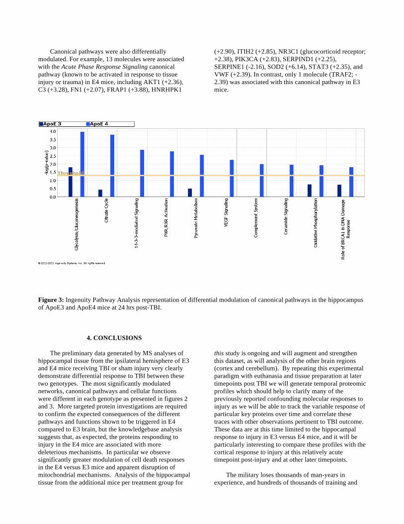
Canonical pathways were also differentially modulated. For example, 13 molecules were associated with the Acute Phase Response Signaling canonical pathway (known to be activated in response to tissue injury or trauma) in E4 mice, including AKT1 (+2.36), C3 (+3.28), FN1 (+2.07), FRAP1 (+3.88), HNRHPK1
(+2.90), ITIH2 (+2.85), NR3C1 (glucocorticoid receptor; +2.38), PIK3CA (+2.83), SERPIND1 (+2.25), SERPINE1 (-2.16), SOD2 (+6.14), STAT3 (+2.35), and VWF (+2.39). In contrast, only 1 molecule (TRAF2; -2.39) was associated with this canonical pathway in E3 mice.
Figure 3: Ingenuity Pathway Analysis representation of differential modulation of canonical pathways in the hippocampus of ApoE3 and ApoE4 mice at 24 hrs post-TBI.
4. CONCLUSIONS
The preliminary data generated by MS analyses of hippocampal tissue from the ipsilateral hemisphere of E3 and E4 mice receiving TBI or sham injury very clearly demonstrate differential response to TBI between these two genotypes. The most significantly modulated networks, canonical pathways and cellular functions were different in each genotype as presented in figures 2 and 3. More targeted protein investigations are required to confirm the expected consequences of the different pathways and functions shown to be triggered in E4 compared to E3 brain, but the knowledgebase analysis suggests that, as expected, the proteins responding to injury in the E4 mice are associated with more deleterious mechanisms. In particular we observe significantly greater modulation of cell death responses in the E4 versus E3 mice and apparent disruption of mitochondrial mechanisms. Analysis of the hippocampal tissue from the additional mice per treatment group for
this study is ongoing and will augment and strengthen this dataset, as will analysis of the other brain regions (cortex and cerebellum). By repeating this experimental paradigm with euthanasia and tissue preparation at later timepoints post TBI we will generate temporal proteomic profiles which should help to clarify many of the previously reported confounding molecular responses to injury as we will be able to track the variable response of particular key proteins over time and correlate these traces with other observations pertinent to TBI outcome. These data are at this time limited to the hippocampal response to injury in E3 versus E4 mice, and it will be particularly interesting to compare these profiles with the cortical response to injury at this relatively acute timepoint post-injury and at other later timepoints.
The military loses thousands of man-years in
experience, and hundreds of thousands of training and
education dollars each year, due to the effects of traumatic brain injuries in soldiers, including those prematurely returned to active duty as well as soldiers who cannot return to service. Many young adults never return to premorbid skills or responsibilities after TBI, despite intensive and comprehensive rehabilitation efforts on their behalf. In addition, the sequelae of TBI are a mixture of cognitive psycho motor and emotional (psychiatric) signs and symptoms and the emotional and psychological burden on patients and caregivers can be enormous. Immediate prospects for improved management and treatment of TBI lie in the development of strategies for limiting the consequences of brain injury and repairing the damage. Therefore, we are using state of the art technology, as described above, to identify functionally correlated protein responses to TBI and molecular targets for enhancing repair processes and/or inhibiting neurodegenerative targets which are amenable to therapeutic intervention.
We are studying response to TBI by comparing the
proteome of injured mouse brain with uninjured mouse brain. The results presented here represent the proteomic response to TBI in E3 versus E4 mice at an acute post-injury timepoint (24 hrs). Our results indicate a significantly different proteomic response to injury across APOE genotypes. As our program progresses we will generate databases of proteomic response to injury in specific brain regions from APOE3 and APOE4 mice at different timepoints post-TBI, different ages of mice and with more severe levels of injury such as 1.8mm depth impact. We also plan neuropathological examination of similarly exposed mice in order to correlate those observations with the proteomic profiles. Another area of interest is to investigate any gender specific responses to TBI. In the paradigms analyzing more chronic response to injury than the 24hr timepoint reported herein we will be also be able to correlate proteomic profiles with neurobehavioral performance determined by measures such as the Rotarod or Morris Water Maze.
Using data mining and knowledgebases such as the
Ingenuity Pathway Analysis software described in this manuscript, we will identify molecular functions and pathways which correlate with the E3 and E4 response to injury and with the neuropathological and neurobehavioral outcomes. Where such pathways can be specifically associated with either a negative or a positive outcome, key molecules in those pathways will represent potential targets for therapeutic intervention. Where appropriate inhibitors or enhancers are available, or mice genetically modified at the loci of interest, we will develop treatment paradigms to test our hypotheses.
The optimized protocols for iTRAQ analysis
described in this manuscript have recently been
pioneered in our laboratories, and the data presented here represent the very first data generated by MS analyses for this program; we therefore eagerly anticipate the growing database of proteomic profiles which will be generated over the coming months. This strategy of proteomic profiling in mice which (by virtue of their genetic modifications) demonstrate different outcomes in response to the same level of TBI, will enable us to focus the data analyses and rapidly generate testable hypotheses, moving us toward the ultimate goal of this research - preclinical testing of valid therapeutic strategies.
ACKNOWLEDGMENTS
This work is supported by a contract from the Telemedicine and Advanced Technology Research Center (TATRC) of the U.S. Army Medical Research and Materiel Command (MRMC).
REFERENCES Blennow K, Hesse C, Fredman P, 1994: Cerebrospinal
fluid apolipoprotein E is reduced in Alzheimer's disease, Neuroreport, 5:2534-6.
Bramlett HM, Kraydieh S, Green EJ, Dietrich WD, 1997: Temporal and regional patterns of axonal damage following traumatic brain injury: a beta-amyloid precursor protein immunocytochemical study in rats, J Neuropathol Exp Neurol, 56:1132-41.
Cernak I, 2006: Recent advances in neuroprotection for treating traumatic brain injury, Expert Opin Investig Drugs, 15:1371-81.
Crawford F, Vanderploeg R, Freeman M, Singh S, Waisman M, Michaels L, Abdullah L, Warden D, Lipsky R, Salazar A, Mullan M, 2002; APOE genotype influences acquisition and recall following traumatic brain injury, Neurology, 58(7):1115-8.
Crawford F, Wood M, Ferguson S, Mathura V, Faza B, Wilson S, Fan T, O’Steen B, Ait-Ghezala G, Hayes R, Mullan M, 2007: Genomic analysis of response to traumatic brain injury in a mouse model of Alzheimer’s disease (APPsw), Brain Research, 1185:45-58.
DeKosky ST, Taffe KM, Abrahamson EE, Dixon CE, Kochanek PM, Ikonomovic MD, 2004: Time course analysis of hippocampal nerve growth factor and antioxidant enzyme activity following lateral controlled cortical impact brain injury in the rat, J Neurotrauma, 21:491-500.
Gong JS, Kobayashi M, Hayashi H, Zou K, Sawamura N, Fujita SC, Yanagisawa K, Michikawa M, 2002: Apolipoprotein E (ApoE) isoform-dependent lipid release from astrocytes prepared from human ApoE3

and ApoE4 knock-in mice, J Biol Chem, 277:29919-26.
Haqqani AS, Hutchison JS, Ward R, Stanimirovic DB, 2007: Biomarkers and Diagnosis; Protein Biomarkers in Serum of Pediatric Patients with Severe Traumatic Brain Injury Identified by ICAT-LC-MS/MS. J Neurotrauma, 24:54-74.
Kay A, Petzold A, Kerr M, Keir G, Thompson E, Nicoll J, 2003: Temporal alterations in cerebrospinal fluid amyloid beta-protein and apolipoprotein E after subarachnoid hemorrhage, Stroke, 34:e240-3.
Olsson A, Csajbok L, Ost M, Hoglund K, Nylen K, Rosengren L, Nellgard B, Blennow K, 2004: Marked increase of beta-amyloid(1-42) and amyloid precursor protein in ventricular cerebrospinal fluid after severe traumatic brain injury, J Neurol, 251:870-6.
Rapp A, Gmeiner B, Huttinger M, 2006: Implication of apoE isoforms in cholesterol metabolism by primary rat hippocampal neurons and astrocytes, Biochimie, 88:473-83.
Tang Y, Lu A, Aronow BJ, Wagner KR, Sharp FR, 2002: Genomic responses of the brain to ischemic stroke, intracerebral haemorrhage, kainate seizures, hypoglycemia, and hypoxia, Eur J Neurosci, 15:1937-52.
Wang H, Durham L, Dawson H, Song P, Warner DS, Sullivan PM, Vitek MP, Laskowitz DT, 2007: An apolipoprotein E-based therapeutic improves outcome and reduces Alzheimer's disease pathology following closed head injury: Evidence of pharmacogenomic interaction, Neuroscience, 144:1324-33.