Identification of Novel Small Molecules that bind to the … · Identification of Novel Small...
14
Identification of Novel Small Molecules that bind to the Loop2 Region of Sclerostin –An In silico Computational Analysis KARTHIKEYAN MUTHUSAMY 1 , MOHAN SUBBURAMAN 2 , SELVARAMAN NAGAMANI 1 , CHANDRASEKHAR KESAVAN 2 1 Department of Bioinformatics, Alagappa University, Karaikudi 630 004, India. 2 Musculoskeletal Disease Center, VA Loma Linda Healthcare system, Loma Linda, CA 92357, USA. Running title: Small molecules binding to sclerostin Corresponding Authors: Chandrasekhar Kesavan Assistant Research Professor Musculoskeletal Disease Center VA Loma Linda Healthcare System Research Service 11201 Benton St Loma Linda, CA 92357, USA. Email: [email protected] Fax No: 0119097961680
Transcript of Identification of Novel Small Molecules that bind to the … · Identification of Novel Small...
Identification of Novel Small Molecules that bind to the Loop2
Region of Sclerostin –An In silico Computational Analysis
KARTHIKEYAN MUTHUSAMY1, MOHAN SUBBURAMAN
2, SELVARAMAN NAGAMANI
1,
CHANDRASEKHAR KESAVAN 2
1Department of Bioinformatics, Alagappa University, Karaikudi 630 004, India.
2Musculoskeletal Disease Center, VA Loma Linda Healthcare system, Loma Linda, CA 92357,
USA.
Running title: Small molecules binding to sclerostin
Corresponding Authors: Chandrasekhar Kesavan
Assistant Research Professor
Musculoskeletal Disease Center
VA Loma Linda Healthcare System
Research Service
11201 Benton St
Loma Linda, CA 92357, USA.
Email: [email protected]
Fax No: 0119097961680
Zdenka.Stadnikova
Pre-press

Summary
The goal of this study was to identify small molecular weight compounds that bind to sclerostin using in-silico methods because of
the established importance of sclerostin-based therapies for the treatment of disease characterized by low bone mass. The zinc
database (Zdb) revealed that nine potential molecules bind to the loop2 region (functional site) of sclerostin with ADME/T properties
that are within an acceptable range defined for human use. Compounds 30160056 and 56871042 showed the highest docking
score. Density functional theory (by HOMO, LUMO and MESP analysis) and MM/GBSA analysis showed that four compounds
30160056, 56871042, 72112226 and 43920281 exhibit high stability among the nine small molecules identified. Induced Docking Fit
and Pymol software analyses revealed that the identified compounds differ in the interaction with amino acids in the loop2 region of
sclerostin. Six compound exhibited interaction with Ile95 and 2 compounds with Asn93, an amino acid in the loop2 region known to
be involved in sclerostin’s inhibitory effect, suggesting that the identified compounds have the potential to bind and neutralize
sclerostin function. Furthermore, compound 43920281 showed a low risk of toxicity and drug-like characteristic features compared
to all nine identified compounds. In conclusion, in silico analysis identified a novel compound 43920281 as a potent anti-sclerostin
therapeutic for drug development for the treatment of osteoporosis.
Key words: Osteoporosis, Sclerostin (SOST), Wnt (wingless-type MMTV integration site family) antagonist, Computer-aided drug
design (CADD).
Main body
Osteoporosis is a disease characterized by low bone mineral density (BMD) and poor bone quality resulting
in reduced bone strength and an increased risk of bone fractures (Drake, et al. 2015, Kobayashi and Kronenberg
2014, Lombardi et al. 2010, Olney 2009). To date, millions of individuals suffer from osteoporosis due to a variety
of causes including aging, lack of exercise and endocrine/ metabolic disturbances (Golob and Laya 2015, Preisinger
et al. 1996). According to the “National Osteoporosis Foundation”, osteoporosis is responsible for two million
broken bones and $19 million in related healthcare costs every year, and by 2025 the bone fracture numbers are
expected to raise and the annual associated costs to increase to 25.5 billion. Currently, moderate exercise, vitamin
based therapy and parathyroid hormone (PTH), a Food and Drug administration (FDA) established bone anabolic
therapeutic agent, that helps to maintain bone mass and reduce the incidence of osteoporosis (Augustine and
Horwitz 2013, Bhutani and Gupta 2013, Iwamoto 2014, Laktasic-Zerjavic 2014, Prentice 2004), however the
amount of new bone formation stimulated with these agents is less effective in individuals who have lost a
tremendous amount of bone (postmenopausal women and in the elderly). Therefore, there is a need for the
identification and development of additional agents as therapeutics to stimulate new bone formation not only to
reduce the incidence of osteoporosis but also to treat bone injuries.
In osteoporosis, higher osteoclast activity enhances bone resorption and reduces osteoblast activity, which
reduces bone formation, are major causes for the pathogenesis of osteoporosis. Recent studies have shown that
sclerostin, a protein encoded by the SOST gene, expressed in osteocytes, is a key player in the pathophysiology of
osteoporosis. Studies using animal and human models have delineated that mutations in the sclerostin gene resulted
in increased bone mass and bone strength (de Vernejoul and Kornak 2010, Li et al. 2008). Subsequently, clinical
studies revealed that postmenopausal women with decreased bone mass and increased bone loss showed high levels
of sclerostin in the serum (Ardawi et al. 2012). In terms of underlying mechanisms to account for bone loss, studies
using in vitro and in vivo models have shown that Sclerostin binds to Low density lipoprotein (LRP) 5/6 and thereby
inhibits wingless (WNT) signaling, a pathway well established in promoting new bone formation (Kim et al. 2013).
Likewise, other studies have shown that Sclerostin blocks the osteogenic effect induced by blocking Bone
Morphogenic Protein (BMP) ligand interaction with BMP receptors (van Bezooijen et al. 2007, Winkler et al.
2003). These data from independent studies led to the conclusion that sclerostin is a key contributor to osteoporosis
and that blocking sclerostin should increase new bone formation. Accordingly, animal and human model studies
have provided evidence that blocking sclerostin using antibody based therapies promoted new bone formation and
reduced the risk of osteoporosis (Clarke 2014, Lewiecki 2011). Although antibody based therapies have been
efficient, this type of therapy is expensive and thus has a significant impact on health care costs. Therefore
identifying novel therapeutics that are less costly and show a potential for blocking sclerostin function will have a
significant impact not only in reducing the detrimental effects of osteoporosis on our aging population but also on
future health care costs. Therefore, we undertook a computational analysis using in-silico and molecular docking
analyses to identify small molecules that could inhibit sclerostin actions.
All computational analyses were carried out on a Red hat 5.1 Linux platform running on a Lenovo Intel
core 2 duo processor with 2 GB of RAM. We used different chemical databases such as the Asinex
(www.asinex.com) (55958 compounds), Chembridge (www.chembridge.com) (511324 compounds) and Zinc
databases (www.zinc.docking.org) (155819 compounds) to identify molecules that dock into the active site of
sclerostin to screen compounds which could inhibit sclerostin actions. The crystal structure of sclerostin (Protein
database (PDB) id – 2k8p) was downloaded from the PDB (Veverka et al. 2009) and prepared using the ‘protein
preparation module’ (Schrödinger, LLC, New York, 2011). In order to evaluate the over-lapping atoms, Hydrogen
atoms were added to the crystal structure. Furthermore, all the “structural” waters molecules were removed since
water molecules have not been shown to be critical to the function of the protein-ligand interaction. Partial atomic
charges were created due to the asymmetric distribution of electrons in chemical bonds. In order to determine the
atomic charges, the Schrodinger software used the OPLS_2005 (optimized potentials liquid for simulations) force
field, which contains different parameters for treating different atoms. Furthermore, the energy was minimized until
the average root mean square deviation of non-hydrogen atoms reached 0.30 Å, to avoid structure deviations. Once
the structure was prepared, a receptor grid was generated. The grid enclosing box was centered over the active
amino acids (Proline (Pro)92, Asparagine (Asp)93, Alanine (Ala)94, Isoleucine (Ile)95 and glycine (Gly)96),
represented as PNAIG, in the loop-2 region of sclerostin (Veverka et al. 2009) so that they were enclosed within 3
Å from the center of the residues. The scaling Van der walls radii were set at 1.0 Å in the receptor grid generation.
The grid was enclosed with the bounding box set at 20 Å. A virtual screening workflow was used to screen the large
collections of compounds against the defined target in the sclerostin molecule. This workflow included Ligprep (for
ligand preparation, version 2.5, Schrodinger, LLC, New York, 2011), Qikprop (ADME/T (Adsorption Distribution
Metabolism Excretion / Toxicity predictions, version 3.5, Schrodinger, LLC, New York, 2011), HTVS (high
throughput virtual screening) and other structural properties (HTVS, Schrodinger, LLC, New York, 2011) [11, 12].
Asinex, TOSLab collections and Zinc databases which contain a total of 723,101 compounds were screened based
on Glide score, Glide energy and hydrogen bond interactions (Schrodinger). To further confirm the docking results,
we re-ran the whole docking process using “Induced Fit Docking” (mixed molecular docking/molecular dynamics
protocol) which keeps receptors flexible (Singh et al. 2012). In this protocol both the protein and ligand molecules
were kept flexible. We set the same default parameters as mentioned in the normal Glide XP docking method.
Subsequently, the prime MM/GBSA (Molecular Mechanics Generalized Born Surface Area) method was used to
calculate the free energy of binding for a set of ligands to a receptor. The energy minimization was carried out using
the local optimization feature in Prime, and the free energies of the complex were calculated using the OPLS_2005
force field program and GB/SA continuum solvent model. The binding free energy was calculated as follows (Das et
al. 2009, Lyne et al. 2006),
∆Gbind = ∆E + ∆Gsolv + ∆GSA
∆E = Ecomplex – Eprotein – Eligand
where Ecomplex, Eprotein and Eligand are the minimized energies of the protein-ligand complex, protein and
ligand, respectively.
∆Gsolv = Gsolv(complex) – Gsolv(protein) – Gsolv(ligand)
where Gsolv(complex), Gsolv(protein) and Gsolv(ligand) are the solvation free energies of the complex, protein and
ligand respectively.
∆GSA = GSA(complex) – GSA(protein) – GSA(ligand)
Where GSA(complex), GSA(protein) and GSA(ligand) are the surface area energies for the complex, protein and ligand
respectively.
Additionally, all the identified compounds were subjected to density functional theory (DFT) calculations
(Lee et al. 1988). DFT calculations are typically used for studying electronic molecular features to define a
molecular electrostatic map, with the electron density, and frontier molecular orbital density fields (i.e. HOMO,
LUMO) which can predict the molecular properties and biological activity of a compound. The DFT calculations
were performed with Jaguar version 8.1 software, which sets the solvation state. The DFT was analyzed through
Becke’s three-parameter exchange potential and the Lee-Yang-Parr correlation functional (B3LYP) basic set at a 3-
21G* level (Seminario 1996), using “Poisson Boltzmann Finite” (PBF) solvation. In the present study, the 3D-
molecular electrostatic potentials (MESP) V(r) at a point r because of a molecular system with nuclear charges
located at and the electron density ρ (r) were derived using the following equation,
|'|
')'(
||)(
3
1 rr
rdr
Rr
ZrV
N
A A
A . In this equation N represents the total number of nuclei in the molecule and
the two terms refer to the bare nuclear potential and the electronic contributions, respectively. We computed the
Jaguar Dipole moment, Molecular electrostatic properties, Lowest Unoccupied Molecular Orbital (LUMO),
including MESP (Molecular electrostatic potential) and Highest Occupied Molecular Orbital (HOMO) energy. We
calculated the electrostatic potentials by van der Waals contact surface area of the molecule (Jaquar, version 8.1,
Schrodinger, LLC, New York, 2011).
To identify novel molecules that showed a high binding affinity to sclerostin, we used the published crystal
structure of the human sclerostin protein (PBD id-2k8p) for the molecular docking analysis. In particular, we used
the loop2 region of sclerostin because LRP5/6 binds to amino acids in the loop2 region of sclerostin, known as the
PNAIG motif, thus, inhibiting LRP5/6 activity. LRP5/6 activity is required for activation of WNT signaling and
subsequent bone formation. Additionally, a report has shown that a peptide derived from the loop2 region of
sclerostin blocked the interaction of sclerostin with LRP5/6 (Holdsworth et al. 2012). Based on this information, we
used the loop2 region of sclerostin to form a glide-grid for the screening analysis. Different databases including the
Asinex, TOSLab and Zinc database were screened against the loop2 region of sclerostin using a virtual screening
workflow in the maestro (Schrodinger, LLC, New York, 2009) for hit identification and lead compound
optimization. The database ligands were prepared at pH 7.0 ± 2.0 using an epik state (enumerates ligand protonation
states and tautomers in biological conditions) and the large penalties for high energy ionization of tautomer states
were removed. In the default constraint parameters the protein has been kept as a scaling van der Walls radius of 1.0
Å with a partial atomic charge set at less than 0.25 Å. The Glide HTVS was carried out with a flexible docking
algorithm using selected constraints for each grid in the OPLS_2005 force field. We finalized the best lead
molecules based on glide score rather than on glide energy. This is because glide energy includes only modified
Coulomb-van der Waals interaction energy while the glide score includes other parameters such as hydrogen bond
interactions, a lipophilic term and coulomb energy. The docking analysis revealed nine compounds that show a

potential binding affinity towards the loop2 region of sclerostin (Table-1A).The 2D structure of each identified lead
molecules is displayed in Figure 1 (supplement data). We further confirmed the docking analysis using an Induced
Fit Docking analyses which revealed similar types of interactions.
To further characterize the physiochemical properties (ADME/T) of the nine compounds, Qikprop
simulation was performed on the identified compounds using Schrodinger software (QikProp version 3.5,
Schrodinger 2012). Compounds that have high molecular weights are likely to have low solubility and have
difficulty passing through the cell membrane. In our study, the molecular weight of all identified compound are
within an acceptable range to have a high solubility and a high probably of being able to enter a cell. Similarly,
lipophilicity (a ratio of the molecule’s solubility in octanol to solubility in water (QPlogPo/w), an analysis that
determines how a compound is distributed within the body after absorption and how rapidly it is metabolized and
excreted, was performed on the identified compounds. This analysis revealed that all the values of compounds (C2
to C9) are within the standard range. In addition, the human oral absorption rate of each compound was determined.
We found that seven of the nine compounds (C1 to C6 and C9) were within the acceptable range generally observed
for drugs while compound 34853727 and compound 64634013 fell outside the acceptable range (Table-1A).
Importantly, the identified compounds demonstrated small variations within the ADME/T properties but all
compounds fell within an acceptable range defined for human use. In addition to ADEM/T, we also performed
density functional theory analysis on the identified compounds to determine the molecular stability of the compound
which is important for achieving a biological function. The small HOMO-LUMO gap (HLG) suggests reasonable
stability of all the identified compounds (Table-1A). Subsequently, we also analyzed the toxicity and drug-likeness
properties with OSIRIS Property Explorer software ("OSIRIS property explorer, www.organic-
chemistry.org/prog/peop/,"). Though the identified compounds showed acceptable ADME/T properties and drug-
like scores (a combination of CloP, LogS, TPSA analysis), toxicity screening results revealed that the identified
compounds possess varying risks of mutagenesis and tumorigenesis (Table-1B). In particular, three compounds
(30160056, 56871042, 72112226) that showed a high docking score, ADME/T properties and oral absorption rates
possessed a medium to high mutagenesis rate and a high risk for tumorigenesis. While the compound 43920281
showed a moderate docking score and compounds (34853723, 64634013, 35636232) showing a low docking score,
possessed a very low risk for toxicity based on parameters when compared to other identified compounds. Thus,
data from these analyses suggest that further experiments should be focused on compounds that have moderate and
low docking scores with a low risk of toxicity.
Additionally, we extended our study by determining whether the identified compounds show similar or
different amino acid interactions in the loop2 region of sclerostin using Pymol software (Version 1.5.0.4
Schrodinger, LLC). Our analysis revealed that the identified compounds differ in their interactions with amino acids
in the loop2 region of sclerostin (Table-2). We found that one compound 77270363 showed 6 interaction sites, 3
compounds (30160056, 72112226 and 64634013) had 5 interaction sites and 4 compounds (56871042, 43920281,
72693278, 34853727) had 4 interaction sites within the loop2 region of sclerostin. In addition, we found one
compound (35636232) with only 3 interactions (Table-2). Overall, these data suggest that even though these
compounds differ in the interactions with amino acids in the loop2 region of sclerostin, they all have the potential to
alter sclerostin function. To this end, studies have shown that the amino acids (Pro92, Asp93, Ala94, Ile95, Gly96 )
in the loop2 region of sclerostin, identified as the PNAIG motif binds specifically to LRP5/6 and thereby blocks the
WNT signaling pathway (Kim et al. 2013). In particular, it has been shown that this inhibitor effect is largely
mediated by an interaction with two amino acids (Asn93 and Ile95) in the PNAIG region of the loop2 site of
sclerostin (Holdsworth et al. 2012). In our study, the induced fit docking analysis and Pymol software found that the
eight out of nine compounds interact with the same amino acids, Ile95 or Asn93, respectively, in the PNAIG
sequence of the loop2 region of sclerostin that blocks WNT signaling (Table-2). This computational analysis
substantiates that the identified compounds could act as potent anti-sclerostin therapeutic drugs.
In conclusion, our pilot study has predicted three molecules that show strong binding affinity towards the
loop2 region of sclerostin. Moreover the ADME/T predictions also revealed that the identified compounds are in an
acceptable range for human consumption. However, further functional studies are necessary to validate this
computational analysis and to determine if the identified compounds, in particular, compound 43920281 block
sclerostin function without exerting severe side effects.
Acknowledgements

We thank Department of Bioinformatics, Science Block, Alagappa University, Karaikudi – 630 004, Tamil Nadu,
India, for undertaking the computational work as a part of the collaboration. All the analyses and manuscript were
prepared at VA Loma Linda Health care system. We thank Dr. Donna Strong for proof reading the manuscript.
CONFLICT OF INTEREST
The authors declare no conflicts of interest.
References
ARDAWI MS, ROUZI AA, AL-SIBIANI SA, AL-SENANI NS, QARI MH, MOUSA SA: High serum sclerostin predicts the
occurrence of osteoporotic fractures in postmenopausal women: the Center of Excellence for Osteoporosis Research Study. J Bone Miner Re 27 2592-2602, 2012.
AUGUSTINE M, HORWITZ MJ: Parathyroid hormone and parathyroid hormone-related protein analogs as therapies
for osteoporosis. Curr Osteoporos Rep 11 400-406, 2013.
BHUTANI G, GUPTA MC: Emerging therapies for the treatment of osteoporosis. J Midlife Health 4 147-152, 2013.
CLARKE BL: Anti-sclerostin antibodies: utility in treatment of osteoporosis. Maturitas 78 199-204, 2014.
DAS D, KOH Y, TOJO Y, GHOSH AK, MITSUYA H: Prediction of potency of protease inhibitors using free energy
simulations with polarizable quantum mechanics-based ligand charges and a hybrid water model. J Chem Inf Model 49 2851-2862, 2009.
DE VERNEJOUL MC, KORNAK U: Heritable sclerosing bone disorders: presentation and new molecular mechanisms.
Ann N Y Acad Sci 1192 269-277, 2010.
DRAKE, MT, CLARKE BL, LEWIECKI EM: The Pathophysiology and Treatment of Osteoporosis. Clin Ther, 2015.
GOLOB AL, LAYA MB: Osteoporosis: Screening, Prevention, and Management. Med Clin North Am 99 587-606,
2015.
HOLDSWORTH G, SLOCOMBE P, DOYLE C, SWEENEY B, VEVERKA V, LE RICHE K, ROBINSON MK: Characterization of
the interaction of sclerostin with the low density lipoprotein receptor-related protein (LRP) family of Wnt co-receptors. J Biol Chem 287 26464-26477, 2012.
IWAMOTO J: Vitamin K(2) therapy for postmenopausal osteoporosis. Nutrients 6 1971-1980, 2014.
KIM JH, LIU X, WANG J, CHEN X, ZHANG H, KIM SH, HE TC: Wnt signaling in bone formation and its therapeutic
potential for bone diseases. Ther Adv Musculoskelet Dis 5 13-31, 2013.
KOBAYASHI T, KRONENBERG HM: Overview of skeletal development. Methods Mol Biol 1130 3-12, 2014.
LAKTASIC-ZERJAVIC N: The role of vitamin D and calcium in the management of osteoporosis. Reumatizam 61 80-
88, 2014.
LEE C, YANG W AND PARR RG: Development of the Colle-Salvetti correlation-energy formula into a functional of
the electron density. Phys Rev B Condens Matter 37 785-789, 1988.
LEWIECKI EM: Sclerostin: a novel target for intervention in the treatment of osteoporosis. Discov Med 12 263-
273, 2011.
LI X, OMINSKY MS, NIU QT, SUN N, DAUGHERTY B, D'AGOSTIN D, PASZTY C: Targeted deletion of the sclerostin
gene in mice results in increased bone formation and bone strength. J Bone Miner Res 23 860-869, 2008.
LOMBARDI G, DI SOMMA C, VUOLO L, GUERRA E, SCARANO E AND COLAO A: Role of IGF-I on PTH effects on bone.
J Endocrinol Invest 33 22-26, 2010.

LYNE PD, LAMB ML AND SAEH JC: Accurate prediction of the relative potencies of members of a series of kinase
inhibitors using molecular docking and MM-GBSA scoring. J Med Chem 49 4805-4808, 2006.
OLNEY RC: Mechanisms of impaired growth: effect of steroids on bone and cartilage. Horm Res 72 Suppl 1, 30-35,
2009. OSIRIS property explorer, www.organic-chemistry.org/prog/peop/.
PREISINGER E, ALACAMLIOGLU Y, PILS K, BOSINA E, METKA M, SCHNEIDER B AND ERNST E: Exercise therapy for
osteoporosis: results of a randomised controlled trial. Br J Sports Med 30 209-212, 1996.
PRENTICE A: Diet, nutrition and the prevention of osteoporosis. Public Health Nutr 7 227-243, 2004.
SEMINARIO J: Recent developments and applications of modern density functional theory 1st ed ed, Vol. 1
Amsterdam: Elsevier Science, 1996.
SCHRODINGER. Software, LLC, New York, http://www.schrodinger.com/citations/
SINGH KH D, KIRUBAKARAN P, NAGARAJAN S, SAKKIAH S, MUTHUSAMY K, VELMURGAN D AND JEYAKANTHAN J: Homology modeling, molecular dynamics, e-pharmacophore mapping and docking study of Chikungunya virus nsP2 protease. J Mol Model 18 39-51, 2012.
VAN BEZOOIJEN RL, SVENSSON JP, EEFTING D, VISSER A, VAN DER HORST G, KARPERIEN M, LOWIK CW: Wnt but
not BMP signaling is involved in the inhibitory action of sclerostin on BMP-stimulated bone formation. J Bone Miner Res 22 19-28, 2007.
VEVERKA V, HENRY AJ, SLOCOMBE PM, VENTOM A, MULLOY B, MUSKETT FW, CARR MD: Characterization of the
structural features and interactions of sclerostin: molecular insight into a key regulator of Wnt-mediated bone formation. J Biol Chem 284 10890-10900, 2009.
WINKLER DG, SUTHERLAND MK, GEOGHEGAN JC, YU C, HAYES T, SKONIER JE, LATHAM JA: Osteocyte control of
bone formation via sclerostin, a novel BMP antagonist. EMBO J 22 6267-6276, 2003.

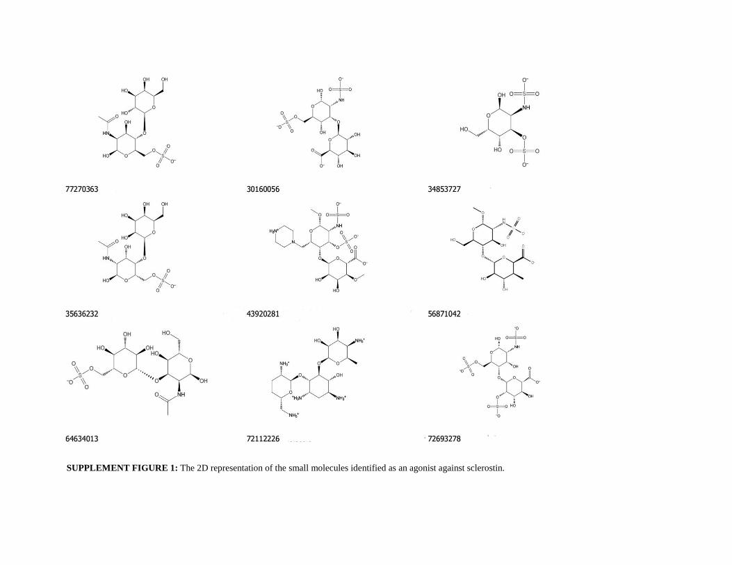
SUPPLEMENT FIGURE 1: The 2D representation of the small molecules identified as an agonist against sclerostin.
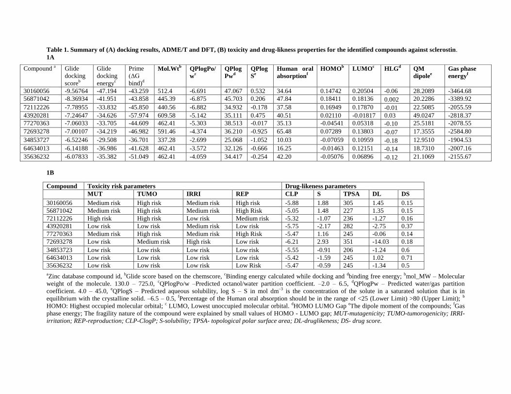
Table 1. Summary of (A) docking results, ADME/T and DFT, (B) toxicity and drug-likness properties for the identified compounds against sclerostin.
1A
1B
aZinc database compound id,
bGlide score based on the chemscore,
cBinding energy calculated while docking and
dbinding free energy;
bmol_MW – Molecular
weight of the molecule. 130.0 – 725.0, cQPlogPo/w –Predicted octanol/water partition coefficient. –2.0 – 6.5,
dQPlogPw – Predicted water/gas partition
coefficient. 4.0 – 45.0, eQPlogS – Predicted aqueous solubility, log S – S in mol dm
–3 is the concentration of the solute in a saturated solution that is in
equilibrium with the crystalline solid. –6.5 – 0.5, fPercentage of the Human oral absorption should be in the range of <25 (Lower Limit) >80 (Upper Limit);
b
HOMO: Highest occupied molecular orbital; c LUMO, Lowest unoccupied molecular orbital.
dHOMO LUMO Gap
eThe dipole moment of the compounds;
fGas
phase energy; The fragility nature of the compound were explained by small values of HOMO - LUMO gap; MUT-mutagenicity; TUMO-tumorogenicity; IRRI-
irritation; REP-reproduction; CLP-ClogP; S-solubility; TPSA- topological polar surface area; DL-druglikeness; DS- drug score.
Compound a Glide
docking
scoreb
Glide
docking
energyc
Prime
(ΔG
bind)d
Mol.Wtb QPlogPo/
wc
QPlog
Pwd
QPlog
Se
Human oral
absorptionf
HOMOb LUMO
c HLG
d QM
dipolee
Gas phase
energyf
30160056 -9.56764 -47.194 -43.259 512.4 -6.691 47.067 0.532 34.64 0.14742 0.20504 -0.06 28.2089 -3464.68
56871042 -8.36934 -41.951 -43.858 445.39 -6.875 45.703 0.206 47.84 0.18411 0.18136 0.002 20.2286 -3389.92
72112226 -7.78955 -33.832 -45.850 440.56 -6.882 34.932 -0.178 37.58 0.16949 0.17870 -0.01 22.5085 -2055.59
43920281 -7.24647 -34.626 -57.974 609.58 -5.142 35.111 0.475 40.51 0.02110 -0.01817 0.03 49.0247 -2818.37
77270363 -7.06033 -33.705 -44.609 462.41 -5.303 38.513 -0.017 35.13 -0.04541 0.05318 -0.10 25.5181 -2078.55
72693278 -7.00107 -34.219 -46.982 591.46 -4.374 36.210 -0.925 65.48 0.07289 0.13803 -0.07 17.3555 -2584.80
34853727 -6.52246 -29.508 -36.701 337.28 -2.699 25.068 -1.052 10.03 -0.07059 0.10959 -0.18 12.9510 -1904.53
64634013 -6.14188 -36.986 -41.628 462.41 -3.572 32.126 -0.666 16.25 -0.01463 0.12151 -0.14 18.7310 -2007.16
35636232 -6.07833 -35.382 -51.049 462.41 -4.059 34.417 -0.254 42.20 -0.05076 0.06896 -0.12 21.1069 -2155.67
Compound Toxicity risk parameters Drug-likeness parameters
MUT TUMO IRRI REP CLP S TPSA DL DS
30160056 Medium risk High risk Medium risk High risk -5.88 1.88 305 1.45 0.15
56871042 Medium risk High risk Medium risk High Risk -5.05 1.48 227 1.35 0.15
72112226 High risk High risk Low risk Medium risk -5.32 -1.07 236 -1.27 0.16
43920281 Low risk Low risk Medium risk Low risk -5.75 -2.17 282 -2.75 0.37
77270363 Medium risk High risk Medium risk High Risk -5.47 1.16 245 -0.06 0.14
72693278 Low risk Medium risk High risk Low risk -6.21 2.93 351 -14.03 0.18
34853723 Low risk Low risk Low risk Low risk -5.55 -0.91 206 -1.24 0.6
64634013 Low risk Low risk Low risk Low risk -5.42 -1.59 245 1.02 0.71
35636232 Low risk Low risk Low risk Low Risk -5.47 -0.59 245 -1.34 0.5
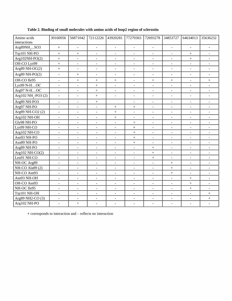
Table 2. Binding of small molecules with amino acids of loop2 region of sclerostin
+ corresponds to interaction and – reflects no interaction
Amino acids
interactions
30160056 56871042 72112226 43920281 77270363 72693278 34853727 64634013 35636232
Arg89NH2...SO3 + - - - - - - - -
Trp101 NH-PO + + - - - - - + -
Arg102NH-PO(2) + - - - - - - + -
OH-CO Lys99 + - - - - - - - -
Arg89 NH-OC(2) + - - - - - - - -
Arg89 NH-PO(2) - + - - - - - - -
OH-CO Ile95 - + + + - + + - +
Lys99 N-H…OC - - + - - - - - -
Arg97 N-H…OC - - + - - - - - -
Arg102 NH2-PO3 (2) - - + - - - - - -
Arg89 NH-PO3 - - + - - - - -
Arg97 NH-PO - - - + + - - - -
Arg89 NH-CO2 (2) - - - + - - - - -
Arg102 NH-OH - - - + - - - - -
Gly98 NH-PO - - - - + - - - -
Lys99 NH-CO - - - - + - - - -
Arg102 NH-CO - - - - + - - - -
Asn93 NH-PO - - - - + - - - -
Asn89 NH-PO - - - - + - - - -
Arg89 NH-PO - - - - - + - - -
Arg102 NH-CO(2) - - - - - + - - -
Leu91 NH-CO - - - - - + - - -
NH-OC Arg89 - - - - - - + - -
NH-CO Ala89 (2) - - - - - - + - -
NH-CO Asn93 - - - - - - + - -
Asn93 NH-OH - - - - - - - + -
OH-CO Asn93 - - - - - - - + -
NH-OC Ile95 - - - - - - - + -
Trp101 NH-OH - - - - - - - - +
Arg89 NH2-CO (3) - - - - - - - - +
Arg102 NH-PO - + - - - - - - -