Identification of myxobacteria-derived HIV inhibitors by a
9
RESEARCH Open Access Identification of myxobacteria-derived HIV inhibitors by a high-throughput two-step infectivity assay Javier P Martinez 1 , Bettina Hinkelmann 2 , Eric Fleta-Soriano 1 , Heinrich Steinmetz 3 , Rolf Jansen 3 , Juana Diez 4 , Ronald Frank 2 , Florenz Sasse 2 and Andreas Meyerhans 1,5* Abstract Background: Drug-resistance and therapy failure due to drug-drug interactions are the main challenges in current treatment against Human Immunodeficiency Virus (HIV) infection. As such, there is a continuous need for the development of new and more potent anti-HIV drugs. Here we established a high-throughput screen based on the highly permissive TZM-bl cell line to identify novel HIV inhibitors. The assay allows discriminating compounds acting on early and/or late steps of the HIV replication cycle. Results: The platform was used to screen a unique library of secondary metabolites derived from myxobacteria. Several hits with good anti-HIV profiles were identified. Five of the initial hits were tested for their antiviral potency. Four myxobacterial compounds, sulfangolid C, soraphen F, epothilon D and spirangien B, showed EC 50 values in the nM range with SI > 15. Interestingly, we found a high amount of overlapping hits compared with a previous screen for Hepatitis C Virus (HCV) using the same library. Conclusion: The unique structures and mode-of-actions of these natural compounds make myxobacteria an attractive source of chemicals for the development of broad-spectrum antivirals. Further biological and structural studies of our initial hits might help recognize smaller drug-like derivatives that in turn could be synthesized and further optimized. Introduction Current Human Immunodeficiency Virus (HIV) treat- ment comprises a combination of three or more anti- retroviral drugs, which often lead to drug-resistance and therapy failure due to drug-drug interactions and toxic effects, especially in patients with HIV-associated co- infections [1-5]. As such, there is a continuous need for the development of new and more potent anti-HIV drugs. Here we describe the establishment of a two-step high-throughput screening (HTS) platform to identify molecules against HIV infection. The assay is based on the highly permissive TZM-bl cell line [6]. These are modified HeLa cells expressing endogenous CD4, CXCR4 and CCR5 receptors, and an integrated Tat- dependent firefly luciferase gene. The TZM-bl cells in combination with HIV pseudoviruses have been exten- sively used in antibody neutralization tests with highly reproducible results [7] and in a previous siRNA screen [8]. Upon infection, the viral RNA genome is reversed transcribed into DNA and integrated into the host-cell as a provirus. Then, the proviral-produced Tat protein mediates the activation of the LTR-driven luciferase gene. Thus, the amount of luciferase signal is in direct relationship with the efficiency of infection and the anti- viral activity of test compounds can be measured as a function of reductions in luciferase expression compared to un-treated or drug-solvent controls. The two-step cell-based screen is shown in Figure 1 and described in Materials and Methods. Briefly, TZM- bl cells seeded in 384-well plates are incubated with test compounds and infected with HIV LAI at a multiplicity of infection (MOI) of 0.5. To monitor compound-related toxicity in parallel, TZM-bl cells are left uninfected and * Correspondence: [email protected] 1 Infection Biology Group, Department of Experimental and Health Sciences, Universitat Pompeu Fabra, Barcelona, Spain 5 Institució Catalana de Recerca i Estudis Avançats (ICREA), Barcelona, Spain Full list of author information is available at the end of the article © 2013 Martinez et al.; licensee BioMed Central Ltd. This is an Open Access article distributed under the terms of the Creative Commons Attribution License (http://creativecommons.org/licenses/by/2.0), which permits unrestricted use, distribution, and reproduction in any medium, provided the original work is properly cited. Martinez et al. Microbial Cell Factories 2013, 12:85 http://www.microbialcellfactories.com/content/12/1/85
Transcript of Identification of myxobacteria-derived HIV inhibitors by a

Martinez et al. Microbial Cell Factories 2013, 12:85http://www.microbialcellfactories.com/content/12/1/85
RESEARCH Open Access
Identification of myxobacteria-derived HIVinhibitors by a high-throughput two-stepinfectivity assayJavier P Martinez1, Bettina Hinkelmann2, Eric Fleta-Soriano1, Heinrich Steinmetz3, Rolf Jansen3, Juana Diez4,Ronald Frank2, Florenz Sasse2 and Andreas Meyerhans1,5*
Abstract
Background: Drug-resistance and therapy failure due to drug-drug interactions are the main challenges in currenttreatment against Human Immunodeficiency Virus (HIV) infection. As such, there is a continuous need for thedevelopment of new and more potent anti-HIV drugs. Here we established a high-throughput screen based on thehighly permissive TZM-bl cell line to identify novel HIV inhibitors. The assay allows discriminating compoundsacting on early and/or late steps of the HIV replication cycle.
Results: The platform was used to screen a unique library of secondary metabolites derived from myxobacteria.Several hits with good anti-HIV profiles were identified. Five of the initial hits were tested for their antiviral potency.Four myxobacterial compounds, sulfangolid C, soraphen F, epothilon D and spirangien B, showed EC50 values inthe nM range with SI > 15. Interestingly, we found a high amount of overlapping hits compared with a previousscreen for Hepatitis C Virus (HCV) using the same library.
Conclusion: The unique structures and mode-of-actions of these natural compounds make myxobacteria anattractive source of chemicals for the development of broad-spectrum antivirals. Further biological and structuralstudies of our initial hits might help recognize smaller drug-like derivatives that in turn could be synthesized andfurther optimized.
IntroductionCurrent Human Immunodeficiency Virus (HIV) treat-ment comprises a combination of three or more anti-retroviral drugs, which often lead to drug-resistance andtherapy failure due to drug-drug interactions and toxiceffects, especially in patients with HIV-associated co-infections [1-5]. As such, there is a continuous need forthe development of new and more potent anti-HIVdrugs. Here we describe the establishment of a two-stephigh-throughput screening (HTS) platform to identifymolecules against HIV infection. The assay is based onthe highly permissive TZM-bl cell line [6]. These aremodified HeLa cells expressing endogenous CD4,CXCR4 and CCR5 receptors, and an integrated Tat-
* Correspondence: [email protected] Biology Group, Department of Experimental and Health Sciences,Universitat Pompeu Fabra, Barcelona, Spain5Institució Catalana de Recerca i Estudis Avançats (ICREA), Barcelona, SpainFull list of author information is available at the end of the article
© 2013 Martinez et al.; licensee BioMed CentraCommons Attribution License (http://creativecreproduction in any medium, provided the or
dependent firefly luciferase gene. The TZM-bl cells incombination with HIV pseudoviruses have been exten-sively used in antibody neutralization tests with highlyreproducible results [7] and in a previous siRNA screen[8]. Upon infection, the viral RNA genome is reversedtranscribed into DNA and integrated into the host-cellas a provirus. Then, the proviral-produced Tat proteinmediates the activation of the LTR-driven luciferasegene. Thus, the amount of luciferase signal is in directrelationship with the efficiency of infection and the anti-viral activity of test compounds can be measured as afunction of reductions in luciferase expression comparedto un-treated or drug-solvent controls.The two-step cell-based screen is shown in Figure 1
and described in Materials and Methods. Briefly, TZM-bl cells seeded in 384-well plates are incubated with testcompounds and infected with HIVLAI at a multiplicity ofinfection (MOI) of 0.5. To monitor compound-relatedtoxicity in parallel, TZM-bl cells are left uninfected and
l Ltd. This is an Open Access article distributed under the terms of the Creativeommons.org/licenses/by/2.0), which permits unrestricted use, distribution, andiginal work is properly cited.

Figure 1 Overview of the HIV screen assay. (A) Two step infection approach used for the primary screen. In Part 1, TZM-bl cells are seeded on384-well plates, incubated with the test compounds for 2 h and infected with HIV. 48 h post-infection, supernatants from infected cells are usedto infect fresh TZM-bl cells (beginning of Part 2). Cells from Part 1 are assayed for Tat-dependent luciferase expression and, in parallel, forcompound-related toxicity. 48 h after re-infection, cells of Part 2 are assayed in the same manner (See text for details). (B) Scheme showing thepossible outcomes of the two-step HIV screen. A compound inactive against HIV will not show significant reductions in Tat-dependent luciferaseexpression compared to solvent controls for both Part 1 and Part 2 of the screen (left). A compound truly acting on early steps of the HIV cyclewill show reductions in luciferase expression in Part 1 that will be reflected in Part 2 of the screen (middle). A compound acting on late eventsmight show little or no decrease in relative luciferase in Part 1, but it will show a significant reduction of luciferase expression in cells assayed inPart 2.
Martinez et al. Microbial Cell Factories 2013, 12:85 Page 2 of 9http://www.microbialcellfactories.com/content/12/1/85
incubated with test compounds (Part 1 of the screen).48 h after initial infection, virus-containing supernatantsare used to infect fresh TZM-bl cells (Part 2) and cellsfrom Part 1 are assayed for Tat-dependent luciferaseexpression (Figure 1A). Compound-related toxicity isquantified in parallel plates by a commercial ATP assay.48 h after re-infection of fresh TZM-bl cells (part 2 ofthe screen), plates are assayed as in part 1. This ap-proach is able to identify molecules acting on early HIVsteps (from entry to translation) by detecting luciferasereductions in cells from Part 1, and compounds actingon late HIV steps (such as trafficking, assembly, releaseand maturation), which will be detected in re-infected
cells of Part 2 of the screen (Figure 1B). The assay set-up is similar to a previous anti-HIV assay using MAGIcells [9]. The effects of test compounds on infectivityand cell viability are quantified by normalizing the meanluciferase expression units to the solvent controls andhits are determined by calculating a robust Z-score asdescribed [9] (see Materials and Methods).
Results and discussionTo validate the two-step screen, we tested the effect ofthree FDA-approved anti-HIV drugs: the nucleosidereverse transcriptase inhibitor Zidovudine, the fusioninhibitor Enfuvirtide and the protease inhibitor Indinavir
Martinez et al. Microbial Cell Factories 2013, 12:85 Page 3 of 9http://www.microbialcellfactories.com/content/12/1/85
on HIV infectivity and cell viability (Figure 2A). Theassay yielded an excellent separation between solventand drug controls (assay Z-factor of 0.9) [10]. The plat-form was then used to screen a small library of around150 myxobacterial secondary metabolites from theHelmholtz Centre for Infection Research, Braunschweig,Germany [11,12]. Myxobacteria are among the top pro-ducers of natural products, matching those produced bymarine bacteria [13]. These true microbial cell factoriesare known to manufacture highly active antimicrobialswith novel chemical structures [14,15]. The library wasscreened using a single concentration of each test sub-stance (and solvent controls) assayed in quadruple.Robust Z-score values were calculated from the mean ofthe replicates as described [9,16], and plotted against the% mean of the solvent controls. Hits are defined as com-pounds inhibiting 50% infectivity with Z-scores < 0 and,conversely, compounds having less than 70% toxiceffects with Z-scores > 0 (Figure 2B-D).After discarding compounds without significant antiviral
and/or toxic effects, we analyzed hit ranking by comparingtheir infectivity and viability Z-scores for both Part 1 andPart 2 of the screen (Figure 3). Among the top hits(i.e compounds having infectivity Z-scores < -1) in Part 1,we found clustering of derivatives of the tubulinpolymerization inhibitors disorazoles, polyketides isolated
Figure 2 Screen validation and hit identification. (A) The screen protocEnfuvirtide (T20) and Indinavir (IND). Values are plotted as % mean luciferasmyxobacterial library screen are shown for Viability (B), infectivity Part 1 (C)of the control is plotted vs. the calculated robust Z-score. Hits are defined(infectivity Z-score < 0) and with less than 30% compound-related toxicity (quadruple measurements.
from Sorangium cellulosum [17], and tubulysins, unusualpeptides isolated from Archangium gephyra [18]. In theprimary screen both compounds inhibited HIV by 80 - 90%with infectivity and viability Z-scores of < -1.3 and > 0,respectively (Table 1). The tubulin polymerization en-hancers epothilones, macrolides isolated from Sorangiumcellulosum [19], were also found among the strongestinhibitors in Part 1 of the screen (Figure 3 and Table 1).Other hits were the ATPase inhibitors apicularen andarchazolid [18]. Due to their known function, compoundsspirangien B and soraphen F were the most interesting hitsfrom Part 1 of the screen. Spirangien B is an inhibitor ofIkBα, a key regulator of the NF-κB signaling pathway [20].Repressing IkBα has been also shown to inhibit HIV inJurkat cells [21]. Soraphens are acetyl-CoA carboxylateinhibitors [22]. This enzyme is also suggested to play a rolein HIV infection [23] and other viruses requiring fatty acidsfor replication [24].In order to further analyse the results of the primary
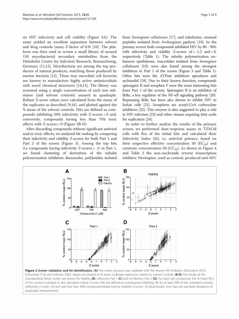
screen, we performed dose-response assays in TZM-blcells with five of the initial hits and calculated theirSelectivity Index (SI), i.e. antiviral potency, based ontheir respective effective concentration 50 (EC50) andcytotoxic concentration 50 (CC50). As shown in Figure 4and Table 2 the non-nucleoside reverse transcriptaseinhibitor Nevirapine, used as control, produced anti-HIV
ol was validated with the known HIV inhibitors Zidovudine (AZT),e expression relative to solvent controls. (B-D) The results of theand re-infection Part 2 (D). For each test compound, the % mean RLU
as compounds inhibiting HIV by at least 50% of the untreated controlsviability Z-score > 0) (blue boxes). Error bars are standard deviations of

Figure 3 Summary of the strongest myxobacterial hits. The plot shows the viability Z-score vs. the infectivity Z-score of compounds havingthe strongest antiviral activity (Z score < -1) for both Part 1 and Part 2 (inlay). The figure shows a clear clustering of compounds based onstructural similarities and known function (right). The commercial drugs AZT, T20 and IND are included as positive controls. Compounds areabbreviated to prevent figure congestion. Corresponding compound names are given in Table 1.
Martinez et al. Microbial Cell Factories 2013, 12:85 Page 4 of 9http://www.microbialcellfactories.com/content/12/1/85
SI values similar to those previously described [25]. Themyxobacterial compounds sulfangolid C and soraphen Fshowed EC50 and CC50 values comparable to each other,with SI ranging from 16 to 20. Both epothilon D andspirangien B showed lower EC50 values than Nevirapine,while spirangien exhibited the highest SI value of the fivetested myxobacteria metabolites (>50) (Figure 4 andTable 2). Kulkenon was not selective in the follow-up, witha low SI of around 5.The most interesting hit from Part 2 of the screen was
rhizopodin, an actin inhibitor isolated from the myxo-bacterium Myxococcus stipitatus [26]. Actin filamentsare known to be essential for virological synapse forma-tion and HIV cell-to-cell transmission, the main routeof HIV infection in naïve cells in vivo [27,28]. Anotherhit was leupyrrin B1, a macrodiol with a unique pyrrolering that has been shown to inhibit DNA, RNA and pro-tein synthesis in yeast [29] (Figure 3 and Table 1). Inter-estingly, highly substituted pyrroles have been shown totarget the HIV reverse transcriptase [30] and, in our pri-mary screen, leupyrrin B1 also showed a mild effect onPart 1 (data not shown). However, the studies needed totest the real potency of these late-acting hits are beyondthis report.In summary, we have established a two-step HTS plat-
form based on the TZM-bl cell line for the screening ofnovel HIV inhibitors. The advantage of this method is thatit can distinguish between compounds acting on early orlate events of the HIV replication cycle so that initial hintson the mode-of-action can be already obtained fromprimary screens. With this assay, a small library of
secondary metabolites derived from myxobacteria wasscreened and interesting primary hits were found. In gen-eral, the intrinsic antiviral potency was lower in thefollow-up experiments. Unfortunately, these observationsare common in HTS campaings, from which few initialhits are confirmed in subsequent assays [31,32]. In par-ticular, comparative low SI values have been commonlyobserved for natural products of diverse origin [33-38],and it has also been noted that a compound´s biologicalefficacy is not due to in vitro toxicity when the SI > 10[39]. In our experiments, even when dose response assayswith five of the initial hits showed a rather low SI com-pared to a standard FDA-approved drug, four com-pounds exhibited SI values >15. The screening fornatural compounds allows for the discovery of mole-cules with good effectiveness, which could be furtheroptimized by synthetic methods. The myxobacteria sec-ondary metabolism comprises compounds with largechemical components that might “mask” the actual anti-viral pharmacophore (see structures in Table 2). There-fore, further structural studies of our initial hits mighthelp recognize smaller drug-like derivatives that in turncould be synthesized and further optimized.
ConclusionsWe have established a robust high-throughput anti-HIVassay that allows, already from the primary screen, todiscriminate compounds acting on early and/or late stepsof the virus replication cycle. We identified several second-ary metabolites derived from myxobacteria with goodanti-HIV profiles. Remarkably, compared to a previous

Table 1 Robust Z-score ranking of the myxobacterial hits for both part 1 (upper) and part 2 (bottom) of the screen
Hits from part 1 Infectivity Viability
Name Abbr. Z-score % Mean SD Z-score % Mean SD
Tubulysin l* Tlsl −2.47 3.70 1.27 −0.08 92.04 4.38
Kulkenon Kulk −2.41 5.15 1.09 0.03 93.01 18.62
Epothilon A* EpoA −2.38 0.49 0.15 0.24 87.72 10.27
Spirangien B SpiB −2.38 1.09 0.19 0.08 85.71 4.14
Epothilon C* EpoC −2.37 0.54 0.14 −0.25 81.63 4.07
Epothilon D* EpoD −2.37 1.52 0.54 1.00 97.33 11.03
Terrestribisamid A TerA −2.35 6.70 0.26 0.34 95.99 10.58
Enfuvirtide (Control) T20 −2.30 2.26 0.18 1.36 97.21 0.05
Sulfangolid C SulfC −2.29 8.13 1.26 1.73 109.20 13.03
Zidovudine (Control) AZT −2.24 4.29 0.21 1.59 98.62 1.59
Apicularen B* ApiB −1.93 13.77 2.40 −0.16 82.77 4.37
Disorazol A4* DszA4 −1.78 20.37 2.01 0.21 94.79 14.99
Tubulysin B* TlsB −1.61 24.04 1.01 0.38 89.55 10.43
Disorazol B1* DszB1 −1.55 25.89 3.60 1.25 104.59 15.96
Disorazol E1* DszE1 −1.54 26.22 3.20 −0.13 91.48 18.83
Stigmatelin A StgA −1.43 29.56 2.80 −0.34 80.43 3.79
Tubulysin A* TlsA −1.42 29.95 4.87 −0.11 83.33 9.78
Archazolid A* ArcA −1.40 30.47 4.01 −0.35 80.30 4.86
Tubulysin G* TlsG −1.34 32.46 3.73 0.34 88.98 2.54
Soraphen F SorphF −1.32 31.43 7.72 1.58 107.80 6.05
Disorazol E3* DszE3 −1.31 31.74 4.50 −0.05 92.32 3.12
Maltepolid B MalB −1.16 35.34 8.71 −0.11 91.76 7.42
Crocacin A CroA −1.06 41.32 6.39 0.15 86.66 2.43
Noricumazol B NoricB −1.01 43.03 6.34 0.86 95.47 5.54
Socein A SoceinA −0.97 40.09 7.31 0.68 99.20 13.69
Hits from part 2 Infectivity Viability
Name Abbr. Z-score % Mean SD Z-score % Mean SD
Rhizopodin A RhiA −2.11 0.00 0.00 −1.38 67.39 11.37
Indinavir (control) IND −1.93 2.73 0.10 1.43 91.21 8.50
Leupyrrin B1 LeuB1 −1.85 3.12 0.24 −0.01 67.54 11.23
Crocapeptin B* CrpB −1.80 11.38 1.78 −1.40 67.17 6.95
Soraphen A SorA −1.49 22.24 2.69 0.15 86.61 6.68
Cruentaren A CruA −1.25 31.05 5.38 0.66 93.01 5.04
Corresponding % relative infectivity and cell viability is shown. Compounds are listed by infectivity Z-scores. Control drugs are shown for comparison.*: Compounds with anti-HCV activities according to [40].
Martinez et al. Microbial Cell Factories 2013, 12:85 Page 5 of 9http://www.microbialcellfactories.com/content/12/1/85
screen for Hepatitis C Virus (HCV) using the same library,we found a high amount of overlapping hits (Table 1 and[40]), suggesting that these compounds may target com-monly used host factors or pathways necessary for viralreplication. Although the intrinsic antiviral potency ofmost of these compounds remains to be elucidated, theunique structures and mode-of-actions of these naturalcompounds make myxobacteria an attractive source ofchemicals for the development of broad-spectrumantivirals [14].
Materials and methodsCells and culture mediumTZM-bl, a modified HeLa cell-line susceptible to infectionwith different HIV-1 isolates, was obtained from the NIHAIDS Research and Reference Reagent Program, Cat#8129) and maintained in Dulbecco’s modified Eagle’smedium (DMEM; Invitrogen, Karlsruhe, Germany)supplemented with 10% heat-inactivated FCS, HEPES25 mM and 0.5% gentamycin. PM1 cells (NIH AIDS Re-search and Reference Reagent Program, Cat# 3038) were
Figure 4 Dose response curves of five of the HIV hits. TZM-bl cells were pre-incubated with 10-fold serial dilutions of compounds andvehicle control for 2 h and infected with HIVLAI at an MOI = 0.5. 48 h after initial infection luciferase expression was quantified (see Materials andMethods). Cell toxicity was measured in parallel plates. Curves show the drug dose vs. the response normalized to the control. Error bars arestandard error of the mean.
Martinez et al. Microbial Cell Factories 2013, 12:85 Page 6 of 9http://www.microbialcellfactories.com/content/12/1/85
maintained with RPMI medium supplemented with 10%heat-inactivated FCS and 1% of penicillin-streptomycin.Both cell-lines were cultured at 37°C, 5% CO2.
Virus stocks and infectionsHIV-1LAI isolate was obtained from the Centre for AIDSReagents, NIBSC, UK. Virus was propagated in PM1 cellsand titrated in TZM-bl cells as described [41]. 1 mLaliquots of virus stocks were stored at -80°C until use.Infection experiments in TZM-bl cells were performed inquadruple at a multiplicity of infection (MOI) of 0.5. Fordrug-response assays TZM-bl cells were plated (104 cells/well) in Nunc® MicroWell 96 well optical bottom plates(Sigma) and incubated for 1 h with increasing concentra-tions of test compounds in 10-fold dilutions or with thecorresponding vehicle (DMSO or MeOH) as negative con-trol in triplicates. After drug incubation, cells wereinfected with HIVLAI and 48 h after infection luciferaseactivity was measured using Britelite Plus™ (PerkinElmer,
Waltham, USA). In parallel, cell viability of TZM-bl cellswas determined with an ATP quantification method usingthe commercial kit CellTiter-Glo® Luminescent Cell Via-bility Assay (Promega, Madison, USA). ATP is a marker ofthe presence of metabolically active cells [42]. Therefore,the ATP levels relative to the untreated control are ameasure of drug-induced cytotoxicity. Mean luciferasevalues were normalized to untreated controls and EffectiveConcentration 50 (EC50) and Cytotoxic Concentration 50(CC50) were calculated in GraphPad Prism (GraphPadSoftware, San Diego, CA, USA) by analyzing the logdose vs.normalized response. The Selectivity Index (SI) refers tothe antiviral potency of a drug and is calculated as theratio of CC50 to EC50 [43,44].
Test compoundsThe library of 154 myxobacterial secondary metabolitesused for the screening belongs to a collection of naturalcompounds isolated at the Helmholtz Centre for Infection

Table 2 Compound names, structures, EC50, CC50 and SI values for five of the preliminary hits
Compound name Structure EC50 (μM) CC50 (μM) SI*
Nevirapine 0.07 81.8 >1000
Sulfangolid C 0.41 8.18 20.2
Soraphen F 0.30 5.02 16.5
Epothilon D 0.0005 0.012 24.4
Spirangien B 0.007 0.35 52
Kulkenon 0.07 0.36 5.3
*SI: Selectivity Index.
Martinez et al. Microbial Cell Factories 2013, 12:85 Page 7 of 9http://www.microbialcellfactories.com/content/12/1/85
Research, Braunschweig, Germany [11,12,45]. Compoundsof > 95% purity as measured by LC-MS were provided in96-well screening plates in a concentration of 1 mMin puriss.p.a. dimethyl sulfoxide (DMSO) or methanol(MeOH). The fusion inhibitor Enfuvirtide (Fuzeon, Roche,Basel Switzerland) and the nucleoside reverse transcript-ase inhibitor Zidovudine (NIH AIDS Research and Refer-ence Reagent Program, Cat# 3485) were used as positivecontrols at final concentrations of 1 μM.
Two-step TZM-bl based high-throughput screening assayFor the Part 1 of the screen, TZM-bl cells were seeded in384-well plates at a density of 2500 cells per well in 30 μL
of culture medium and incubated overnight at 37°C and5% CO2. After incubation, 50 to 70 nL of the test com-pounds and DMSO and/or MeOH controls from the96-well screening plates were dispensed in quadruple tothe cells with the PinTool of an Evolution P3 pipettingplatform (PerkinElmer, Zaventem, Belgium). Final concen-tration of compounds was around 1.5 to 2 μM with acontent of around 0.2% of DMSO or MeOH in all cases.DMSO or MeOH alone were used as controls. 2 h afteraddition of compounds, cells were infected with 50 μL ofHIVLAI at a MOI of 0.5. In parallel, duplicate plateswere left uninfected for quantification of compound-related toxicity. Plates were incubated at 37°C and 5% CO2.
Martinez et al. Microbial Cell Factories 2013, 12:85 Page 8 of 9http://www.microbialcellfactories.com/content/12/1/85
Forty-eight hours after virus addition, 50 μL supernatantfrom cells of Part 1 was transferred to fresh TZM-bl cellsseeded in 384-wells the day before. Cells of Part 1 plateswere assayed for Tat-dependent luciferase expression byadding an equal volume of Britelite Plus™ (PerkinElmer,Waltham, USA) according to the manufacturer´s in-structions. Compound-related toxicity in duplicate plateswas quantified with the CellTiter-Glo® Luminescent CellViability Assay (Promega, Madison, USA) according tothe manufacturer´s instructions. ATP is a marker of thepresence of metabolically active cells [42]. Following thesame procedure, cells from Part 2 of the screen wereassayed forty-eight hours after supernatant addition. Lu-ciferase expression was measured with a TECAN InfiniteM1000PRO microplate reader (Tecan, Switzerland). Thepossible outcomes of the screen are depicted in Figure S1.
Data analysis and statisticsData was analyzed by obtaining the % mean luciferasevalues normalized to solvent controls (% control meanRLU) and by calculating the assay Z-factor (a measurefor assay quality) and samples (robust) Z-scores (a meas-ure of hit quality) as described [9,10,16]. Briefly, back-ground levels were substracted from the mean luciferaseof samples and values were normalized to those of thesolvent controls (set to 100% expression). The assayZ-factor was calculated by dividing 3X standard devia-tions of controls by the sum of their means as described(see Table 1 in [10]). For hit determination, we adapted apreviously described robust Z-score calculation [9] toadjust for interplate variation by dividing the absolutedeviation of the mean of the quadruple data points(4 wells for each compound) by the median absolutedeviation of each plate. Unless stated otherwise, errorsare given as ± SD.
Competing interestThe authors declare that they have no competing interests
Authors’ contributionsJPM and BH established and performed the screening assay; JPM and EFSperformed drug-response assays; HS and RJ provided the myxobacteriallibrary and additional compounds; JPM, JD, RF, FS and AM designed theexperimental approach and wrote the manuscript. All listed authors read andapproved the final manuscript.
AcknowledgementsThe research is supported by grants from the Bill and Melinda GatesFoundation, Institució Catalana de Recerca i Estudis Avancats (ICREA), theSpanish Ministry of Science and Innovation (SAF2010-21336 and BFU2010-20803), FPI grant number BES-2011-048569.
Author details1Infection Biology Group, Department of Experimental and Health Sciences,Universitat Pompeu Fabra, Barcelona, Spain. 2Department of ChemicalBiology, Helmholtz Centre for Infection Research, Braunschweig, Germany.3Department of Microbial Drugs, Helmholtz Centre for Infection Research,Braunschweig, Germany. 4Molecular Virology Group, Department ofExperimental and Health Sciences, Universitat Pompeu Fabra, Barcelona,
Spain. 5Institució Catalana de Recerca i Estudis Avançats (ICREA), Barcelona,Spain.
Received: 6 August 2013 Accepted: 19 September 2013Published: 24 September 2013
References1. Arts EJ, Hazuda DJ: HIV-1 antiretroviral drug therapy. Cold Spring Harbor
perspectives in medicine 2012, 2(4):a007161.2. Back D: New drug interactions in HIV and HCV. Retrovirology 2012, 9:I8.3. de Maat MM, Ekhart GC, Huitema AD, Koks CH, Mulder JW, Beijnen JH: Drug
interactions between antiretroviral drugs and comedicated agents.Clinical pharmacokinetics 2003, 42:223–282.
4. McIlleron H, Meintjes G, Burman WJ, Maartens G: Complications ofantiretroviral therapy in patients with tuberculosis: drug interactions,toxicity, and immune reconstitution inflammatory syndrome. J Infect Dis2007, 196:S63–S75.
5. Tseng A, Foisy M: Important drug-drug interactions in HIV-infectedpersons on antiretroviral therapy: an update on new interactionsbetween HIV and non-HIV drugs. Current infectious disease reports 2012,14:67–82.
6. Wei X, Decker JM, Liu H, Zhang Z, Arani RB, Kilby JM, Saag MS, Wu X, ShawGM, Kappes JC: Emergence of resistant human immunodeficiency virustype 1 in patients receiving fusion inhibitor (T-20) monotherapy.Antimicrob Agents Chemother 2002, 46:1896–1905.
7. Montefiori DC: Evaluating neutralizing antibodies against HIV, SIV, andSHIV in luciferase reporter gene assays. Curr Protoc Immunol 2005,Chapter 12:Unit 12–11.
8. Brass AL, Dykxhoorn DM, Benita Y, Yan N, Engelman A, Xavier RJ, LiebermanJ, Elledge SJ: Identification of host proteins required for HIV infectionthrough a functional genomic screen. Science 2008, 319:921–926.
9. Tan X, Hu L, Luquette LJ 3rd, Gao G, Liu Y, Qu H, Xi R, Lu ZJ, Park PJ, Elledge SJ:Systematic identification of synergistic drug pairs targeting HIV. NatBiotechnol 2012, 30:1125–1130.
10. Zhang JH, Chung TD, Oldenburg KR: A simple statistical parameter for usein evaluation and validation of high throughput screening assays.J Biomol Screen 1999, 4:67–73.
11. Reichenbach H: Myxobacteria, producers of novel bioactive substances.J Ind Microbiol Biotechnol 2001, 27:149–156.
12. Reichenbach H, Höfle G: Myxobacteria as producers of secondarymetabolites. In Drug discovery from nature. 1999:149–179.
13. Pandey S, Sree A, Dash SS, Sethi DP, Chowdhury L: Diversity of marinebacteria producing beta-glucosidase inhibitors. Microb Cell Fact 2013,12:35.
14. Diez J, Martinez JP, Mestres J, Sasse F, Frank R, Meyerhans A: Myxobacteria:natural pharmaceutical factories. Microb Cell Fact 2012, 11:52.
15. Huttel S, Muller R: Methods to optimize myxobacterial fermentationsusing off-gas analysis. Microb Cell Fact 2012, 11:59.
16. Malo N, Hanley JA, Cerquozzi S, Pelletier J, Nadon R: Statistical practice inhigh-throughput screening data analysis. Nat Biotechnol 2006, 24:167–175.
17. Hopkins CD, Wipf P: Isolation, biology and chemistry of the disorazoles:new anti-cancer macrodiolides. Nat Prod Rep 2009, 26:585–601.
18. Weissman KJ, Muller R: Myxobacterial secondary metabolites: bioactivitiesand modes-of-action. Nat Prod Rep 2010, 27:1276–1295.
19. Reichenbach H, Hofle G: Discovery and development of the epothilones:a novel class of antineoplastic drugs. Drugs R D 2008, 9:1–10.
20. Reboll MR, Ritter B, Sasse F, Niggemann J, Frank R, Nourbakhsh M: Themyxobacterial compounds spirangien a and spirangien M522 are potentinhibitors of IL‐8 expression. ChemBioChem 2012, 13:409–415.
21. Kwon H, Pelletier N, DeLuca C, Genin P, Cisternas S, Lin R, Wainberg MA,Hiscott J: Inducible expression of IkappaBalpha repressor mutantsinterferes with NF-kappaB activity and HIV-1 replication in Jurkat T cells.J Biol Chem 1998, 273:7431–7440.
22. Jump DB, Torres-Gonzalez M, Olson LK: Soraphen A, an inhibitor of acetylCoA carboxylase activity, interferes with fatty acid elongation.Biochemical pharmacology 2011, 81:649–660.
23. Steger DJ, Eberharter A, John S, Grant PA, Workman JL: Purified histoneacetyltransferase complexes stimulate HIV-1 transcription frompreassembled nucleosomal arrays. Proc Natl Acad Sci 1998,95:12924–12929.
Martinez et al. Microbial Cell Factories 2013, 12:85 Page 9 of 9http://www.microbialcellfactories.com/content/12/1/85
24. Heaton NS, Perera R, Berger KL, Khadka S, LaCount DJ, Kuhn RJ, Randall G:Dengue virus nonstructural protein 3 redistributes fatty acid synthase tosites of viral replication and increases cellular fatty acid synthesis.Proc Natl Acad Sci 2010, 107:17345–17350.
25. Koh Y, Haim H, Engelman A: Identification and characterization ofpersistent intracellular human immunodeficiency virus type 1 integrasestrand transfer inhibitor activity. Antimicrobial agents and chemotherapy2011, 55:42–49.
26. Sasse F, Steinmetz H, Höfle G, Reichenbach H: Rhizopodin, a new compoundfrom myxococcus stipitatus (myxobacteria) causes formation of rhizopodia-like structures in animal cell cultures. Production, isolation, physico-chemical and biological properties. J Antibiot (Tokyo) 1993, 46:741.
27. Felts RL, Narayan K, Estes JD, Shi D, Trubey CM, Fu J, Hartnell LM, Ruthel GT,Schneider DK, Nagashima K: 3D visualization of HIV transfer at thevirological synapse between dendritic cells and T cells. Proc Natl Acad Sci2010, 107:13336–13341.
28. Jolly C, Kashefi K, Hollinshead M, Sattentau QJ: HIV-1 cell to cell transferacross an Env-induced, actin-dependent synapse. J Exp Med 2004,199:283–293.
29. Bode HB, Irschik H, Wenzel SC, Reichenbach H, Muller R, Hofle G: Theleupyrrins: a structurally unique family of secondary metabolites from themyxobacterium Sorangium cellulosum. J Nat Prod 2003, 66:1203–1206.
30. Antonucci T, Warmus J, Hodges J, Nickell D: Characterization of theantiviral activity of highly substituted pyrroles: a novel class of non-nucleoside HIV-1 reverse transcriptase inhibitor. Antiviral chemistry &chemotherapy 1995, 6:98–108.
31. Malo N, Hanley JA, Cerquozzi S, Pelletier J, Nadon R: Statistical practice inhigh-throughput screening data analysis. Nat Biotechnol 2006, 24(2):167–175.
32. Smith T, Ho P-i, Yue K, Itkin Z, MacDougall D, Paolucci M, Hill A, Auld DS:Comparison of compound administration methods in biochemicalassays: effects on apparent compound potency using either assay-readycompound plates or pin tool -delivered compounds. J Biomol Screen2013, 18(1):14–25.
33. Asres K, Bucar F, Kartnig T, Witvrouw M, Pannecouque C, De Clercq E:Antiviral activity against human immunodeficiency virus type 1 (HIV‐1)and type 2 (HIV‐2) of ethnobotanically selected Ethiopian medicinalplants. Phytother Res 2001, 15:62–69.
34. Cos P, Vlietinck AJ, Berghe DV, Maes L: Anti-infective potential of naturalproducts: how to develop a stronger in vitro ‘proof-of-concept’. Journalof ethnopharmacology 2006, 106:290–302.
35. Crance JM, Scaramozzino N, Jouan A, Garin D: Interferon, ribavirin,6-azauridine and glycyrrhizin: antiviral compounds active againstpathogenic flaviviruses. Antiviral research 2003, 58:73–79.
36. Fiore C, Eisenhut M, Krausse R, Ragazzi E, Pellati D, Armanini D, Bielenberg J:Antiviral effects of Glycyrrhiza species. Phytother Res 2008, 22:141–148.
37. Hayashi K, Minoda K, Nagaoka Y, Hayashi T, Uesato S: Antiviral activity ofberberine and related compounds against human cytomegalovirus.Bioorganic & medicinal chemistry letters 2007, 17:1562–1564.
38. Sun Y, Song M, Niu L, Bai X, Sun N, Zhao X, Jiang J, He J, Li H: Antiviraleffects of the constituents derived from Chinese herb medicines oninfectious bursal disease virus. Pharmaceutical biology 2013,51(9):1127–1143.
39. Vonthron-Sénécheau C, Weniger B, Ouattara M, Bi FT, Kamenan A, Lobstein A,Brun R, Anton R: In vitro antiplasmodial activity and cytotoxicity ofethnobotanically selected Ivorian plants. Journal of ethnopharmacology 2003,87:221–225.
40. Gentzsch J, Hinkelmann B, Kaderali L, Irschik H, Jansen R, Sasse F, Frank R,Pietschmann T: Hepatitis C virus complete life cycle screen foridentification of small molecules with pro- or antiviral activity. AntiviralRes 2011, 89:136–148.
41. Montefiori DC: Evaluating neutralizing antibodies against HIV, SIV, andSHIV in luciferase reporter gene assays. Current protocols in immunology2005, Chap.12:Unit 12.11.
42. Crouch SP, Kozlowski R, Slater KJ, Fletcher J: The use of ATPbioluminescence as a measure of cell proliferation and cytotoxicity.J Immunol Methods 1993, 160:81–88.
43. Tamamura H, Omagari A, Oishi S, Kanamoto T, Yamamoto N, Peiper SC,Nakashima H, Fujii N: Pharmacophore identification of a specific CXCR4inhibitor, T140, leads to development of efective anti-HIV agents withvery high selectivity indexes. Bioorg Med Chem 2000, 10:2633–2637.
44. Pauwels R, Andries K, Debyser Z, Van Daele P, Schols D, Stoffels P, De Vreese K,Woestenborghs R, Vandamme aM, Janssen CG: Potent and highly selectivehuman immunodeficiency virus type 1 (HIV-1) inhibition by a series ofalpha-anilinophenylacetamide derivatives targeted at HIV-1 reversetranscriptase. PNAS 1993, 90:1711–1715.
45. Reichenbach H: Myxobacteria. In Encyclopedia of Bioprocess Technology. Vol.1-5th edition. Wiley-Interscience; 1992.
doi:10.1186/1475-2859-12-85Cite this article as: Martinez et al.: Identification of myxobacteria-derivedHIV inhibitors by a high-throughput two-step infectivity assay. MicrobialCell Factories 2013 12:85.
Submit your next manuscript to BioMed Centraland take full advantage of:
• Convenient online submission
• Thorough peer review
• No space constraints or color figure charges
• Immediate publication on acceptance
• Inclusion in PubMed, CAS, Scopus and Google Scholar
• Research which is freely available for redistribution
Submit your manuscript at www.biomedcentral.com/submit