Identification of Histamine Receptors in the Canine ...
67
Mississippi State University Mississippi State University Scholars Junction Scholars Junction Theses and Dissertations Theses and Dissertations 1-1-2016 Identification of Histamine Receptors in the Canine Identification of Histamine Receptors in the Canine Gastrointestinal Tract Gastrointestinal Tract Alyssa Martin Sullivant Follow this and additional works at: https://scholarsjunction.msstate.edu/td Recommended Citation Recommended Citation Sullivant, Alyssa Martin, "Identification of Histamine Receptors in the Canine Gastrointestinal Tract" (2016). Theses and Dissertations. 2549. https://scholarsjunction.msstate.edu/td/2549 This Graduate Thesis - Open Access is brought to you for free and open access by the Theses and Dissertations at Scholars Junction. It has been accepted for inclusion in Theses and Dissertations by an authorized administrator of Scholars Junction. For more information, please contact [email protected].
Transcript of Identification of Histamine Receptors in the Canine ...
Mississippi State University Mississippi State University
Scholars Junction Scholars Junction
Theses and Dissertations Theses and Dissertations
1-1-2016
Identification of Histamine Receptors in the Canine Identification of Histamine Receptors in the Canine
Gastrointestinal Tract Gastrointestinal Tract
Alyssa Martin Sullivant
Follow this and additional works at: https://scholarsjunction.msstate.edu/td
Recommended Citation Recommended Citation Sullivant, Alyssa Martin, "Identification of Histamine Receptors in the Canine Gastrointestinal Tract" (2016). Theses and Dissertations. 2549. https://scholarsjunction.msstate.edu/td/2549
This Graduate Thesis - Open Access is brought to you for free and open access by the Theses and Dissertations at Scholars Junction. It has been accepted for inclusion in Theses and Dissertations by an authorized administrator of Scholars Junction. For more information, please contact [email protected].
Template B v3.0 (beta): Created by J. Nail 06/2015
Identification of histamine receptors in the canine gastrointestinal tract
By TITLE PAGE
Alyssa Martin Sullivant
A Thesis Submitted to the Faculty of Mississippi State University
in Partial Fulfillment of the Requirements for the Degree of Master of Science
in Veterinary Medical Research in the College of Veterinary Medicine
Mississippi State, Mississippi
December 2016

Copyright by COPYRIGHT PAGE
Alyssa Martin Sullivant
2016

Identification of histamine receptors in the canine gastrointestinal tract
By APPROVAL PAGE
Alyssa Martin Sullivant
Approved:
____________________________________ Todd M. Archer
(Major Professor)
____________________________________ Andrew J. Mackin
(Committee Member/ Graduate Coordinator)
____________________________________ G. Todd Pharr
(Committee Member)
____________________________________ Avery James Cooley (Committee Member)
____________________________________ Kent H. Hoblet
Dean College of Veterinary Medicine
Name: Alyssa Martin Sullivant ABSTRACT
Date of Degree: December 9, 2016
Institution: Mississippi State University
Major Field: Veterinary Medical Research
Major Professor: Todd M. Archer
Title of Study: Identification of histamine receptors in the canine gastrointestinal tract
Pages in Study: 57
Candidate for Degree of Master of Science
The role of histamine in chronic gastrointestinal diseases has been increasingly
recognized in humans, but the role of histamine in the canine gastrointestinal tract has not
been thoroughly investigated. The presence and distribution of all 4 histamine receptors
(H1, H2, H3, and H4) in the stomach, duodenum, ileum, jejunum, and colon of healthy
dogs were evaluated with a commonly employed immunohistochemistry technique using
antibodies predicted to cross react with canine histamine receptors. All 4 histamine
receptors were identified in the canine gastrointestinal tract, and differed in location and
density within sections of the canine gastrointestinal tract. Antibody specificity was
evaluated with Western blot. With the establishment of a method to study histamine
receptors in the canine gastrointestinal tract, additional research to evaluate histamine
receptors in dogs is warranted to further understand the pathophysiology and treatment of
chronic canine enteropathies.

ii
DEDICATION
I would like to dedicate this research to my parents, who have loved me and
supported me in every way possible. I will never be able to thank them enough. I would
also like to dedicate this to my precious husband, Hayden, who graciously allowed me to
chase my dreams and cheered me on the whole way. And lastly, I dedicate this to our
beautiful daughter, Riley, who brings me endless joy with her quick wit and joyful spirit.

iii
ACKNOWLEDGEMENTS
I would like to thank Dr. Todd Archer and Dr. Andrew Mackin for the
tremendous amount of support and guidance throughout my research and residency. They
inspire me to be the best internist and teacher I can be, and I feel very fortunate to work
with them every day. I am also very grateful for the expertise and patience of Drs. Jim
Cooley, Todd Pharr, and Bob Wills as well. The success of this project would have never
been possible without each and every one of my committee members, and I am proud to
work amongst such wonderful clinicians, researchers, and educators. A special thank you
also goes to Mrs. Stephany Mays and the rest of the Mississippi State University
immunohistochemistry lab for the many hours dedicated to teaching and helping me learn
the ropes in immunohistochemistry. Most importantly, I am grateful for the endless
blessings bestowed upon me by God, both personally and professionally.
iv
TABLE OF CONTENTS
DEDICATION .................................................................................................................... ii
ACKNOWLEDGEMENTS ............................................................................................... iii
TABLE OF CONTENTS ................................................................................................... iv
LIST OF FIGURES ........................................................................................................... vi
CHAPTER
I. INTRODUCTION ................................................................................................1
Histamine Discovery and Biology .........................................................................1 Histamine Receptors ..............................................................................................2
H1 Receptor .....................................................................................................3 H2 Receptor .....................................................................................................4 H3 Receptor .....................................................................................................5 H4 Receptor .....................................................................................................7
Emerging Roles of Histamine and Histamine Receptors ......................................8
Histamine in Inflammation ..............................................................................9 Effects of Histamine on Immune Cells .....................................................9 Histamine in Asthma and Allergic Inflammation ...................................11 Histamine in Gastrointestinal Inflammation ...........................................12
Histamine in Neoplasia ..................................................................................15 Importance of the Research .................................................................................15
Canine Chronic Enteropathies .......................................................................16 Histamine Research Techniques ....................................................................17
References Cited ..................................................................................................19
II. IDENTIFICATION OF HISTAMINE RECEPTORS IN THE CANINE GASTROINTESTINAL TRACT ...........................................................27
Introduction .........................................................................................................27 Materials and Methods ........................................................................................30
Tissue Collection and Evaluation ..................................................................30
Evaluation of Gastrointestinal Inflammation ................................................31 Immunohistochemistry ..................................................................................31 Histamine Receptor Scoring ..........................................................................33

v
Western Blot Technique ................................................................................33
Statistical Analysis ........................................................................................34 Results .................................................................................................................35
WSAVA Assessment of Gastrointestinal Inflammation ...............................35 Western Blot Validation of Histamine Receptor Antibodies ........................35 Histamine Receptor Immunohistochemistry Descriptive Findings ...............37 Histamine Receptor Immunohistochemistry Statistical Analysis .................40 Differences in Histamine Receptor Distribution ...........................................41
Discussion ............................................................................................................42 Conclusion ...........................................................................................................45 Footnotes .............................................................................................................46 References Cited ..................................................................................................47
III. CONCLUSION ...................................................................................................49
References Cited ..................................................................................................56
vi
LIST OF FIGURES
2.1 Histamine receptor scoring in the canine gastrointestinal tract .......................33
2.2 Western blots for histamine receptor antibodies ..............................................36
2.3 Histamine receptor staining patterns in the canine gastrointestinal tract. ........39
2.4 Distribution of histamine receptors in the canine gastrointestinal tract ...........40
1
INTRODUCTION
Histamine Discovery and Biology
In 1910, Henry Dale isolated and identified 2- (4-imidazole)-ethylamine, now
known as histamine, from an extract of ergot.1 Since its discovery, histamine has been
linked to well over 20 different physiological responses.1, 2 Initial histamine research led
to groundbreaking discoveries in the realm of human and veterinary medicine, including
the development of specific therapies for anaphylaxis, asthma, gastric ulcers, and allergic
reactions. More recently, the roles of histamine and its receptors in inflammatory,
immune-mediated, and neoplastic diseases have been intensely investigated in human
medicine.3 Human research using new histamine antagonists has advanced our
understanding of these complex conditions and offers exciting therapeutic potentials,
particularly in regards to the treatment of complicated inflammatory conditions.
Histamine is a primary amine synthesized intracellularly through oxidative
decarboxylation of the amino acid histidine and stored in granules within mast cells and
basophils.2 The enzyme L-histadine decarboxylase catalyzes and regulates histamine
production, and the gene for this enzyme is expressed intracellularly in many cell types.4
Mast cells, which are located throughout the body but are especially concentrated in the
skin, lungs, and gastrointestinal tract, are the main sites of histamine synthesis.5
Enterochromaffin-like cells of the gastric mucosa, endothelial cells, platelets, basophils,

2
dendritic cells, T cells, and histaminergic neurons also synthesize and secrete
histamine.2,4,6,7 Histamine is present in varying concentrations in most organs,
particularly within the stomach, lymph nodes, and thymus.7 Histamine is released
following immunological and non-immunological stimulation of histamine-producing
cells, and exerts its diverse biological actions through binding of four different G-protein
coupled histamine receptors (H1, H2, H3, and H4) located throughout the body.3 Overall,
only 2-3% of histamine is excreted unchanged in the urine.3 The majority of histamine
(50-80%) is metabolized to its primary metabolite, N-methylhistamine, by the cytosolic
enzyme N-methyltransferase, which degrades intracellular histamine but spares stored
histamine.2,7,8 The remainder of histamine is metabolized by the enzyme diamine
oxidase.4,8
Histamine Receptors
Investigation of histamine receptors has to date been limited in the veterinary
literature, but these receptors have been studied in human medicine for over 75 years.1
Histamine exerts its actions via binding of four different G-protein coupled receptors,
named H1, H2, H3, and H4, which are members of the largest family of membrane
proteins in the human genome.5 Upon histamine binding, these receptors transmit the
extracellular signal to intracellular second messengers that trigger a cascade of molecular
events that direct a particular physiological response, which, in turn, depends on the
subtype and location of the engaged histamine receptor.2 Each receptor varies in its
affinity for histamine and its level of expression throughout the body.9 Although there are
many similarities in receptor distribution across species, differences between species in
receptor location and function have been documented.1,2 In the following section,
3
location of histamine receptors is based on research in humans and laboratory animals
primarily, unless otherwise noted.
H1 Receptor
The H1 histamine receptor was the first of the four histamine receptors to be
discovered.5 It was characterized in 1937, and the bovine H1 receptor was cloned in
1991.10 The receptor was later cloned in several other species, including the dog, guinea
pig, and human.1 Histamine binding of the H1 receptor stimulates the inositol phosphate
signaling pathway, which leads to increased intracellular calcium.4 Stimulation of the H1
receptor also induces synthesis of prostacyclin and platelet-activating factor, activation of
NF-кB transcription factor, and release of von Willebrand factor and nitrous oxide.4
The H1 receptor is ubiquitously expressed in vascular endothelium, vascular and
airway smooth muscle, hepatocytes, chondrocytes, epithelial cells, neurons, glial cells
and immune cells, including dendritic cells, monocytes, neutrophils, T cells and B
cells.3,4 It has also been identified in the gastrointestinal tract, genitourinary tract, heart,
skin and adrenal medulla.11 Biological actions mediated by the H1 receptor include the
well-known Type 1 hypersensitivity reaction, including smooth muscle contraction in the
intestines responsible for abdominal cramping, pruritus, bronchoconstriction, potent
vasodilation, anaphylactic shock, increased vascular permeability and edema, and release
of catecholamines from the adrenal medulla.2,4 H1 receptor-mediated excitation in the
central nervous system is the major mechanism for cortical activation and wakefulness in
the wake-sleep cycle.5
Common pathological processes mediated by the H1 receptor include allergic
rhinitis, conjunctivitis, asthma, urticaria, atopic or allergic dermatitis, and anaphylaxis.2,4
4
The well-known antihistamines chlorpheniramine, diphenhydramine, and clemastine are
H1 receptor antagonists that have been used for over 70 years for the treatment of allergic
conditions and anaphylactic shock.12 These anti-allergy medications inhibit the effects of
histamine on H1 receptors and stabilize mast cells.9 Because of the sedating effects of
these first-generation H1 antagonists, non-sedating second-generation antagonists such as
cetirizine, lorantadine, and desrolorantadine have been developed.12,13
H2 Receptor
In 1972, the H2 receptor was identified after researchers noticed that several
effects of histamine, such as the stimulatory effect of histamine on gastric acid secretion,
were not inhibited by the H1 antagonist mepyramine.5 The H2 receptor was cloned from
the dog in 1991, and was subsequently cloned from the rat, mouse, guinea pig, and
human.13,14 The human H2 receptor is coupled to both adenylate cyclase and
phosphoinositide second messenger systems.4 Binding of histamine to the receptor may
produce either a stimulatory or inhibitory action on H2 receptor-expressing cells.15.
Like the H1 receptor, the H2 receptor is widespread throughout the body. It is
abundant on the parietal cells of the gastric mucosa and on the endothelial cells,
astrocytes, and neurons in the brain.1 It is also located in smooth muscle cells, myocytes,
enteric neurons, neutrophils, eosinophils, monocytes, macrophages, dendritic cells, and T
and B lymphocytes.2,11,15
The H2 receptor is best known for its role in controlling gastric acid secretion.
Gastrin secreted by the gastric antrum, acetylcholine released from the vagal nerve, and
histamine released by gastric enterochromaffin-like (ECL) cells stimulate hydrochloric
acid release from parietal cells.16 Gastrin initiates the process through stimulation of ECL
5
cells, which synthesize and secrete histamine that binds to the H2 receptor on parietal
cells.16 This binding then activates the H/K-ATPase pump on the parietal cell.16 This
pump is the common target for all stimulatory mechanisms that increase gastric acid
production.16 Without the central role of histamine and its corresponding H2 receptor on
parietal cells, gastric acid production is significantly hampered.17
In addition to its role in gastric acid secretion, the H2 receptor also mediates
vasodilation, inotropy and chronotropy, and smooth muscle relaxation in the uterus,
vasculature, and airways.4,18 Unlike the H1 receptor, which has a pro-inflammatory role,
the H2 receptor has an anti-inflammatory role through the inhibition of cytokine
production and chemotaxis and dampening of T cell responses.2,19 Therefore H2
antagonism may predispose to or exacerbate allergy, inflammation, or infection,
particularly in the gastrointestinal (GI) tract.19,20
Cimetidine was the first H2 antagonist developed in 1977, and it revolutionized
the understanding and treatment of peptic ulcers, dyspepsia, and gastrointestinal reflux.16
More potent H2 antagonists such as ranitidine and famotidine were later developed, and
all are considered safe, with very little affinity for H1 receptors.5 Other potential uses of
H2 antagonists include cardioprotection in ischemic conditions and improvement of
cognitive function.18,21
H3 Receptor
The third histamine receptor was characterized in 1983, and cloned in humans in
1999.22 It was later cloned in the rat, guinea pig, mouse, and monkey, and has been found
to be over 90% conserved between species.1,5 This receptor shows low sequence
homology with the H1 and H2 receptors, and different isoforms of the receptor have been
6
identified.22,23 The H3 receptor was discovered as a presynaptic receptor on
histaminergic nerve terminals in the central nervous system, where it functions to control
the synthesis and release of histamine.12 When the H3 receptor is bound to its G-protein
coupled receptor, adenylate cyclase activity is inhibited along with calcium channels at
certain nerve endings, thereby controlling neurotransmitter release.2
Primary locations of the H3 receptor in the central nervous system (CNS) include
the hypothalamus, thalamus, frontal cortex, hippocampus, caudate nucleus, and basal
ganglia.2,3 This receptor is also located in the peripheral nervous system, particularly in
the enteric nervous system.3 The H3 receptor has also been found in other non-
neurological tissues, including the respiratory epithelium, gastric and intestinal mucosa,
skin, vascular endothelium, thymus, heart, and kidney of the rat.5,24 The H3 receptor is
involved primarily in negative feedback-mediated inhibition of the synthesis and release
of not only histamine but also other neurotransmitters such as dopamine, serotonin,
glutamate, acetylcholine, and norepinephrine.1,2 Neurogenic inflammation may also be
controlled in part through the interaction of histamine on mast cell H3 receptors through
a neuron-mast cell feedback loop.15,25 In addition to its control of histamine and
neurotransmitter synthesis and release, the H3 receptor is also involved in the sleep-wake
cycle, memory, appetite, and cognition.4,26 Gastrointestinal motility is altered by the H3
receptor due to influence on the release various neurotransmitters.2,27 The H3 receptor
also appears to have a protective role against the formation of gastrointestinal ulcers.28,29
Several H3 agonists and antagonists have been developed, and all are in the
preclinical phases of study.5 Thioperamide is a particularly useful, potent H3 antagonist
that has had a pivotal role in investigation of the H3 receptor.12 Treatment of several brain

7
disorders with H3 receptor antagonists have been investigated, including narcolepsy,
Alzheimer’s, and attention disorders, , and the results support their use in these complex
disorders.15 Treatment of pain with H3 antagonists has yielded conflicting results, but in a
recent study an experimental H3 antagonist was safe and efficacious for neuropathic or
osteoarthritic pain.1,30
H4 Receptor
In 2000, the H4 receptor was discovered, and it has since been cloned in the rat,
mouse, dog, guinea pig, monkey, and pig.5, 31 Sequence homology between the canine
and human H4 receptor is 71%.31 Histamine has a high affinity for the H4 receptor, but
species differences in ligand binding and downstream signaling are evident.5 There is an
approximately 40% sequence homology between the H3 and H4 receptor, and less
homology between the H1 and H2 receptors.26 Similar to the H3 receptor, the H4 receptor
inhibits cAMP formation after it is bound by histamine.5,15
Expression of H4 receptors is highest in the bone marrow and peripheral
hematopoietic cells.4 The receptor is found on neutrophils, eosinophils, mast cells,
dendritic cells, basophils, T cells and B cells.1,2,4 H4 receptors have been identified in the
spleen, thymus, brain, liver, lung, heart, epidermis, peripheral nerves, enteric nerves, and
the large and small intestines.1,4 In 2011, H4 receptor mRNA was first identified in
healthy canine skin, colon, liver, spleen and kidney, and showed similar tissue
distribution to that of humans and rodent models.32 New sites of H4 receptor expression
are emerging as research with this receptor continues at a rapid pace, but the dynamic
expression levels of the receptor, particularly in inflammation, have led to inconsistent
findings.5,33
8
The primary function of the H4 receptor is mediation of inflammation through
induction of cytokine release and chemotaxis of inflammatory cells, especially
eosinophils and mast cells.3,5 Overall, the H4 receptor appears to exert a pro-
inflammatory state, and evidence for its role in chronic inflammatory diseases and
autoimmunity is abundant.1,3,4 The H4 receptor mediates increased pruritus and promotes
allergic inflammation, along with H1 receptor.2,34,35 Immune cell proliferation and
differentiation is also affected by the H4 receptor.5 H4 receptor dysregulation has also
been linked to certain neoplasia, such as breast and colon cancer.1,4
H4 receptor antagonists have been developed for research and most are in phase I
preclinical trials.5,22 However, the antagonist JNJ-39758979 has recently been advanced
into clinical trials, and has shown efficacy in treating allergic conditions that have
previously not responded fully to H1 antagonism.36 This antagonist along with another
well-studied H4 antagonist, JNJ 7777120, has also been effective in various animal
models of inflammation such as asthma, dermatitis, and colitis.22,36,37 H4 antagonism
improved pruritus in a mouse model of atopic dermatitis, and in dogs with inflammatory
skin disease.5,37 Efficacy of H4 antagonists in chronic pain has recently been
demonstrated as well.5
Emerging Roles of Histamine and Histamine Receptors
With the development of new histamine receptor agonists and antagonists over
the last two decades, especially in the case of the H3 and H4 receptors, histamine’s role
in numerous biological processes has proven to be far more diverse than ever imagined.38
Newly discovered effects of histamine on the immune response are complex, and they
offer exciting new therapeutic opportunities in chronic inflammatory disease and
9
immune-mediated disorders.4 Furthermore, involvement of different histamine receptors
in certain gastrointestinal disorders, allergic diseases, and cancers is being unraveled.1-5
Histamine in Inflammation
Histamine is intimately involved in many functions of the immune system, and
directly affects the activity of individual immune cells.15 Even wound healing, cellular
proliferation and differentiation, and tissue regeneration involve histamine.4 Histamine
not only mediates several aspects of acute inflammation, including cytokine release,
chemotaxis, and the function of individual immune cells, but also modulates chronic
inflammatory events.3,4,39,40 Consequently, our understanding of several complex and
debilitating inflammatory conditions has been enhanced through histamine research.
Effects of Histamine on Immune Cells
Mast cells and basophils are the primary source of histamine storage in normal
tissues.2 Histamine is released from these cells after immunological activation of their
immunoglobulin E receptor, which binds antibody or antigen and incites histamine
release.2 Non-immunological means of histamine release include cytotoxic and non-
cytotoxic stimulation by cytokines, free radicals, and products of the complement
system.38 Endothelial cells express histamine receptors, and histamine incites
upregulation of adhesion molecule expression and regulates epithelial barrier function
and permeability, mostly via the H1 receptor.4 Mast cells, basophils, monocytes, and
lymphocytes express H1, H2, and H4 receptors, and their cellular functions are affected
by histamine.2,41 Furthermore, histamine modulates cytokine production.2,3,41 Notably,
histamine increases IL-1 and IL-6 production and induces IL-10 synthesis.41,42,43
10
Perhaps one of the most consequential effects of histamine on the immune system
is its role in chemotaxis and accumulation of granulocytes at target sites.5 The primary
location of the H4 receptor on virtually every immune cell highlights the importance of
this particular receptor in inflammation. Overall, the H1 and H4 receptors mediate a pro-
inflammatory role, and the H2 receptor mediates downregulation of the immune
system.4,19 The H3 receptor primarily mediates control of histamine release, therefore
indirectly influences the immune response.5 Both the H1 and H4 receptors enhance
eosinophil chemotaxis, while the H2 receptor is inhibitory.5,19,40 Eosinophil adhesion to
the endothelium, which is a pivotal step in eosinophil migration, is mediated by the H4
receptor.44 Similarly, mast cell migration is induced by the H4 receptor.45,46 Histamine-
mediated accumulation of eosinophils and mast cells promotes chronic inflammation and
tissue damage, and the use of H4 antagonists with or without concurrent H1 antagonists
may improve outcome in chronic inflammatory conditions. On the other hand, the H2
receptor suppresses neutrophil chemotaxis and function, and H2 antagonism may permit
further inflammation.5,19
Antigen-presenting dendritic cells express all 4 histamine receptors, and are
differentially influenced by each receptor.15 Both the H1 and H4 receptor mediate
chemotaxis of natural killer cells and dendritic cells, and promote increased antigen-
presenting capacity and pro-inflammatory cytokine release.15,47,48 Histamine also provides
a link between the innate and adaptive immune response through its influence on the
dendritic cell/T cell interaction and subsequent polarization into Th1 and Th2 cells.41
Specifically, the H1 receptor increases cellular immunity via Th1 cells, and the H4
receptor promotes Th2 polarization through release of the key Th2 cytokine, IL-4, as well
11
as IL-10 release from dendritic cells.40,41,42,49 H4 antagonism reduces production of the
Th2 cytokines IL4, 5, and 13, as well as production of the pro-inflammatory IL-6 and IL-
17, emphasizing the powerful role of histamine in T cell function.41 Th2 cells favor IgE
production, further increasing histamine secretion by mast cells. Hence, histamine’s role
in T cell mediated diseases such as rheumatoid arthritis, asthma, and multiple sclerosis
can be explained by histamine’s influence on innate and adaptive immunity, and is
supported by the reduction of inflammation by H4 antagonists in experimental models of
the diseases.40
Pro-inflammatory effects of histamine are counterbalanced by the H2 receptor,
which enhances humoral immunity and IL-10 production, and dampens both Th1 and
Th2 cell-associated cytokines and responses.15,42,49 The anti-inflammatory effects of H2
receptor are consistent with its proposed role in peripheral tolerance and/or suppression
of the inflammatory response.15,50 In fact, H2 antagonists have benefited immune-
suppressed patients battling secondary infections.51
Histamine in Asthma and Allergic Inflammation
Despite clear evidence that histamine is involved in asthma, efficacy of H1
antagonists in the treatment of asthma has been poor.4 However, H4 antagonists may
offer new hope. Because the H4 receptor mediates CD4+ T cell activation and induces
eosinophil chemotaxis, the receptor mediates allergic responses.41 H4 receptor deficient
mice and mice treated with H4 antagonists display lower concentrations of eosinophils
and lymphocytes in their lungs and have decreased Th2 responses.41 In further support of
the role of H4 receptors in asthma, in vivo studies have shown that the highly selective
12
H4 receptor antagonist JNJ7777120 blocks histamine-mediated mast cell accumulation
in respiratory tissues.52
Historically, the H1 receptor was identified as the histamine receptor responsible
for allergic dermatitis and pruritus. However, involvement of the H3 and H4 receptor in
the itch sensation was demonstrated in 2011, creating new therapeutic options for
pruritus.35 Use of H4 antagonists has relieved chronic pruritus in mouse models of atopic
dermatitis, and the H4 receptor now appears to be more involved in pruritus than the H1
receptor.34 Similarly, clinical data with the H4 receptor antagonist JNJ 39758979 support
its efficacy in reducing pruritus in humans.42
Histamine in Gastrointestinal Inflammation
Histamine’s contribution to gastrointestinal pathology has been a productive focus
of research. In addition to its central role in gastric acid secretion, histamine has an
important role in many functions of the gastrointestinal tract, including
neurotransmission, visceral nociception, and mucosal immunity.2,53 Because of
histamine’s far-reaching effects on numerous physiological processes, an increase in
histamine or a dysregulation of its receptors has a significant impact on normal organ
structure and function.
Hyperhistaminemia is a well-established risk factor for gastroduodenal ulceration
and perforation.2 Ion transport across the intestinal epithelium is also increased by
histamine/H1 receptor interactions, contributing to diarrhea.54,55 The influence of
histamine on the enteric nervous system via all 4 histamine receptors may contribute to
increased motility and cramping.54,55 Levels of histamine in the gut lumen of human
patients with inflammatory bowel disease are elevated, likely because gastrointestinal
13
lesions in these patients have dense accumulations of histamine-producing mast cells.26
Intestinal tissue samples and secretions of patients with chronic enteropathies such as
Crohn’s disease have elevated histamine levels, and the histamine levels correlate with
disease severity.56,57,58,59 Gastrointestinal mucosal IgE and mast cells, as well as urinary
N-methylhistamine levels, are elevated some human and canine inflammatory bowel
disease (IBD) patients.42,56,60,61
In 2006, the H1, H2, and H4 receptors were demonstrated, through PCR and
immunohistochemistry techniques, in healthy and diseased human GI tract biopsies.11H3
receptor expression was only detected in 2 patients in this study.11 One year later,
Breunig, et al discovered that all 4 histamine receptors were involved in excitation of the
human enteric nervous system.27 The H1 receptor is involved in intestinal smooth muscle
contraction, mucosal ion transport, and nociception.62 A recent study showed that visceral
hypersensitivity in irritable bowel patients is mediated by the H1 receptor, and that
administration of the H1 receptor antagonist, ebastine, substantially reduces abdominal
pain by blocking a common nociceptor involved in visceral hypersensitivity.62
The H2 receptor is widely dispersed in the gastrointestinal tract.1,2 In addition to
its previously described role in gastric acid secretion, it downregulates the immune
response.19 It promotes an anti-inflammatory cytokine profile and therefore is protective
in the realm of gastrointestinal immunity.19 Indeed, antagonism of the H2 receptor has led
to exacerbations of clinical signs of Crohn’s disease, has been linked to worsening of
intestinal bacterial inflammation, and has been associated with susceptibility to immune-
mediated inflammation.19,20
14
Regulation of histamine and intestinal neurotransmitter release is mediated in part
by the H3 receptors, which are densely concentrated in the enteric nervous system.63
Additionally, the H3 receptor may be involved in gastroprotection, but current data is
somewhat conflicting.28,63 New H3 antagonists are, however, showing good results for
relief of chronic pain, and may have a future role in treatment of visceral
hypersensitivity.1,30
Many of histamine’s effects on GI mucosal immunity are mediated by the H4
receptor. For example, the H4 receptor is a strong mediator of eosinophil chemotaxis, and
eosinophilic enteritis is a frequent finding in gastrointestinal tissue of both human and
canine IBD patients.64 Toll-like receptors (TLRs), which are implicated in the
pathogenesis of inflammatory bowel disease, interact with the H4 receptor to influence
cytokine and chemokine production by dendritic cells 26,41,65 Treatment of experimentally
induced peritonitis with an H4 antagonist blocks TLR-mediated inflammation.26 The
trinitrobenze sulphonic acid model of colonic inflammation, which mimics Crohn’s
disease, involves TLR, T cell and dendritic cell pathology that is effectively reduced by
H4 antagonism.26 H4 antagonists also inhibit TNF-α and IL-6, cytokines involved in the
pathogenesis colonic inflammation.53 Similar to the H3 receptor, the H4 receptor may
have a role in controlling gastric acid secretion.28,29 Research suggests it might promote
healing of gastrointestinal ulcers alone or in combination with the H3 receptor.28,29,53
These developments in histamine research are exciting for the future of human and
veterinary gastroenterology, particularly in regards to the potential of H4 antagonists.
15
Histamine in Neoplasia
Histamine’s effects on cell proliferation and differentiation led to investigation of
histamine receptors in certain malignancies. Indeed, histamine receptors H2 and H4 have
been identified on a variety of malignant cells.1,5 H2 receptors have been documented in
breast cancer cells and significantly altered populations of H1, H2, and H4 receptors have
been detected in colorectal cancers.66 Decreased expression of the H4 receptor has been
noted in colorectal carcinoma.67 H2 antagonism has been shown to decrease growth of
some tumor cell lines.68 Due to the immune-suppressive effects of the H2 receptor,
treatment with H2 antagonists in order to stimulate a more robust immune attack on
tumor cells has been explored.69 Also, the influence of H4 receptor-mediated inhibition of
proliferation has been demonstrated in melanoma and breast cancer cells.70
Importance of the Research
Recently, H4 receptor mRNA was identified in the canine colon for the first
time.11 The presence of all four histamine receptors in the canine GI tract has not been
documented to our knowledge. Histamine receptor research in humans has led to the
development of new histamine receptor antagonists that show promise in treating chronic
intestinal inflammation. Further documentation of histamine receptors in the canine
gastrointestinal tract will provide additional research opportunities to further our
understanding of the pathophysiology of the canine counterparts of chronic enteropathies,
particularly the role of histamine receptors in these diseases d to potentially explore new
histamine antagonist treatment strategies for these common yet highly variable diseases.

16
Canine Chronic Enteropathies
Inflammatory bowel disease is one of the most common causes of chronic
gastrointestinal signs in dogs. The predisposition of certain breeds to IBD supports a
possible genetic cause in some patients.71 The Yorkshire terrier, German shepherd,
Rottweiler, Basenji, Norwegian Lundehund, soft-coated Wheaten terrier, Irish setter and
boxer dog are common breeds that are predisposed to IBD. In two separate studies of
intestinal biopsies from dogs with chronic GI signs, nearly 25% had some form of
IBD.72,73 While the exact etiopathogenesis of IBD is unknown, a breakdown of
immunological self-tolerance to dietary and bacterial antigens in the GI lumen is widely
accepted to play a central role in the pathogenesis of the condition.74 Clinical signs and
severity in dogs varies significantly in affected patients, and different sections of the GI
tract are often variably affected.
Highly variable patient responsiveness to current therapy highlights the need for a
greater understanding of the pathogenesis of canine IBD, in order that better treatment
options can be explored. Exploration of the role of GI histamine receptors in the canine
gastrointestinal tract is an important step in this process. Canine IBD shares many
similarities with Crohn’s disease and ulcerative colitis in humans. Both diseases involve
immune-mediated inflammation in the gut wall that responds most consistently, but not
always, to medications that suppress the immune system and dietary changes. Histamine
certainly appears to have a role in contributing to the progression of IBD in dogs.61,74
Dogs with IBD have been shown to have increased intestinal mast cell density, and some
also have high levels of fecal and urinary N-methlyhistamine.61,74 The large body of
evidence supporting the role of histamine and its receptors in intestinal inflammation in

17
humans suggests a potential therapeutic role for histamine receptor antagonists,
particularly H4 antagonists, in chronic canine intestinal diseases.
Histamine Research Techniques
Histamine receptors are dispersed throughout the body, with each different
receptor mediating unique responses in the body. Documentation of histamine receptors
is limited in the veterinary literature, but is abundant in human medicine. The majority of
work has been performed in humans, mice, rats, monkeys, and guinea pigs.1,3,4,5 The
function of each receptor has been uncovered through the use of ligands, specifically
histamine receptor agonists and antagonists.12 Radiolabelled agonists have also been used
to study the location of various receptors in tissues.12 Successful cloning of all 4 receptors
opened the door for researchers to use the polymerase chain reaction techniques to search
tissues containing histamine receptor cDNA.1,3,4,5
Assessment of histamine and/or N-methylhistamine levels is a non-invasive
means of indirectly studying histamine.75 Recently, a method for quantitating plasma
histamine in dogs, mice, rats, and humans was validated.76 Histamine has a short half-life
in circulation, buts it major metabolite, N-methylhistamine (NMH), is stable and
measureable.75 A method for NMH measurement in human plasma was described in
1997, and this was modified by Ruaux, et al to develop a chromatography-mass
spectrometry assay for measurement of fecal and urinary N-methylhistamine levels in
dogs.75.77 Despite the correlation of urinary NMH with disease severity in Crohn’s
disease, NMH levels appear to be inconsistently elevated in dogs with chronic
enteropathies and do not predict mast cell activation or clinical signs.61,78
18
Arguably, immunohistochemistry and/or immunofluorescence techniques are
some of the most clinically useful means for determining the location and density of
histamine receptors in target tissues.79 These methods depend on detection of receptor
expression, versus molecular methods such as PCR that detect mRNA of the receptors.
The presence of mRNA does not necessarily equate to the level or location of protein
expression in a certain disease or inflammatory state. In order to investigate the location
and density of histamine receptors in canine tissues, development of a straightforward,
affordable histopathologic immunohistochemical technique is ideal. Then, the same
tissue samples obtained for routine histopathology could be submitted for evaluation of
histamine receptors via immunohistochemistry. Although there are currently no
commercially available canine histamine receptor antibodies, cross reactivity amongst
species is supported based on sequence homologies.22,32 Establishment of an
immunohistochemical technique can be used to not only study canine intestinal tissues,
but also other tissues such as skin and lung in order to evaluate the role of histamine
receptors in dermatological and respiratory diseases, respectively.

19
References Cited
1. Thurmond, R.L., 2010. Histamine in Inflammation, Landes Bioscience Springer Science and Business Media, pp. 1-136.
2. Peters, L.J., Kovacic, J.P., 2009. Histamine: metabolism, physiology, and pathophysiology with applications in veterinary medicine. J. Vet. Emerg. Crit. Care. 19, 311-328
3. Shahid, M., Tripathi, T., Sobia, F., Moin, S., Siddiqui, M., Khan, R.A., 2009. Histamine, histamine receptors, and their role in immunomodulation: an updated systematic review. The Open Immunol. J. 2, 9-41
4. Jutel, M., Akdis, M., Akdis, C.A., 2009. Histamine, histamine receptors and their role in immune pathology. Clin. Exp. Aller. 39, 1786-1800.
5. Panula, P., Chazot, P.L., Cowart, M., Gutzmer, R., Leurs, R., Wai, L.S., Liu, H.S., Thurmond, R.L., Haas, H.L., International union of basic and clinical pharmacology. XCVIII. Histamine receptors. Pharmacol. Rev. 67, 601-655.
6. Prinz, C., Zanner, R., Gerhard, M., Mahr, S., Neumayer, N., Hohne-Zell, B. Gratz, M. 1999. The mechanism of histamine secretion from gastric enterochromaffin-like cells. Am. J. Physiol. 227, C845-C855.
7. Zimmerman, A.S., Buhenne, H., Kaever, V., Seifert, R., Neumann, D., 2011. Sytemic analysis of histamine and N-methylhistamine concentrations in organs from two common laboratory mouse stains: C57B1/6 and Balb/c. Inflamm. Res. 60, 1153-1159.
8. Huetz, G.N., Schwelberger, H.G., 2003. Simultaneous purification of the histamine degrading enzyme diamine oxidase and histamine N-methyltransferase from the same tissue. Inflamm. Res. 52 (Suppl 1), S65-66.
9. Migalovich-Sheikhet, H., Friedman, S., Mankuta, D., Levi-Schaffer, F. 2012. Novel identified receptors on mast cells. Front. Immunol. 3, 1-17 (article 238).
10. Yamashita, M, Fukui, H., Sugama, K., Horio, Y., Ito, S. Mizuguchi, H., Wada, H., 1991. Expression cloning of a cDNA encoding the bovine histamine H1 receptor. Proc. Natl. Acad. Sci. 88, 11515-11519.
11. Sander, L.E., Lorentz, A., Sellge, G., Coeffeir, M., Neipp, M., Veres, T., Frieling, T., Meier, P.N., Manns, M.P., Bischoff, S.C., 2006. Selective expression of histamine receptors H1R, H2R, and H4R, but not H3R, in the human intestinal trt. Gut. 55, 498-504
20
12. Van der Goot, H., Timmerman, H., 2000. Selective ligands as tools to study histamine receptors. Eur. J. Med. Chem. 35, 5-20.
13. Jie., Q., Kodithuwakku, N.D., Yaun, X., He, G., Chen, M., Xu, S., Wu, Y., 2015. Anti-allergic andanti-inflammatorypropertiesofapotenthistamine H1 receptor antagonist,desloratadine citrate disodium injection, and its anti-inflammatory mechanism on EA.hy926 endothelial cells. Eur. J. Pharmacol. 754, 1-10.
14. Gantz, I., Schaffer, M., DelValle, J., Logsdon, C., Campbell, V., Uhler, M., Yamada, T., 1991. Molecular cloning of a gene encoding the histamine H2 receptor. Proc. Natl., Acad. Sci. 88, 429-433.
15. Akdis, C.A., Simons, F.E.R., 2006. Histamine receptors are hot in pharmacology. Eur. J. Pharmacol. 553, 69-76.
16. Sach, G., Prinz, C, Loo, D., Bamberg, K, Besancon, M., Shin, J.M. 1994. Gastric acid secretion: activation and inhibition. Yale J. Biol. Med. 67, 81-95.
17. Furutani, K, Aihara, T.N.E., Tanaka, S., Ichikawa, A., Ohtsu, H., Okabe, S., 2003.Crucial role of histamine for regulation of gastric acid secretion ascertained by histidine decarboxylase-knockout mice. J. Pharm. Exp. Ther. 307, 331-338.
18. Asanama, H., Minamino, T., Ogai, A., Kim, J., Asakura, M., Komamua, K., Sanada, S., Fujita, M., Hirata, A., Masakatsu, W., Tsukamoto, O., Shinozaki, Y., Myoishi, M., Takashima, S., Tomoike, H., Kitakaze, M., 2006. Blockade of histamine H2 receptors protects the heart against ischemia and reperfusion injury in dogs. J. Mol. Cell. Cardiol. 40, 666-674
19. Gao, C., Major, A., Rendon, D., Lugo, M., Jackson, V., Shi, Z., Mori-Akiyama, Y., Versalovic, J., 2015. Histamine H2 receptor-mediated suppression of intestinal inflammation by probiotic Lactobacillus reuteri. M. Bio. 6, e01358-15
20. Teuscher, C., Poynter, M.E., Offner, H., Zamora, A., Watanabe, T., Fillmore, P.D., Zachary, J.F., Blankenhorn, E.P., 2004. Attenuation of Th1 effector cell responses and susceptibility to experimental allergic encephalomyelitis in histamine H2 receptor. knockout mice is due to dysregulation of cytokine production by antigen-presenting cells. Am. J. Pathol. 164, 883-892.
21. Selbach, O, Brown, R.E., Haas, H.L., 1997. Long term increase in hippocampal excitability by histamine and cyclic AMP. Neuropharmacol. 36, 1539-1548.
22. Parsons, M.E., Ganellin, C.R., 2006. Histamine and its receptors. Br. J. Pharmacol. 147, S127-S135.
21
23. Drutel, G., Peitsaro, N., Karlstedt, K., Wieland, K., Smit, M.J., Timmerman, H., Panula, P., Leurs, R., 2001. Identification of rat H3 receptor isoforms with different brain expression and signaling properties. Mol. Pharmacol. 59, 1-8.
24. Heron, A., Rouleau, A., Cochois, V., Pillot, C., Schwartz, J.C., Arrang, J.M., 2001. Expression analysis of the H3 receptor in developing rat tissues. Mech. Dev. 105, 167-173.
25. Kohno, S., Nakao, S., Ogawa, K., Yamamua, H., Nabe, T., Ohata, K., 1994. Possible participation of histamine H3-receptors in the regulation of anaphylactic histamine release from isolated rat peritoneal mast cells. Jpn. J. Pharmacol. 66, 173-180.
26. Zhang, M., Thurmond, R.L., Dunford, P.J., 2007. The histamine H4 receptor: a novel modulator of inflammatory and immune disorders. Pharmacol. Therapeut. 113, 594-606.
27. Breunig, E., Michel, K., Zeller, F., Seidl, S., Hann v.Werhern, C.W., Schemann, M., 2007. Histamine exerts neurons in the human submucous plexus through activation of H1, H2, H3, and H4 receptors. J. Physiol. 583, 731-742.
28. Adami, M., Pozzoli, C., Leurs, R., Stark, H., Corruzi, G., 2010. Histamine H3 receptors are involved in the protective effect of ghrelin against HCl-induced gastric damage in rats. Pharmacol. 86, 259-266.
29. Coruzzi, G., Adami, M., Pozzoli, C., deEsch, I.J.P., Smits, R., Leurs, R., 2011. Selective histamine H3 and H4 receptor agonists exert opposite effects against the gastric lesions induced by HCl in the rat stomach. Eur. J. Pharmacol. 669, 121-127
30. Cowart, M., Hsieh, G.,Black, L.A., Zhan, C., Gomez, E.J., Pai. M., Strakhova, M., Manelli, A., Carr, T., Wetter, J., Lee, A., Diaz, G., Garrison, T., Brioni, J.D., 2012. Pharmacological characterization of A-960656, a histamine H3 receptor antagonist with efficacy in animal models of osteoarthritis and neuropathic pain. Eur. J. Pharmacol. 684, 87-94.
31. Jiang, W., Lim, H.D., Zhang, M., Desai, P., Dai, H., Colling, P.M., Leurs, R., Thurmond, R.L., 2008. Cloning and pharmacological characterization of the dog histamine H4 receptor. Eur. J. Pharmacol. 592, 26-32.
32. Eisenschenk, M.N.C., Torres, S.M.F., Oliveira, S., Been, C.S., 2010. The expression of histamine H4 receptor mRNA in the skin and other tissues of normal dogs. Vet. Derm. 22, 296-400.
22
33. Feliszek, M., Speckmann, V., Schacht, D., von Lehe, M., Stark, H., Schlicker, E., 2015. A search for functional histamine H4 receptors in the human guinea pig and mouse brain. Naunyn-Schmiedeberg’s Arch. Pharmacol. 388, 11-17.
34. Dunford, P.J., Williams, K.N., Desai, P.J., Karlsson, L, McQueen, D., Thurmond, R.L., 2007. Histamine H4 receptor antagonists are superior to traditional antihistamines in the attenuation of experimental pruritus. J. Allergy Clin. Immunol. 119, 176-183.
35. Rossbach, K., Nassenstein, C., Gschwandter, M., Schnell, D., Sander, K., Seifert, R., Stark, H., Kietzmann, M., Baumer, W., 2011. Histamine H1, H3, and H4 receptors are involved in pruritus. Neuroscience. 190, 89-102.
36. Thurmond, R.L, Chen, B., Dunford, P.J., Greenspan, A.J., Karlsson, L., La, D., Ward, P., Xu, X.L., 2014. Clinical and preclinical characterization of the histamine H4 antagonist JNJ-39758979. J. Pharmacol. Exp. Ther. 349, 176-184.
37. Rossbach, K., Stark, H., Sander, K., Leurs, R., Kietzmann, Baumer, W., 2009. The histamine H4 receptor as a new target for treatment of canine inflammatory skin disease. Vet. Derm. 20, 555-561.
38. Smuda, C., Bryce, P.J., 2011. New developments in the use of histamine and histamine receptors. Curr. Allergy Asthma Rep. 11, 94-100.
39. Xie, H., He, S., 2005. Roles of histamine and its receptors in allergic and inflammatory bowel disease. World J. Gastroenterol. 11, 2851-2857.
40. Thurmond, R.L., 2015. The histamine H4 receptor: from orphan to the clinic. Fronti. Pharmacol. 65, 1-8.
41. Dunford, P.J., O’Donnell, M., Riley, J.P., Williams, K.N., Karlsson, L., Thurmond, R.L., 2006. The histamine H4 receptor mediates allergic airway inflammation by regulating the activation of CD4+ T cells. J. Immunol. 176, 7062-7070.
42. Bischoff, S.C., 2009. Physiological and pathophysiological functions of intestinal mast cells. Semin. Immunopathol. 31, 185-205.
43. Cowden, J.M., Riley, J.P., Ma, J.Y., Thurmond, R.L., Dunford, P.J., 2010. Histamine H4 receptor antagonism diminishes existing airway inflammation and dysfunction via modulation of Th2 cytokines. Resp. Res. 11, 86-98.
44. Grosicki, M., Wojcik, T., Chlopicki, S., Kiec-Kononowich, K., 2016. In vitro study of histamine and histamine receptor ligands influence on the adhesion of purified human eosinophils to endothelium. Eur. J. Pharmacol. 777, 49-59.
23
45. Hofstra, C.L., Desai, P.J., Thurmond, R.L., Fung-Leung, W.P., 2003. Histamine H4 receptor mediates chemotaxis and calcium mobilization of mast cells. J. Pharmacol. Exp. Ther.305, 1212-1221.
46. Thurmond, R.L., Desai, P.J., Dunford, P.J., Fung-Leung, W., Hofstra, C.L., Jiang, W., Nguyen, S., Riley, J.P., Sun, S., Williams, K.N., Edwards, J.P., Karlsson, L., 2004. A potent and selective histamine H4 receptor antagonist with anti-inflammatory properties. J. Pharmacol. Exp. Ther. 309, 404-413.
47. Baumer, W.B., Wendorff, S., Gutzmer, R., Werfel, T., Dijkstra, D., Chazo, P., Stark, H., Kietzmann, M., 2008. Histamine H4 receptors modulate dendritic cell migration through skin-immunomodulatory role of histamine. Allergy 63, 1387-1394.
48. Ehling S., Rosbach, K., Dunston, S.M., Stark, H., Baumer, W., 2016. Allergic inflammation is augmented via H4 receptor activation: the role of natural killer cells in vitro and in vivo. J. Derm. Sci. 83, 106-115.
49. Walter, M., Kottke, T., Stark, H., 2011. The histamine H4 receptor: targeting inflammatory disorders. Eur. J. Pharmacol. 668, 1-5.
50. O’Keefe, G.E., Gentileelo, L.M., Maier, R.V., 1998. Incidence of infectious complications associated with the use of histamine H2-receptor antagonism in critically ill trauma patients. Ann. Surg. 227, 120-125.
51. Moharana, A.K, Bhattacharya, S.K., Mediratta, P.K., Sharma, K.K., 2000. Possible role of histamine receptors in the central regulation of immune responses. Indian J. Physiol. Phamarcol. 44: 153-160.
52. Somma, T., Cinci, L., Formicola, G., Pini, I., Thurmond, R.L., Ennis, M., Bani, D., Masini E., 2013. A selective antagonist of H4 receptors prevents antige-induced airway inflammation and bronchoconstriction in guinea pigs: involvement of lipocortin-1. Br.J.Pharmacol. 170, 200-213.
53. Deireren, A., De Man, J.G., Pelckmans, P.A., De Winter, B.Y., 2014. Histamine H4 receptors in the gastrointestinal tract. Br. J. Pharmacol. 172, 1165-1178.
54. Rijnierse, A., Nijkamp, F.P., Kraneveld, A.D., 2007. Mast cells and nerves tickle in the tummy. Implications for inflammatory bowel disease and irritable bowel syndrome. Pharm. Ther. 116, 207-235.
55. He, S., 2004. Key role of mast cells and their major secretory products in inflammatory bowel disease. World J. Gastroenterol. 10, 309-318.
24
56. Nolte, H., Spjeldnaes, N., Kruse, A., Windelborg, 1990. Histamine release from gut mast cells from patients with inflammatory bowel diseases. Gut. 31, 791-794.
57. Baenkler, H.W., Lux, G., Gunthner, R., Kohlhaufl, M., Matek, W., 1987. Biopsy histamine in ulcerative colitis and Crohn’s disease. Hepatogastroenterol. 34, 289-290.
58. Raithel, M., Nagel, A., Zopf, Y., deRossi, Th., Stengel, Ch., Hagel, A., Kressel, J., Hahn, E.G., Konturek, P., 2010. Plasma histamine levels during adjunctive H1 receptor antagonist treatment with lorantadine in patients with active inflammatory bowel disease. Inflamm.Res. 59 (suppl 2), S257-S258.
59. Kimpel, S., Nägel, A. Kestler, C., Backhaus, B., Straube, S., Buchwald, F., Schultis, H.W., Kressel, J., Hahn, E.G., Raithel, M., 2007. Evaluation of N-methylhistamine excretion during a long-term follow up of patients with inactive Crohn’s disease. Inflamm.Res. 56 (suppl 1), S61-62.
60. Bischoff, S.C., Wedemeyer, J., Herrmann., A., Meier, P.N., Trautwein, C., Cetin, Y., Maschek, H., Stolte, M., Gebel, M., Manns, M.P., 1996. Quantitative assessment of intestinal eosinophils and mast cells in inflammatory bowel disease. 28, 1-13.
61. Berghoff, N., Hill, S., Parnell, N.K., Mansell, J. Suchodolski, J.S., Steiner, J.M., 2014. Fecal and urinary N-methylhistamine concentrations in dogs with chronic gastrointestinal disease. Vet. J. 201, 289-294.
62. Wouters, M.M., Balemans, D., Van Wanrooy, S., 2016. Histamine receptor H1-mediated sensitization of TRPV1 mediates visceral hypersensitivity and symptoms in patients with irritable bowel syndrome. Gastroenterol. 150, 875-887.
63. Coruzzi, G., Morini, M., Adami, M., Grandi, D., 2001. Role of histamine H3 receptors in the regulation of gastric functions. J. Physiol. 52, 539-553.
64. Ling, P., Ngo, K., , Nguyen, S.,, Thurmond, R.L., Edwards, J.P., Karlsson, L., Fung-Leung, W.P., 2004. Histamine H4 receptor mediates eosinophil chemotaxis with cell shape change and adhesion molecule upregulation. Br. J. Pharmacol. 142, 161-171.
65. Frosali, S., Pagliari, D., Gambassi, G., Landolfi, R., Pandolfi, F., Cianci, R., 2015. How the intricate interaction among toll-like receptors, microbiota, and intestinal immunity can influence gastrointestinal pathology. J. Immunol. Res.
https://www.ncbi.nlm.nih.gov/pubmed/?term=Backhaus%20B%5BAuthor%5D&cauthor=true&cauthor_uid=17806183
https://www.ncbi.nlm.nih.gov/pubmed/?term=Buchwald%20F%5BAuthor%5D&cauthor=true&cauthor_uid=17806183
https://www.ncbi.nlm.nih.gov/pubmed/?term=Edwards%20JP%5BAuthor%5D&cauthor=true&cauthor_uid=15131002
25
66. Boer, K., Helinger, E., Helinger, A., Pocza, P., Pos, Z., Demeter, P., Baranyai, Z., Dede, K., Darvas, Z., Falus, A., 2008. Decreased expression of histamine H1 and H4 receptors suggests disturbance of local regulation in human colorectal tumours by histamine. Eur. J. Cell Biol. 87, 227-236.
67. Kiss, R., Keseru, G.M., 2012. Histamine H4 receptor ligands and their potential therapeutic applications: an update. Expert Opin. Ther. Patents 22, 205-221.
68. Vila-Leahey, A., Oldford, S.A., Marignani, P.A., Wang, J., Haidl, I.D., Marshall, J.S., 2016. Ranitidine modifies myeloid cell populations and inhibits breast tumor development and spread in mice. Oncoimmunol. 5, e1151591.
69. Tomita, K., Nakamura, E., Okabe, S., 2005. Histamine regulates growth of malignant melanoma implants via H2 receptors in mice. Inflammapharmacol. 13, 281-289.
70. Massari, N.A., Medina, V.A., Lamas, D.J.M., Cricco, G.P., Croci, M., Sambuco, L., Bergoc, R.M., Rivera, E.S., 2011. Role of H4 receptor in histamine-mediated responses in human melanoma. Melanoma Res. 21, 395-404.
71. Simpson, K.W., Jergens, A.E., 2011. Pitfalls and progress in the diagnosis and management of canine inflammatory bowel disease. Vet Clin North Am Small Anim Pract 41, 381-398.
72. Allenspach, K., Wieland, B., Grone, A., Gaschen, F., 2007. Chronic enteropathies in dogs: evaluation of risk factors for negative outcome. J Vet Intern Med 21,700-708.
73. Van der Gaag, I.H.R., 1990. The histological appearance of peroral small intestinal biopsies in clinically healthy dogs and dogs with chronic diarrhea. Zentralblatt fur Veterinarmedizin 37, 401-416.
74. Locher, C., Tipold, A., Welle, M., Busato, A., Zurbriggen, A., Griot-Wenk, M.E., 2001. Quantitative assessment of mast cells and expression of IgE protein and mRNA for IgE and interleukin 4 in the gastrointestinal tract of healthy dogs with inflammatory bowel disease. Am. J. Vet. Res. 62, 211-216 .
75. Ruaux, C.G., Wright, J.M., Steiner, J.M., Williams, D.A., 2009. Gas chromatography-mass spectrometry assay for determination of N-methylhistamine concentration in canine urine specimens and fecal extracts. Am. J. Vet. Res. 70, 167-171.
76. Liu, J., Wang, L., Hu, W., Chen, X., Zhong, D., 2014. Development of a UHPLC-MS/MS method for the determination of plasma histamine in various mammalian species. J. Chromatogr. 971, 35-42.
26
77. Tredget, E.E., Iwashina, T., Scott, P.G., 1997. Determination of plasma N-methylhistamine in vivo by isotope dilution using benchtop gas chromatography-mass spectrometry. J. Chromatogr. B. Biomed. Sci. Appl. 694, 1-9.
78. Anfinsen, K.P., Berghoff, N., Priestnall, S.L., Suchodolski, J.S., Steiner, J.M., Allenspach, K., 2014. Urinary and faecal N-methylhistamine concentrations do not serve as markers for mast cell activation or clinical disease activity in dogs with chronic enteropathies. 56, 952-961.
79. Grandi. D., Shenton, F.C., Chazot, P.L., Morini, G., 2008. Immunolocalization of histamine H3 receptors on endocrine cells in the rat gastrointestinal tract. 23, 789-798.

27
IDENTIFICATION OF HISTAMINE RECEPTORS IN THE CANINE
GASTROINTESTINAL TRACT
Introduction
The role of histamine in allergic responses, anaphylaxis, and gastric acid secretion
has been well described in both human and veterinary medicine. New roles for histamine
and its receptors, however, have been elucidated over the last two decades, and novel
histamine receptor antagonists have also recently been developed.1,2 Histamine has been
shown to have an important role in many functions of the gastrointestinal (GI) tract,
including neurotransmission, visceral nociception, and mucosal immunity.3,4
Histamine is a primary amine that is synthesized via decarboxylation of the amino acid
histidine in mast cells, platelets, basophils, histaminergic neurons, and enterochromaffin
cells, and then stored within intracellular vesicles.3 Mast cells, which are abundant
throughout the body, but especially in the skin and GI tract, are the predominant storage
site of histamine.5 Immunological and non-immunological stimulation of histamine-
producing cells triggers release of histamine. Histamine then exerts its actions via binding
to four different G-protein coupled receptors (H1, H2, H3, and H4) throughout the body.6
Histamine mediates many different physiologic responses, of which the best-known is the
Type I hypersensitivity reaction. In the GI tract, histamine is involved in regulation of
gastric acid production, smooth muscle motility, and mucosal ion transport.7 Histamine
28
also has a role in mucosal defense and neurotransmission.3 Importantly, histamine is a
potent mediator of inflammatory responses, inciting cytokine and chemokine release and
inducing chemotaxis and adhesion molecule production.5,8,9
Histamine receptors are dispersed throughout the body, with each individual
receptor mediating unique responses. Investigation of histamine receptors has to date
been limited in the veterinary literature, but these receptors have been more thoroughly
explored in human medicine. In humans, histamine receptors have been demonstrated
with polymerase chain reaction (PCR), Western blotting, and immunohistochemistry
techniques.7 Approximately 50 years ago, it was first recognized that not all histamine
receptors were alike, and that various cells expressed different histamine receptors.2
The first two receptors to be identified and categorized in humans were the H1
and H2 receptors which are both widely distributed throughout the body.2 H1 receptors
mediate smooth muscle contraction, particularly in the airways and GI tract, and are
heavily involved in hypersensitivity reactions such as asthma.3 H1 receptors are also
involved in the pathogenesis of some of the classic features of the inflammatory reaction,
such as vasodilation and increased vascular permeability.5 The best-known function of
the H2 receptor is regulation of gastric acid secretion. The H2 receptor also influences
intestinal secretion and neurotransmission, and has been shown to mediate both pro-
inflammatory and anti-inflammatory processes in the immune response.1,8
The presence of the H3 receptor in humans was confirmed approximately 25
years ago, and the receptor was successfully cloned in 1999.2 The H3 receptor is unique,
as it controls feedback of histamine release, particularly in the central nervous system,
where it modulates neurotransmitter release and acts at the presynaptic histaminergic

29
neurons, inhibiting release and synthesis of histamine.3 H3 receptors are also involved in
mucosal defense and inhibition of neurotransmitter release within the GI tract in
humans.10,11
The newest histamine receptor to be discovered is the H4 receptor, which was
identified in humans and cloned in 2000.2 Genetically, the H4 receptor is closely related
to the H3 receptor, but is restricted to cells within the spleen, intestines and thymus, and
to immune cells such as T cells, mast cells, neutrophils and eosinophils.2 In humans, the
H4 receptor plays a significant role in regulation of inflammatory responses by inducing
inflammatory cell chemotaxis and cytokine release from inflammatory cells.9,12
Histamine and histamine receptors have recently become a focus of intense
interest in the area of chronic GI disease in people. Many different cell types store and
secrete histamine in the GI tract, especially mast cells. Mucosal mast cell accumulation
and increased mucosal and luminal concentrations of histamine have been documented in
humans with chronic GI diseases such as Crohn’s disease, ulcerative colitis, irritable
bowel syndrome and allergic enteropathy.1,2,8 In the first study to document expression of
all histamine receptors in the human GI tract, mRNA associated with H1, H2, and H4
receptors was distributed equally in the mucosa, submucosa and muscular layers, and
immunostaining patterns correlated with mRNA expression.7 Later studies also
confirmed the presence of the H3 receptor in the human GI tract.10 Human patients with
irritable bowel syndrome and food allergy have significantly higher expression of H1 and
H2 mRNA within the GI tract compared to unaffected individuals.7
Rodent models have also been used to study GI tract histamine receptors. Studies
using a commonly employed rodent model, in which the oxidizing hapten TNBS
30
(trinitrobenzenesulfonic acid) induces GI inflammation that mimics Crohn’s disease,
have demonstrated increased expression of the H4 receptor in the GI tract (and
correlation of colitis progression with receptor density.11,13
The role of histamine in canine inflammatory bowel disease (IBD) has not been
thoroughly investigated, but dogs with IBD have been shown to have increased GI tract
mast cell density, and some have high levels of urine N-methylhistamine, a metabolite of
histamine.14 In 2008, the canine H4 receptor was cloned, and PCR techniques were used
to identify H4 receptor expression in the canine small intestine.15 More recently, H4
receptor mRNA was identified in the canine colon.16 To the authors’ knowledge, the
specific locations and distribution of all 4 histamine receptors in the entire canine GI tract
have never been documented. The purpose of this study was to identify the presence and
location of all four histamine receptors in the normal canine GI tract using
immunohistochemical techniques. This information will provide additional research
opportunities into the pathophysiology and treatment of canine chronic enteropathies.
Materials and Methods
Tissue Collection and Evaluation
Multiple biopsies were obtained from the GI tract of 6 adult, clinically healthy
purpose-bred, colony-kept, research dogs immediately after humane euthanasia. These
dogs were deemed healthy based on physical examination and history. A packed cell
volume and total protein were performed prior to anesthesia in all dogs and were in
normal range. All dogs were fed a standardized diet for at least 6 weeks before being
euthanized as an endpoint of an unrelated project approved by the university’s
Institutional Animal Care and Use Committee (IACUC).1 Specifically, for each dog, 2
31
full-thickness wedge biopsies and 1 full-thickness 8 mm punch biopsy of the stomach,
duodenum, ileum, jejunum, and colon were obtained immediately after humane
euthanasia. All tissue sections were fixed in 10% neutral buffered formalin, routinely
processed, embedded in paraffin and sectioned at 5μm for either staining with
hematoxylin and eosin or for immunohistochemistry. Similarly, formalin-fixed paraffin-
embedded tissue sections from a high grade malignancy (Grade III) canine cutaneous
mast cell tumor were used as a positive control for H1, H2, and H4 receptor
identification.17Peripheral nervous system tissue (intestinal ganglia) obtained from the
full-thickness GI biopsies served as the positive control for H3 receptor identification.2
Evaluation of Gastrointestinal Inflammation
Gastric, duodenal, and colonic tissues from each of the 6 control dogs were
evaluated for inflammation according to World Small Animal Veterinary Association
(WSAVA) guidelines.18Tissue samples were sectioned at 5 μm and stained with
hematoxylin and eosin and evaluated with light microscopy. Mucosal pathology and
inflammatory cell infiltrates of the lamina propria were described according to WSAVA
standards as normal, mild, moderate, or marked.18
Immunohistochemistry
A standard avidin-biotin-peroxidase complex technique was applied according to
manufacturer recommendations (Dako LSAB2 System; Agilent Technologies, CA, USA)
for immunostaining of histamine receptors. Briefly, all tissue sections were
deparaffinized and rehydrated prior to antigen retrieval with heat and citrate buffer.
Slides were rinsed in Tris-buffered saline (TBS) and endogenous peroxidase was
32
quenched with 3% H2O2 for 10 minutes at room temperature. Sections were rinsed with
TBS and blocked with Dako protein block serum-free solution (Dako; Agilent
Technologies, CA, USA) for 1 hour before incubation with primary polyclonal goat or
rabbit histamine receptor antibodies.
Optimal primary antibody incubation times and concentrations were determined
for the H1, H2, H3, and H4 receptor antibody. Sections were incubated at room
temperature for 8 hours with the H1 (goat) and H2 (rabbit) primary antibodies (Acris
Antibodies; CA, USA) at a dilution of 10 μg/ml. Sections were incubated at room
temperature for 4 hours with the H3 (rabbit) primary antibody (Acris Antibodies; CA,
USA) at a dilution of 5 μg/ml, and with the H4 (rabbit) primary antibody (Santa Cruz
Biotechnology; CA, USA) for 2 hours at a dilution of 3 μg/ml. Negative controls were
established for immunohistochemical evaluation by omission of the primary antibody for
all experiments with the 4 histamine receptors.
After incubation with primary antibodies, sections were washed with TBS and
incubated with a biotinylated goat anti-rabbit and goat anti-mouse secondary antibody for
30 minutes at room temperature, according to manufacturer recommendations (Dako;
LSAB2 System-HRP; Agilent Technologies, CA, USA). After another rinse with TBS,
the antigen-antibody complex was visualized with 3, 3̓ diaminobenzidine (DAB)
chromogen solution (Dako; Agilent Technologies, CA, USA). Tissues were
counterstained with hematoxylin and dehydrated with a series of ethanol and xylene
before being mounted with cover slips. Tissue sections were examined under an optical
microscope.

33
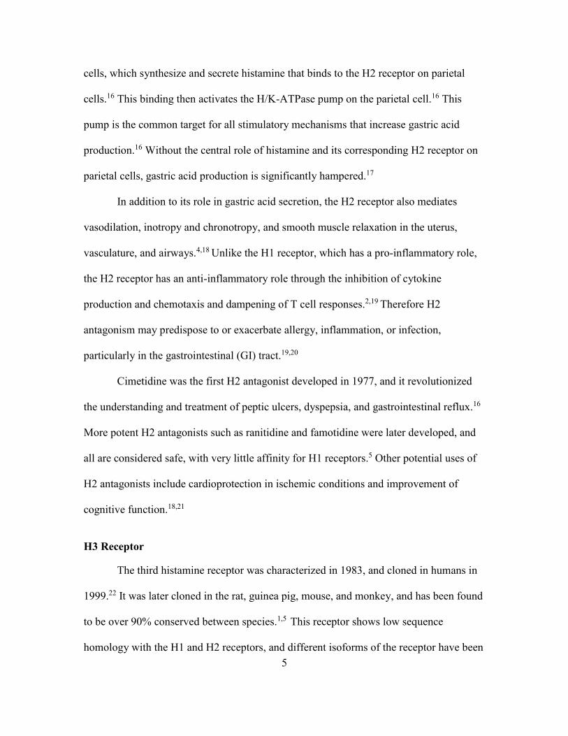
Histamine Receptor Scoring
Immunostaining intensity of H1, H2, H3, and H4 receptors were subjectively
scored by a board-certified pathologist as follows: 0, negative; 1+, weak; 2+, moderate;
and 3+, strong (Figure 2.1). Layers and tissues within the stomach, duodenum, jejunum,
ileum, and colon tissue sections of each dog were evaluated and scored individually.
Specifically, the superficial and deep mucosal layers and submucosa, smooth muscle,
submucosal ganglia, and myenteric ganglia were scored in each section of the GI tract in
all 6 dogs. Staining that was suspected to be non-specific was not scored.
Figure 2.1 Histamine receptor scoring in the canine gastrointestinal tract
Immunostaining of histamine receptors in the canine gastrointestinal tract was scored as 0; negative (A), 1; mild staining (B), 2; moderate staining (C), or 3; strong staining (D).
Western Blot Technique
Histamine receptor expression was evaluated using Western blotting as previously
described.19Gastric, small intestine, and colonic tissue samples were lysed with
radioimmunoprecipitation assay (RIPA) buffer (3 ml RIPA per 1 gram of tissue) (Santa
Cruz Biotechnology; CA, USA). For each ml of RIPA buffer, 10 μLs each of
phenylmethylsulfonyl fluoride (PMSF) solution, sodium orthovanadate solution, and
34
protease inhibitor cocktail solution (Santa Cruz Biotechnology; CA, USA) were added.
Protein quantification of the lysates was achieved using the bicinchoninic acid (BCA)
assay according to the manufacturer’s instructions (Pierce Biotechnology; IL USA).
Tissue lysates were resolved on a 10% SDS-PAGE and proteins transferred to
nitrocellulose membranes. The membranes were blocked overnight and incubated for one
hour at room temperature with the primary H1, H2, and H3 receptor antibodies at
concentrations of 2 μg/mL and overnight at 4°C with the primary H4 receptor antibody at
a concentration of 1 μg/mL. Membranes were also incubated overnight with
commercially available smooth muscle actin monoclonal mouse antibody according to
manufacturer recommendations at 1 μg/mL at 4°C (Dako; Agilent Technologies, CA,
USA). This antibody is widely used in immunohistochemistry and served as a positive
control for gastrointestinal tissue lysates.20Membranes were washed and then incubated
with the appropriate alkaline phosphatase-conjugated secondary antibody (Sigma-
Aldrich; MO, USA) for one hour and then washed. Immunoreactivities were revealed
with the 5-bromo-4-chloro-3-indolyl-phosphate/nitro blue tetrazolium (BCIP/NBT)
substrate (Sigma-Aldrich; MO, USA).
Statistical Analysis
To determine if there were differences in histamine receptor immunostaining
scores among the gastrointestinal wall layers/tissues within the different sections of the
gastrointestinal tract (stomach, duodenum, jejunum, ileum, and colon), a method similar
to the non-parametric Friedman’s test was conducted. Separate analyses were conducted
for each layer/tissue. For each histamine receptor scored (H1, H2, H3, and H4) within a
section of the gastrointestinal tract, the data was first ranked within each dog. An
35
analysis of variance using PROC MIXED in SAS for Windows v9.4 (SAS Institute Inc.;
Cary, NC) was then conducted on the ranked data with dog and layer/tissue as fixed
effects. To prevent type I errors due to multiple comparisons, differences in least squares
means with Bonferroni adjustment were determined for histamine receptor scores.
Results
WSAVA Assessment of Gastrointestinal Inflammation
Based on WSAVA guidelines, gastric tissue was normal in all dogs except Dog 5,
which had mild lymphocytic plasmacytic gastritis, mild mucosal atrophy, and mild
hyperplasia of submucosal Peyer’s patch lymphoid follicles. All dogs had mild
lymphocytic plasmacytic duodenal inflammation, and Dog 4 had mild eosinophilic
inflammation as well. Duodenal mucosal morphology was normal in all dogs. Mild
eosinophilic colitis was present in Dog 4 and Dog 6. Colonic tissue in the remaining dogs
was normal.
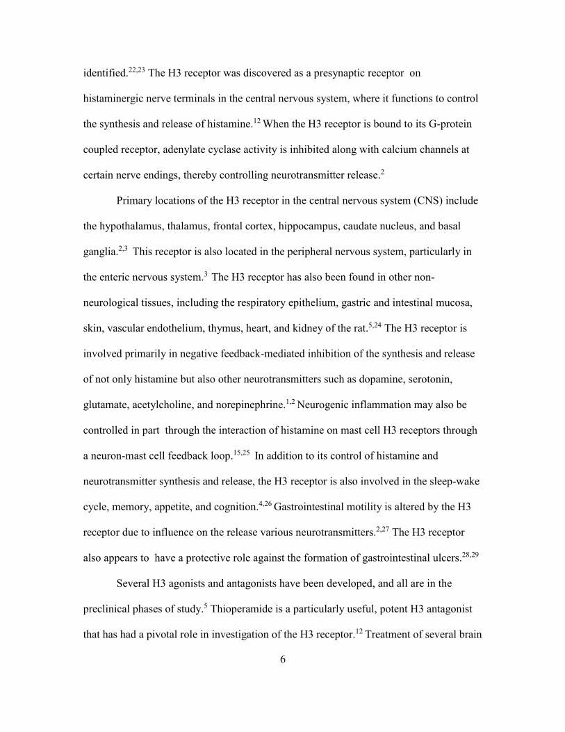
Western Blot Validation of Histamine Receptor Antibodies
Expression of the histamine receptor protein in gastric, small intestine and colonic
lysates was examined by Western blotting (Figures 2.2A-2.2D). Expression of the smooth
muscle actin protein at 50 kDa in all lysates was confirmed. The H1 receptor and H3
receptor antibodies yielded bands at the expected molecular weight of approximately 50
kDa. However, additional bands were present, suggesting non-specific binding of the H1
and H3 receptor antibody. Successful utilization of the H2 receptor antibody for Western
blotting was not possible with any of the lysates, despite the known presence of H2
receptors in the canine stomach. For the H4 receptor antibody, a band was present at the

36
expected molecular weight of approximately 50 kDa in the gastric, small intestinal, and
colonic tissue lysates (Figure 2.2 D). This finding was repeatable, as was the absence of
this band using the control anti-rabbit antibody, indicating excellent specificity of the H4
receptor antibody.
Figure 2.2 Western blots for histamine receptor antibodies
A A band (see arrow) at the expected molecular weight (approximately 55 kDa) of the H1 receptor was present in lysates of the stomach (Lane 1) and the duodenum (Lane 2). Additional bands are also present, consistent with non-specific staining noted with immunohistochemistry. B A band at the expected molecular weight (approximately 50 kDa) of the H2 receptor was not obtained in lysates of the stomach (Lane 1) or the duodenum (Lane 2), despite excellent immunohistochemistry results. C A band (see arrows) at the expected molecular weight (approximately 70 kDa) of the H3 receptor was present in lysates of the stomach (Lane 1) and the duodenum (Lane 2). Additional bands are also present, consistent with non-specific staining noted with immunohistochemistry. D A band (see arrow) at the expected molecular weight (47-50 kDa) of the H4 receptor was present in lysates of the stomach (Lane 1), duodenum (Lane 2), and the colon (Lane 3) using the H4 receptor antibody but not with the rabbit control antibody. A strong band at the expected molecular weight of smooth muscle actin (42 kDa) in all tissue lysates was present.

37
Histamine Receptor Immunohistochemistry Descriptive Findings
Moderate to strong immunostaining of H1, H2, and H3 receptors and weaker
staining of H4 receptors in mast cells was identified in the control mast cell tumor tissue
sections. Moderate to strong endothelial staining of H1, H2, and H4 receptors was
identified in the mast cell tumor sections. Strong immunostaining of the H3 receptor was
present in control intestinal ganglia sections.
The presence and distribution of H1, H2, H3, and H4 receptors varied among
layers and tissues within the canine GI tract. When present in a specific layer or tissue,
the receptors were typically found consistently in all 6 dogs with minimal variation in
location and staining intensity amongst dogs. No submucosal staining of histamine
receptors was identified in any dog. Overall, the distribution of H1, H2, and H3 receptors
was greater than that of H4 receptors throughout the entire GI tract.
The H1 receptor was located in all sections of the GI tract. Superficial and/or deep
mucosal staining was only present in the stomach except in Dog 2, which had mild
staining in the superficial colonic mucosa. Staining in the stomach was intermittent and
included staining of parietal cell, chief cells and superficial epithelial cells (Figure 2.3A).
Deep gastric mucosal staining was strongest and more uniform in the dog with mild
gastritis (Dog 5) (Figure 2.3B). Lymphoid follicles in the ileum and colon were positive
for the H1 receptor, with the strongest staining in lymphoblasts and germinal centers
(Figure 2.3C). The H1 receptor was identified in all smooth muscle and ganglia. Non-
specific staining of the H1 receptor antibody was identified in collagen and in enterocyte
nuclei.

38
Distribution of the H2 receptor in the GI tract was widespread in the mucosa of
the stomach, duodenum, jejunum, ileum, and colon (Figure 2.3D-2.3F). Gastric mucosal
staining was strong, especially in the parietal and chief cells (Figure 2.3D). Moderate to
strong staining of lymphoid tissue was present in the duodenum, jejunum, ileum and
colon. H2 receptors were not identified in smooth muscle but were present in all ganglia.
Subjectively, the specificity of the H2 immunostaining was excellent, with consistently
specific staining of parietal cells, chief cells and enterocytes in the small intestine and
colon. No background or non-specific immunostaining was suspected.
The H3 receptor was most frequently located in the gastric mucosa and all ganglia
(Figure 2.3G). Mucosal staining was present in the stomach and colon, but not in the
small intestine. Gastric mucosal staining was typically greater in the superficial mucosal
enterocytes, but the dog with mild gastritis (Dog 5) had strong staining in both the
superficial and deep mucosa (Figure 2.3H). Lymphoid follicles of the ileum and colon
were positive for the H3 receptor, and strong staining of a gastric lymphoid follicle was
also present in the dog with gastritis (Figure 2.3I). The H3 receptor was identified in all
smooth muscle and ganglia. Non-specific nuclear and collagen staining was present with
the H3 receptor antibody, similar to that seen with the H1 receptor antibody.
Immunostaining for the H4 receptor was identified in gastric and colonic mucosa,
but not in the small intestinal mucosa (Figure 2.3J and 2.3K). The strongest gastric
staining was in the deep mucosa of the dog with mild gastritis (Dog 5). Lymphoid
follicles in the ileum were weakly positive in Dog 1 and Dog 2 (Figure 2.3L). Smooth
muscle and ganglia staining for the H4 receptor was mild to absent throughout the GI
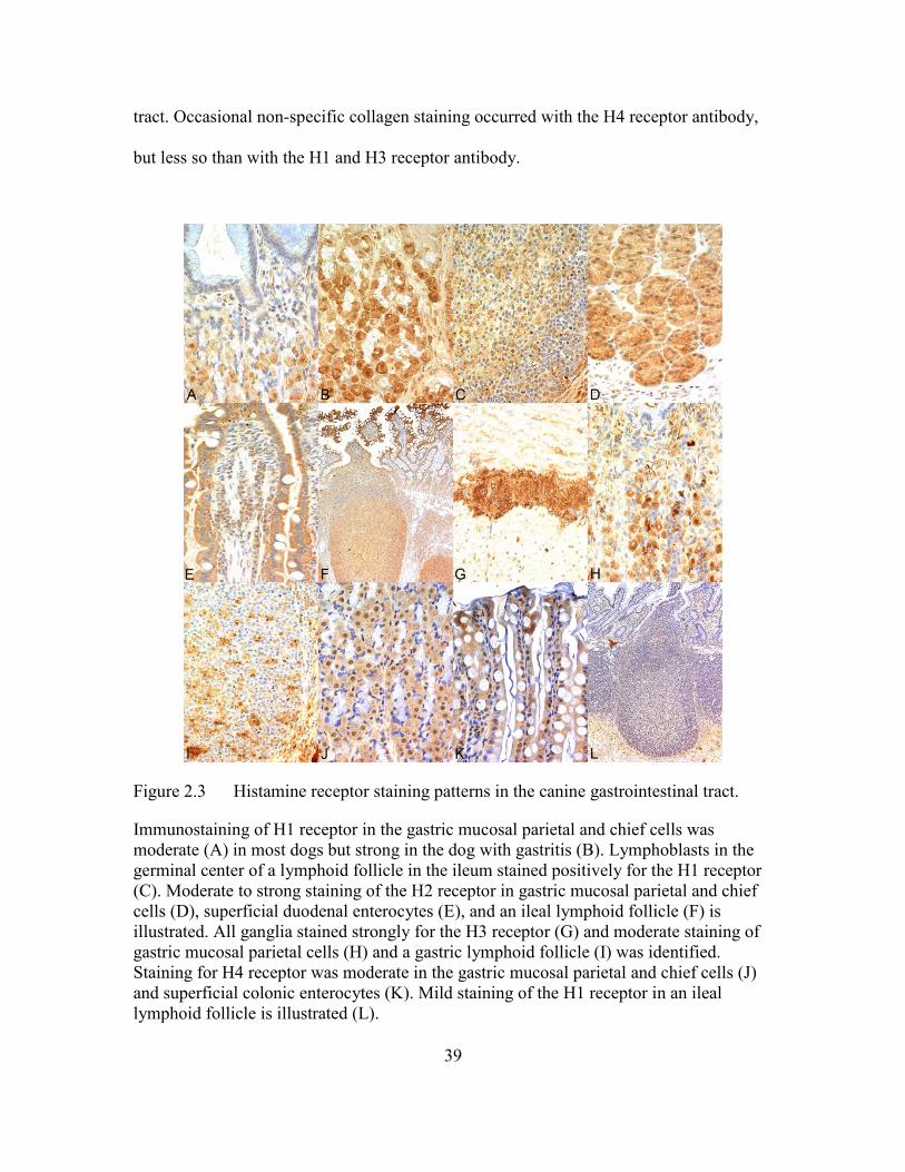
39
tract. Occasional non-specific collagen staining occurred with the H4 receptor antibody,
but less so than with the H1 and H3 receptor antibody.
Figure 2.3 Histamine receptor staining patterns in the canine gastrointestinal tract.
Immunostaining of H1 receptor in the gastric mucosal parietal and chief cells was moderate (A) in most dogs but strong in the dog with gastritis (B). Lymphoblasts in the germinal center of a lymphoid follicle in the ileum stained positively for the H1 receptor (C). Moderate to strong staining of the H2 receptor in gastric mucosal parietal and chief cells (D), superficial duodenal enterocytes (E), and an ileal lymphoid follicle (F) is illustrated. All ganglia stained strongly for the H3 receptor (G) and moderate staining of gastric mucosal parietal cells (H) and a gastric lymphoid follicle (I) was identified. Staining for H4 receptor was moderate in the gastric mucosal parietal and chief cells (J) and superficial colonic enterocytes (K). Mild staining of the H1 receptor in an ileal lymphoid follicle is illustrated (L).
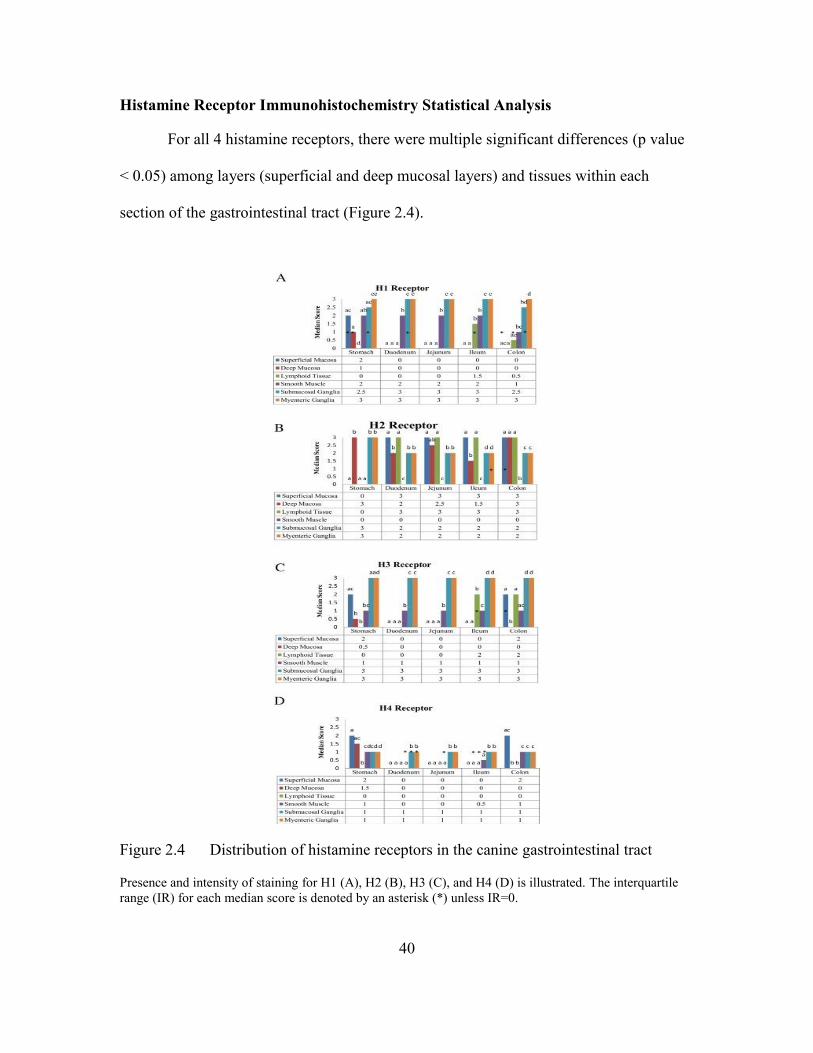
40
Histamine Receptor Immunohistochemistry Statistical Analysis
For all 4 histamine receptors, there were multiple significant differences (p value
< 0.05) among layers (superficial and deep mucosal layers) and tissues within each
section of the gastrointestinal tract (Figure 2.4).
Figure 2.4 Distribution of histamine receptors in the canine gastrointestinal tract
Presence and intensity of staining for H1 (A), H2 (B), H3 (C), and H4 (D) is illustrated. The interquartile range (IR) for each median score is denoted by an asterisk (*) unless IR=0.
41
Differences in Histamine Receptor Distribution
In the stomach, there were significantly less H1 receptors in lymphoid tissue
compared to all other targets. In the small intestine, significant differences in H1 receptor
locations were comparable within the duodenum and jejunum. In contrast to the
duodenum and jejunum, ileal lymphoid tissue had significantly more H1 receptors than
the superficial and deep mucosa.
Gastric deep mucosa and ganglia had significantly more H2 receptors than other
gastric layers/tissues. In the small intestine, there were significantly more H2 receptors in
the duodenal superficial mucosa and lymphoid tissue than all other duodenal
layers/tissues. Significant differences in H2 receptors were similar in the jejunum and
ileum. H2 receptors in the colon were significantly greater in the superficial and deep
mucosa and lymphoid tissue than in the smooth muscle and ganglia.
In the stomach, there were significantly more H3 receptors in the superficial
mucosa than the deep mucosa. In the duodenum, jejunum, ileum, and colon, H3 receptors
were significantly greater in the ganglia than other sites.
H4 receptors in the gastric superficial mucosa were significantly greater than all
other sites except the deep mucosa. The lymphoid tissue had significantly less H4
receptors than all other layers/tissues. In the colon, H4 receptors in the superficial mucosa
were significantly greater than the deep mucosa.
The authors refer the reader to Figures 3A-3D, which demonstrate additional
statistically significant differences (p value < 0.05) in histamine receptor distribution
among the different layers/tissues.
42
Discussion
Functions of histamine and histamine receptors have been studied for many years,
and H1 and H2 receptor antagonists such as diphenhydramine and famotidine are used
commonly in both human and veterinary medicine to treat a variety of conditions,
including anaphylaxis, allergies, pruritus and GI ulcers. With the discovery of the H3 and
H4 receptors, new roles for histamine have been elucidated, and new drug therapies are
being explored.3 An understanding of histamine receptor location and function is
necessary for the development of novel histamine receptor antagonists and agonists.
The current study identified the presence and location of all four histamine
receptors in the GI tract of 6 healthy adult dogs. Although no clinical signs of GI disease
were noted, mild inflammation was noted in at least one section of the GI tract in all dogs
based on WSAVA guidelines. Interpretation of GI inflammation in asymptomatic dogs is
challenging for several reasons. A lack of agreement in regards to normal histology
exists, despite development of the WSAVA guidelines.21 Significant variation in
intraepithelial lymphocyte numbers occurs between normal dogs as well.18 Furthermore,
in the absence of clinical signs or other clinical criteria for inflammatory bowel disease,
the current veterinary literature does not explain the significance of histopathological
abnormalities in the GI tract of healthy dogs.18
Distribution of histamine receptors in the intestines varies among species, but our
findings are similar to those previously reported in humans and guinea pigs.2,9 H1
receptors were located mainly in the gastric mucosa, smooth muscle, and lymphoid
tissue, corresponding to the known function of this receptor in smooth muscle contraction
and humoral immunity.5 H2 receptors were most abundant in the gastric mucosa,
43
supporting the major role of this receptor in controlling gastric acid secretion. The H3
receptor predominantly mediates neurotransmission, and findings of intense staining of
the H3 receptor antibody in all ganglia of the canine GI tract are consistent with this
function.10 Similar to other species, the H4 receptor was not as widely distributed in the
canine GI tract as the other histamine receptors.11 H4 receptors were, however, located in
the gastric and colonic mucosa, as well as in lymphoid tissue. In humans, the H4 receptor
appears to have a major influence on many aspects of the immune response, including
chemotaxis of neutrophils and eosinophils, and T cell responses.4 Therefore, presence of
the H4 receptor in lymphoid tissue was expected.
To date, canine-specific histamine receptor antibodies are not commercially
available. In our study, we used antibodies predicted by the manufacturers to cross react
with canine histamine receptors. The specificity of the H4 receptor antibody was
confirmed with Western blotting, which was consistent with the limited amount of non-
specific staining observed on immunohistochemistry. Non-specific staining of nuclei and
collagen was noted with the H1 and H3 receptor antibodies on immunohistochemistry
and, although Western blotting demonstrated bands at the expected molecular weights of
these histamine receptors, additional bands were also present. In addition to non-specific
binding of collagen and nuclear material, the additional bands could be due to the
presence of isoforms of the H1 and H3 antibodies.8 Despite some degree of non-specific
staining, immunohistochemistry results with the H1 and H3 antibodies were typically
consistent and repeatable among all dogs, and were also consistent with previously
published findings in other species, including humans.
44
The H2 receptor antibody yielded excellent immunohistochemical staining with
no non-specific background staining, and results were consistent and repeatable.
Immunohistochemical findings in the dogs in our study were also very comparable with
previously documented H2 receptor distribution in other species.2 Despite multiple
attempts, a band at the correct molecular weight of the H2 receptor could not be obtained
with Western blotting. The authors suspect that the rabbit H2 receptor antibody used in
our study recognizes a native conformational protein instead of a linear sequence
produced as a result of necessary reducing conditions for Western blot, and is, therefore,
not compatible with Western blot methodology. To confirm the specificity of the H2
antibody, an additional method, such as immunoprecipitation followed by mass
spectrophotometry, would need to be performed.
Whether histamine receptor distribution is altered in canine GI diseases remains
unknown, but human research has documented altered histamine receptor density in
certain GI diseases.4,7 Additionally, a novel H4 receptor antagonist, JNJ7777120, has
been shown to reduce histological inflammation and clinical signs in experimentally-
induced colitis.22 Interestingly, the dog in our study with histologic evidence of mild
gastritis had stronger mucosal staining for the H1 receptor and stronger lymphoid tissue
staining for the H3 receptor compared to the dogs with normal gastric biopsies.
One limitation of our study was that all control dogs had histologic evidence of
mild GI inflammation according to established WSAVA guidelines despite being
apparently healthy purpose-bred research dogs housed in a controlled environment with
no history of vomiting, diarrhea or other signs of GI disease, and despite having normal
physical examinations and negative fecal flotations.18 These findings are similar to
45
previous reports of mild histopathology changes in asymptomatic healthy, control
dogs.23,24 Histology of the canine GI tract is affected by many factors, including age,
intestinal parasites, diet, intestinal flora, medications, and environmental allergens.18,24
All dogs received monthly topical parasiticide2 and deworming every 3 months;3
therefore intestinal parasitism is an unlikely explanation for the mild GI inflammation
noted. The authors agree with the observation by Junginger et al, that mild
histopathological abnormalities may simply represent background variation in the GI
tract of normal dogs.23 We, therefore, believe that, with the possible exception of the dog
with mild gastritis, the histologic findings in the dogs in our study are consistent with a
healthy GI tract, and that the histamine receptor distribution reported in our study reflects
the distribution in clinically normal dogs.
Our study has established immunohistochemical techniques for the staining of
H1, H2, H3, and H4 receptors in canine GI tissue, and described the distribution of these
receptors in normal dogs. The results of our study provide the foundation for similar
work in dogs with GI disease. Future research in the role of histamine receptors in canine
IBD, for example, may provide evidence to suggest additional treatment options for a
common, yet sometimes refractory, disease.
Conclusion
All four histamine receptors were located throughout the canine GI tract using
immunohistochemistry. Differences in their distribution patterns were appreciated
amongst the different segments of the canine GI tract, as well as in the different
histologic layers and tissues. The presence of each receptor was typically consistent
among and within dogs. Based on our findings, H1, H3, and H4 receptor density may be

46
increased with gastritis, but further investigation is necessary, as only one of the dogs in
our study had mild gastritis. The results of our study will provide valuable baseline
information in healthy dogs as a basis for additional histamine receptor research.
Histamine blockade with novel antagonists may have therapeutic implications for GI
inflammation in the dog, and further investigation of the distribution of histamine
receptors in dogs with GI diseases such as IBD is, therefore, warranted.
Footnotes
1. Institutional Animal Care and Use Committee (IACUC) #12-065, approval granted on
8-21-2012 and expired on 8-20-2015 for the 4th year veterinary student elective course,
Advanced Small Animal Surgery CVM 5754
2. Advantage Multi®; Bayer, Kansas, USA.
3. Drontal® Plus; Bayer, Kansas, USA
47
References Cited
1. Smuda, C., Bryce, P.J., 2011. New developments in the use of histamine and histamine receptors. Curr. Allergy Asthma Rep. 11, 94-100.
2. Thurmond, R.L., 2010. Histamine in Inflammation, Landes Bioscience Springer Science and Business Media, pp. 1-136.
3. Peters, L.J., Kovacic, J.P., 2009. Histamine: metabolism, physiology, and pathophysiology with applications in veterinary medicine. J. Vet. Emerg. Crit. Care. 19, 311-328.
4. Deiteren, A., De Man, J.G., Pelckmans, P.A., De Winter, B.Y., 2014. Histamine H4 receptors in the gastrointestinal tract. Br. J. Pharmacol. 172, 1165-1178.
5. Shahid, M., Tripathi, T., Sobia, F., Moin, S., Siddiqui, M., Khan, R.A., 2009. Histamine, histamine receptors, and their role in immunomodulation: an updated systematic review. The Open Immunol J. 2, 9-41
6. Parsons, M.E., Ganellin, C.R., 2006. Histamine and its receptors. Br. J. Pharmacol. 147, S127-S135.
7. Sander, L.E., Lorentz, A., Sellge, G., Coeffeir, M., Neipp, M., Veres, T., Frieling, T., Meier, P.N., Manns, M.P., Bischoff, S.C., 2006. Selective expression of histamine receptors H1R, H2R, and H4R, but not H3R, in the human intestinal tract. Gut. 55, 498-504.
8. Xie, H., He, S., 2005. Roles of histamine and its receptors in allergic and inflammatory bowel disease. World J. Gastroenterol. 11, 2851-2857.
9. Thurmond, R.L., 2015. The histamine H4 receptor: from orphan to the clinic. Fronti. Pharmacol. 65, 1-8.
10. Breunig, E., Michel, K., Zeller, F., Seidl, S., Hann v.Werhern, C.W., Schemann, M., 2007. Histamine exerts neurons in the human submucous plexus through activation of H1, H2, H3, and H4 receptors. J. Physiol. 583, 731-742.
11. Coruzzi, G., Adami, M., Pozzoli, C., 2012. Role of histamine H4 receptors in the gastrointestinal tract. Front. Biosc. S4, 226-239.
12. Walter, M., Kottke, T., Stark, H., 2011. The histamine H4 receptors: targeting inflammatory disorders. Eur. J.Pharmacol. 668, 1-5.
13. Elson, C.O., Sartor, R.B., Tennyson, G.S., Riddell, R.H., 1995. Experimental models of inflammatory bowel disease. Gastroenterology 109, 1344-1367.
48
14. Berghoff, N., Hill, S., Parnell, N.K., Mansell, J. Suchodolski, J.S., Steiner, J.M., 2014. Fecal and urinary N-methylhistamine concentrations in dogs with chronic gastrointestinal disease. Vet. J. 201, 289-294.
15. Jiang, W., Lim, H.D., Zhang, M., Desai, P., Dai, H., Colling, P.M., Leurs, R., Thurmond, R.L., 2008. Cloning and pharmacological characterization of the dog histamine H4 receptor. Eur. J. Pharmacol. 592, 26-32.
16. Eisenschenk, M.N.C., Torres, S.M.F., Oliveira, S., Been, C.S., 2010. The expression of histamine H4 receptor mRNA in the skin and other tissues of normal dogs. Vet. Derm. 22, 296-400.
17. Migalovich-Sheikhet, H., Friedman, S., Mankuta, D., Levi-Schaffer, F., 2012. Novel identified receptors on mast cells. Front. Immunol. 238, 1-17.
18. Washabau, R.J., Day, M.J., Willard, M.D., Hall, E.J., Jergens, A.E., Mansell, J., Minami, T., Bilzer, T.W., 2010. Endoscopic, biopsy, and histopathologic guidelines for the evaluation of gastrointestinal inflammation in companion animals. J. Vet. Intern. Med. 24, 10-26.
19. Wan, X., Branton, S.L., Hughlett, M.B., Hanson, L.A., Pharr, G.T., 2004. Expression and subcellular location of a leucine aminopeptidase of Mycoplasma gallinarum. Int. J. Poult. Sci. 3, 70-74.
20. Skalli, O., Ropraz, P., Trzeciak, A., Benzonana, G., Gillessen, D., Gabbiani, G., 1986. A monoclonal antibody against alpha-smooth muscle actin: a new probe for smooth muscle differentiation. J. Cell Biol. 103, 2787-2796.
21. Willard, M., Mansell, J., 2011. Correlating clinical activity and histopathologic assessment of gastrointestinal lesion severity: current challenges. Vet. Clin. Small Anim. 41, 457-463.
22. Schirmer, B., Rezniczek, T., Seifert, R., Neumann, D., 2015. Proinflammatory role of the histamine H4 receptor in dextrane sodium sulfate-induced acute colitis. Biochem Pharmacol. 98, 102-109.
23. Junginger, J., Schwittlick, U., Lemensieck, F., Nolte, I., Hewicker-Trautwein, M., 2012. Immunohistochemical investigation of Foxp3 expression in the intestine in healthy and diseased dogs. Vet. Res. 43 (1), 23.
24. Haas, E., Rutgen, B.C., Gerner, W., Richter, B., Tichy, A., Galler, A., Bilek, A., Thalhammer, J.G., Saalmuller, A., Luckschander-Zeller, N., 2014. Phenotypic characterization of canine intestinal intraepithelial lymphocytes in dogs with inflammatory bowel disease. J. Vet. Intern. Med. 28, 1708-1715.
49
CONCLUSION
In this study, the presence and distribution of all four histamine receptors (H1,
H2, H3 and H4) were documented in the dog for the first time. An immunohistochemical
technique to study canine histamine receptors was established using commercial goat
and/or rabbit antibodies predicted to cross react with the canine histamine receptors.
Despite some non-specific staining with the H1 and H3 receptor antibodies, interpretation
of staining patterns was straightforward, and results were repeatable with all 4 receptor
antibodies. The location and distribution of histamine receptors in 6 clinically normal
adult dogs were very similar to findings in humans.1,2 Furthermore, findings were
consistent with the mRNA distribution of the histamine receptors in previous canine
research.1,3
Additional research opportunities may now be explored utilizing the
immunohistochemical technique established in normal dogs. Of particular interest is the
possible role of histamine receptors in canine inflammatory bowel disease, and the
potential to explore the use of new histamine receptor antagonists in the dog. In
collaboration with the Texas A&M University Gastroenterology Laboratory, the
correlation of canine urinary and/or fecal N-methylhistamine levels with gastrointestinal
histamine receptor expression in canine enteropathies may now be investigated. Similar
research in cats has not been conducted in veterinary medicine, but the current study
50
provides a foundation for future work in this species. In addition to advancing research in
veterinary gastroenterology, histamine receptor research also opens the door for
advancing scientific knowledge in small animal veterinary dermatology, oncology, and
respiratory medicine.
Use of validated histamine receptor antibodies for additional research is important
for producing reliable and repeatable results. Initial validation methods include
verification of antibody specificity with western blotting to confirm a band at the
expected molecular weight of the antibody’s target protein. Use of positive and negative
controls with the test antibodies also provides valuable information about antibody
specificityl. Staining patterns with the antibodies should be consistent with known
biological functions. Furthermore, an important criterion for antibody validation is
reproducibility of staining patterns with different lots of antibodies and between multiple
runs.
To date, commercial canine-specific histamine receptor antibodies are not
available. The H1 and H3 histamine receptor antibodies used in the current study are
derived from the goat and rabbit; however, based on shared genetic sequences, these
antibodies should cross react with the canine species. Staining was present in positive
control tissues, was repeatable between antibody lots and runs with these antibodies, and
was consistent with previously established functions of the H1 and H3 receptors.
However, since there was a degree of non-specific staining using current antibodies,
exploration of other available H1 and H3 receptor antibodies for clinical use in further
canine research would be ideal. Current literature documents particular challenges with
the H4 receptor antibodies with regards to poor specificity and cross-reactivity across

51
species.1,4,5 However, western blot results for the H4 receptor antibody used in the current
study were encouraging, indicating good specificity in canine intestinal tissue.
Immunostaining with the H2 receptor antibody was superior to all other antibodies,
despite failure to yield a band on western blot after undergoing standard denaturing
processing. Therefore, additional validation methods will be sought for this antibody,
which appears very specific for canine histamine receptors. Both the H2 and H4 receptor
antibodies demonstrated excellent and consistent staining between antibody lots and runs,
and staining correlated with published biological functions and tissue distribution,
including in positive control tissues. In the author’s opinion, these antibodies are worthy
of continued use in clinical canine histamine research using the described
immunohistochemistry technique.
Results of the study provide valuable information necessary to move forward with
further research in the canine gastrointestinal tract. Of particular interest is the role of
these receptors in canine inflammatory bowel disease, which shares similarities with
Crohn’s disease and ulcerative colitis. Inflammatory bowel disease (IBD) is a common
diagnosis in young adult and middle-aged dogs displaying mild to severe gastrointestinal
(GI) signs. A definitive diagnosis is based on a combination of clinical signs and
histologic analysis of biopsy samples obtained from the GI tract. There is considerable
variation in clinical signs and histologic disease severity amongst canine IBD patients.
Treatment for canine IBD typically involves specific dietary changes and modulation of
the immune system. Unfortunately, not all patients respond to standard treatment
modalities. Individual-to-individual differences in disease severity and treatment
responsiveness in our canine IBD patients highlight the pressing need to better
52
understand the pathogenesis of this important condition. Clinically, patients who have
comparable histological findings on GI biopsy can respond very differently to similar
treatment regimens: some dogs have minimal response to aggressive therapy, while other
respond well. Commonly used canine IBD protocols have been shown to lead to only
partial or no treatment responses in up to 50% of patients.6 In one large retrospective
study of canine IBD, 13% of affected dogs had intractable disease and were euthanized,
and a further 50% still had intermittent clinical signs despite treatment with a variety of
immunosuppressants and antibiotics.7 In another large prospective study in dogs with
chronic enteropathies, 18% of dogs were euthanized due to refractory disease.8 This lack
of predictability in response to therapy serves to underscore our limited understanding of
certain aspects of IBD in dogs, including the role of histamine receptors.
Histamine receptor distribution within the human GI tract has been extensively
studied over the past decade, and alterations in histamine levels and histamine receptor
expression have been described in human patients with chronic enteropathies such as
ulcerative colitis and Crohn’s.1,9 Human patients with irritable bowel syndrome and food
allergy have higher expression of H1 and H2 mRNA within the GI tract compared to
unaffected individuals.2 Rodent models of inflammation have also been used to study GI
tract histamine receptors. Studies using a common mouse model of GI inflammation that
mimics Crohn’s disease in humans, for example, have demonstrated increased expression
of the H4 receptor in the GI tract, and a correlation of colitis progression with receptor
density.9,10
Application of the current technique to evaluate histamine receptor distribution in
canine IBD is an important next step in this research. Findings may not only yield
53
valuable knowledge in the realm of veterinary gastroenterology but also in human
gastroenterology, since canine and human chronic enteropathies appear to share similar
etiologies. A naturally-occurring canine model of inflammatory bowel disease could
prove more valuable, for instance, than an experimental rodent model. The current
immunohistochemical technique can readily be applied to full-thickness biopsies
obtained routinely in dogs with chronic enteropathies. Application of the technique to
gastrointestinal endoscopic biopsies will help determine the feasibility of using mucosal
pinch biopsy tissue samples in future research. A retrospective study using archived
tissue samples, as well as prospective studies, can be conducted using the current staining
technique. Immunostaining patterns in affected dogs can be compared to those of the six
clinically normal dogs in the current study, in order to determine if significant differences
in histamine receptor distribution and density occur in dogs with chronic enteropathies
such as IBD. In a prospective study, concurrent evaluation of urinary and fecal N-
methylhistamine levels would further aid our understanding of the pathophysiological
role of histamine in canine IBD, and help determine the usefulness of this metabolite as a
potential biomarker of gastrointestinal disease in dogs.
The large body of evidence supporting the role of histamine and histamine
receptors in intestinal inflammation in human patients and rodent models suggests a
potential therapeutic use for histamine receptor antagonists in chronic intestinal
diseases.11,12 The histamine receptors vary in location, histamine binding ability,
signaling, and functions; therefore antagonism of these receptors has a variety of
therapeutic potentials.13 In particular, the recently developed histamine H4 receptor
antagonists have gained increasing interest as a novel therapeutic approach to a wide
54
range of inflammatory conditions. This is due to the unique location of the H4 receptors
on the immune cells that play an integral role in the pathogenesis of inflammatory
conditions such Crohn’s disease.14 H4 receptor ligands have been used in multiple studies
over the last decade to demonstrate the role of the H4 receptors in gastric acid secretion,
intestinal motility, mucosal defense, abdominal pain, intestinal immunity, and
carcinogenesis.9 In experimentally-induced GI inflammation in a rodent model, a
beneficial effect of H4 antagonists was seen in reducing intestinal damage, inflammatory
cell infiltration, and release of IL-6 and TNFα, two cytokines known to play an important
role in the pathogenesis of human IBD.10 Additionally, in a mouse model of peritonitis,
H1 and H4 receptor antagonists both decreased influx of inflammatory cells.11 The H1
receptor antagonist pyrilamine has been shown to inhibit histamine release and ion
transport in patients with inflammatory bowel disease, suggesting a potential role for
antihistamines in treating the clinical signs associated with IBD.15 The H1 receptor
antagonists loratadine and ketotifen reportedly decrease clinical signs of IBD such as
abdominal pain and bloody stool in people with active IBD (loratadine) and children with
ulcerative colitis (ketotifen).16
If continued research into histamine receptors in canine chronic enteropathies
suggests involvement of/or alterrations in gastrointestinal histamine receptors, then
evaluation of new histamine antagonists in dogs will be warranted. The anti-
inflammatory properties of H4 receptor antagonists, in particular, are intriguing, and may
be relevant to the treatment of canine inflammatory bowel disease. Pharmacokinetic
studies determining efficacy and safety of the H4 antagonists have not been conducted in
veterinary medicine. This work could prove to be revolutionary in the treatment of
55
chronic canine enteropathies, given the promising results associated with H4 antagonists
in experimentally-induced gastrointestinal inflammation in rodent models and in pre-
clinical human trials.
Certainly, histamine receptors have a significant role in human gastrointestinal
immunology, and scientific evidence supports their role in gastrointestinal pathology.
There is a pressing need to better understand the pathogenesis of chronic intestinal
diseases in the dog, and to develop new treatment strategies, particularly for
inflammatory bowel disease. The newly established immunohistochemistry technique
and results of the current study will now serve as the basis for future histamine receptor
research in veterinary medicine.

56
References Cited
1. Deiteren, A., De Man, J.G., Pelckmans, P.A., De Winter, B.Y., 2014. Histamine H4 receptors in the gastrointestinal tract. Br. J. Pharmacol. 172, 1165-1178.
2. Sander, L.E., Lorentz, A., Sellge, G., Coeffeir, M., Neipp, M., Veres, T., Frieling, T., Meier, P.N., Manns, M.P., Bischoff, S.C., 2006. Selective expression of histamine receptors H1R, H2R, and H4R, but not H3R, in the human intestinal tract. Gut. 55, 498-504.
3. Peters, L.J., Kovacic, J.P., 2009. Histamine: metabolism, physiology, and pathophysiology with applications in veterinary medicine. J. Vet. Emerg. Crit. Care. 19, 311-328.
4. Panula, P., Chazot, P.L., Cowart, M., Gutzmer, R., Leurs, R., Wai, L.S., Liu, H.S., Thurmond, R.L., Haas, H.L., International union of basic and clinical pharmacology. XCVIII. Histamine receptors. Pharmacol. Rev. 67, 601-655.
5. Thurmond, R.L., 2015. The histamine H4 receptor: from orphan to the clinic. Front. Pharmacol. 65, 1-8.
6. Jergens, A.E., Crandell, J., Morrison, J.A., Deitz, K., Pressel, M., Ackermann, M., Suchodolski, J.S., Steiner, J.M., Evans, R., 2010. Comparison of oral prednisone and prednisone combined with metronidazole for induction therapy of canine inflammatory bowel disease: a randomized-controlled trial. J Vet. Intern. Med. 24, 269-277.
7. Craven, M., Simpson, J.W., Ridyard, A.E., Chandler, M.L., 2004. Canine inflammatory bowel disease: retrospective analysis of diagnosis and outcome in 80 cases (1995-2002). J Small Anim. Pract. 45, 336-342.
8. Allenspach, K., Wieland, B., Grone, A., Gaschen, F., 2007. Chronic enteropathies in dogs: evaluation of risk factors for negative outcome. J Vet. Intern. Med. 21,700-708.
9. Coruzzi, G., Adami, M., Pozzoli, C., 2012. Role of histamine H4 receptors in the gastrointestinal tract. Front. Bio. S4, 226-239.
10. Schirmer, B., Rezniczek, T., Seifert R., Neumann, D., 2015. Proinflammatory role of the histamine H4 receptor in dextrane sodium sulfate-induced acute colitis. Biochem. Pharmaocol. 98, 102-109.
11. Smuda, C., Bryce, P.J., 2011. New developments in the use of histamine and histamine receptors. Curr. Allergy Asthma Rep. 11, 94-100.
12. Akdis, C.A., Simons, F.E.R., 2006. Histamine receptors are hot in pharmacology. Eur. J. Pharmacol. 553, 69-76.
57
13. Zhang, M., Thurmond, R.L., Dunford, P.J., 2007. The histamine H4 receptor: a novel modulator of inflammatory and immune disorders. Pharmacol. Therapeut. 113, 594-606
14. Kiss, R., Keseru, G.M., 2012. Histamine H4 receptor ligands and their potential therapeutic applications: an update. Expert Opin. Ther. Patents 22, 205-221.
15. Crowe, S.E., Luthra, G.K., Perdue, M.H., 1997. Mast cell mediated ion transport in intestine from patients with and without inflammatory bowel disease. Gut 41, 785-792.
16. Raithel, M., Nagel, A., Zopf, Y., deRossi, Th., Stengel, Ch., Hagel, A., Kressel, J., Hahn, E.G., Konturek, P., 2010. Plasma histamine levels during adjunctive H1 receptor antagonist treatment with lorantadine in patients with active inflammatory bowel disease. Inflamm.Res. 59 (suppl 2), S257-S258.


![GPCR agonist binding revealed by modeling and crystallography · [11] and most recently histamine H1 (H1R) [12] receptors in complex with stabilizing antagonists provide a long-sought](https://static.fdocuments.net/doc/165x107/5fec05119ab70f6340545b1c/gpcr-agonist-binding-revealed-by-modeling-and-crystallography-11-and-most-recently.jpg)