Identification of DNA Repair Pathways That Affect the...
10
Identification of DNA Repair Pathways That Affect the Survival of Ovarian Cancer Cells Treated with a Poly(ADP-Ribose) Polymerase Inhibitor in a Novel Drug Combination □ S Amelia M. Huehls, Jill M. Wagner, Catherine J. Huntoon, and Larry M. Karnitz Division of Oncology Research (A.M.H., J.M.W., C.J.H., L.M.K.), Department of Molecular Pharmacology and Experimental Therapeutics (A.M.H., L.M.K.), and Department of Radiation Oncology (L.M.K.), Mayo Clinic, College of Medicine, Rochester, Minnesota Received June 14, 2012; accepted July 24, 2012 ABSTRACT Floxuridine (5-fluorodeoxyuridine, FdUrd), a U.S. Food and Drug Administration-approved drug and metabolite of 5-fluo- rouracil, causes DNA damage that is repaired by base excision repair (BER). Thus, poly(ADP-ribose) polymerase (PARP) inhib- itors, which disrupt BER, markedly sensitize ovarian cancer cells to FdUrd, suggesting that this combination may have activity in this disease. It remains unclear, however, which DNA repair and checkpoint signaling pathways affect killing by these agents individually and in combination. Here we show that depleting ATR, BRCA1, BRCA2, or RAD51 sensitized to ABT- 888 (veliparib) alone, FdUrd alone, and FdUrd ABT-888 (FA), suggesting that homologous recombination (HR) repair protects cells exposed to these agents. In contrast, disabling the mismatch, nucleotide excision, Fanconi anemia, nonhomologous end joining, or translesion synthesis repair pathways did not sen- sitize to these agents alone (including ABT-888) or in combination. Further studies demonstrated that in BRCA1-depleted cells, FA was more effective than other chemotherapyABT-888 combina- tions. Taken together, these studies 1) identify DNA repair and checkpoint pathways that are important in ovarian cancer cells treated with FdUrd, ABT-888, and FA, 2) show that disabling HR at the level of ATR, BRCA1, BRCA2, or RAD51, but not Chk1, ATM, PTEN, or FANCD2, sensitizes cells to ABT-888, and 3) demonstrate that even though ABT-888 sensitizes ovarian tumor cells with functional HR to FdUrd, the effects of this drug combi- nation are more profound in tumors with HR defects, even com- pared with other chemotherapy ABT-888 combinations, includ- ing cisplatin ABT-888. Introduction FdUrd, a metabolite of 5-fluorouracil, is a U.S. Food and Drug Administration-approved therapy for hepatic metasta- ses of colorectal and other tumors of the gastrointestinal tract (Power and Kemeny, 2009). Although FdUrd is a me- tabolite of 5-fluorouracil, multiple studies have demon- strated that these agents have disparate mechanisms of ac- tion in human tumor cells (Wyatt and Wilson, 2009). After uptake, 5-fluorouracil is converted to metabolites that dis- rupt RNA and DNA metabolism (Longley et al., 2003), but recent studies have found that its ability to disrupt DNA metabolism has minimal effects on cytotoxicity in some cell lines, suggesting that toxicity is caused by disruption of RNA metabolism (Gmeiner et al., 2010; Geng et al., 2011; Huehls et al., 2011; Pettersen et al., 2011). In contrast, FdUrd pri- marily kills cells by disrupting DNA metabolism after its conversion to two active metabolites, 5-fluoro-2-deoxyuri- dine monophosphate and 5-fluoro-2-deoxyuridine triphos- phate (Wyatt and Wilson, 2009). 5-Fluoro-2-deoxyuridine monophosphate inhibits thymidylate synthase, thereby dis- rupting dNTP ratios and causing massive accumulation of dUTP. This dUTP, along with 5-fluoro-2-deoxyuridine triphosphate, is directly incorporated by replicative DNA polymerases, leading to the accumulation of uracil and 5-flu- orouracil in the genome. Collectively, the disruption of dNTPs and accumulation of genomic uracil and 5-fluorouracil activate the ATR and ATM checkpoint signaling pathways (Parsels et al., 2004; Wilsker and Bunz, 2007; Liu et al., 2008; Jardim et al., 2009; Geng et al., 2011; Huehls et al., 2011). Uracil and 5-fluorouracil sub- This work was supported by the National Institutes of Health National Cancer Institute [Grants R01-CA084321, P50-CA136393]; the National Insti- tutes of Health National Institute for General Medical Sciences [Grant T32- GM072474]; a Mayo Clinic Eagles Pilot Project Award; and the Mayo Clinic. A.M.H., J.M.W., and C.J.H. contributed equally to this study. Article, publication date, and citation information can be found at http://molpharm.aspetjournals.org. http://dx.doi.org/10.1124/mol.112.080614. □ S The online version of this article (available at http://molpharm. aspetjournals.org) contains supplemental material. ABBREVIATIONS: FdUrd, 5-fluorodeoxyuridine, floxuridine; BER, base excision repair; PARP, poly(ADP-ribose) polymerase; AZD2281, olaparib; PTEN, phosphatase and tensin homolog; ABT-888, veliparib; FA, FdUrd ABT-888; siRNA, small interfering RNA; DSB, DNA double-strand break. 1521-0111/12/8204-767–776$25.00 MOLECULAR PHARMACOLOGY Vol. 82, No. 4 Copyright © 2012 The American Society for Pharmacology and Experimental Therapeutics 80614/3797689 Mol Pharmacol 82:767–776, 2012 767 http://molpharm.aspetjournals.org/content/suppl/2012/07/25/mol.112.080614.DC1 Supplemental material to this article can be found at: at ASPET Journals on June 24, 2018 molpharm.aspetjournals.org Downloaded from
Transcript of Identification of DNA Repair Pathways That Affect the...
Identification of DNA Repair Pathways That Affect the Survivalof Ovarian Cancer Cells Treated with a Poly(ADP-Ribose)Polymerase Inhibitor in a Novel Drug Combination□S
Amelia M. Huehls, Jill M. Wagner, Catherine J. Huntoon, and Larry M. KarnitzDivision of Oncology Research (A.M.H., J.M.W., C.J.H., L.M.K.), Department of Molecular Pharmacology and ExperimentalTherapeutics (A.M.H., L.M.K.), and Department of Radiation Oncology (L.M.K.), Mayo Clinic, College of Medicine, Rochester,Minnesota
Received June 14, 2012; accepted July 24, 2012
ABSTRACTFloxuridine (5-fluorodeoxyuridine, FdUrd), a U.S. Food andDrug Administration-approved drug and metabolite of 5-fluo-rouracil, causes DNA damage that is repaired by base excisionrepair (BER). Thus, poly(ADP-ribose) polymerase (PARP) inhib-itors, which disrupt BER, markedly sensitize ovarian cancercells to FdUrd, suggesting that this combination may haveactivity in this disease. It remains unclear, however, which DNArepair and checkpoint signaling pathways affect killing by theseagents individually and in combination. Here we show thatdepleting ATR, BRCA1, BRCA2, or RAD51 sensitized to ABT-888 (veliparib) alone, FdUrd alone, and FdUrd � ABT-888(F�A), suggesting that homologous recombination (HR) repairprotects cells exposed to these agents. In contrast, disabling themismatch, nucleotide excision, Fanconi anemia, nonhomologous
end joining, or translesion synthesis repair pathways did not sen-sitize to these agents alone (including ABT-888) or in combination.Further studies demonstrated that in BRCA1-depleted cells, F�Awas more effective than other chemotherapy�ABT-888 combina-tions. Taken together, these studies 1) identify DNA repair andcheckpoint pathways that are important in ovarian cancer cellstreated with FdUrd, ABT-888, and F�A, 2) show that disabling HRat the level of ATR, BRCA1, BRCA2, or RAD51, but not Chk1,ATM, PTEN, or FANCD2, sensitizes cells to ABT-888, and 3)demonstrate that even though ABT-888 sensitizes ovarian tumorcells with functional HR to FdUrd, the effects of this drug combi-nation are more profound in tumors with HR defects, even com-pared with other chemotherapy � ABT-888 combinations, includ-ing cisplatin � ABT-888.
IntroductionFdUrd, a metabolite of 5-fluorouracil, is a U.S. Food and
Drug Administration-approved therapy for hepatic metasta-ses of colorectal and other tumors of the gastrointestinaltract (Power and Kemeny, 2009). Although FdUrd is a me-tabolite of 5-fluorouracil, multiple studies have demon-strated that these agents have disparate mechanisms of ac-tion in human tumor cells (Wyatt and Wilson, 2009). Afteruptake, 5-fluorouracil is converted to metabolites that dis-rupt RNA and DNA metabolism (Longley et al., 2003), but
recent studies have found that its ability to disrupt DNAmetabolism has minimal effects on cytotoxicity in some celllines, suggesting that toxicity is caused by disruption of RNAmetabolism (Gmeiner et al., 2010; Geng et al., 2011; Huehlset al., 2011; Pettersen et al., 2011). In contrast, FdUrd pri-marily kills cells by disrupting DNA metabolism after itsconversion to two active metabolites, 5�-fluoro-2�-deoxyuri-dine monophosphate and 5�-fluoro-2�-deoxyuridine triphos-phate (Wyatt and Wilson, 2009). 5�-Fluoro-2�-deoxyuridinemonophosphate inhibits thymidylate synthase, thereby dis-rupting dNTP ratios and causing massive accumulation ofdUTP. This dUTP, along with 5�-fluoro-2�-deoxyuridinetriphosphate, is directly incorporated by replicative DNApolymerases, leading to the accumulation of uracil and 5-flu-orouracil in the genome.
Collectively, the disruption of dNTPs and accumulation ofgenomic uracil and 5-fluorouracil activate the ATR and ATMcheckpoint signaling pathways (Parsels et al., 2004; Wilskerand Bunz, 2007; Liu et al., 2008; Jardim et al., 2009; Geng etal., 2011; Huehls et al., 2011). Uracil and 5-fluorouracil sub-
This work was supported by the National Institutes of Health NationalCancer Institute [Grants R01-CA084321, P50-CA136393]; the National Insti-tutes of Health National Institute for General Medical Sciences [Grant T32-GM072474]; a Mayo Clinic Eagles Pilot Project Award; and the Mayo Clinic.
A.M.H., J.M.W., and C.J.H. contributed equally to this study.Article, publication date, and citation information can be found at
http://molpharm.aspetjournals.org.http://dx.doi.org/10.1124/mol.112.080614.□S The online version of this article (available at http://molpharm.
aspetjournals.org) contains supplemental material.
ABBREVIATIONS: FdUrd, 5-fluorodeoxyuridine, floxuridine; BER, base excision repair; PARP, poly(ADP-ribose) polymerase; AZD2281, olaparib; PTEN,phosphatase and tensin homolog; ABT-888, veliparib; F�A, FdUrd � ABT-888; siRNA, small interfering RNA; DSB, DNA double-strand break.
1521-0111/12/8204-767–776$25.00MOLECULAR PHARMACOLOGY Vol. 82, No. 4Copyright © 2012 The American Society for Pharmacology and Experimental Therapeutics 80614/3797689Mol Pharmacol 82:767–776, 2012
767
http://molpharm.aspetjournals.org/content/suppl/2012/07/25/mol.112.080614.DC1Supplemental material to this article can be found at:
at ASPE
T Journals on June 24, 2018
molpharm
.aspetjournals.orgD
ownloaded from
stitutions are also targeted by DNA repair pathways (Wyattand Wilson, 2009). 5-Fluorouracil mispairs may be recog-nized by the mismatch repair pathway, an event thought toreduce survival of FdUrd-exposed cells (Meyers et al., 2001;Liu et al., 2008; Jardim et al., 2009). Alternatively, bothuracil and 5-fluorouracil are targets of base excision repair(BER), which is initiated by uracil glycosylases that removethese lesions, leaving an abasic site. The abasic site is pro-cessed by an apurinic/apyridinic endonuclease (APE1), cre-ating a nick that attracts poly(ADP-ribose) polymerase(PARP) and XRCC1, a scaffold protein that recruits addi-tional repair proteins. Consistent with the idea that BERproductively repairs these lesions, disabling the repair pro-teins APE1, XRCC1, or PARP increases cell killing by FdUrd(McNeill et al., 2009; Geng et al., 2011; Huehls et al., 2011).Likewise, small-molecule PARP inhibitors sensitize ovarianand colon cancer cell lines to FdUrd but not to 5-fluorouracil(Geng et al., 2011; Huehls et al., 2011).
PARP inhibitors have garnered significant attention asantitumor agents, especially since the demonstration thatthe PARP inhibitor olaparib (AZD2281) has single-agentactivity in ovarian tumors with mutations in BRCA1 andBRCA2 (Kummar et al., 2012). Likewise, PARP inhibitorsmay be useful in tumors that lack mutations in BRCA1 orBRCA2 but that have defects in HR repair, a featureknown as “BRCAness” (Turner et al., 2004). Although itremains unclear what causes BRCAness, it has been re-ported that reduced BRCA1/2 expression, defects in signal-ing pathways that influence HR repair (e.g., ATM, ATR,and Chk1), or defects in other proteins that regulate orparticipate in HR (e.g., Fanconi anemia pathway members,RAD51, and PTEN) sensitize to PARP inhibitors, thussuggesting that these defects may contribute to BRCAness(McCabe et al., 2006).
In addition to using PARP inhibitors as monotherapies,there is interest in combining these agents with conven-tional chemotherapy agents. Indeed, we recently reportedthat PARP inhibitors synergize remarkably with FdUrd,with toxicities that exceed those seen with other chemo-therapy agents used to treat ovarian cancer. Accordingly,such results suggest that combining FdUrd with PARPinhibitors may be worthy of clinical studies, especially inovarian cancer in which both FdUrd and PARP inhibitorshave activity as single agents (Muggia et al., 1996; Kum-mar et al., 2012).
Before launching such trials, it is important to understandhow the individual drugs and the drug combination affecttumor cells. This is especially important because combining aDNA-damaging agent, such as FdUrd, with an agent [velipa-rib (ABT-888)] that inhibits the repair of those lesions maycreate DNA damage that differs from the damage caused byFdUrd alone. Thus, different DNA repair and/or checkpointpathways may assume importance in cells exposed to F�A.We therefore undertook a systematic analysis of the majorDNA repair and checkpoint signaling pathways to determinewhich of these pathways affect the survival of ovarian cancercells treated with FdUrd, ABT-888, and the F�A combina-tion. Our studies reveal novel insights into the DNA repairpathways that affect FdUrd, ABT-888, and F�A tumor cellkilling.
Materials and MethodsCell Lines and Culture. OVCAR-8, a gift from Dominic Scud-
ierio (National Cancer Institute, Bethesda, MD), and SKOV-3(American Type Culture Collection, Manassas, VA) were cultured at37°C in 5% CO2 with 8 or 10% fetal bovine serum (Atlanta Biologi-cals, Norcross, GA) in RPMI 1640 medium (Mediatech, Herndon,VA). For clonogenic assays, media were supplemented with 100 U/mlpenicillin and 100 �g/ml streptomycin (Mediatech). Cell lines werereinitiated every 3 months from cryopreserved stocks prepared uponreceipt from the indicated sources.
Materials. Reagents were from the following suppliers: FdUrd,Bedford Laboratories (Bedford, OH); ABT-888 [Selleck Chemicals(Houston, TX) and ChemieTek (Indianapolis, IN)]; cisplatin, TevaPharmaceuticals (Irvine, CA) gemcitabine, Eli Lilly & Co. (Indianap-olis, IN); doxorubicin, melphalan, and topotecan, Sigma-Aldrich (St.Louis, MO); and SuperSignal Pico West, Thermo Fisher Scientific,Waltham, MA). All other materials were from Sigma-Aldrich.
Antibodies to the following antigens were obtained as follows:phospho-Ser317-Chk1, R&D Systems (Minneapolis, MN); phospho-Thr68-Chk2, ATR, AKT, phospho-Ser473-AKT, BRCA1, BRCA2,KU80, PTEN, horseradish peroxidase-linked rabbit IgG, and horse-radish peroxidase-linked mouse IgG, Cell Signaling Technology, Inc.(Danvers, MA); Chk1, Santa Cruz Biotechnology, Inc. (Santa Cruz,CA); Chk2, MSH2, RAD51, and ATM, Epitomics, (Burlingame, CA);phospho-Ser139-H2AX, Millipore Corporation (Billerica, MA);XRCC1, Bethyl Laboratories (Montgomery, TX); FANCD2, GeneTex(Irvine, CA); RAD18, Novus Biologicals, Inc. (Littleton, CA); XPA,Neomarkers (Fremont, CA); fluorescein-conjugated goat anti-mouseIgG, Invitrogen (Carlsbad, CA); and HSP90 D. Toft (Mayo Clinic,Rochester, MN).
Cell Transfections and siRNAs. Cells were transfected as de-scribed previously (Huehls et al., 2011) and cultured 48 h before use.Sequences of siRNAs used were as follows: ATM-1, 5�-AAGCAC-CAGTCCAGTATTGGC-3� (Wang and Qin, 2003); ATR-2, 5�-CCTCCGTGATGTTGCTTGA-3� (Casper et al., 2004); Chk1-1, 5�-AAGCGTGCCGTAGACTGTCCA-3� (Zhao et al., 2002); BRCA1-1,5�-GUGGGUGUUGGACAGUGUA-3� (Bartz et al., 2006);BRCA2-1, 5�-GACUCUAGGUCAAGAUUUA-3� (Bartz et al.,2006); FANCD2-1, 5�-GGUCAGAGCUGUAUUAUUC-3� (Wag-ner and Karnitz, 2009); Ku80-1, 5�-GCGAGUAACCAG-CUCAUAA-3� (Nimura et al., 2007); MSH2-1, 5�-CTGAAGT-AATAGCAAAGAA-3� (Geng et al., 2011); PTEN-1, 5�-AAGAGGAUGGAUUCGACUUAGAC-3� (Hamada et al., 2005);RAD18-1, 5�-GCTCTCTGATCGTGATTTA-3� (Geng et al., 2011);RAD51, ON-TARGETplus SMARTpool-human RAD51 (Dharma-con RNA Technologies, Lafayette, CO); XPA-1, 5�-GTCAA-GAAGCATTAGAAGA-3� (Biard et al., 2005); XRCC1-2, 5�-CUCGACUCACUGUGCAGAAUU-3� (Luo et al., 2004); andluciferase, 5�-CTTACGCUGAGUACUUCGA-3� (Elbashir et al.,2001).
Clonogenic Assays, Cell Lysis, Immunoblotting, Phospho-H2AX Staining, and Cell Irradiation. Clonogenic assays, celllysis, and immunoblotting were performed as described previously(Wagner and Karnitz, 2009). For clonogenic assays, percentage sur-vival at each drug concentration was normalized to the vehicle-treated control for the given siRNA. For phospho-H2AX analysis,cells were stained as described but with 2 �g/ml anti-phospho-H2AXantibody (Lansiaux et al., 2007). Cells were exposed to ionizing andultraviolet radiation using a RS-2000 Biological Irradiator (RadSource, Suwanee, GA) and a UVC-515 Ultraviolet Multilinker(Ultra-Lum, Carson, CA), respectively, 4 to 6 h after cell plating.
ResultsPARP Inhibition Enhances FdUrd-Induced Chk1
and Chk2 Activation. We have shown previously thatFdUrd activates the ATR and ATM checkpoint signaling
768 Huehls et al.
at ASPE
T Journals on June 24, 2018
molpharm
.aspetjournals.orgD
ownloaded from

pathways and that ATR, but not ATM, promotes survival ofovarian and colon cancer cells treated with FdUrd (Geng etal., 2011; Huehls et al., 2011). However, these studies did notassess whether these checkpoint pathways are important incells treated with the drug combination, which could induceDNA damage that differs from that induced by FdUrd alone.Moreover, they did not address the role of Chk1, an ATRsubstrate that is activated by other nucleoside analogs andantimetabolites and that protects tumor cells from the toxiceffects of these agents. Correspondingly, there is intenseinterest in combining small-molecule Chk1 inhibitors, whichare currently in clinical trials, with various chemotherapyagents. To examine the roles of these pathways in ovariancancer cells exposed to FdUrd � ABT-888 (F�A), we assessedwhether they were activated by ABT-888 (veliparib) alone,FdUrd alone, or the combination F�A. For these studies, weused mismatch repair-proficient OVCAR-8 and mismatch re-pair-deficient SKOV-3 cells (Roschke et al., 2002), which arederived from serous epithelial ovarian cancers. These celllines have wild-type BRCA1, BRCA2, and PTEN (Ikediobi etal., 2006; Garnett et al., 2012) and very limited sensitivity toPARP inhibitors, indicating that they have functional HRrepair (Huehls et al., 2011).
On the basis of our previous finding that continuous ABT-888 treatment after a 24-h exposure to F�A markedly in-creased toxicity (Huehls et al., 2011), two exposure para-digms were used for these studies. In the first, cells wereexposed to FdUrd alone, ABT-888 alone, or F�A for 24 h andthen analyzed for phosphorylation of Chk1 and Chk2, whichare markers for ATR and ATM activation, respectively. Inthe second, cells were exposed to the same agents for 24 h andwashed, and ABT-888 was readded to the samples that orig-inally contained ABT-888. The cells were then analyzed afterculturing for an additional 24 h (indicated as 24 � 24 h). Asshown in Fig. 1A, ABT-888 alone did not provoke Chk1 orChk2 phosphorylation under either exposure paradigm.When ABT-888 was added with FdUrd, the PARP inhibitorincreased FdUrd-induced Chk1 and Chk2 phosphorylation(Fig. 1A, cf. lanes 3 and 4) at the 24-h time point. Afterremoval of FdUrd [with the continued presence of ABT-888(24 � 24 h)], Chk1 phosphorylation was markedly reducedcompared with that seen after the 24-h exposure, and ABT-
888 did not increase Chk1 phosphorylation. In contrast,FdUrd-induced Chk2 phosphorylation persisted in the 24 �24-h samples, again with increased levels in the cells co-treated with ABT-888. Taken together, these results showthat ABT-888 increases Chk1 and Chk2 activation, suggest-ing that PARP inhibition blocks the repair of lesions inflictedby FdUrd.
ABT-888 Blocks the Repair of FdUrd-Induced DNADamage. To assess DNA damage caused by these agents, weexamined histone H2AX phosphorylation, which serves as asurrogate marker for DNA damage such as double-strandedDNA breaks, replication stress, and other types of DNA dam-age. ABT-888 alone did not induce H2AX phosphorylation(Fig. 1B). In contrast, FdUrd triggered robust H2AX phos-phorylation at 24 h, which was enhanced when ABT-888 wasadded with FdUrd. Analysis of the 24 � 24-h samples showedthat after the removal of FdUrd, the level of H2AX phosphor-ylation was decreased in the cells treated with FdUrd alone,suggesting that the DNA damage was being repaired. Inter-estingly, however, in the FdUrd-treated cells that were cul-tured in the continued presence of ABT-888, phospho-H2AXlevels remained high, indicating that ABT-888 slowed therepair of lesions induced by FdUrd and/or produced newlesions that are repaired more slowly. Taken together, theseresults suggest that PARP inhibition increases FdUrd-in-duced activation of the Chk1 and Chk2 signaling and causespersistence of FdUrd-induced DNA damage.
ATR, but Not Chk1 or ATM, Plays a Critical Role inOvarian Cancer Cells Treated with FdUrd, ABT-888, orF�A. Given that the ATR and ATM pathways are hyperac-tivated when PARP is inhibited, we asked whether depletionof ATM, ATR, or Chk1 affected proliferation after exposure toFdUrd, ABT-888, or F�A. Consistent with previous reports(Geng et al., 2011; Huehls et al., 2011), ATR depletion sen-sitized OVCAR-8 cells to ABT-888 (Fig. 2A, right). ATR-depleted cells were also markedly sensitized to FdUrd alone,and these cells were even more sensitive to the F�A combi-nation (Fig. 2A, left), indicating that ATR protects these cellsfrom damage inflicted by each agent individually and incombination. Analyses of cells depleted of ATM revealed avery different result. Although ATM-depleted OVCAR-8 cellswere more sensitive to ionizing radiation (Fig. 2B, right,
Fig. 1. PARP inhibition increasesFdUrd-induced Chk1 and Chk2 acti-vation and causes persistent DNAdamage. OVCAR-8 cells were treatedwith 2 �M FdUrd and 3 �M ABT-888.After 24 h of incubation, one set of cells(labeled 24 h, lanes 2–4) was collected.The second set of cells (labeled 24 �24 h, lanes 5–7) was washed after 24 hto remove FdUrd, and 3 �M ABT-888was readded to samples that initiallycontained ABT-888. These cells werethen cultured an additional 24 h. A, celllysates were immunoblotted for the indi-cated antigens. B, cells were stained todetect phospho-Ser139-H2AX (P-H2AX)and analyzed by flow cytometry. P, phos-pho; HSP90, heat shock protein 90.
BRCA1/2 and Rad51 Protect from Floxuridine and ABT-888 769
at ASPE
T Journals on June 24, 2018
molpharm
.aspetjournals.orgD
ownloaded from
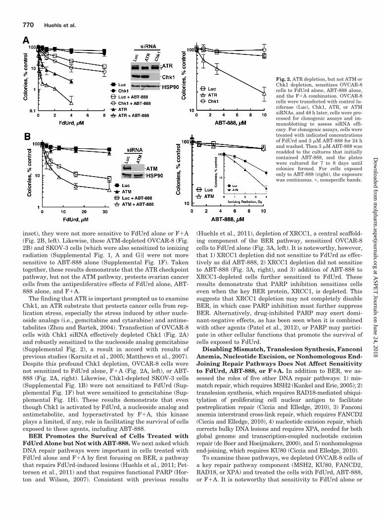
inset), they were not more sensitive to FdUrd alone or F�A(Fig. 2B, left). Likewise, these ATM-depleted OVCAR-8 (Fig.2B) and SKOV-3 cells [which were also sensitized to ionizingradiation (Supplemental Fig. 1, A and G)] were not moresensitive to ABT-888 alone (Supplemental Fig. 1F). Takentogether, these results demonstrate that the ATR checkpointpathway, but not the ATM pathway, protects ovarian cancercells from the antiproliferative effects of FdUrd alone, ABT-888 alone, and F�A.
The finding that ATR is important prompted us to examineChk1, an ATR substrate that protects cancer cells from rep-lication stress, especially the stress induced by other nucle-oside analogs (i.e., gemcitabine and cytarabine) and antime-tabolites (Zhou and Bartek, 2004). Transfection of OVCAR-8cells with Chk1 siRNA effectively depleted Chk1 (Fig. 2A)and robustly sensitized to the nucleoside analog gemcitabine(Supplemental Fig. 2), a result in accord with results ofprevious studies (Karnitz et al., 2005; Matthews et al., 2007).Despite this profound Chk1 depletion, OVCAR-8 cells werenot sensitized to FdUrd alone, F�A (Fig. 2A, left), or ABT-888 (Fig. 2A, right). Likewise, Chk1-depleted SKOV-3 cells(Supplemental Fig. 1B) were not sensitized to FdUrd (Sup-plemental Fig. 1F) but were sensitized to gemcitabine (Sup-plemental Fig. 1H). These results demonstrate that eventhough Chk1 is activated by FdUrd, a nucleoside analog andantimetabolite, and hyperactivated by F�A, this kinaseplays a limited, if any, role in facilitating the survival of cellsexposed to these agents, including ABT-888.
BER Promotes the Survival of Cells Treated withFdUrd Alone but Not with ABT-888. We next asked whichDNA repair pathways were important in cells treated withFdUrd alone and F�A by first focusing on BER, a pathwaythat repairs FdUrd-induced lesions (Huehls et al., 2011; Pet-tersen et al., 2011) and that requires functional PARP (Hor-ton and Wilson, 2007). Consistent with previous results
(Huehls et al., 2011), depletion of XRCC1, a central scaffold-ing component of the BER pathway, sensitized OVCAR-8cells to FdUrd alone (Fig. 3A, left). It is noteworthy, however,that 1) XRCC1 depletion did not sensitize to FdUrd as effec-tively as did ABT-888, 2) XRCC1 depletion did not sensitizeto ABT-888 (Fig. 3A, right), and 3) addition of ABT-888 toXRCC1-depleted cells further sensitized to FdUrd. Theseresults demonstrate that PARP inhibition sensitizes cellseven when the key BER protein, XRCC1, is depleted. Thissuggests that XRCC1 depletion may not completely disableBER, in which case PARP inhibition must further suppressBER. Alternatively, drug-inhibited PARP may exert domi-nant-negative effects, as has been seen when it is combinedwith other agents (Patel et al., 2012), or PARP may partici-pate in other cellular functions that promote the survival ofcells exposed to FdUrd.
Disabling Mismatch, Translesion Synthesis, FanconiAnemia, Nucleotide Excision, or Nonhomologous End-Joining Repair Pathways Does Not Affect Sensitivityto FdUrd, ABT-888, or F�A. In addition to BER, we as-sessed the roles of five other DNA repair pathways: 1) mis-match repair, which requires MSH2 (Kunkel and Erie, 2005); 2)translesion synthesis, which requires RAD18-mediated ubiqui-tylation of proliferating cell nuclear antigen to facilitatepostreplication repair (Ciccia and Elledge, 2010), 3) Fanconianemia interstrand cross-link repair, which requires FANCD2(Ciccia and Elledge, 2010), 4) nucleotide excision repair, whichcorrects bulky DNA lesions and requires XPA, needed for bothglobal genome and transcription-coupled nucleotide excisionrepair (de Boer and Hoeijmakers, 2000), and 5) nonhomologousend-joining, which requires KU80 (Ciccia and Elledge, 2010).
To examine these pathways, we depleted OVCAR-8 cells ofa key repair pathway component (MSH2, KU80, FANCD2,RAD18, or XPA) and treated the cells with FdUrd, ABT-888,or F�A. It is noteworthy that sensitivity to FdUrd alone or
Fig. 2. ATR depletion, but not ATM orChk1 depletion, sensitizes OVCAR-8cells to FdUrd alone, ABT-888 alone,and the F�A combination. OVCAR-8cells were transfected with control lu-ciferase (Luc), Chk1, ATR, or ATMsiRNAs, and 48 h later, cells were pro-cessed for clonogenic assays and im-munoblotting to assess siRNA effi-cacy. For clonogenic assays, cells weretreated with indicated concentrationsof FdUrd and 3 �M ABT-888 for 24 hand washed. Then 3 �M ABT-888 wasreadded to the cultures that initiallycontained ABT-888, and the plateswere cultured for 7 to 8 days untilcolonies formed. For cells exposedonly to ABT-888 (right), the exposurewas continuous. �, nonspecific bands.
770 Huehls et al.
at ASPE
T Journals on June 24, 2018
molpharm
.aspetjournals.orgD
ownloaded from

F�A was not affected by depletion of MSH2 (Fig. 3B), KU80(Fig. 4A), XPA (Fig. 4B), FANCD2 (Fig. 4C), or RAD18 (Fig.4D), even though depletion of each protein was sufficient toaffect the sensitivity of these cells to control DNA-damagingagents [N-methyl-N�-nitro-N-nitrosoguanidine for MSH2(Fig. 3B, right, inset), ionizing radiation for KU80 (Supple-mental Fig. 3A), ultraviolet light for XPA (Supplemental Fig.3B), cisplatin for RAD18, and FANCD2 (Supplemental Fig. 3,C and D)]. Similarly, the sensitivity of OVCAR-8 cells to
ABT-888 was not affected by depleting MSH2, KU80,FANCD2, or RAD18 (Fig. 3B, right; Supplemental Fig. 4,A–C), whereas depletion of BRCA1 markedly sensitized tothis agent (Supplemental Fig. 4D). The observation thatFANCD2 depletion did not sensitize to ABT-888 was unex-pected, because mouse fibroblasts lacking FANCD2 are sen-sitive to PARP inhibitors (McCabe et al., 2006). We thereforedetermined the impact of depleting FANCD2 in SKOV-3 cells(Supplemental Fig. 1D), which also were not sensitized to
Fig. 3. The BER pathway, but not themismatch repair pathway, protectscells from FdUrd and F�A. OVCAR-8cells were transfected with controlluciferase (Luc) and XRCC1 (A) orMSH2 (B) siRNAs and plated as singlecells, allowed to adhere, exposed to theindicated concentrations of FdUrd withor without 3 �M ABT-888 for 24 h, andwashed. Then 3 �M ABT-888 was read-ded to the cultures that initially con-tained ABT-888, and the plates werecultured until colonies formed. For cellsexposed only to ABT-888 (right), the ex-posure was continuous.
Fig. 4. Nonhomologous end-joining,Fanconi anemia, translesion synthe-sis, and nucleotide excision repairpathways do not protect cells fromFdUrd, ABT-888, or F�A. OVCAR-8cells were transfected with control lu-ciferase (Luc), KU80 (A), XPA (B),FANCD2 (C), or RAD18 (D) siRNAs,and 48 h after transfections cells wereexposed to the indicated concentra-tions of FdUrd with or without 3 �MABT-888 for 24 h and washed. Then 3�M ABT-888 was readded to the cul-tures that initially contained ABT-888, and the plates were cultured un-til colonies formed. �, nonspecificband. RAD18 migrates as two bands,with the slowest migrating as a resultof monoubiquitylation.
BRCA1/2 and Rad51 Protect from Floxuridine and ABT-888 771
at ASPE
T Journals on June 24, 2018
molpharm
.aspetjournals.orgD
ownloaded from

ABT-888 (Supplemental Fig. 1F). Taken together, these re-sults indicate that the DNA damage inflicted by F�A isprobably not acted on productively by these DNA repairpathways. Furthermore, they demonstrate that depletingFANCD2 does not affect sensitivity to ABT-888 in OVCAR-8and SKOV-3 cells.
Depletion of HR Repair Proteins BRCA1, BRCA2,and RAD51 Sensitizes to FdUrd, ABT-888, and F�A.Our results so far indicate that inhibition of PARP in cellstreated with FdUrd causes increased DNA damage (Fig. 1B)that is not repaired by translesion synthesis, nucleotide ex-cision, Fanconi anemia, or nonhomologous end-joining repairpathways. Moreover, our results also demonstrate that ATR,but not the ATR substrate Chk1, plays a critical role in cellstreated with F�A (Fig. 2A). The observation that Chk1 de-pletion did not affect sensitivity of the cells to FdUrd, ABT-888, or F�A suggests that the function of ATR in cells treatedwith these agents may be channeled through other sub-strates. Although hundreds of ATR substrates have beenidentified, we turned our attention to HR repair proteinsbecause 1) several HR proteins are ATR substrates, includingBRCA1 and BRCA2 (Tibbetts et al., 1999; Gatei et al., 2001;Ciccia and Elledge, 2010), 2) BRCA1, BRCA2, and RAD51play important roles at stalled replication forks and at DSBs,two genotoxic events that occur when cells are treated withFdUrd, and 3) defects in HR sensitize to ABT-888 alone(Martin et al., 2008).
For these studies, we depleted RAD51, which is requiredfor HR (Ciccia and Elledge, 2010), and BRCA1 or BRCA2, twogenes that are frequently mutated in ovarian cancer (CancerGenome Atlas Research Network, 2011) and that play pivotalroles in HR (Ciccia and Elledge, 2010). OVCAR-8 cells de-pleted of RAD51, BRCA1, or BRCA2 (Fig. 5, A–C, insets)were sensitive to ABT-888 alone (Fig. 5, A–C, right), asexpected for cells with defective HR. Surprisingly, depletionof these three HR proteins also markedly sensitized cells toFdUrd alone (Fig. 5, A–C, left), indicating that FdUrd causesdamage that is repaired by the HR pathway. Most impor-tantly, OVCAR-8 cells depleted of RAD51, BRCA1, or BRCA2(Fig. 5, A–C, left), as well as SKOV-3 cells depleted of BRCA1(Supplemental Fig. 1C), were exquisitely sensitive to F�A(Supplemental Fig. 1J). Taken together these results suggestthat this combination of chemotherapy agents may be espe-cially toxic in ovarian cancers with defects in HR.
PTEN Depletion Does Not Affect Sensitivity toFdUrd, ABT-888, or F�A. Cells with disabled PTEN havebeen reported to be sensitive to PARP inhibition and to havereduced RAD51 and HR repair (Shen et al., 2007; Mendes-Pereira et al., 2009; Dedes et al., 2010; McEllin et al., 2010).Given that PTEN may regulate RAD51 and is frequentlymutated in ovarian cancer (Cancer Genome Atlas ResearchNetwork, 2011), we depleted PTEN from OVCAR-8 andSKOV-3 cells, which have wild-type PTEN (Ikediobi et al.,2006). Immunoblotting demonstrated that PTEN levels werereduced in OVCAR-8 (Fig. 5D, inset) and SKOV-3 cells (Sup-plemental Fig. 1E). Accordingly, activating AKT phosphoryla-tion on Ser473 was increased, indicating that PTEN depletioncaused accumulation of its substrate (phosphatidylinositol3,4,5-trisphosphate), the second messenger that activates AKT.Despite this profound PTEN depletion, RAD51 levels were un-altered in OVCAR-8 (Fig. 5D) or SKOV-3 cells (SupplementalFig. 1E). Consistent with the lack of an effect on RAD51 levels,
PTEN-depleted OVCAR-8 cells were not sensitized to FdUrd orF�A (Fig. 5D, left). OVCAR-8 (Fig. 5D, right) and SKOV-3(Supplemental Fig. 1F) cells depleted of PTEN were also notsensitized to ABT-888.
F�A Is More Cytotoxic Than Other Chemotherapy �ABT-888 Combinations in BRCA1-Depleted OvarianCancer Cells. The results in Fig. 5 (and Supplemental Figs.1 and 4) show that cells with disabled RAD51, BRCA1, orBRCA2 are very sensitive to F�A. However, this sensitivitymay be no more than would be seen when ABT-888 is com-bined with other chemotherapy agents. To address this, wecompared the antiproliferative activities of multiple chemo-therapy agents used to treat ovarian cancer alone and com-bined with ABT-888 in BRCA1-depleted OVCAR-8 cells(Fig. 6). These results demonstrated several points. First,BRCA1 depletion sensitized to ABT-888, demonstrating thatthe BRCA1 depletions were sufficient to affect sensitivity toa PARP inhibitor. Second, BRCA1 depletion sensitized to allgenotoxic agents except gemcitabine. Third, in cells withfunctional HR (luciferase siRNA-transfected cells), PARP in-hibition did not sensitize to cisplatin or doxorubicin. Fourth,PARP inhibition did not further sensitize BRCA1-depletedcells to doxorubicin, cisplatin, or melphalan. Fifth, and insharp contrast to what was observed with cisplatin, ABT-888further sensitized BRCA1-depleted cells to FdUrd and topo-tecan, with the sensitization to FdUrd being the most pro-found. Additional experiments demonstrated that BRCA1depletion in SKOV-3 also sensitized to FdUrd and ABT-888and that these depleted cells were exceptionally sensitive tothe F�A combination (Supplemental Fig. 1J).
DiscussionWe initiated these studies to determine whether defects in
DNA repair or checkpoint signaling pathways influence thetoxicity of F�A. We reasoned that if this drug combinationmoves into clinical trials, it will be useful to determine whatDNA repair and/or checkpoint pathways affect tumor cellsurvival, because such information may help identify pa-tients most likely to respond to the drug combination. Giventhat FdUrd and PARP inhibitors have activity in ovariancancer, our studies focused on this tumor type.
FdUrd disrupts dNTP ratios and causes misaccumulationof uracil and 5-fluorouracil in the genome. These misincor-porated bases are recognized by the BER pathway. However,it is not known whether these lesions are substrates for otherrepair pathways that could productively repair them. Nor isit known whether attempted repair and/or replication ofthese lesions creates DNA damage that is more toxic, apossibility, considering that FdUrd induces double-strandDNA breaks (Yoshioka et al., 1987; Tang et al., 1996; Meyerset al., 2001). Finally, it is not clear whether PARP inhibition,which disrupts DNA repair, alters the lesions that are ulti-mately induced by FdUrd. Thus, rather than directly assess-ing the types of damage produced by each agent alone and incombination, an approach that requires the development ofmany assays, we instead focused on identifying which DNArepair pathways affect cell killing. To that end, we system-atically inactivated the major signaling and repair pathwaysand examined the effects on cells exposed to FdUrd, ABT-888, and F�A.
772 Huehls et al.
at ASPE
T Journals on June 24, 2018
molpharm
.aspetjournals.orgD
ownloaded from

Our initial studies focused on the mismatch repair path-way, because defective mismatch repair has been reported toincrease resistance to FdUrd-induced death, probably by pre-venting the attempted (but ultimately futile) mismatch re-pair of fluorouracil-guanine mispairs (Meyers et al., 2001;Liu et al., 2008). In contrast to these previous reports, weshowed that effectively depleting MSH2 did not affect sensi-tivity to FdUrd or F�A but did reduce sensitivity to N-
methyl-N�-nitro-N-nitrosoguanidine, an agent that produceslesions that are substrates for mismatch repair. It is note-worthy that similar results, showing that defects in mis-match repair in colon cancer cells do not (or only minimally)affect sensitivity to FdUrd have been recently reported (Pet-tersen et al., 2011). Taken together, these results suggestthat defective mismatch repair is not universally linked toaltered FdUrd sensitivity.
Fig. 5. Disruption of HR repair sensi-tizes ovarian cancer cells to FdUrd,ABT-888, and F�A. OVCAR-8 cellswere transfected with control lu-ciferase (Luc), RAD51 (A), BRCA1 (B),BRCA2 (C), or PTEN (D) siRNAs, and48 h after transfections cells were ex-posed to the indicated concentrationsof FdUrd with or without 3 �M orABT-888 for 24 h and washed. Then 3�M ABT-888 was readded to the cul-tures that initially contained ABT-888, and the plates were cultured un-til colonies formed. For cells exposedonly to ABT-888 (right), the exposurewas continuous.
BRCA1/2 and Rad51 Protect from Floxuridine and ABT-888 773
at ASPE
T Journals on June 24, 2018
molpharm
.aspetjournals.orgD
ownloaded from

We also found that disabling mismatch, nucleotide excision,Fanconi anemia, nonhomologous end-joining, or translesionsynthesis repair pathways did not sensitize cells to FdUrd orF�A, indicating that these pathways do not productivelyrepair lesions induced directly or indirectly by FdUrd or evenby F�A. Our results also demonstrate that these repairpathways do not make lesions induced by FdUrd alone orF�A more toxic. Given that FdUrd activates ATM and
causes DSBs (Yoshioka et al., 1987; Tang et al., 1996; Meyerset al., 2001; Parsels et al., 2004; Wilsker and Bunz, 2007; Liuet al., 2008; Jardim et al., 2009; Geng et al., 2011; Huehls etal., 2011) and given that F�A induces more H2AX phosphor-ylation than FdUrd alone (see Fig. 1B), we were surprisedthat depletion of ATM or KU80, two proteins that participatein signaling from and repair of DSBs, did not affect thesurvival of cells treated with these agents. In contrast, dis-
Fig. 6. The combination F�A ishighly cytotoxic in BRCA1-deficientovarian cancer cells compared withother ABT-888�chemotherapy combina-tions. OVCAR-8 cells transfected withcontrol luciferase (Luc) or BRCA1 siRNAwere exposed to the indicated concentra-tions of gemcitabine (A), doxorubicin (B),cisplatin (C), melphalan (D), FdUrd (E),or topotecan (F) and 3 �M ABT-888 for24 h and washed. Then 3 �M ABT-888was readded to the cultures that initiallycontained ABT-888, and the plates werecultured until colonies formed.
774 Huehls et al.
at ASPE
T Journals on June 24, 2018
molpharm
.aspetjournals.orgD
ownloaded from
abling ATR, which responds to replication stress, sensitizedcells to FdUrd alone and F�A, suggesting that these treat-ments are creating damage that causes replication stressrather than double-stranded DNA breaks.
It is noteworthy that, despite the critical role of ATR,depletion of Chk1, a key ATR substrate, did not affect sensi-tivity to FdUrd or F�A (but did sensitize to gemcitabine).These findings indicate that the replication stress caused byFdUrd must differ substantially from the stress created byother nucleoside analogs and suggest that other ATR sub-strates play critical roles in cells treated with this agent.Indeed, several proteins that participate in HR are phosphor-ylated and regulated by ATR. Therefore, we found that de-pletion of BRCA1, BRCA2, or RAD51 markedly sensitized toFdUrd. Although these HR proteins could participate in therepair of FdUrd-induced DSBs, they may also participate inHR-dependent resolution of stalled replication forks and/orstabilize the forks, an emerging role for these proteins(Schlacher et al., 2011; Feng and Zhang, 2012).
Although the major goal of these studies was to identify thecheckpoint and DNA repair pathways important in cells treatedwith FdUrd and F�A, the present findings also shed light on therole of these pathways in ovarian cancer cells treated with ABT-888. In particular, we found that depletion of ATR, BRCA1,BRCA2, and RAD51 sensitized to ABT-888. In contrast, depletionof ATM, Chk1, FANCD2, RAD18, KU80, and XPA did not sensi-tize to ABT-888. These findings indicate several important pointsrelated to ovarian cancer cells exposed to a PARP inhibitor. Theyare consistent with previous results showing that disruption ofBRCA1, BRCA2, or RAD51 markedly sensitizes cells to PARPinhibitors. They also agree with observations that disabling ATR,which regulates BRCA1, BRCA2, RAD51, and HR, sensitizes tu-mor cells to PARP inhibitors.
It is noteworthy, however, that the present findings differin several important ways from those of other studies thathave examined which DNA repair and checkpoint pathwaysaffect sensitivity to PARP inhibitors. First, depletion ofPTEN, which was shown previously to regulate RAD51 levels(Shen et al., 2007), did not sensitize these ovarian cell lines toPARP inhibitors or decrease RAD51 levels. Although thisresult differs from results of studies in which disabling PTENsensitized to PARP inhibitors (Mendes-Pereira et al., 2009;Dedes et al., 2010; McEllin et al., 2010), our studies agreewith recent reports showing that genetic disruption of PTENor depletion of PTEN did not affect RAD51 levels, HR, orsensitivity to PARP inhibitors (Gupta et al., 2009; Fraser etal., 2012). Second, we found that depleting ATM did notsensitize to ABT-888, a result that contrasts with others, inwhich disruption of this kinase sensitized to PARP inhibitors(McCabe et al., 2006; Weston et al., 2010; Williamson et al.,2010, 2012; Golla et al., 2011). Third, our studies showed thatdepletion of FANCD2, a central participant in the Fanconianemia repair pathway, did not sensitize to ABT-888. This findingdiffers from published findings, which showed that Fancd2(�/�)[as well as Fanca(�/�) and Fancc(�/�)] mouse fibroblasts weresensitive to PARP inhibitors (McCabe et al., 2006). Fourth, wefound that profound Chk1 depletion did not affect ABT-888 sensi-tivity. This result contrasts with studies showing that Chk1 deple-tion or inhibition sensitizes tumor cells to PARP inhibition in otherexperimental systems (McCabe et al., 2006; Mitchell et al., 2010;Tang et al., 2012), thus suggesting that the requirement for Chk1in PARP-inhibited cells may vary significantly among tumor cell
types. Finally, the observation that Chk1 depletion did not influ-ence FdUrd toxicity was unexpected given that Chk1 has beenimplicated in the regulation of RAD51 and HR (Sørensen et al.,2005), which we showed markedly protect cells from FdUrd. Theapparent lack of need for Chk1 suggests that ATR may regulateBRCA1/BRCA2/RAD51 independently of Chk1.
There are several possible explanations for why our resultsdiffer from those of previous studies. On the one hand, thesesiRNA-mediated depletions may not be sufficiently effectiveto uncover potential roles in ABT-888-exposed cells. Thisseems unlikely given that the depletions profoundly reducedprotein expression and sensitized to appropriate DNA-dam-aging agents (or, in the case of PTEN, increased activatingphosphorylation of AKT). On the other hand, serous epithe-lial ovarian cancer cells may differ with respect to the exper-imental models used in the previous studies. This consider-ation is particularly relevant because 1) the genes encodingATM, Fanconi anemia pathway members, and PTEN are mu-tated in some ovarian cancers (Cancer Genome Atlas ResearchNetwork, 2011), and 2) defects in these pathways have beenlinked to BRCAness, which predicts sensitivity to PARP inhib-itors. If disabling these pathways does not sensitize to PARPinhibitors in all settings, our results suggest that caution iswarranted when evaluating the effectiveness of PARP inhibi-tors against ovarian tumors with mutations in these genes.
In summary, our studies have furthered our understand-ing of the DNA repair pathways that affect the survival ofovarian cancer cells treated with FdUrd and F�A, specifi-cally demonstrating that disabling HR increases ovarian can-cer cell killing by FdUrd alone and ABT-888 alone. Moreimpressive, our studies demonstrate that even though ABT-888 sensitizes cells with functional HR to FdUrd, a combina-tion of F�A may be most effective in tumors with defects inHR and that this combination may be more effective thanother chemotherapy � ABT-888 combinations.
Acknowledgments
We thank D. Scuderio for OVCAR-8 cells, S. Kaufmann and P.Haluska for insightful discussion, D. Toft for antibodies, P. Beckerfor manuscript preparation; and the Mayo Flow Cytometry/OpticalMorphology Core facility.
Authorship Contributions
Participated in research design: Huehls, Wagner, Huntoon, andKarnitz.
Conducted experiments: Huehls, Wagner, Huntoon, and Karnitz.Contributed new reagents or analytic tools: Huehls, Wagner,
Huntoon, and Karnitz.Performed data analysis: Huehls and Karnitz.Wrote or contributed to the writing of the manuscript: Huehls and
Karnitz.
ReferencesBartz SR, Zhang Z, Burchard J, Imakura M, Martin M, Palmieri A, Needham R, Guo
J, Gordon M, Chung N, et al. (2006) Small interfering RNA screens reveal en-hanced cisplatin cytotoxicity in tumor cells having both BRCA network and TP53disruptions. Mol Cell Biol 26:9377–9386.
Biard DS, Despras E, Sarasin A, and Angulo JF (2005) Development of new EBV-based vectors for stable expression of small interfering RNA to mimick humansyndromes: application to NER gene silencing. Mol Cancer Res 3:519–529.
Cancer Genome Atlas Research Network (2011) Integrated genomic analyses ofovarian carcinoma. Nature 474:609–615.
Casper AM, Durkin SG, Arlt MF, and Glover TW (2004) Chromosomal instability atcommon fragile sites in Seckel syndrome. Am J Hum Genet 75:654–660.
Ciccia A and Elledge SJ (2010) The DNA damage response: making it safe to playwith knives. Mol Cell 40:179–204.
BRCA1/2 and Rad51 Protect from Floxuridine and ABT-888 775
at ASPE
T Journals on June 24, 2018
molpharm
.aspetjournals.orgD
ownloaded from

de Boer J and Hoeijmakers JH (2000) Nucleotide excision repair and human syn-dromes. Carcinogenesis 21:453–460.
Dedes KJ, Wetterskog D, Mendes-Pereira AM, Natrajan R, Lambros MB, Geyer FC,Vatcheva R, Savage K, Mackay A, Lord CJ, et al. (2010) PTEN deficiency inendometrioid endometrial adenocarcinomas predicts sensitivity to PARP inhibi-tors. Sci Transl Med 2:53ra75.
Elbashir SM, Harborth J, Lendeckel W, Yalcin A, Weber K, and Tuschl T (2001)Duplexes of 21-nucleotide RNAs mediate RNA interference in cultured mamma-lian cells. Nature 411:494–498.
Feng Z and Zhang J (2012) A dual role of BRCA1 in two distinct homologousrecombination mediated repair in response to replication arrest. Nucleic Acids Res40:726–738.
Fraser M, Zhao H, Luoto KR, Lundin C, Coackley C, Chan N, Joshua AM, BismarTA, Evans A, Helleday T, et al. (2012) PTEN deletion in prostate cancer cells doesnot associate with loss of RAD51 function: implications for radiotherapy andchemotherapy. Clin Cancer Res 18:1015–1027.
Garnett MJ, Edelman EJ, Heidorn SJ, Greenman CD, Dastur A, Lau KW, GreningerP, Thompson IR, Luo X, Soares J, et al. (2012) Systematic identification of genomicmarkers of drug sensitivity in cancer cells. Nature 483:570–575.
Gatei M, Zhou BB, Hobson K, Scott S, Young D, and Khanna KK (2001) Ataxiatelangiectasia mutated (ATM) kinase and ATM and Rad3 related kinase mediatephosphorylation of Brca1 at distinct and overlapping sites. In vivo assessmentusing phospho-specific antibodies. J Biol Chem 276:17276–17280.
Geng L, Huehls AM, Wagner JM, Huntoon CJ, and Karnitz LM (2011) Checkpointsignaling, base excision repair, and PARP promote survival of colon cancer cellstreated with 5-fluorodeoxyuridine but not 5-fluorouracil. PLoS ONE 6:e28862.
Gmeiner WH, Reinhold WC, and Pommier Y (2010) Genome-wide mRNA and microRNAprofiling of the NCI 60 cell-line screen and comparison of FdUMP[10] with fluorouracil,floxuridine, and topoisomerase 1 poisons. Mol Cancer Ther 9:3105–3114.
Golla RM, Li M, Shen Y, Ji M, Yan Y, Fu K, Greiner TC, McKeithan TW and Chan WC(2011) Inhibition of poly(ADP-ribose) polymerase (PARP) and ataxia telangiectasia mu-tated (ATM) on the chemosensitivity of mantle cell lymphoma to agents that induce DNAstrand breaks. Hematol Oncol http://dx.doi.org/10.1002/hon.1020.
Gupta A, Yang Q, Pandita RK, Hunt CR, Xiang T, Misri S, Zeng S, Pagan J, JefferyJ, Puc J, et al. (2009) Cell cycle checkpoint defects contribute to genomic instabilityin PTEN deficient cells independent of DNA DSB repair. Cell Cycle 8:2198–2210.
Hamada K, Sasaki T, Koni PA, Natsui M, Kishimoto H, Sasaki J, Yajima N, HorieY, Hasegawa G, Naito M, et al. (2005) The PTEN/PI3K pathway governs normalvascular development and tumor angiogenesis. Genes Dev 19:2054–2065.
Horton JK and Wilson SH (2007) Hypersensitivity phenotypes associated withgenetic and synthetic inhibitor-induced base excision repair deficiency. DNA Re-pair (Amst) 6:530–543.
Huehls AM, Wagner JM, Huntoon CJ, Geng L, Erlichman C, Patel AG, KaufmannSH, and Karnitz LM (2011) Poly(ADP-ribose) polymerase inhibition synergizeswith 5-fluorodeoxyuridine but not 5-fluorouracil in ovarian cancer cells. CancerRes 71:4944–4954.
Ikediobi ON, Davies H, Bignell G, Edkins S, Stevens C, O’Meara S, Santarius T, AvisT, Barthorpe S, Brackenbury L, et al. (2006) Mutation analysis of 24 known cancergenes in the NCI-60 cell line set. Mol Cancer Ther 5:2606–2612.
Jardim MJ, Wang Q, Furumai R, Wakeman T, Goodman BK, and Wang XF (2009)Reduced ATR or Chk1 expression leads to chromosome instability and chemosen-sitization of mismatch repair-deficient colorectal cancer cells. Mol Biol Cell 20:3801–3809.
Karnitz LM, Flatten KS, Wagner JM, Loegering D, Hackbarth JS, Arlander SJ,Vroman BT, Thomas MB, Baek YU, Hopkins KM, et al. (2005) Gemcitabine-induced activation of checkpoint signaling pathways that affect tumor cell sur-vival. Mol Pharmacol 68:1636–1644.
Kummar S, Chen A, Parchment RE, Kinders RJ, Ji J, Tomaszewski JE, and Doroshow JH(2012) Advances in using PARP inhibitors to treat cancer. BMC Med 10:25.
Kunkel TA and Erie DA (2005) DNA mismatch repair. Annu Rev Biochem 74:681–710.
Lansiaux A, Leonce S, Kraus-Berthier L, Bal-Mahieu C, Mazinghien R, Didier S,David-Cordonnier MH, Hautefaye P, Lavielle G, Bailly C, et al. (2007) Novel stablecamptothecin derivatives replacing the E-ring lactone by a ketone function arepotent inhibitors of topoisomerase I and promising antitumor drugs. Mol Phar-macol 72:311–319.
Liu A, Yoshioka K, Salerno V, and Hsieh P (2008) The mismatch repair-mediated cellcycle checkpoint response to fluorodeoxyuridine. J Cell Biochem 105:245–254.
Longley DB, Harkin DP, and Johnston PG (2003) 5-Fluorouracil: mechanisms ofaction and clinical strategies. Nat Rev Cancer 3:330–338.
Luo H, Chan DW, Yang T, Rodriguez M, Chen BP, Leng M, Mu JJ, Chen D, Songyang Z,Wang Y, et al. (2004) A new XRCC1-containing complex and its role in cellular survival ofmethyl methanesulfonate treatment. Mol Cell Biol 24:8356–8365.
Martin SA, Lord CJ, and Ashworth A (2008) DNA repair deficiency as a therapeutictarget in cancer. Curr Opin Genet Dev 18:80–86.
Matthews DJ, Yakes FM, Chen J, Tadano M, Bornheim L, Clary DO, Tai A, Wagner JM,Miller N, Kim YD, et al. (2007) Pharmacological abrogation of S-phase checkpoint en-hances the anti-tumor activity of gemcitabine in vivo. Cell Cycle 6:104–110.
McCabe N, Turner NC, Lord CJ, Kluzek K, Bialkowska A, Swift S, Giavara S,O’Connor MJ, Tutt AN, Zdzienicka MZ, et al. (2006) Deficiency in the repair ofDNA damage by homologous recombination and sensitivity to poly(ADP-ribose)polymerase inhibition. Cancer Res 66:8109–8115.
McEllin B, Camacho CV, Mukherjee B, Hahm B, Tomimatsu N, Bachoo RM, and Burma S(2010) PTEN loss compromises homologous recombination repair in astrocytes: implica-tions for glioblastoma therapy with temozolomide or poly(ADP-ribose) polymerase inhib-itors. Cancer Res 70:5457–5464.
McNeill DR, Lam W, DeWeese TL, Cheng YC, and Wilson DM 3rd (2009) Impair-ment of APE1 function enhances cellular sensitivity to clinically relevant alkyla-tors and antimetabolites. Mol Cancer Res 7:897–906.
Mendes-Pereira AM, Martin SA, Brough R, McCarthy A, Taylor JR, Kim JS, Wald-
man T, Lord CJ, and Ashworth A (2009) Synthetic lethal targeting of PTENmutant cells with PARP inhibitors. EMBO Mol Med 1:315–322.
Meyers M, Wagner MW, Hwang HS, Kinsella TJ, and Boothman DA (2001) Role ofthe hMLH1 DNA mismatch repair protein in fluoropyrimidine-mediated cell deathand cell cycle responses. Cancer Res 61:5193–5201.
Mitchell C, Park M, Eulitt P, Yang C, Yacoub A, and Dent P (2010) Poly(ADP-ribose)polymerase 1 modulates the lethality of CHK1 inhibitors in carcinoma cells. MolPharmacol 78:909–917.
Muggia FM, Liu PY, Alberts DS, Wallace DL, O’Toole RV, Terada KY, Franklin EW,Herrer GW, Goldberg DA, and Hannigan EV (1996) Intraperitoneal mitoxantroneor floxuridine: effects on time-to-failure and survival in patients with minimalresidual ovarian cancer after second-look laparotomy–a randomized phase II studyby the Southwest Oncology Group. Gynecol Oncol 61:395–402.
Nimura Y, Kawata T, Uzawa K, Okamura J, Liu C, Saito M, Shimada H, Seki N,Nakagawara A, Ito H, et al. (2007) Silencing Ku80 using small interfering RNAenhanced radiation sensitivity in vitro and in vivo. Int J Oncol 30:1477–1484.
Parsels LA, Parsels JD, Tai DC, Coughlin DJ, and Maybaum J (2004) 5-Fluoro-2�-deoxyuridine-induced cdc25A accumulation correlates with premature mitotic en-try and clonogenic death in human colon cancer cells. Cancer Res 64:6588–6594.
Patel AG, Flatten KS, Schneider PA, Dai NT, McDonald JS, Poirier GG, and Kauf-mann SH (2012) Enhanced killing of cancer cells by poly(ADP-ribose) polymeraseinhibitors and topoisomerase I inhibitors reflects poisoning of both enzymes. J BiolChem 287:4198–4210.
Pettersen HS, Visnes T, Vågbø CB, Svaasand EK, Doseth B, Slupphaug G, Kavli B,and Krokan HE (2011) UNG-initiated base excision repair is the major repairroute for 5-fluorouracil in DNA, but 5-fluorouracil cytotoxicity depends mainly onRNA incorporation. Nucleic Acids Res 39:8430–8444.
Power DG and Kemeny NE (2009) The role of floxuridine in metastatic liver disease.Mol Cancer Ther 8:1015–1025.
Roschke AV, Stover K, Tonon G, Schaffer AA, and Kirsch IR (2002) Stable karyo-types in epithelial cancer cell lines despite high rates of ongoing structural andnumerical chromosomal instability. Neoplasia 4:19–31.
Schlacher K, Christ N, Siaud N, Egashira A, Wu H, and Jasin M (2011) Double-strand break repair-independent role for BRCA2 in blocking stalled replicationfork degradation by MRE11. Cell 145:529–542.
Shen WH, Balajee AS, Wang J, Wu H, Eng C, Pandolfi PP, and Yin Y (2007) Essential rolefor nuclear PTEN in maintaining chromosomal integrity. Cell 128:157–170.
Sørensen CS, Hansen LT, Dziegielewski J, Syljuåsen RG, Lundin C, Bartek J, andHelleday T (2005) The cell-cycle checkpoint kinase Chk1 is required for mamma-lian homologous recombination repair. Nat Cell Biol 7:195–201.
Tang HY, Weber KL, Lawrence TS, Merchant AK, and Maybaum J (1996) Depen-dence of fluorodeoxyuridine-induced cytotoxicity and megabase DNA fragmentformation on S phase progression in HT29 cells. Cancer Chemother Pharmacol37:486–490.
Tang Y, Hamed HA, Poklepovic A, Dai Y, Grant S, and Dent P (2012) Poly(ADP-ribose) polymerase 1 modulates the lethality of CHK1 inhibitors in mammarytumors. Mol Pharmacol 82:322–332.
Tibbetts RS, Brumbaugh KM, Williams JM, Sarkaria JN, Cliby WA, Shieh SY, TayaY, Prives C, and Abraham RT (1999) A role for ATR in the DNA damage-inducedphosphorylation of p53. Genes Dev 13:152–157.
Turner N, Tutt A, and Ashworth A (2004) Hallmarks of ‘BRCAness’ in sporadiccancers. Nat Rev Cancer 4:814–819.
Wagner JM and Karnitz LM (2009) Cisplatin-induced DNA damage activates repli-cation checkpoint signaling components that differentially affect tumor cell sur-vival. Mol Pharmacol 76:208–214.
Wang Y and Qin J (2003) MSH2 and ATR form a signaling module and regulate twobranches of the damage response to DNA methylation. Proc Natl Acad Sci USA100:15387–15392.
Weston VJ, Oldreive CE, Skowronska A, Oscier DG, Pratt G, Dyer MJ, Smith G,Powell JE, Rudzki Z, Kearns P, et al. (2010) The PARP inhibitor olaparib inducessignificant killing of ATM-deficient lymphoid tumor cells in vitro and in vivo.Blood 116:4578–4587.
Williamson CT, Kubota E, Hamill JD, Klimowicz A, Ye R, Muzik H, Dean M, Tu L,Gilley D, Magliocco AM, et al. (2012) Enhanced cytotoxicity of PARP inhibition inmantle cell lymphoma harbouring mutations in both ATM and p53. EMBO MolMed 4:515–527.
Williamson CT, Muzik H, Turhan AG, Zamo A, O’Connor MJ, Bebb DG, and Lees-Miller SP (2010) ATM deficiency sensitizes mantle cell lymphoma cells to poly-(ADP-ribose) polymerase-1 inhibitors. Mol Cancer Ther 9:347–357.
Wilsker D and Bunz F (2007) Loss of ataxia telangiectasia mutated- and Rad3-related function potentiates the effects of chemotherapeutic drugs on cancer cellsurvival. Mol Cancer Ther 6:1406–1413.
Wyatt MD and Wilson DM, 3rd (2009) Participation of DNA repair in the responseto 5-fluorouracil. Cell Mol Life Sci 66:788–799.
Yoshioka A, Tanaka S, Hiraoka O, Koyama Y, Hirota Y, Ayusawa D, Seno T, GarrettC, and Wataya Y (1987) Deoxyribonucleoside triphosphate imbalance. 5-Fluoro-deoxyuridine-induced DNA double strand breaks in mouse FM3A cells and themechanism of cell death. J Biol Chem 262:8235–8241.
Zhao H, Watkins JL, and Piwnica-Worms H (2002) Disruption of the checkpointkinase 1/cell division cycle 25A pathway abrogates ionizing radiation-induced Sand G2 checkpoints. Proc Natl Acad Sci USA 99:14795–14800.
Zhou BB and Bartek J (2004) Targeting the checkpoint kinases: chemosensitizationversus chemoprotection. Nat Rev Cancer 4:216–225.
Address correspondence to: Dr. Larry M. Karnitz, Oncology Research,Mayo Clinic, 200 First St. SW, Gonda 19-300, Rochester, MN 55905. E-mail:[email protected]
776 Huehls et al.
at ASPE
T Journals on June 24, 2018
molpharm
.aspetjournals.orgD
ownloaded from