Identification of common predisposing loci to ...
28
RESEARCH ARTICLE Identification of common predisposing loci to hematopoietic cancers in four dog breeds Benoı ˆt He ´ danID 1 *,E ´ douard Cadieu 1 , Maud Rimbault ID 1 , Amaury VaysseID 1 , Caroline Dufaure de Citres 2 , Patrick Devauchelle 3 , Nadine Botherel 1 , Je ´ro ˆ me Abadie ID 4 , Pascale Quignon 1 , Thomas DerrienID 1 , Catherine Andre ´ 1 1 Univ Rennes, CNRS, IGDR (Institut de Ge ´ne ´ tique et De ´ veloppement de Rennes)–UMR6290, Rennes, France, 2 Antagene, La Tour-de-Salvagny, France, 3 Micen Vet, Cre ´ teil, France, 4 Oniris, Laboniris— Department of Biology, Pathology and Food Sciences, Nantes, France * [email protected] Abstract Histiocytic sarcoma (HS) is a rare but aggressive cancer in both humans and dogs. The spontaneous canine model, which has clinical, epidemiological, and histological similarities with human HS and specific breed predispositions, provides a unique opportunity to unravel the genetic basis of this cancer. In this study, we aimed to identify germline risk factors asso- ciated with the development of HS in canine-predisposed breeds. We used a methodology that combined several genome-wide association studies in a multi-breed and multi-cancer approach as well as targeted next-generation sequencing, and imputation We combined several dog breeds (Bernese mountain dogs, Rottweilers, flat-coated retrievers, and golden retrievers), and three hematopoietic cancers (HS, lymphoma, and mast cell tumor). Results showed that we not only refined the previously identified HS risk CDKN2A locus, but also identified new loci on canine chromosomes 2, 5, 14, and 20. Capture and targeted sequenc- ing of specific loci suggested the existence of regulatory variants in non-coding regions and methylation mechanisms linked to risk haplotypes, which lead to strong cancer predisposi- tion in specific dog breeds. We also showed that these canine cancer predisposing loci appeared to be due to the additive effect of several risk haplotypes involved in other hemato- poietic cancers such as lymphoma or mast cell tumors as well. This illustrates the pleiotropic nature of these canine cancer loci as observed in human oncology, thereby reinforcing the interest of predisposed dog breeds to study cancer initiation and progression. Author summary Because of specific breed structures and artificial selection, pet dogs suffer from numerous genetic diseases, including cancers and represent a unique spontaneous model of human cancers. Here, we focused on histiocytic sarcoma (HS), a rare and highly aggressive cancer in humans. In this study, we have used spontaneous affected and unaffected dogs from three predisposed dog breeds to identify loci involved in HS predisposition. Through genetic analyses, we showed that these canine cancer predispositions are due to the addi- tive effect of several risk haplotypes also involved in the predisposition of other PLOS Genetics | https://doi.org/10.1371/journal.pgen.1009395 April 1, 2021 1 / 28 a1111111111 a1111111111 a1111111111 a1111111111 a1111111111 OPEN ACCESS Citation: He ´dan B, Cadieu E ´ , Rimbault M, Vaysse A, Dufaure de Citres C, Devauchelle P, et al. (2021) Identification of common predisposing loci to hematopoietic cancers in four dog breeds. PLoS Genet 17(4): e1009395. https://doi.org/10.1371/ journal.pgen.1009395 Editor: Carlos Alvarez, Abigail Wexner Research Institute at Nationwide Children’s Hospital, UNITED STATES Received: July 28, 2020 Accepted: February 3, 2021 Published: April 1, 2021 Peer Review History: PLOS recognizes the benefits of transparency in the peer review process; therefore, we enable the publication of all of the content of peer review and author responses alongside final, published articles. The editorial history of this article is available here: https://doi.org/10.1371/journal.pgen.1009395 Copyright: © 2021 He ´dan et al. This is an open access article distributed under the terms of the Creative Commons Attribution License, which permits unrestricted use, distribution, and reproduction in any medium, provided the original author and source are credited. Data Availability Statement: All genotyping data ia available at doi:10.5061/dryad.hx3ffbgd4.
Transcript of Identification of common predisposing loci to ...

RESEARCH ARTICLE
Identification of common predisposing loci to
hematopoietic cancers in four dog breeds
Benoıt HedanID1*, Edouard Cadieu1, Maud RimbaultID
1, Amaury VaysseID1,
Caroline Dufaure de Citres2, Patrick Devauchelle3, Nadine Botherel1, Jerome AbadieID4,
Pascale Quignon1, Thomas DerrienID1, Catherine Andre1
1 Univ Rennes, CNRS, IGDR (Institut de Genetique et Developpement de Rennes)–UMR6290, Rennes,
France, 2 Antagene, La Tour-de-Salvagny, France, 3 Micen Vet, Creteil, France, 4 Oniris, Laboniris—
Department of Biology, Pathology and Food Sciences, Nantes, France
Abstract
Histiocytic sarcoma (HS) is a rare but aggressive cancer in both humans and dogs. The
spontaneous canine model, which has clinical, epidemiological, and histological similarities
with human HS and specific breed predispositions, provides a unique opportunity to unravel
the genetic basis of this cancer. In this study, we aimed to identify germline risk factors asso-
ciated with the development of HS in canine-predisposed breeds. We used a methodology
that combined several genome-wide association studies in a multi-breed and multi-cancer
approach as well as targeted next-generation sequencing, and imputation We combined
several dog breeds (Bernese mountain dogs, Rottweilers, flat-coated retrievers, and golden
retrievers), and three hematopoietic cancers (HS, lymphoma, and mast cell tumor). Results
showed that we not only refined the previously identified HS risk CDKN2A locus, but also
identified new loci on canine chromosomes 2, 5, 14, and 20. Capture and targeted sequenc-
ing of specific loci suggested the existence of regulatory variants in non-coding regions and
methylation mechanisms linked to risk haplotypes, which lead to strong cancer predisposi-
tion in specific dog breeds. We also showed that these canine cancer predisposing loci
appeared to be due to the additive effect of several risk haplotypes involved in other hemato-
poietic cancers such as lymphoma or mast cell tumors as well. This illustrates the pleiotropic
nature of these canine cancer loci as observed in human oncology, thereby reinforcing the
interest of predisposed dog breeds to study cancer initiation and progression.
Author summary
Because of specific breed structures and artificial selection, pet dogs suffer from numerous
genetic diseases, including cancers and represent a unique spontaneous model of human
cancers. Here, we focused on histiocytic sarcoma (HS), a rare and highly aggressive cancer
in humans. In this study, we have used spontaneous affected and unaffected dogs from
three predisposed dog breeds to identify loci involved in HS predisposition. Through
genetic analyses, we showed that these canine cancer predispositions are due to the addi-
tive effect of several risk haplotypes also involved in the predisposition of other
PLOS Genetics | https://doi.org/10.1371/journal.pgen.1009395 April 1, 2021 1 / 28
a1111111111
a1111111111
a1111111111
a1111111111
a1111111111
OPEN ACCESS
Citation: Hedan B, Cadieu E, Rimbault M, Vaysse
A, Dufaure de Citres C, Devauchelle P, et al. (2021)
Identification of common predisposing loci to
hematopoietic cancers in four dog breeds. PLoS
Genet 17(4): e1009395. https://doi.org/10.1371/
journal.pgen.1009395
Editor: Carlos Alvarez, Abigail Wexner Research
Institute at Nationwide Children’s Hospital, UNITED
STATES
Received: July 28, 2020
Accepted: February 3, 2021
Published: April 1, 2021
Peer Review History: PLOS recognizes the
benefits of transparency in the peer review
process; therefore, we enable the publication of
all of the content of peer review and author
responses alongside final, published articles. The
editorial history of this article is available here:
https://doi.org/10.1371/journal.pgen.1009395
Copyright: © 2021 Hedan et al. This is an open
access article distributed under the terms of the
Creative Commons Attribution License, which
permits unrestricted use, distribution, and
reproduction in any medium, provided the original
author and source are credited.
Data Availability Statement: All genotyping data ia
available at doi:10.5061/dryad.hx3ffbgd4.

hematopoietic cancers. The corresponding chromosomal regions in humans are involved
in the predisposition of several cancers and are also associated with immune traits. This
study demonstrates the pleiotropic nature of these canine cancer loci as observed in
human oncology, thereby reinforcing the interest of predisposed dog breeds to study
mechanisms involved in cancer initiation.
1. Introduction
Over the past decade, dogs have emerged as a relevant and under-used spontaneous model for
the analysis of cancer predisposition and progression as well as development and trials of more
efficient therapies for many human cancers [1–10]. With over 4.2 million dogs diagnosed with
cancer annually in the USA [8], canine cancers represent a unique source of spontaneous
tumors. Canine cancers share strong similarities with human cancers, based on both the bio-
logical behavior and histopathological features [11–14]. Thus, spontaneous canine models are
a natural and ethical model i.e., a non-experimental model to decipher the genetic basis of can-
cers. Given the incomplete penetrance and genetic heterogeneity of human cancers, identify-
ing their genetic predisposition is complex [6], and almost impossible in rare cancers. Because
of specific breed structures and artificial selection, dog breeds have gained numerous suscepti-
bilities to genetic diseases, and a limited number of their critical genes are involved in complex
diseases such as cancers [6]. Further, numerous genome-wide association studies (GWAS) in
dogs have illustrated that with complex traits, such as body size and cancer, a small number of
loci with strong effects are involved in dogs, as compared to humans, thereby facilitating their
identification. With large intra-breed linkage disequilibrium (LD), cancer loci have been suc-
cessfully identified even in studies with a small number of cases and controls [15–20]. Thus,
spontaneously affected pet dogs, with breed-specific cancers, provide efficient natural models
to identify the genetics underlying several dog-human homologous cancers.
Histiocytic sarcoma (HS), which involves histiocytic cells (dendritic or monocytic/macro-
phagic lineages), is extremely rare in humans, and is associated with a limited response to che-
motherapy and high mortality. Due to the rarity of this cancer, there is no consensus on its
prognostic factors and standard treatment [21]; therefore, models are urgently needed to better
understand this aggressive cancer. In the entire dog species, HS is also a relatively rare cancer;
however, a few popular breeds, such as Bernese mountain dogs (BMD), Rottweilers, retrievers
(especially flat coated retrievers [FCR]), are highly predisposed to this cancer with breed-spe-
cific clinical presentations. Interestingly, the clinical presentation and histopathology of this
canine cancer are similar to those observed in humans [22,23]. These breed-specific predispo-
sitions have allowed to sample numerous cases and led to the successful identification of
shared somatic mutations between human and canine HS involving the mitogen-activated
protein kinase (MAPK) pathway [24,25]. We recently showed that the same mutations of pro-
tein tyrosine phosphatase non-receptor type 11 (PTPN11), the most frequently altered gene of
the MAPK pathway, are found in both human and canine HS [25]. Most importantly, thanks
to these breed predispositions, we have previously shown that somatic mutations of PTPN11found in half of the HS canine cases are linked to an aggressive HS clinical subgroup in both
dogs and humans [25]. Regarding predisposition of HS, a previous study with 236 cases and
228 controls has highlighted that S-methyl-50-thioadenosine phosphorylase (MTAP)—cyclin-
dependent kinase inhibitor 2A (CDKN2A) genomic region is one of the main loci that confers
susceptibility to HS in BMD [26]. Nevertheless, HS is a multifactorial cancer, and other loci
are expected to be involved in HS predisposition. This is in accordance with the fact that,
PLOS GENETICS Histiocytic sarcoma and hematopoietic cancer predispositions in dog
PLOS Genetics | https://doi.org/10.1371/journal.pgen.1009395 April 1, 2021 2 / 28
Funding: CA received fundings from INCa PLBio
(https://www.e-cancer.fr/Institut-national-du-
cancer/) (Grant "canine rare tumours” funding (N˚
2012-103; 2012-2016) and Aviesan (https://
aviesan.fr/) (Grant MTS 2012-06) for the work
describe here. BH received fundings from
American Kennel Club Canine Health fundation
(https://www.akcchf.org/) (Grant N 2446). This
research is also funded by ANR (Grant ANR-11-
INBS-0003). The funders had no role in study
design, data collection and analysis, decision to
publish, or preparation of the manuscript.
Competing interests: The authors have declared
that no competing interests exist.

despite the awareness and attempts to select against HS for 20 years, breeders have not suc-
ceeded in reducing the prevalence of this devastating cancer because of its strong heritability
in BMD [27]. In addition, it is suspected that HS-predisposed breeds (BMD, Rottweiler, and
retrievers) share common risk alleles due to common ancestors; thus, cases from close breeds
can accelerate the identification of common loci by reducing the haplotype of these critical
regions [28]. Finally, it is worth noting that these HS-predisposed breeds also present a high
risk of developing other cancers such as lymphomas, mast cell tumors, hemangiosarcomas,
osteosarcomas, or melanomas [26,29,30]. It is estimated that a high proportion of deaths in
these HS-predisposed breeds are due to several neoplasms (BMD: 45–76%, golden retriever:
39–50%,Labrador retriever: 31–34%,FCR: 54%, and Rottweiler: 30–45%) [30–32].
This study aimed to extend previous studies by deciphering the genetic basis of HS based
on a multi-breed approach. We performed exhaustive GWAS with a substantially increased
numbers of cases and controls from three different breeds, and with higher density single
nucleotide variation (SNV) arrays. Our results not only strengthen the crucial role of the
CDKN2A locus in HS, but also shed light on secondary loci located on canine chromosomes 2,
5, 14 and 20 containing relevant novel candidate genes. They point toward the existence of reg-
ulatory variants in non-coding regions and/or methylation mechanisms linked to risk haplo-
types, which ultimately lead to strong cancer predisposition in specific dog breeds.
2. Results
To decipher the genetic basis of HS in the dog model system, we took advantage of the well-
known HS-predisposed breeds. We combined data from GWAS with high-density genotyped
and imputed SNV data from BMD, FCR, and Rottweiler with HS, lymphomas, and mast cell
tumors as well as publicly available data from lymphoma and mast cell tumors in golden
retrievers [15,16] (Table 1).
2.1. Identification of loci linked to HS risk development in BMD breed
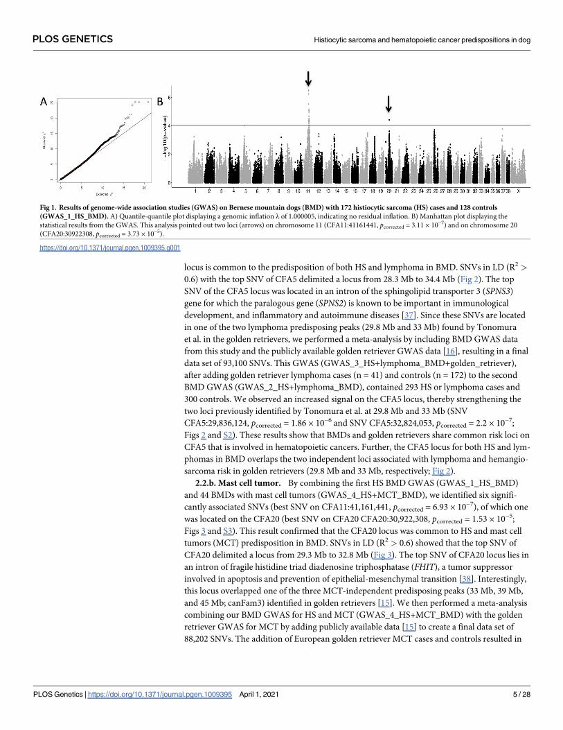
Using BMD DNA from 172 HS cases and 128 controls, we performed the first round of
GWAS (GWAS_1_HS_BMD) by correcting for population stratification and cryptic related-
ness (Fig 1). From 10,3487 SNVs left after applying filters, we identified 21 SNVs that were sig-
nificantly associated with HS, including 20 SNVs on chromosome 11 spanning 40.3–47.2 Mb
(strongest associated SNV was CFA11:41,161,441, pcorrected = 3.11 × 10⁻⁷) and one SNV on
chromosome 20 (CFA20:30,922,308, pcorrected = 3.73 × 10⁻⁵). Moreover, an additional SNV on
chromosome 5 (CFA5:30,496,048, pcorrected = 9.48. × 10−5) was close to the genome-wide sig-
nificance, and was suspected to be associated with HS. This GWAS confirmed that the main
locus linked to HS was located on CFA11, overlapping the MTAP-CDKN2A region, a locus
previously associated with HS [26]. The analysis also identified a new locus on CFA20 and sug-
gested the existence of another locus on CFA5. Interestingly, these three regions were previ-
ously identified for cancer predisposition in dogs: CFA11 (41.3–41.4 Mb) in osteosarcoma,
CFA5 (29.6–34.1 Mb) in lymphomas and hemangiosarcomas, CFA20 (30.9–50.1 Mb) in mast
cell tumors [15–17]. Indeed, Tonomura et al. has identified two independent peaks on CFA5
involved in lymphomas and hemangiosarcomas [16] overlapping the CFA5 locus suspected in
HS; while, Arendt et al. has identified at least two independent peaks on CFA20 involved in
mast cell predisposition [15], which also overlap the CFA20 locus found in HS in the BMD
breed. Thus, we hypothesized that, because of strong breed selection, the significant associa-
tions detected for HS could be due to the cumulative risk alleles/haplotypes that can also be at
risk for other hematopoietic cancers. This hypothesis was reinforced by the strong predisposi-
tion of BMD to hematopoietic cancers such as lymphomas [33,34] and by the fact that a
PLOS GENETICS Histiocytic sarcoma and hematopoietic cancer predispositions in dog
PLOS Genetics | https://doi.org/10.1371/journal.pgen.1009395 April 1, 2021 3 / 28

majority of BMDs (58.3% to 66.5%) succumb to cancer, in first place HS, lymphomas, or mast
tumors [26,30,35]. Moreover, we observed that in HS-affected BMD families, relatives of HS
affected dogs are frequently affected by other hematopoietic cancers such as mast cell tumors
and lymphomas [36] (S1 Fig).
2.2. Involvement of HS loci in other hematopoietic cancers
To test whether these HS predisposing loci could also be involved in the predisposition of lym-
phomas or mast cell tumors, we added lymphoma or mast cell tumor cases to the previous HS
GWAS.
2.2.a. Lymphoma. We performed a second GWAS (GWAS_2_HS+lymphoma_BMD) by
adding 80 lymphoma-affected BMDs to the first GWAS (GWAS_1_HS_BMD). We identified
six significantly associated SNVs, one of which was located on the CFA5 (best SNV on
CFA5:30,496,048, pcorrected = 5.88 × 10⁻⁶; Figs 2 and S2). This result confirmed that the CFA5
Table 1. Characteristics of the genome-wide association studies (GWAS) analyses performed in this study. � Dogs genotyped on the Affymetrix Axiom Canine Geno-
typing Array were also available in the Illumina 173K SNV Canine HD array format.
GWAS Name Paragrah cancers breeds SNP arrays format Number of
cases after QC
Number of
controls after
QC
GWAS_1_HS_BMD §2.1 HS BMD Illumina 173k SNV Canine HD
array
172 128
GWAS_2_HS+lymphoma_BMD §2.2a HS and
lymphoma
BMD Illumina 173k SNV Canine HD
array
252 128
GWAS_3_HS+lymphoma_BMD
+golden_retriever
§2.2a HS and
lymphoma
BMD, golden
retriever
Illumina 173k SNV Canine HD
array
293 300
GWAS_4_HS+MCT_BMD §2.2b HS and mast
cell tumor
BMD Illumina 173k SNV Canine HD
array
216 128
GWAS_5_HS+MCT_BMD+golden_retriever §2.2b HS and mast
cell tumor
BMD, golden
retriever
Illumina 173k SNV Canine HD
array
285 202
GWAS_6_HS_BMD_with_imputed_SNV §2.3 HS BMD Affymetrix Axiom Canine
Genotyping array 1.1M SV
(n = 113)�
Illumina 20k SNV Canine HD
array imputed for 1.1M SV
(n = 464)
Illumina 173k SNV Canine HD
array imputed for 1.1M SV
(n = 300)
403 347
GWAS_7_HS_BMD+FCR_with_imputed_SNV §2.3 HS BMD, FCR Affymetrix Axiom Canine
Genotyping array 1.1M SV
(n = 134)�
Illumina 20k SNV Canine HD
array imputed for 1.1M SV
(n = 464)
Illumina 173k SNV Canine HD
array imputed for 1.1M SV
(n = 328)
416 362
GWAS_8_HS_BMD+FCR
+Rottweiler_with_imputed_SNV
§2.3 HS BMD, FCR,
Rottweiler
Affymetrix Axiom Canine
Genotyping array 1.1M SV
(n = 134)�
Illumina 20k SNV Canine HD
array imputed for 1.1M SV
(n = 464)
Illumina 173k SNV Canine HD
array imputed for 1.1M SV
(n = 388)
453 385
https://doi.org/10.1371/journal.pgen.1009395.t001
PLOS GENETICS Histiocytic sarcoma and hematopoietic cancer predispositions in dog
PLOS Genetics | https://doi.org/10.1371/journal.pgen.1009395 April 1, 2021 4 / 28
locus is common to the predisposition of both HS and lymphoma in BMD. SNVs in LD (R2 >
0.6) with the top SNV of CFA5 delimited a locus from 28.3 Mb to 34.4 Mb (Fig 2). The top
SNV of the CFA5 locus was located in an intron of the sphingolipid transporter 3 (SPNS3)
gene for which the paralogous gene (SPNS2) is known to be important in immunological
development, and inflammatory and autoimmune diseases [37]. Since these SNVs are located
in one of the two lymphoma predisposing peaks (29.8 Mb and 33 Mb) found by Tonomura
et al. in the golden retrievers, we performed a meta-analysis by including BMD GWAS data
from this study and the publicly available golden retriever GWAS data [16], resulting in a final
data set of 93,100 SNVs. This GWAS (GWAS_3_HS+lymphoma_BMD+golden_retriever),
after adding golden retriever lymphoma cases (n = 41) and controls (n = 172) to the second
BMD GWAS (GWAS_2_HS+lymphoma_BMD), contained 293 HS or lymphoma cases and
300 controls. We observed an increased signal on the CFA5 locus, thereby strengthening the
two loci previously identified by Tonomura et al. at 29.8 Mb and 33 Mb (SNV
CFA5:29,836,124, pcorrected = 1.86 × 10⁻⁶ and SNV CFA5:32,824,053, pcorrected = 2.2 × 10⁻⁷;
Figs 2 and S2). These results show that BMDs and golden retrievers share common risk loci on
CFA5 that is involved in hematopoietic cancers. Further, the CFA5 locus for both HS and lym-
phomas in BMD overlaps the two independent loci associated with lymphoma and hemangio-
sarcoma risk in golden retrievers (29.8 Mb and 33 Mb, respectively; Fig 2).
2.2.b. Mast cell tumor. By combining the first HS BMD GWAS (GWAS_1_HS_BMD)
and 44 BMDs with mast cell tumors (GWAS_4_HS+MCT_BMD), we identified six signifi-
cantly associated SNVs (best SNV on CFA11:41,161,441, pcorrected = 6.93 × 10⁻⁷), of which one
was located on the CFA20 (best SNV on CFA20 CFA20:30,922,308, pcorrected = 1.53 × 10⁻⁵;
Figs 3 and S3). This result confirmed that the CFA20 locus was common to HS and mast cell
tumors (MCT) predisposition in BMD. SNVs in LD (R2 > 0.6) showed that the top SNV of
CFA20 delimited a locus from 29.3 Mb to 32.8 Mb (Fig 3). The top SNV of CFA20 locus lies in
an intron of fragile histidine triad diadenosine triphosphatase (FHIT), a tumor suppressor
involved in apoptosis and prevention of epithelial-mesenchymal transition [38]. Interestingly,
this locus overlapped one of the three MCT-independent predisposing peaks (33 Mb, 39 Mb,
and 45 Mb; canFam3) identified in golden retrievers [15]. We then performed a meta-analysis
combining our BMD GWAS for HS and MCT (GWAS_4_HS+MCT_BMD) with the golden
retriever GWAS for MCT by adding publicly available data [15] to create a final data set of
88,202 SNVs. The addition of European golden retriever MCT cases and controls resulted in
Fig 1. Results of genome-wide association studies (GWAS) on Bernese mountain dogs (BMD) with 172 histiocytic sarcoma (HS) cases and 128 controls
(GWAS_1_HS_BMD). A) Quantile-quantile plot displaying a genomic inflation λ of 1.000005, indicating no residual inflation. B) Manhattan plot displaying the
statistical results from the GWAS. This analysis pointed out two loci (arrows) on chromosome 11 (CFA11:41161441, pcorrected = 3.11 × 10⁻⁷) and on chromosome 20
(CFA20:30922308, pcorrected = 3.73 × 10⁻⁵).
https://doi.org/10.1371/journal.pgen.1009395.g001
PLOS GENETICS Histiocytic sarcoma and hematopoietic cancer predispositions in dog
PLOS Genetics | https://doi.org/10.1371/journal.pgen.1009395 April 1, 2021 5 / 28
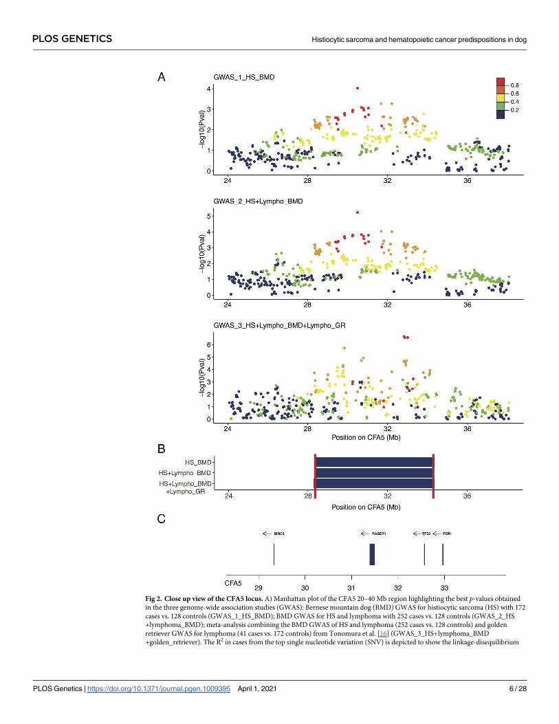
Fig 2. Close up view of the CFA5 locus. A) Manhattan plot of the CFA5 20–40 Mb region highlighting the best p-values obtained
in the three genome-wide association studies (GWAS): Bernese mountain dog (BMD) GWAS for histiocytic sarcoma (HS) with 172
cases vs. 128 controls (GWAS_1_HS_BMD); BMD GWAS for HS and lymphoma with 252 cases vs. 128 controls (GWAS_2_HS
+lymphoma_BMD); meta-analysis combining the BMD GWAS of HS and lymphoma (252 cases vs. 128 controls) and golden
retriever GWAS for lymphoma (41 cases vs. 172 controls) from Tonomura et al. [16] (GWAS_3_HS+lymphoma_BMD
+golden_retriever). The R2 in cases from the top single nucleotide variation (SNV) is depicted to show the linkage-disequilibrium
PLOS GENETICS Histiocytic sarcoma and hematopoietic cancer predispositions in dog
PLOS Genetics | https://doi.org/10.1371/journal.pgen.1009395 April 1, 2021 6 / 28

an increased association signal in the CFA20 locus, and clearly pointed out the CFA20 locus at
33 Mb (best SNV on CFA20:33,321,282, pcorrected = 4.79 × 10⁻⁷) in rho guanine nucleotide
exchange factor 3 (ARHGEF3) and close to interleukin 17 receptor D (IL17RD), one of the
three peaks identified by Arendt et al. These results show that the BMD and golden retriever
breeds share common inherited risk factors on CFA20 for HS and MCT (Figs 3 and S3). These
results also confirmed that the association of CFA20 with MCT in the golden retrievers is due
to the additional effect of at least three risk haplotypes (33 Mb, 39 Mb, and 45 Mb).
In conclusion, it appears that the loci linked to cancer in dogs can have pleiotropic effects
associated with the risk of several cancers in several breeds, and can be due to the additive
effects of several risk peaks. Thus, to more precisely identify the HS risk haplotypes and reduce
the loci size, we added new HS cases from BMD and other HS predisposed breeds to the first
BMD GWAS.
2.3. Refining HS loci by multiple-breed analyses and imputation on higher
density SNV array
To increase the power of the GWAS and refine the HS loci, we added HS cases and controls
from FCR and Rottweiler breeds genotyped on Illumina 173K SNV Canine HD. To increase
the density of markers, 134 dogs (113 BMDs and 21 FCRs) were re-genotyped on the higher
density Affymetrix Axiome Canine Genotyping array (1.1M SNV), and were used as a refer-
ence panel to impute these SNVs on the Illumina 173K SNV Canine HD. In addition, we also
added data from previously published BMD cases and controls [26] by imputing their geno-
types from the Canine SNP20 Bead-Chip panel (Illumina -22K SNV) to the higher density
Axiome Canine Genotyping array (1.1M SNV). The quality of imputation was evaluated by
masking imputed SNVs on half of the BMDs genotyped on the high-density Affymetrix
Axiome Canine Genotyping array. The mean concordances between the masked autosomal
SNVs and imputed SNVs were 91.97% and 95.86% for SNVs imputed from the Illumina -22k
SNV panel to the higher density Affymetrix Axiome Canine Genotyping array and from the
Illumina 173K SNV Canine HD panel to the higher density Affymetrix Axiome Canine Geno-
typing array, respectively. These concordances are similar to those described by the work of
Friedenberg and Meurs, which describes a genotype concordance of up to 92.4% with Beagle
software [39].
The addition of BMD cases and controls to the first BMD GWAS (GWAS_1_HS_BMD)
resulted in a total of 403 cases and 347 controls imputed to form a final data set of 488,872
SNVs (GWAS_6_HS_BMD_with_imputed_SNV). Statistical analysis allowed the identifica-
tion of 1,730 SNVs significantly associated with HS (Table 2, Fig 4A and 4B). This GWAS,
after increasing the number of BMD cases and controls, confirmed the involvement of the
CFA11 locus as well as the role of other loci (CFA5 and CFA14), and identified a new locus on
chromosome 2 in HS BMD predisposition.
By the addition of 28 FCRs (13 cases and 15 controls) to GWAS_6_HS_BMD_with_impu-
ted_SNV to form a final dataset of 525,657 SNVs (GWAS_7_HS_BMD+-
FCR_with_imputed_SNV), we identified 1,558 SNVs that were significantly associated with
HS (Table 2, Fig 4C and 4D), confirming the involvement of the CFA2, CFA5, CFA11, and
CFA14 loci in HS predisposition.
(LD) structure. B) Regions delimitated by the SNPs in LD with the best GWAS SNVs (R2 > 0.6) in cases; minimal region between
the three GWAS (CFA5:28309815–34321500) is delimitated by red lines. C) Close up view of the genes (with available symbols)
located in this minimal region (28–34 Mb) of CFA5.
https://doi.org/10.1371/journal.pgen.1009395.g002
PLOS GENETICS Histiocytic sarcoma and hematopoietic cancer predispositions in dog
PLOS Genetics | https://doi.org/10.1371/journal.pgen.1009395 April 1, 2021 7 / 28
PLOS GENETICS Histiocytic sarcoma and hematopoietic cancer predispositions in dog
PLOS Genetics | https://doi.org/10.1371/journal.pgen.1009395 April 1, 2021 8 / 28

The addition of 60 Rottweilers (37 cases and 23 controls) to GWAS_7_HS_BMD+-
FCR_with_imputed_SNV formed a final dataset of 532,053 SNVs (GWAS_8_HS_BMD+FCR
+Rottweiler_with_imputed_SNV), and led to the identification of 1,505 SNVs that were signif-
icantly associated with HS (Table 2, Fig 4E and 4F). This GWAS confirmed the involvement of
the CFA2, CFA5, CFA11, and CFA14 loci in HS predisposition. SNVs in LD with significant
SNVs in cases (R2� 0.6) allowed us to identify large regions spanning several Mb (up to 23
Mb for CFA11; Table 2). The analysis of these SNVs within the three breeds allowed us to
reduce the CFA11 locus region to 38.4–41.4 Mb (Fig 4G).
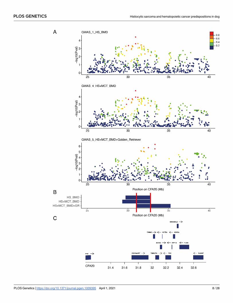
These analyses identified SNVs that were significantly associated with HS risk and were
shared between the three predisposed breeds on at least chromosomes 2, 5, 11, and 14. To
Fig 3. Close up view of the CFA20 locus. A) Manhattan plot of the CFA20 20–45 Mb region highlighting the best p-values obtained in
the three genome-wide association study (GWAS): Bernese Mountain dogs (BMD) GWAS for histiocytic sarcoma (HS) with 172 cases vs.
128 controls (GWAS_1_HS_BMD); BMD GWAS for HS and mast cell tumor with 216 cases vs. 128 controls (GWAS_4_HS
+MCT_BMD); meta-analysis combining BMD GWAS for HS and mast cell tumor with European golden retriever (69 cases vs. 74
controls) from Arendt et al. [15] (GWAS_5_HS+MCT_BMD+golden_retriever). The R2 in cases from top single nucleotide variation
(SNVs) show the linkage disequilibrium (LD) structure. B) Regions delimitated by SNVs in LD with the best GWAS SNVs (R2 > 0.6) in
cases; minimal region between the three GWAS (CFA20:31036863–32778949) is delimitated by red lines. C) Close up view of the genes
(with available symbols) located in this minimal region (31–33 Mb) of CFA20.
https://doi.org/10.1371/journal.pgen.1009395.g003
Table 2. Significant loci identified by genome-wide association studies (GWAS) after imputation on high-density single nucleotide variation (SNV) array. Number
of associated SNVs with the best SNV and the corresponding corrected p-value are presented for each locus and each GWAS.
GWAS
Chromosome BMD BMD+FCR BMD+FCR+Rott
2 Number of associated SNV 1 4 7
Localisation of the best SNV 29716535 29716535 29716535
pcorrected = 3.24 x 10⁻⁴ pcorrected = 6.02 x 10⁻⁵ pcorrected = 3.58 x 10⁻⁵
Region delimited by significant associated SNVs 29716535 29653137–29978776 29507029–34223001
Region delimited by significant associated SNVs and SNVs in LD in cases
(R2�0.6)
29154373–30121047 29154373–30121047 29385904–35075340
5 Number of associated SNV 292 320 322
Localisation of the best SNV 30496048 33823740 33823740
pcorrected = 8.22 x 10⁻⁶ pcorrected = 2.52 x 10⁻⁶ pcorrected = 2.4 x 10⁻⁶
Region delimited by significant associated SNVs 25628485–34513401 25522718–34477045 25566642–34477045
Region delimited by significant associated SNVs and SNVs in LD in cases
(R2�0.6)
25402068–37781406 25402068–34513401 25517580–34513401
11 Number of associated SNV 1145 984 930
Localisation of the best SNV 41215628 41252822 41252822
pcorrected = 1.46 x
10−13pcorrected = 1.49 x
10−13pcorrected = 2.02 x
10−14
Region delimited by significant associated SNVs 29934486–52418087 29978631–52418087 29978631–52418087
Region delimited by significant associated SNVs and SNVs in LD in cases
(R2�0.6)
29047449–52471659 29315057–52471659 29341449–52418087
14 Number of associated SNV 292 250 246
Localisation of the best SNVs 6567456 6567456 6566022
pcorrected = 4.05 x 10⁻⁶ pcorrected = 1.37 x 10⁻⁶ pcorrected = 1.09 x 10⁻⁶
10231328 10665001 11021670
pcorrected = 9.19 x 10⁻⁶ pcorrected = 2.78 x 10⁻⁶ pcorrected = 1.52 x 10⁻⁶
Region delimited by significant associated SNVs 561549–11111293 561549–11111293 561549–11111293
Region delimited by significant associated SNVs and SNVs in LD in cases
(R2�0.6)
475090–11638599 475090–11379670 475090–11379670
https://doi.org/10.1371/journal.pgen.1009395.t002
PLOS GENETICS Histiocytic sarcoma and hematopoietic cancer predispositions in dog
PLOS Genetics | https://doi.org/10.1371/journal.pgen.1009395 April 1, 2021 9 / 28

determine the proportion of HS risk that could be explained by these loci, we performed a
restricted maximum likelihood (REML) analysis using GCTA software [40]. All chromosomes
together could explain at least 61.8% of the phenotype (p-value� 4.93 × 10⁻²⁹; S1 Table).
SNVs of the CFA11 locus could explain over 10.3% (p-value� 3.5 × 10⁻¹⁹) of the HS pheno-
type; while, SNVs of the CFA14 locus explained a similar part of the phenotype, and the CFA5
could explain only 4.8–6.7% of the phenotype.
Fig 4. Genome-wide association studies (GWAS) of Bernese mountain dogs (BMD) and other predisposed breeds on histiocytic sarcoma
(HS) with the imputation of single nucleotide variations (SNV) on a higher density SNV array. A–B) BMD GWAS results based on 403 cases
and 347 controls (GWAS_6_HS_BMD_with_imputed_SNV). A) Quantile-quantile plot displaying a genomic inflation λ of 1.000023, indicating
no residual inflation. B) Manhattan plot displaying the statistical results from the GWAS. This analysis shows four loci (arrows) on chromosome
2 (best SNV at CFA2:29716535, pcorrected = 3.25 × 10⁻⁴), chromosome 5 (best SNV at Chr5:30496048, pcorrected = 8.22 × 10⁻⁶), chromosome 11
(best SNV at Chr11:41215628, pcorrected = 1.45 × 10⁻¹³), and chromosome 14 (CFA14:6567456, pcorrected = 4.04 × 10⁻⁶). C–D. GWAS results for
HS combining BMDs (403 cases vs. 347 controls) and flat-coated retrievers (FCRs; 13 cases vs. 15 controls; GWAS_7_HS_BMD
+FCR_with_imputed_SNV). C) Quantile-quantile plot displaying a genomic inflation λ of 1.000018, indicating no residual inflation. D)
Manhattan plot displaying the statistical results from the GWAS. This analysis shows four loci (arrows) on chromosome 2 (best SNV at
CFA2:29716535, pcorrected = 6.02 × 10⁻⁵), chromosome 5 (best SNV at CFA5:33823740, pcorrected = 2.52 × 10⁻⁶), chromosome 11 (best SNV at
CFA11:41252822, pcorrected = 1.49 × 10⁻¹³), and chromosome 14 (CFA14:6567456, pcorrected = 1.37 × 10⁻⁶). E–F) GWAS results for HS combining
BMDs (403 cases vs. 347 controls), FCRs (13 cases vs. 15 controls), and Rottweilers (37 cases vs. 23 controls; GWAS_8_HS_BMD+FCR
+Rottweiler_with_imputed_SNV). E) Quantile-quantile plot displaying a genomic inflation λ of 1.000013, indicating no residual inflation. F)
Manhattan plot displaying the statistical results from the GWAS. This analysis shows four loci (arrows) on chromosome 2 (best SNV at
CFA2:29716535, pcorrected = 3.58 × 10⁻⁵), chromosome 5 (best SNV at CFA5:33823740, pcorrected = 2.4 × 10⁻⁶), chromosome 11 (best SNV at
CFA11:41252822, pcorrected = 2.04 × 10⁻¹⁴), and chromosome 14 (best SNV at CFA14:6566022, pcorrected = 1.09 × 10⁻⁶). G) Close up view of the
CFA11 locus highlighting the best p-values obtained in the three GWAS: BMDs GWAS (GWAS_6_HS_BMD_with_imputed_SNV), BMDs plus
FCRs GWAS (GWAS_7_HS_BMD+FCR_with_imputed_SNV), and BMDs plus Rottweilers and FCRs GWAS (GWAS_8_HS_BMD+FCR
+Rottweiler_with_imputed_SNV). R2 in cases from top SNV is depicted to show the linkage disequilibrium (LD) structure. H) Regions
delimitated by SNVs in LD with the best GWAS SNVs (R2 > 0.6) in cases, minimal region between the three GWAS (CFA11: 38435917–
41701130) is delimitated by red lines. I) Close up view of the genes (with available symbols) located in this minimal region (38–42 Mb).
https://doi.org/10.1371/journal.pgen.1009395.g004
PLOS GENETICS Histiocytic sarcoma and hematopoietic cancer predispositions in dog
PLOS Genetics | https://doi.org/10.1371/journal.pgen.1009395 April 1, 2021 10 / 28
2.4. Haplotype analyses of HS loci identified risk haplotypes shared
between breeds
To identify risk haplotypes tagged by the best SNVs and shared between HS cases, we deter-
mined the haplotype blocks including the best SNVs (Cf Materials and Methods), in each breed.
On CFA11, we identified a haplotype block containing the best CFA11 SNV (41,252,822)
that was more frequent in HS cases in BMD and Rottweiler breeds than in the controls (0.79 vs.
0.55 and 0.77 vs. 0.54 in BMD and Rottweiler, respectively), and significantly linked to the risk
of developing HS (odds ratio = 3.03, p-value = 7.03 × 10⁻²³ for BMDs; odds ratio = 2.61, p-
value = 0.0175 for Rottweilers; Table 3). This block was frequently present in the FCR breed
(53.8% and 56.6% in FCR cases and controls, respectively), and 75% of FCRs carried at least one
copy of this risk haplotype. However, while the number of FCRs in the GWAS remained low,
this haplotype did not appear to be enriched in HS cases (odds ratio = 0.89, p-value = 0.83). We
identified a second independent CFA11 HS locus between 44 Mb and 45 Mb in the GWAS of
the three breeds (GWAS_8_HS_BMD+FCR+Rottweiler_with_imputed_SNV; Fig 4G), which
had already been identified in previous studies [26]. The best SNV in this region
(CFA11:44,150,645) was not in LD with the best SNV of CFA11 (CFA11:41,252,822; R2 of the
three breeds = 0.41), indicating that there were at least two independent peaks on CFA11
involved in HS predisposition. Haplotype analysis of the CFA11:44 Mb region indicated a com-
mon risk haplotype enriched in the cases of the three breeds (0.66 vs. 0.44), and was significantly
associated with the risk of developing HS (odds ratio = 2.45, p-value = 3.2 × 10⁻¹⁹; Table 3).
For CFA5, the GWAS analysis indicated that a large locus (25–35 Mb) overlapped the two
CFA5 lymphoma loci, as previously identified by Tonomura et al. (29 Mb and 33 Mb) [16].
Similarly, the haplotype analysis of the best SNVs indicated that the common risk haplotype
was delimited by 18 SNVs (33,839,057–34,234,461) in the three breeds. This haplotype was sig-
nificantly enriched in BMD and FCR cases, and was common in Rottweilers (69% and 63% in
cases and controls, respectively; Table 3). In the three breeds, this risk haplotype was associated
with a significantly increased risk of developing HS (odds ratio = 2.05, p-value = 6.58 × 10⁻¹²).
For CFA14, the third main HS risk locus, GWAS analysis revealed a large region with signifi-
cant SNVs spanning from 0.4 to 11.3 Mb. The associated SNVs in this region were clustered in at
least two peaks located 4.4 Mb apart (Table 2). The top SNVs of these two peaks were located at
6,566,022 and 11,021,670 with pcorrected = 1.09 × 10⁻⁶ and 1.52 × 10⁻⁶, respectively, in the GWAS
including the three predisposed breeds (GWAS_8_HS_BMD+FCR+-
Rottweiler_with_imputed_SNV). With an R2 of 0.11 between these two SNVs across the three
breeds, the data suggested that these peaks were independent. When considering the best SNV of
CFA11 (CFA11:41,252,822), the two best SNVs of CFA14 were located at 10,665,001 (pcorrected =
1.14 × 10⁻⁷) and 11,021,670 (pcorrected = 1.16 × 10⁻⁷). Haplotype analysis of the region showed that
the same risk haplotype was enriched in BMD and Rottweiler cases but not in FCR cases. Surpris-
ingly, another haplotype, 50-GCCAACGTATAAGT-30, was enriched in the controls of the three
breeds and was significantly associated with a decreased risk of developing HS in these predis-
posed breeds (odds ratio = 0.34, p-value = 9.08 × 10⁻¹¹). These results suggested that the CFA14
locus contains a protective allele, which is shared by the three predisposed breeds (Table 3).
Overall, this imputation with high density of SNVs allowed the identification of shared risk
loci between the three HS predisposed breeds. We identified common risk or protective haplo-
types shared by the predisposed breeds on major loci localized on CFA11, CFA14, and CFA5.
2.5. HS risk results from cumulative risk haplotypes
In these three breeds, cases had risk alleles on these three chromosomes, especially in BMD
and Rottweilers, for which most cases had at least five risk copies (Table 4). In this cohort, no
PLOS GENETICS Histiocytic sarcoma and hematopoietic cancer predispositions in dog
PLOS Genetics | https://doi.org/10.1371/journal.pgen.1009395 April 1, 2021 11 / 28
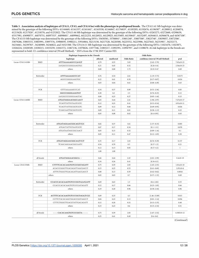
Table 3. Association analysis of haplotypes of CFA11, CFA5, and CFA14 loci with the phenotype in predisposed breeds. The CFA11:41 Mb haplotype was deter-
mined by the genotype of the following SNVs: 41144469, 41161357, 41161441�, 41163558, 41166847, 41176819�, 41185205, 41192676, 41196587�, 41200012, 41204074,
41215628, 41217026�, 41218376, and 41252822. The CFA11:44 Mb haplotype was determined by the genotype of the following SNVs: 43263273, 43272660, 43290639,
43317981, 43690075�, 44070751, 44097553�, 44098601�, 44099662, 44121253, 44126921, 44129025, 44135605, 44150645�, 44152497, 44364615, 44366678, and 44367106�.
The CFA5:33 Mb haplotype was determined by the genotype of the following SNVs: 33650581, 33788841�, 33801240�, 33807500�, 33823740�, 33839057, 33872506�,
33879286, 33883533, 33883991, 33897576, 33900059, 33906211, 33916864, 34213158, 34217528, 34220300, 34225552, 34225906, 34227643, 34231901�, 34232173,
34234461, 34239783�, 34240989, 34246822, and 34321500. The CFA14:11 Mb haplotype was determined by the genotype of the following SNVs: 11021670, 11023871,
11026224, 11028349, 11030215, 11033259, 11041572, 11067136, 11070626, 11077346, 11083217, 11092391, 11094795�, and 11108670. At risk haplotype in the breeds are
represented in bold. CI: confidence interval (Woolf Method). � SNVs from the 173K SNV Canine HD.
Haplotype frequencies in the 3 breeds Odds Ratio
haplotype affected unaffected Odds Ratio confidence interval (Woolf Method) pval
Locus: CFA11:41Mb BMD ATTTAAAAAGCCC{A/G}T 0,79 0,55 3.03 [2.42–3.79] 7.03x10⁻²³
{A/G}GCCCGGGGAAGTGC 0,21 0,45 0.32 [0.26–0.4] 2.56x10⁻²³
others 0,00 0,00 1.73 [0.32–9.47 0.69
Rottweiler ATTTAAAAAGCCCAT 0,76 0,54 2.61 [1.19–5.73] 0.0175
AGCCCGGGGAAGTGC 0,22 0,41 0.39 [0.17–0.87] 0.024
others 0,03 0,04 0.61 [0.08–4.49] 0.63
FCR GTTTAAAAAGCCCAT 0,54 0,57 0.89 [0.31–2.56] 0.83
GGCCCGGGGAAGTGT 0,38 0,2 2.5 [0.76–8.25] 0.12
{A/G}GCCCGGGGAAGTAC 0,08 0,23 0.27 [0.05–1.44] 0.15
Locus: CFA11:44Mb BMD ATGATAGGAACGGCAACT 0,65 0,44 2.41 [1.96–2.97] 7.27x10⁻¹⁷
TCAGTTATTGTAATCGTC 0,12 0,25 0.41 [0.31–0.54] 4.91x10⁻¹¹
TCAGTTATTGCGGTCGTC 0,09 0,12 0.68 [0.49–0.95] 0.026
TCAGCAATTGCGGTCGTC 0,09 0,11 0.81 [0.58–1.13] 0.23
others 0,05 0,08 0.62 [0.4–0.95] 0.03
Rottweiler ATGATAGGAACGGCAG{T/C}C 0,82 0,57 3.61 [1.57–8.33] 0.003
ATGACTATTGTAATCGTC 0,07 0,17 0.34 [0.1–1.11] 0.078
ATGATAATAGCGGCAACT 0,05 0,15 0.32 [0.09–1.16] 0.1
others 0,05 0,11 0.47 [0.12–1.85] 0.30
FCR ATGATAGGAACGGCAA{T/C}T 0,35 0,17 2.65 [0.76–9.29] 0.137
TCAGCAGGAACGGCAATC 0,54 0,70 0.5 [0.17–1.5] 0.21
others 0,12 0,13 0.85 [0.17–4.2] 1
1,00 1,00
all breeds ATGATAGGAACGGCA--- 0,66 0,44 2.45 [2.01–2.99] 3.2x10⁻¹⁹
others 0,34 0,56 0.41 [0.34–0.5]
Locus: CFA5:33Mb BMD CTTTTCACACAAGTGTCCCGGTAGATT 0,75 0,59 2.05 [1.65–2.55] 1.01x10⁻¹⁰
ACACCCGGGTTGACAGATTAACGACCT 0,15 0,25 0.53 [0.41–0.69] 1.87x10⁻⁶
ATTTCTGGGTTGACAGATTAACGACCT 0,08 0,13 0.59 [0.42–0.82] 0.0016
others 0,02 0,03 0.7 [0.37–1.32] 0.69
Rottweiler CCACCCACACAAGTGTCCCGGTA{A/G}ATT 0,69 0,63 1.3 [0.6–2.82] 0.55
CCACCCACACAAGTGTCCCGATAGATT 0,12 0,17 0.66 [0.23–1.85] 0.66
others 0,19 0,20 0.96 [0.38–2.44] 0.96
FCR ACTTTCACACAAGTGTCCCGGTAGA{T/C}C 0,69 0,33 4.5 [1.46–13.89] 0.015
CCTTCTACACAAGTAGACCGGTAACCT 0,04 0,23 0.13 [0.01–1.14] 0.056
CTTTCTGGGTTGACGTATTGACAGATT 0,12 0,20 0.52 [0.12–2.33] 0.48
others 0,15 0,23 0.6 [0.15–2.34] 0.51
all breeds ------CACACAAGTGTCCCGGTA------- 0,75 0,59 2.05 [1.67–2.52] 6.58X10⁻¹²
others 0,25 0,41 0.49 [0.4–0.6]
(Continued)
PLOS GENETICS Histiocytic sarcoma and hematopoietic cancer predispositions in dog
PLOS Genetics | https://doi.org/10.1371/journal.pgen.1009395 April 1, 2021 12 / 28
FCR controls had over three risk haplotypes. In the three breeds, the cumulative risk alleles on
the three main loci (CFA11, CFA5, and CFA14) strongly impacted the probability of develop-
ing HS: carrying 5/6 risk alleles is associated with HS with an odds ratio of 5.27 (p-value = 1.52 × 10⁻³⁰). Stepwise model selections of the 1,505 significant SNVs identified by
GWAS_8_HS_BMD+FCR+Rottweiler_with_imputed_SNV were performed to create general-
ized risk-models for HS (S2 Table). Both the Stepwise Forward model selection or the Stepwise
Table 3. (Continued)
Haplotype frequencies in the 3 breeds Odds Ratio
haplotype affected unaffected Odds Ratio confidence interval (Woolf Method) pval
Locus: CFA14:11Mb BMD ATAGGAACCCGCGT 0,74 0,61 1.79 [1.44–2.23] 1.86x10⁻⁷
ACCAACGCACGCAT 0,17 0,19 0.85 [0.65–1.1] 0.25
GCCAACGTATAAGT 0,05 0,15 0.34 [0.23–0.49] 2.65x10⁻⁹
ATAGGAACCCGCAT 0,04 0,04 0.8 [0.48–1.34] 0.43
others 0,01 0,01 0.64 [0.22–1.85] 0.43
Rottweiler ATAGGAACCCGCGT 0,57 0,43 1.77 [0.85–3.69] 0.14
ATAGGAACCCGCAT 0,35 0,41 0.77 [0.36–1.64] 0.56
GCCAACGTATAAGT 0,07 0,11 0.59 [0.16–2.16] 0.59
others 0,01 0,04 0.2 [0.02–1.98] 0.15
FCR ACCAACGCACGCAC 0,46 0,20 3.43 [1.05–11.17] 0.036
GCCAACGTATAAGT 0,23 0,53 0.26 [0.08–0.83] 0.02
ATAGGAACCCGCAC 0,23 0,23 0.99 [0.29–3.44] 0.98
others 0,08 0,03 2.42 [0.21–28.34] 0.59
all breeds GCCAACGTATAAGT 0,06 0,16 0.34 [0.24–0.47] 9.08x10⁻¹¹
others 0,94 0,84 2.92 [2.09–4.08]
https://doi.org/10.1371/journal.pgen.1009395.t003
Table 4. Association of CFA5, CFA11, and CFA14 with the phenotype in predisposed breeds. The number of risk alleles was determined with the genotype of the fol-
lowing SNVs: CFA5:30496048, CFA11:41252822, and CFA14:11021670. CI: confidence interval (Woolf Method).
frequencies of risk alleles in the 3 breeds Odds Ratio
number of risk alleles affected unaffected Odds Ratio confidence interval (Woolf Method) pval
BMD �5 risk alleles 0,72 0,31 5.67 [4.14–7.76] 1.44x10⁻²⁹
4 risk alleles 0,23 0,4 0.46 [0.34–0.63] 1.779x10⁻⁶
� 3 risk alleles 0,05 0,29 0.12 [0.07–0.2] 1.34x10—20
Rottweiler �5 risk alleles 0,74 0,43 3.51 [1.17–10.53] 0.03
4 risk alleles 0,18 0,26 0.66 [0.19–2.29] 0.53
�3 risk alleles 0,08 0,3 0.2 [0.05–0.88] 0.034
FCR 4 risk alleles 0,38 0 NA NA 0.013
3 risk alleles 0,54 0,53 1.02 [0.23–4.52] 1
�2risk alleles 0,08 0,47 0.1 [0.01–0.98] 0.037
all breeds �5 risk alleles 0,70 0,31 5.27 [3.92–7.08] 1.52X10⁻³⁰
4 risk alleles 0,23 0,37 0.51 [0.38–0.69] 1.45x10⁻⁵
�3 risk alleles 0,07 0,32 0.15 [0.1–0.23] 8.1X10⁻²²
https://doi.org/10.1371/journal.pgen.1009395.t004
PLOS GENETICS Histiocytic sarcoma and hematopoietic cancer predispositions in dog
PLOS Genetics | https://doi.org/10.1371/journal.pgen.1009395 April 1, 2021 13 / 28
Forward and Backward model selection included markers of CFA2, CFA5, CFA11, and
CFA14, with several markers spaced more than 4 Mb apart. The fact that some markers are
separated by several Mbases on each chromosome, shows the cumulative effect of several inde-
pendent risk haplotypes.
2.6. Capture and targeted sequencing of the three best HS candidate loci
(CFA5, CFA11, and CFA14)
Since common haplotypes were detected in the three predisposed breeds on the three main
loci (CFA5, CFA11, and CFA14), we sequenced these three regions to identify putative com-
mon variants. DNA samples from 16 dogs (10 BMDs, 4 Rottweilers, and 2 FCRs) from the
three predisposed breeds, with a balanced distribution of risk and protective haplotypes, were
selected for targeted sequencing. An average of 9,458 SNVs and 2,674 indels per sample were
identified with a mean depth of 142 x per sample. These variants (SNVs and indels) were
imputed on the remaining dog samples (453 HS cases and 385 controls from BMD, Rottweiler,
and FCR breeds). A total of 2,608 significant variants (of which 1,886 were on CFA11) were
identified while performing statistical analysis with the imputed genotypes. For CFA11, no
coding variants were significantly associated with HS risk, and four of the top ten variants
associated with HS predisposition were imputed genotypes and localized within 100 kb (S3
Table). The best-associated variant remained CFA11:41,252,822, which was already identified
in the previous GWAS (see section 2.3.). CFA11:41,252,822 is localized in a non-coding tran-
script (CFRNASEQ_UC_00018829) that overlaps CDKN2A and CDKN2B antisense RNA
(CDKN2B-AS1). Interestingly, looking at RNASeq data from Hoeppner et al [41], we found
that CFRNASEQ_UC_00018829 was highly expressed in blood than in eight other tissues.
Moreover, the human orthologous region of this SNV (chr9:21,996,622, hg38), close to the
CpG island (chr9:21,994,103–21,995,911, hg38) and DNAse I hypersensitivity peak cluster
(chr9:21,994,641–21,996,130, hg38), overlaps an enhancer (GH09J021996, chr9:21,996,543–
21,996,791, hg38), which regulates CDKN2A and cyclin-dependent kinase 4 inhibitor B
(CDKN2B). The fourth top variant was an indel that is localized in a non-coding transcript
(CFRNASEQ_IGNC_00021613), 6,500 bp upstream of the CDKN2A transcript (S3 Table).
Since it was previously shown that the expression of CDKN2A correlated with CFA11:41 Mb
risk haplotype [26], we strongly suspected that these non-coding SNVs could deregulate the
expression of CDKN2A.
To identify the best candidate variants in the secondary loci, a complementary association
analysis was performed by including the genotypes of the best variant (CFA11:41,252,822) as a
covariate (S3 Table). We identified 2,413 variants (of which 21 SNVs were on CFA2, 1,533
SNVs were on CFA5, and 859 SNVs were on CF14) with significant residual associations in
the three breeds. Residual association was found for the CFA11:44–45 Mb locus with the best
SNV CFA11:45,941,548 (p-value = 0.008), close to C9orf72, and not in LD with
CFA11:41,252,822 (R2 of the three breeds = 0.0899). This result confirmed the existence of two
independent risk loci on CFA11 between 41–45 Mb.
For the CFA14 locus, seven of the top ten variants were imputed variants, and were local-
ized between positions 10,665,001 and 11,042,153. This locus overlapped POT1 antisense
RNA 1 (POT1-AS1) and protection of telomeres 1 (POT1).
In the CFA5 locus, nine of the top ten variants of CFA5 that were found to be associated
with the risk of developing HS were imputed variants; they were localized between positions
30,483,338 and 30,496,048 that overlap the SPNS3 gene (S3 Table and S4 Fig). Interestingly,
the second (CFA5:30,489,203) and third (CFA5:30,489,217) variants were localized in a region
containing DNA methylation marks, and one of them included the CFA5:30,489,217 variant.
PLOS GENETICS Histiocytic sarcoma and hematopoietic cancer predispositions in dog
PLOS Genetics | https://doi.org/10.1371/journal.pgen.1009395 April 1, 2021 14 / 28
Moreover, the nearby CFA5:30,489,203 variant created a CpG site. Thus, we hypothesized that
these two close SNVs could be associated with allele-specific methylation in histiocytic cells.
Bisulfite sequencing of HS cell lines confirmed that these two variants (CFA5:30,489,203 and
CFA5:30,489,217) presented with specific alleles (CFA5:30,489,203-G and CFA5:30,489,217-C,
respectively), thereby creating CpG sites with methylation in histiocytic cells (S3 Fig). This was
also the case for the best HS GWAS SNV on CFA2 with the CFA2:29,716,535-G allele (S5 Fig).
We hypothesized that these specific methylation alleles could be associated with modifications
of regulation of neighboring genes expression.
2.7. Validation of major loci on independent cohorts
Since risk haplotypes were detected in the three predisposed breeds because of the imputed data,
we genotyped variants of the three major loci on an independent cohort of BMD (186 cases and
176 controls) to validate the major role of these loci in HS (S4 Table). This analysis confirmed
that the risk alleles of the top variants of CFA5, CFA11, and CFA14 (see section 2.4) significantly
increased the risk of developing HS (odds ratios = 2.56–3.94, p-value = 2.4 × 10⁻⁶–6.93 × 10⁻¹⁶).
Moreover, when a BMD case from this cohort carried 5/6 risk alleles, it strongly impacted the
probability of developing this cancer with an odds ratio of 9.71 (p-value = 3.44 × 10⁻²³).
Since a majority of BMDs succumb to cancer, mostly HS, lymphomas, or mast tumors
[26,30,35], we suspected that carrying these risk alleles would impact the life span of BMDs.
We thus analyzed the risk alleles on an independent cohort of 317 dogs (age <10 years and
without pathological diagnosis of HS (see Materials and methods) and correlated longevity
with the number of risk alleles. This analysis confirmed that, independently of the known clini-
cal status, carrying 5/6 risk alleles significantly impacted the longevity at the BMD population
level (median = 7.5 vs 9.17 years, p-value = 0.0015, log-rank test; S6 Fig).
3. Discussion
3.1. Dog breeds: unique models to detangle the genetic features of human
cancers
Several genetic studies of canine cancers have shown that high-risk dog breeds can lead to the
advancement in genetics of rare cancers in humans [6,8,25]. Indeed, a limited number of criti-
cal loci have been identified in canine cancers [6,15–17,26], wherein some loci are shared by
several canine cancers, and are also well-known in human cancers. Moreover, somatic alter-
ations identified to date in canine tumors through genome-wide approaches, are found in the
same genes [25,42,43]. HS affects a few dog breeds with incredibly high frequencies (BMD,
Rottweiler, retrievers), and these breeds appear to be a perfect example to study the genetics
underlying such a strong HS predisposition in dogs.
Further, dissecting the genetic factors for rare cancers such as HS is challenging in humans.
Hence, we proposed that dog models of HS could help in deciphering the genetic predisposi-
tion factors of this rare cancer. Previously, we had successfully identified a relevant locus on
canine chromosome 11, and this locus is also well-known in canine osteosarcoma and several
human cancers (human chr.9p21 locus) [17,44]. In this study, we presented the GWAS on a
large cohort of several dog breeds affected by HS. We applied a multiple-breed approach to
not only refine the previously identified locus on CFA11 but also identified additional loci on
CFA2, CFA5, CFA14, and CFA20. Moreover, this study highlighted the fact that besides an ini-
tial association signal peak found in canine cancer GWAS, the independent risk haplotypes
can be cumulative and shared by several dog breeds and in several cancers.
PLOS GENETICS Histiocytic sarcoma and hematopoietic cancer predispositions in dog
PLOS Genetics | https://doi.org/10.1371/journal.pgen.1009395 April 1, 2021 15 / 28

3.2. Genetic predisposition of canine cancers: cumulative risk haplotypes
In humans, the genetic architecture of cancer risk is usually described by a combination of rare
variations in families with dominant inheritance patterns, and common variants with small-effect
sizes in the population. Within-breed canine GWAS usually identifies fewer variants with stron-
ger effects [18] because of breed structure and artificial selection of variants with strong effects,
which is not necessarily the rule for cancer development. Indeed, because of the genetic drift of
canine breeds resulting from the strong selection of the morphological criteria of sires and dams
used for reproduction, deleterious alleles could be involuntarily selected and enriched in specific
populations. Consequently, significant associations detected with cancer in dog GWAS could be
due to cumulative risk alleles. In such conditions, it would be surprising if HS predisposition was
only due to one risk haplotype, especially since at least 33 loci have been identified by previous
GWAS for canine osteosarcoma predisposition [17]. Our results confirmed the main role of the
CFA11 locus, along with the cumulative effects of at least two different risk haplotypes as sug-
gested in our previous study [26]. Here, a multi-breed approach allowed to refine the main risk
haplotype on CFA11 to a region of ~74 kb that was shared between BMDs and Rottweilers.
Moreover, this study allowed the identification of a strong candidate variant overlapping
CDKN2B-AS1 that regulates CDKN2A. Additional GWAS peaks were also identified on CFA2,
CFA5, CFA11, CFA14, and CFA20. Some of these loci were shared between the three predis-
posed breeds, mostly between Rottweilers and BMDs, which was expected considering the
close phylogenetic relationship between these two breeds [45]. FCR are a small number of
dogs in France; thus, the number of FCR samples included in the study was low, and further
GWAS will be needed to better decipher the shared predispositions between FCR and other
breeds. Nevertheless, in these three predisposed breeds, the cumulative risk alleles on the three
main loci (CFA11, CFA5, CFA14) strongly impacted the probability of developing this cancer
with an odds ratio of 5.27, i.e., to be affected by HS when dogs carry 5/6 risk alleles. This study
illustrated that the GWAS association detected between a cancer and a locus in dogs could
hide the cumulative risk of several haplotypes. This study also confirmed previous findings in
the golden retriever breed by Arendt et al. (2015) and Tonomura et al. (2015) who described
that there were at least two independent risk haplotypes on the CFA20 locus for mast cell
tumor and on the CFA5 locus for lymphoma, respectively [15,16].
3.3. Multi-cancer loci identified through a multi-breed approach
Additionally, we confirmed that some risk haplotypes were also involved in several cancers, as
suggested by Tonomura et al., based on the association of the CFA5 locus with hemangiosar-
coma and lymphoma [16]. Such pleiotropy at cancer risk loci has also been observed in human
cancers, where one-third of the SNVs mapped to genomic loci are associated with multiple
cancers [44]. Here, we confirmed that the multi-cancer effect of loci for CFA5, CFA11, and
CFA20 influenced the risk of HS, lymphomas, osteosarcomas, and mast cell tumors in BMD
or golden retrievers. However, further studies with higher SNV density in the golden retriever
breed are needed to confirm whether the same predisposing risk alleles are shared with BMD.
The CDKN2A locus was detected in Rottweilers and BMDs, and a neighboring region of the
locus (CFA11:41.37 Mb; canFam3) was associated with osteosarcoma and was fixed in the
Rottweiler population [17]. This shows that HS-affected Rottweilers were accumulating risk
haplotypes for at least two cancers (osteosarcoma and HS) at this locus. Co-occurrence of two
different risk haplotypes for HS and osteosarcoma across 200 kb also perfectly illustrates that a
given locus can harbor multi-cancer risk because of different risk haplotypes as observed in
humans. Indeed, in human GWAS, for some loci such as 8q24.21 (containing MYC), different
risk SNVs are associated with different risk cancers, although they might ultimately converge
PLOS GENETICS Histiocytic sarcoma and hematopoietic cancer predispositions in dog
PLOS Genetics | https://doi.org/10.1371/journal.pgen.1009395 April 1, 2021 16 / 28
toward the same oncogenic mechanism [44]. In such a situation, using GWAS in a multi-
breed strategy can help decipher risk haplotypes when several dog breeds share several
predispositions.
3.4. Pleiotropic effect of loci
Cancers are multigenic diseases wherein cumulative alterations in key pathways are consid-
ered as hallmarks of cancer [46]. HS involves histiocytic immune cells, and is suspected to
be at the crossroads of immune dysregulation and cancer predisposition in dogs. HS predis-
posed breeds, especially BMD, are also predisposed to reactive histiocytic diseases [47], and
other immune or inflammatory diseases such as glomerulonephritis, aseptic meningitis,
and inflammatory bowel disease (https://www. bmdca.org/health/diseases.php) [48,49].
While no causal relationship was found between inflammation and HS, inflammation is
suspected to contribute to HS development [50–52]. Thus, it is not surprising that HS
GWAS hits overlap not only candidate tumor suppressor genes (TUSC1; tumor suppressor
candidate 1) or other well-known tumor suppressors involved in cell cycle (CDKN2A),
genome stability (telomere protection: POT1, replication stress/DNA damage: FHIT) but
also inflammation (IL17rd, SPNS3, ARHGEF3; S5 Table). The GWAS hits highlighted an
enrichment of genes involved in cancer pathways (cell cycle, p-value = 9.28 × 10⁻³; cellular
senescence, p-value = 0.013; bladder cancer, p-value = 0.049; aging, p-value = 0.0024; signal-
ing pathways regulating pluripotency of stem cells, p-value = 0.0066; TP53 network, p-value = 0.03) and lipid metabolism (regulation of lipid metabolism by peroxisome prolifera-
tor-activated receptor alpha [PPARalpha], p-value = 0.016; response to leptin, p-value = 0.0066). Expanding our search for HS association signals clearly showed overlaps
with human GWAS signals (S5 Table). A number of these genes are not only known to be
involved in the predisposition of several cancers (CDKN2A, POT1, FHIT) but are also asso-
ciated with immune traits (monocyte, platelet, etc.), cholesterol, high density lipids/light
density lipids, and allergens in humans. This suggests that the pleiotropic nature of these
loci is not limited to cancer risk. This is apparent in humans; for instance, loci associated
with N-glycosylation of human immunoglobulin G show pleiotropy with autoimmune dis-
eases and hematological cancers [53]. Concomitantly to this work, a study by Labadie et al.
(2020) confirmed the pleiotropic effect of these canine cancer loci by identifying a shared
region for canine T zone lymphoma, mast cell tumors, and hypothyroidism in golden
retriever; one of these loci on CFA14 that is involved in mast cell tumors and canine T zone
lymphoma is ~800 kb downstream of POT1 locus identified in this study [54].
3.5. Genetic predisposition of HS in three most at risk dog breeds (CFA2,
CFA5, CFA11, CFA14, and CFA20)
In addition to the CFA11 major locus, this study identified other HS loci on CFA2, CFA5,
CFA14, and CFA20. Imputation was done to perform GWAS on a larger cohort of cases and
controls from the three predisposed breeds with a higher density of SNVs. To avoid incorrect
genotypes and potentially unreliable results after imputation, we used parameters previously
shown to give accurate imputation in dogs [39]. Both BMD and FCR breeds were included in
the reference panel of the higher density SNVs, imputation was performed within the same
breeds, and simulations showed a good concordance of the imputed genotypes (91.97–
95.86%). However, while there is a close genetic relationship between BMDs and Rottweilers
[45,55], the lack of Rottweilers in the reference panel may be a potential limitation. Neverthe-
less, the shared risk haplotypes identified in the three predisposed breeds had SNVs only from
the Illumina 173K SNV Canine HD array; thus, none of them were imputed in the FCR or
PLOS GENETICS Histiocytic sarcoma and hematopoietic cancer predispositions in dog
PLOS Genetics | https://doi.org/10.1371/journal.pgen.1009395 April 1, 2021 17 / 28

Rottweiler cohorts (Table 3). Finally, we replicated these results on two independent cohorts
of BMDs, and showed the reliability of the loci and risk alleles identified in this study, and
their impact on the longevity of BMDs in the whole population.
The CFA5 locus is associated with HS and lymphoma risk in BMDs, and the top SNVs of this
locus are located in an intron of the SPNS3 gene. However, very little is known about this gene
although its paralogous gene (SPNS2) is important in immunological development, and plays a
critical role in inflammatory and autoimmune diseases, influences lymphocyte trafficking and
lymphatic vessel network organization, and drives defective macrophage phagocytic functions
[37,56]. Moreover, this region is associated in human GWAS with chemokine CLL2 [57]. Thus,
SPNS3 is a strong candidate gene that can explain the predisposition to HS and lymphoma.
Further, the CFA14 locus has already been suggested in our previous study [26]. Here, the
best SNVs are located in the introns of POT1-AS1 and POT1. POT1 encodes a nuclear protein
that is involved in telomere maintenance and in the predisposition and development of
numerous cancers (S5 Table). This study identified a potential protective haplotype that was
shared between the three predisposed breeds. The difference in age during onset of cases carry-
ing zero or one copy, and the difference in age at death of controls carrying none, one, or two
copies of this protective haplotype suggested that this shared protective haplotype was most
probably involved in longevity, and thereby in the age of onset of HS (S7 Fig).
CFA20 locus is associated with HS and mast cell tumor risk in BMDs. The top SNV here is
present in the intron of FHIT, a tumor suppressor involved in apoptosis and prevention of the
epithelial-mesenchymal transition, and one of the earliest and most frequently altered genes in
most human cancers [38], including predisposition in breast cancer [57]. Moreover, stable
nuclear localization of FHIT is a special marker for histiocytes, suggesting another function of
FHIT as a signaling molecule related to antiproliferation function [58].
3.6. Causal variants
Like previous studies on canine HS predisposition [26] and human cancers [44,59], capture and
targeted sequencing of the three HS candidate loci did not allow the identification of potential
coding variants and risk variants that are common in the predisposed breeds. While the geno-
type concordance of imputed SNVs with the Beagle software was reliable, imputation of SNVs
identified by the capture could limit the detection of causal variants. However, as in humans,
majority of the loci identified from cancer GWAS do not directly affect the amino acid sequence
of the expressed protein, and thus elucidation of causal variants is challenging, since all closely
linked variants that are in LD with the best GWAS SNV are relevant candidates [44]. In this
study, the best SNVs were either in the intronic part of the candidate genes (FHIT, SPNS3, etc.),
upstream of a candidate gene (POT1), or overlapped long non-coding RNAs near a strong can-
didate gene (CDKN2A, POT1). Finally, we showed that the best variants linked to SPNS3 or
PFKFB3 belonged to CpG sites methylated in histiocytic cells. This strongly suggests that HS
predisposing variants in dogs are non-coding variants with regulatory effects. Under these con-
ditions, the local cumulative risk haplotypes could reflect complex regulatory interactions such
as the MYC locus, for which recent Hi-C analysis of its genomic region has demonstrated a
complicated regulatory mechanism, thereby implicating that various large intergenic non-cod-
ing RNAs may mediate effects at the risk loci [44]. Therefore, further functional studies are
needed to identify the involvement of such variants in the regulation of candidate genes.
3.7. Conclusions
In conclusion, we presented the largest GWAS of HS in dog cohorts through a multi-breed
approach, and confirmed the main role of the CDKN2A locus (CFA11/HSA9q21), and
PLOS GENETICS Histiocytic sarcoma and hematopoietic cancer predispositions in dog
PLOS Genetics | https://doi.org/10.1371/journal.pgen.1009395 April 1, 2021 18 / 28
identified four new loci (CFA2, CFA5, CFA14, and CFA20). We used multiple breeds and can-
cers to highlight the cumulative effect of different risk haplotypes behind each locus and their
pleiotropic nature. Finally, while capture and targeted sequencing of specific loci did not lead
to the identification of straightforward variants linked to cancer predisposition, our results
pointed toward strong candidate genes and to the existence of regulatory variants in the non-
coding regions and CpG islands linked to risk haplotypes, which lead to strong cancer predis-
positions in specific dog breeds.
4. Materials and methods
4.1 Ethics statement
The study was approved by the Centre National de la Recherche Scientifique (CNRS) ethical
board, France (35-238-13).
4.2. Sample collection
Blood and tissue biopsy samples from cancer-affected and unaffected dogs were collected by a
network of veterinarians through the Cani-DNA Biological Resource Center (http://dog-
genetics.genouest.org), and DNA and RNA samples were extracted as previously described
[10]. Samples were collected by veterinarians during the medical care of the dogs, with the
informed consent of the owners. Blood and tissue samples were collected at a medical visit or
during surgery, and then stored in tubes containing EDTA or RNAlater, respectively. We
selected dogs based on their breeds i.e., BMD, Rottweiler, and FCR. For the cases, we included
dogs with a pathology report confirming diagnosis of hematopoietic cancer (HS, mast cell
tumor, or lymphoma), and for controls, we included dogs >10 years who were without cancer.
4.3. GWAS
DNA samples were genotyped on an Illumina 173K SNV Canine HD array at the Centre
National de Genotypage (Evry, France) and on Affymetrix Axiom Canine Genotyping Array
Set A and B 1.1M SNV array at Affymetrix, Inc. (Santa Clara, CA, USA; Table 1). SNV geno-
types were filtered with pre-sample call rate>95%, per SNV call rate>95%, and minor allele
frequency (MAF) = 0.01. Breed check was performed with a cluster tree and genetic matrix
distances obtained from common SNVs of our data and in publicly available data [15,16]. Sex
check was performed via the Plink 1.9 option “—check-sex “[60]. For GWAS including multi-
ple breeds, we conducted this quality control protocol for each breed before merging the data-
set. We used mixed linear model analyses by taking into account the population structure and
kinship with R package “Eigenstrat–GenABEL” 1.8 [61] on R studio software (version 1.1.463;
Vienna, Austria). P-values corrected for inflation factor λ were used, and we identified all
SNVs with significant association exceeding 95% confidence intervals as defined empirically
using 1,000 random phenotype permutations with the Eigenstrat–GenABEL 1.8 software [61].
For imputation, the Beagle software [62] version 4.1 was used to impute the Illumina 20K
SNV and Illumina 170K SNV arrays to the Affymetrix 712K SNV array format as well as to
input 21,614 variants identified from the capture and targeted sequencing of 16 dogs. Thereaf-
ter, 113 BMDs and 21 FCRs, representative of the BMD and FCR populations, were selected
and genotyped on the Affymetrix 712K SNV array. These dogs were used as a reference panel
for the imputation. SNVs for imputation were filtered for MAF > 0.05 and Hardy-Weinberg
Equilibrium p-value> 1×10⁻⁷, as previously described by Friedenberg and Meurs [39]. All
default settings of the Beagle software were used except for the following options: nitera-tions = 50 window = 3000.
PLOS GENETICS Histiocytic sarcoma and hematopoietic cancer predispositions in dog
PLOS Genetics | https://doi.org/10.1371/journal.pgen.1009395 April 1, 2021 19 / 28
4.4. Capture and sequencing of targeted canine GWAS loci
In total, four loci were captured and sequenced for 16 dogs selected according to their haplo-
type: one localized on CFA5 (29,805,467–34,459,320), two on CFA11 (41,148,019–41,237,204
and 43,520,875–46,778,525), and one on CFA14 (9,949,911–11,524,424).
Capture, sequencing, variant detection, and annotation were performed by IntegraGen S.A.
(Evry, France). Genomic DNA was captured using Agilent in-solution enrichment methodol-
ogy via the Agilent SureSelect Target Enrichment System kit (Agilent technology, Santa Clara,
California, USA). The SureSelect Target Enrichment workflow is a solution-based system that
uses ultralong 120-mer-biotinylated cRNA baits to capture regions of interest by enriching
them out of a next-generation sequencing genomic fragment library. Library preparation and
capture were followed by paired-end 75 base massive parallel sequencing on an Illumina
HiSeq 2000 sequencer [63].
A custom-made SureSelect oligonucleotide probe library was designed to capture the loci of
interest according to Agilent’s recommendations with 1× and 2× tiling densities using the eAr-
ray web-based probe design tool (https://earray.chem.agilent.com/earray). A total of 57,205
RNA probes were synthesized by Agilent Technologies, Santa Clara, CA, USA. Sequence cap-
ture, enrichment, and elution were performed according to the manufacturer’s instructions
and protocols (SureSelect, Agilent) without any modification, except library preparation,
which was performed instead with NEBNext Ultra kit (New England Biolabs). For the library
preparation, 600 ng genomic DNA was fragmented by sonication and purified to yield frag-
ments of 150–200 bp. Paired-end adaptor oligonucleotides from the NEB kit were ligated on
repaired A-tailed fragments, then purified and enriched by eight polymerase chain reaction
(PCR) cycles. Next, 1200 ng of these purified libraries were hybridized to the SureSelect oligo
probe capture library for 72 h. After hybridization, washing, and elution, the eluted fractions
were PCR-amplified with nine cycles, purified, and quantified by quantitative PCR (qPCR).
Based on this quantification, an equimolar pool was acquired and quantified again by qPCR.
Finally, the pool was sequenced on an Illumina HiSeq 2000 platform as paired-end 75 bp
reads. Image analysis and base determination were performed using the Illumina RTA soft-
ware version 1.12.4.2 with default settings.
Bioinformatics analyses of sequencing data were based on the Illumina pipeline (CASAVA
1.8.2). CASAVA performs an alignment of a sequencing run to a reference genome (can-
Fam3), calls SNVs based on allele calls and read depth, and detects variants (SNVs and indels).
The alignment algorithm used was ELANDv2 (Maloney alignment and multi-seed reducing
artifact mismatches). Only the positions included in the bait coordinates were conserved.
Genetic variation annotation was performed using IntegraGen in-house pipelines. It consisted
of gene annotations (RefSeq), detection of known polymorphisms (dbSNP), and variant anno-
tation (exonic, intronic, silent, nonsense, etc.).
4.5. Predictive modeling
For model selection, we extracted the data only for the 1,505 significant SNVs identified by
GWAS_8_HS_BMD+FCR+Rottweiler_with_imputed_SNV, and recoded them as 0 for AA
genotype, 1 for AB genotype, and 2 for BB genotype. For the first model selection method, we
used a Stepwise Forward selection based on a 0.05 alpha inclusion and exclusion threshold
inspired from Zapata et al [20]. Briefly, the selection process began with a model with no
terms. Independent variables were sequentially added based on their lowest p-values in the
generalized model. Before adding the next term, the selection method would remove any vari-
ables that became non-significant after the inclusion of the previous term. The selection pro-
cess was terminated when no more terms could be added or removed from the model. For the
PLOS GENETICS Histiocytic sarcoma and hematopoietic cancer predispositions in dog
PLOS Genetics | https://doi.org/10.1371/journal.pgen.1009395 April 1, 2021 20 / 28

second model selection, we used a stepwise both forward and backward model selection using
the R library MASS [64] to select the best predictive model. To reduce the number of SNV to
test, a first selection was performed by adding in the generalized model all significant SNVs
identified in Table 2 and removing any variables that became non-significant. The Stepwise
both forward and backward model selection was performed on the 44 remaining significant
SNVs.
4.6. REML analysis
Estimation of the phenotypic variance for HS based on genetic variance was performed by
REML analysis using GCTA [40] with default settings. In our analyses, variance of a genetic
factor was determined by the genotypes of SNVs on all autosomes and within the associated
regions on chromosomes 5, 11, and 14. Log-likelihood ratio tests were performed with no esti-
mated prevalence, since the prevalence of HS is unknown in Rottweiler and FCR breeds, and
is 0.25 for BMDs.
4.7. Haplotype analyses
Minimal risk haplotypes for different breeds were identified on the associated loci. First, vari-
ants in strong LD (R2 > 0.8) with the top SNVs were identified in each breed using PLINK 1.9
[60] LD clumping, and used as an input for haplotype phasing in each breed with fastPHASE
1.4. [65]. Risk haplotypes enriched in cases were identified based on the top SNV genotype.
Starting from the top SNV localization, and then moving both up- and downstream, we identi-
fied the SNV positions where the risk haplotype was broken by a recombination event (i.e.,
two alternative alleles were present on both the risk and non-risk haplotypes). This was done
separately for each breed, and thereafter, the minimal shared risk haplotype across breeds was
defined.
4.8. Gene-set enrichment analysis
Approved symbols of closest genes to the strongest significant signal per chromosome and per
GWAS were analyzed with the online tool Webegestalt [66] to identify overrepresentation of
pathways in the identified loci of the GWAS.
4.9. Methylation analyses
Methylation analyses were performed on 12 HS cell lines, including one commercial cell line
(DH82; American Tissue Type Culture, CRL-10389; RRID:CVCL_2018) and 11 cell lines
developed from HS-affected fresh dog tissue samples. These eleven cell lines are available on
request (S6 Table). The cells were cultivated in complete Roswell Park Memorial Institute
Medium (RPMI) medium containing RPMI 1640 GlutaMAX supplemented medium (Gibco
Life Technologies) with 10% fetal bovine serum (HyClone, GE Healthcare, Life Sciences,
Logan, UT) and 0.025% primocin (InvivoGen, Toulouse, France) at 37˚C in a humidified 5%
CO2 incubator. All cell lines were tested for mycoplasma using the MycoAlert Plus kit (Lonza,
Rockland, ME) by following the manufacturer’s protocol, and were found to be mycoplasma-
free cells. SNVs of CFA5 and CFA2 were sequenced by Sanger sequencing as previously
described [10] with the following primers: CFA2_29716535-F: 50-GGTGTACTTTCGGGTCC
AAC-30, CFA2_29716535-R: 50-CCCTGTCATTCGATGTCCTT-30, CFA5_30489203–3048
9217_F: 50-CCTGAGTGAGTGGAATGAGGA-30, CFA5_30489203–30489217_R, 50-CTTC
CTGCGACCTGCTGT-30 in absence and/or (CFA2_29716242–29716795_FM: 50-TAGGTGT
TGGGTTTATATTGTTAGG-30, CFA2_29716242–29716795_RM: 50-CTTCCTGCGACCT
PLOS GENETICS Histiocytic sarcoma and hematopoietic cancer predispositions in dog
PLOS Genetics | https://doi.org/10.1371/journal.pgen.1009395 April 1, 2021 21 / 28
GCTGT-30, CFA5_30488738–30489339_FM: 50-TAGGTGTTGGGTTTATATTGTTAGG-30,
CFA5_30488738–30489339_RM: AAACCTATTCTCTTTTTCTAATTCACTTTA) in the
presence of bisulfite conversion (EZ DNA Methylation-Gold Kit, Ozyme, St Cyr–l’ecole,
France).
4.10. Genotyping for validation on independent cohorts
Genotyping of the SNVs of Chr5_30488886, Chr11_41252822, and Chr14_11021670 was per-
formed by targeted sequencing using Ion AmpliSeq technology with Ion GeneStudio S5 Prime
System (Life Technology, ThermoFisher Scientific, Courtaboeuf, France). Probes were
designed using the AmpliSeq design service, and Torrent Suite Software (v5.12) was used for
sequencing data processing. Torrent Mapping Alignment Program (TMAP) software was used
to perform read processing and mapping on the loaded genome (canFam3) using default
parameters. Variant Caller Plugin was used for variant calling, and two independent cohorts
were selected from the BMD samples collected between 2012–2020. The first independent
cohort for genetic analyses was made of 186 cases and 176 controls. The second independent
cohort for survival analyses comprised 317 dogs without a pathological report of HS and youn-
ger than 10 years (thus, independent of the first cohort). Survival probability was estimated
using the Kaplan–Meier method, and the differences in longevity according to the number of
risk alleles were tested using the log-rank test via the “survival” package of R [67].
Supporting information
S1 Fig. Pedigrees of a Bernese mountain dog family showing the co-segregation of lym-
phoma (blue) and mast cell tumor (green) with histiocytic sarcoma (black).
(TIFF)
S2 Fig. Genome-wide association studies (GWAS) on histiocytic sarcoma (HS) and lym-
phoma. A–B. Bernese mountain dog (BMD) GWAS results for HS with 172 cases and 128
controls (GWAS_1_HS_BMD). A) Quantile-quantile plot displaying a genomic inflation λ of
1.000005, indicating no residual inflation. B) Manhattan plot displaying the statistical results
from the GWAS. This analysis shows two loci (arrows) on chromosome 11 (CFA11:41161441,
pcorrected = 3.11 × 10⁻⁷) and chromosome 20 (CFA20:30922308, pcorrected = 3.73 × 10⁻⁵). C–D.
BMD GWAS results for HS and lymphoma with 252 cases vs. 128 controls (GWAS_2_HS+-
lymphoma_BMD). C) Quantile-quantile plot displaying a genomic inflation λ of 1.000005,
indicating no residual inflation. D) Manhattan plot displaying the statistical results from the
GWAS. This analysis shows two loci (arrows) on chromosomes 11 (CFA11:41161441, pcorrected
= 1.5 × 10⁻⁶) and 5 (CFA5:30496048, pcorrected = 5.88 × 10⁻⁶). E–F. Meta-analysis combining
the BMD GWAS for HS and lymphoma (252 cases vs. 128 controls) and the golden retrievers
GWAS for lymphoma (41 cases vs. 172 controls) from Tonomura et al. [16] (GWAS_3_HS+-
lymphoma_BMD+golden_retriever). E) Quantile-quantile plot displaying a genomic inflation
λ of 1, indicating no residual inflation. F) Manhattan plot displaying the statistical results from
the GWAS. This analysis shows the locus on chromosome 5 (CFA5:32824053, pcorrected =
2.2 × 10⁻⁷).
(TIFF)
S3 Fig. Genome-wide association studies (GWAS) on histiocytic sarcoma (HS) and mast
cell tumor. A–B. Bernese mountain dog (BMD) GWAS results for HS with 172 cases and 128
controls (GWAS_1_HS_BMD). A) Quantile-quantile plot displaying a genomic inflation λ of
1.000005, indicating no residual inflation. B) Manhattan plot displaying the statistical results
from the GWAS. This analysis shows two loci (arrows) on chromosome 11 (CFA11:41161441,
PLOS GENETICS Histiocytic sarcoma and hematopoietic cancer predispositions in dog
PLOS Genetics | https://doi.org/10.1371/journal.pgen.1009395 April 1, 2021 22 / 28
pcorrected = 3.11 × 10⁻⁷) and chromosome 20 (CFA20:30922308, pcorrected = 3.73 × 10⁻⁵). C–D.
BMD GWAS results for HS and mast cell tumor with 216 cases vs. 128 controls (GWAS_4_HS
+MCT_BMD). C) Quantile-quantile plot displaying a genomic inflation λ of 1.000005, indicat-
ing no residual inflation. D) Manhattan plot displaying the statistical results from the GWAS.
This analysis shows two loci (arrows) on chromosomes 11 (CFA11:41161441, pcorrected =
6.94 × 10⁻⁷) and 20 (CFA20:30922308, pcorrected = 1.53 × 10⁻⁵). E–F. Meta-analysis combining
the BMD GWAS for HS and mast cell tumor (216 cases vs. 128 controls) and European golden
retriever GWAS for mast cell tumor (69 cases vs. 74 controls) from Arendt et al. [15]
(GWAS_5_HS+MCT_BMD+golden_retriever). E) Quantile-quantile plot displaying a geno-
mic inflation λ of 1, indicating no residual inflation. F) Manhattan plot displaying the statisti-
cal results from the GWAS. This analysis shows the locus on chromosome 20
(CFA20:33321282, pcorrected = 4.79 × 10⁻⁷).
(TIFF)
S4 Fig. Identification of DNA methylation sites at the single nucleotide variations (SNVs)
on the CFA5 locus included in CpG islands. The UCSC track of chr5:30,489,183–30,489,230
with methylation track (Dog-MDCK-Meth) and Sanger sequencing performed on histiocytic
sarcoma cell lines are represented. Sequencing of homozygous and heterozygous histiocytic
sarcoma cell lines in the absence and presence of bisulfite treatment showed that the two SNVs
present with allele-specific methylation in histiocytic cells.
(TIFF)
S5 Fig. Identification of DNA methylation sites at the single nucleotide variations (SNVs)
on the CFA2 locus included in CpG islands. The UCSC track of chr2:29,716,519–29,716,550
with methylation tracks (Dog-MDCK-Meth, Dog-R3-Sperm-Meth) and Sanger sequencing
performed on histiocytic sarcoma cell lines are represented. Sequencing of homozygous and
heterozygous histiocytic sarcoma cell lines in the absence and presence of bisulfite treatment
showed that the SNV presents with allele-specific methylation in histiocytic cell lines.
(TIFF)
S6 Fig. Impact of risk alleles on the life span of Bernese mountain dogs (BMD). Kaplan–
Meier estimates of BMD longevity and the corresponding hazard ratio is represented accord-
ing to the number of risk alleles (n� 4 or n� 5). Survival mean and survival median are 8.4
and 9.5 years, respectively, for BMDs with� 4 risk alleles; whereas, the survival mean and sur-
vival median are 7.54 and 7.92 years, respectively, for BMDs with�5 risk alleles.
(TIFF)
S7 Fig. Age of onset or death of Bernese mountain dog (BMD) histiocytic sarcoma (HS)
cases and controls according to the number of copies of protective CFA14 haplotype. Cases
with zero copies, n = 183, mean age = 6.22 years. Cases with one copy, n = 24, mean age = 7.23
years. Cases with two copies, n = 1, mean age = 6.9 years. Controls with zero copies, n = 132,
mean age = 11.21 years. Controls with one copy, n = 50, mean age = 11.27 years. Controls with
two copies, n = 5, mean age = 11.97 years. One-sided Wilcoxon rank sum test was conducted.
(TIFF)
S1 Table. Variance explained by chromosomes 5, 11, 14, or all autosomes, as estimated by
restricted maximum likelihood (REML) analysis.
(XLSX)
S2 Table. Risk modeling of histiocytic sarcoma (HS).
(XLSX)
PLOS GENETICS Histiocytic sarcoma and hematopoietic cancer predispositions in dog
PLOS Genetics | https://doi.org/10.1371/journal.pgen.1009395 April 1, 2021 23 / 28

S3 Table. Top 10 variants identified in chromosomes 11, 14, and 5 loci after imputation of
captured variants on 455 histiocytic sarcoma (HS) cases and 408 controls from Bernese
mountain dog (BMD), Rottweiler, and flat-coated retriever (FCR) breeds. For chromo-
somes 14 and 5, the association analyses were performed by taking into account the informa-
tion of the best SNV of chromosome 11 (CFA11:41252822).
(XLSX)
S4 Table. Association analysis of CFA11, CFA5, and CFA14 loci with the phenotype in an
independent validation cohort of Bernese mountain dog (BMD; 186 cases and 176 con-
trols). At-risk alleles are represented in bold. CI: confidence interval (Woolf method).
(XLSX)
S5 Table. List of candidate genes identified by genome-wide association studies (GWAS)
performed on several dog breeds and several cancers. Indicated genes correspond to the
closest genes with the best single nucleotide variation (SNV) per GWAS experiment. The
involvement of these genes in human cancer or inflammation is summarized according to the
NIH gene database (https://www.ncbi.nlm.nih.gov/gene). Their association with specific traits
in human GWAS is summarized according to the PheGenI database (https://www.ncbi.nlm.
nih.gov/gap/phegeni).
(XLSX)
S6 Table. List of cell lines developed from HS-affected fresh dog tissue samples. The breed,
the tumoral tissue and mutation status for PTPN11, KRAS and BRAF are indicated.
(XLSX)
Acknowledgments
We thank the veterinarians who provided anatomopathological diagnoses, especially Olivier
Albaric and Laetitia Dorso (Laboniris, Oniris, Ecole Nationale Veterinaire de Nantes, France),
Marie-Odile SEMIN (LAPV, Amboise, France), Caroline Laprie (Vet-Histo, Marseille,
France), Marie Lagadic (Idexx Alfort, France), and Frederique Degorce-Rubiale (LAPVSO,
Toulouse, France). We also thank the veterinarians for providing us with clinical data and
samples as well as the dog owners, breeders, and breed clubs, especially the French club AFBS,
European clubs, American club BMDCA, IWG International Working Group (for Bernese
mountain dogs), and US Berner-Garde foundation, and especially Pat Long for her dedicated
trust and follow-up of our study. We also warmly thank Clotilde de Brito, Laetitia Lagoutte,
Annabelle Garand, Anne Sophie Guillory, Melanie Rault, and Ronan Ulve (IGDR, Rennes,
France) for their technical support and helpful discussions, Stephane Dreano (IGDR, Rennes,
France) for Sanger sequencing and Biogenouest bioinformatics platform. Finally, we thank
Guillaume Queney and Anne Thomas from Antagene (Animal Genetics Company, La Tour
de Salvagny, France) for providing samples and genetic sample characterization, as well as
Eleonore Thierry, for her follow-up of BMD dogs in the frame of her DVM at Antagene.
Author Contributions
Conceptualization: Benoıt Hedan, Patrick Devauchelle, Jerome Abadie, Catherine Andre.
Data curation: Benoıt Hedan, Maud Rimbault.
Formal analysis: Amaury Vaysse, Nadine Botherel.
Funding acquisition: Benoıt Hedan, Catherine Andre.
Investigation: Benoıt Hedan, Maud Rimbault, Thomas Derrien.
PLOS GENETICS Histiocytic sarcoma and hematopoietic cancer predispositions in dog
PLOS Genetics | https://doi.org/10.1371/journal.pgen.1009395 April 1, 2021 24 / 28

Methodology: Benoıt Hedan, Edouard Cadieu, Thomas Derrien.
Project administration: Benoıt Hedan.
Supervision: Benoıt Hedan.
Validation: Edouard Cadieu, Caroline Dufaure de Citres.
Writing – original draft: Benoıt Hedan, Maud Rimbault, Pascale Quignon, Thomas Derrien,
Catherine Andre.
References1. Pinho SS, Carvalho S, Cabral J, Reis CA, Gartner F. Canine tumors: a spontaneous animal model of
human carcinogenesis. -DEPTH Cover Cardiovasc IMAGING. 2012; 159: 165–172. https://doi.org/10.
1016/j.trsl.2011.11.005 PMID: 22340765
2. LeBlanc AK, Breen M, Choyke P, Dewhirst M, Fan TM, Gustafson DL, et al. Perspectives from man’s
best friend: National Academy of Medicine’s Workshop on Comparative Oncology. Sci Transl Med.
2016; 8: 324ps5. https://doi.org/10.1126/scitranslmed.aaf0746 PMID: 26843188
3. Rowell JL, McCarthy DO, Alvarez CE. Dog models of naturally occurring cancer. Trends Mol Med.
2011; 17: 380–388. https://doi.org/10.1016/j.molmed.2011.02.004 PMID: 21439907
4. Rao SR, Somarelli JA, Altunel E, Selmic LE, Byrum M, Sheth MU, et al. From the Clinic to the Bench
and Back Again in One Dog Year: How a Cross-Species Pipeline to Identify New Treatments for Sar-
coma Illuminates the Path Forward in Precision Medicine. Front Oncol. 2020; 10: 117. https://doi.org/
10.3389/fonc.2020.00117 PMID: 32117764
5. DeWeerdt S. How dogs are teaching researchers new tricks for treating cancer. Nature. 2018; 563:
S50–S51. https://doi.org/10.1038/d41586-018-07365-2 PMID: 30429569
6. Ostrander EA, Dreger DL, Evans JM. Canine Cancer Genomics: Lessons for Canine and Human
Health. Annu Rev Anim Biosci. 2019; 7: 449–472. https://doi.org/10.1146/annurev-animal-030117-
014523 PMID: 30418802
7. Megquier K, Turner-Maier J, Swofford R, Kim J-H, Sarver AL, Wang C, et al. Comparative Genomics
Reveals Shared Mutational Landscape in Canine Hemangiosarcoma and Human Angiosarcoma. Mol
Cancer Res MCR. 2019; 17: 2410–2421. https://doi.org/10.1158/1541-7786.MCR-19-0221 PMID:
31570656
8. Schiffman JD, Breen M. Comparative oncology: what dogs and other species can teach us about
humans with cancer. Philos Trans R Soc Lond B Biol Sci. 2015;370. https://doi.org/10.1098/rstb.2014.
0231 PMID: 26056372
9. Alvarez CE. Naturally occurring cancers in dogs: insights for translational genetics and medicine. ILAR
J Natl Res Counc Inst Lab Anim Resour. 2014; 55. https://doi.org/10.1093/ilar/ilu010 PMID: 24936028
10. Ulve R, Rault M, Bahin M, Lagoutte L, Abadie J, De Brito C, et al. Discovery of Human-Similar Gene
Fusions in Canine Cancers. Cancer Res. 2017; 77: 5721–5727. https://doi.org/10.1158/0008-5472.
CAN-16-2691 PMID: 28883003
11. Porrello A, Cardelli P, Spugnini EP. Oncology of companion animals as a model for humans. an over-
view of tumor histotypes. J Exp Clin Cancer Res CR. 2006;25. PMID: 16761625
12. Vail DM, MacEwen EG. Spontaneously occurring tumors of companion animals as models for human
cancer. Cancer Invest. 2000; 18: 781–792. https://doi.org/10.3109/07357900009012210 PMID:
11107448
13. De Vico G, Maiolino P. Canine and Feline Models for Cancer. In: Conn PM, editor. Sourcebook of Mod-
els for Biomedical Research. Humana Press; 2008. pp. 677–682. Available: http://dx.doi.org/10.1007/
978-1-59745-285-4_70
14. Prouteau A, Andre C. Canine Melanomas as Models for Human Melanomas: Clinical, Histological, and
Genetic Comparison. Genes. 2019; 10. https://doi.org/10.3390/genes10070501 PMID: 31262050
15. Arendt ML, Melin M, Tonomura N, Koltookian M, Courtay-Cahen C, Flindall N, et al. Genome-Wide
Association Study of Golden Retrievers Identifies Germ-Line Risk Factors Predisposing to Mast Cell
Tumours. PLoS Genet. 2015; 11: e1005647. https://doi.org/10.1371/journal.pgen.1005647 PMID:
26588071
16. Tonomura N, Elvers I, Thomas R, Megquier K, Turner-Maier J, Howald C, et al. Genome-wide associa-
tion study identifies shared risk loci common to two malignancies in golden retrievers. PLoS Genet.
2015; 11: e1004922. https://doi.org/10.1371/journal.pgen.1004922 PMID: 25642983
PLOS GENETICS Histiocytic sarcoma and hematopoietic cancer predispositions in dog
PLOS Genetics | https://doi.org/10.1371/journal.pgen.1009395 April 1, 2021 25 / 28

17. Karlsson EK, Sigurdsson S, Ivansson E, Thomas R, Elvers I, Wright J, et al. Genome-wide analyses
implicate 33 loci in heritable dog osteosarcoma, including regulatory variants near CDKN2A/B. Genome
Biol. 2013; 14. https://doi.org/10.1186/gb-2013-14-12-r132 PMID: 24330828
18. Rimbault M, Beale HC, Schoenebeck JJ, Hoopes BC, Allen JJ, Kilroy-Glynn P, et al. Derived variants at
six genes explain nearly half of size reduction in dog breeds. Genome Res. 2013; 23: 1985–1995.
https://doi.org/10.1101/gr.157339.113 PMID: 24026177
19. Hayward JJ, Castelhano MG, Oliveira KC, Corey E, Balkman C, Baxter TL, et al. Complex disease and
phenotype mapping in the domestic dog. Nat Commun. 2016; 7: 10460. https://doi.org/10.1038/
ncomms10460 PMID: 26795439
20. Zapata I, Moraes LE, Fiala EM, Zaldivar-Lopez S, Couto CG, Rowell JL, et al. Risk-modeling of dog
osteosarcoma genome scans shows individuals with Mendelian-level polygenic risk are common. BMC
Genomics. 2019; 20: 226. https://doi.org/10.1186/s12864-019-5531-6 PMID: 30890123
21. Takahashi E, Nakamura S. Histiocytic sarcoma: an updated literature review based on the 2008 WHO
classification. J Clin Exp Hematop JCEH. 2013; 53: 1–8. https://doi.org/10.3960/jslrt.53.1 PMID:
23801128
22. Hedan B, Thomas R, Motsinger-Reif A, Abadie J, Andre C, Cullen J, et al. Molecular cytogenetic char-
acterization of canine histiocytic sarcoma: A spontaneous model for human histiocytic cancer identifies
deletion of tumor suppressor genes and highlights influence of genetic background on tumor behavior.
BMC Cancer. 2011;11. https://doi.org/10.1186/1471-2407-11-11 PMID: 21223585
23. Kennedy K, Thomas R, Breen M. Canine Histiocytic Malignancies—Challenges and Opportunities. Vet
Sci. 2016: 2. https://doi.org/10.3390/vetsci3010002 PMID: 29056712
24. Thaiwong T, Sirivisoot S, Takada M, Yuzbasiyan-Gurkan V, Kiupel M. Gain-of-function mutation in
PTPN11 in histiocytic sarcomas of Bernese Mountain Dogs. Vet Comp Oncol. 2018; 16: 220–228.
https://doi.org/10.1111/vco.12357 PMID: 28929581
25. Hedan B, Rault M, Abadie J, Ulve R, Botherel N, Devauchelle P, et al. PTPN11 mutations in canine and
human disseminated histiocytic sarcoma. Int J Cancer. 2020; 147. https://doi.org/10.1002/ijc.32991
PMID: 32212266
26. Shearin AL, Hedan B, Cadieu E, Erich SA, Schmidt EV, Faden DL, et al. The MTAP-CDKN2A locus
confers susceptibility to a naturally occurring canine cancer. Cancer Epidemiol Biomark Prev Publ Am
Assoc Cancer Res Cosponsored Am Soc Prev Oncol. 2012; 21: 1019–1027. https://doi.org/10.1158/
1055-9965.EPI-12-0190-T PMID: 22623710
27. Padgett GA, Madewell BR, Keller ET, Jodar L, Packard M. Inheritance of histiocytosis in Bernese moun-
tain dogs. J Small Anim Pract. 1995; 36: 93–98. https://doi.org/10.1111/j.1748-5827.1995.tb02838.x
PMID: 7783441
28. Lindblad-Toh K. What animals can teach us about evolution, the human genome, and human disease.
Ups J Med Sci. 2020; 125: 1–9. https://doi.org/10.1080/03009734.2020.1722298 PMID: 32054372
29. Erich SA, Rutteman GR, Teske E. Causes of death and the impact of histiocytic sarcoma on the life
expectancy of the Dutch population of Bernese mountain dogs and Flat-coated retrievers. Vet J Lond
Engl 1997. 2013; 198: 678–683. https://doi.org/10.1016/j.tvjl.2013.09.062 PMID: 24231621
30. Klopfenstein M, Howard J, Rossetti M, Geissbuhler U. Life expectancy and causes of death in Bernese
mountain dogs in Switzerland. BMC Vet Res. 2016; 12: 153. https://doi.org/10.1186/s12917-016-0782-
9 PMID: 27457701
31. Adams VJ, Evans KM, Sampson J, Wood JLN. Methods and mortality results of a health survey of pure-
bred dogs in the UK. J Small Anim Pract. 2010; 51: 512–524. https://doi.org/10.1111/j.1748-5827.2010.
00974.x PMID: 21029096
32. Fleming JM, Creevy KE, Promislow DEL. Mortality in north american dogs from 1984 to 2004: an inves-
tigation into age-, size-, and breed-related causes of death. J Vet Intern Med Am Coll Vet Intern Med.
2011; 25: 187–198. https://doi.org/10.1111/j.1939-1676.2011.0695.x PMID: 21352376
33. Ramsey IK, McKay JS, Rudorf H, Dobson JM. Malignant histiocytosis in three Bernese mountain dogs.
Vet Rec. 1996; 138: 440–444. https://doi.org/10.1136/vr.138.18.440 PMID: 8735262
34. Comazzi S, Marelli S, Cozzi M, Rizzi R, Finotello R, Henriques J, et al. Breed-associated risks for devel-
oping canine lymphoma differ among countries: an European canine lymphoma network study. BMC
Vet Res. 2018; 14: 232. https://doi.org/10.1186/s12917-018-1557-2 PMID: 30081964
35. BMDCA-survey 2005. Available: https://www.bmdca.org/health/pdf/2005_Health_Survey.pdf
36. Abadie J, Hedan B, Cadieu E, De Brito C, Devauchelle P, Bourgain C, et al. Epidemiology, pathology,
and genetics of histiocytic sarcoma in the Bernese mountain dog breed. J Hered. 2009; 100 Suppl 1:
S19–27. https://doi.org/10.1093/jhered/esp039 PMID: 19531730
37. SPNS2.Database: Gene [Internet]. Available: https://www.ncbi.nlm.nih.gov/gene/124976
PLOS GENETICS Histiocytic sarcoma and hematopoietic cancer predispositions in dog
PLOS Genetics | https://doi.org/10.1371/journal.pgen.1009395 April 1, 2021 26 / 28
38. Waters CE, Saldivar JC, Hosseini SA, Huebner K. The FHIT gene product: tumor suppressor and
genome “caretaker”. Cell Mol Life Sci CMLS. 2014; 71: 4577–4587. https://doi.org/10.1007/s00018-
014-1722-0 PMID: 25283145
39. Friedenberg SG, Meurs KM. Genotype imputation in the domestic dog. Mamm Genome Off J Int
Mamm Genome Soc. 2016; 27: 485–494. https://doi.org/10.1007/s00335-016-9636-9 PMID: 27129452
40. Yang J, Lee SH, Goddard ME, Visscher PM. GCTA: a tool for genome-wide complex trait analysis. Am
J Hum Genet. 2011; 88: 76–82. https://doi.org/10.1016/j.ajhg.2010.11.011 PMID: 21167468
41. Hoeppner MP, Lundquist A, Pirun M, Meadows JRS, Zamani N, Johnson J, et al. An improved canine
genome and a comprehensive catalogue of coding genes and non-coding transcripts. PloS One. 2014;
9: e91172. https://doi.org/10.1371/journal.pone.0091172 PMID: 24625832
42. Elvers I, Turner-Maier J, Swofford R, Koltookian M, Johnson J, Stewart C, et al. Exome sequencing of
lymphomas from three dog breeds reveals somatic mutation patterns reflecting genetic background.
Genome Res. 2015; 25: 1634–1645. https://doi.org/10.1101/gr.194449.115 PMID: 26377837
43. Decker B, Parker HG, Dhawan D, Kwon EM, Karlins E, Davis BW, et al. Homologous Mutation to
Human BRAF V600E Is Common in Naturally Occurring Canine Bladder Cancer—Evidence for a Rele-
vant Model System and Urine-Based Diagnostic Test. Mol Cancer Res MCR. 2015; 13: 993–1002.
https://doi.org/10.1158/1541-7786.MCR-14-0689 PMID: 25767210
44. Sud A, Kinnersley B, Houlston RS. Genome-wide association studies of cancer: current insights and
future perspectives. Nat Rev Cancer. 2017; 17: 692–704. https://doi.org/10.1038/nrc.2017.82 PMID:
29026206
45. Vaysse A, Ratnakumar A, Derrien T, Axelsson E, Rosengren Pielberg G, Sigurdsson S, et al. Identifica-
tion of genomic regions associated with phenotypic variation between dog breeds using selection map-
ping. PLoS Genet. 2011; 7: e1002316. https://doi.org/10.1371/journal.pgen.1002316 PMID: 22022279
46. Hanahan D, Weinberg RA. Hallmarks of cancer: the next generation. Cell. 2011; 144: 646–674. https://
doi.org/10.1016/j.cell.2011.02.013 PMID: 21376230
47. Moore PF. A review of histiocytic diseases of dogs and cats. Vet Pathol. 2014; 51: 167–184. https://doi.
org/10.1177/0300985813510413 PMID: 24395976
48. Lau J, Nettifee JA, Early PJ, Mariani CL, Olby NJ, Muñana KR. Clinical characteristics, breed differ-
ences, and quality of life in North American dogs with acute steroid-responsive meningitis-arteritis. J
Vet Intern Med. 2019; 33: 1719–1727. https://doi.org/10.1111/jvim.15543 PMID: 31175683
49. Gerber B, Eichenberger S, Wittenbrink MM, Reusch CE. Increased prevalence of Borrelia burgdorferi
infections in Bernese Mountain Dogs: a possible breed predisposition. BMC Vet Res. 2007; 3: 15.
https://doi.org/10.1186/1746-6148-3-15 PMID: 17626630
50. Manor EK, Craig LE, Sun X, Cannon CM. Prior joint disease is associated with increased risk of periarti-
cular histiocytic sarcoma in dogs. Vet Comp Oncol. 2018; 16: E83–E88. https://doi.org/10.1111/vco.
12338 PMID: 28836371
51. van Kuijk L, van Ginkel K, de Vos JP, Brearley MJ, Butinar J, Gielen I, et al. Peri-articular histiocytic sar-
coma and previous joint disease in Bernese Mountain Dogs. J Vet Intern Med. 2013; 27: 293–299.
https://doi.org/10.1111/jvim.12059 PMID: 23458888
52. Ruple A, Morley PS. Risk Factors Associated with Development of Histiocytic Sarcoma in Bernese
Mountain Dogs. J Vet Intern Med. 2016; 30: 1197–1203. https://doi.org/10.1111/jvim.13964 PMID:
27160025
53. Lauc G, Huffman JE, PučićM, Zgaga L, Adamczyk B, Muzinić A, et al. Loci associated with N-glycosyla-
tion of human immunoglobulin G show pleiotropy with autoimmune diseases and haematological can-
cers. PLoS Genet. 2013; 9: e1003225. https://doi.org/10.1371/journal.pgen.1003225 PMID: 23382691
54. Labadie JD, Elvers I, Feigelson HS, Magzamen S, Yoshimoto J, Dossey J, et al. Genome-wide associa-
tion analysis of canine T zone lymphoma identifies link to hypothyroidism and a shared association with
mast-cell tumors. BMC Genomics. 2020; 21: 464. https://doi.org/10.1186/s12864-020-06872-9 PMID:
32631225
55. Ostrander EA, Wang G-D, Larson G, vonHoldt BM, Davis BW, Jagannathan V, et al. Dog10K: an inter-
national sequencing effort to advance studies of canine domestication, phenotypes and health. Natl Sci
Rev. 2019; 6: 810–824. https://doi.org/10.1093/nsr/nwz049 PMID: 31598383
56. Zhu X, Ren K, Zeng Y-Z, Zheng Z, Yi G-H. Biological function of SPNS2: From zebrafish to human. Mol
Immunol. 2018; 103: 55–62. https://doi.org/10.1016/j.molimm.2018.08.025 PMID: 30196234
57. Ramos EM, Hoffman D, Junkins HA, Maglott D, Phan L, Sherry ST, et al. Phenotype-Genotype Integra-
tor (PheGenI): synthesizing genome-wide association study (GWAS) data with existing genomic
resources. Eur J Hum Genet EJHG. 2014; 22: 144–147. https://doi.org/10.1038/ejhg.2013.96 PMID:
23695286
PLOS GENETICS Histiocytic sarcoma and hematopoietic cancer predispositions in dog
PLOS Genetics | https://doi.org/10.1371/journal.pgen.1009395 April 1, 2021 27 / 28

58. Zhao P, Hou N, Lu Y. Fhit protein is preferentially expressed in the nucleus of monocyte-derived cells
and its possible biological significance. Histol Histopathol. 2006; 21: 915–923. https://doi.org/10.14670/
HH-21.915 PMID: 16763940
59. Manolio TA, Collins FS, Cox NJ, Goldstein DB, Hindorff LA, Hunter DJ, et al. Finding the missing herita-
bility of complex diseases. Nature. 2009; 461: 747–753. https://doi.org/10.1038/nature08494 PMID:
19812666
60. Chang CC, Chow CC, Tellier LC, Vattikuti S, Purcell SM, Lee JJ. Second-generation PLINK: rising to
the challenge of larger and richer datasets. GigaScience. 2015; 4: 7. https://doi.org/10.1186/s13742-
015-0047-8 PMID: 25722852
61. Aulchenko YS, Ripke S, Isaacs A, van Duijn CM. GenABEL: an R library for genome-wide association
analysis. Bioinforma Oxf Engl. 2007; 23: 1294–1296. https://doi.org/10.1093/bioinformatics/btm108
PMID: 17384015
62. Browning SR, Browning BL. Rapid and accurate haplotype phasing and missing-data inference for
whole-genome association studies by use of localized haplotype clustering. Am J Hum Genet. 2007;
81: 1084–1097. https://doi.org/10.1086/521987 PMID: 17924348
63. Gnirke A, Melnikov A, Maguire J, Rogov P, LeProust EM, Brockman W, et al. Solution hybrid selection
with ultra-long oligonucleotides for massively parallel targeted sequencing. Nat Biotechnol. 2009; 27:
182–189. https://doi.org/10.1038/nbt.1523 PMID: 19182786
64. Venables WN, Ripley BD. Modern Applied Statistics with S. 4th edition. New York: Springer; 2002.
Available: http://www.stats.ox.ac.uk/pub/MASS4/.
65. Scheet P, Stephens M. A fast and flexible statistical model for large-scale population genotype data:
applications to inferring missing genotypes and haplotypic phase. Am J Hum Genet. 2006; 78: 629–
644. https://doi.org/10.1086/502802 PMID: 16532393
66. Liao Y, Wang J, Jaehnig EJ, Shi Z, Zhang B. WebGestalt 2019: gene set analysis toolkit with revamped
UIs and APIs. Nucleic Acids Res. 2019; 47: W199–W205. https://doi.org/10.1093/nar/gkz401 PMID:
31114916
67. Therneau. A package for survival analysis in R. 2020. Available: https://CRAN.R-project.org/package=
survival.
PLOS GENETICS Histiocytic sarcoma and hematopoietic cancer predispositions in dog
PLOS Genetics | https://doi.org/10.1371/journal.pgen.1009395 April 1, 2021 28 / 28