Identification of a retroviroid-like element from plants
5
Proc. Natl. Acad. Sci. USA Vol. 92, pp. 6856-6860, July 1995 Biochemistry Identification of a retroviroid-like element from plants (viroids/viroid-like satellite RNAs/hammerhead structures/pararetroviruses/retroelements) JOSEi A. DAROS AND RICARDO FLORES* Instituto de Biologia Molecular y Celular de Plantas (UPV-CSIC), Universidad Politecnica de Valencia, Camino de Vera s/n, 46022 Valencia, Spain Communicated by Theodor 0. Diener, University of Maryland, College Park, MD, April 7, 1995 ABSTRACT The biological nature of carnation small viroid-like RNA (CarSV RNA), a 275-nt circular molecule with self-cleaving hammerhead structures in its strands of both polarities, was investigated. The lack of infectivity observed in a series of transmission assays in carnation indicates that CarSV RNA, in spite of sharing structural similarities with viroid and viroid-like satellite RNAs from plants, does not belong to either of these two groups. Additional evidence in this direction comes from the observation that CarSV RNA also exists in carnation plants as DNA tandem repeats. In this respect, CarSV RNA is similar to a small transcript of a tandemly repeated DNA sequence of the newt genome. More- over, CarSV and newt RNAs have similarities in their se- quences as well as in some characteristics of their correspond- ing hammerhead structures. Further analyses have revealed that CarSV DNA is found directly fused to DNA sequences of carnation etched ring caulimovirus, a pararetrovirus, most likely in the form of an extrachromosomal element. The properties of the CarSV RNA/DNA system are those of a retroviroid-like element having some features in common with viroid and viroid-like satellite RNAs from plants and others with the newt transcript. Viroid and viroid-like satellite RNAs from plants (1, 2) and the hepatitis $ virus (HDV) RNA (3) form a group of small, infectious, circular molecules that replicate by a rolling-circle mechanism with only RNA intermediates (4-8). Whereas viroids are endowed with autonomous replication, viroid-like satellite RNAs and HDV RNA are functionally dependent on several plant RNA viruses and on hepatitis B virus, respec- tively. Carnation small viroid-like RNA (CarSV RNAt) is a 275-nt RNA (10) that resembles the aforementioned subviral plant RNAs in size, circularity, and sequence (10, 11). More- over, plus and minus CarSV RNAs self-cleave in vitro through hammerhead structures (10), as do two viroids and several viroid-like satellite RNAs (12-14). These self-cleaving do- mains are very probably also operative in vivo and mediate processing of oligomeric RNAs, the postulated replicative intermediates of the rolling-circle mechanism. The structural similarities that CarSV RNA shares with viroid and viroid-like satellite RNAs suggested to us that it could be an additional member of either of these two groups of small pathogenic RNAs (10). More specifically, analysis of trimer frequencies indicated the possibility that CarSV RNA may be related to viroid-like satellite RNAs (11). Here we report a series of studies aimed at defining the biological nature of CarSV RNA. Our results show that this viroid-like RNA is unique in having a DNA counterpart, an observation that has led us to the proposal that the CarSV RNA and DNA represent the two forms of a retroviroid-like element. This type of retroelement, to our knowledge, has been previously undescribed. MATERIALS AND METHODS Purification, PCR Amplification, and Southern Analysis of DNA. Total leaf DNA from carnation (Dianthus caryophyllus L.) was extracted as described (15) and purified by CsCl equilibrium sedimentation. When indicated, DNA samples (4 ,ug) were subjected, prior to PCR amplification, to the follow- ing alternative treatments: (i) incubation with 0.5 M NaOH at 65°C for 1 hr and neutralization or (ii) digestion at 37°C for 1 hr with either 1 ,ug of RNase A (DNase-free) per ,ul or with 1 unit of DNase I (RNase-free) per ,ul, heating at 70°C for 15 min, and extraction with phenol/chloroform. Aliquots (1/40) of these preparations and of untreated controls were PCR- amplified (16) using a CarSV-specific pair of adjacent primers, PI and PII, complementary and identical to nt 257-14 and nt 15-39 of CarSV RNA, respectively, Taq DNA polymerase in 1.5 mM MgCl2, and 30 cycles of 40 s at 94°C, 30 s at 60°C, and 2 min at 72°C, followed by a final extension of 10 min at 72°C. Aliquots were analyzed by PAGE in 5% gels and Southern blot hybridization. Membranes were hybridized (55°C, 50% form- amide) and washed as reported (17). The probe was a radio- active full-length RNA complementary to CarSV RNA ob- tained by transcription with T3 RNA polymerase of the recombinant plasmid pCA15 that contains a complete mono- mer of CarSV DNA in the Sma I site of pBluescript II KS+ (10). Fractionation of DNA. Samples (10 jig) of total DNA (cv. "Indios"), purified by CsCl equilibrium sedimentation and then digested with either RNase plus Sau3A or with RNase only, were applied on 10-40% sucrose density gradients. After centrifugation in a Beckman SW40 rotor at 26,000 rpm for 18 hr at 20°C, the DNA from 1-ml fractions was recovered by ethanol precipitation. Aliquots were electrophoresed in 0.8% agarose gels with TBE buffer (89 mM Tris/89 mM boric acid/2.5 mM EDTA) and stained with ethidium bromide. Other aliquots containing equal volumes of the DNA prepa- rations from contiguous fractions were assayed by PCR am- plification using primers PII and PIII (identical and comple- mentary to nt 15-39 and 143-174 of CarSV RNA, respectively) and the products were analyzed as indicated above. The Sau3A site in CarSV DNA encompasses nt 209-212 (the same numbers are used in the DNA as in the RNA) and is located outside the fragment amplified with primers PII and PIII. Nested PCR Amplification. Fractions 1 and 2 from the sucrose gradient containing the DNA treated only with RNase were pooled and the DNA was digested with Hae III that generates blunt ends and does not cut within the CarSV DNA. The fragments were ligated to a double-stranded oligonucle- Abbreviations: CarSV RNA, carnation small viroid-like RNA (see t footnote); CarMV, carnation mottle virus; CERV, carnation etched ring caulimovirus. *To whom reprint requests should be addressed. tCarSAV was initially proposed as an acronym for carnation stunt- associated viroid (9). Since this RNA is not infectious, it does not seem to be associated with a stunting syndrome (10), and because it also exists as a DNA, we prefer to name it carnation small viroid-like RNA and to use the abbreviations CarSV RNA/DNA for the corresponding forms. 6856 The publication costs of this article were defrayed in part by page charge payment. This article must therefore be hereby marked "advertisement" in accordance with 18 U.S.C. §1734 solely to indicate this fact.
Transcript of Identification of a retroviroid-like element from plants

Proc. Natl. Acad. Sci. USAVol. 92, pp. 6856-6860, July 1995Biochemistry
Identification of a retroviroid-like element from plants(viroids/viroid-like satellite RNAs/hammerhead structures/pararetroviruses/retroelements)
JOSEi A. DAROS AND RICARDO FLORES*
Instituto de Biologia Molecular y Celular de Plantas (UPV-CSIC), Universidad Politecnica de Valencia, Camino de Vera s/n, 46022 Valencia, Spain
Communicated by Theodor 0. Diener, University of Maryland, College Park, MD, April 7, 1995
ABSTRACT The biological nature of carnation smallviroid-like RNA (CarSV RNA), a 275-nt circular molecule withself-cleaving hammerhead structures in its strands of bothpolarities, was investigated. The lack of infectivity observed ina series of transmission assays in carnation indicates thatCarSV RNA, in spite of sharing structural similarities withviroid and viroid-like satellite RNAs from plants, does notbelong to either of these two groups. Additional evidence inthis direction comes from the observation that CarSV RNAalso exists in carnation plants as DNA tandem repeats. In thisrespect, CarSV RNA is similar to a small transcript of atandemly repeated DNA sequence of the newt genome. More-over, CarSV and newt RNAs have similarities in their se-quences as well as in some characteristics of their correspond-ing hammerhead structures. Further analyses have revealedthat CarSV DNA is found directly fused to DNA sequences ofcarnation etched ring caulimovirus, a pararetrovirus, mostlikely in the form of an extrachromosomal element. Theproperties of the CarSV RNA/DNA system are those of aretroviroid-like element having some features in common withviroid and viroid-like satellite RNAs from plants and otherswith the newt transcript.
Viroid and viroid-like satellite RNAs from plants (1, 2) and thehepatitis $ virus (HDV) RNA (3) form a group of small,infectious, circular molecules that replicate by a rolling-circlemechanism with only RNA intermediates (4-8). Whereasviroids are endowed with autonomous replication, viroid-likesatellite RNAs and HDV RNA are functionally dependent onseveral plant RNA viruses and on hepatitis B virus, respec-tively. Carnation small viroid-like RNA (CarSV RNAt) is a275-nt RNA (10) that resembles the aforementioned subviralplant RNAs in size, circularity, and sequence (10, 11). More-over, plus and minus CarSV RNAs self-cleave in vitro throughhammerhead structures (10), as do two viroids and severalviroid-like satellite RNAs (12-14). These self-cleaving do-mains are very probably also operative in vivo and mediateprocessing of oligomeric RNAs, the postulated replicativeintermediates of the rolling-circle mechanism.The structural similarities that CarSV RNA shares with
viroid and viroid-like satellite RNAs suggested to us that itcould be an additional member of either of these two groupsof small pathogenic RNAs (10). More specifically, analysis oftrimer frequencies indicated the possibility that CarSV RNAmay be related to viroid-like satellite RNAs (11). Here wereport a series of studies aimed at defining the biologicalnature of CarSV RNA. Our results show that this viroid-likeRNA is unique in having a DNA counterpart, an observationthat has led us to the proposal that the CarSV RNA and DNArepresent the two forms of a retroviroid-like element. Thistype of retroelement, to our knowledge, has been previouslyundescribed.
MATERIALS AND METHODSPurification, PCR Amplification, and Southern Analysis of
DNA. Total leaf DNA from carnation (Dianthus caryophyllusL.) was extracted as described (15) and purified by CsClequilibrium sedimentation. When indicated, DNA samples (4,ug) were subjected, prior to PCR amplification, to the follow-ing alternative treatments: (i) incubation with 0.5 M NaOH at65°C for 1 hr and neutralization or (ii) digestion at 37°C for 1hr with either 1 ,ug of RNase A (DNase-free) per ,ul or with1 unit of DNase I (RNase-free) per ,ul, heating at 70°C for 15min, and extraction with phenol/chloroform. Aliquots (1/40)of these preparations and of untreated controls were PCR-amplified (16) using a CarSV-specific pair of adjacent primers,PI and PII, complementary and identical to nt 257-14 and nt15-39 of CarSV RNA, respectively, Taq DNA polymerase in1.5 mM MgCl2, and 30 cycles of 40 s at 94°C, 30 s at 60°C, and2 min at 72°C, followed by a final extension of 10 min at 72°C.Aliquots were analyzed by PAGE in 5% gels and Southern blothybridization. Membranes were hybridized (55°C, 50% form-amide) and washed as reported (17). The probe was a radio-active full-length RNA complementary to CarSV RNA ob-tained by transcription with T3 RNA polymerase of therecombinant plasmid pCA15 that contains a complete mono-mer of CarSV DNA in the Sma I site of pBluescript II KS+(10).
Fractionation of DNA. Samples (10 jig) of total DNA (cv."Indios"), purified by CsCl equilibrium sedimentation andthen digested with either RNase plus Sau3A or with RNaseonly, were applied on 10-40% sucrose density gradients. Aftercentrifugation in a Beckman SW40 rotor at 26,000 rpm for 18hr at 20°C, the DNA from 1-ml fractions was recovered byethanol precipitation. Aliquots were electrophoresed in 0.8%agarose gels with TBE buffer (89 mM Tris/89 mM boricacid/2.5 mM EDTA) and stained with ethidium bromide.Other aliquots containing equal volumes of the DNA prepa-rations from contiguous fractions were assayed by PCR am-plification using primers PII and PIII (identical and comple-mentary to nt 15-39 and 143-174 of CarSV RNA, respectively)and the products were analyzed as indicated above. The Sau3Asite in CarSV DNA encompasses nt 209-212 (the samenumbers are used in the DNA as in the RNA) and is locatedoutside the fragment amplified with primers PII and PIII.
Nested PCR Amplification. Fractions 1 and 2 from thesucrose gradient containing the DNA treated only with RNasewere pooled and the DNA was digested with Hae III thatgenerates blunt ends and does not cut within the CarSV DNA.The fragments were ligated to a double-stranded oligonucle-
Abbreviations: CarSV RNA, carnation small viroid-like RNA (see tfootnote); CarMV, carnation mottle virus; CERV, carnation etchedring caulimovirus.*To whom reprint requests should be addressed.tCarSAV was initially proposed as an acronym for carnation stunt-associated viroid (9). Since this RNA is not infectious, it does notseem to be associated with a stunting syndrome (10), and because italso exists as a DNA, we prefer to name it carnation small viroid-likeRNA and to use the abbreviations CarSV RNA/DNA for thecorresponding forms.
6856
The publication costs of this article were defrayed in part by page chargepayment. This article must therefore be hereby marked "advertisement" inaccordance with 18 U.S.C. §1734 solely to indicate this fact.

Proc. Natl. Acad. Sci. USA 92 (1995) 6857
otide with blunt and protruding ends obtained by annealingPIV, TATCAGGATCCAGTCGAATTCACGG, and PV,CCGTGAATTCGACTGGATCCTGAT. Regions containingCarSV and non-CarSV sequences were amplified by twoconsecutive PCRs using first primers PIV and PVI (comple-mentary to nt 83-102 of CarSV RNA) and then PIV and PI(complementary to nt 257-14 of CarSV RNA). PCR productswere examined by PAGE and Southern blot hybridizationusing a CarSV-specific RNA probe, and the DNA generatingthe strongest signal was eluted, cloned, and sequenced follow-ing established protocols (18).Northern Blot Hybridization. Nucleic acid preparations
enriched in CarSV RNA sequences were obtained as described(10). RNAs were examined in urea/TBE gels (89 mM Tris/89mM boric acid/2.5 mM EDTA, pH 8.3, for the electrode bufferand the same plus 8 M urea for the gel buffer). Before loading,the nucleic acids were dissolved in the gel buffer, denatured at100°C for 1.5 min, and snap-cooled on ice. After ethidiumbromide staining the RNAs were electroblotted to nylonmembranes and fixed by UV irradiation. The membranes werethen hybridized as indicated above with the exceptions that thehybridization temperature was 70°C and that plus and minusradioactive CarSV RNAs of full-length, synthesized by tran-scription of plasmid pCA15 with T7 and T3 RNA polymerases,respectively (10), were used as probes.
Sequence Comparisons. The fragments of highest similarityscores were obtained with the BESTFIT program (gap weight, 5;length weight, 0.3) of the Genetics Computer Group package(19). The statistical significance of the alignment was tested bya procedure (20) that estimates the probability, as a functionof the sequence length, that a given alignment, includingmismatches or not, is due to randomness.
RESULTS AND DISCUSSIONTransmission Assays. In preliminary experiments using
purified circular CarSV RNA, no infectivity was observed incarnation plants (10). Repeated attempts using this inoculumor a dimeric CarSV RNA obtained by in vitro transcriptionwere also negative (data not shown). These results suggestedthat CarSV RNA could be a satellite RNA depending on ahelper virus for its replication and/or transmission, instead ofan autonomously replicating viroid. The best candidate forsuch a virus was carnation mottle virus (CarMV), since at leastpart of the CarSV RNA appears to be physically associatedwith the CarMV particles. To test this hypothesis, the fol-lowing inocula were prepared from plants positive for CarSVRNA and CarMV by dot-blot hybridization: crude homoge-nates, purified CarMV virions, RNAs from virions (supple-mented with purified CarSV RNA), and total plant nucleicacids (also supplemented with a cloned dimeric CarSV DNA,or the corresponding sense and antisense dimeric RNAs).Inoculations were made on carborundum-dusted carnationplants negative for CarSV RNA and either positive or negativefor CarMV. While CarMV or its RNA was transmissible in allinstances, CarSV RNA was not. Grafting pieces of tissue fromcarnation plants positive for CarMV and CarSV RNA ontoplants negative for both of them also failed in transmittingCarSV RNA, whereas CarMV was detected shortly afterinoculation. On the other hand, we observed that somecarnation plants contained naturally CarSV RNA but notCarMV. The absence of CarMV was confirmed by bioassay inChenopodium quinoa and, moreover, some of these plants wereseedlings and no seed transmission of CarMV has been found(21). Therefore, CarSV RNA was not horizontally transmis-sible and its propagation in the plant was not dependent on
CarMV. However, CarSV RNA was transmissible throughovules and pollen since it was detected in 30 (F1) seedlingsanalyzed from a carrier plant pollinated by a noncarrier one,whereas the reciprocal cross (fertilization of a noncarrier plantwith pollen from a carrier one) led to 25 carrier and 5noncarrier seedlings. Attempts to eliminate CarSV RNA bymeristem propagation have been so far negative.
Detection of a CarSV DNA. These results led us to considerthe existence of a CarSV DNA form in some carnation plants.Another hint in this direction was our previous finding that thehammerhead structure of the plus CarSV RNA is more relatedto that of the 330-nt transcript of a tandemly repeated se-quence scattered throughout the newt chromosomes, the onlyknown animal RNA with a hammerhead structure (22), thanto the remaining 15 hammerhead structures of plant origin(10). Fig. LA shows that the hammerhead structures of the plusCarSV RNA and the newt transcript are identical in havinghelix III of 2 bp closed by a loop of only two C and G residuesand in having helix II, but not helix I, closed by a short loop.Moreover, a search for the fragments of highest similaritybetween CarSV and newt RNAs revealed an observed lengthhigher than the minimum expected significant one (Fig. 1B)and also higher than the lengths observed previously betweenthe CarSV RNA and some viroid and viroid-like satelliteRNAs (10).To determine whether a DNA form of CarSV exists, total
carnation DNA was electrophoresed in agarose and polyacryl-amide gels and analyzed by Southern blot hybridization usingCarSV-specific DNA and RNA probes. No hybridizations wereobserved with undigested DNA or DNA digested with severalenzymes, includingAsp 700, HindII, and Sau3A, each of whichcuts inside the CarSV sequence and would be expected toproduce unit-length CarSV DNA (data not shown). Underthese conditions 10 pg of cloned CarSV DNA was clearlydetectable, indicating that, if present, only low amounts ofCarSV DNA accumulate in carnation.As a more sensitive approach to the question of CarSV
DNA, carnation DNAwas assayed by PCR amplification usingprimers PI and PII (complementary and identical, respectively,to two adjacent regions of the CarSV RNA sequence) and then
A I~~~~G carSV RNA
AA MA C / 6 3A G G G C GCAGAG-/
UG CCC G, :2C G U U 5
CG N IRN
41
U~~~C iNc wtRN 3
ACCC[U CUAGG-/AGUGGGA OrrJG A U C C-5,
BCarBV RNL 34-UC--CACGSGUCGUCAGCUCUGUA __VC-72
.. ..... . .. .. ..... ... ... ....Newt REN 195- ChUGU CUGGCCAGCACUC-235
FIG. 1. Structural and sequence similarities shared by the CarSVRNA and the transcript of the newt. (A) Hammerhead structures ofthe plus CarSV RNA (10) and of the newt transcript (22). Thestructures are represented using the adopted convention (23) with the13 conserved nucleotides present in most hammerhead structuresboxed and the self-cleavage sites indicated by arrows. (B) Fragmentsof highest similarity between the CarSV and the newt RNAs. Thepercent similarity was 71.8%, the observed length was 41, and theminimum expected significant length (at the 0.05 level) was 31. Thesequence of the newt transcript was obtained from the EMBL database (accession no. X04478).
tDar6s, J. A. & Flores, R., Ninth International Congress on Virology,Aug. 8-13, 1993, Glasgow, Scotland, abstr. P67-12, p. 353.
Biochemistry: Dar'os and Flores
6858 Biochemistry: Dar6s and Flores
A 1
bp,
600-400 -
200- gj
100-
2 3 4 5 6:: |i 3! '
ns .S.. :> ¢:ffi-e. :S<:".':. z :,.,' ,.: SZf$-,,,i''SS$.='''-'!1's* .. ... . s. .. - U'\ !-s^>rAY>!
*%.>-';jtisgS,.-, , : : :'., ;. , ,I .. j, i E B 1 2 3 4bp
600-400-
200-
100-
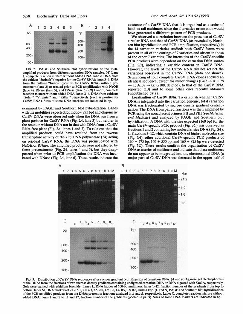
FIG. 2. PAGE and Southern blot hybridizations of the PCR-amplified products from different carnation DNA samples. (A) Lane1, complete reaction mixture without added DNA; lane 2, DNA fromthe cultivar "Sarinah" (negative for the CarSV RNA); lanes 3-6, DNAfrom the cultivar "Indios" (positive for CarSV RNA) without pre-treatment (lane 3) or treated prior to PCR amplification with NaOH(lane 4), RNase (lane 5), and DNase (lane 6). (B) Lane 1, completereaction mixture without added DNA; lanes 2-4, DNA from cultivars"Solar," "Virginie," and "Killer," respectively (each is positive forCarSV RNA). Sizes of some DNA markers are indicated in bp.
examined by PAGE and Southem blot hybridization. Bandswith the mobilities expected for mono- (275 bp) and oligomericCarSV DNAs were observed only when the DNA was from aplant positive for CarSV RNA (Fig. 2A, lane 3) but neither inthe reaction without DNA nor in that with DNA from a CarSVRNA-free plant (Fig. 14, lanes 1 and 2). To rule out that theamplified products could have resulted from the reversetranscriptase activity of the Taq DNA polymerase (24) actingon residual CarSV RNA, the DNA was preincubated withNaOH or RNase. The amplified products were not affected bythese pretreatments (Fig. 14, lanes 4 and 5), but they disap-peared when prior to PCR amplification the DNA was incu-bated with DNase (Fig. 14, lane 6). These results indicate the
existence of a CarSV DNA that it is organized as a series ofhead-to-tail multimers, since the alternative orientation wouldhave generated a different pattern of PCR products.We observed a correlation between the presence of CarSV
circular RNA and that of CarSV DNA (as revealed by North-ern blot hybridization and PCR amplification, respectively) inthe 14 carnation varieties studied: both CarSV forms werepresent in all of the cuttings of 7 varieties and absent in thoseof the other 7 varieties. The intensities of the CarSV-specificPCR products were dependent on the carnation DNA source(Fig. 2B), indicating a variable content in CarSV DNA.However, the levels of the CarSV RNA did not reflect thevariations observed in the CarSV DNA (data not shown).Sequencing of four complete CarSV DNA clones showed anidentical sequence, except for minor changes (G67 -- A; C78
> T; A137 -* G; G108, deleted), to that of the CarSV RNAreported (10) and to some other ones recently obtained(unpublished data).
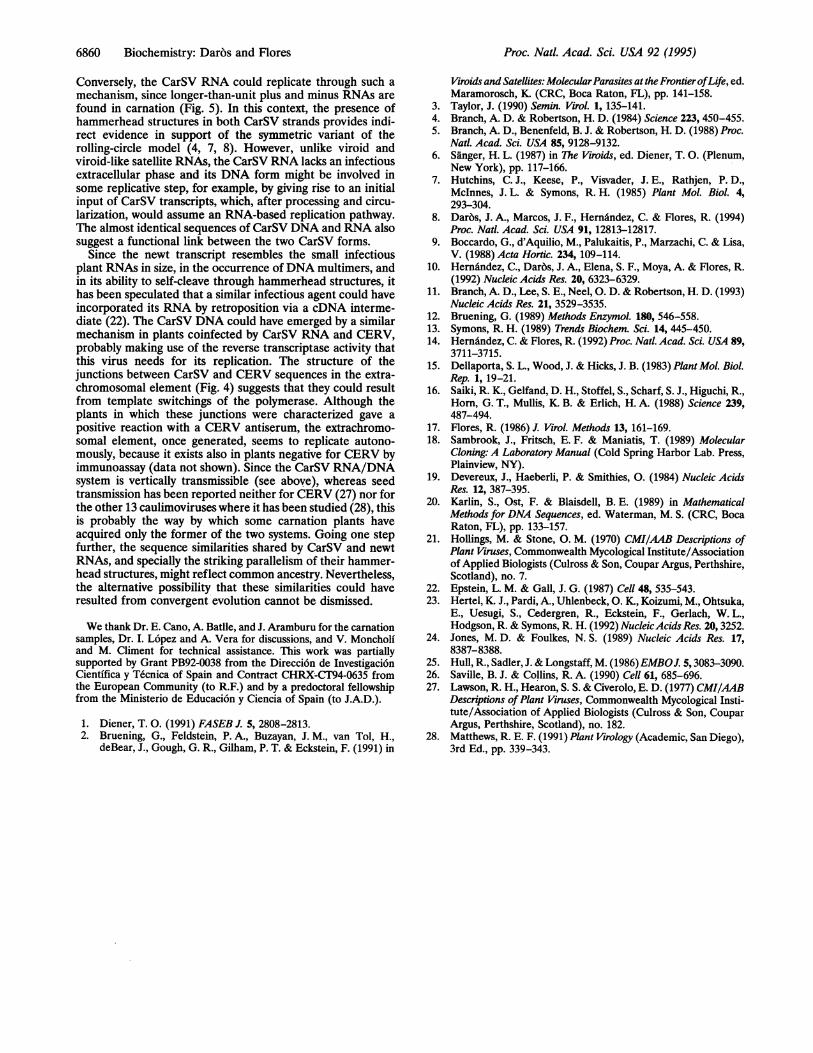
Localization of CarSV DNA. To establish whether CarSVDNA is integrated into the carnation genome, total carnationDNA was fractionated by sucrose density gradient centrifu-gation. The DNA from paired fractions was then amplified byPCR using the nonadjacent primers PII and PIII (see Materialsand Methods) and analyzed by PAGE and Southern blothybridization. A DNA with the size expected (160 bp) for themain CarSV-specific PCR product (Fig. 3C) was observed infractions 1 and 2 containing low molecular size DNA (Fig. 3A).In fractions 3-12, which contain DNA of higher molecular size(Fig. 3A), other additional CarSV-specific PCR products of160 + 275 bp, 160 + 550 bp, and 160 + 825 bp were detected(Fig. 3C). These results confirm the organization of CarSVDNA as a series of multimers and indicate that these multimersdo not appear to be integrated into the chromosomal DNA (amajor part of CarSV DNA was detected in the upper half of
AL 1 2 3 4 5 6 7 8 9 10 11 12 M
CC 2 34 56 78 10 12
*:..' '.. ..':.
bp
600-400-
200-
100-.........1 00-~~~~~~~~~~~~~~~~~~~~~~~~~Nn
BL 1 2 3 4 5 6 78 9 1011 12M
kbp- 21.2
- 3.5
-1.6
-0.6
D 1 2 3 4 5 6 7 8 910112
bp - :.... .... .l;,
600 -
400-
100-
FIG. 3. Distribution of CarSV DNA sequences after sucrose gradient centrifugation of carnation DNA. (A and B) Agarose gel electrophoresisof the DNAs from the fractions of two sucrose density gradients containing undigested carnation DNA or DNA digested with Sau3A, respectively.Gels were stained with ethidium bromide. Lanes L, DNA ladder of 100-bp multimers; lanes 1-12, fraction number of the gradients from top tobottom; lanes M, DNA markers of 21.2, 5.1, 5.0, 4.3, 3.5, 2.0, 1.9, 1.6, 1.4, 0.9, 0.8, 0.6, and 0.1 kbp. (C and D) PAGE and Southern blot hybridizationsof the PCR-amplified products from the DNAs present in fractions analyzed inA and B, respectively. Lanes C, complete reaction mixture withoutadded DNA; lanes 1 and 2 to 11 and 12, fraction number of the gradients (pooled in pairs). Sizes of some DNA markers are indicated in bp.
Proc. Natl. Acad. Sci. USA 92 (1995)
i"Mg 'U i: i.

Proc. Natl. Acad. Sci. USA 92 (1995) 6859
the gradient, whereas the bulk of cellular DNA was found inthe bottom half of the gradient and, moreover, CarSV DNAmultimers were fractionated in the gradient according to theirsizes). More likely, CarSV DNA exists in a smaller extra-chromosomal element consisting of a variable number ofCarSV DNA repeats that could also contain additional non-CarSV sequences. The non-Mendelian inheritance of theCarSV system through ovules and pollen (see above) alsosupports an extrachromosomal location for CarSV DNA,although we cannot exclude that a minor fraction of it couldbe integrated into the plant genome. Susceptibility of CarSVDNA to digestion with Sau3A demonstrated that it is double-stranded (Fig. 3D).
Analysis of the Non-CarSV DNA Sequences of the Extra-chromosomal Element. Digestion of total carnation DNA withHae III, which does not cut within the CarSV DNA sequence,displaced CarSV DNA signals to fractions of the sucrosegradient of lower molecular sizes (data not shown), indicatingthe presence of non-CarSV DNA sequences in the extrachro-mosomal element. Sequence analysis of the products obtainedfollowing nested PCR amplification revealed that these non-CarSV sequences are homologous to regions of the DNA ofcarnation etched ring caulimovirus (CERV), a plant pararet-rovirus (25) (Fig. 4A4). Direct PCR amplification of totalcarnation DNA with CarSV- and CERV-specific primers andsequencing of the products confirmed this observation andshowed additional junctions (Fig. 4B). These junctions werecharacterized by 4-6 nt shared by the CarSV and CERVsequences. Interestingly, CarSV sequence starts in two of themat nt 1, which is the position immediately following theself-cleavage site of plus CarSV RNA (10). With regard toCERV sequences flanking the junctions, one of them rich inG residues (Fig. 4A) is located around the presumed gap 2 ofCERV (25), one of the two regions where synthesis of thesecond strand of the DNA is initiated. The identification ofCarSV DNA fused with genuine pararetroviral sequences isconsistent with its extrachromosomal location (since the DNAof pararetroviruses does not become integrated into their hostchromosomes) and, on the other hand, supports the contentionthat the CarSV RNA and DNA constitute a retroviroid-likesystem.
A
A12
.,. .........
dl -
ml-
B12:t. ':. S.. ',f-
FIG. 5. Northern blot hybridization of carnation RNAs separatedby PAGE in a 5% denaturing gel. Autoradiograms obtained usingRNA probes for plus (A) and minus (B) CarSV sequences are shown.Lanes 1 and 2, nucleic acid extracts from the carnation cultivars"Sarinah" and "Indios" (negative and positive for CarSV RNA,respectively). The positions ofmonomeric circular (mc) and linear (ml)and of the apparent dimeric circular (dc) and linear (dl) CarSV RNAsare indicated. Other prominent bands moving faster than the 275-ntmc and ml CarSV forms (A, lane 2) correspond to circular and linearCarSV RNAs that are smaller than unit length as a result of containingdeletions (unpublished data). The hybridization solutions had thesame acid-precipitable cpm of either plus or minus probes and thefilms were exposed for the same time.
Relationships of the CarSV RNA/DNA System with OtherSmall Circular RNAs. The existence of DNA tandem repeatsof the CarSV RNA has no precedent in the small infectiouscircular RNAs either from plants, such as viroid and viroid-likesatellite RNAs (1, 2), or from animal origin, such as the human8 virus RNA (3). The closest correlate to CarSV RNA is thesmall newt transcript (22), although this RNA is linear and hasa hammerhead structure only in the plus strand. Some parallelalso exists with the VS DNA and RNA found in the mito-chondria of an isolate of Neurospora (26). The VS RNA is asingle-stranded circular molecule complementary to onestrand of a low-copy double-stranded circular VS DNA pop-ulation organized as a series of head-to-tail multimers. The VSRNA is able to self-cleave in vitro but lacks a hammerheadstructure and is unlikely to replicate by a rolling-circle mech-anism, because no minus VS RNA could be detected (26).
230 CarSV 256
PIV-CCTGAACCAATTAACATCACTTCAAACCAACATCTCTTTTTAGAAGAGGG13GGG TCMTCTTTGCCTGATGAGCC-PI
4180190 CarSV
FIG. 4. Primary structure of the extrachromosomal element around several junctions (boxed) between CarSV and non-CarSV DNA sequences.
(A) Product formed by CERV nt 3827-3881 and CarSV nt 230-256 resulting from two consecutive PCR amplifications, the second one with primersPI (complementary to nt 257-14 of CarSV RNA) and PIV (see text). Outlined fonts indicate substitutions with respect to CERV DNA reported(25). CERV sequence starts with the dinucleotide CC as expected from the initial digestion with Hae III. (B) Other products from direct PCRamplifications with primers PVI (complementary to nt 83-102 of CarSV RNA) and PVII (identical to nt 3827-3854 of CERV with the threesubstitutions indicated above) or with PVIII (complementary to nt 143-174 of CarSV RNA) and PVII. With the last pair of primers, two fragmentsof similar size but with different junctions were amplified. Heavy lines indicate CarSV and CERV sequences, which in the first case were identicalto CarSV RNA reported (10) and in the second one showed 91-92% similarity with CERV DNA reported (25). CERV sequences inA and B were
identical within the shared interval.
3827
BPVII
CERV
3855
381
PVII_
CERV
1
3855
CarSV
IGCAGAq4180
PVI
3
CERV
I
1855
CarSVKGCAGAi|
82
PVI
142l
PVIII
142
CERV 4086
PVIII
I
I
I
I
..I bim.
Biochemistry: Dar'os and Flores
|TTGAl 07,; IZi11-
6860 Biochemistry: Daros and Flores
Conversely, the CarSV RNA could replicate through such amechanism, since longer-than-unit plus and minus RNAs arefound in carnation (Fig. 5). In this context, the presence ofhammerhead structures in both CarSV strands provides indi-rect evidence in support of the symmetric variant of therolling-circle model (4, 7, 8). However, unlike viroid andviroid-like satellite RNAs, the CarSV RNA lacks an infectiousextracellular phase and its DNA form might be involved insome replicative step, for example, by giving rise to an initialinput of CarSV transcripts, which, after processing and circu-larization, would assume an RNA-based replication pathway.The almost identical sequences of CarSV DNA and RNA alsosuggest a functional link between the two CarSV forms.
Since the newt transcript resembles the small infectiousplant RNAs in size, in the occurrence ofDNA multimers, andin its ability to self-cleave through hammerhead structures, ithas been speculated that a similar infectious agent could haveincorporated its RNA by retroposition via a cDNA interme-diate (22). The CarSV DNA could have emerged by a similarmechanism in plants coinfected by CarSV RNA and CERV,probably making use of the reverse transcriptase activity thatthis virus needs for its replication. The structure of thejunctions between CarSV and CERV sequences in the extra-chromosomal element (Fig. 4) suggests that they could resultfrom template switchings of the polymerase. Although theplants in which these junctions were characterized gave apositive reaction with a CERV antiserum, the extrachromo-somal element, once generated, seems to replicate autono-mously, because it exists also in plants negative for CERV byimmunoassay (data not shown). Since the CarSV RNA/DNAsystem is vertically transmissible (see above), whereas seedtransmission has been reported neither for CERV (27) nor forthe other 13 caulimoviruses where it has been studied (28), thisis probably the way by which some carnation plants haveacquired only the former of the two systems. Going one stepfurther, the sequence similarities shared by CarSV and newtRNAs, and specially the striking parallelism of their hammer-head structures, might reflect common ancestry. Nevertheless,the alternative possibility that these similarities could haveresulted from convergent evolution cannot be dismissed.
We thank Dr. E. Cano, A. BatHle, and J. Aramburu for the carnationsamples, Dr. I. Lopez and A. Vera for discussions, and V. Moncholiand M. Climent for technical assistance. This work was partiallysupported by Grant PB92-0038 from the Direccion de Investigaci6nCientifica y Tecnica of Spain and Contract CHRX-CT94-0635 fromthe European Community (to R.F.) and by a predoctoral fellowshipfrom the Ministerio de Educaci6n y Ciencia of Spain (to J.A.D.).
1. Diener, T. 0. (1991) FASEB J. 5, 2808-2813.2. Bruening, G., Feldstein, P. A., Buzayan, J. M., van Tol, H.,
deBear, J., Gough, G. R., Gilham, P. T. & Eckstein, F. (1991) in
Viroids and Satellites: MolecularParasites at the FrontierofLife, ed.Maramorosch, K (CRC, Boca Raton, FL), pp. 141-158.
3. Taylor, J. (1990) Semin. Virol. 1, 135-141.4. Branch, A. D. & Robertson, H. D. (1984) Science 223, 450-455.5. Branch, A. D., Benenfeld, B. J. & Robertson, H. D. (1988) Proc.
Natl. Acad. Sci. USA 85, 9128-9132.6. Siinger, H. L. (1987) in The Viroids, ed. Diener, T. 0. (Plenum,
New York), pp. 117-166.7. Hutchins, C. J., Keese, P., Visvader, J. E., Rathjen, P. D.,
McInnes, J. L. & Symons, R. H. (1985) Plant Mol. Biol. 4,293-304.
8. Daros, J. A., Marcos, J. F., Hernandez, C. & Flores, R. (1994)Proc. Natl. Acad. Sci. USA 91, 12813-12817.
9. Boccardo, G., d'Aquilio, M., Palukaitis, P., Marzachi, C. & Lisa,V. (1988) Acta Hortic. 234, 109-114.
10. Hernandez, C., Darbs, J. A., Elena, S. F., Moya, A. & Flores, R.(1992) Nucleic Acids Res. 20, 6323-6329.
11. Branch, A. D., Lee, S. E., Neel, 0. D. & Robertson, H. D. (1993)Nucleic Acids Res. 21, 3529-3535.
12. Bruening, G. (1989) Methods Enzymol. 180, 546-558.13. Symons, R. H. (1989) Trends Biochem. Sci. 14, 445-450.14. Hema'ndez, C. & Flores, R. (1992) Proc. Natl. Acad. Sci. USA 89,
3711-3715.15. Dellaporta, S. L., Wood, J. & Hicks, J. B. (1983) Plant Mol. Biol.
Rep. 1, 19-21.16. Saiki, R. K., Gelfand, D. H., Stoffel, S., Scharf, S. J., Higuchi, R.,
Horn, G. T., Mullis, K. B. & Erlich, H. A. (1988) Science 239,487-494.
17. Flores, R. (1986) J. Virol. Methods 13, 161-169.18. Sambrook, J., Fritsch, E. F. & Maniatis, T. (1989) Molecular
Cloning: A Laboratory Manual (Cold Spring Harbor Lab. Press,Plainview, NY).
19. Devereux, J., Haeberli, P. & Smithies, 0. (1984) Nucleic AcidsRes. 12, 387-395.
20. Karlin, S., Ost, F. & Blaisdell, B. E. (1989) in MathematicalMethods for DNA Sequences, ed. Waterman, M. S. (CRC, BocaRaton, FL), pp. 133-157.
21. Hollings, M. & Stone, 0. M. (1970) CMI/AAB Descriptions ofPlant Viruses, Commonwealth Mycological Institute/Associationof Applied Biologists (Culross & Son, Coupar Argus, Perthshire,Scotland), no. 7.
22. Epstein, L. M. & Gall, J. G. (1987) Cell 48, 535-543.23. Hertel, K. J., Pardi, A., Uhlenbeck, 0. K., Koizumi, M., Ohtsuka,
E., Uesugi, S., Cedergren, R., Eckstein, F., Gerlach, W. L.,Hodgson, R. & Symons, R. H. (1992) NucleicAcids Res. 20, 3252.
24. Jones, M. D. & Foulkes, N. S. (1989) Nucleic Acids Res. 17,8387-8388.
25. Hull, R., Sadler, J. & Longstaff, M. (1986)EMBOJ. 5,3083-3090.26. Saville, B. J. & Collins, R. A. (1990) Cell 61, 685-696.27. Lawson, R. H., Hearon, S. S. & Civerolo, E. D. (1977) CMI/AAB
Descriptions ofPlant Viruses, Commonwealth Mycological Insti-tute/Association of Applied Biologists (Culross & Son, CouparArgus, Perthshire, Scotland), no. 182.
28. Matthews, R. E. F. (1991) Plant Virology (Academic, San Diego),3rd Ed., pp. 339-343.
Proc. Natl. Acad. Sci. USA 92 (1995)