
Identification of a gain-of-function mutation of the ...Identification of a gain-of-function...
6
Identification of a gain-of-function mutation of the prolactin receptor in women with benign breast tumors Roman L. Bogorad* † , Carine Courtillot* †‡ , Chidi Mestayer* † , Sophie Bernichtein* † , Lilya Harutyunyan ‡ , Jean-Baptiste Jomain* † , Anne Bachelot ‡ , Fre ´de ´ rique Kuttenn* †‡ , Paul A. Kelly* † , Vincent Goffin* †§ , Philippe Touraine* †‡§¶ , and the Benign Breast Diseases Study Group ‡ *Institut National de la Sante ´ et de la Recherche Me ´ dicale (INSERM) U845, Equipe ‘‘Prl, GH et tumeurs,’’ Centre de Recherche Croissance et Signalisation, F-75015 Paris, France; † Faculte ´ de Me ´ decine, site Necker, Universite ´ Paris Descartes, F-75015 Paris, France; and ‡ De ´ partement d’Endocrinologie et Me ´ decine de la Reproduction, Centre des Maladies Rares Gyne ´ cologiques, Assistance Publique–Ho ˆ pitaux de Paris, Groupe Hospitalier Pitie ´ Salpe ˆ trie ` re, F-75651 Paris Cedex 13, France Edited by John D. Baxter, University of California, San Francisco, CA, and approved July 17, 2008 (received for review January 23, 2008) There is currently no known genetic disease linked to prolactin (Prl) or its receptor (PrlR) in humans. Given the essential role of this hormonal system in breast physiology, we reasoned that genetic anomalies of Prl/PrlR genes may be related to the occurrence of breast diseases with high proliferative potential. Multiple fibro- adenomas (MFA) are benign breast tumors which appear most frequently in young women, including at puberty, when Prl has well-recognized proliferative actions on the breast. In a prospec- tive study involving 74 MFA patients and 170 control subjects, we identified four patients harboring a heterozygous single nucleo- tide polymorphism in exon 6 of the PrlR gene, encoding Ile146 3Leu substitution in its extracellular domain. This sole substitution was sufficient to confer constitutive activity to the receptor variant (PrlRI146L ), as assessed in three reconstituted cell models (Ba/F3, HEK293 and MCF-7 cells) by Prl-independent (i) PrlR tyrosine phosphorylation, (ii) activation of signal transducer and activator of transcription 5 (STAT5) signaling, (iii) transcriptional activity toward a Prl-responsive reporter gene, and (iv) cell proliferation and protection from cell death. Constitutive activity of PrlRI146L in the breast sample from a patient was supported by increased STAT5 signaling. This is a unique description of a functional mutation of the PrlR associated with a human disease. Hallmarks of constitutive activity were all reversed by a specific PrlR antag- onist, which opens potential therapeutic approaches for MFA, or any other disease that could be associated with this mutation in future. antagonist breast diseases human mutation constitutive activity cytokine receptor T he role of prolactin (Prl) in breast physiology has been recognized for decades. In synergy with various hormones and growth factors, Prl plays a critical role in many steps of breast development (1). Given its potent activity on breast cell prolif- eration and differentiation, it has been long assumed that mutations affecting the properties of Prl or of its receptor (PrlR) should have clinical impact on the breast. Although genetically modified animal models fully support this assumption (2), an unequivocal answer to this question is lacking in humans. This is due in part to the fact that the rare studies that were performed to date to identify coding mutations of the PrlR gene in breast cancer patients either failed to find any (3), or reported a single nucleotide polymorphism (SNP) that remained uncharacterized at the functional level and involved too small cohorts to achieve significance (4). To date, only association studies between noncoding variations in PrlR gene and breast cancer have been reported (5, 6). More generally, there is no known loss- or gain-of-function genetic pathology yet reported for Prl or its receptor. Besides breast cancer, several benign breast diseases (BBD) affecting the human breast remain poorly understood (7, 8). These diseases are marked by lobuloalveolar growth and/or differentiation disorders, including abnormally high prolifera- tion of the epithelium as observed in fibroadenomas (FA) (8). Multiple FA (MFA) is defined by more than 3 FA in one breast (9) (Fig. 1), which are not histologically different from isolated FAs. Receptors for estrogen, progesterone, and various growth factors have been suggested as potential candidates involved in tumor appearance/growth (8). The PrlR is another candidate, as its expression is maintained, or even increased in various benign breast lesions (10, 11). According to the poorly understood etiology of all BBDs, current treatments are mostly empirical. One of the most currently used involves progestins, although the question of whether they are beneficial or instead deleterious for the breast is still a matter of debate (12). Some have tried antiestrogen therapy with tamoxifen in BBDs, but no evaluation of such practice has ever been reported. Concerning the use of inhibitors of Prl secretion (dopamine agonists), no long-term treatment study has been reported for patients with BBDs, especially FAs or MFAs (13). In such a context, the development of new therapeutic approaches capable of reducing the prolif- eration observed in benign breast tumors should be relevant. Recently, genetic predisposition has been proposed as a causal factor in benign breast disorders (7), which could apply to any regulator of breast morphogenesis, including Prl. We thus ini- tiated a prospective clinical study aimed at identifying any coding alterations of Prl and PrlR genes in the largest MFA cohort ever Author contributions: F.K., P.A.K., V.G., and P.T. designed research; R.L.B., C.C., C.M., S.B., L.H., A.B., V.G., P.T., and the Benign Breast Diseases Study Group performed research; R.L.B., S.B., J.-B.J., and V.G. contributed new reagents/analytic tools; R.L.B., C.C., S.B., A.B., P.A.K., V.G., and P.T. analyzed data; and R.L.B., C.C., P.A.K., V.G., and P.T. wrote the paper. The authors declare no conflict of interest. This article is a PNAS Direct Submission. Data deposition: The data reported in this paper have been deposited in the Single Nucleotide Polymorphism database (dbSNP), www.ncbi.nlm.nih.gov/sites/entrez?dbsnp (SNP ID no. ss102734470). Freely available online through the PNAS open access option. § V.G. and P.T. contributed equally to this work. ¶ To whom correspondence should be addressed. E-mail: [email protected]. Benign Breast Diseases Study Group: A. Bachelot, B. Belaroussi, J. Bensimhon, J. Berdah, M. J. Blin, A. Boudinet, B. Brethon, C. Bricaire, J. Caby, G. Caillaud, J. C. Carel, N. Chabbert- Buffet, H. Charitanski, C. Chretien, K. Clough, C. Courtillot, G. Delattre, I. Denys, K. Desthieux-Ngo, M. Detoeuf, C. Dhainault, C. Duflos, O. Fiori, C. Genestie, G. Gibaud, A. Gompel, C. Gracia, A. Grimard, C. Hofman, H. Hofman, F. Kuttenn, F. Laki, C. Lanty, J. P. Lefranc, M. A. Le Frere-Belda, D. Leger, F. Martinez, A. May, L. Meng, C. Nos, D. Pelletier, A. Perrin, G. Plu-Bureau, B. Raccah-Tebbeca, J. C. Saiovici, R. Salmon, M. Sibout, B. Sigal-Zafrani, J. C. Thalabard, E. Thibaud, A. Thoury, P. Touraine, K. B. Triana-Rabi, S. Uzan, J. Viriot, and S. Yacoub. This article contains supporting information online at www.pnas.org/cgi/content/full/ 0800685105/DCSupplemental. © 2008 by The National Academy of Sciences of the USA www.pnas.orgcgidoi10.1073pnas.0800685105 PNAS September 23, 2008 vol. 105 no. 38 14533–14538 MEDICAL SCIENCES Downloaded by guest on March 11, 2020
Transcript of Identification of a gain-of-function mutation of the ...Identification of a gain-of-function...

Identification of a gain-of-function mutationof the prolactin receptor in womenwith benign breast tumorsRoman L. Bogorad*†, Carine Courtillot*†‡, Chidi Mestayer*†, Sophie Bernichtein*†, Lilya Harutyunyan‡,Jean-Baptiste Jomain*†, Anne Bachelot‡, Frederique Kuttenn*†‡, Paul A. Kelly*†, Vincent Goffin*†§,Philippe Touraine*†‡§¶, and the Benign Breast Diseases Study Group‡�
*Institut National de la Sante et de la Recherche Medicale (INSERM) U845, Equipe ‘‘Prl, GH et tumeurs,’’ Centre de Recherche Croissance et Signalisation,F-75015 Paris, France; †Faculte de Medecine, site Necker, Universite Paris Descartes, F-75015 Paris, France; and ‡Departement d’Endocrinologie etMedecine de la Reproduction, Centre des Maladies Rares Gynecologiques, Assistance Publique–Hopitaux de Paris, Groupe Hospitalier PitieSalpetriere, F-75651 Paris Cedex 13, France
Edited by John D. Baxter, University of California, San Francisco, CA, and approved July 17, 2008 (received for review January 23, 2008)
There is currently no known genetic disease linked to prolactin (Prl)or its receptor (PrlR) in humans. Given the essential role of thishormonal system in breast physiology, we reasoned that geneticanomalies of Prl/PrlR genes may be related to the occurrence ofbreast diseases with high proliferative potential. Multiple fibro-adenomas (MFA) are benign breast tumors which appear mostfrequently in young women, including at puberty, when Prl haswell-recognized proliferative actions on the breast. In a prospec-tive study involving 74 MFA patients and 170 control subjects, weidentified four patients harboring a heterozygous single nucleo-tide polymorphism in exon 6 of the PrlR gene, encoding Ile1463Leusubstitution in its extracellular domain. This sole substitution wassufficient to confer constitutive activity to the receptor variant(PrlRI146L), as assessed in three reconstituted cell models (Ba/F3,HEK293 and MCF-7 cells) by Prl-independent (i) PrlR tyrosinephosphorylation, (ii) activation of signal transducer and activatorof transcription 5 (STAT5) signaling, (iii) transcriptional activitytoward a Prl-responsive reporter gene, and (iv) cell proliferationand protection from cell death. Constitutive activity of PrlRI146L inthe breast sample from a patient was supported by increasedSTAT5 signaling. This is a unique description of a functionalmutation of the PrlR associated with a human disease. Hallmarksof constitutive activity were all reversed by a specific PrlR antag-onist, which opens potential therapeutic approaches for MFA, orany other disease that could be associated with this mutation infuture.
antagonist � breast diseases � human mutation �constitutive activity � cytokine receptor
The role of prolactin (Prl) in breast physiology has beenrecognized for decades. In synergy with various hormones
and growth factors, Prl plays a critical role in many steps of breastdevelopment (1). Given its potent activity on breast cell prolif-eration and differentiation, it has been long assumed thatmutations affecting the properties of Prl or of its receptor (PrlR)should have clinical impact on the breast. Although geneticallymodified animal models fully support this assumption (2), anunequivocal answer to this question is lacking in humans. This isdue in part to the fact that the rare studies that were performedto date to identify coding mutations of the PrlR gene in breastcancer patients either failed to find any (3), or reported a singlenucleotide polymorphism (SNP) that remained uncharacterizedat the functional level and involved too small cohorts to achievesignificance (4). To date, only association studies betweennoncoding variations in PrlR gene and breast cancer have beenreported (5, 6). More generally, there is no known loss- orgain-of-function genetic pathology yet reported for Prl or itsreceptor. Besides breast cancer, several benign breast diseases(BBD) affecting the human breast remain poorly understood (7,
8). These diseases are marked by lobuloalveolar growth and/ordifferentiation disorders, including abnormally high prolifera-tion of the epithelium as observed in fibroadenomas (FA) (8).Multiple FA (MFA) is defined by more than 3 FA in one breast(9) (Fig. 1), which are not histologically different from isolatedFAs. Receptors for estrogen, progesterone, and various growthfactors have been suggested as potential candidates involved intumor appearance/growth (8). The PrlR is another candidate, asits expression is maintained, or even increased in various benignbreast lesions (10, 11). According to the poorly understoodetiology of all BBDs, current treatments are mostly empirical.One of the most currently used involves progestins, although thequestion of whether they are beneficial or instead deleterious forthe breast is still a matter of debate (12). Some have triedantiestrogen therapy with tamoxifen in BBDs, but no evaluationof such practice has ever been reported. Concerning the use ofinhibitors of Prl secretion (dopamine agonists), no long-termtreatment study has been reported for patients with BBDs,especially FAs or MFAs (13). In such a context, the developmentof new therapeutic approaches capable of reducing the prolif-eration observed in benign breast tumors should be relevant.
Recently, genetic predisposition has been proposed as a causalfactor in benign breast disorders (7), which could apply to anyregulator of breast morphogenesis, including Prl. We thus ini-tiated a prospective clinical study aimed at identifying any codingalterations of Prl and PrlR genes in the largest MFA cohort ever
Author contributions: F.K., P.A.K., V.G., and P.T. designed research; R.L.B., C.C., C.M., S.B.,L.H., A.B., V.G., P.T., and the Benign Breast Diseases Study Group performed research; R.L.B.,S.B., J.-B.J., and V.G. contributed new reagents/analytic tools; R.L.B., C.C., S.B., A.B., P.A.K.,V.G., and P.T. analyzed data; and R.L.B., C.C., P.A.K., V.G., and P.T. wrote the paper.
The authors declare no conflict of interest.
This article is a PNAS Direct Submission.
Data deposition: The data reported in this paper have been deposited in the SingleNucleotide Polymorphism database (dbSNP), www.ncbi.nlm.nih.gov/sites/entrez?db�snp(SNP ID no. ss102734470).
Freely available online through the PNAS open access option.
§V.G. and P.T. contributed equally to this work.
¶To whom correspondence should be addressed. E-mail: [email protected].
�Benign Breast Diseases Study Group: A. Bachelot, B. Belaroussi, J. Bensimhon, J. Berdah,M. J. Blin, A. Boudinet, B. Brethon, C. Bricaire, J. Caby, G. Caillaud, J. C. Carel, N. Chabbert-Buffet, H. Charitanski, C. Chretien, K. Clough, C. Courtillot, G. Delattre, I. Denys, K.Desthieux-Ngo, M. Detoeuf, C. Dhainault, C. Duflos, O. Fiori, C. Genestie, G. Gibaud, A.Gompel, C. Gracia, A. Grimard, C. Hofman, H. Hofman, F. Kuttenn, F. Laki, C. Lanty, J. P.Lefranc, M. A. Le Frere-Belda, D. Leger, F. Martinez, A. May, L. Meng, C. Nos, D. Pelletier,A. Perrin, G. Plu-Bureau, B. Raccah-Tebbeca, J. C. Saiovici, R. Salmon, M. Sibout, B.Sigal-Zafrani, J. C. Thalabard, E. Thibaud, A. Thoury, P. Touraine, K. B. Triana-Rabi, S. Uzan,J. Viriot, and S. Yacoub.
This article contains supporting information online at www.pnas.org/cgi/content/full/0800685105/DCSupplemental.
© 2008 by The National Academy of Sciences of the USA
www.pnas.org�cgi�doi�10.1073�pnas.0800685105 PNAS � September 23, 2008 � vol. 105 � no. 38 � 14533–14538
MED
ICA
LSC
IEN
CES
Dow
nloa
ded
by g
uest
on
Mar
ch 1
1, 2
020

reported. In four patients, we identified a heterozygous missensemutation in the PrlR gene, leading to a constitutively activereceptor.
ResultsPatients and Controls. Seventy-four Caucasian women with MFAwere consecutively recruited by the BBDs study group, from 9different centers, and referred to our outpatient clinic. Inclusionwas possible based on the existence of at least 3 FAs in one breastin patients receiving no treatment influencing gonadal axis forat least 1 month. All underwent basic clinical investigations. Forthose who underwent surgery, FA and adjacent tissue wereobtained whenever possible.
A cohort of 96 control Caucasian subjects was constitutedbased on stringent inclusion criteria including no history ofbenign or malignant breast disease, no pituitary disorder, normalPrl levels. To minimize the risk of including young subjects whocould later develop an MFA, we fixed the cut-off age at 35. Arandom population of 74 unrelated control women (no inclusioncriteria) was also analyzed.
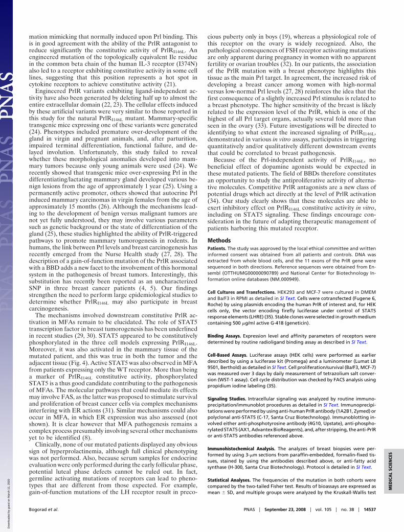
PrlR Genotype Associated with MFAs. No missense single nucleotidepolymorphism (SNP) was identified in the Prl gene of any patient.With respect to the PrlR, we found in both MFA patients andcontrol subjects (with no difference) the sole coding SNP reportedin the NCBI database (rs16871473, C/T in exon 5, encoding I76Vsubstitution at protein level), as well as many known SNPs inintronic regions bordering exons [supporting information (SI) Text].In four unrelated patients (5.6%), we found a coding SNP that wasnot reported in the NCBI database (deposit procedure in progress).It involves A-to-C substitution in exon 6 (Fig. 2A and Fig. S1), andsubstitutes Leu for Ile146 in the second cytokine receptor homologymotif of the PrlR ligand-binding domain (Fig. 2B) (14). This SNPwas found in none of 96 control women ascertained to be free ofany history of Prl disorder and/or breast diseases (P � 0.034), andit was also absent in a random population of 74 women (totalcontrols � 170).
Establishment of Cell Models to Study I146L SNP. The impact ofI146L substitution on PrlR properties (mutant is referred to asPrlRI146L) was characterized by using transfected cell models andmultiple well-established readouts for structure-function studiesof the PrlR (Ba/F3 mouse lymphoid cells, HEK293 humanembryonic kidney fibroblasts). To avoid any bias, stable clones(HEK) or populations (Ba/F3) to be compared were generatedand selected based on similar expression levels of WT andmutated PrlRs as determined by semiquantitative PrlR immu-
noblotting (Fig. S2) and/or radioligand receptor assay. HEK-PrlRWT and HEK-PrlRI146L clones expressed approximately5,000 surface receptors/cell, whereas Ba/F-PrlRWT and Ba/F-PrlRI146L populations expressed many fewer surface receptors(approximately 500 PrlR/cell as determined by ligand bindingassay) which were not detectable by immunoblot. The mutatedreceptor exhibited unchanged affinity for human Prl comparedto PrlRWT (Fig. S3 and text). To generate a model in which bothWT and mutated PrlRs are co-expressed in a mammary context(as in MFAs of heterozygous patients), MCF-7 human breastcancer cells were stably transfected by using expression vectorencoding PrlRI146L or PrlRWT to get comparable clones regard-ing the level of PrlR expression (Fig. S4). As MCF-7 expressendogenous PrlRWT, stable clones were noted MCF7-PrlRWT,WTversus MCF7-PrlRWT,I146L.
Mutation I146L Encodes a Constitutively Active PrlR. In stable HEK-PrlRWT and MCF7-PrlRWT,WT clones, Prl stimulation inducedtyrosine phosphorylation of the PrlR (Fig. 3 A and B), which isknown to be mediated by the receptor-associated tyrosinekinase, JAK2 (14). Otherwise, strong receptor phosphorylationwas observed in non Prl-stimulated HEK-PrlRI146L cells, but notin HEK-PrlRWT cells (Fig. 3A). This was also observed inMCF7-PrlRWT,I146L (Fig. 3B), highlighting that receptor phos-phorylation persisted in the heterozygous context. Signalingstudies were performed in serum-free media but we failed todetect production of endogenous Prl in these cell lines (not
Fig. 1. MRI of a MFA patient showing several fibroadenomas (arrows) mainlylocated in the left breast.
I146L
D1
D2
Hormone
Human PRLR extrac ellular
domai n
B
5p13-p1 2
w ild ty pe (homoz y gous)
mutated (heteroz y gous)
1 2 3 4 5 6 7 8 9 10 11
Signa l peptide
Extrac ell u lar domai n
Intercellu lar domai n TM
.T G T A T G AA TT C G A T. A C
.T G T A T G AAA TT C G A T.
UTR A
Fig. 2. Exon 6 mutation in human PrlR gene leads to I146L missense substi-tution. (A) Schematic representation of the 11 exons of the human PrlR geneand the corresponding protein domains. Examples of exon 6 sense sequencesobtained from one homozygous patient (2 WT alleles) and one heterozygouspatient harboring both WT and A-to-C mutated alleles are shown. (B) The 3Dstructure of the human PrlR extracellular domain (blue) complexed to growthhormone (orange) (PDB ID code 1BP3) is used to locate the I146L substitutionon the folded receptor. D1 and D2 indicate cytokine receptor homologydomains 1 and 2. UTR, untranslated region; TM, transmembrane.
14534 � www.pnas.org�cgi�doi�10.1073�pnas.0800685105 Bogorad et al.
Dow
nloa
ded
by g
uest
on
Mar
ch 1
1, 2
020
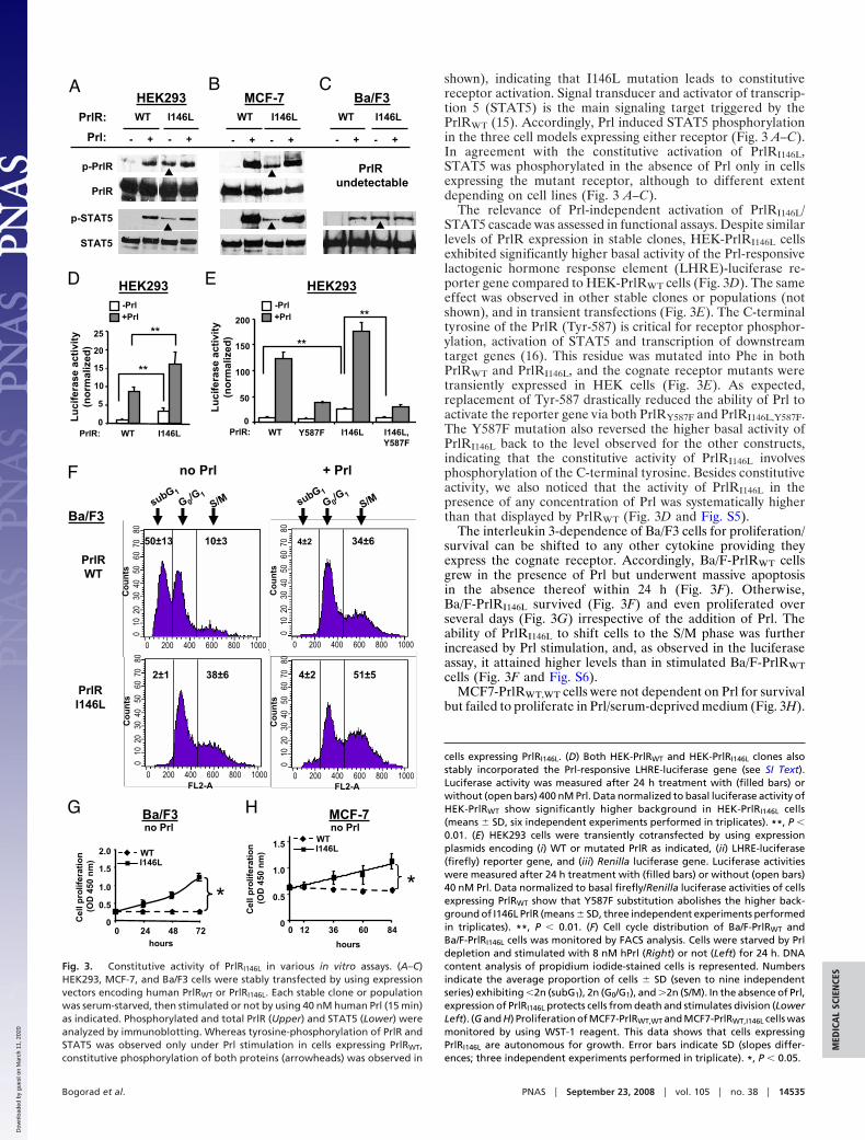
shown), indicating that I146L mutation leads to constitutivereceptor activation. Signal transducer and activator of transcrip-tion 5 (STAT5) is the main signaling target triggered by thePrlRWT (15). Accordingly, Prl induced STAT5 phosphorylationin the three cell models expressing either receptor (Fig. 3 A–C).In agreement with the constitutive activation of PrlRI146L,STAT5 was phosphorylated in the absence of Prl only in cellsexpressing the mutant receptor, although to different extentdepending on cell lines (Fig. 3 A–C).
The relevance of Prl-independent activation of PrlRI146L/STAT5 cascade was assessed in functional assays. Despite similarlevels of PrlR expression in stable clones, HEK-PrlRI146L cellsexhibited significantly higher basal activity of the Prl-responsivelactogenic hormone response element (LHRE)-luciferase re-porter gene compared to HEK-PrlRWT cells (Fig. 3D). The sameeffect was observed in other stable clones or populations (notshown), and in transient transfections (Fig. 3E). The C-terminaltyrosine of the PrlR (Tyr-587) is critical for receptor phosphor-ylation, activation of STAT5 and transcription of downstreamtarget genes (16). This residue was mutated into Phe in bothPrlRWT and PrlRI146L, and the cognate receptor mutants weretransiently expressed in HEK cells (Fig. 3E). As expected,replacement of Tyr-587 drastically reduced the ability of Prl toactivate the reporter gene via both PrlRY587F and PrlRI146L,Y587F.The Y587F mutation also reversed the higher basal activity ofPrlRI146L back to the level observed for the other constructs,indicating that the constitutive activity of PrlRI146L involvesphosphorylation of the C-terminal tyrosine. Besides constitutiveactivity, we also noticed that the activity of PrlRI146L in thepresence of any concentration of Prl was systematically higherthan that displayed by PrlRWT (Fig. 3D and Fig. S5).
The interleukin 3-dependence of Ba/F3 cells for proliferation/survival can be shifted to any other cytokine providing theyexpress the cognate receptor. Accordingly, Ba/F-PrlRWT cellsgrew in the presence of Prl but underwent massive apoptosisin the absence thereof within 24 h (Fig. 3F). Otherwise,Ba/F-PrlRI146L survived (Fig. 3F) and even proliferated overseveral days (Fig. 3G) irrespective of the addition of Prl. Theability of PrlRI146L to shift cells to the S/M phase was furtherincreased by Prl stimulation, and, as observed in the luciferaseassay, it attained higher levels than in stimulated Ba/F-PrlRWTcells (Fig. 3F and Fig. S6).
MCF7-PrlRWT,WT cells were not dependent on Prl for survivalbut failed to proliferate in Prl/serum-deprived medium (Fig. 3H).
cells expressing PrlRI146L. (D) Both HEK-PrlRWT and HEK-PrlRI146L clones alsostably incorporated the Prl-responsive LHRE-luciferase gene (see SI Text).Luciferase activity was measured after 24 h treatment with (filled bars) orwithout (open bars) 400 nM Prl. Data normalized to basal luciferase activity ofHEK-PrlRWT show significantly higher background in HEK-PrlRI146L cells(means � SD, six independent experiments performed in triplicates). **, P �0.01. (E) HEK293 cells were transiently cotransfected by using expressionplasmids encoding (i) WT or mutated PrlR as indicated, (ii) LHRE-luciferase(firefly) reporter gene, and (iii) Renilla luciferase gene. Luciferase activitieswere measured after 24 h treatment with (filled bars) or without (open bars)40 nM Prl. Data normalized to basal firefly/Renilla luciferase activities of cellsexpressing PrlRWT show that Y587F substitution abolishes the higher back-ground of I146L PrlR (means � SD, three independent experiments performedin triplicates). **, P � 0.01. (F) Cell cycle distribution of Ba/F-PrlRWT andBa/F-PrlRI146L cells was monitored by FACS analysis. Cells were starved by Prldepletion and stimulated with 8 nM hPrl (Right) or not (Left) for 24 h. DNAcontent analysis of propidium iodide-stained cells is represented. Numbersindicate the average proportion of cells � SD (seven to nine independentseries) exhibiting �2n (subG1), 2n (G0/G1), and �2n (S/M). In the absence of Prl,expression of PrlRI146L protects cells from death and stimulates division (LowerLeft). (G and H) Proliferation of MCF7-PrlRWT,WT and MCF7-PrlRWT,I146L cells wasmonitored by using WST-1 reagent. This data shows that cells expressingPrlRI146L are autonomous for growth. Error bars indicate SD (slopes differ-ences; three independent experiments performed in triplicate). *, P � 0.05.
A
p-PrlR
PrlR
p-STAT5
STAT5
WT I146L
Prl: - + - +
HEK293
PrlRundetectable
ED
subG 1
+ Prl
Co
un
ts
0 200 400 600 800 1000FL2-A
Co
un
ts
0 200 400 600 800 1000FL2-A
51±54±22±1 38±6
0 1
0 2
0 3
0 4
0 5
0 6
0 7
0 8
0C
ou
nts
0 200 400 600 800 1000
0 1
0 2
0 3
0 4
0 5
0 6
0 7
0 8
0C
ou
nts
0 200 400 600 800 1000
G 0/G 1
S/M
4±2 34±650±13 10±3
subG 1
G 0/G 1
S/M
no Prl
PrlRI146L
PrlRWT
no Prl
F
0
50
100
150
200
WT I146LY587F I146L,Y587F
**
H
- + - +
MCF-7
- + - +
Ba/F3
PrlR: WT I146L WT I146L
Lu
cife
rase
acti
vity
(no
rmal
ized
)
G
HEK293 HEK293
Ba/F3
Ba/F3
hours
0
0.5
1.0
1.5
2.0
0 24 48 72
*
Cel
l pro
lifer
atio
n
(OD
450
nm
)
hours
0
0.5
1.0
1.5
0 12 36 60 84
*
Cel
l pro
lifer
atio
n
(OD
450
nm
)
MCF-7no Prl
-Prl+Prl
-Prl+Prl
**
PrlR: PrlR:
WTI146LWT
I146L
Lu
cife
rase
acti
vity
(no
rmal
ized
)
0
5
10
15
20
25
WT I146L
**
**
0 1
0 2
0 3
0 4
0 5
0 6
0 7
0 8
0
0 1
0 2
0 3
0 4
0 5
0 6
0 7
0 8
0
CB
Fig. 3. Constitutive activity of PrlRI146L in various in vitro assays. (A–C)HEK293, MCF-7, and Ba/F3 cells were stably transfected by using expressionvectors encoding human PrlRWT or PrlRI146L. Each stable clone or populationwas serum-starved, then stimulated or not by using 40 nM human Prl (15 min)as indicated. Phosphorylated and total PrlR (Upper) and STAT5 (Lower) wereanalyzed by immunoblotting. Whereas tyrosine-phosphorylation of PrlR andSTAT5 was observed only under Prl stimulation in cells expressing PrlRWT,constitutive phosphorylation of both proteins (arrowheads) was observed in
Bogorad et al. PNAS � September 23, 2008 � vol. 105 � no. 38 � 14535
MED
ICA
LSC
IEN
CES
Dow
nloa
ded
by g
uest
on
Mar
ch 1
1, 2
020

In the same conditions, MCF7-PrlRWT,I146L cells proliferated toa submaximal level, as Prl could further enhance cell division(not shown). This demonstrates that the growth-promotingeffect of PrlRI146L, as observed for receptor and STAT5 phos-phorylation, occurs irrespectively of co-expression of the WTreceptor, which is particularly important regarding the fact thatthe mutation is heterozygous in our MFA patients.
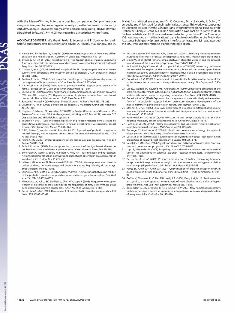
Increased Stat5 Signaling in Breast Tissue from Mutated Patients.Biopsies of MFA and adjacent tissue from one of these fourheterozygous patients (PrlRWT,I146L) was available for histolog-ical studies and was compared to samples from two homozygouspatients (PrlRWT,WT). Activation of the PrlR-STAT5 pathwaywas investigated by immunohistochemical analysis of the recep-tor, STAT5, phospho-STAT5, and fatty-acid synthase (FAS), adownstream target gene of STAT5 in mammary cells (17). InMFA samples, irrespective of the PrlR genotype, the nuclearlocation of phospho-STAT5 labeling (arrows on Fig. 4) and theintense FAS labeling supported activation of that pathway intumors. Remarkably, phospho-STAT5 and FAS labeling wasalso observed in adjacent tissue from the mutated patient but notin those from non mutated patients, suggesting increased acti-vation of PrlR-triggered cascades also occurs in healthy tissueexpressing the mutated receptor. PrlR and STAT5 labeling weresimilar in all samples analyzed (not shown), indicating thatactivation of STAT5 cascade was not caused by over-expressionof these proteins.
Inhibition of PrlRI146L Constitutive Activity by PrlR Signaling Inhibitors.We investigated whether strategies known to inhibit Prl-inducedactivation of PrlRWT could down-regulate the constitutive ac-tivity of PrlRI146L. Tyrphostin B42 (AG490) is a pharmacologicalinhibitor of JAK2 activity and Del1–9-G129R-hPrl is a specific,competitive PrlR antagonist (18). Both inhibited constitutivephosphorylation of PrlRI146L stably expressed in HEK cells (Fig.5A). A single treatment with the PrlR antagonist markedly
reduced STAT5 activation in Ba/F-PrlRI146L cells over 24 h (Fig.5B) as well as the number of spontaneously dividing (S/M) cells(from 41 � 3% to 22 � 4% in favor of G0/G1 cells; 3 independentexperiments, data not shown). In agreement, three-day prolif-eration of Ba/F-PrlRI146L cells was also significantly reduced bya single treatment with the PrlR antagonist (Fig. 5C). Similargrowth inhibition by the antagonist was observed on MCF7-PrlRWT,I146L cells (Fig. 5D).
DiscussionOur data represent a unique functional characterization of agenetic anomaly of the PrlR gene associated with a humandisease. The multiple functional assays used in this study unam-biguously converge to the evidence that PrlRI146L exhibits con-stitutive activity, highlighting the remarkable effect of this singlesubstitution on the biological properties of the PrlR. Impor-tantly, these conclusions were confirmed by using mammaryepithelial cells (MCF-7) co-expressing both PrlRWT (endoge-nous) and PrlRI146L (exogenous), which is presumably the mostrepresentative model of the situation found in the breast tissueof the heterozygous patients.
The molecular mechanism by which I146L mutation confersconstitutive activity to the PrlR is currently unknown. This is notthe first example of a membrane receptor on which such aconservative substitution has functional consequences (19).Based on the three-dimensional structure of the dimerized ratPrlR (20), Ile146 is located just under the surface of interactionof both receptor molecules (Fig. S7). It is reasonable to postulatethat I146L mutation could force the PrlR to fold in a confor-
Fig. 4. Immunohistochemical analysis of breast biopsies (MFA and adjacenttissue). STAT5 phosphorylation (Top) and FAS expression (Bottom) were an-alyzed in MFA (Right) and adjacent tissue (Left) from one homozygous patient(PrlRWT,WT) and one heterozygous patient harboring the mutated PrlR allele(PrlRWT,I146L). (Middle) Nonspecific staining obtained without the addition ofprimary antibodies. Phospho-STAT5 was predominantly found in cell nuclei(arrows). Both phospho-STAT5 and FAS were up-regulated in adjacent tissueof the patient harboring PrlRI146L allele and in MFAs.
C
A HEK-PrlRI146L B Ba/F-PrlRI146L
p-PrlR
PrlR
Antagonist: - - + AG490: - + - + antagonist
Cel
l pro
lifer
atio
n
(OD
450
nm
)
day 0 day 3
WT
0
0.5
1.0
1.5
2.0*I146L
Ba/F3no Prl
Antagonist: - - +
MCF-7
day 0 day 3
*
WT
I146L
0
0.5
1.0
1.5
2.0
no Prl
Cel
l pro
lifer
atio
n
(OD
450
nm
)
Antagonist: - - +
D
no Prlno Prl
Time (h): 0 0.25 0.5 1 2 4 5 12 24
p-STAT5
STAT5
Fig. 5. Inhibition of PrlRI146L constitutive activity by a PrlR antagonist. (A)Constitutive phosphorylation of the mutated PrlR in HEK-PrlRI146L cells wasinhibited by treating cells with the JAK2 inhibitor AG490 (50 �M, 1 h), whichidentifies this tyrosine kinase as involved in constitutive PrlR phosphosylation.The same effect was obtained by using the PrlR antagonist Del1–9-G129R-hPrl(0.8 �M, 1 h), which demonstrates that abolition of constitutive activation canalso be achieved by this PrlR-specific inhibitor. (B) Constitutive phosphoryla-tion of STAT5 in Ba/F-PrlRI146L cells was inhibited in a time-dependent mannerby the addition of PrlR antagonist Del1–9-G129R-hPrl (0.16 �M) and remainedbelow starting levels for at least 24 h. (C and D) Ba/F-PrlRWT and Ba/F-PrlRI146L
cells (C) and MCF7-PrlRWT,WT and MCF7-PrlRWT,I146L cells (D) were treated byusing the PrlR antagonist Del1–9-G129R-hPrl (0.8 �M), and cell proliferation(growth/survival ratio) was monitored after 3-day treatment using WST-1.Although the PrlR antagonist did not affect survival of cells expressing the WTPrlR (indicating absence of toxicity), it significantly reduced the growth of cellsexpressing the PrlR mutant (means � SD, three independent experimentsperformed in triplicate). *, P � 0.05.
14536 � www.pnas.org�cgi�doi�10.1073�pnas.0800685105 Bogorad et al.
Dow
nloa
ded
by g
uest
on
Mar
ch 1
1, 2
020
mation mimicking that normally induced upon Prl binding. Thisis in good agreement with the ability of the PrlR antagonist toreduce significantly the constitutive activity of PrlRI146L. Anengineered mutation of the topologically equivalent Ile residuein the common beta chain of the human IL-3 receptor (I374N)also led to a receptor exhibiting constitutive activity in some celllines, suggesting that this position represents a hot spot incytokine receptors to achieve constitutive activity (21).
Engineered PrlR variants exhibiting ligand-independent ac-tivity have also been generated by deleting half up to almost theentire extracellular domain (22, 23). The cellular effects inducedby these artificial variants were very similar to those reported inthis study for the natural PrlRI146L mutant. Mammary-specifictransgenic mice expressing one of these variants were generated(24). Phenotypes included premature over-development of thegland in virgin and pregnant animals, and, after parturition,impaired terminal differentiation, functional failure, and de-layed involution. Unfortunately, this study failed to revealwhether these morphological anomalies developed into mam-mary tumors because only young animals were used (24). Werecently showed that transgenic mice over-expressing Prl in thedifferentiating/lactating mammary gland developed various be-nign lesions from the age of approximately 1 year (25). Using apermanently active promoter, others showed that autocrine Prlinduced mammary carcinomas in virgin females from the age ofapproximately 15 months (26). Although the mechanisms lead-ing to the development of benign versus malignant tumors arenot yet fully understood, they may involve various parameterssuch as genetic background or the state of differentiation of thegland (25), these studies highlighted the ability of PrlR-triggeredpathways to promote mammary tumorogenesis in rodents. Inhumans, the link between Prl levels and breast carcinogenesis hasrecently emerged from the Nurse Health study (27, 28). Thedescription of a gain-of-function mutation of the PrlR associatedwith a BBD adds a new facet to the involvement of this hormonalsystem in the pathogenesis of breast tumors. Interestingly, thissubstitution has recently been reported as an uncharacterizedSNP in three breast cancer patients (4, 5). Our findingsstrengthen the need to perform large epidemiological studies todetermine whether PrlRI146L may also participate in breastcarcinogenesis.
The mechanisms involved downstream constitutive PrlR ac-tivation in MFAs remain to be elucidated. The role of STAT5transcription factor in breast tumorogenesis has been underlinedin recent studies (29, 30). STAT5 appeared to be constitutivelyphosphorylated in the three cell models expressing PrlRI146L.Moreover, it was also activated in the mammary tissue of themutated patient, and this was true in both the tumor and theadjacent tissue (Fig. 4). Active STAT5 was also observed in MFAfrom patients expressing only the WT receptor. More than beinga marker of PrlRI146L constitutive activity, phosphorylatedSTAT5 is a thus good candidate contributing to the pathogenesisof MFAs. The molecular pathways that could mediate its effectsmay involve FAS, as the latter was proposed to stimulate survivaland proliferation of breast cancer cells via complex mechanismsinterfering with ER actions (31). Similar mechanisms could alsooccur in MFA, in which ER expression was also assessed (notshown). It is clear however that MFA pathogenesis remains acomplex process presumably involving several other mechanismsyet to be identified (8).
Clinically, none of our mutated patients displayed any obvioussign of hyperprolactinemia, although full clinical phenotypingwas not performed. Also, because serum samples for endocrineevaluation were only performed during the early follicular phase,potential luteal phase defects cannot be ruled out. In fact,germline activating mutations of receptors can lead to pheno-types that are different from those expected. For example,gain-of-function mutations of the LH receptor result in preco-
cious puberty only in boys (19), whereas a physiological role ofthis receptor on the ovary is widely recognized. Also, thepathological consequences of FSH receptor activating mutationsare only apparent during pregnancy in women with no apparentfertility or ovarian troubles (32). In our patients, the associationof the PrlR mutation with a breast phenotype highlights thistissue as the main Prl target. In agreement, the increased risk ofdeveloping a breast cancer among women with high-normalversus low-normal Prl levels (27, 28) reinforces the idea that thefirst consequence of a slightly increased Prl stimulus is related toa breast phenotype. The higher sensitivity of the breast is likelyrelated to the expression level of the PrlR, which is one of thehighest of all Prl target organs, actually several fold more thanseen in the ovary (33). Future investigations will be directed toidentifying to what extent the increased signaling of PrlRI146L,demonstrated in various in vitro assays, participates in triggeringquantitatively and/or qualitatively different downstream eventsthat could be correlated to breast pathogenesis.
Because of the Prl-independent activity of PrlRI146L, nobeneficial effect of dopamine agonists would be expected inthese mutated patients. The field of BBDs therefore constitutesan opportunity to study the antiproliferative activity of alterna-tive molecules. Competitive PrlR antagonists are a new class ofpotential drugs which act directly at the level of PrlR activation(34). Our study clearly shows that these molecules are able toexert inhibitory effect on PrlRI146L constitutive activity in vitro,including on STAT5 signaling. These findings encourage con-sideration in the future of adapting therapeutic management ofpatients harboring this mutated receptor.
MethodsPatients. The study was approved by the local ethical committee and writteninformed consent was obtained from all patients and controls. DNA wasextracted from whole blood cells, and the 11 exons of the PrlR gene weresequenced in both directions. Reference sequences were obtained from En-sembl (OTTHUMG00000090789) and National Center for Biotechnology In-formation online databases (NM�000949).
Cell Cultures and Transfections. HEK293 and MCF-7 were cultured in DMEMand Ba/F3 in RPMI as detailed in SI Text. Cells were cotransfected (Fugene 6,Roche) by using plasmids encoding the human PrlR of interest and, for HEKcells only, the vector encoding firefly luciferase under control of STAT5response elements (LHRE) (35). Stable clones were selected in growth mediumcontaining 500 �g/ml active G-418 (geneticin).
Binding Assays. Expression level and affinity parameters of receptors weredetermined by routine radioligand binding assay as described in SI Text.
Cell-Based Assays. Luciferase assays (HEK cells) were performed as earlierdescribed by using a luciferase kit (Promega) and a luminometer (Lumat LB9501, Berthold) as detailed in SI Text. Cell proliferation/survival (Ba/F3, MCF-7)was measured over 3 days by daily measurement of tetrazolium salt conver-sion (WST-1 assay). Cell cycle distribution was checked by FACS analysis usingpropidium iodine labeling (35).
Signaling Studies. Intracellular signaling was analyzed by routine immuno-precipitation/immunoblot procedures as detailed in SI Text. Immunoprecipi-tations were performed by using anti-human PrlR antibody (1A2B1, Zymed) orpolyclonal anti-STAT5 (C-17, Santa Cruz Biotechnology). Immunoblotting in-volved either anti-phosphotyrosine antibody (4G10, Upstate), anti-phospho-rylated STAT5 (AX1, Advantex BioReagents), and, after stripping, the anti-PrlRor anti-STAT5 antibodies referenced above.
Immunohistochemical Analysis. The analyzes of breast biopsies were per-formed by using 3-�m sections from paraffin-embedded, formalin-fixed tis-sues, stained by using the antibodies described above, or anti-fatty acidsynthase (H-300, Santa Cruz Biotechnology). Protocol is detailed in SI Text.
Statistical Analyses. The frequencies of the mutation in both cohorts werecompared by the two-tailed Fisher test. Results of bioassays are expressed asmean � SD, and multiple groups were analyzed by the Kruskall-Wallis test
Bogorad et al. PNAS � September 23, 2008 � vol. 105 � no. 38 � 14537
MED
ICA
LSC
IEN
CES
Dow
nloa
ded
by g
uest
on
Mar
ch 1
1, 2
020
with the Mann–Whitney U test as a post hoc comparison. Cell proliferationassay was analyzed by linear regression analysis, with comparison of slopes byANCOVA analysis. Statistical analysis was performed by using PRISM software(GraphPad Software). P � 0.05 was regarded as statistically significant.
ACKNOWLEDGMENTS. We thank Profs. S. Lyonnet and F. Soubrier for theirhelpful and constructive discussions and advice; A. Rouxel, M.L. Tanguy, and A.
Mallet for statistical analyses; and Dr. C. Coussieu, Dr. K. Laborde, J. Dulon, Y.Lerouzic, and V. Michaud for their technical assistance. This work was supportedby Direction de la Recherche Clinique Paris, Programme Regional Hospitalier deRecherche Clinique Grant AOR03057 and Institut National de la Sante et de laRecherche Medicale. R.L.B. received an unrestricted grant from Pfizer Company,V.G. was awarded an Institut National de la Sante et de la Recherche Medicale–Assistance Publique–Hopitaux de Paris Interface contract, and S.B. was awardedthe 2007 Prix Societe Francaise d’Endocrinologie–Ipsen.
1. Neville MC, McFadden TB, Forsyth I (2002) Hormonal regulation of mammary differ-entiation and milk secretion. J Mammary Gland Biol Neoplasia 7:49–66.
2. Ormandy CJ, et al. (2003) Investigation of the transcriptional changes underlyingfunctional defects in the mammary glands of prolactin receptor knockout mice. RecentProg Horm Res 58:297–323.
3. Glasow A, et al. (2001) Mutational analysis of the PRL receptor gene in human breasttumors with differential PRL receptor protein expression. J Clin Endocrinol Metab86:3826–3832.
4. Canbay E, et al. (2004) Could prolactin receptor gene polymorphism play a role inpathogenesis of breast carcinoma? Curr Med Res Opin 20:533–540.
5. Vaclavicek A, et al. (2006) Association of prolactin and its receptor gene regions withfamilial breast cancer. J Clin Endocrinol Metab 91:1513–1519.
6. Lee SA, et al. (2007) A comprehensive analysis of common genetic variation in prolactin(PRL) and PRL receptor (PRLR) genes in relation to plasma prolactin levels and breastcancer risk: The multiethnic cohort. BMC Med Genet 8:72.
7. Santen RJ, Mansel R (2005) Benign breast disorders. N Engl J Med 353:275–285.8. Courtillot C, et al. (2005) Benign breast diseases. J Mammary Gland Biol Neoplasia
10:325–335.9. Hughes, LE, Mansel, RE, Webster, DJT (2000) in Benign Disorders and Diseases of the
Breast—Concepts and Clinical Management, eds Hughes LE, Mansel RE, Webster DJT(WB Saunders Ltd, Philadelphia), pp 21–34.
10. Touraine P, et al. (1998) Increased expression of prolactin receptor gene assessed byquantitative polymerase chain reaction in human breast tumors versus normal breasttissues. J Clin Endocrinol Metab 83:667–674.
11. Gill S, Peston D, Vonderhaar BK, Shousha S (2001) Expression of prolactin receptors innormal, benign, and malignant breast tissue: An immunohistological study. J ClinPathol 54:956–960.
12. Fabre A, et al. (2007) Oral progestagens before menopause and breast cancer risk. Br JCancer 96:841–844.
13. Parlati E, et al. (1987) Bromocriptine for treatment of benign breast disease: Adouble-blind clinical trial versus placebo. Acta Obstet Gynecol Scand 66:483–488.
14. Bole-Feysot C, Goffin V, Edery M, Binart N, Kelly PA (1998) Prolactin and its receptor:Actions, signal transduction pathways and phenotypes observed in prolactin receptorknockout mice. Endocr Rev 19:225–268.
15. LeBaron MJ, Ahonen TJ, Nevalainen MT, Rui H (2007) In vivo response-based identifi-cation of direct hormone target cell populations using high-density tissue arrays.Endocrinology 148:989–1008.
16. Lebrun JJ, Ali S, Goffin V, Ullrich A, Kelly PA (1995) A single phosphotyrosine residueof the prolactin receptor is responsible for activation of gene transcription. Proc NatlAcad Sci USA 92:4031–4035.
17. Menendez JA, Peirce SK, Siddiqui L, Chen WY, Lupu R (2005) Progesterone receptorisoform B exacerbates prolactin-induced up-regulation of fatty acid synthase (FAS)gene expression in breast cancer cells. AACR Meeting Abstracts:873–874.
18. Bernichtein S, et al. (2003) Development of pure prolactin receptor antagonists. J BiolChem 278:35988–35999.
19. Wu SM, Leschek EW, Rennert OM, Chan WY (2000) Luteinizing hormone receptormutations in disorders of sexual development and cancer. Front Biosci 5:D343–D352.
20. Elkins PA, et al. (2000) Ternary complex between placental lactogen and the extracel-lular domain of the prolactin receptor. Nat Struct Biol 7:808–815.
21. Jenkins BJ, Bagley CJ, Woodcock J, Lopez AF, Gonda TJ (1996) Interacting residues inthe extracellular region of the common beta subunit of the human granulocyte-macrophage colony-stimulating factor, interleukin (IL)-3, and IL-5 receptors involved inconstitutive activation. J Biol Chem 271:29707–29714.
22. Gourdou I, et al. (1996) Development of a constitutively active mutant form of theprolactin receptor, a member of the cytokine receptor family. Mol Endocrinol 10:45–56.
23. Lee RC, Walters JA, Reyland ME, Anderson SM (1999) Constitutive activation of theprolactin receptor results in the induction of growth factor-independent proliferationand constitutive activation of signaling molecules. J Biol Chem 274:10024–10034.
24. Gourdou I, et al. (2004) Expression by transgenesis of a constitutively active mutantform of the prolactin receptor induces premature abnormal development of themouse mammary gland and lactation failure. Biol Reprod 70:718–728.
25. Manhes C, et al. (2006) Local over-expression of prolactin in differentiating mousemammary gland induces functional defects and benign lesions, but no carcinoma. JEndocrinol 190:271–285.
26. Rose-Hellekant TA, et al. (2003) Prolactin induces ERalpha-positive and ERalpha-negative mammary cancer in transgenic mice. Oncogene 22:4664–4674.
27. Hankinson SE, et al. (1999) Plasma prolactin levels and subsequent risk of breast cancerin postmenopausal women. J Natl Cancer Inst 91:629–634.
28. Tworoger SS, Hankinson SE (2008) Prolactin and breast cancer etiology: An epidemi-ologic perspective. J Mammary Gland Biol Neoplasia 13:41–53.
29. Cotarla I, et al. (2004) Stat5a is tyrosine phosphorylated and nuclear localized in a highproportion of human breast cancers. Int J Cancer 108:665–671.
30. Nevalainen MT, et al. (2004) Signal transducer and activator of transcription-5 activa-tion and breast cancer prognosis. J Clin Oncol 22:2053–2060.
31. Lupu R, Menendez JA (2006) Targeting fatty acid synthase in breast and endometrialcancer: An alternative to selective estrogen receptor modulators? Endocrinology147:4056–4066.
32. De Leener A, et al. (2006) Presence and absence of follicle-stimulating hormonereceptor mutations provide some insights into spontaneous ovarian hyperstimulationsyndrome physiopathology. J Clin Endocrinol Metab 91:555–562.
33. Peirce SK, Chen WY, Chen WY (2001) Quantification of prolactin receptor mRNA inmultiple human tissues and cancer cell lines by real time RT-PCR. J Endocrinol 171:R1–R4.
34. Goffin V, Touraine P, Culler MD, Kelly PA (2006) Drug insight: Prolactin-receptorantagonists, a novel approach to treatment of unresolved systemic and local hyper-prolactinemia? Nat Clin Pract Endocrinol Metab 2:571–581.
35. Bernichtein S, Jeay S, Vaudry R, Kelly PA, Goffin V (2003) New homologous bioassaysfor human lactogens show that agonism or antagonism of various analogs is a functionof assay sensitivity. Endocrine 20:177–190.
14538 � www.pnas.org�cgi�doi�10.1073�pnas.0800685105 Bogorad et al.
Dow
nloa
ded
by g
uest
on
Mar
ch 1
1, 2
020


![Activacion of Plant Immune Responses by a Gain-Of-function Mutation in an Atypical Receptor-like Kinase-Adolfo Jeueves 23 de Septiembre Del 2010[1]](https://static.fdocuments.net/doc/165x107/577d360e1a28ab3a6b920bce/activacion-of-plant-immune-responses-by-a-gain-of-function-mutation-in-an-atypical.jpg)


![[EXPRESS] A novel gain-of-function Nav1.7 mutation in a ... · A novel gain-of-function Na v1.7 mutation in a carbamazepine-responsive patient with adult-onset painful peripheral](https://static.fdocuments.net/doc/165x107/5f024b9b7e708231d4038ed2/express-a-novel-gain-of-function-nav17-mutation-in-a-a-novel-gain-of-function.jpg)


