![ECO&PETfelt COLOR FABRIC 01 A - slalom-it.com€¦ · info@slalom-it.com - - T +39 039 61 80 571 | F +39 039 68 82 097 | via Rossi 69 | 20862 Arcore [MB] | Italia SLALOM srl 01_A](https://static.fdocuments.net/doc/165x107/5ec48167d22b524ef82adb24/ecopetfelt-color-fabric-01-a-slalom-itcom-infoslalom-itcom-t-39-039.jpg)
Idegi Sejtdifferenciáció II. - MTA KOKIkornyei/Glia_eloadasok_2016_osz/01_A glia_tortenete... ·...
50
Idegi Sejtdifferenciáció II. azaz GLIA sejtbiológia, fiziológia, patofiziológia 2016/17. 1. félév Dr Környei Zsuzsanna MTA KOKI Neuroimmunológia Kutatócsoport Előadások itt (folyamatosan frissítve): http://www.koki.hu/~kornyei/
Transcript of Idegi Sejtdifferenciáció II. - MTA KOKIkornyei/Glia_eloadasok_2016_osz/01_A glia_tortenete... ·...

Idegi Sejtdifferenciáció II.
azaz
GLIA sejtbiológia, fiziológia, patofiziológia
2016/17. 1. félév
Dr Környei Zsuzsanna
MTA KOKI
Neuroimmunológia Kutatócsoport
Előadások itt (folyamatosan frissítve): http://www.koki.hu/~kornyei/

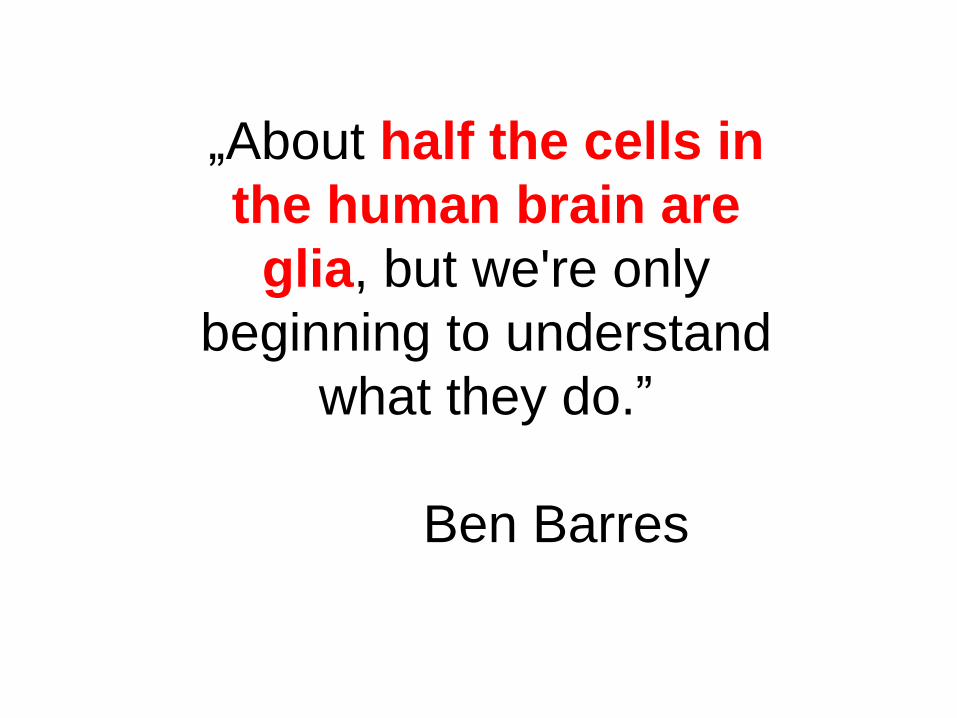
„About half the cells in
the human brain are
glia, but we're only
beginning to understand
what they do.”
Ben Barres

Philip Haydon
2014 2014 Gliafarmakológia… első cég

1858, Rudolf Ludwig Karl Virchow (37 évesen) a Berlini
Egyetem Patológiai Intézetében előadássorozatot tartott
13. előadásában (‘Spinal cord and the brain’) említette
meg az „agyi kötőszövetet” („nervenkitt, nerve-
cement”), melyet neurogliának nevezett el
A ‘GLIA’ felfedezése glia glia (görög): ragadós
Virchow gondolatai az előadásaiból készült „Cellular
Pathologie” c. könyvben jelennek meg, 1858-ban. Rudolf
Virchow és Karl Weigert szerint – a neuroglia szerepe csak a
helykitöltés a neuronok között (‘neuroglia holds nervous
elements together and gives the whole its form’)
A gliát Virchow így definiálta „ .. connective substance,
which forms in the brain, in the spinal cord and in the higher
sensory nerves as a sort of nervenkitt (neuroglia), in which
the nervous system elements are embedded”. For Virchow,
glia was a connective tissue devoid of cellular elements.
Neuroglia elnevezés először !
1821–1902, German doctor, anthropologist,
pathologist, prehistorian,
biologist and politician

A ‘GLIA’ felfedezése
1843–1926,
Italian physician, pathologist,
scientist, and Nobel laureate
1873, Camillo Golgi kifejlesztette az ezüst-kromát festést – ez
volt a kezdete a hisztológia forradalmának, hiszen eddig csak
festetlen preparátumokat vizsgáltak. Sokféle gliasejtet írt le.
(Nobel prize: from 1895)
Protoplazmás
asztroglia sejtek a
szürke-
állományban.
Festette és rajzolta:
Golgi; 1883.
Golgi már 1871-ben felismerte, hogy a
gliasejtek az idegsejtektől eltérő sejtes
populációt alkotnak. Megfigyelte, hogy a
gliasejtek nyúlványokat nyújtanak a
vérdeényekhez és azokon végtalpakat
létesítenek. Ő javasolta először, hogy a
gliasejtek metabolikus anyagokat
továbbítanak az erekből az agy-
parenchimába (1875; 1894).

1888-tól, Ramon y Cajal fejlesztette ki az arany-klorid
szublimálásos technikát, ami festette a rostos és plazmás
asztrocitákat (e festés targetje IF, GFAP !)
A ‘GLIA’ felfedezése
1852-1934,
Spanish pathologist,
histologist, neuroscientist,
Nobel laureate (1906)
Ramón y Cajal eredeti rajzai
gliasejtekről:
“Neuroglia of the superficial layers of the
cerebrum; child of two months. Method of
Golgi. A, B, [C], D, neuroglial cells of the
plexiform layer; E, F, [G, H, K], R,
neuroglial cells of the second and third
layers; V, blood vessel; I, J, neuroglial
cells with vascular [pedicles].”
Verkhratsky, Butt 2007
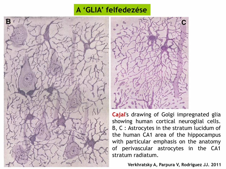
Cajal's drawing of Golgi impregnated glia
showing human cortical neuroglial cells.
B, C : Astrocytes in the stratum lucidum of
the human CA1 area of the hippocampus
with particular emphasis on the anatomy
of perivascular astrocytes in the CA1
stratum radiatum.
A ‘GLIA’ felfedezése
Verkhratsky A, Parpura V, Rodríguez JJ. 2011
A ‘GLIA’ felfedezése
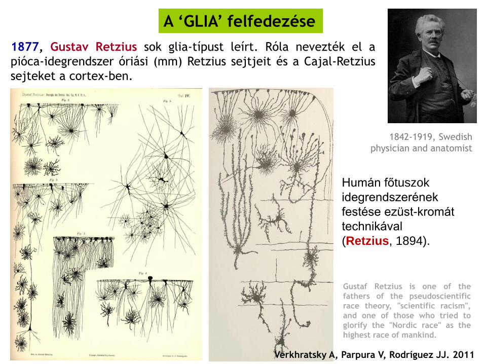
1842-1919, Swedish
physician and anatomist
1877, Gustav Retzius sok glia-típust leírt. Róla nevezték el a
pióca-idegrendszer óriási (mm) Retzius sejtjeit és a Cajal-Retzius
sejteket a cortex-ben.
Humán főtuszok
idegrendszerének
festése ezüst-kromát
technikával
(Retzius, 1894).
Verkhratsky A, Parpura V, Rodríguez JJ. 2011
Gustaf Retzius is one of the
fathers of the pseudoscientific
race theory, "scientific racism",
and one of those who tried to
glorify the "Nordic race" as the
highest race of mankind.

Morfológiai diverzitás
és a gliasejtek túlsúlya
az agyban – ezt látta
Gustaf Retzius.
Két neuront nyíl jelöl.
A gliasejteket ezüst
impregnációs
technikával festette.
The image shows a drawing
from Retzius’ book
Biologische Untersuchungen
(Stockholm: Samson and
Wallin, 1890-1914), Vol. 6
(1894), Plate ii, Figure 5.
(The image was kindly
provided by Professor Helmut
Kettenmann, MDC, Berlin)
A ‘GLIA’ felfedezése
Verkhratsky, Butt 2007

Emsley, Macklis 2006
Astroglial
heterogeneity closely
reflects the neuronal-
defined anatomy of
the adult murine CNS.
hGFAP-GFP
2001

Neuronális heterogenitás: Cajal rajzai alapján
Electrofiziológia: 1940’s (voltage clamp) és 1970’s (patch clamp) +
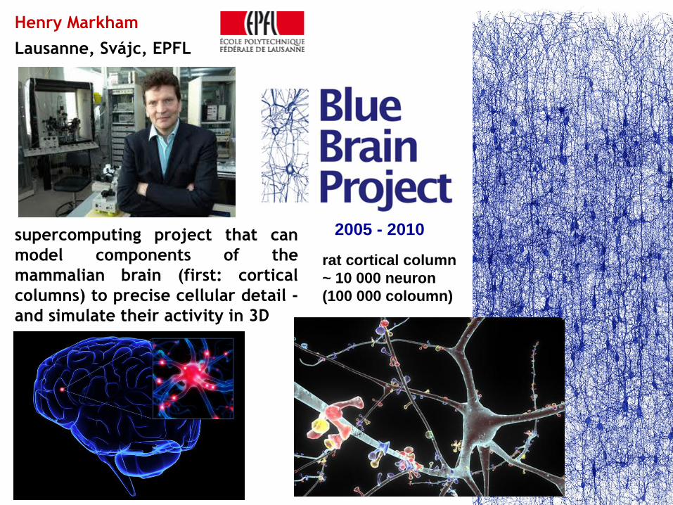
Henry Markham
Lausanne, Svájc, EPFL
supercomputing project that can
model components of the
mammalian brain (first: cortical
columns) to precise cellular detail -
and simulate their activity in 3D
2005 - 2010
rat cortical column
~ 10 000 neuron
(100 000 coloumn)

Morphological Classes of Neocortical Neurons
Henry Markham
PC=pyramidal cell; SSC=spiny stellate cell; LBC=large basket cell; SBC=small basket cell; NBC=nest basket cell;
BTC=bitufted cell; BPC=bipolar cell; e-BPC=excitatory bipolar cell (putative); DBC=double bouquet cell; e-
DBC=excitatory bitufted cell (putative); NGC=neurogliaform cell; MC=Martinotti cell; CRC=Cajal-Retzius cell;
ChC=chandelier cell…
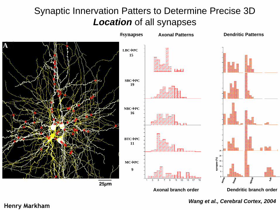
Synaptic Innervation Patters to Determine Precise 3D
Location of all synapses #synapses
LBCPC
SBCPC
NBCPC
BTCPC
15
19
16
11
A
MCPC
9
Axonal branch order Dendritic branch order
Axonal Patterns Dendritic Patterns
Wang et al., Cerebral Cortex, 2004 Henry Markham

Diversity of Electrical Behaviors of Neocortical Neurons
Henry Markham

Soma Targeting INTS LBC_bA D
LBC_bIS
LBC_bNA
LBC_cA D
LBC_cFS
LBC_cST
LBC_c NA
LBC_dFS
LBC_dST
NBC _bNA
NBC _cA D
NBC-bADNBC _cFS
NBC _c NA
NBC _dFS
NBC _dST
NBC-cST
NBC-dAD
NBC-bSTSBC _bNA
SBC _cA D
SBC _cFS
SBC _c NA
SBC _dFS
Axon Targeting INTS ChC _cA D
ChC _cFS
ChC -dNA
Excitatory Neurons SSC _cA D
SSC _cST
L2PC-cAD
L3PC-cAD
L4PC-cAD
L4SP-cAD
l4SS-cAD
L5CSPC-cAD
L5CHPC-cAD
L6CTPC-cAD
L6CCPC-cAD
L6CSPC-cAD
Bipolar
Bitufted
Multiple Electrical Types in Each Morphological Class
Henry Markham
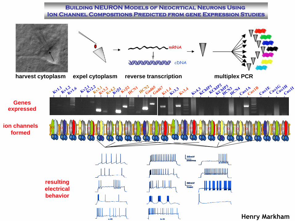
Building NEURON Models of Neocrtical Neurons Using
Ion Channel Compositions Predicted from gene Expression Studies
harvest cytoplasm expel cytoplasm reverse transcription multiplex PCR
Genes expressed
ion channels
formed
resulting
electrical
behavior
Henry Markham
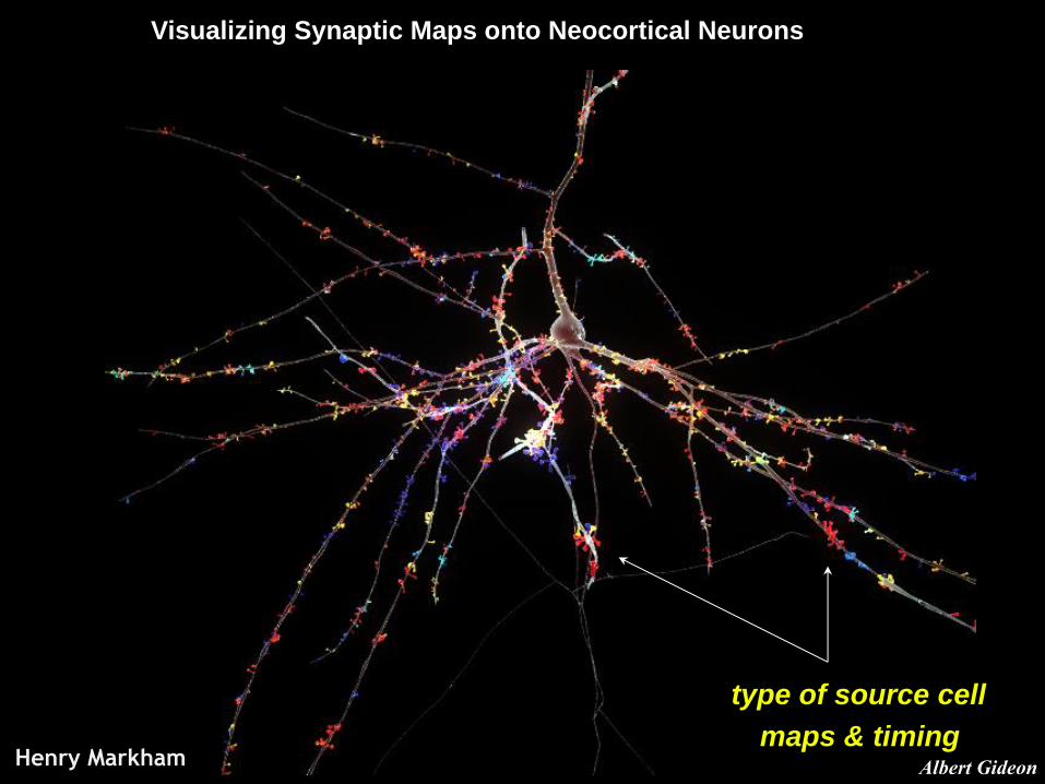
Visualizing Synaptic Maps onto Neocortical Neurons
Albert Gideon
type of source cell
maps & timing Henry Markham

Supercomputing…
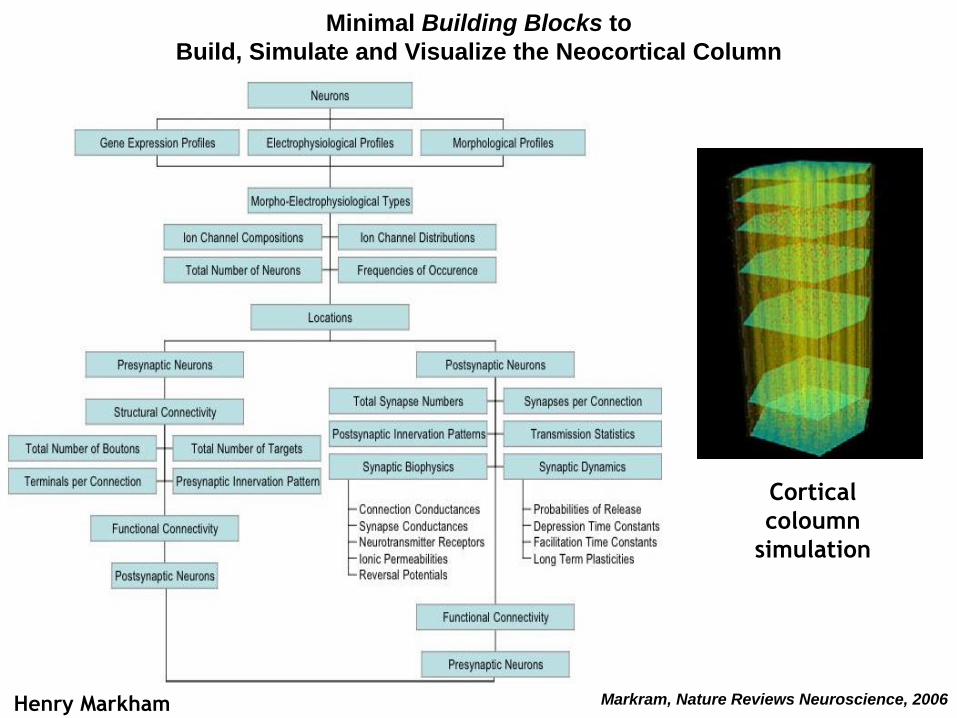
Minimal Building Blocks to
Build, Simulate and Visualize the Neocortical Column
Markram, Nature Reviews Neuroscience, 2006 Henry Markham
Cortical
coloumn
simulation
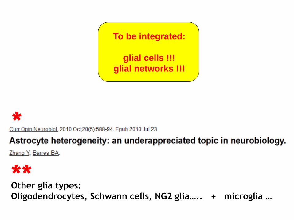
Other glia types:
Oligodendrocytes, Schwann cells, NG2 glia….. + microglia …
*
**
To be integrated:
glial cells !!!
glial networks !!!
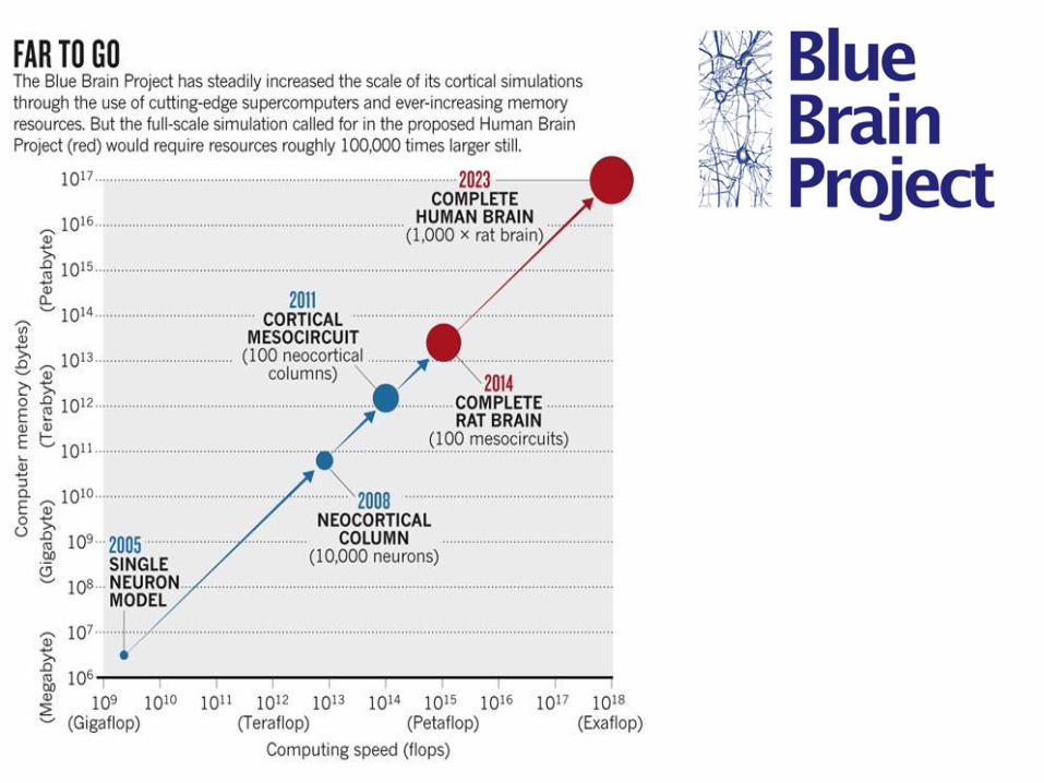

Human Brain Project
2013-2023
~1,4 milliárd Euro
Led by Dr. Henry Markram, The Blue
Brain Project recently joined with
other 12 partners to propose the
Human Brain Project – a very large 10
year project that will pursue precisely
these aims. The new grouping has just
been awarded a Eur 1.4 billion
European grant to formulate a detailed
proposal.

http://www.neurofuture.eu/
Ugyanakkor a Human Brain Projectről más vélemény is van..: read it !
https://forbetterscience.wordpress.com/2016/07/15/the-laborious-delivery-of-markrams-brainchild/

https://www.humanbrainproject.eu/

1863-1937, was an Hungarian anatomist and histologist
uncle to Albert Szent-Györgyi (1893-1986)
A ‘GLIA’ felfedezése
Lenhossék Mihály vezette be az asztrocita szót, utalva a neuroglia sejtek
alakjára. Ő írta az első terjedelmes összefoglalást a gliasejtekről egy könyvben,
mely az idegrendszer finom-szerkezetéről szólt, a gerincvelőt állítva fókuszba.
Állította, hogy a gliasejtek, melyeket oly sokan leírtak, egy vegyes populáció, és
valójában több sejttípusból áll. Terminológiai változtatásokat javasolt (Lenhossek,
1893):
„ I would suggest that all supportive cells be named as spongiocytes, and the most common
form in vertebrates be named spider cells or astrocytes, and use the term neuroglia only
cum grano salis (with a grain of salt), at least until we have a clearer view... Astrocytes are
the small elements, which form the supportive system of the spinal cord. They are star
shaped and indeed no other comparison describes their form so clearly. While the term
spider cell introduced by Jastrowitz has become popular and gives a proper impression of
the cells, one should regard Gierke’s note, namely that nobody has seen a spider with so
many feet as these cells have processes.”
In 1893, Mihály Lenhossék vezette be az asztrocita
kifejezést
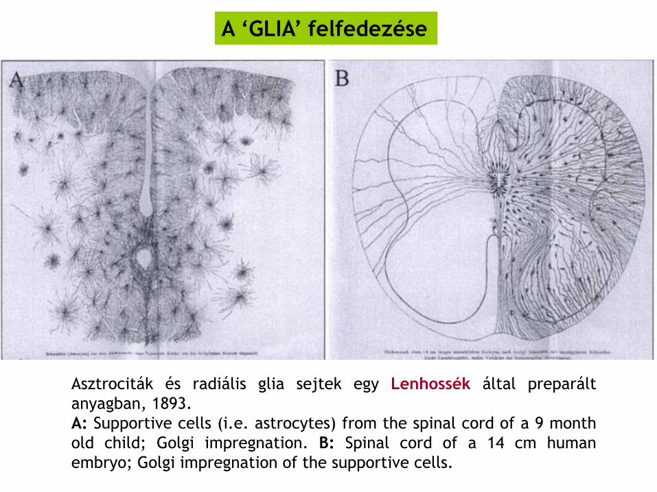
Asztrociták és radiális glia sejtek egy Lenhossék által preparált
anyagban, 1893.
A: Supportive cells (i.e. astrocytes) from the spinal cord of a 9 month
old child; Golgi impregnation. B: Spinal cord of a 14 cm human
embryo; Golgi impregnation of the supportive cells.
A ‘GLIA’ felfedezése

Neuroglia cells of brain shown by Golgi’s method. A. Cell with
branched processes. B. Spider cell with unbranched processes.
(After Andriezen.) in Henry Gray (1825–1861). Anatomy of the
Human Body. 1918.
A ‘GLIA’ felfedezése
Protoplazmás
asztrocita
Rostos
asztrocita
szürkeállomány fehérállomány
1895, a rostos és protoplazmás asztrocitákat Albert von Kölliker és
Andriezen írta le .
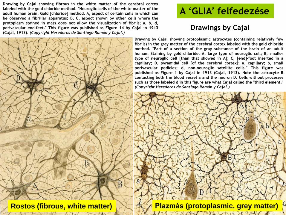
Drawing by Cajal showing fibrous in the white matter of the cerebral cortex
labeled with the gold chloride method. "Neuroglic cells of the white matter of the
adult human brain. Gold [chloride] method. A, aspect of certain cells in which can
be observed a fibrillar apparatus; B, C, aspect shown by other cells where the
protoplasm stained in mass does not allow the visualization of fibrils; a, b, d,
perivascular end-feet." This figure was published as Figure 14 by Cajal in 1913
(Cajal, 1913). (Copyright Herederos de Santiago Ramón y Cajal.)
Drawing by Cajal showing protoplasmic astrocytes (containing relatively few
fibrils) in the gray matter of the cerebral cortex labeled with the gold chloride
method. "Part of a section of the gray substance of the brain of an adult
human. Staining by gold chloride. A, large type of neuroglic cell; B, smaller
type of neuroglic cell [than that showed in A]; C, [end]-foot inserted in a
capillary; D, pyramidal cell [of the cerebral cortex]; a, capillary; b, small
perivascular pedicles; d, non-neuroglic satellite cells." This figure was
published as Figure 1 by Cajal in 1913 (Cajal, 1913). Note the astrocyte B
contacting both the blood vessel a and the neuron D. Cells without processes
such as those labeled d in this figure are what Cajal called the "third element."
(Copyright Herederos de Santiago Ramón y Cajal.)
A ‘GLIA’ felfedezése
Rostos (fibrous, white matter) Plazmás (protoplasmic, grey matter)
Drawings by Cajal
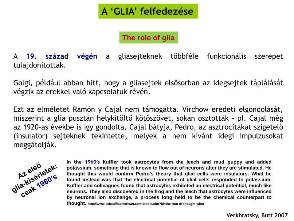
A 19. század végén a gliasejteknek többféle funkcionális szerepet
tulajdonítottak.
Golgi, például abban hitt, hogy a gliasejtek elsősorban az idegsejtek táplálását
végzik az erekkel való kapcsolatuk révén.
Ezt az elméletet Ramón y Cajal nem támogatta. Virchow eredeti elgondolását,
miszerint a glia pusztán helykitöltő kötőszövet, sokan osztották - pl. Cajal még
az 1920-as évekbe is így gondolta. Cajal bátyja, Pedro, az asztrocitákat szigetelő
(insulator) sejteknek tekintette, melyek a nem kívánt idegi impulzusokat
meggátolják.
A ‘GLIA’ felfedezése
Verkhratsky, Butt 2007
The role of glia
In the 1960’s Kuffler took astrocytes from the leech and mud puppy and added
potassium, something that is known to flow out of neurons after they are stimulated. He
thought this would confirm Pedro's theory that glial cells were insulators. What he
found instead was that the electrical potential of glial cells responded to potassium.
Kuffler and colleagues found that astrocytes exhibited an electrical potential, much like
neurons. They also discovered in the frog and the leech that astrocytes were influenced
by neuronal ion exchange, a process long held to be the chemical counterpart to
thought. http://www.scientificamerican.com/article.cfm?id=the-root-of-thought-what
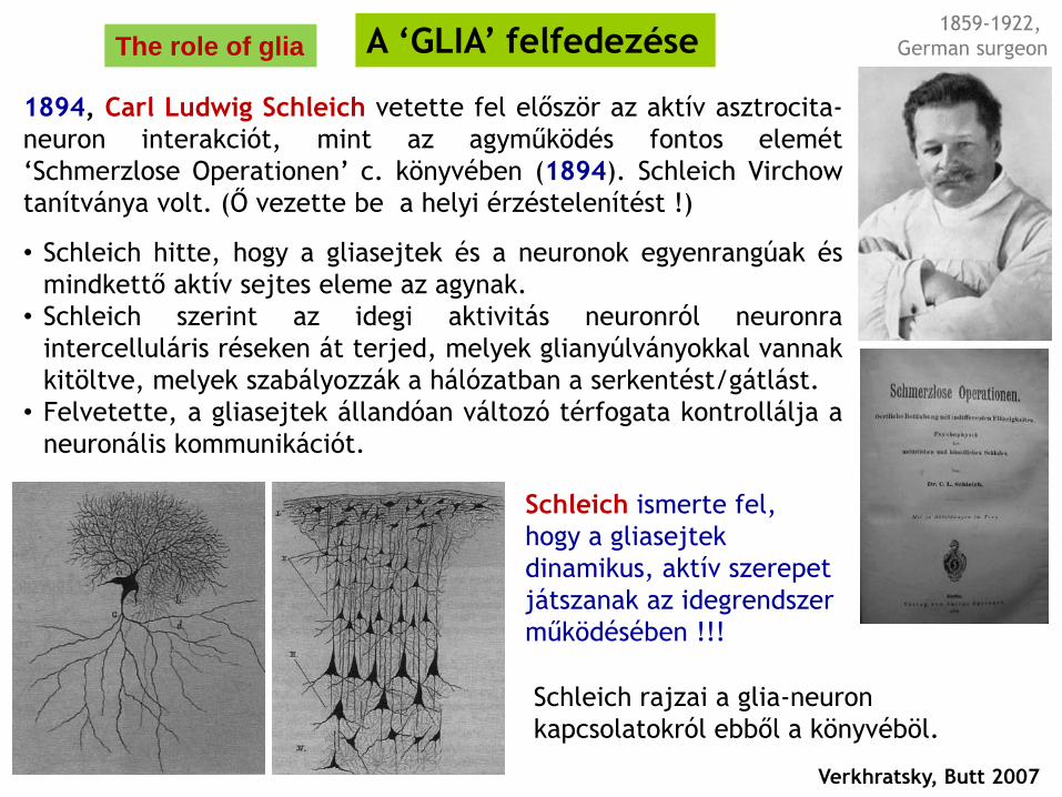
Schleich rajzai a glia-neuron
kapcsolatokról ebből a könyvéböl.
A ‘GLIA’ felfedezése
Verkhratsky, Butt 2007
1894, Carl Ludwig Schleich vetette fel először az aktív asztrocita-
neuron interakciót, mint az agyműködés fontos elemét
‘Schmerzlose Operationen’ c. könyvében (1894). Schleich Virchow
tanítványa volt. (Ő vezette be a helyi érzéstelenítést !)
• Schleich hitte, hogy a gliasejtek és a neuronok egyenrangúak és
mindkettő aktív sejtes eleme az agynak.
• Schleich szerint az idegi aktivitás neuronról neuronra
intercelluláris réseken át terjed, melyek glianyúlványokkal vannak
kitöltve, melyek szabályozzák a hálózatban a serkentést/gátlást.
• Felvetette, a gliasejtek állandóan változó térfogata kontrollálja a
neuronális kommunikációt.
The role of glia
Schleich ismerte fel,
hogy a gliasejtek
dinamikus, aktív szerepet
játszanak az idegrendszer
működésében !!!
1859-1922,
German surgeon
A ‘GLIA’ felfedezése
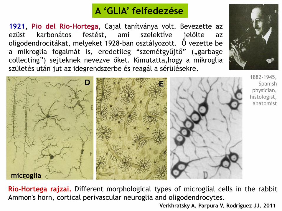
Río-Hortega rajzai. Different morphological types of microglial cells in the rabbit
Ammon's horn, cortical perivascular neuroglia and oligodendrocytes. Verkhratsky A, Parpura V, Rodríguez JJ. 2011
microglia
1921, Pio del Rio-Hortega, Cajal tanítványa volt. Bevezette az
ezüst karbonátos festést, ami szelektíve jelölte az
oligodendrocitákat, melyeket 1928-ban osztályozott. Ő vezette be
a mikroglia fogalmát is, eredetileg “szemétgyűjtő” („garbage
collecting”) sejteknek nevezve őket. Kimutatta,hogy a mikroglia
születés után jut az idegrendszerbe és reagál a sérülésekre. 1882-1945,
Spanish
physician,
histologist,
anatomist
A ‘GLIA’ felfedezése
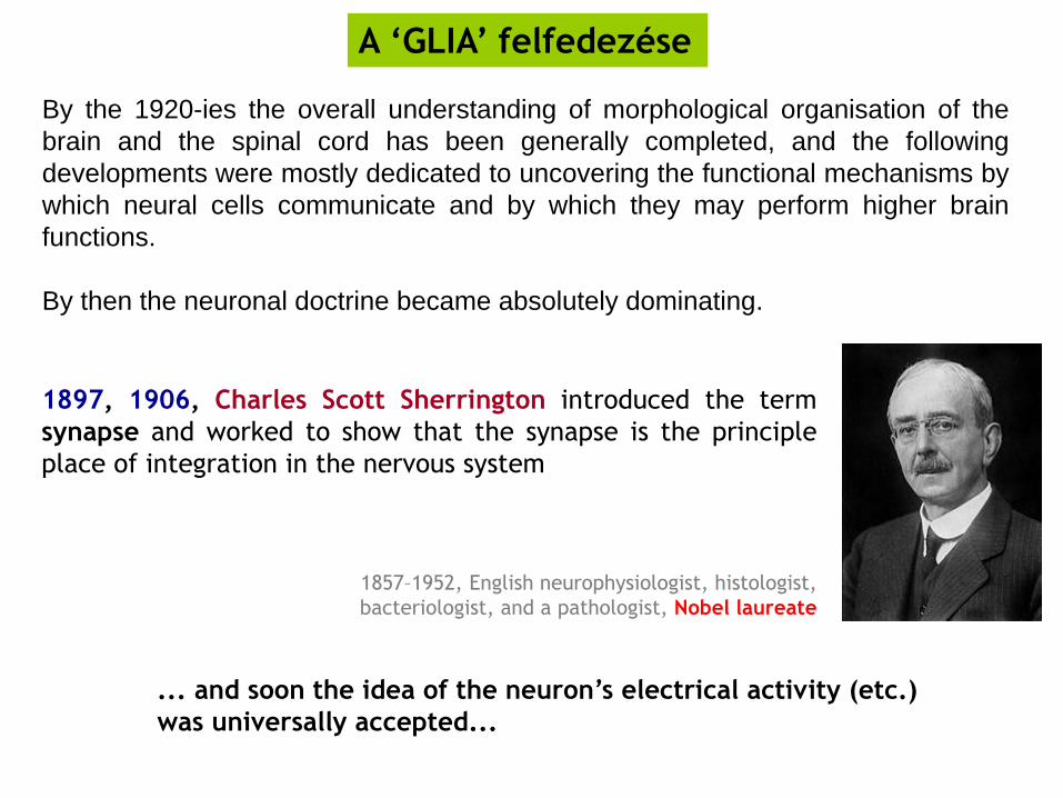
By the 1920-ies the overall understanding of morphological organisation of the
brain and the spinal cord has been generally completed, and the following
developments were mostly dedicated to uncovering the functional mechanisms by
which neural cells communicate and by which they may perform higher brain
functions.
By then the neuronal doctrine became absolutely dominating.
1897, 1906, Charles Scott Sherrington introduced the term
synapse and worked to show that the synapse is the principle
place of integration in the nervous system
1857–1952, English neurophysiologist, histologist,
bacteriologist, and a pathologist, Nobel laureate
... and soon the idea of the neuron’s electrical activity (etc.)
was universally accepted...

1954, Ben Geren B. Schwann cells
myelinate
1966, Steven Kuffler, John Nicolls, Richard Orkand demonstrated coupling
between glial cells
1969, Milton Brightman, Tom Reese identified structures connecting glial
networks (which we know now as gap junctions)
A ‘GLIA’ felfedezése
Verkhratsky, Butt 2007
The role of glia
Nonetheless, for the following two decades glial cells were still regarded as passive
elements of the NS, bearing mostly supportive and nutritional roles. The advent of
modern physiological techniques, most notably those of the patch-clamp and
fluorescent calcium dyes, has dramatically changed this image of glia as ‘silent’
brain cells.
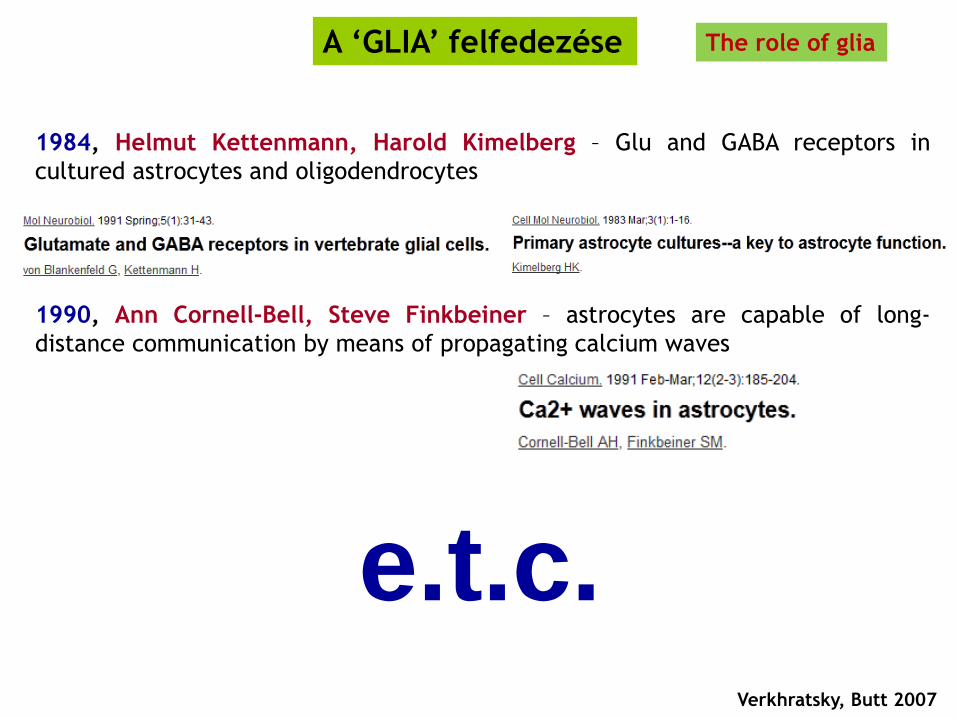
1984, Helmut Kettenmann, Harold Kimelberg – Glu and GABA receptors in
cultured astrocytes and oligodendrocytes
1990, Ann Cornell-Bell, Steve Finkbeiner – astrocytes are capable of long-
distance communication by means of propagating calcium waves
A ‘GLIA’ felfedezése
Verkhratsky, Butt 2007
The role of glia
e.t.c.

Neuroglia – Evolúciós aspektusok
Verkhratsky A, Parpura V, Rodríguez JJ. 2011 Tree of life, Haeckel 1879.
• Gliasejtek robbanásszerű
térhódítása hominidákban
• Proto-myelináló sejtek megjelenése
és a myelin-hüvely kialakulása
(ganglionok majd központosított
idegrendszer szerveződése)
• Asztrociták is megjelennek a
ganglionokban
• Asztrociták képezik a kezdeti vér-agy-
gátat
• Immunsejtek belépnek a ganglionokba
az ősi mikrogliát reprezentálva
• A glia megjelenése (valószínűleg már a
diffúz idegrendszer szintjén)

fejgerinchúrosok
ízeltlábúak
puhatestűek
laposférgek
kerekesférgek
tüskésbőrűek
laposféreg-szerűek
kétoldali szimmetriájúak
Jelenleg mindkettő lehetséges:
glia közös
eredetű
(Acoelomorpha..)
glia többször is
kialakul
Neuroglia –
Evolúciós
aspektusok
50% 50%

BBB
• blood–brain barrier (BBB): csak
asztrocitákból áll rákokban, rovarokban,
fejlábúakban és cápákban, rájákban,
tokhalakban
• későbbiekben a BBB funkciót endotélsejtek
felé tolódik, de asztrociták kontrollja alatt
marad
• Bundgaard és Abbott szerint az ősi
gerinceseknek is gliális BBB-ja volt !
Blue: glial BBB
Red: endothelial BBB
Alternatív modellek a gerinces BBB
törzsfejlődésére
Neuroglia – Evolúciós aspektusok

Teljes sejtszám felnőtt
hermafrodita C. elegans-ban:
959
302 neuron
50 glia (5 %)
24 sheath glia,
26 socket glia
C. elegans glia
C: Red: sheath glia, green: neuronal cilia
Blue: socket glia
Amphid: fő kemoszenzoros szerv a
nematodákban (kutikula beidegzett
betűrődései). Minden amphid-ot 12
érző neuron és 2 glia (kék/piros)
alkot.

Gliasejtek ablációja:
(lézeres mikrosebészet...)
- nincs neuronpusztulás
- de az érzőneuronok dendrit és
axonnövekedés zavart lesz

AFD = termoszenzoros neuron, az amphid
12 neuronjának egyike. Ezt is AMsh glia
öleli körbe.
A KCC-3 K+/Cl- transzporter az AMsh
gliasejt AFD neuron mikrovillusait
körbevevő részein lokalizálódik és
szabályozza a K+/Cl- szintet az ec. térben.
A Cl- ionok direkt módon gátolják a neuron
receptor guanylyl-ciklázt (GCY-8), ami
cGMPt szintetizál.
A magas cGMP szint gátolja az aktin
regulátor WSP-1/WASP-ot, ami blokkolja a
neuronális növekedési kúp elongációját.
Components of this pathway are broadly
expressed throughout the nervous system,
suggesting that ionic regulation of the NRE
microenvironment may be a conserved
mechanism by which glia control
neuron shape and function.

Agy mérete
elefánt, bálna agya nagyobb mint emberé
Agy/testtömeg arány
kis emlősöknél, pl. mókuscickánynál nagyobb az agy/test tömeg aránya, mint embernél
Agykéreg mérete
delfinek, bálnák abszolút és relatív agykéreg mérete is nagyobb lehet, mint az emberé
Milyen gének expresszálódnak inkább a humán agyban a főemlősökéhez képest ?
miért vagyunk „okosabbak” ??
több glia-related gén, mint neuronális !
Gliasejtek mérete/morfológiája
emberben elágazóbb, jóval nagyobb (asztrociták)
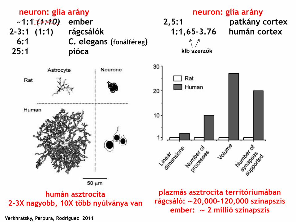
neuron: glia arány
~1:1 (1:10) ember
2-3:1 (1:1) rágcsálók
6:1 C. elegans (fonálféreg)
25:1 pióca
Verkhratsky, Parpura, Rodríguez 2011
neuron: glia arány
2,5:1 patkány cortex
1:1,65-3.76 humán cortex
humán asztrocita
2-3X nagyobb, 10X több nyúlványa van
plazmás asztrocita territóriumában
rágcsáló: ∼20,000–120,000 szinapszis
ember: ∼ 2 millió szinapszis
klb szerzők

cortical
human
astrocyte
[Image: Nancy
Ann Oberheim
and Takahiro
Takano,
University of
Rochester
Medical School]

DAPI
NeuN (Fox3)
B = billion.
! N 1:3.7 G
N 4,3:1 G
N 1,02:1 G
N 1:11,2 G
Teljes humán
agy neuron:glia
arány = 1:1 !!!

„Humanized mice”


Neuroglia – The function...
The first biophysically realistic computational model
based on experimental results in HC – including GLIA !
in the hippocampus for every neuron
there are two to five glia cells
"Glia cells are like the brain's
supervisors. By regulating the synapses,
they control the transfer of information
between neurons, affecting how the
brain processes information and
learns."

Glial Neurobiology: A Textbook
Alexei Verkhratsky and Arthur Butt
© 2007 John Wiley & Sons, Ltd ISBN 978-0-470-01564-3 (HB); 978-0-
470-51740-6 (PB)
Ajánlott Irodalom
Glia
Huszti Zsuzsa - Kálmán Mihály
Akadémiai Kiadó, 2008
Pl.: http://www.ted.com/talks/henry_markram_supercomputing_the_brain_s_secrets.html
TED: “Technology, Entertainment, Design”,
from 1984 – now broader scope
The top five neuroscience project:
http://www.33rdsquare.com/2012/01/top-five-neuroscience-projects.html
5. The Human Connectome Project
4. Numenta / Hierarchical Temporal Memory Theory
3. SyNAPSE
2. The Human Brain Project
1. The Blue Brain Project


![01 A proposal for Sustainable Employer Brandemijournal.cz/wp-content/uploads/2015/08/01_A-proposal... · 2017-10-30 · (0, 9ro ,vvxh ,661 3ulqw ; 2qolqh zzz hplmrxuqdo f] pdqdjhphqw](https://static.fdocuments.net/doc/165x107/5f511b1c4b58806e11096b87/01-a-proposal-for-sustainable-employer-2017-10-30-0-9ro-vvxh-661-3ulqw-2qolqh.jpg)