IB Chemistry on Mass Spectrometry, Index Hydrogen Deficiency and Isotopes
31
Index Hydrogen Deficiency (IHD) = Degree unsaturation H H H H H H ׀ ׀ ׀ ׀ ׀ ׀H - C - C – C – C – C – C – H ׀ ׀ ׀ ׀ ׀ ׀H H H H H H H H H H H H ׀ ׀ ׀ ׀ ׀ ׀H - C - C = C – C – C – C – H ׀ ׀ ׀ ׀H H H H H H H H H H ׀ ׀ ׀ ׀ ׀ ׀H - C - C – C = C – C – C – H ׀ ׀ ׀ ׀H H H H Hexane C 6 H 14 IHD = 0 Hex-2-ene C 6 H 12 IHD = 1 Hex-3-ene C 6 H 12 IHD = 1 Cyclohexane C 6 H 12 IHD = 1 How many double bonds/rings present Any double/triple bond/cyclic - number hydrogen atoms decrease How many H 2 need to convert molecule to saturated/non cyclic Saturated hydrocarbon = C n H 2n+2 Saturated IDH = 0 Unsaturated (1 π bond) 2 H deficiency IHD = 1 H H ׀ ׀C = C ׀ ׀H H Unsaturated (2 π bond) 4 H deficiency IHD = 2 Unsaturated (1 ring) 2 H deficiency IHD = 1 Unsaturated (3 π bond) + 1 ring 8 H deficiency IHD = 4 IHD = 1 1 π bond or 1 ring structure Unsaturated (2 π bond) + 1 ring 6 H deficiency IHD = 3 Unsaturated (4 π bond) + 1 ring 10 H deficiency IHD = 5 IHD = 2 2 π bond / 1 π bond + 1 ring IHD = 3 3 π bond / 2 π bond + 1 ring / 1 π bond+ 2 ring Unsaturated (4 π bond) + 1 ring 10 H deficiency IHD = 5 Unsaturated (6 π bond) + 2 ring 16 H deficiency IHD = 8 Click here IHD (Khan Academy)
-
Upload
lawrence-kok -
Category
Education
-
view
934 -
download
7
Transcript of IB Chemistry on Mass Spectrometry, Index Hydrogen Deficiency and Isotopes

Index Hydrogen Deficiency (IHD) = Degree unsaturation
H H H H H H
׀ ׀ ׀ ׀ ׀ ׀
H - C - C – C – C – C – C – H
׀ ׀ ׀ ׀ ׀ ׀
H H H H H H
H H H H H H
׀ ׀ ׀ ׀ ׀ ׀
H - C - C = C – C – C – C – H
׀ ׀ ׀ ׀
H H H H
H H H H H H
׀ ׀ ׀ ׀ ׀ ׀
H - C - C – C = C – C – C – H
׀ ׀ ׀ ׀
H H H H
Hexane C6H14 IHD = 0
Hex-2-ene C6H12 IHD = 1
Hex-3-ene C6H12 IHD = 1
Cyclohexane C6H12 IHD = 1
How many double bonds/rings present Any double/triple bond/cyclic - number hydrogen atoms decrease
How many H2 need to convert molecule to saturated/non cyclic
Saturated hydrocarbon = CnH2n+2
Saturated IDH = 0
Unsaturated (1 π bond)
2 H deficiency IHD = 1
H H
׀ ׀
C = C
׀ ׀
H H
Unsaturated (2 π bond)
4 H deficiency IHD = 2
Unsaturated (1 ring)
2 H deficiency IHD = 1
Unsaturated (3 π bond) + 1 ring
8 H deficiency IHD = 4
IHD = 1 1 π bond or 1 ring structure
Unsaturated (2 π bond) + 1 ring
6 H deficiency IHD = 3
Unsaturated (4 π bond) + 1 ring
10 H deficiency IHD = 5
IHD = 2 2 π bond / 1 π bond + 1 ring
IHD = 3 3 π bond / 2 π bond + 1 ring / 1 π bond+ 2 ring
Unsaturated (4 π bond) + 1 ring
10 H deficiency IHD = 5
Unsaturated (6 π bond) + 2 ring
16 H deficiency IHD = 8
Click here IHD (Khan Academy)

2
22 yxIHD
H H H H H H
׀ ׀ ׀ ׀ ׀ ׀
H - C - C – C – C – C – C – H
׀ ׀ ׀ ׀ ׀ ׀
H H H H H H
Hexane C6H14 IHD = 0
Hex-2-ene C6H12 IHD = 1
Hex-3-ene C6H12 IHD = 1
Cyclohexane C6H12 IHD = 1
2
22 rsyxIHD
How many double bonds/rings present Any double/triple bond/cyclic - number hydrogen atoms decrease
How many H2 need to convert molecule to saturated/non cyclic
IHD = 1 1 π bond or 1 ring structure
IHD = 2 2 π bond / 1 π bond + 1 ring
IHD = 3 3 π bond / 2 π bond + 1 ring / 1 π bond+ 2 ring
Molecule with C and H Molecule with N, O, S or Halogen
yx HCsrqyx XNOHC
C6H12 C2H2
1
2
)12262(
2
22
IHD
IHD
yxIHD
or
2
2
)2222(
2
22
IHD
IHD
yxIHD
(1 π bond / 1 ring) (2 π bond)
C2H3CI C5H7N
1
2
)13222(
2
22
IHD
IHD
syxIHD
3
2
)17252(
2
22
IHD
IHD
ryxIHD
H - C = C - CI
׀ ׀ H H
(1 π bond) (3 π bond / 2 π bond + 1 ring)
or
H H H H H H
׀ ׀ ׀ ׀ ׀ ׀
H - C - C = C – C – C – C – H
׀ ׀ ׀ ׀
H H H H
H H H H H H
׀ ׀ ׀ ׀ ׀ ׀
H - C - C – C = C – C – C – H
׀ ׀ ׀ ׀
H H H H
H H H H H H
׀ ׀ ׀ ׀ ׀ ׀
H - C - C = C – C – C – C – H
׀ ׀ ׀ ׀
H H H H
Index Hydrogen Deficiency (IHD) = Degree unsaturation Saturated hydrocarbon = CnH2n+2

Molecule Index H2 Deficiency
C2H2 2
Oxygen and Sulfur - No effect on IHD Halogen - like H – CHCI3 = CH4 , C2H5CI = C2H6
Nitrogen – add one to C and one to H - CH5N same IHD as C2H6
Index Hydrogen Deficiency (IHD) = Degree unsaturation
How many double bonds/rings present Any double/triple bond/cyclic - number hydrogen atoms decrease
Molecule with N, O, S or Halogen
srqyx XNOHC
2
22 rsyxIHD
Click here IHD video
1
2
)4222(
2
22
yxIHD
2
2
)2222(
2
22
yxIHD
1
2
)4222(
2
22
yxIHD
0
2
)15222(
2
22
syxIHD
4
2
)6262(
2
22
yxIHD
Molecule Index H2 Deficiency
C2H4 1
Molecule Index H2 Deficiency
C2H4O 1
Molecule Index H2 Deficiency
C2H5CI 0
Molecule Index H2 Deficiency
C6H6 4
Molecule Index H2 Deficiency
C7H6O2 5
5
2
)6272(
2
22
yxIHD
Molecule Index H2 Deficiency
C7H9N2CI3 3
1
2
)19242(
2
22
ryxIHD
Molecule Index H2 Deficiency
C4H9N 1
3
2
)239272(
2
22
rsyxIHD
Molecule Index H2 Deficiency
C6H9NOCI2 2
2
2
)129262(
2
22
rsyxIHD
Molecule Index H2 Deficiency
C4H8CIF 0
0
2
)28242(
2
22
syxIHD
Click here IHD (Khan Academy)
Molecule Index H2 Deficiency
C6H12O6 1
1
2
)12262(
2
22
yxIHD
Halogen

Click here spectroscopy database (NIST)
Weighted average calculation RAM calculation Video on Isotopes Video on weighted average
Relative Atomic Mass
Weighted average mass- due to presence of isotopes
Relative Isotopic Mass, (Ar) of an element: •Relative isotopic mass = Average mass of one atom of element 1/12 x mass of one carbon-12 • Relative isotopic mass, carbon = 12.01
RAM = 12.01 Relative Abundance
13
Why RAM is not a whole number?
Relative Isotopic Mass: = (Mass 12C x % Ab) + (Mass 13C x % Ab) = (12 x 98.9/100) + (13 x 1.07/100) = 12.01
Video on Isotopes
12
Isotopes are present
C C C 12.01
98.9% 1.07%
Click here spectroscopy database (Ohio State)

Mg - 3 Isotopes
24 Mg – (100/127.2) x 100% - 78.6% 25 Mg – (12.8/127.2) x 100% - 10.0% 26 Mg – (14.4/127.2) x 100% - 11.3%
Relative Isotopic Mass: = (Mass 24Mg x % Ab) + (Mass 25Mg x % Ab) + (Mass 26Mg x % Ab) = (24 x 78.6/100) + (25 x 10.0/100) + (26 x 11.3/100) = 24.30
Relative Abundance % Abundance
Pb - 4 Isotopes
204Pb – (0.2/10) x 100% - 2% 206Pb – (2.4/10) x 100% - 24% 207Pb – (2.2/10) x 100% - 22% 208Pb – (5.2/10) x 100% - 52%
Relative Isotopic Mass = (Mass 204Pb x % Ab) + (Mass 206Pb x % Ab) + (Mass 207Pb x % Ab) + (Mass 208Pb x % Ab) = (204 x 2/100) + (206 x 24/100) + (207 x 22/100) + (208 x 52/100) = 207.20
Convert relative abundance to % abundance
Convert relative abundance to % abundance
Relative Abundance % Abundance
Relative Isotopic Mass
24 25 26 24 25 26 Mg Mg

Mass Spectrometer
Uses mass spectrometer
Presence of isotopes and its abundance
Relative atomic mass of an element
Relative Molecular mass of a molecule
Structure of organic compound
Distinguish bet structural isomers
CH3CH2CH2OH OH | CH3CHCH3
CH3 | CH3C-CH3
| CH3
CO2
structural formula
Organic structure determination
24 25 26
Mg Mg

Detail notes from chem msu Click here notes from chemguide
Mass Spectrometer
Parts of Mass Spectrometer
Sample injection
Vaporization Chamber • Sample heat to vapour state
Ionization Chamber • Molecule bombard with electron form positive ion
Accelerator Chamber • M+ ion accelerated by Electric field
Deflector • M+ ion deflected by magnetic field
Detector • Convert amt M+ ion to current. • M+ ion neutralize by electron (more e need - higher current – higher intensity of peak) • Intensity of peak show -relative abundance of ion
Sample X bombard by electron • Form positive M+ ion • Accelerated (Electric Field) • Deflected (Magnetic Field) and Detected X + e- → X+ + 2e-
Vaporization Ionization Accelerator Deflector Detector 3 2 1 5 4
2
1
3 4
5

Click here for simulation
Mass Spectrometer
Parts of Mass Spectrometer
Vaporization Ionization Accelerator Deflector Detector 3 2 1 5 4
Vaporization Injection/ vaporization of sample liquid state gaseous
Ionization Form cation, M+
Acceleration M+ ion accelerate by Electric field
Deflection M+ ion deflect
by magnetic field
Deflection depend: mass/charge (m/z) ratio: (m/z) ratio HIGH↑ - Deflection LOW↓
Deflection depend: mass/charge (m/z) ratio: (m/z) ratio LOW↓- Deflection HIGH ↑
37CI+
35CI+
35CI2+
2
3 4
1
5 Detector • Convert abundance M+ ion to current. • M+ ion neutralize by electron (more e need - high current – high intensity of peak) • Peak Intensity –relative abundance of ion
Video Mass spectrometer Video Ionization/fragmentation Video how MS works
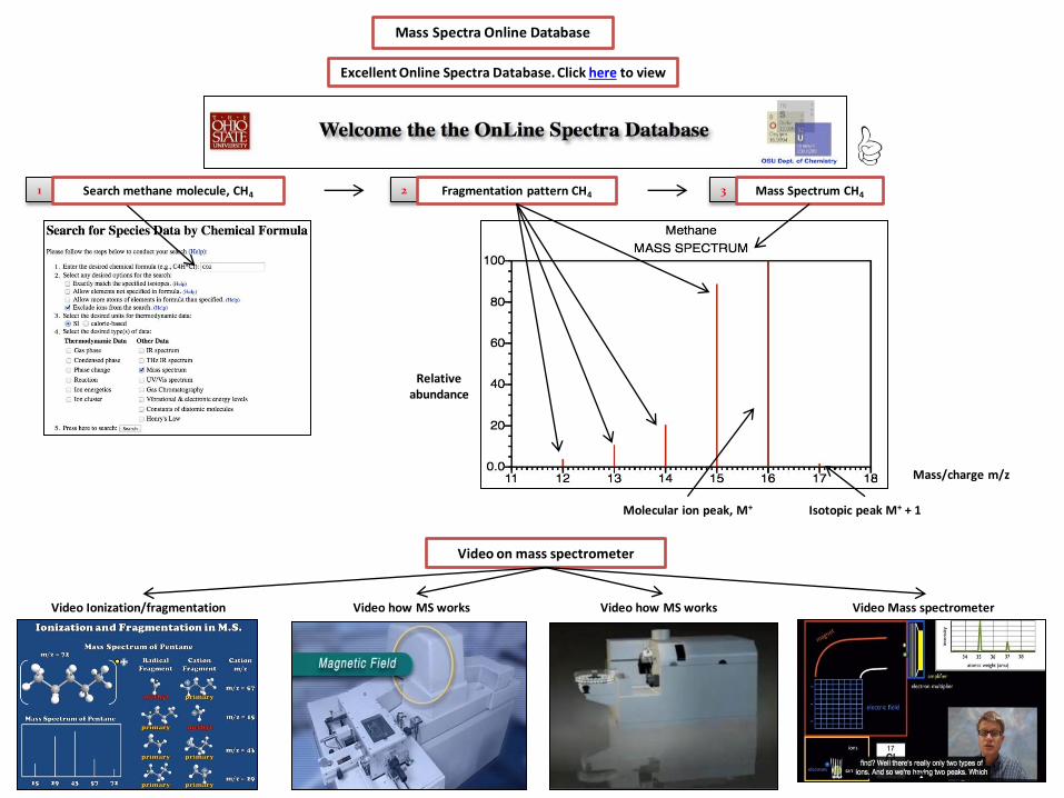
Excellent Online Spectra Database. Click here to view
Mass Spectra Online Database
1 Search methane molecule, CH4
Video on mass spectrometer
Mass/charge m/z
Relative abundance
Isotopic peak M+ + 1 Molecular ion peak, M+
2 Fragmentation pattern CH4 3 Mass Spectrum CH4
Video how MS works
Mg - 3 Isotopes
26 Mg - 11.3% - m/z highest – deflect LEAST 25 Mg - 10.0% 24 Mg – 78.6% - m/z lowest – deflect MOST
Relative Isotopic Mass: = (24Mg x % Ab) + (25Mg x % Ab) + (26Mg x % Ab) = (24 x 78.6/100) + (25 x 10.0/100) + (26 x 11.3/100) = 24.30
Mass spectrometry to determine Relative Isotopic Mass
Deflect MOST
Deflect LEAST
Pb - 4 Isotopes
208Pb – 52% - m/z highest – deflect LEAST 207Pb - 22% 206Pb - 24% 204Pb – 2% - m/z lowest – deflect MOST
Relative Isotopic Mass = (204Pb x % Ab) + (206Pb x % Ab) + (207Pb x % Ab) + (208Pb x % Ab) = (204 x 2/100) + (206 x 24/100) + (207 x 22/100) + (208 x 52/100) = 207.20
Deflect MOST
Deflect LEAST
24 Mg 26 Mg
204Pb 208Pb

CI - 2 Isotopes
37 CI – 24.5% - m/z highest – deflect LEAST 35 CI – 75.5% - m/z lowest – deflect MOST
Relative Isotopic Mass: = (35CI x % Ab) + (37CI x % Ab) = (35 x 75.5/100) + (37 x 24.5/100) = 35.5
Deflect MOST
Deflect LEAST
Br - 2 Isotopes
81Br – 49.3% - m/z highest – deflect LEAST 79Br – 50.6% - m/z lowest – deflect MOST
Deflect MOST
Deflect LEAST
35CI 37CI
Relative Isotopic Mass: = (79Br x % Ab) + (81Br x % Ab) = (79 x 50.6/100) + (81 x 49.3/100) = 79.9
79Br 81Br
Mass spectrometry to determine Relative Isotopic Mass
35 CI 37 CI
79Br 81Br

H - 3 Isotopes
3H – trace amt 2H – 0.015% - m/z highest – deflect LEAST 1H – 99.9% - m/z lowest – deflect MOST
Relative Isotopic Mass: = (1H x % Ab) + (2H x % Ab) = (1 x 99.9/100) + (2 x 0.015/100) = 1.007
Deflect MOST
Deflect LEAST
C - 3 Isotopes
14C- trace amt
13C – 1.1% - m/z highest – deflect LEAST 12C – 98.9% - m/z lowest – deflect MOST
Deflect MOST
Deflect LEAST
1H 2H
Relative Isotopic Mass: = (12C x % Ab) + (813Cx % Ab) = (12 x 98.9/100) + (13 x 1.1/100) = 12.01
12C 13C
3H
14C
Mass spectrometry to determine Relative Isotopic Mass
1H 2H
12C 13C

Ionization and Fragmentation
Ionization forming M+
CH3CH2CH2 : CH3 + e → CH3CH2CH2+.CH3 + 2e
• Fragmentation of M+ producing 43 CH3CH2CH2
+·CH3 → CH3CH2CH2+ + ·CH3
• Fragmentation of M+ producing 15 CH3CH2CH2
+·CH3 → CH3CH2CH2· + +CH3
Ionization and Fragmentation Process- CH3CH2CH2CH3
Ionization Process - CH3CH2CH2CH3 • Bombard by electron form cation • Molecular ion, M+ = 58 • (CH3CH2CH2CH3)+ = 58
Fragmentation Process CH3CH2CH2CH3 • Molecular ion, M+ undergo fragmentation • Cation and Radical form • Cation - Detected • Radical –Not detected (No charged)
H H
׀ ׀ CH3CH2CH2 C:H + e → CH3CH2CH2 C+.H + 2e ׀ ׀ H H
Ionization forming M+
CH3CH2:CH2CH3 + e → CH3CH2+·CH2CH3 + 2e
• Fragmentation of M+ producing 29 CH3CH2
+·CH2CH3 → CH3CH2+ + .CH2CH3
Ionization M+, m/z = 58
CH3CH2CH2CH3 + e → CH3CH2CH2CH3+ + 2e
Ionization and Fragmentation of M+ • Form - m/z = 58, 43 and 15
m/z = 58
m/z = 43
m/z = 15
Ionization and Fragmentation of M+ • Form- m/z = 58 and 29
m/z = 58
m/z = 58
m/z = 29
Unpair electron Positively charged
Will ACCELARATED NOT move
cation radical

CH3CH2CH2CH3
CH3CH2CH2CH3+- 58 - m/z highest –deflect LEAST
CH3CH2CH2+ – 43
CH3CH2+ – 29
CH3+ –15 - m/z lowest– deflect MOST
Ionization/ Fragmentation pattern CH3CH2CH2CH3
Deflect MOST
Deflect LEAST
CH3CH2CH2CH3+
CH3CH2CH2+
ionization
CH3+
Ionization and Fragmentation Process
Fragmentation
Ionization CH3CH2CH2CH3
CH3CH2CH2CH3 + e → CH3CH2CH2CH3+ + 2e → 58
or CH3CH2:CH2CH3 + e → CH3CH2
+·CH2CH3 + 2e → 58
Mass spectrum CH3CH2CH2CH3 Ionization CH3CH2CH2CH3
CH3CH2+
Fragmentation of M+ CH3CH2CH2
+·CH3 → CH3CH2CH2+ - 43
CH3CH2
+·CH2CH3 → CH3CH2+ – 29
CH3CH2CH2
+·CH3 → +CH3 - 15
CH3CH2CH2CH3+- 58
CH3CH2CH2+ – 43
CH3CH2+ – 29
CH3+ – 15
CH3+ CH3CH2CH2CH3
+

CH3CH2CH2OH
CH3CH2CH2OH+- 60 - m/z highest –deflect LEAST CH2CH2OH+ – 45 CH2OH+ - 31 CH3CH2
+ – 29 CH3
+ –15 - m/z lowest– deflect MOST
Ionization/ Fragmentation pattern CH3CH2CH2OH
Deflect MOST Deflect LEAST
CH3CH2CH2OH+
ionization
CH3 +
Fragmentation
Ionization CH3CH2CH2OH
CH3CH2CH2OH + e → CH3CH2CH2OH+ + 2e → 60 or CH3CH2CH2OH + e → CH3CH2
+. CH2OH + 2e → 60
Mass spectrum CH3CH2CH2OH CH3CH2CH2OH
CH3CH2+
Fragmentation of M+ CH3
+.CH2CH2OH → +CH2CH2OH - 45
CH3CH2
+·CH2OH → +CH2OH – 31
CH3CH2
+·CH2OH → CH3CH2+ – 29
CH3
+.CH2CH2OH → +CH3 - 15
CH2CH2OH+ CH2OH+
15 60
CH3CH2CH2OH+- 60 CH2CH2OH+ – 45 CH2OH+ - 31 CH3CH2
+ – 29 CH3
+ – 15
15 60
Ionization and Fragmentation Process
Ionization
CH3+ CH3CH2CH2OH+

CH3CH(CH3)CH2CH3+- 72 - m/z highest –deflect LEAST
CH3CH(CH3)CH2+ – 57
CH3CH(CH3)+ - 43
CH3CH2+ – 29
CH3+ –15 - m/z lowest– deflect MOST
Ionization/ Fragmentation pattern CH3CH(CH3)CH2CH3
Deflect MOST
Deflect LEAST
CH3CH(CH3)CH2CH3+
Ionization
CH3+
Fragmentation
Ionization of CH3CH(CH3)CH2CH3
CH3CH(CH3)CH2CH3 + e → CH3CH(CH3)CH2CH3 + + 2e → 72
or CH3CH(CH3)CH2CH3 + e → CH3CH(CH3)CH2
+.CH3 + 2e → 72 or CH3CH(CH3)CH2CH3 + e → CH3CH(CH3)+.CH2CH3 + 2e → 72
Mass spectrum CH3CH(CH3)CH2CH3 Ionization CH3CH(CH3)CH2CH3
Fragmentation of M+ CH3CH(CH3)CH2
+ - 57 CH3CH(CH3)
+ – 43
CH3CH2+ – 29
CH3+ - 15
CH3CH(CH3)+
15
CH3CH(CH3)CH2+
CH3CH(CH3)CH2CH3+
CH3CH2+
CH3CH(CH3)CH2CH3+- 72
CH3CH(CH3)CH2+ – 57
CH3CH(CH3)+ - 43
CH3CH2+ – 29
CH3+ – 15
Ionization and Fragmentation Process
CH3+ CH3CH(CH3)CH2CH3
+
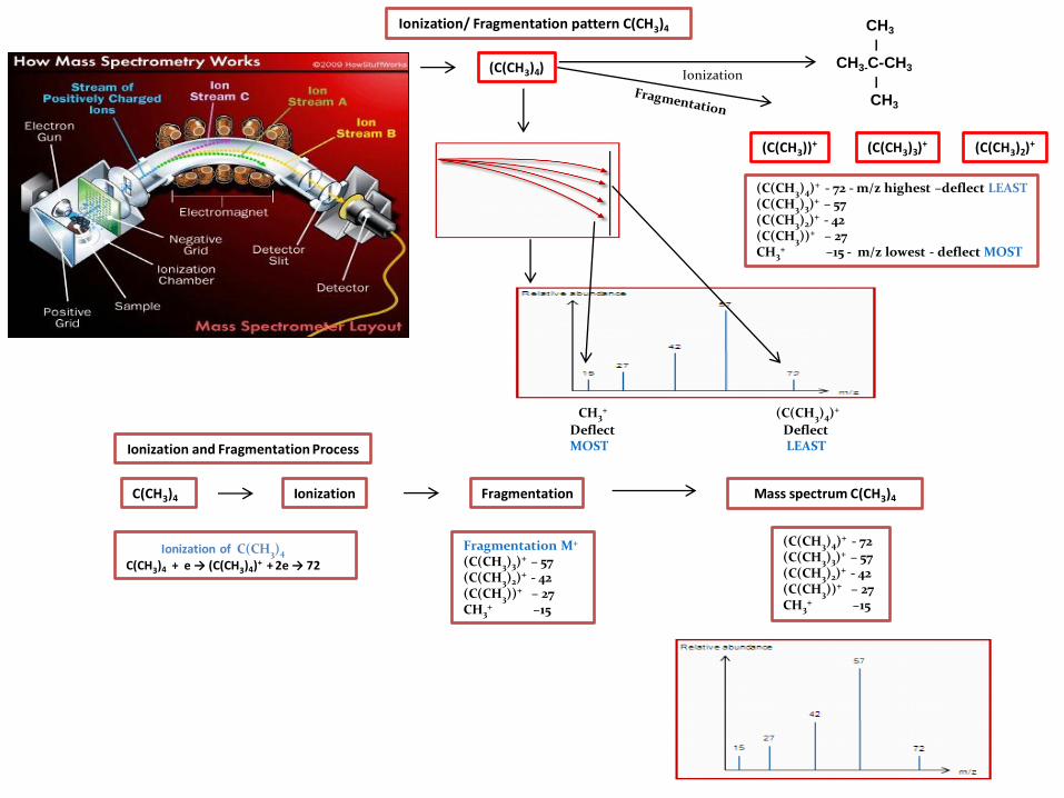
(C(CH3)4)+ - 72 - m/z highest –deflect LEAST (C(CH3)3)
+ – 57 (C(CH3)2)
+ - 42 (C(CH3))+ – 27 CH3
+ –15 - m/z lowest - deflect MOST
Ionization/ Fragmentation pattern C(CH3)4
Deflect MOST
Deflect LEAST
Ionization
Fragmentation
Ionization of C(CH3)4
C(CH3)4 + e → (C(CH3)4)+ + 2e → 72
Mass spectrum C(CH3)4 Ionization C(CH3)4
(C(CH3)3)+
(C(CH3)4)
(C(CH3)2)+ (C(CH3))+
(C(CH3)4)+ - 72 (C(CH3)3)
+ – 57 (C(CH3)2)
+ - 42 (C(CH3))+ – 27 CH3
+ –15
Fragmentation M+
(C(CH3)3)+ – 57
(C(CH3)2)+ - 42
(C(CH3))+ – 27 CH3
+ –15
Ionization and Fragmentation Process
CH3+ (C(CH3)4)+
CH3
׀
CH3-C-CH3
׀
CH3
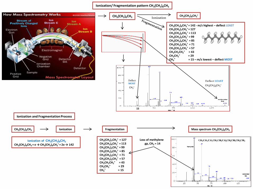
Ionization/ Fragmentation pattern CH3(CH2)8CH3
Ionization
Fragmentation
Ionization of CH3(CH2)8CH3
CH3(CH2)8CH3 + e → CH3(CH2)8CH3+ + 2e → 142
Mass spectrum CH3(CH2)8CH3 Ionization
CH3(CH2)8CH3 CH3(CH2)8CH3
+
CH3(CH2)8CH3+ = 142 - m/z highest – deflect LEAST
CH3(CH2)7CH2+ = 127
CH3(CH2)6CH2+ = 113
CH3(CH2)5CH2+ = 99
CH3(CH2)4CH2+ = 85
CH3(CH2)3CH3+ = 71
CH3(CH2)2CH2+ = 57
CH3CH2CH2+ = 43
CH3CH2+ = 29
CH3+ = 15 – m/z lowest – deflect MOST
Loss of methylene gp, CH2 = 14
CH3(CH2)8CH3
CH3(CH2)7CH2+ = 127
CH3(CH2)6CH2+ = 113
CH3(CH2)5CH2+ = 99
CH3(CH2)4CH2+ = 85
CH3(CH2)3CH3+ = 71
CH3(CH2)2CH2+ = 57
CH3CH2CH2+ = 43
CH3CH2+ = 29
CH3+ = 15
Deflect LEAST
CH3+
Deflect
MOST CH3(CH2)8CH3
+
Ionization and Fragmentation Process
15

Ionization/ Fragmentation pattern CH3(CH2)8CH3
Ionization
Fragmentation
Ionization of C6H5CH2OH
C6H5CH2OH + e → C6H5CH2OH+ + 2e → 108
Mass spectrum CH3(CH2)8CH3 Ionization
C6H5CH2OH+ = 108 - m/z highest – deflect LEAST C6H5CH2
+ = 91 C6H5
+ = 77 CH2OH+ = 31 OH+ = 17 – m/z lowest – deflect MOST
C6H5CH2OH C6H5CH2OH+
C6H5CH2OH
C6H5CH2OH+
C6H5CH2+ = 91
C6H5+ = 77
CH2OH+ = 31 OH+ = 17
C6H5CH2OH+ = 108 C6H5CH2
+ = 91 C6H5
+ = 77 CH2OH+ = 31 OH+ = 17
OH+
Deflect
MOST
Deflect LEAST
Ionization and Fragmentation Process
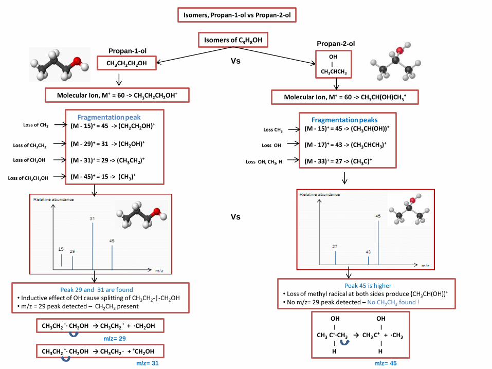
Isomers, Propan-1-ol vs Propan-2-ol
Peak 45 is higher • Loss of methyl radical at both sides produce (CH3CH(OH))+ • No m/z= 29 peak detected – No CH2CH3 found !
Fragmentation peak (M - 15)+ = 45 -> (CH2CH2OH)+
(M - 29)+ = 31 -> (CH2OH)+ (M - 31)+ = 29 -> (CH3CH2)+ (M - 45)+ = 15 -> (CH3)+
Isomers of C3H8OH
Fragmentation peaks (M - 15)+ = 45 -> (CH3CH(OH))+ (M - 17)+ = 43 -> (CH3CHCH3)+ (M - 33)+ = 27 -> (CH3C)+
Vs
Loss of CH3
Loss of CH3CH2
Loss of CH2OH
Loss of CH2CH2OH
Loss CH3
OH OH ׀ ׀ CH3 C+·CH3 → CH3 C
+ + ·CH3
׀ ׀ H H
m/z= 45
CH3CH2CH2OH
OH | CH3CHCH3
Loss OH
Loss OH, CH3, H
Peak 29 and 31 are found • Inductive effect of OH cause splitting of CH3CH2-|-CH2OH • m/z = 29 peak detected – CH2CH3 present
CH3CH2 +· CH2OH → CH3CH2
+ + ·CH2OH
m/z= 29
CH3CH2 +· CH2OH → CH3CH2 ·
+ +CH2OH
m/z= 31
Propan-1-ol Propan-2-ol
15
Vs
Molecular Ion, M+ = 60 -> CH3CH2CH2OH+ Molecular Ion, M+ = 60 -> CH3CH(OH)CH3+

Isomers, 2 methylbutane vs 2, 2 dimethylpropane
CH3
׀ CH3CHCH2CH3
CH3 | CH3C-CH3
| CH3
Peak 29 absent • No CH3CH2 Peak 57 is higher • Loss of methyl radical produce tertiary carbocation • Tertiary carbocation – More stable
Fragmentation peaks (M - 15)+ = 57 -> CH3CH(CH3)CH2
+ (M - 29)+ = 43 -> CH3CH(CH3)
+ (M - 43)+ = 29 -> CH3CH2
+ (M - 57)+ = 15 -> CH3
+
Isomers of C5H12
Fragmentation peaks (M - 15)+ = 57 -> C(CH3)3
+ (M - 30)+ = 42 -> C(CH3)2
+ (M - 45)+ = 27 -> CH3C+ (M - 57)+ = 15 -> CH3
+
Vs
Loss of CH3
Loss of CH3CH2
Loss of CH3CH(CH3)
Loss of CH3CH(CH3)CH2
Loss of CH3
Loss of TWO CH3
Loss of THREE CH3
CH3 ׀ CH3C+·CH3 ׀ CH3
m/z= 57
CH3
׀ CH3 C
+ + ·CH3
׀ CH3
2 methylbutane
2, 2 dimethylpropane
Loss of C(CH3)3
Vs
Peak 29 absent • CH3CH2 present
Molecular Ion, M+ = 72 -> CH3CH(CH3)CH2CH3+ Molecular Ion, M+ = 72 -> C(CH3)4
+

Normal Vs High Resolution Mass spectrometer
Normal Mass Spectrometer
• Molecular formula by adding all RAM
• RMM for molecule = Sum of all RAM • RMM O2 = 16 + 16 = 32 • RMM N2H4 = (14 x 2) + (1 x 4) = 32 • RMM CH3OH = (12 + 3 + 16 + 1) = 32 • Molecular ion peak - O2, N2H4, CH3OH - SAME = 32
RAM, O = 16 RAM, N = 14 RAM, H = 1 RAM, C = 12
High Resolution Mass Spectrometer Measure RMM to 4/5 decimal places
• Molecular formula by adding all RAM • RMM for molecule = Sum of all RAM • RMM O2 = 15.9949 + 15.9949 = 31.9898 • RMM N2H4 = (14.0031 x 2) + (1.0078 x 4) = 32.0375 • RMM CH3OH = (12.0000 )+ (3 x 1.0078) + 15.9949 = 32.0262 • Molecular ion peak- O2, N2H4, CH3OH is the NOT the same
RAM, O = 15.9949 RAM, N = 14.0031 RAM, H = 1.0078 RAM, C = 12.0000
Vs
Vs
O2, N2H4, CH3OH
Same 32
O2 N2H4 CH3OH
different
Video how MS works
High resolution Mass spectrum

37CI+ 35CI+
CI2 molecule
37CI-37CI - 74 - m/z highest – deflect LEAST 35CI-37CI –72 35CI-35CI –70 37CI –37 35CI –35 - m/z lowest– deflect MOST
Ionization/ Fragmentation pattern CI2
Deflect MOST
Deflect LEAST
35CI-35CI+
35CI+
35CI-37CI+
37CI-37CI+
Ionization
37CI+
37CI-37CI+
Fragmentation
Fragmentation of CI2+ into CI+
CI+.CI → [35CI+ + 35CI·] + 2e – 35 CI+.CI → [37CI+ + 37CI·] + 2e –37
Ionization of CI2 to CI2+
CI:CI + e- →[35CI+.35CI] + 2e – 70 CI:CI + e- →[35CI+.37CI] + 2e – 72 CI:CI + e- →[37CI+.37CI] + 2e – 74
m/z = 37
m/z = 35
Ratio (35CI : 37CI) - 3:1
Mass spectrum CI2 / CI atom
Ratio (35CI35CI: 35CI37CI: 37CI37CI) - 9:6:1
Ionization CI2 molecule
37CI-37CI - 74 35CI-37CI – 72 35CI-35CI – 70 37CI – 37 35CI – 35
Ionization and Fragmentation Process

Br2 molecule
81Br-81Br - 162 - m/z highest – deflect LEAST 79Br-81Br –160 79Br-79Br –158 81Br –81 79Br –79 - m/z lowest– deflect MOST
Deflect MOST Deflect LEAST
79Br-79Br+
79Br+
79Br-81Br+
81Br-81Br+
Ionization
81Br+
79Br+
81Br-81Br+
Fragmentation
Fragmentation of Br2+ to Br+
Br+.Br → [81Br+ + 81Br·] – 81 Br+.Br →[79Br+ + 79Br·] – 79
Ionization of Br2 to Br2+
Br:Br + e- →[81Br+.81Br] + 2e – 162 Br:Br + e- →[79Br+.81Br] + 2e – 160 Br:Br + e- →[79Br+.79Br] + 2e – 158
m/z = 79
m/z = 81
Ratio (79Br : 81Br) - 1:1
Mass spectrum Br2 / Br atoms
Ratio (79Br79Br: 79Br81Br: 81Br81Br) – 1:2:1
Ionization Br2 molecule
81Br-81Br - 162 79Br-81Br –160 79Br-79Br –158 81Br – 81 79Br – 79
Ionization/ Fragmentation pattern Br2
Ionization and Fragmentation Process

Ionization/ Fragmentation pattern CH3CH(CI)CH3
Ionization
Ionization
Ionization CH3CH(CI)CH3
CH3CH(CI)CH3+ e → CH3CH(CI)CH3+ + 2e → 78/80
Presence isotope 35CI and 37CI
CH3CH(37CI)CH3+ = 80 - m/z highest – deflect LEAST
CH3CH(35CI)CH3+ = 78
CH3CH(37CI)+ = 65 CH3CH(35CI)+ = 63 CH3CHCH3
+ = 43 CH3C
+ = 27 - m/z lowest – deflect MOST
CH3CH(37CI)+ = 65 CH3CH(35CI)+ = 63 CH3CHCH3
+ = 43 CH3C
+ = 27
CH3CH(CI)CH3 CH3CH(CI)CH3+
CH3CH(CI)CH3+
Isotopic peak (M+)= 78
Isotopic peak (M++2) = 80
CH3CH(35CI)CH3 CH3CH(37CI)CH3
Isotopic peak 63
Isotopic peak 65
CH3CH(35CI)+ CH3CH(37CI)+
CH3CH(CI)CH3 Fragmentation
CH3C+
Deflect MOST Deflect LEAST
Presence M+ and (M++ 2) peak
Presence of Isotopes
Ionization and Fragmentation Process

CH3CH2CH279Br CH3CH2CH2
81Br CH2CH279Br CH2CH2
81Br
Ionization/ Fragmentation pattern CH3CH2CH3Br
Ionization
Ionization
Ionization CH3CH2CH2Br
CH3CH2CH2Br + e → CH3CH2CH2Br+ + 2e → 122/124
Presence isotope 79Br and 81Br
CH3CH2CH281Br+ = 124 - m/z highest – deflect LEAST
CH3CH2CH279Br + = 122
CH2CH281Br+ = 109
CH2CH279Br+ = 107
CH281Br+ = 95
CH279Br+ = 93
CH3CH2CH2+ = 43
CH3C + = 27 - m/z lowest – deflect MOST
Isotopic peak (M+) = 122
Isotopic peak (M++2) = 124
Isotopic peak 107
Isotopic peak 109
Fragmentation
CH3C+
Deflect MOST Deflect LEAST
CH3CH2CH2Br CH3CH2CH2Br+
CH3CH2CH3Br
CH3CH2CH2Br+
CH2CH281Br+ = 109
CH2CH279Br+ = 107
CH281Br+ = 95
CH279Br+ = 93
CH3CH2CH2+ = 43
CH3C + = 27
CH3C+
Deflect
LEAST
CH3CH2CH2Br+
Presence of M+ and (M++ 2) peak
Presence of Isotopes
Deflect
MOST
Ionization and Fragmentation Process
TI
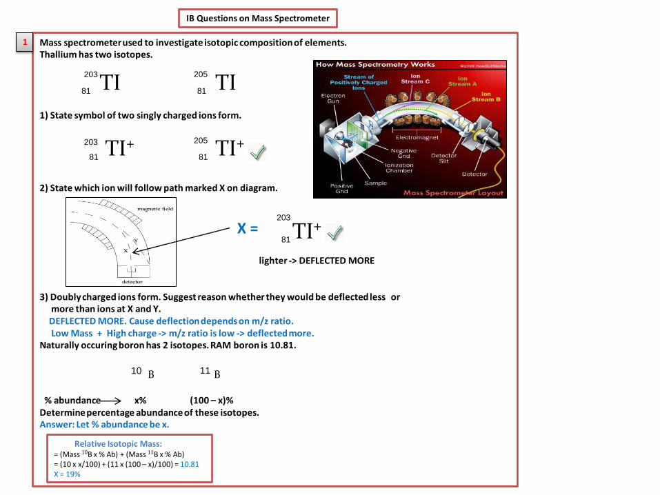
IB Questions on Mass Spectrometer
Mass spectrometer used to investigate isotopic composition of elements. Thallium has two isotopes. 1) State symbol of two singly charged ions form. 2) State which ion will follow path marked X on diagram. lighter -> DEFLECTED MORE 3) Doubly charged ions form. Suggest reason whether they would be deflected less or more than ions at X and Y. DEFLECTED MORE. Cause deflection depends on m/z ratio. Low Mass + High charge -> m/z ratio is low -> deflected more. Naturally occuring boron has 2 isotopes. RAM boron is 10.81.
% abundance x% (100 – x)% Determine percentage abundance of these isotopes. Answer: Let % abundance be x.
1
TI TI 203 205
81 81
X =
B 10 B 11
Relative Isotopic Mass: = (Mass 10B x % Ab) + (Mass 11B x % Ab) = (10 x x/100) + (11 x (100 – x)/100) = 10.81 X = 19%
TI+ 81
203 TI+ 205
81
TI+ 203
81

IB Questions on Mass Spectrometer
Germanium is analysed in mass spec. The first and last processes are vaporization and detection. 1) State the names of other 3 processes in order in which they occur Answer: Ionization -> Acceleration -> Deflection 2) For each of processes named in a (i), outline how process occur Ionization -> Sample bombarded with high energy/high speed electrons Acceleration -> Cations (+ve charged ions) accelerated by an electric field Deflection -> Cations deflected by a magnetic field 3) Germanium found to have following composition i)Define relative atomic mass. Average / weighted masses of all isotopes of an element. ii) Calculate RAM, giving answer to two decimal places.
2
Relative Isotopic Mass = (Mass 70Ge x % Ab) + (Mass 72Ge x % Ab) + (Mass 74Ge x % Ab) + (Mass 76Ge x % Ab) = (70 x 22.60/100) + (72 x 25.45/100) + (74 x 36.73/100) + (76 x 15.22/100) = 72.89

IB Questions on Mass Spectrometer
Shows a mass spectrometer. 1)Identify the parts labelled A, B and C.
2)State and explain which one will undergo greatest deflection. Answer : Greatest deflection -> lowest mass + highest charged -> m/z -> lowest 3) Mass spectrum shown below: i) Explain why there is more than one peak. Existence of isotopes ii) Calculate RAM.
3
Relative Isotopic Mass
= (Mass 24Y x % Ab) + (Mass 25Y x % Ab) + (Mass 26Yx % Ab) = (24 x 79/100) + (25 x 10/100) + (26 x 11/100) = 24.32
• electron gun • ionisation chamber • ionizer
• Electric field • Charged plates • Potential difference
• Magnetic field • Magnet • Electromagnet
greatest deflection – low mass, high charged
smallest deflection – high mass, low charged
A
C
B
Li+
Li2+
7
6

IB Questions on Mass Spectrometer
Vaporized magnesium is introduced into mass spec. One of the ions that reaches detector shown below. 1)Identify number of protons, neutron and electrons Answer : 12 protons, 13 neutrons, 11 electrons
2) State how ion is accelerated in mass spectrometer. Using a strong electric field/strong opposite charged plate/potential difference 3) The ion is also detected by changing the magnetic field. Deduce and explain by reference to m/z values of these two ions of magnesium, which of the ions and is detected using a stronger magnetic field. Answer: - due to lower charge -> m/z is higher -> deflected less -> needs a stronger magnetic field to deflect.
4
Cations (+ve) accelerated by (-ve) plates
25
12
25Mg 2+
smallest deflection – high mass, low charged
Strong magnet/magnetic field to deflect it to bottom
Mg +
25Mg 2+ 25Mg +
25Mg +
25Mg +

Rubidium contains two stable isotopes. RAM for rubidium is 85.47 1)Calculate % of each isotope in rubidium. Answer : Let % abundance be x %.
% Abundance x% (100 – x)%
76.5% 23.5%
2) Vaporized sample is ionized and accelerated. How the use of magnetic field and detector enables percentage of two isotopes to be determined.
5
85 87
Relative Isotopic Mass: = (Mass 85Rb x % Ab) + (Mass 87Rb x % Ab) = (85 x x/100) + (87 x (100 – x)/100) = 85.47 X = 76.5%
Rb
Detector • Convert abundance M+ ions to current. • M+ ions neutralize by electrons (more e needed - higher current – higher intensity of peak) •Ratio of intensity peaks show ratio of ions in sample •Ratio of height of peaks due to 85Rb : 87Rb –> 76.5 : 23.5
Magnetic field/Deflector • M+ ion deflected by magnetic field
- lighter -> deflected more
- heavier -> deflected less
IB Questions on Mass Spectrometer
Rb Rb
85 Rb 87 Rb
85 Rb 87 Rb
85 Rb
87 Rb