IB Assessment Statement H.2.1 State that digestive juices are secreted into the alimentary canal by...
63
IB Assessment Statement • H.2.1 State that digestive juices are secreted into the alimentary canal by glands, including salivary glands, gastric glands in the stomach wall, the pancreas and the wall of the small intestine.
-
Upload
thomasina-lyons -
Category
Documents
-
view
215 -
download
0
Transcript of IB Assessment Statement H.2.1 State that digestive juices are secreted into the alimentary canal by...
IB Assessment Statement
• H.2.1 State that digestive juices are secreted into the alimentary canal by glands, including salivary glands, gastric glands in the stomach wall, the pancreas and the wall of the small intestine.

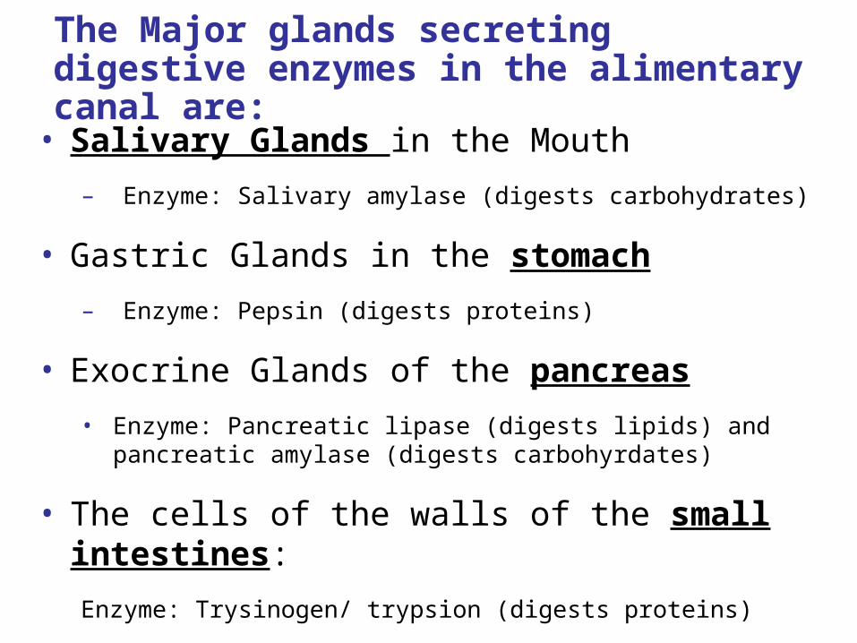
The Major glands secreting digestive enzymes in the alimentary canal are:
• Salivary Glands in the Mouth
– Enzyme: Salivary amylase (digests carbohydrates)
• Gastric Glands in the stomach
– Enzyme: Pepsin (digests proteins)
• Exocrine Glands of the pancreas
• Enzyme: Pancreatic lipase (digests lipids) and pancreatic amylase (digests carbohyrdates)
• The cells of the walls of the small intestines:
Enzyme: Trysinogen/ trypsion (digests proteins)
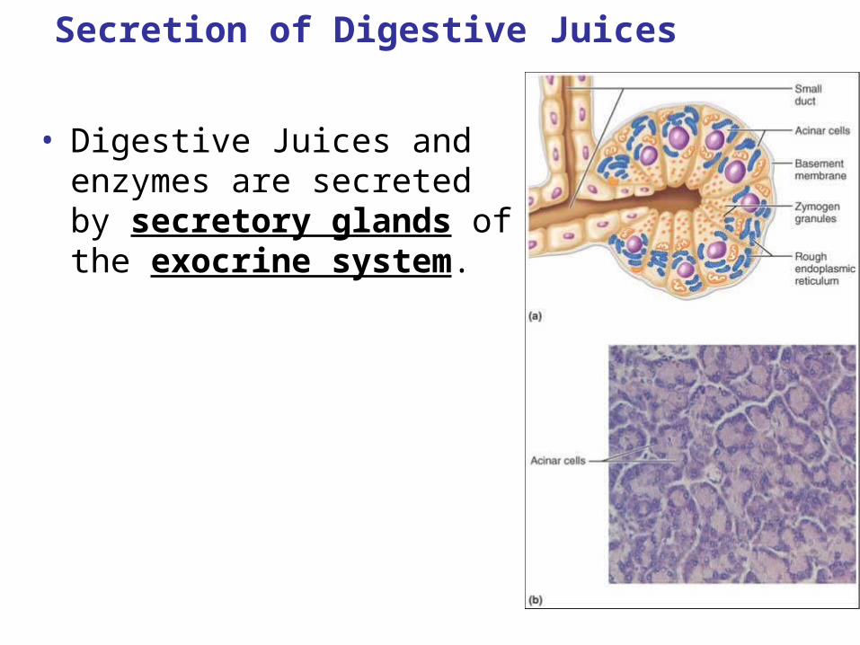
Secretion of Digestive Juices
• Digestive Juices and enzymes are secreted by secretory glands of the exocrine system.
IB Learning Objective: Identify and describe the parts of the digestive system that secretes digestive enzymes.
Gastric secretion Animations
•http://highered.mheducation.com/sites/0072437316/student_view0/chapter43/animations.html#
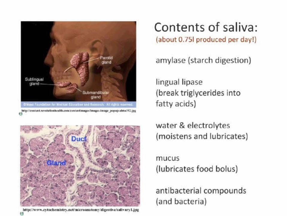
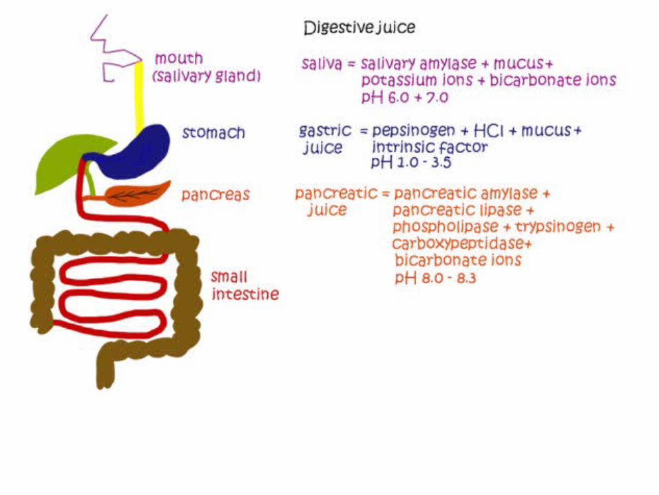
Summary of Saliva & Salivary Gland
• Secrete in mouth
• pH 6.5-7.5
• Contents
– Amylase – function to digest starch
– Mucus –lubricate food.

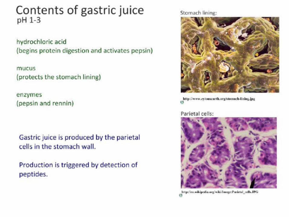
Summary of Gastric Juice & Stomach gastric glands
• Secrete in stomach
• pH 2
• Contents
– Pepsin – function to digest protein
– Hydrochloric Acid – creates acidic environments to kill bacteria
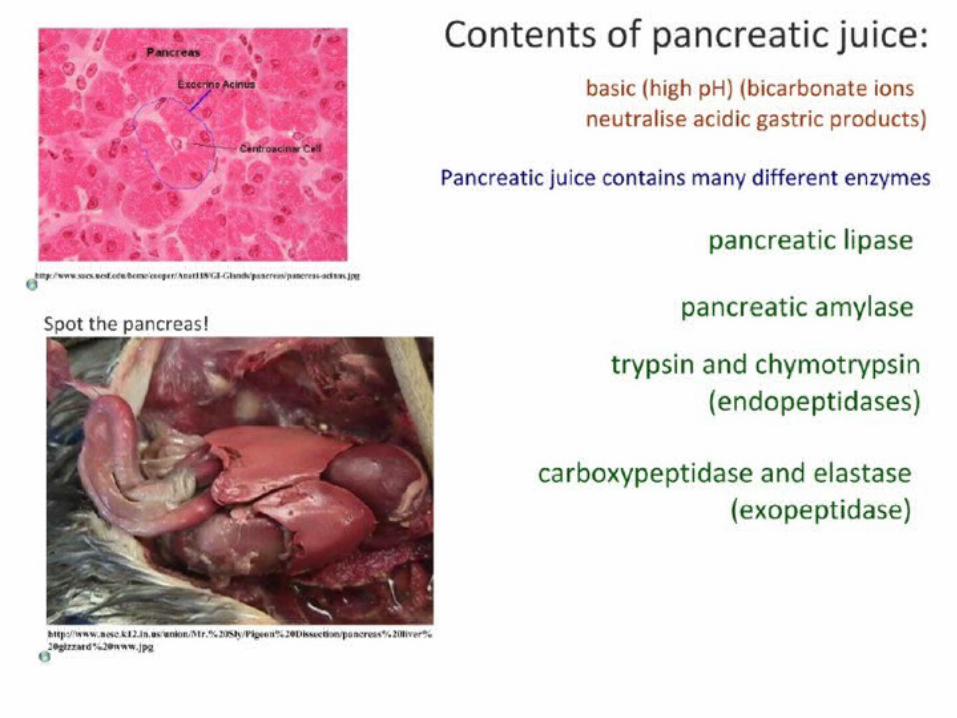
Summary of Pancreatic Juice & Pancreas
• Secrete into small intestine
• pH 7
• Contents
– amylase – function to digest carbohydrates
– Protease (trypsin) – function to digest proteins
– Lipase – functions to digests lipids
– Nucleases – functions to digest nucleic acid
IB Assessment Statement
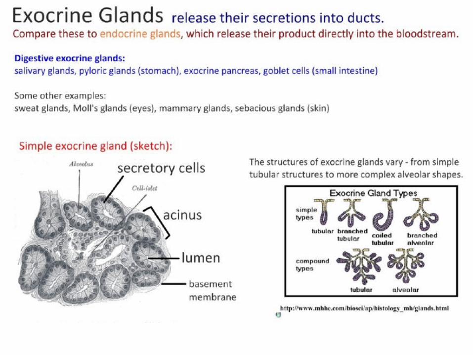
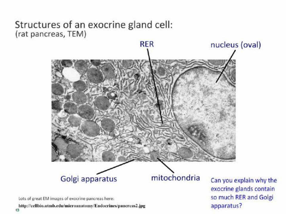
• H.2.2 Explain the structural features of exocrine gland cells.
– Include the secretory cells grouped into acini and ducts, and the ultrastructure of secretory cells as seen in the electron micrographs
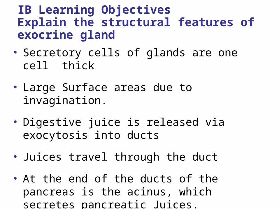
IB Learning Objectives Explain the structural features of exocrine gland
• Secretory cells of glands are one cell thick
• Large Surface areas due to invagination.
• Digestive juice is released via exocytosis into ducts
• Juices travel through the duct
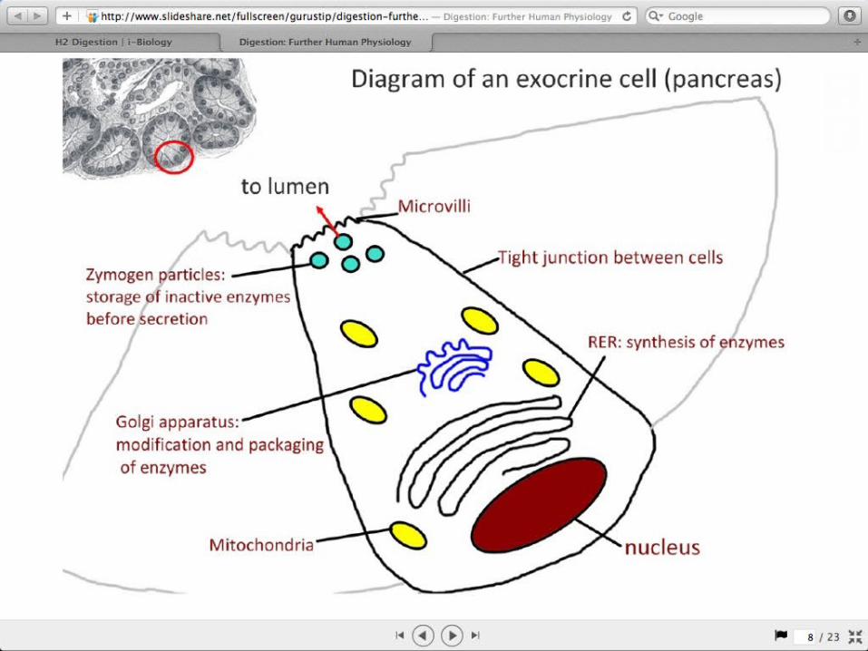
• At the end of the ducts of the pancreas is the acinus, which secretes pancreatic Juices.
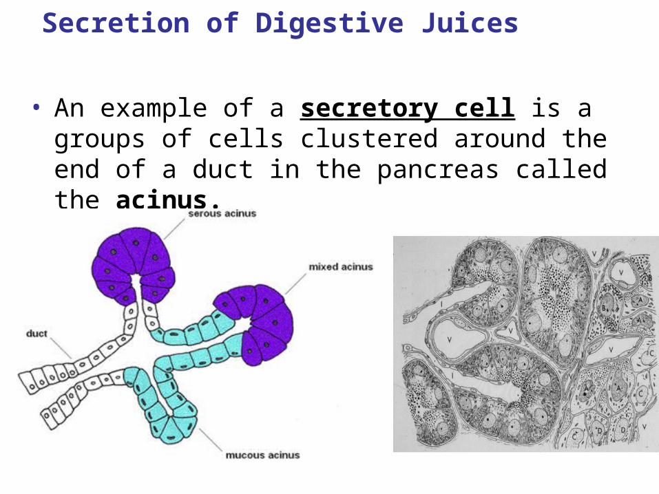
Secretion of Digestive Juices
• An example of a secretory cell is a groups of cells clustered around the end of a duct in the pancreas called the acinus.
Secretion of Digestive Juices
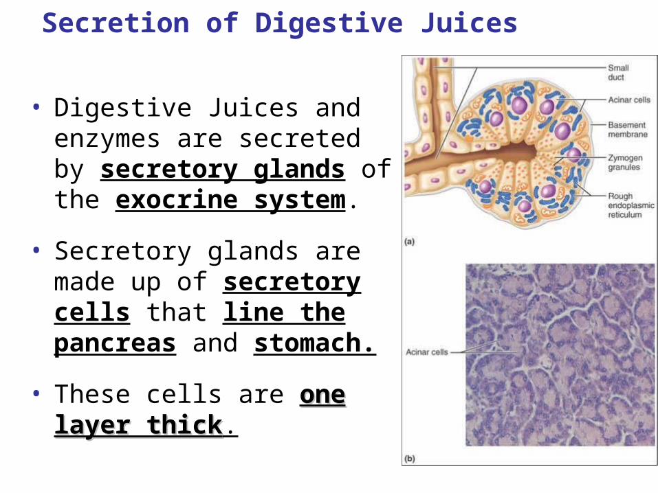
• Digestive Juices and enzymes are secreted by secretory glands of the exocrine system.
• Secretory glands are made up of secretory cells that line the pancreas and stomach.
• These cells are one layer one layer thickthick.
Secretion of Digestive Juices
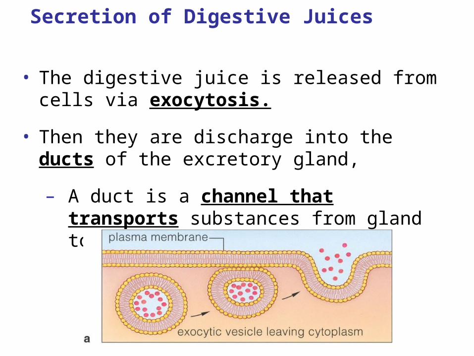
• The digestive juice is released from cells via exocytosis.
• Then they are discharge into the ducts of the excretory gland,
– A duct is a channel that transports substances from gland to organ.
Secretion of Digestive Juices
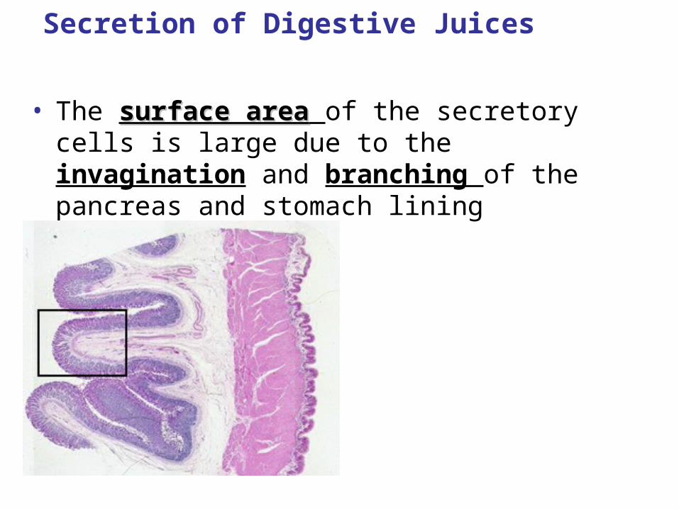
• The surface areasurface area of the secretory cells is large due to the invagination and branching of the pancreas and stomach lining
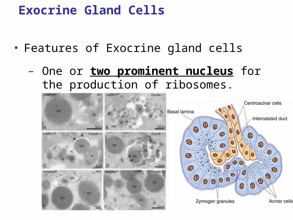
Exocrine Gland Cells
• Features of Exocrine gland cells
– One or two prominent nucleus for the production of ribosomes.
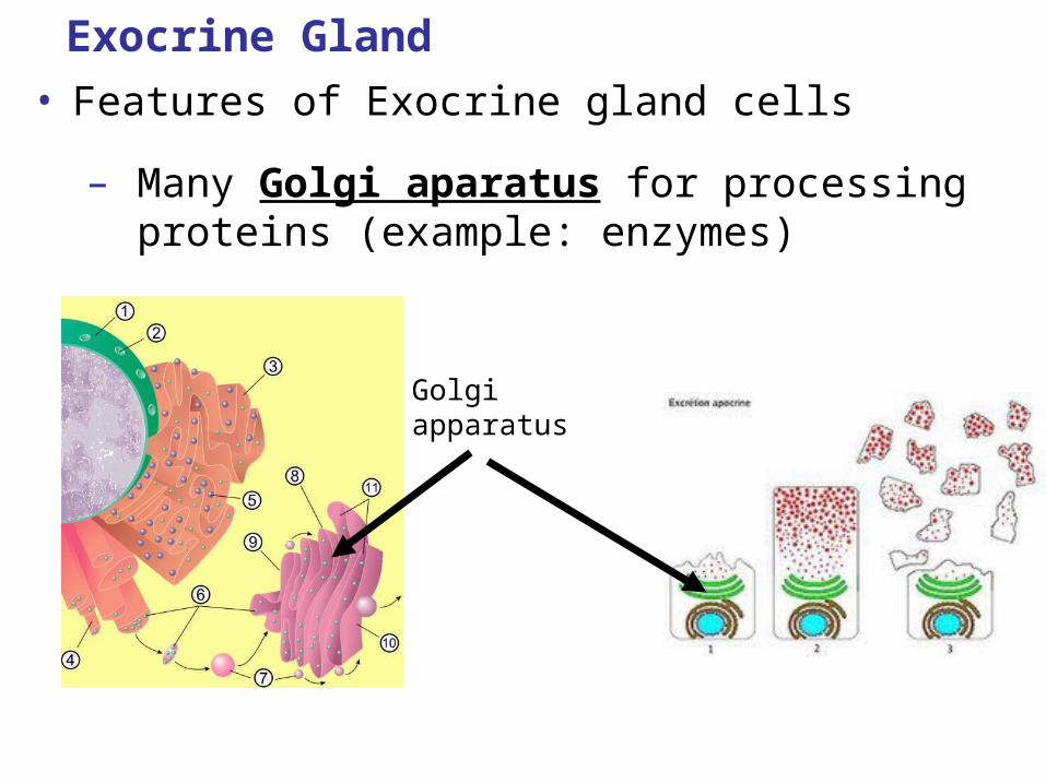
Exocrine Gland
• Features of Exocrine gland cells
– Many Golgi aparatus for processing proteins (example: enzymes)
Golgi apparatus
Exocrine Gland
• Features of Exocrine gland cells
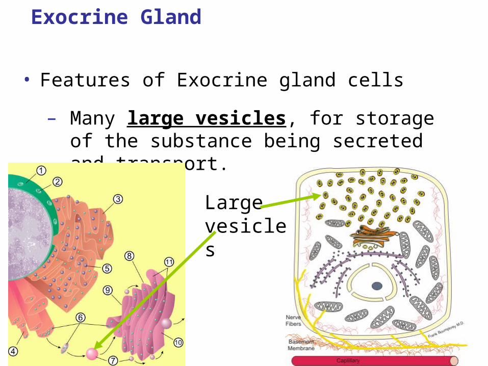
– Many large vesicles, for storage of the substance being secreted and transport.
Large vesicles
Exocrine Gland
• Features of Exocrine gland cells
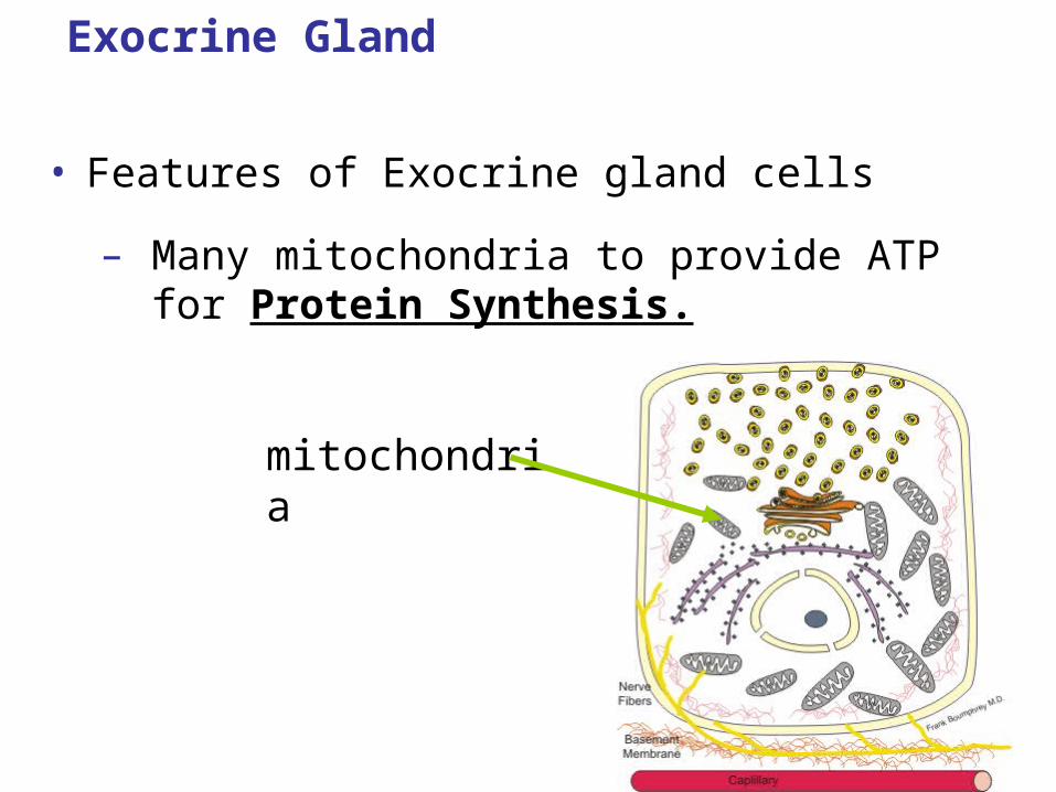
– Many mitochondria to provide ATP for Protein Synthesis.
mitochondria
IB LEARNING OBJECTIVE
• H.2.3 Compare the composition of saliva, gastric juice and pancreatic juice.
IB LEARNING OBJECTTIVE
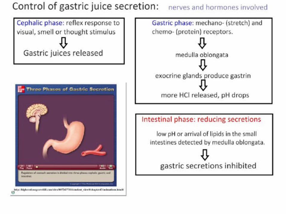
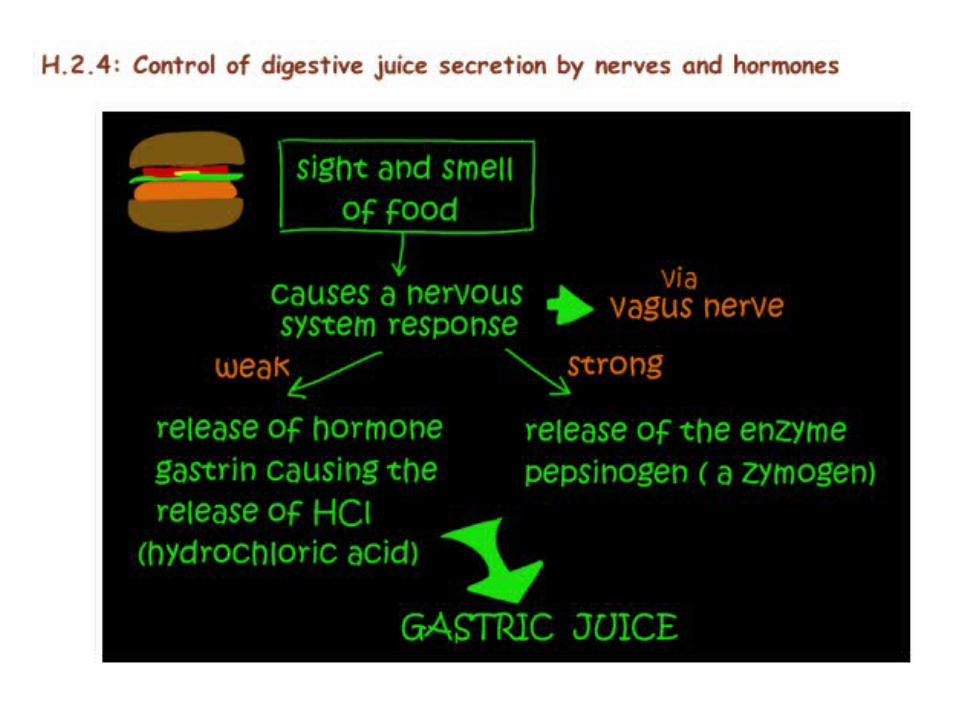
• H.2.4 Outline the control of digestive juice secretion by nerves and hormones, using the example of secretion of gastric juice.

The control of Secretion of Digestions Juices
• The secretion of digestive juices is regulated by nervous or/ and hormonal mechanisms.
The control of Secretion of Digestions Juices
Nervous Response
• The sight or smell of food stimulates a nervous impulse from the brain to the exocrine glands of the walls of the stomach to start gastric secretions.
• More gastric secretions are released when food enters the stomach
– Chemoreceptors and touch receptors in the lining of the stomach
• These receptors send impulse to brain, which sends impulses to the exocrine gland cells.
The control of Secretion of Digestions Juices
Hormonal Response – ENDOCRINE SYSTEM RESPONSE
– When food enters the stomach nervous impulse are sent to endocrine glands in stomach lining
– Endocrine glands release gastrin hormone
– Gastrin stimulates exocrine gland cells to secret hydrochloric acid (HCl)
• This lowers the pH in stomach to 3
IB Learning Objective:
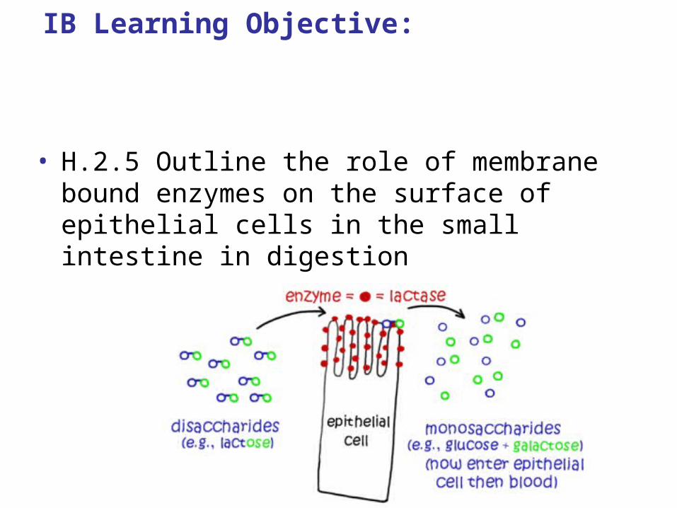
• H.2.5 Outline the role of membrane bound enzymes on the surface of epithelial cells in the small intestine in digestion
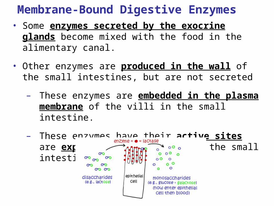
Membrane-Bound Digestive Enzymes• Some enzymes secreted by the exocrine glands
become mixed with the food in the alimentary canal.
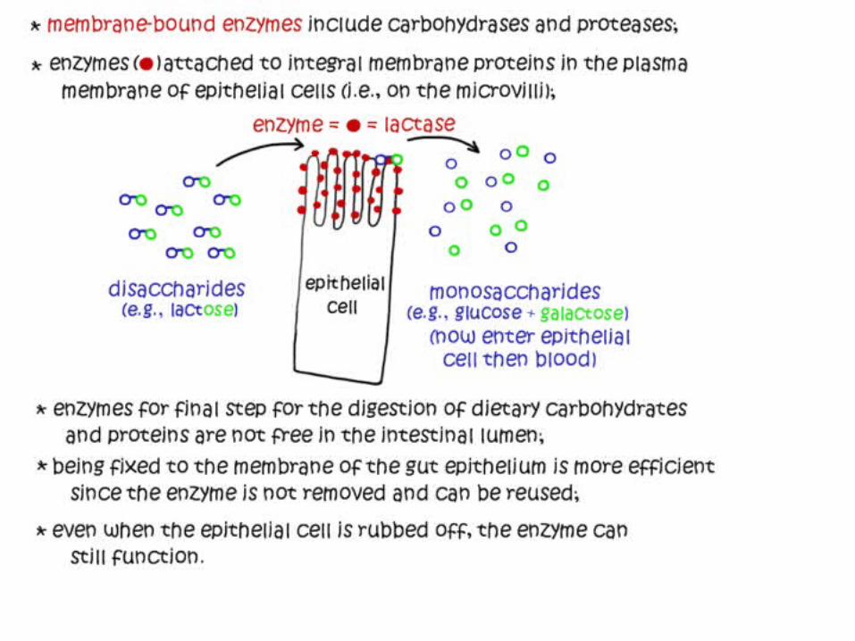
• Other enzymes are produced in the wall of the small intestines, but are not secreted
– These enzymes are embedded in the plasma membrane of the villi in the small intestine.
– These enzymes have their active sites are exposed to the food inside the small intestines.
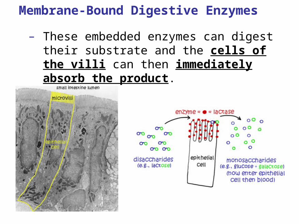
Membrane-Bound Digestive Enzymes
– These embedded enzymes can digest their substrate and the cells of the villi can then immediately absorb the product.
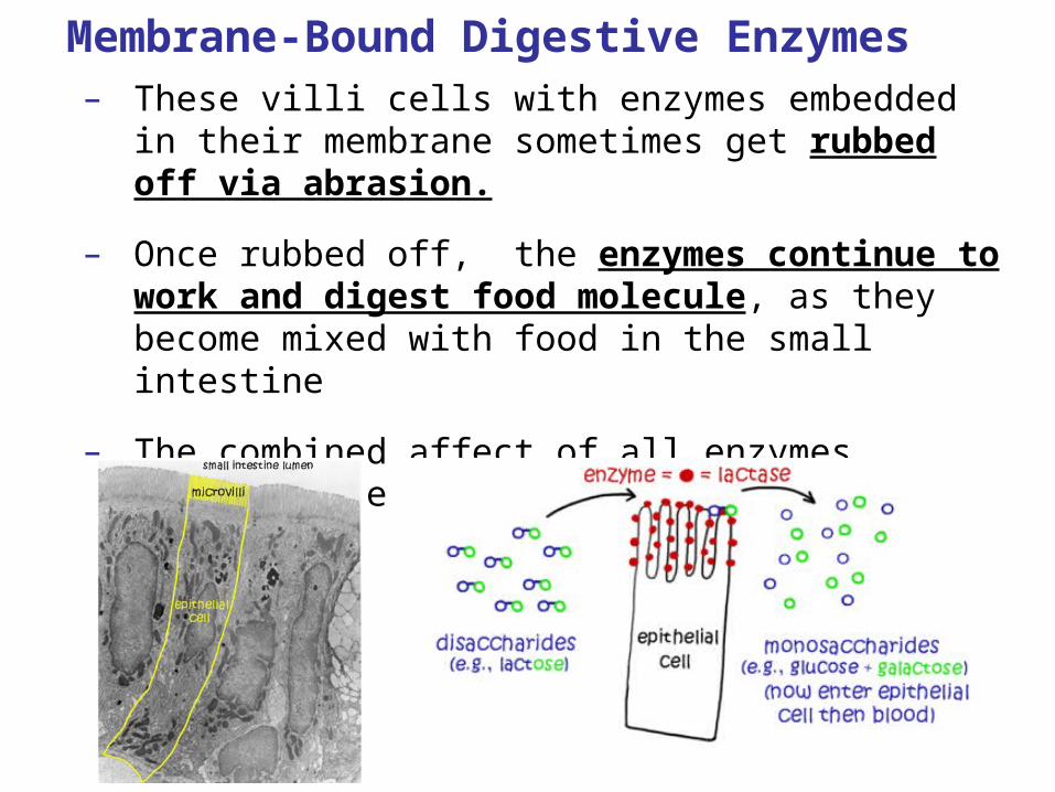
Membrane-Bound Digestive Enzymes– These villi cells with enzymes embedded in their
membrane sometimes get rubbed off via abrasion.
– Once rubbed off, the enzymes continue to work and digest food molecule, as they become mixed with food in the small intestine
– The combined affect of all enzymes complete the process of digestions.

IB Assessment Statement
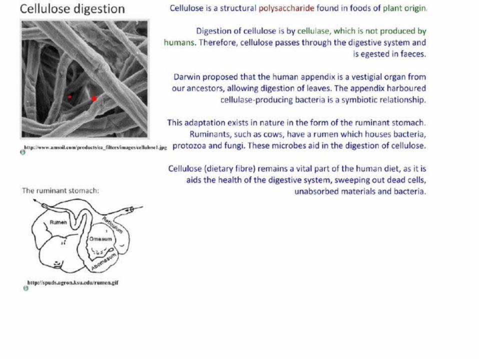
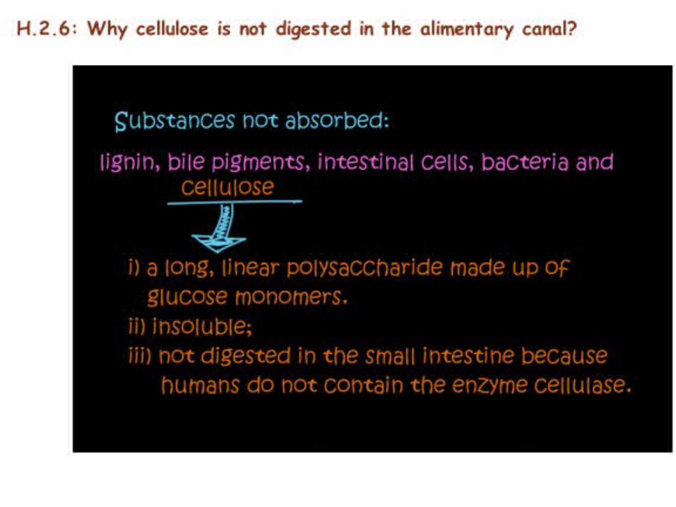
• H.2.6 Outline the reasons for cellulose not being digested in the alimentary canal.
IB Assessment Statement
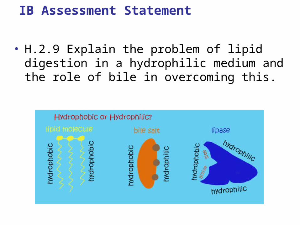
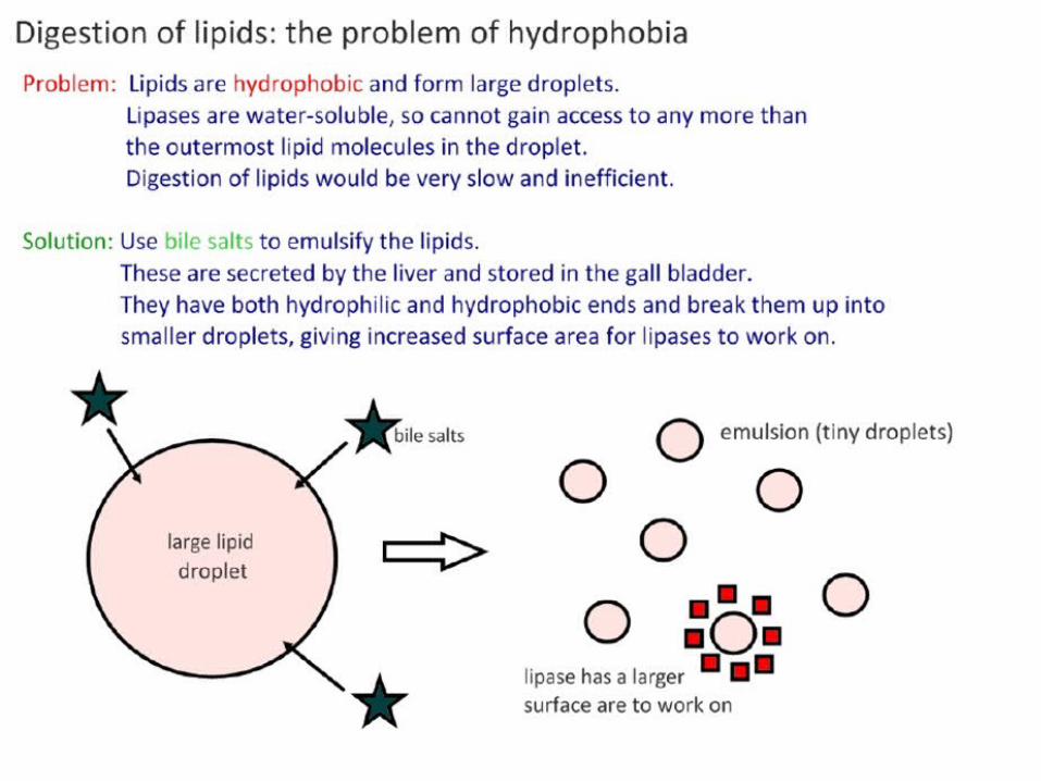
• H.2.9 Explain the problem of lipid digestion in a hydrophilic medium and the role of bile in overcoming this.
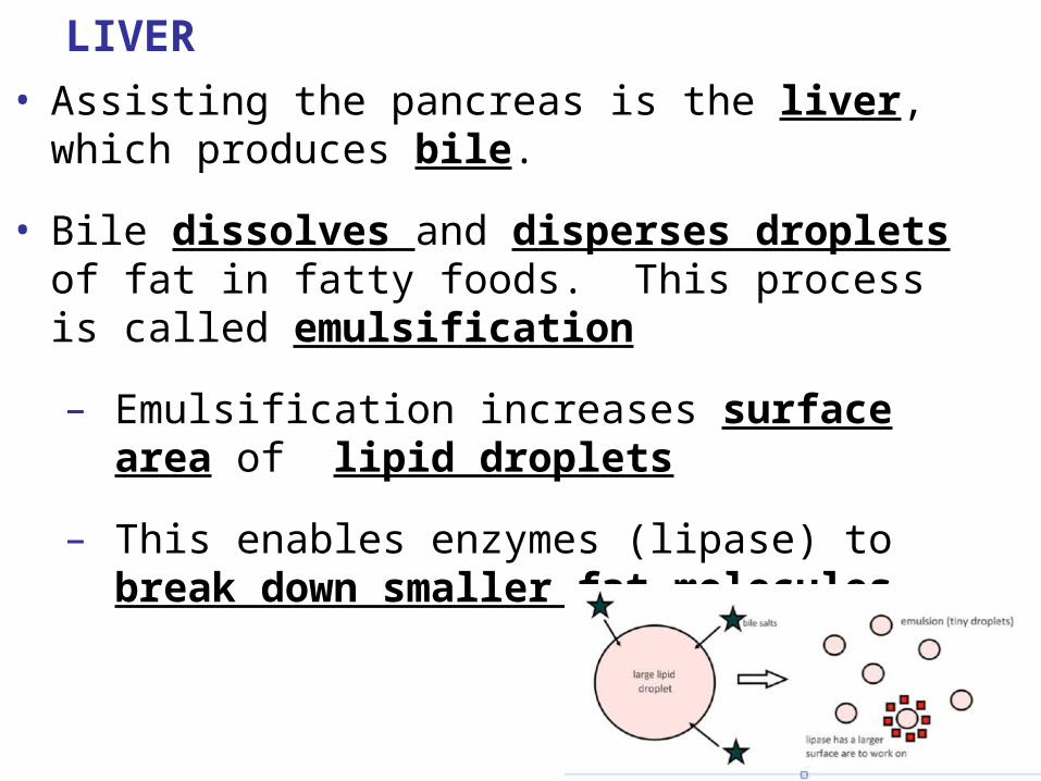
LIVER
• Assisting the pancreas is the liver, which produces bile.
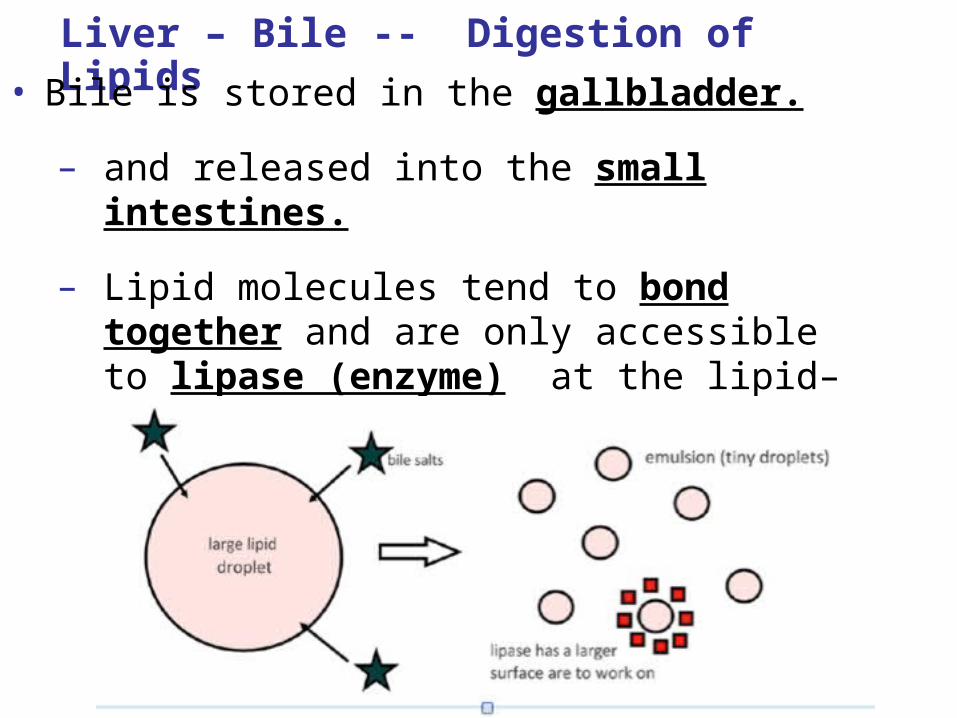
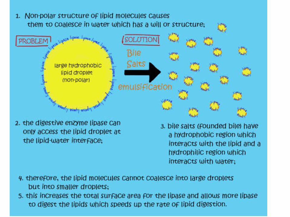
• Bile dissolves and disperses droplets of fat in fatty foods. This process is called emulsification
– Emulsification increases surface area of lipid droplets
– This enables enzymes (lipase) to break down smaller fat molecules.
Liver – Bile -- Digestion of Lipids
• Bile is stored in the gallbladder.
– and released into the small intestines.
– Lipid molecules tend to bond together and are only accessible to lipase (enzyme) at the lipid–water interface/ boundary.
Liver – Bile -- Digestion of Lipids
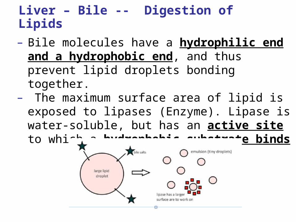
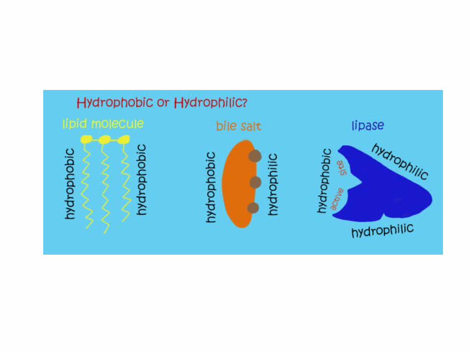
– Bile molecules have a hydrophilic end and a hydrophobic end, and thus prevent lipid droplets bonding together.
– The maximum surface area of lipid is exposed to lipases (Enzyme). Lipase is water-soluble, but has an active site to which a hydrophobic substrate binds
Digestion of Lipids Animation
• http://www.wiley.com/legacy/college/boyer/0470003790/animations/fatty_acid_metabolism/fatty_acid_metabolism.htm
IB Learning Objective
H.2.7 Explain why pepsin and trypsin are initially synthesized as inactive precursors and how they are subsequently activated
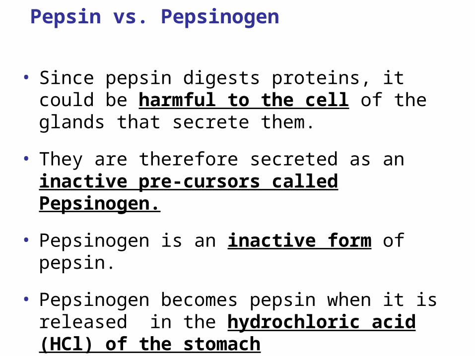
Pepsin vs. Pepsinogen
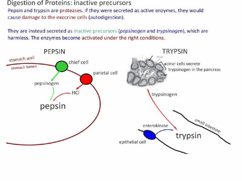
• Since pepsin digests proteins, it could be harmful to the cell of the glands that secrete them.
• They are therefore secreted as an inactive pre-cursors called Pepsinogen.
• Pepsinogen is an inactive form of pepsin.
• Pepsinogen becomes pepsin when it is released in the hydrochloric acid (HCl) of the stomach
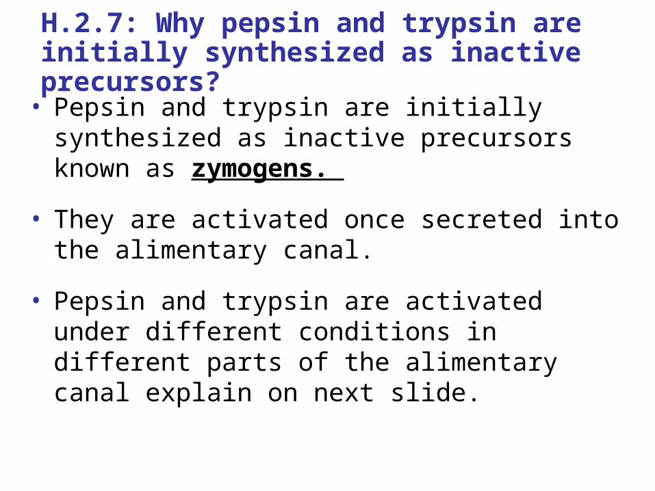
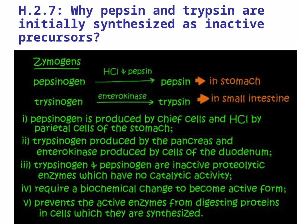
H.2.7: Why pepsin and trypsin are initially synthesized as inactive precursors?
• Pepsin and trypsin are initially synthesized as inactive precursors known as zymogens.
• They are activated once secreted into the alimentary canal.
• Pepsin and trypsin are activated under different conditions in different parts of the alimentary canal explain on next slide.
H.2.7: Why pepsin and trypsin are initially synthesized as inactive precursors?
IB Learning Objective
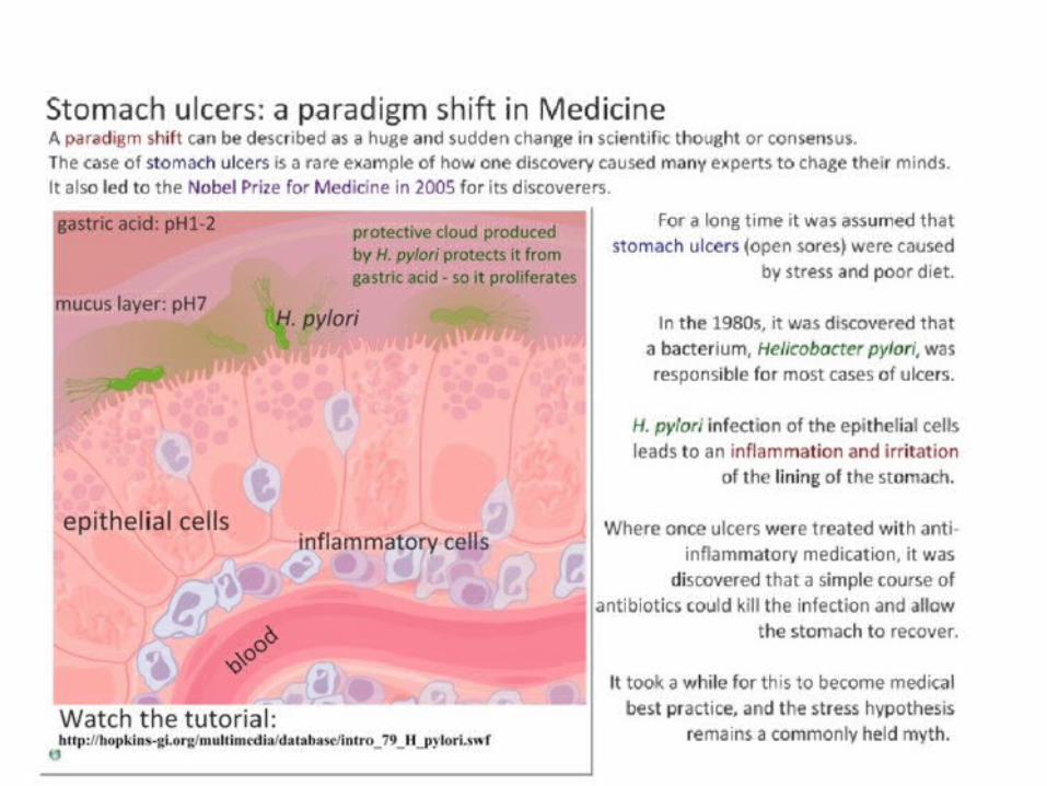
H.2.8 Discuss the roles of gastric acid and Helicobacter pylori in the development of stomach ulcers and stomach cancers
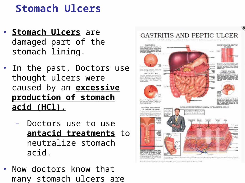
Stomach Ulcers
• Stomach Ulcers are damaged part of the stomach lining.
• In the past, Doctors use thought ulcers were caused by an excessive production of stomach acid (HCl).
– Doctors use to use antacid treatments to neutralize stomach acid.
• Now doctors know that many stomach ulcers are caused by an acid tolerant bacteria called, Helicobacter pylori (H. pylori).
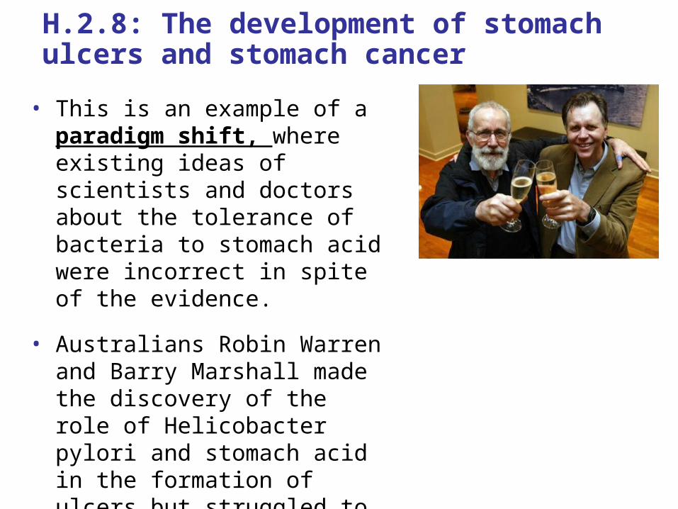
H.2.8: The development of stomach ulcers and stomach cancer
• This is an example of a paradigm shift, where existing ideas of scientists and doctors about the tolerance of bacteria to stomach acid were incorrect in spite of the evidence.
• Australians Robin Warren and Barry Marshall made the discovery of the role of Helicobacter pylori and stomach acid in the formation of ulcers but struggled to convince the scientific and medical community.
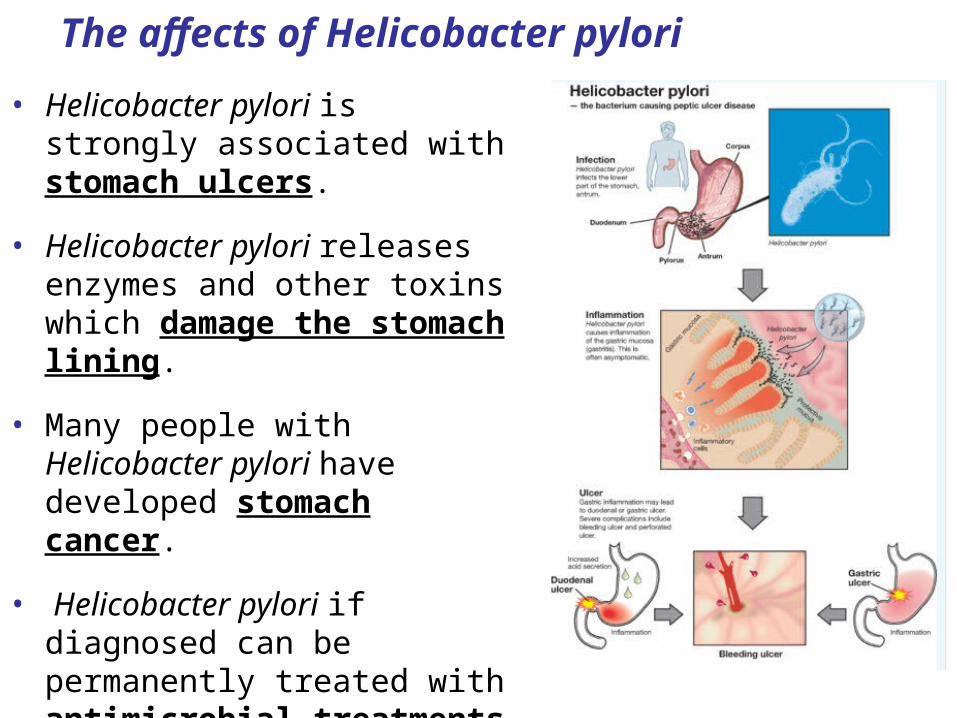
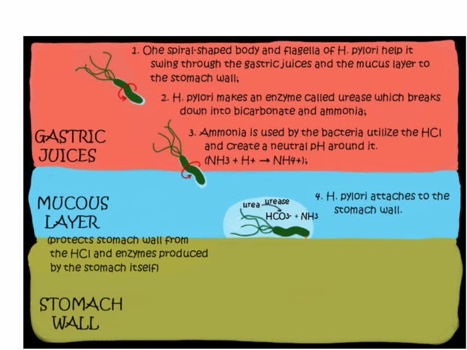
The affects of Helicobacter pylori
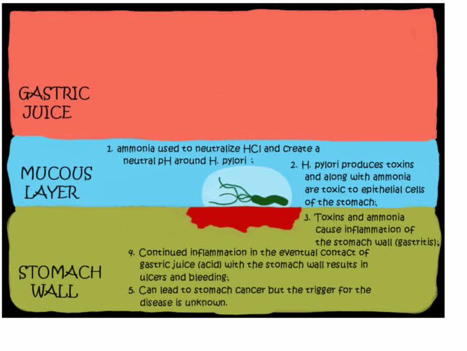
• Helicobacter pylori is strongly associated with stomach ulcers.
• Helicobacter pylori releases enzymes and other toxins which damage the stomach lining.
• Many people with Helicobacter pylori have developed stomach cancer.
• Helicobacter pylori if diagnosed can be permanently treated with antimicrobial treatments that eliminate this bacterial infection.