IAEA HUMAN HEALTH SERIES IAEA HUMAN HEALTH SERIES
140
IAEA HUMAN HEALTH SERIES No. 20 Practical Guidance on Peptide Receptor Radionuclide Therapy (PRRNT) for Neuroendocrine Tumours IAEA
Transcript of IAEA HUMAN HEALTH SERIES IAEA HUMAN HEALTH SERIES
INTERNATIONAL ATOMIC ENERGY AGENCYVIENNA
ISBN 978–92–0–129210–0ISSN 2075–3772
Practical G
uidance on Peptide R
eceptor Radionuclide Therapy (P
RR
NT) for N
euroendocrine Tumours
This publication provides comprehensive, multidisciplinary guidance on the use of peptide receptor radionuclide therapy (PRRNT) in the treatment of patients with neuroendocrine tumours (NETs) and gastroenteropancreatic cancers, taking into account the recent international classifi cations of NETs. It provides comprehensive protocols for employing 90Y or 177Lu tagged somatostatin receptor targeting peptides as well as clinically assessed protocols for renal protection. It provides comprehensive, evidence based clinical guidelines, with input from experienced and renowned medical professionals in the fi eld. The various sections of the book cover clinical presentation, patient eligibility criteria and means of assessing the effectiveness of therapy using molecular and morphological medical imaging techniques.
Practical G
uidance on Peptide R
eceptor Radionuclide Therapy (P
RR
NT) for N
euroendocrine Tumours
IAEA HUMAN HEALTH SERIES IAEA HUMAN HEALTH SERIESNo. 20
IAEA HUMAN HEALTH SERIES No. 20
Practical Guidance on Peptide Receptor
Radionuclide Therapy (PRRNT) for
Neuroendocrine Tumours
IAEA HUMAN HEALTH SERIES
RELATED PUBLICATIONS
www.iaea.org/books
Nuclear MediciNe resources MaNualSTI/PUB/1198 (532 pp.; 2006)ISBN 92–0–107504–9 Price: €65.00
Quality MaNageMeNt audits iN Nuclear MediciNe PracticesSTI/PUB/1371 (57 pp.; 2008)ISBN 978–92–0–112108–0 Price: €25.00
a guide to cliNical Pet iN oNcology: iMProviNg cliNical MaNageMeNt of caNcer PatieNtsiaea tecdoc series No. 1605IAEA-TECDOC-1605 (55 pp.; 2008)ISBN 978–92–0–110608–7 Price: €15.00
criteria for PalliatioN of BoNe Metastases – cliNical aPPlicatioNsiaea tecdoc series No. 1549IAEA-TECDOC CD-1549 (55 pp.; 2007)ISBN 92–0–104507–7 Price: €15.00
Nuclear MediciNe iN thyroid caNcer MaNageMeNt: a Practical aPProachiaea tecdoc series No. 1608IAEA TECDOC-1608 (273 pp.; 2009)ISBN 978–92–0–113108–9 Price: €15.00
release of PatieNts after radioNuclide theraPysafety reports series No. 63STI/PUB/1417 (77 pp.; 2009)ISBN 978–92–0–108909–0 Price: €28.00
Quality assuraNce for radioactive MeasureMeNt iN Nuclear MediciNetechnical reports series No. 454STI/DOC/010/454 (81 pp.; 2006)ISBN 92–0–105306–1 Price: €42.00
IAEA HUMAN HEALTH SERIES PUBLICATIONS
The mandate of the IAEA human health programme originates from Article II of its Statute, which states that the “Agency shall seek to accelerate and enlarge the contribution of atomic energy to peace, health and prosperity throughout the world”. The main objective of the human health programme is to enhance the capabilities of IAEA Member States in addressing issues related to the prevention, diagnosis and treatment of health problems through the development and application of nuclear techniques, within a framework of quality assurance.
Publications in the IAEA Human Health Series provide information in the areas of: radiation medicine, including diagnostic radiology, diagnostic and therapeutic nuclear medicine, and radiation therapy; dosimetry and medical radiation physics; and stable isotope techniques and other nuclear applications in nutrition. The publications have a broad readership and are aimed at medical practitioners, researchers and other professionals. International experts assist the IAEA Secretariat in drafting and reviewing these publications. Some of the publications in this series may also be endorsed or co-sponsored by international organizations and professional societies active in the relevant fields. There are two categories of publications in this series:
IAEA HUMAN HEALTH SERIESPublications in this category present analyses or provide information of an
advisory nature, for example guidelines, codes and standards of practice, and quality assurance manuals. Monographs and high level educational material, such as graduate texts, are also published in this series.
IAEA HUMAN HEALTH REPORTSHuman Health Reports complement information published in the IAEA Human
Health Series in areas of radiation medicine, dosimetry and medical radiation physics, and nutrition. These publications include reports of technical meetings, the results of IAEA coordinated research projects, interim reports on IAEA projects, and educational material compiled for IAEA training courses dealing with human health related subjects. In some cases, these reports may provide supporting material relating to publications issued in the IAEA Human Health Series.
All of these publications can be downloaded cost free from the IAEA web site:http://www.iaea.org/Publications/index.html
Further information is available from:Marketing and Sales UnitInternational Atomic Energy AgencyVienna International CentrePO Box 1001400 Vienna, Austria
Readers are invited to provide their impressions on these publications. Information may be provided via the IAEA web site, by mail at the address given above, or by email to:

PRACTICAL GUIDANCE ONPEPTIDE RECEPTOR
RADIONUCLIDE THERAPY (PRRNT)FOR NEUROENDOCRINE TUMOURS
The following States are Members of the International Atomic Energy Agency:
AFGHANISTANALBANIAALGERIAANGOLAARGENTINAARMENIAAUSTRALIAAUSTRIAAZERBAIJANBAHRAINBANGLADESHBELARUSBELGIUMBELIZEBENINBOLIVIABOSNIA AND HERZEGOVINABOTSWANABRAZILBULGARIABURKINA FASOBURUNDICAMBODIACAMEROONCANADACENTRAL AFRICAN
REPUBLICCHADCHILECHINACOLOMBIACONGOCOSTA RICACÔTE D’IVOIRECROATIACUBACYPRUSCZECH REPUBLICDEMOCRATIC REPUBLIC
OF THE CONGODENMARKDOMINICADOMINICAN REPUBLICECUADOREGYPTEL SALVADORERITREA
GREECEGUATEMALAHAITIHOLY SEEHONDURASHUNGARYICELANDINDIAINDONESIAIRAN, ISLAMIC REPUBLIC OF IRAQIRELANDISRAELITALYJAMAICAJAPANJORDANKAZAKHSTANKENYAKOREA, REPUBLIC OFKUWAITKYRGYZSTANLAO PEOPLE’S DEMOCRATIC
REPUBLICLATVIALEBANONLESOTHOLIBERIALIBYALIECHTENSTEINLITHUANIALUXEMBOURGMADAGASCARMALAWIMALAYSIAMALIMALTAMARSHALL ISLANDSMAURITANIAMAURITIUSMEXICOMONACOMONGOLIAMONTENEGROMOROCCOMOZAMBIQUEMYANMAR
PAKISTANPALAUPANAMAPAPUA NEW GUINEAPARAGUAYPERUPHILIPPINESPOLANDPORTUGALQATARREPUBLIC OF MOLDOVAROMANIARUSSIAN FEDERATIONRWANDA SAUDI ARABIASENEGALSERBIASEYCHELLESSIERRA LEONESINGAPORESLOVAKIASLOVENIASOUTH AFRICASPAINSRI LANKASUDANSWEDENSWAZILANDSWITZERLANDSYRIAN ARAB REPUBLICTAJIKISTANTHAILANDTHE FORMER YUGOSLAV
REPUBLIC OF MACEDONIATOGOTRINIDAD AND TOBAGOTUNISIATURKEYUGANDAUKRAINEUNITED ARAB EMIRATESUNITED KINGDOM OF
GREAT BRITAIN AND NORTHERN IRELAND
UNITED REPUBLIC OF TANZANIA
The Agency’s Statute was approved on 23 October 1956 by the Conference on the Statute of thIAEA held at United Nations Headquarters, New York; it entered into force on 29 July 1957. ThHeadquarters of the Agency are situated in Vienna. Its principal objective is “to accelerate and enlarge thcontribution of atomic energy to peace, health and prosperity throughout the world’’.
ESTONIAETHIOPIAFIJIFINLANDFRANCEGABONGEORGIAGERMANYGHANA
NAMIBIANEPAL NETHERLANDSNEW ZEALANDNICARAGUANIGERNIGERIANORWAYOMAN
UNITED STATES OF AMERICAURUGUAYUZBEKISTANVENEZUELAVIETNAMYEMENZAMBIAZIMBABWE
e e e
IAEA HUMAN HEALTH SERIES No. 20
PRACTICAL GUIDANCE ONPEPTIDE RECEPTOR
RADIONUCLIDE THERAPY (PRRNT) FOR NEUROENDOCRINE TUMOURS
INTERNATIONAL ATOMIC ENERGY AGENCYVIENNA, 2013
IAEA Library Cataloguing in Publication Data
Practical guidance on peptide receptor radionuclide therapy (PRRNT) for neuroendocrine tumours. — Vienna : International Atomic Energy Agency, 2013.
COPYRIGHT NOTICE
All IAEA scientific and technical publications are protected by the terms of the Universal Copyright Convention as adopted in 1952 (Berne) and as revised in 1972 (Paris). The copyright has since been extended by the World Intellectual Property Organization (Geneva) to include electronic and virtual intellectual property. Permission to use whole or parts of texts contained in IAEA publications in printed or electronic form must be obtained and is usually subject to royalty agreements. Proposals for non-commercial reproductions and translations are welcomed and considered on a case-by-case basis. Enquiries should be addressed to the IAEA Publishing Section at:
Marketing and Sales Unit, Publishing SectionInternational Atomic Energy AgencyVienna International CentrePO Box 1001400 Vienna, Austriafax: +43 1 2600 29302tel.: +43 1 2600 22417email: [email protected] http://www.iaea.org/books
© IAEA, 2013
Printed by the IAEA in AustriaMarch 2013
STI/PUB/1560
p. ; 24 cm. — (IAEA human health series, ISSN 2075–3772 ; no. 20)STI/PUB/1560ISBN 978–92–0–129210–0Includes bibliographical references.
1. Nuclear medicine. 2. Neuroendocrine tumors. 3. Radioisotopes — Therapeutic use. I. International Atomic Energy Agency. II. Series.
IAEAL 13–00788
FOREWORD
Peptide receptor radionuclide therapy (PRRNT) using 90Y-DOTATOC was first administered in 1996 in Basel, Switzerland, to a 40 year old patient with a gastroenteropancreatic neuroendocrine tumour (NET). The objective was to stabilize the progression of the tumour, which had proven refractory to conventional chemotherapy. The excellent subjective and objective responses after several treatment cycles prompted exhaustive pre-clinical and clinical research to explore the therapeutic potential of PRRNT for the treatment of NETs. Since then, PRRNT using 90Y- or 177Lu-DOTATOC has acquired wide acceptance and is now used in many medical centres in Europe and other parts of the world.
NET is a unique subclass of cancer in which a good percentage of affected patients may experience disease control following several cycles of PRRNT, with improvement of symptoms and quality of life in the majority of cases. This book is a practical reference for specialists in clinical oncology and nuclear medicine embarking on deploying and executing a comprehensive programme for treating patients with NETs. It is part of a larger endeavour of the IAEA to enable medical centres in Member States to introduce therapeutic applications of unsealed radioisotopes in clinical routine practice.
This publication provides comprehensive, multidisciplinary guidance on the use of PRRNT in order to enhance the effective, safe and standardized implementation of best practice for treating patients with NETs and gastroenteropancreatic cancers, with due regard to the recent international classifications of NETs. It provides comprehensive protocols for employing either 90Y or 177Lu tagged somatostatin receptor targeting peptides, as well as clinically assessed protocols for renal protection. It is a comprehensive compilation of clinically based evidence with input from experienced and renowned medical professionals in this field. The various sections cover clinical presentations, patient eligibility criteria and means of assessing the effectiveness of therapy utilizing molecular and morphological medical imaging techniques.
The decision of whether or not to prescribe PRRNT is to be made by the treating medical physicians after considering histological reports, anatomical and functional imaging, previous therapeutic regimens, cumulative irradiation dose to critical organs and existing risk factors in susceptible patients. In selected patients, however, it may be appropriate for the conscientious physician to adopt a treatment strategy different from that set out in this book, tailored to the
condition of the patient or governed by other circumstances such as the availability of radiopharmaceuticals or advances in knowledge made since the publication of this guidance.The IAEA wishes to acknowledge the many individuals who contributed to and reviewed this manuscript for sharing their invaluable knowledge and time,

and for their efforts to achieve a consensus on the guidance provided here. The IAEA officer responsible for this publication was J.J. Zaknun of the Division of Human Health.
EDITORIAL NOTE
Although great care has been taken to maintain the accuracy of information contained in this publication, neither the IAEA nor its Member States assume any responsibility for consequences which may arise from its use.
The mention of names of specific companies or products (whether or not indicated as registered) does not imply any intention to infringe proprietary rights, nor should it be construed as an endorsement or recommendation on the part of the IAEA.
The authors are responsible for having obtained the necessary permission for the IAEA to reproduce, translate or use material from sources already protected by copyrights.
Opinions expressed by the authors represent their personal views and are not necessarily the position of the IAEA nor are they to be construed as representing Regulatory requirements which may vary from nation to nation. Clinical practitioners, physicists and radiation safety personnel are advised to communicate with their local regulatory authorities
concerning specific issues about administration of radioactive materials to humans as well as discharge of patients who receive radioactive material and limits on radioactivity released to the environment.The IAEA has no responsibility for the persistence or accuracy of URLs for external or third party Internet web sites referred to in this book and does not guarantee that any content on such web sites is, or will remain, accurate or appropriate.
CONTENTS
1. INTRODUCTION . . . . . . . . . . . . . . . . . . . . . . . . . . . . . . . . . . . . . . . . 1
1.1. Background . . . . . . . . . . . . . . . . . . . . . . . . . . . . . . . . . . . . . . . . . 11.2. Objective . . . . . . . . . . . . . . . . . . . . . . . . . . . . . . . . . . . . . . . . . . . 11.3. Scope. . . . . . . . . . . . . . . . . . . . . . . . . . . . . . . . . . . . . . . . . . . . . . 11.4. Structure . . . . . . . . . . . . . . . . . . . . . . . . . . . . . . . . . . . . . . . . . . . 2
2. NEUROENDOCRINE TUMOURS AND PRRNT. . . . . . . . . . . . . . . 3
2.1. Rationale . . . . . . . . . . . . . . . . . . . . . . . . . . . . . . . . . . . . . . . . . . . 32.2. Epidemiology . . . . . . . . . . . . . . . . . . . . . . . . . . . . . . . . . . . . . . . 42.3. Introduction to classification systems. . . . . . . . . . . . . . . . . . . . . 52.4. Clinical presentation . . . . . . . . . . . . . . . . . . . . . . . . . . . . . . . . . . 8
2.4.1. Introduction. . . . . . . . . . . . . . . . . . . . . . . . . . . . . . . . . . . 82.4.2. Clinical syndromes . . . . . . . . . . . . . . . . . . . . . . . . . . . . . 11
2.5. Clinical course and prognosis . . . . . . . . . . . . . . . . . . . . . . . . . . . 132.6. Confirmation of diagnosis and staging . . . . . . . . . . . . . . . . . . . . 14
2.6.1. Revision of histopathology specimens . . . . . . . . . . . . . . 142.6.2. Biochemical assays in functional tumours . . . . . . . . . . . 142.6.3. General tumour markers . . . . . . . . . . . . . . . . . . . . . . . . . 18
References to Section 2 . . . . . . . . . . . . . . . . . . . . . . . . . . . . . . . . . . . . 19
3. SPECIAL CONSIDERATION OF PRRNTIN CHILDREN AND ADOLESCENTS . . . . . . . . . . . . . . . . . . . . . . . 23
3.1. Introduction. . . . . . . . . . . . . . . . . . . . . . . . . . . . . . . . . . . . . . . . . 233.2. Epidemiology . . . . . . . . . . . . . . . . . . . . . . . . . . . . . . . . . . . . . . . 243.3. Tumours in children eligible for PRRNT . . . . . . . . . . . . . . . . . . 24
3.3.1. Carcinoid (neuroendocrine tumour) . . . . . . . . . . . . . . . . 243.3.2. Medullary carcinoma . . . . . . . . . . . . . . . . . . . . . . . . . . . 253.3.3. Multiple endocrine neoplasia . . . . . . . . . . . . . . . . . . . . . 253.3.4. Munchausen’s syndrome by proxy . . . . . . . . . . . . . . . . . 263.3.5. Neuroblastoma . . . . . . . . . . . . . . . . . . . . . . . . . . . . . . . . 26
3.3.6. Pheochromocytoma and paraganglioma . . . . . . . . . . . . . 263.3.7. Small cell carcinoma. . . . . . . . . . . . . . . . . . . . . . . . . . . . 273.4. Administration of PRRNT in children and young adults . . . . . . 273.5. Teaching points . . . . . . . . . . . . . . . . . . . . . . . . . . . . . . . . . . . . . . 28References to Section 3 . . . . . . . . . . . . . . . . . . . . . . . . . . . . . . . . . . . . 28

4. ANATOMICAL IMAGING . . . . . . . . . . . . . . . . . . . . . . . . . . . . . . . . 31
4.1. Introduction. . . . . . . . . . . . . . . . . . . . . . . . . . . . . . . . . . . . . . . . . 314.2. Endoscopy, ultrasound and endoscopic ultrasound . . . . . . . . . . 314.3. Computed tomography and MRI . . . . . . . . . . . . . . . . . . . . . . . . 32References to Section 4 . . . . . . . . . . . . . . . . . . . . . . . . . . . . . . . . . . . . 33
5. MOLECULAR IMAGING . . . . . . . . . . . . . . . . . . . . . . . . . . . . . . . . . 34
5.1. Introduction. . . . . . . . . . . . . . . . . . . . . . . . . . . . . . . . . . . . . . . . . 345.1.1. Withdrawal of somatostatin analogues . . . . . . . . . . . . . . 34
5.2. SPECT imaging of NETs . . . . . . . . . . . . . . . . . . . . . . . . . . . . . . 345.2.1. 111In-Octreoscan . . . . . . . . . . . . . . . . . . . . . . . . . . . . . . . 345.2.2. 99mTc-HYNIC-TOC. . . . . . . . . . . . . . . . . . . . . . . . . . . . . 35
5.3. PET/CT imaging of NETs. . . . . . . . . . . . . . . . . . . . . . . . . . . . . . 355.3.1. 68Ga-PET/CT for somatostatin receptor imaging . . . . . . 355.3.2. 18F-FDG . . . . . . . . . . . . . . . . . . . . . . . . . . . . . . . . . . . . . 375.3.3. 18F-DOPA . . . . . . . . . . . . . . . . . . . . . . . . . . . . . . . . . . . . 37
5.4. Other molecular targets. . . . . . . . . . . . . . . . . . . . . . . . . . . . . . . . 38References to Section 5 . . . . . . . . . . . . . . . . . . . . . . . . . . . . . . . . . . . . 38
6. OPTIONS FOR THE CARE OF NET PATIENTS. . . . . . . . . . . . . . . 41
6.1. The role of surgery . . . . . . . . . . . . . . . . . . . . . . . . . . . . . . . . . . . 416.2. The role of somatostatin analogues. . . . . . . . . . . . . . . . . . . . . . . 446.3. Molecular basis for the action of somatostatin analogues . . . . . 446.4. Antisecretory treatment. . . . . . . . . . . . . . . . . . . . . . . . . . . . . . . . 45
6.4.1. Carcinoid syndrome . . . . . . . . . . . . . . . . . . . . . . . . . . . . 456.4.2. Prevention and therapy of carcinoid crisis . . . . . . . . . . . 456.4.3. Octreotide medication in endocrine pancreatic
tumours . . . . . . . . . . . . . . . . . . . . . . . . . . . . . . . . . . . . . . 456.4.4. Antiproliferative treatment . . . . . . . . . . . . . . . . . . . . . . . 466.4.5. Pharmacokinetics and application of
somatostatin analogues . . . . . . . . . . . . . . . . . . . . . . . . . . 466.5. Side effects . . . . . . . . . . . . . . . . . . . . . . . . . . . . . . . . . . . . . . . . . 506.6. Interferon. . . . . . . . . . . . . . . . . . . . . . . . . . . . . . . . . . . . . . . . . . . 51
6.7. Concluding remarks on the use of somatostatin analogues . . . . 526.8. Chemotherapy . . . . . . . . . . . . . . . . . . . . . . . . . . . . . . . . . . . . . . . 52
6.9. Molecular targeted therapies. . . . . . . . . . . . . . . . . . . . . . . . . . . . 536.9.1. Introduction. . . . . . . . . . . . . . . . . . . . . . . . . . . . . . . . . . . 536.9.2. Anti-angiogenic pharmaceuticals . . . . . . . . . . . . . . . . . . 546.9.3. mTOR pathway targeting molecules. . . . . . . . . . . . . . . . 55
6.10. Locoregional approaches . . . . . . . . . . . . . . . . . . . . . . . . . . . . . . 576.11. Supportive and palliative care. . . . . . . . . . . . . . . . . . . . . . . . . . . 59
6.11.1. Nutrition . . . . . . . . . . . . . . . . . . . . . . . . . . . . . . . . . . . . . 596.11.2. Pain control . . . . . . . . . . . . . . . . . . . . . . . . . . . . . . . . . . . 596.11.3. Evaluation and treatment of bone metastases . . . . . . . . . 606.11.4. Family counselling and patient support . . . . . . . . . . . . . 60
References to Section 6 . . . . . . . . . . . . . . . . . . . . . . . . . . . . . . . . . . . . 62
7. PEPTIDE RECEPTOR RADIONUCLIDE THERAPY (PRRNT) . . . 66
7.1. Introduction. . . . . . . . . . . . . . . . . . . . . . . . . . . . . . . . . . . . . . . . . 667.1.1. Historical development . . . . . . . . . . . . . . . . . . . . . . . . . . 667.1.2. Rationale . . . . . . . . . . . . . . . . . . . . . . . . . . . . . . . . . . . . . 677.1.3. Peptide affinity and pharmacokinetics . . . . . . . . . . . . . . 677.1.4. Tumours suitable for PRRNT . . . . . . . . . . . . . . . . . . . . . 697.1.5. Outcomes: Response, survival and toxicity . . . . . . . . . . 71
7.2. Multidisciplinary approaches to PRRNT . . . . . . . . . . . . . . . . . . 757.3. Patient eligibility for PRRNT . . . . . . . . . . . . . . . . . . . . . . . . . . . 76
7.3.1. Inclusion criteria . . . . . . . . . . . . . . . . . . . . . . . . . . . . . . . 767.3.2. Aggravating conditions (caveats) . . . . . . . . . . . . . . . . . . 777.3.3. Exclusion criteria . . . . . . . . . . . . . . . . . . . . . . . . . . . . . . 77
7.4. Implementing PRRNT . . . . . . . . . . . . . . . . . . . . . . . . . . . . . . . . 787.4.1. Withdrawal of somatostatin analogues . . . . . . . . . . . . . . 787.4.2. Pre-medications for PRRNT . . . . . . . . . . . . . . . . . . . . . . 787.4.3. Treatment regimens using
90Y-DOTATATE/90Y-DOTATOC. . . . . . . . . . . . . . . . . . . 797.4.4. Treatment regimen using
177Lu-DOTATATE/177Lu-DOTATOC . . . . . . . . . . . . . . . 797.4.5. Combination 177Lu/90Y-peptide therapy regimens . . . . . 797.4.6. Additional measures in compromised patients . . . . . . . . 797.4.7. Special considerations for PRRNT in children . . . . . . . . 807.4.8. Re-treatment options . . . . . . . . . . . . . . . . . . . . . . . . . . . . 80
References to Section 7 . . . . . . . . . . . . . . . . . . . . . . . . . . . . . . . . . . . . 81
8. EVALUATION OF RESPONSE. . . . . . . . . . . . . . . . . . . . . . . . . . . . . 83
References to Section 8 . . . . . . . . . . . . . . . . . . . . . . . . . . . . . . . . . . . . 84
9. PRACTICAL ASPECTS OF PRRNT . . . . . . . . . . . . . . . . . . . . . . . . . 85
9.1. Facilities . . . . . . . . . . . . . . . . . . . . . . . . . . . . . . . . . . . . . . . . . . . 859.2. Administration of therapeutic radiopharmaceuticals . . . . . . . . . 859.3. Renal protection . . . . . . . . . . . . . . . . . . . . . . . . . . . . . . . . . . . . . 86
9.3.1. Physiology of renal irradiation by PRRNT. . . . . . . . . . . 869.3.2. Amino acid protection protocols. . . . . . . . . . . . . . . . . . . 869.3.3. Gelofusin renal protection protocol . . . . . . . . . . . . . . . . 889.3.4. Precautions in special clinical conditions . . . . . . . . . . . . 88
9.4. Post-therapy imaging . . . . . . . . . . . . . . . . . . . . . . . . . . . . . . . . . 889.4.1. 177Lu-DOTATATE . . . . . . . . . . . . . . . . . . . . . . . . . . . . . . 899.4.2. 90Y-DOTATOC . . . . . . . . . . . . . . . . . . . . . . . . . . . . . . . . 89
9.5. Post-therapy monitoring and management . . . . . . . . . . . . . . . . . 909.5.1. Between cycles . . . . . . . . . . . . . . . . . . . . . . . . . . . . . . . . 909.5.2. Intermediate and long term follow-up . . . . . . . . . . . . . . 95
References to Section 9 . . . . . . . . . . . . . . . . . . . . . . . . . . . . . . . . . . . . 95
10. DOSIMETRY . . . . . . . . . . . . . . . . . . . . . . . . . . . . . . . . . . . . . . . . . . . 97
10.1. Introduction. . . . . . . . . . . . . . . . . . . . . . . . . . . . . . . . . . . . . . . . . 9710.2. Biologically effective dose concept . . . . . . . . . . . . . . . . . . . . . . 97References to Section 10 . . . . . . . . . . . . . . . . . . . . . . . . . . . . . . . . . . . 98
ANNEX I: DOSIMETRIC METHODS . . . . . . . . . . . . . . . . . . . . . . . . . 99
ANNEX II: KARNOFSKY PERFORMANCE SCALE . . . . . . . . . . . . . 104
ANNEX III: EXAMPLE OF A STRUCTURED CLINICALHISTORY FORM FOR NET PATIENTS . . . . . . . . . . . . . . 105
ANNEX IV: INFORMED CONSENT. . . . . . . . . . . . . . . . . . . . . . . . . . . . 109
ACRONYMS AND ABBREVIATIONS . . . . . . . . . . . . . . . . . . . . . . . . . . . 119CONTRIBUTORS TO DRAFTING AND REVIEW . . . . . . . . . . . . . . . . . 123
1. INTRODUCTION
1.1. BACKGROUND
Peptide receptor radionuclide therapy (PRRNT) is an established treatment of neuroendocrine tumours (NETs) in Europe and is emerging in other areas around the world. NETs are rare, but their incidence is increasing worldwide. Because these tumours can arise in any tissue with endocrine cells and symptoms of disease are vague and variable, the identification of the primary tumour is difficult. Molecular imaging of the whole body with receptor targeted radionuclides (111In, 99mTc and 68Ga) has greatly enhanced the diagnosis of both primary and metastatic lesions, as well as providing prediction of the response to PRRNT. Image guided PRRNT is an effective therapy for NETs that are unresponsive to conventional chemotherapy.
1.2. OBJECTIVE
The purpose of this publication is to enable multidisciplinary teams in IAEA Member States to implement this novel therapy in a safe and effective manner for the treatment of NETs. The publication provides theoretical and practical information on the biology, indications, diagnosis and current therapeutic options for the treatment of NETs. It also provides a framework for the integration of PRRNT into current practice of conventional cancer treatment modalities, including surgery, chemotherapy, external beam radiotherapy, and biological, locoregional and molecular targeted treatment approaches.
The ultimate objective is to enable cancer care facilities in IAEA Member States to incorporate diagnostic imaging and PRRNT into their battery of treatment options for patients with NETs. The book also aims at harmonizing and achieving a high level of standardization of the treatment protocols used for the delivery of this unique therapy.
1.3. SCOPE
1
This book provides guidance for the diagnosis, imaging and delivery of PRRNT for differentiated and gastroenteropancreatic NETs. Diagnosis is based on the World Health Organization (WHO) guidelines for grading and staging, with an emphasis on the importance of correct diagnosis in relation to PRRNT. Methodology and imaging guidelines are provided for anatomical and functional
imaging of NETs, including the use of positron emission tomography (PET), computed tomography (CT), magnetic resonance imaging (MRI) and ultrasonography. The rationale and protocols for the safe and effective administration of PRRNT are provided in sufficient detail to allow the implementation of this treatment in advanced nuclear medicine facilities in all IAEA Member States.
1.4. STRUCTURE
The various sections in this book discuss the clinical presentation and diagnosis of NETs in adults and children, the appropriate use of anatomical and molecular imaging, holistic care of patients with NETs, and the appropriate indications for the use of PRRNT alone or in combination with other available treatment options. Each section also discusses the assessment of response to treatment. The final section provides a brief overview of the principles of dosimetry.
Annex I summarizes recent relevant publications that can assist in performing dosimetric calculations of radiation absorbed doses to tumours and kidneys. Annex II presents the Karnofsky performance scale. Annex III presents an example of a structured clinical history form for NET patients. Finally, Annex IV addresses informed consent, including the information that must be provided to NET patients about the procedures prior to and following the delivery of PRRNT, and the potential risks and benefits, to enable them to decide whether to undergo this novel treatment.
2
2. NEUROENDOCRINE TUMOURS AND PRRNT
2.1. RATIONALE
PRRNT is the systemic or locoregional administration of a radiopharmaceutical composed of a beta emitting radionuclide chelated to a peptide for the purpose of delivering cytotoxic radiation to a tumour. The peptides used are designed to target cellular proteins, usually cell surface receptors, such as the somatostatin receptor subtype 2 (sstr2). This subclass of receptors is overexpressed in a tumour specific pattern, thus providing specificity to the radiation delivery. In contrast to external beam radiotherapy, PRRNT is a molecularly targeted radiation therapy that is administered over multiple cycles, usually 8–12 weeks apart.
PRRNT is the result of synergistic collaborations between peptide chemists, endocrinologists, gastroenterologists and nuclear medicine physicians. Together, they have designed stable, highly specific peptide analogues of endogenous peptide ligands and have developed chelators to bind the radionuclides with near irreversibility. Initial nuclear medicine imaging studies provided elegant pictures that precisely demonstrated the specificity of peptide ligands for receptors in vivo, providing vivid illustrations of how high energy, cytotoxic radionuclides may be targeted to malignant tumours. The application of peptide receptor pharmacology to functional imaging, and now to molecularly targeted radiotherapy, is a fascinating example of translational medicine.
NETs have proved to be ideal neoplasms in which to exploit PRRNT, as the majority of these slow-growing malignancies overexpress somatostatin receptors. Furthermore, the endogenous ligand, somatostatin, is a small, cyclic peptide that lends itself to both chemical stabilization through substitution of D-amino acids and attachment of a chelating moiety to bind radionuclides, while retaining a high affinity for the target receptor [2.1]. Initial attempts at functional nuclear medicine imaging of NETs provided surprisingly clear demonstrations of the specificity and sensitivity of 111In-DTPA-octreotide for the somatostatin receptor in gastroenteropancreatic tumours [2.2] and paved the way for PRRNT using 90Y-DOTA-Tyr3-octreotide and 177Lu-DOTA-octreotate [2.3, 2.4]. These two radiopharmaceuticals remain the primary agents used for PRRNT in current practice.
3
The purpose of these guidelines is to enable multidisciplinary teams in IAEA Member States to implement this novel therapy in a safe and effective manner for the treatment of NETs. The guidelines also provide a framework for integrating PRRNT into current practice with conventional cancer treatments including surgery, biologics, chemotherapy, locoregional liver treatments, external beam radiotherapy and molecularly targeted therapies.
2.2. EPIDEMIOLOGY
NETs arising from the diffuse endocrine system can occur in any organ of the body. The most common sites are the ileum, pancreas and lung, with NETs in the thymus, breast, stomach, colon, ovary and cervix being less common.
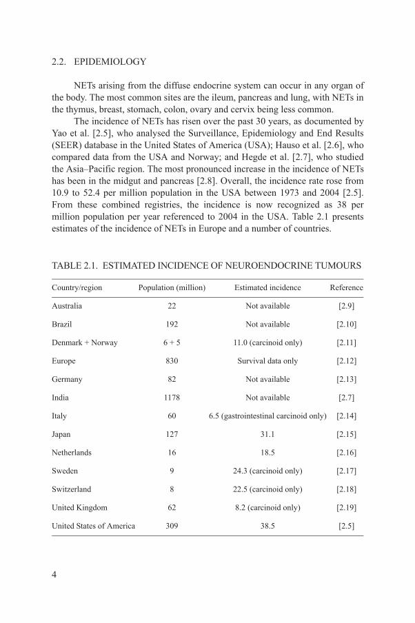
The incidence of NETs has risen over the past 30 years, as documented by Yao et al. [2.5], who analysed the Surveillance, Epidemiology and End Results (SEER) database in the United States of America (USA); Hauso et al. [2.6], who compared data from the USA and Norway; and Hegde et al. [2.7], who studied the Asia–Pacific region. The most pronounced increase in the incidence of NETs has been in the midgut and pancreas [2.8]. Overall, the incidence rate rose from 10.9 to 52.4 per million population in the USA between 1973 and 2004 [2.5]. From these combined registries, the incidence is now recognized as 38 per million population per year referenced to 2004 in the USA. Table 2.1 presents estimates of the incidence of NETs in Europe and a number of countries.
TABLE 2.1. ESTIMATED INCIDENCE OF NEUROENDOCRINE TUMOURS
Country/region Population (million) Estimated incidence Reference
Australia 22 Not available [2.9]
Brazil 192 Not available [2.10]
Denmark + Norway 6 + 5 11.0 (carcinoid only) [2.11]
Europe 830 Survival data only [2.12]
Germany 82 Not available [2.13]
India 1178 Not available [2.7]
Italy 60 6.5 (gastrointestinal carcinoid only) [2.14]
Japan 127 31.1 [2.15]
Netherlands 16 18.5 [2.16]
Sweden 9 24.3 (carcinoid only) [2.17]
4
Switzerland 8 22.5 (carcinoid only) [2.18]
United Kingdom 62 8.2 (carcinoid only) [2.19]
United States of America 309 38.5 [2.5]

Recognizing the slow growth of most NETs and the associated longer survival of these patients, their prevalence is significant. Although survival data were not available to the authors of most of the above studies, Yao et al. estimated the prevalence of NETs in the USA at 103 000 as of 1 January 2004 [2.5], and Ito et al. estimated their prevalence in Japan at 2.23/100.00 in 2005 [2.15].
Many, if not most, patients with NETs can lead high quality lives while being treated. Thus, a new treatment such as PRRNT, which has few side effects, is highly desirable in that it allows these patients to continue as productive members of society [2.20].
2.3. INTRODUCTION TO CLASSIFICATION SYSTEMS
NETs arise from the diffuse endocrine cell system. The term ‘carcinoid’ was first introduced by Oberndorfer in 1907 to describe serotonin-producing tumours in the small intestine with benign behaviour [2.21]. Williams and Sandler first attempted a systematic classification of gastroenteropancreatic NETs in 1963 [2.22]. They subdivided NETs according to their origin in the embryonic gut, and named them foregut, midgut and hindgut NETs. Although this classification is of limited prognostic significance, it is still in use for the anatomical characterization of primary tumour localization in patients with NETs.
In 1980, WHO suggested a classification system in which carcinoid tumours (including NETs derived from gastrin producing G-cells) were separated from pancreatic and a few other endocrine tumours, such as Merkel cell carcinoma, paragangliomas and others. This classification was also unsatisfactory with respect to both adequate histological classification and prognostically relevant clinicopathological categorization.
Also in 1980, Capella et al. [2.23] attempted a new clinicopathological classification system that considered macroscopic features of NETs (e.g. size and metastasis), histopathological features (e.g. cellular differentiation, neuroinvasion, angioinvasion, lymphangioinvasion, proliferation index) and clinical features (e.g. the presence of hormone hypersecretion syndromes), as well as the patient’s hereditary background. This classification generally separated benign NETs from those with uncertain behaviour, and low grade and
5
high grade malignant neuroendocrine carcinomas.In 2000, WHO published a revised classification system for the histological
typing of endocrine tumours [2.24]. This classification distinguishes among well differentiated endocrine tumours (WDETs), well differentiated endocrine carcinomas (WDECs) and poorly differentiated endocrine carcinomas (PDECs),
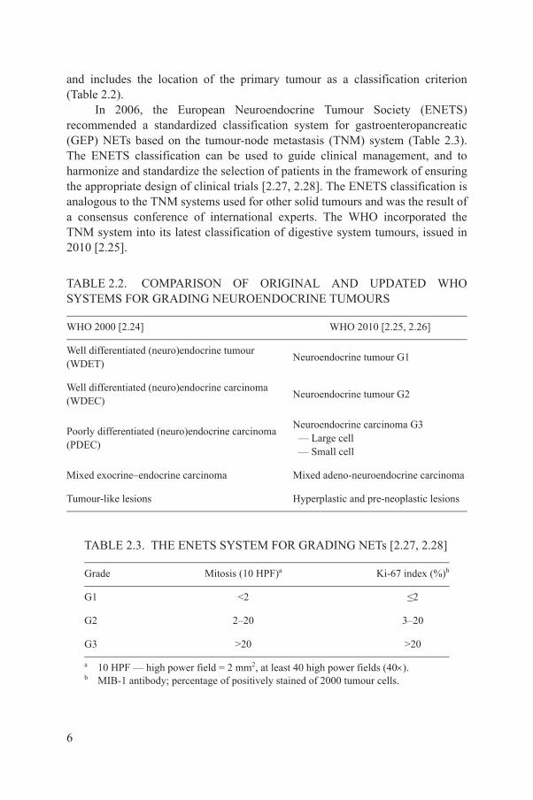
and includes the location of the primary tumour as a classification criterion (Table 2.2).
In 2006, the European Neuroendocrine Tumour Society (ENETS) recommended a standardized classification system for gastroenteropancreatic (GEP) NETs based on the tumour-node metastasis (TNM) system (Table 2.3). The ENETS classification can be used to guide clinical management, and to harmonize and standardize the selection of patients in the framework of ensuring the appropriate design of clinical trials [2.27, 2.28]. The ENETS classification is analogous to the TNM systems used for other solid tumours and was the result of a consensus conference of international experts. The WHO incorporated the TNM system into its latest classification of digestive system tumours, issued in 2010 [2.25].
TABLE 2.2. COMPARISON OF ORIGINAL AND UPDATED WHO SYSTEMS FOR GRADING NEUROENDOCRINE TUMOURS
WHO 2000 [2.24] WHO 2010 [2.25, 2.26]
Well differentiated (neuro)endocrine tumour (WDET)
Neuroendocrine tumour G1
Well differentiated (neuro)endocrine carcinoma (WDEC)
Neuroendocrine tumour G2
Poorly differentiated (neuro)endocrine carcinoma (PDEC)
Neuroendocrine carcinoma G3 — Large cell — Small cell
Mixed exocrine–endocrine carcinoma Mixed adeno-neuroendocrine carcinoma
Tumour-like lesions Hyperplastic and pre-neoplastic lesions
TABLE 2.3. THE ENETS SYSTEM FOR GRADING NETs [2.27, 2.28]
Grade Mitosis (10 HPF)a Ki-67 index (%)b
G1 <2 ≤2
6
G2 2–20 3–20
G3 >20 >20
a 10 HPF — high power field = 2 mm2, at least 40 high power fields (40).b MIB-1 antibody; percentage of positively stained of 2000 tumour cells.
This most recent WHO classification system provides guidelines for both staging and grading, the latter of which characterizes the proliferative potential of NET cells using either the mitotic count or the Ki-67 labelling index [2.26].
The WHO classification is the most advanced and enduring classification; it has been adopted by all European countries and by the ENETS community. It is also the most widely applied. It is important to emphasize that both the WHO and the TNM classifications are currently used in parallel to provide independent prognostic information and for classifying tumours. Information about the latest edition of the TNM Classification of Malignant Tumours can be found on the web site of the Union for International Cancer Control (UICC; www.uicc.org/resources/tnm).
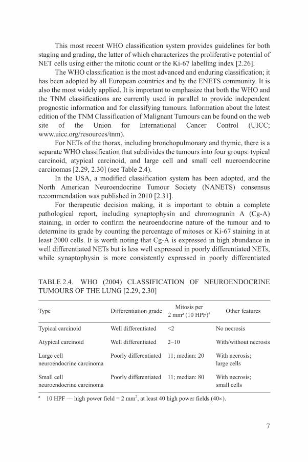
For NETs of the thorax, including bronchopulmonary and thymic, there is a separate WHO classification that subdivides the tumours into four groups: typical carcinoid, atypical carcinoid, and large cell and small cell nueroendocrine carcinomas [2.29, 2.30] (see Table 2.4).
In the USA, a modified classification system has been adopted, and the North American Neuroendocrine Tumour Society (NANETS) consensus recommendation was published in 2010 [2.31].
For therapeutic decision making, it is important to obtain a complete pathological report, including synaptophysin and chromogranin A (Cg-A) staining, in order to confirm the neuroendocrine nature of the tumour and to determine its grade by counting the percentage of mitoses or Ki-67 staining in at least 2000 cells. It is worth noting that Cg-A is expressed in high abundance in well differentiated NETs but is less well expressed in poorly differentiated NETs, while synaptophysin is more consistently expressed in poorly differentiated
TABLE 2.4. WHO (2004) CLASSIFICATION OF NEUROENDOCRINE TUMOURS OF THE LUNG [2.29, 2.30]
Type Differentiation gradeMitosis per
2 mm² (10 HPF)a Other features
Typical carcinoid Well differentiated <2 No necrosis
Atypical carcinoid Well differentiated 2–10 With/without necrosis
7
Large cellneuroendocrine carcinoma
Poorly differentiated 11; median: 20 With necrosis;large cells
Small cellneuroendocrine carcinoma
Poorly differentiated 11; median: 80 With necrosis;small cells
a 10 HPF — high power field = 2 mm2, at least 40 high power fields (40).
NETs. The grading is especially critical to ensure appropriate therapeuticmanagement, as in high grade NETs (>G3, with Ki-67 >20%) chemotherapy becomes the primary option for therapy.
Although not routine in most centres, sstr2 expression by immunohistochemistry can be helpful in determining the differentiation status, as it is expressed in 70–100% of highly differentiated NETs. Clinically, this is best defined by functional scintigraphy (indium 111In pentetreotide/Octreoscan®, 68Ga-PET-CT scan). Both scintigraphy and immunohistochemistry are of limited value in poorly differentiated NETs (>G3, wherein indium 111In pentetreotide/Octreoscan® has only 20–30% sensitivity).
Thus, the minimum histopathological data set can be described as the process that is recommended for the pathologist to provide the clinician with information sufficient to make the best decision possible for the patient’s further care [2.32].
2.4. CLINICAL PRESENTATION
2.4.1. Introduction
The clinical presentation of NETs may vary depending on the site of tumour origin. About 72% of NETs arise in gastrointestinal structures, a further 25% are bronchopulmonary in origin and less than 5% arise at other sites such as the thymus, breast and genital-urinary system.
NETs of the gastroenteropancreatic system consist of cells capable of amine precursor uptake and decarboxylation (APUD) cells previously termed APUDomas. Characteristics of NETs include episodic hormone secretion/release and indolent, slow growth; thus, they may be silent for years.
Although all these tumours express one or more amines and/or polypeptides, only 40–50% are functionally active, resulting in specific clinical symptoms or syndromes. Less than 10% of midgut tumours are associated with carcinoid syndrome, but this is the most frequently observed syndrome among all NETs. It is understood that metastases exist in the presence of the carcinoid syndrome, as most are associated with metastases of the liver.
Those tumours that secrete physiologically important amounts of hormones or amines are termed according to the predominant secretory substance, for
8
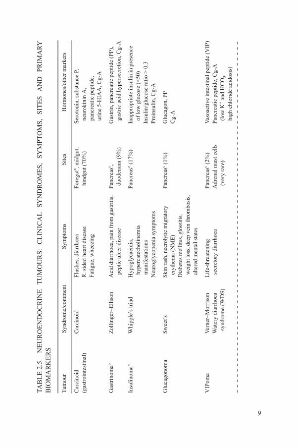
example, Zollinger–Ellison syndrome/gastrinoma (Table 2.5). The most frequent syndromes are carcinoid syndrome (serotonin producing tumours predominantly of the gastrointestinal system), followed by insulinoma and gastrinoma (both predominantly of the pancreas), and gastrinoma associated with the hereditary multiple endocrine neoplasia syndrome type 1 (MEN-1), which is primarily duodenal in origin.
UR
OE
ND
OC
RIN
E
TU
MO
UR
S:
CL
INIC
AL
S
YN
DR
OM
ES
, S
YM
PT
OM
S,
SIT
ES
A
ND
P
RIM
AR
Y
Syn
drom
e/co
mm
ent
Sym
ptom
sS
ites
Hor
mon
es/o
ther
mar
kers
Car
cino
idF
lush
es, d
iarr
hoea
R. s
ided
hea
rt d
isea
se
Fat
igue
, whe
ezin
g
For
egut
a , mid
gut,
hin
dgut
(70
%)
Ser
oton
in, s
ubst
ance
P,
neu
roki
nin
A,
pan
crea
tic
pept
ide,
uri
ne 5
-HIA
A, C
g-A
Zol
linge
r–E
lliso
nA
cid
diar
rhoe
a, p
ain
from
gas
trit
is,
pep
tic
ulce
r di
seas
eP
ancr
easc ,
duo
denu
m (
9%)
Gas
trin
, pan
crea
tic p
epti
de (
PP
),
gas
tric
aci
d hy
pers
ecre
tion
, Cg-
A
Whi
pple
’s tr
iad
Hyp
ogly
caem
ia,
hyp
erca
tech
olin
emia
man
ifes
tati
ons
Neu
rogl
ycop
enia
sym
ptom
s
Pan
crea
sc (17
%)
Inap
prop
riat
e in
suli
n in
pre
senc
e o
f lo
w g
luco
se (
<50
)In
suli
n/gl
ucos
e ra
tio
> 0
.3
Pro
insu
lin,
Cg-
A
Sw
eet’s
Skin
ras
h, n
ecro
lytic
mig
rato
ry
ery
them
a (N
ME
)D
iabe
tes
mel
litus
, glo
ssiti
s,
wei
ght l
oss,
dee
p ve
in th
rom
bosi
s,
alte
red
men
tal s
tate
s
Pan
crea
sc (1%
)G
luca
gon,
PP
Cg-
A
Ver
ner–
Mor
riso
nW
ater
y di
arrh
oea
syn
drom
e (W
DS
)
Lif
e-th
reat
enin
g s
ecre
tory
dia
rrho
eaP
ancr
easc (
2%)
Adr
enal
mas
t cel
ls
(ve
ry r
are)
Vas
oact
ive
inte
stin
al p
eptid
e (V
IP)
Pan
crea
tic
pept
ide,
Cg-
A
(lo
w K
+ a
nd H
CO
3,
hig
h ch
lori
de a
cido
sis)
9
TAB
LE
2.5
. N
EB
IOM
AR
KE
RS
Tum
our
Car
cino
id
(gas
troi
ntes
tina
l)
Gas
trin
omab
Insu
lino
mab
Glu
cago
nom
a
VIP
oma

‘Non
-fun
ctio
nal’
Co-
exis
ts w
ith
othe
r p
ancr
eati
c tu
mou
rs
Dia
rrhe
al w
ith
very
hig
h le
vels
Pan
crea
sc (15
%)
PP,
Cg-
A
‘Non
-fun
ctio
nal’
Dia
bete
s m
ellit
us, c
hole
lith
iasi
s,
ste
ator
rhoe
aP
ancr
easc ,
duo
denu
m (
1%)
Som
atos
tati
nC
g-A
)C
arci
noid
(ver
y ra
re)
Ect
opic
adr
enoc
orti
cotr
opic
hor
mon
e (A
CT
H)
(ra
re)
Cou
gh, p
ain,
pne
umon
itis
, d
iarr
hoea
(ra
re),
flu
shes
(ra
re)
Bro
nchu
s (2
5%)a
Cg-
A (
pred
omin
antl
y)
Pan
crea
tic
pept
ide
AC
TH
Asy
mpt
omat
icD
iarr
hoea
(sp
orad
ic M
TC
)T
hyro
id (
calc
iton
in c
ell)
(7%
of
thyr
oid
canc
er)
Cal
cito
nin
Car
cino
-em
bryo
nic
anti
gen
(CE
A)
is
a de
-dif
fere
ntia
tion
mar
ker
d a)H
yper
tens
ive
cris
isP
allo
r, pa
lpita
tions
, per
spir
atio
n,
hea
dach
eE
piso
dic
hype
rten
sion
; c
an b
e co
nsta
nt in
larg
er tu
mou
rs
Adr
enal
, gan
glia
and
par
agan
glia
Fre
e m
etan
ephr
ine
and
nor
-met
anep
hrin
eC
g-A
ce o
f al
l car
cino
id N
ET
s.ia
ted
wit
h kn
own
ME
N-1
.ce
of
all p
ancr
eati
c N
ET
s.E
N-2
(fa
mil
ial a
nd s
pora
dic)
, von
Hip
pel–
Lin
dau
(VH
L)
synd
rom
e, n
euro
fibr
omat
osis
type
1 (
NF
-1),
all
oft
en b
ilate
ral.
UR
OE
ND
OC
RIN
E
TU
MO
UR
S:
CL
INIC
AL
S
YN
DR
OM
ES
, S
YM
PT
OM
S,
SIT
ES
A
ND
P
RIM
AR
Y
cont
.)
Syn
drom
e/co
mm
ent
Sym
ptom
sS
ites
Hor
mon
es/o
ther
mar
kers
10
PP
oma
Som
atos
tati
nom
a
Car
cino
id
(bro
ncho
-pul
mon
ary
Med
ulla
ry th
yroi
dca
rcin
oma
(MT
C)
Pheo
chro
moc
ytom
a(p
arag
angl
ione
urom
aP
er c
ent o
ccur
ren
b C
omm
only
ass
occ
Per
cen
t occ
urre
nd
Ass
ocia
ted
wit
h M
TAB
LE
2.5
. N
EB
IOM
AR
KE
RS
(
Tum
our
2.4.2. Clinical syndromes
2.4.2.1. Carcinoid syndrome
Carcinoid syndrome is predominantly characterized by flushing (>80% of patients) and diarrhoea (70%); it may also be accompanied by bronchial obstruction (wheezing) in some patients (17%) or by symptomatic hypotension. The main hormones involved in carcinoid syndrome are serotonin and substance P (pain) in midgut tumours, less commonly in foregut NETs (10%), and rarely in hindgut tumours (1%).
Carcinoid heart disease occurs as a long term complication of chronic hyperserotonemia in up to 40% of patients; progressive heart disease, rather than tumour progression, is the primary cause of death once carcinoid heart disease becomes symptomatic.
Serotonergic crisis, which can occur during anaesthesia induction or intervention procedures at the liver metastases, for example, can be a life threatening condition requiring immediate management with intensive care and intravenous somatostatin congeners (see Section 6.4.2).
2.4.2.2. Gastrinoma
Gastrinoma is characterized by hyperacidity due to gastrin hypersecretion from a tumour of the pancreas or duodenum. More than 90% of gastrinomas are found in the gastrinoma triangle, comprising cystic and common ducts, mesenteric vessels and the lateral portion of the duodenal C loop. The symptoms of gastrinoma are associated with peptic ulcer disease, diarrhoea (70%) and reflux oesophagitis. Conditions such as dyspepsia, haemorrhage and abdominal pain are all due to hyperacidity. Some 70–80% of duodenal gastrinomas are associated with MEN-1 (see Section 2.4.2.8, NETs and hereditary syndromes).
The widespread use of proton pump inhibitors (PPIs) may mask many of the typical symptoms of gastrinoma, thereby delaying the diagnosis for months or years. An elevated fasting serum gastrin is a typical finding of gastrinoma.
2.4.2.3. Insulinoma
11
The classic symptoms of insulinoma are expressed as Whipple’s triad: symptomatic hypoglycaemia, biochemically confirmed low blood sugar (<50 mg% or 2.6 mmol/L), and relief of symptoms by glucose ingestion. Sequelae from episodic hyperinsulinemia and/or hypoglycaemia are obesity, hypercatecholanaemic and neuroglucopenic symptoms ranging from sweating and tachycardia to non-specific neurological symptoms, concentration disorders,
focal or generalized seizure, and coma or even death. More than 90% of insulinomas are benign, and almost 100% are located within the pancreas. About 10% of insulinomas are associated with MEN-1 and may be multifocal and plurihormonal.
2.4.2.4. Glucagonoma
Glucagonoma is characterized by necrolytic migratory erythema, which is also associated with acquired diabetes mellitus due to glucagon-hypersecreting tumours. This syndrome is very rare and has been referred to as the ‘4 D’ syndrome, comprising dermatosis (80%), diabetes (80%), deep-vein thrombosis (50%) and depression (50%). Additional symptoms include weight loss (90%), painful glossitis and stomatitis. Elevated glucagon levels establish the diagnosis.
2.4.2.5. VIPoma
Vasoactive intestinal peptide secreting tumour (VIPoma), also known aswatery diarrhoea syndrome (WDS) or Verner–Morrison syndrome, is caused by the abnormal secretion of vasoactive intestinal peptides. This condition is characterized by severe watery (secretory) diarrhoea, hypokalaemia, hypochlorhydria and metabolic acidosis. This often results in severe dehydration, requiring large intravenous fluid (up to 9–10 L/d) and electrolyte replacement. Elevated levels of vasoactive intestinal peptide in the presence of such diarrhoea and metabolic changes establish the diagnosis.
2.4.2.6. Rare functioning tumours
This group of rare tumours includes tumours that secrete growth hormone releasing hormone (GHRH) and adrenocorticotropic hormone (ACTH), leading to acromegaly and Cushing’s syndrome, respectively. Diagnoses are established by means of appropriate endocrine function tests in the presence of a typical physical appearance.
2.4.2.7. Non-functioning tumours
12
Non-functioning tumours account for 50–60% of all NETs. They also include NETs that are clinically silent but secrete a predominant substance (e.g. pancreatic polypeptidoma (PPoma)). Somatostatinoma can be considered non-syndromic [2.33]. Non-functioning tumours are diagnosed either incidentally (e.g. by endoscopy) or from nonspecific symptoms following mass effects, such as liver enlargement, pancreatic duct obstruction or jaundice.
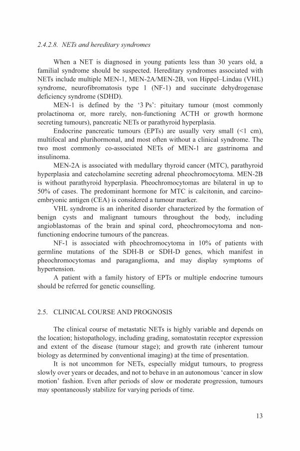
2.4.2.8. NETs and hereditary syndromes
When a NET is diagnosed in young patients less than 30 years old, a familial syndrome should be suspected. Hereditary syndromes associated with NETs include multiple MEN-1, MEN-2A/MEN-2B, von Hippel–Lindau (VHL) syndrome, neurofibromatosis type 1 (NF-1) and succinate dehydrogenase deficiency syndrome (SDHD).
MEN-1 is defined by the ‘3 Ps’: pituitary tumour (most commonly prolactinoma or, more rarely, non-functioning ACTH or growth hormone secreting tumours), pancreatic NETs or parathyroid hyperplasia.
Endocrine pancreatic tumours (EPTs) are usually very small (<1 cm), multifocal and plurihormonal, and most often without a clinical syndrome. The two most commonly co-associated NETs of MEN-1 are gastrinoma and insulinoma.
MEN-2A is associated with medullary thyroid cancer (MTC), parathyroid hyperplasia and catecholamine secreting adrenal pheochromocytoma. MEN-2B is without parathyroid hyperplasia. Pheochromocytomas are bilateral in up to 50% of cases. The predominant hormone for MTC is calcitonin, and carcino-embryonic antigen (CEA) is considered a tumour marker.
VHL syndrome is an inherited disorder characterized by the formation of benign cysts and malignant tumours throughout the body, including angioblastomas of the brain and spinal cord, pheochromocytoma and non-functioning endocrine tumours of the pancreas.
NF-1 is associated with pheochromocytoma in 10% of patients with germline mutations of the SDH-B or SDH-D genes, which manifest in pheochromocytomas and paraganglioma, and may display symptoms of hypertension.
A patient with a family history of EPTs or multiple endocrine tumours should be referred for genetic counselling.
2.5. CLINICAL COURSE AND PROGNOSIS
The clinical course of metastatic NETs is highly variable and depends on the location; histopathology, including grading, somatostatin receptor expression
13
and extent of the disease (tumour stage); and growth rate (inherent tumour biology as determined by conventional imaging) at the time of presentation.
It is not uncommon for NETs, especially midgut tumours, to progress slowly over years or decades, and not to behave in an autonomous ‘cancer in slow motion’ fashion. Even after periods of slow or moderate progression, tumours may spontaneously stabilize for varying periods of time.
In general, pancreatic NETs tend to be more aggressive, leading to shorter median survival times [2.34] in an equivalently paired grading system. Spontaneous tumour remissions are extremely rare.
Prognostically negative outcome parameters include histopathological high grade tumours, advanced tumour stage and high tumour burden, which is also co-associated with rising Cg-A levels [2.35] and low somatostatin receptor density in vivo imaging [2.36].
Survival rates vary between countries, perhaps due to the development of highly specialized multidisciplinary tumour centres. Between 1973 and 2004, median survival rates were 124 months for well differentiated tumours, compared with 64 months for moderately differentiated NETs and 10 months for poorly differentiated NETs, according to the SEER database (http://seer.cancer.gov), as recently reported by Yao et al. [2.5]. According to the SEER database, for the period 1988–2004, the five year survival rate in distant metastatic disease was 27% for pancreatic NETs and 54% for jejunal-ileal NETs. According to national databases and registries in Europe, the five year survival rates for the same NET subgroups of histological differentiation varied between 55 and 70% [2.37–2.41].
The overall five year survival rate in patients with NETs at all stages and all primary locations is about 55% [2.6].
2.6. CONFIRMATION OF DIAGNOSIS AND STAGING
2.6.1. Revision of histopathology specimens
In cases where the histopathological diagnosis is incomplete or unclear, the histological specimens should be evaluated by an experienced pathologist for revising a paraffin-fixed tumour tissue in as much as possible to confirm the neuroendocrine nature of the tumour and to determine the histologic grading, if this is missing. The grading is of prognostic significance, as has been shown for two large groups of NET patients [2.42, 2.43]. If there is a time delay between the initial diagnosis of the disease and the presentation of the patient for decision making, it may be necessary to obtain a core biopsy. Fine needle aspiration (FNA) for this purpose cannot be recommended, however.
14
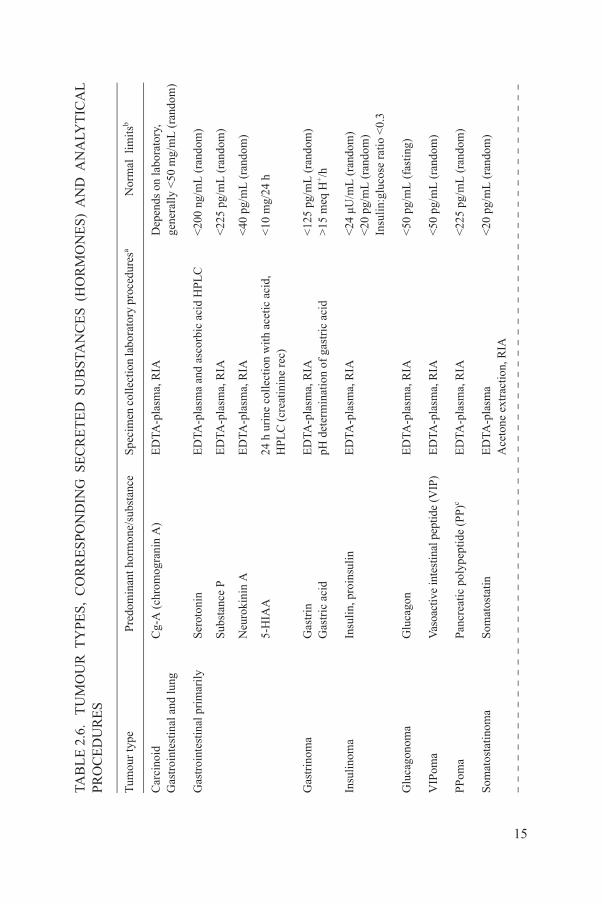
2.6.2. Biochemical assays in functional tumours
The value of biochemical assays lies in the confirmation and the support of the clinical diagnosis and in the follow-up management and response assessment to therapeutic interventions. Such assays may also prove to be of prognostic value (see Table 2.6).
MO
UR
TY
PE
S,
CO
RR
ES
PO
ND
ING
SE
CR
ET
ED
SU
BS
TAN
CE
S (
HO
RM
ON
ES
) A
ND
AN
ALY
TIC
AL
Pre
dom
inan
t hor
mon
e/su
bsta
nce
Spe
cim
en c
olle
ctio
n la
bora
tory
pro
cedu
resa
Nor
mal
lim
itsb
lung
Cg-
A (
chro
mog
rani
n A
)E
DTA
-pla
sma,
RIA
D
epen
ds o
n la
bora
tory
,ge
nera
lly
<50
mg/
mL
(ra
ndom
)
aril
yS
erot
onin
ED
TA-p
lasm
a an
d as
corb
ic a
cid
HP
LC
<20
0 ng
/mL
(ra
ndom
)
Sub
stan
ce P
ED
TA-p
lasm
a, R
IA<
225
pg/m
L (
rand
om)
Neu
roki
nin
AE
DTA
-pla
sma,
RIA
<40
pg/
mL
(ra
ndom
)
5-H
IAA
24 h
uri
ne c
olle
ctio
n w
ith
acet
ic a
cid,
HP
LC
(cr
eati
nine
rec
)<
10 m
g/24
h
Gas
trin
Gas
tric
aci
dE
DTA
-pla
sma,
RIA
pH d
eter
min
atio
n of
gas
tric
aci
d<
125
pg/m
L (
rand
om)
>15
meq
H+/h
Insu
lin,
pro
insu
lin
ED
TA-p
lasm
a, R
IA<
24 μ
U/m
L (
rand
om)
<20
pg/
mL
(ra
ndom
)In
suli
n:gl
ucos
e ra
tio
<0.
3
Glu
cago
nE
DTA
-pla
sma,
RIA
<50
pg/
mL
(fa
stin
g)
Vas
oact
ive
inte
stin
al p
eptid
e (V
IP)
ED
TA-p
lasm
a, R
IA<
50 p
g/m
L (
rand
om)
Pan
crea
tic
poly
pept
ide
(PP
)cE
DTA
-pla
sma,
RIA
<22
5 pg
/mL
(ra
ndom
)
Som
atos
tatin
ED
TA-p
lasm
aA
ceto
ne e
xtra
ctio
n, R
IA<
20 p
g/m
L (
rand
om)
15
TAB
LE
2.6
. T
UP
RO
CE
DU
RE
S
Tum
our
type
Car
cino
id
Gas
troi
ntes
tinal
and
Gas
troi
ntes
tina
l pri
m
Gas
trin
oma
Insu
lino
ma
Glu
cago
nom
a
VIP
oma
PP
oma
Som
atos
tati
nom
a

arci
nom
a)
Cal
cito
nin
Car
cino
-em
bryo
nic
anti
gen
(CE
A)
ED
TA-p
lasm
a, R
IA
CE
A, i
mm
unom
etri
c ch
emil
umin
esce
nce
<20
pg/
mL
(ra
ndom
)<
5 ng
/mL
aF
ree
met
anep
hrin
e,
Fre
e no
r-m
etan
ephr
ine
ED
TA-p
lasm
a, H
PL
C in
US
A;
RIA
in E
urop
e<
0.50
nm
ol/L
<0.
9 nm
ol/L
eopl
asia
Pan
crea
tic tu
mou
r m
arke
rs &
PT
H,
and
pro
lact
inE
DTA
-pla
sma,
bot
h im
mun
omet
ric
chem
ilum
ines
cenc
e<
60 p
g/m
L (
PT
H)
<20
ng/
mL
(pr
olac
tin)
asta
sis
Pan
crea
stat
inE
DTA
-pla
sma,
RIA
<12
5 pg
/mL
unoa
ssay
; HP
LC
— h
igh
perf
orm
ance
liqu
id c
hrom
atog
raph
y.it
s ar
e hi
ghly
var
iabl
e am
ong
labo
rato
ries
. Use
one
spe
cifi
c la
bora
tory
whe
n fo
llow
ing
pati
ent.
unct
ioni
ng N
ET
s.
MO
UR
TY
PE
S,
CO
RR
ES
PO
ND
ING
SE
CR
ET
ED
SU
BS
TAN
CE
S (
HO
RM
ON
ES
) A
ND
AN
ALY
TIC
AL
co
nt.)
Pre
dom
inan
t hor
mon
e/su
bsta
nce
Spe
cim
en c
olle
ctio
n la
bora
tory
pro
cedu
resa
Nor
mal
lim
itsb
16
Med
ulla
ry th
yroi
d c
(M
TC
, cal
citin
oma
Pheo
chro
moc
ytom
a p
arag
angl
ione
urom
Mul
tipl
e en
docr
ine
n t
ype
I (M
EN
-1)
Neu
roen
docr
ine
met
to
live
r
aR
IA —
rad
ioim
mb
Upp
er n
orm
al li
mc
And
for
all
non
-f
TAB
LE
2.6
. T
UP
RO
CE
DU
RE
S (
Tum
our
type

For confirmation and follow-up, markers are routinely measured in the following clinical syndromes: carcinoid syndrome, hypoglycaemia associated syndrome, Zollinger–Ellison syndrome and rare endocrine pancreatic tumour syndromes.
2.6.2.1. Carcinoid syndrome
Carcinoid syndrome is confirmed with measurement of elevated 5-hydroxyindoleacetic acid (5-HIAA) in 24 h urine collections or markedly elevated plasma serotonin. With most assays, 5-HIAA measured by high performance liquid chromatography (HPLC) is more reliable than single measurements of circulating serotonin. It is critical to collect urine on acid, either acetic or hydrochloric acid, according to the specific requirements of the laboratory. Dietary restrictions to avoid serotonin-containing food should be applied. It may be useful to collect creatinine as well, if complete 24 h collections are not feasible [2.44].
2.6.2.2. Hypoglycaemia associated syndrome (insulinoma)
The diagnosis of hypoglycaemia associated syndrome is established by measurements of elevated insulin and C-peptide or proinsulin (the latter, if insulin is not elevated) in conditions of hypoglycaemia. If insulinoma is suspected, a fasting test of up to 72 h with simultaneous measurements of insulin/C-peptide and glucose is the gold standard of diagnosis. Within 48 h, 85–90% (and within 72 h, more than 95%) of patients will develop symptomatic hypoglycaemia. The fasting test terminates in the condition of low blood sugar (<45 mg/dL or 2.5 mmol/L) accompanied by symptoms, although after long-standing repetitive hypoglycaemia the symptoms may be masked. The 72 h fasting test should be performed in an appropriate clinical setting with a patent intravenous line and with clinical monitoring of symptoms and glucose at least every 2–4 h, and more frequently if needed. To exclude factitious hypoglycaemia, measurements of sulphonylurea (urine or blood) and C-peptide should be considered.
2.6.2.3. Zollinger–Ellison syndrome (gastrinoma)
17
Serum gastrin levels are usually markedly elevated in more than 90% of patients. A single very low pH≤2, in combination with elevated gastrin levels, is usually diagnostic of gastrinoma. As an alternative to a single pH determination, a midnight to 6 a.m. or 24 h pH metry demonstrating greater than 15 meq/h gastric acid may also be regarded as a positive test result.

Other conditions leading to isolated hypergastrinemia (in the absence of hyperchlorhydria) include renal failure, pernicious anaemia and, less often, gastric outlet obstruction or Helicobacter pylori infection. In these instances, a secretin provocation test may be required. This test involves injecting a 2 IU/kg bolus of secretin and measuring serum gastrin levels at a baseline (0 min), and at 10, 20 and 30 min after injection. A delta increase in gastrin levels above 200 pg/mL is considered positive in 90% of cases.
2.6.2.4. Rare endocrine pancreatic tumour syndromes
This group comprises VIPoma, glucagonoma and somatostatinoma. In the setting of the clinical syndrome, confirmation of VIPoma is made by measuring vasoactive intestinal peptide (VIP) using ethylene diamine tetraacetic acid (EDTA) tubes containing the protease inhibitor trasylol. After collection, samples should be quickly placed on ice, spun down, separated and frozen.
In glucagonoma, levels of plasma or serum glucagon are determined in a fasting state by means of enzyme linked immunosorbent assay (ELISA) or radioimmunoassay (RIA) in samples gathered using vials containing the protease inhibitor trasylol (500 U/10 mL). After collection, samples should be quickly placed on ice, spun down, separated and frozen.
Somatostatinomas are very rare. Measurement of plasma somatostatin is generally not recommended and very difficult to obtain commercially.
2.6.3. General tumour markers
The most important circulating tumour marker for both functioning and non-functioning NETs is Cg-A. This is an acidic protein that resides and coexists with catecholamines within large chromaffin granules in the vast majority of neuroendocrine cells and their tumours. Cg-A belongs to a unique family of secretory chromogranins that includes chromogranin B and C. It is considered a prohormone without relevant biological function.
In many institutions for gastroenteropancreatic NETs, Cg-A is considered standard care for both determination of diagnosis and monitoring during therapy [2.45]. The circulating level of Cg-A appears to be correlated with the tumour burden [2.46], and Cg-A is more highly expressed and secreted in well
18
differentiated tumours than in poorly differentiated ones. When assessing the success of therapy or a change of management, a delta of 25% or more is considered a significant change, although this should always be viewed in the light of the association between the biochemical value and anatomical imaging.
False positive Cg-A values may be encountered in patients who have been on long term and even short term treatment with antacids, especially PPIs [2.47],

and in patients with chronic atrophic gastritis, renal and heart insufficiency, cardiovascular disease (hypertension, angina pectoris), inflammatory bowel disease or pancreatitis. Intake of PPIs should be interrupted for at least one week and Cg-A determined again before initiating other diagnostic steps [2.48].
Several Cg-A assays are used with variable sensitivity and equal specificity [2.49]. The choice of assay, however, depends on the preferences of different countries. The assays most often used and distributed in Europe are the CisBIO and the Dako ELISA. At least five different Cg-A assays, either RIA or ELISA, are commercially available in the USA, but only the Quest and Interscience Institute assays have been standardized. Cg-A has not been standardized within or between countries. Therefore, and also considering the variability of the different assays, it is essential that the patient be monitored with the same assay and preferably at the same reference laboratory.
Cg-A is not recommended to be used as a screening marker. If Cg-A is not elevated in patients with existing tumours, then alternative markers such as neuron specific enolase (NSE) or pancreatic polypeptide (PP) should be considered.
In future, other markers, such as pancreastatin, might prove useful for prognosis of liver tumour burden [2.50, 2.51]. Pancreastatin is one of the Cg-A derived peptides with known biological inhibitory activity. It induces a general inhibitory secretory effect in many exocrine and endocrine systems.
REFERENCES TO SECTION 2
[2.1] OTTE, A., et al., DOTATOC: A powerful new tool for receptor-mediated radionuclide therapy, Eur. J. Nucl. Med. 24 (1997) 792–795.
[2.2] KRENNING, E.P., et al., Somatostatin receptor scintigraphy with [111In-DTPA-D-Phe1]- and [123I- Tyr3]-octreotide: The Rotterdam experience with more than 1000 patients, Eur. J. Nucl. Med. 20 (1993) 716–731.
[2.3] WALDHERR, C., PLESS, M., MAECKE, H.R., HALDEMANN, A., MUELLER-BRAND, J., The clinical value of [90Y-DOTA]-D-Phe1- Tyr3-octreotide (90Y-DOTATOC) in the treatment of neuroendocrine tumours: A clinical phase II study, Ann. Oncol. 12 (2001) 941–945.
[2.4] KWEKKEBOOM, D.J., et al., Overview of results of peptide receptor radionuclide therapy with 3 radiolabeled somatostatin analogs, J. Nucl. Med. 46 (2005) 62S.
[2.5] YAO, J.C., et al., One hundred years after ‘carcinoid’: Epidemiology of and prognostic
19
factors for neuroendocrine tumours in 35,825 cases in the United States, J. Clin. Oncol. 26 (2008) 3063–3072.
[2.6] HAUSO, O., et al., Neuroendocrine tumour epidemiology: Contrasting Norway and North America, Cancer 113 (2008) 2655–2664.
[2.7] HEGDE, V., MOHANDAS, K.M., RAMADWAR, M., SHUKLA, P., MEHTA, S., Gastric carcinoids — A changing trend, Indian J. Gastroenterol. 22 (2003) 209–211.
[2.8] MODLIN, I.M., et al., Gastroenteropancreatic neuroendocrine tumours, Lancet Oncol. 9 (2008) 61–72.
[2.9] RANGIAH, D.S., COX, M., RICHARDSON, M., TOMPSETT, E., CRAWFORD, M., Small bowel tumours: A 10 year experience in four Sydney teaching hospitals, ANZ J. Surg. 74 (2004) 788–792.
[2.10] YOUNES, R.N., GETNE (GRUPO DE ESTUDO DE TUMORES NEUROENDÓCRINOS), Neuroendocrine tumours: A registry of 1,000 patients, Rev. Assoc. Med. Brasil 54 (2008) 305–307.
[2.11] WESTERGAARD, T., FRISCH, M., MELBYE, M., Carcinoid tumours in Denmark 1978–1989 and the risk of subsequent cancers — A population-based study, Cancer 76(1995) 106–109.
[2.12] LEPAGE, C., et al., EUROCARE working group: European disparities in malignant digestive endocrine tumours survival, Int. J. Cancer 126 (2010) 2928–2934.
[2.13] PLOECKINGER, U., et al., The German NET registry: An audit of the diagnosis and therapy of neuroendocrine tumours, Neuroendocrinology 90 (2009) 349–363.
[2.14] CROCETTI, E., Gastrointestinal carcinoid tumours: A population-based study, Ital. J. Gastroenterol. Hepatol. 29 (1997) 135–137.
[2.15] ITO, T., et al., Epidemiological study of gastroenteropancreatic neuroendocrine tumours in Japan, J. Gastroenterol. 45 (2010) 234–243.
[2.16] TAAL, B.G., VISSER, O., Epidemiology of neuroendocrine tumours, Neuroendocrinology 80 (2004) 3–7.
[2.17] HEMMINKI, K., LI, X., Familial carcinoid tumours and subsequent cancers: A nation-wide epidemiologic study from Sweden, Int. J. Cancer 94 (2001) 444–448.
[2.18] LEVI, F., RANDIMBISON, L., FRANCESCHI, S., LA VECCHIA, C., Descriptive epidemiology of malignant carcinoids in the Swiss Canton of Vaud, Int. J. Cancer 53(1993) 1036–1037.
[2.19] NEWTON, J.N., et al., The epidemiology of carcinoid tumours in England and Scotland, Br. J. Cancer 70 (1994) 939–942.
[2.20] BLOOM, D.E., CANNING, D., SEVILLA, J., The effect of health on economic growth: A production function approach, World Development 32 (2004) 1–13.
[2.21] OBERNDORFER, S., Karzinoide Tumoren des Dünndarmes, Frankfurter Z. Pathol. 1(1907) 426–429.
[2.22] WILLIAMS, E.D., SANDLER, M., The classification of carcinoid tumours, Lancet 1 72–75 (1963) 238–239.
[2.23] CAPELLA, C., HEITZ, P.U., HÖFLER, H., SOLCIA, E., KLÖPPEL, G., Revised classification of neuroendocrine tumours of the lung, pancreas and gut, Virchows Arch. 425 (1995) 547–560.
[2.24] RINDI, G., et al., TNM staging of foregut (neuro)endocrine tumours: A consensus proposal including a grading system, Virchows Arch. 451 (2007) 757–762.
[2.25] BOSMAN, F.T., CARNEIRO, F., HRUBAN, R.H. (Eds), WHO Classification of
20
Tumours of the Digestive System, Vol. 3, IARC Publications, Lyon, France (2010).[2.26] SOLCIA, E., et al., Histological typing of endocrine tumours, WHO International
Histological Classification of Tumours, 2nd edn, Springer, Berlin (2000). [2.27] RINDI, G., et al., TNM staging of midgut and hindgut (neuro) endocrine tumours:
A consensus proposal including a grading system, Virchows Arch. 449 (2006) 395–401.
[2.28] RINDI, G., The ENETS guidelines: The new TNM classification system, Tumouri 96(2010) 806–809.
[2.29] TRAVIS, W.D., et al., International staging committee and participating institutions, J. Thorac. Oncol. 3 (2008) 1213–1223.
[2.30] TRAVIS, W.D., BRAMBILLA, E., MULLER-HERMLINK, H.K., HARRIS, C.C. (Eds), World Health Organization Classification of Tumours: Pathology and Genetics of Tumours of the Lung, Pleura, Thymus and Heart, Lyon, France (2004).
[2.31] DAVID, S., et al., A review of nomenclature, grading, and staging systems: The pathologic classification of neuroendocrine tumours, Pancreas 39 (2010) 707–712.
[2.32] KLIMSTRA, D.S., et al., Pathology reporting of neuroendocrine tumours: Application of the Delphic consensus process to the development of a minimum pathology data set, Am. J. Surg. Pathol. 34 (2010) 300–313.
[2.33] GARBRECHT, N., et al., Somatostatin-producing neuroendocrine tumours of the duodenum and pancreas: Incidence, types, biological behavior, association with inherited syndromes, and functional activity, Endocr. Relat. Cancer 15 (2008) 229–241.
[2.34] YAO, J.C., et al., Daily oral everolimus activity in patients with metastatic pancreatic neuroendocrine tumours after failure of cytotoxic chemotherapy: A phase II trial, J. Clin. Oncol. 1 (2010) 69–76.
[2.35] ARNOLD, R., et al., Plasma chromogranin A as marker for survival in patients with metastatic endocrine gastroenteropancreatic tumours, Clin. Gastroenterol. Hepatol. 6(2008) 820–827.
[2.36] OBERG, K.E., REUBI, J.C., KWEKKEBOOM, D.J., KRENNING, E.P., Role of somatostatins in gastroenteropancreatic neuroendocrine tumour development and therapy, Gastroenterology 139 (2010) 742–753.
[2.37] AHMED, A., et al., Midgut neuroendocrine tumours with liver metastases: Results of the UKINETS study, Endocr. Relat. Cancer 16 (2009) 885–894.
[2.38] EKEBLAD, S., SKOGSEID, B., DUNDER, K., OBERG, K., ERIKSSON, B., Prognostic factors and survival in 324 patients with pancreatic endocrine tumour treated at a single institution, Clin. Cancer Res. 14 (2008) 7798–7803.
[2.39] GARCIA-CARBONERO, R., et al., Incidence, patterns of care and prognostic factors for outcome of gastroenteropancreatic neuroendocrine tumours (GEP-NETs), Results from the National Cancer Registry of Spain (RGETNE), Ann. Oncol. 21 (2010) 1794–1803.
[2.40] JANN, H., et al., Neuroendocrine tumours of midgut and hindgut origin: Tumour-node-metastasis classification determines clinical outcome, Cancer 117 (2011) 3332–3341.
[2.41] SCARPA, A., et al., Pancreatic endocrine tumours: Improved TNM staging and histopathological grading permit a clinically efficient prognostic stratification of patients, Mod. Pathol. 23 (2010) 824–833.
[2.42] PAPE, U.F., et al., Prognostic factors of long-term outcome in gastroenteropancreatic neuroendocrine tumours, Endocr. Relat. Cancer 15 (2008) 1083–1097.
21
[2.43] PANZUTO, F., et al., Metastatic and locally advanced pancreatic endocrine carcinomas: Analysis of factors associated with disease progression, J. Clin. Oncol. (2011).
[2.44] VINIK, A.I., GONZALES, M.R., New and emerging syndromes due to neuroendocrine tumours, Endocrinol. Metab. Clin. North Am. 40 (2011) 19–63.
[2.45] MODLIN, I.M., et al., Chromogranin A – Biological function and clinical utility in neuroendocrine tumour disease, Ann. Surg. Oncol. 17 (2010) 2427–2443.
[2.46] ARNOLD, R., et al., Plasma chromogranin A as marker for survival in patients with metastatic endocrine gastroenteropancreatic tumours, Clin. Gastroenterol. Hepatol. 6(2008) 820–827.
[2.47] GIUSTI, M., SIDOTI, M., AUGERI, C., RABITTI, C., MINUTO, F., Effect of short-term treatment with low dosages of the proton-pump inhibitor omeprazole on serum chromogranin A levels in man, Eur. J. Endocrinol. 150 (2004) 299–303.
[2.48] VINIK, A.I., et al., North American Neuroendocrine Tumour Society (NANETS) consensus guidelines for the diagnosis of neuroendocrine tumour, Pancreas 39 (2010) 713–734.
[2.49] STRIDSBERG, M., ERIKSSON, B., OBERG, K., JANSON, E.T., A comparison between three commercial kits for chromogranin A measurements, J. Endocrinol. 177(2003) 337–341.
[2.50] ARDILL, J., Circulating markers for endocrine tumours of the gastroenteropancreatic tract, Ann. Clin. Biochem. 45 (2008) 539–599.
[2.51] O’DORISIO, T.M., et al., Development of a highly sensitive and specific carboxy-terminal human pancreastatin assay to monitor neuroendocrine tumour behavior, Pancreas 39 (2010) 611–666.
22
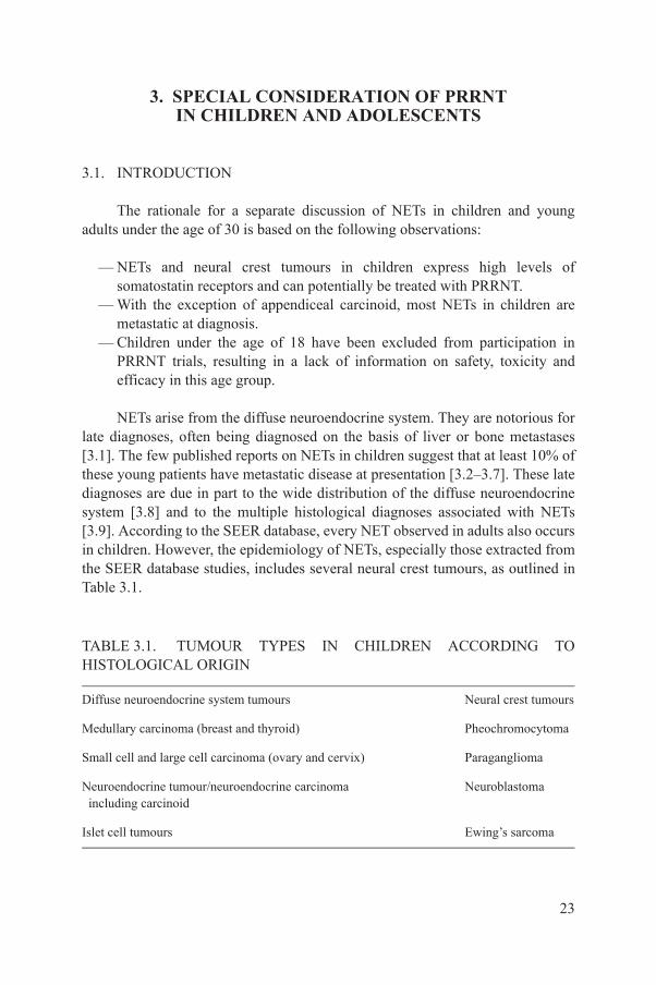
3. SPECIAL CONSIDERATION OF PRRNTIN CHILDREN AND ADOLESCENTS
3.1. INTRODUCTION
The rationale for a separate discussion of NETs in children and young adults under the age of 30 is based on the following observations:
— NETs and neural crest tumours in children express high levels of somatostatin receptors and can potentially be treated with PRRNT.
— With the exception of appendiceal carcinoid, most NETs in children are metastatic at diagnosis.
— Children under the age of 18 have been excluded from participation in PRRNT trials, resulting in a lack of information on safety, toxicity and efficacy in this age group.
NETs arise from the diffuse neuroendocrine system. They are notorious for late diagnoses, often being diagnosed on the basis of liver or bone metastases [3.1]. The few published reports on NETs in children suggest that at least 10% of these young patients have metastatic disease at presentation [3.2–3.7]. These late diagnoses are due in part to the wide distribution of the diffuse neuroendocrine system [3.8] and to the multiple histological diagnoses associated with NETs [3.9]. According to the SEER database, every NET observed in adults also occurs in children. However, the epidemiology of NETs, especially those extracted from the SEER database studies, includes several neural crest tumours, as outlined in Table 3.1.
TABLE 3.1. TUMOUR TYPES IN CHILDREN ACCORDING TO HISTOLOGICAL ORIGIN
Diffuse neuroendocrine system tumours Neural crest tumours
Medullary carcinoma (breast and thyroid) Pheochromocytoma
23
Small cell and large cell carcinoma (ovary and cervix) Paraganglioma
Neuroendocrine tumour/neuroendocrine carcinoma including carcinoid
Neuroblastoma
Islet cell tumours Ewing’s sarcoma
3.2. EPIDEMIOLOGY
Patients who are eventually diagnosed with NETs often have a multi-year history of symptoms prior to identification of the malignancy, with average lag periods of 8–10 years [3.10]. Thus, a 29-year-old adult diagnosed as having a NET may well have been an adolescent when the first symptoms occurred. The incidence of NETs increases with age and nearly 90% of patients in the 0–29 year age group are diagnosed when they are over the age of 20. However, the tendency for late diagnosis, after metastases to the liver or bones, suggests that more than half of NETs diagnosed in this age group probably occurred prior to the age of 21.
The incidence of NETs in children and young adults in the USA has recently been analysed using long term follow-up information provided by the SEER database. Incidence rates, observed survival rates and 31 year limited duration prevalence counts were obtained from SEER for diagnosis years 1975 to 2006 [3.11]. These rates were compared between and within NETs using variables from nine standard SEER registries for patients aged 0–29 years.
The most common NET sites were the lung, breast and appendix, with incidence rates of 0.6 per million for lung, 0.6 per million for breast and 0.5 per million for appendix in the age group 0–29 years. Incidence was less than 0.1 per million for all other NETs. The estimated age-adjusted number of NETs in the USA was 1073 in 2006. NET five year observed survival rates were 84% for the period 2000–2006, and the estimated 31 year limited duration prevalence for NETs as of 1 January 2006 was 7724. The age adjusted multivariate Cox regression demonstrated small cell histology, primary location in the breast and distant stage as major predictors of decreased survival, with five year survival of less than 24% of patients with ovarian small cell NETs.
3.3. TUMOURS IN CHILDREN ELIGIBLE FOR PRRNT
3.3.1. Carcinoid (neuroendocrine tumour)
The lung is the most common location for NETs in young people, and NET is the most common childhood malignancy arising in the lung [3.12]. NETs should be considered in any young person who has a culture negative pneumonia
24
and especially in the case of recurrent, culture negative pneumonia. A chest CT should be performed, and if a lung lesion is found, a biopsy should be obtained. NET biomarker levels (Cg-A, serotonin) should be measured in these children before surgery in order to determine which biomarkers should be utilized in follow-up.
The gastroenteropancreatic NETs together constitute another major group of tumours in children, with the appendix and pancreas the most frequent sites. Both functioning and non-functioning pancreatic NETs occur in children. Biomarkers are also useful in the diagnosis and follow-up of these tumours, as a specific marker may indicate the likely location of the primary (e.g. gastrin, insulin, glucagon, substance P, neurokinin A) or be used to follow liver metastases (pancreastatin). Gastrinoma has been reported in children as young as six years of age [3.7]. Normal fasting gastrin levels are similar in children and adults, making this an easy and extremely useful test. However, even the short term use of PPI medication can raise gastrin and Cg-A levels significantly. PPI should be discontinued for at least 72 h before measuring neuropeptide levels in serum, and some patients may require up to 4 weeks off PPI to allow peptide levels to return to normal [3.13].
Nesidioblastosis is the result of an overactive pancreas (hypertrophy and hyperplasia of the islet cells) and most often presents at birth as hypoglycaemia unresponsive to feeding or intravenous glucose. This most often resolves with close follow-up and octreotide therapy, but may resurface when these children reach puberty [3.14]. Insulinoma has been seen in children as young as five years of age [3.15−3.19]. Insulin and C-peptide levels are measured in blood, and normal levels are similar to those in adults.
Gastroenteropancreatic NETs have high somatostatin receptor expression and an excellent [3.20] response to PRRNT [3.21]. PRRNT may render previously unresectable tumours amenable to surgery. Whether PRRNT should be the initial therapy, followed by surgery and/or chemotherapy such as temozolomide and capecitabine, must be decided on an individual basis.
3.3.2. Medullary carcinoma
Medullary carcinoma occurs more often in the breast than in the thyroid; the primary lesion is often found by the patient, but primary breast location should also be considered in the case of metastatic disease to bone that is found on pathology to be medullary carcinoma. These tumours could be considered non-functional, as none of the usual NET markers are useful. Surgical excision with clear margins is the treatment of choice [3.22]. PRRNT would only be indicated if residual tumour or metastatic disease were found on octreotide scintigraphy.
25
3.3.3. Multiple endocrine neoplasia
MEN-1 occurs in the parathyroid, pancreas and pituitary. MEN-2A occurs in the parathyroid, thyroid (MTC) and adrenal medulla (pheochromocytoma), while MEN-2B includes MTC, pheochromocytoma and neural crest tumours.
Family history and blood pressure measurements are the most important screening tools. Children can be tested and diagnoses made as early as four years of age with blood calcitonin levels; the pentagastrin stimulation test is available, but rarely performed. Urine catecholamines are also important and require 24 h urine collection. Plasma metanephrine measurement is an alternative in young children for whom 24 h urine collection is nearly impossible. Recommendation for thyroid removal is dependent on the family history and the precise characteristics of the RET gene mutation [3.23, 3.24]. PRRNT should only be considered if the primary tumour and any metastatic lesions are positive on an 111In-DTPA-octreotide single photon emission computed tomography (SPECT) scan.
3.3.4. Munchausen’s syndrome by proxy
Diarrhoea, flushing, sweating and fatigue are hallmark symptoms of NETs. However, each of these symptoms is common in normal, healthy children and may be associated with viral infections, topical exposures and allergies. A parent, relative or guardian can easily induce such symptoms in a child. For instance, fictitious iatrogenic diarrhoea can be induced with laxatives and should be included in the screening process. Ricins cause overall irritation of the gastrointestinal tract, while castor oil will induce vomiting as well as some gastrointestinal upset. These substances can be measured in the stool along with pH and stool electrolytes.
3.3.5. Neuroblastoma
Neuroblastoma is a neural crest tumour that shares multiple biomarkers with NETs, including Cg-A, synaptophysin, NSE and sstr2 [3.21]. Some 90% of all neuroblastomas can be imaged with either 111In-DPTA-octreotide or 68Ga-DOTA-octreotate [3.25, 3.26], supporting the use of PRRNT in the treatment of neuroblastoma. Individual case studies validate this theoretical consideration, including minor responses in two subjects with neuroblastoma who participated in a phase I trial of 90Y-DOTA-Tyr3-octreotide in children [3.21]. Diagnostic testing should include vanillylmandelic acid and homovanillic acid (VMA/HVA) in urine, serum Cg-A and either SPECT/CT or PET/CT to
26
localize sstr2 positive disease.
3.3.6. Pheochromocytoma and paraganglioma
Pheochromocytoma is associated with MEN-2A and 2B, VHL syndrome, and NF-1. With the peak incidence between 9 and 12 years of age, nearly 10% of
all pheochromocytomas occur in children, and 10% of these are malignant. Headaches, palpitations, diaphoresis and hypertension are the most common symptoms. Diagnostic testing should include 24 h urine for creatinine, VMA, catecholamines and metanephrine, as well as free-plasma metanephrine and Cg-A. Since pheochromocytoma can be seen in adolescents and young adults, the possibility of drugs interfering with metanephrine testing should be ruled out with a careful review of medication and the patient’s history of illicit drug use. False positive metanephrine test results can be caused by buspirone, benzodiazepines, methyldopa, labetalol, levodopa, tricyclic antidepressants, ethanol, amphetamines, sotalol and chlorpromazine. Those pheochromocytomas and paragangliomas that are positive on an 111In-DPTA-octreotide scan may be amenable to PRRNT [3.27].
3.3.7. Small cell carcinoma
Small cell carcinoma of the cervix and ovary is more common in children and young adults than is small cell carcinoma of the lung. Ovarian small cell carcinoma can be familial [3.28, 3.29] and has a poor prognosis. There is very little information on sstr2 expression in these tumours so that the use of PRRNT must be based on positive sstr2 SPECT or PET imaging.
3.4. ADMINISTRATION OF PRRNT IN CHILDREN ANDYOUNG ADULTS
Only one controlled clinical trial of PRRNT has ever been conducted in children and young adults; this was a phase I trial of 90Y-DOTA-Tyr3-octreotide to determine the dose−toxicity profile in subjects under the age of 25 years with somatostatin receptor positive tumours [3.21]. In this study, a dose escalation design was utilized to determine the highest tolerable dose of 90Y-DOTA-Tyr3-octreotide, while limiting the permitted renal radiation dose to <21 Gy. Activity levels of administered 90Y-DOTA-Tyr3-octreotide were 1.11, 1.48 and 1.85 GBq/m2 per cycle in three cycles at six-week intervals, co-administered with an amino acid infusion for renal protection. The eligibility criteria for participation in the trial included ages 2−25 years, progressive disease,
111
27
positive lesion on an In-DTPA-octreotide scan, glomerular filtration rate (GFR) ≥80 mL·min–1·m–2, bone marrow cellularity >40% or stored autologous hematopoietic stem cells, Lansky play scale >60%, and informed consent (see Annex IV).
In this trial, 17 subjects aged 2−24 years received at least one dose of 90Y-DOTA-Tyr3-octreotide; diagnoses included neuroblastoma, embryonal and
astrocytic brain tumours, paraganglioma, MEN-2B and gastroenteropancreatic NETs. There were no dose-limiting toxicities and no individual dose reductions due to renal or haematological toxicity. The most frequent toxicity was reversible nausea in 70%, even in the presence of antiemetics. There were no complete responses; three subjects experienced partial responses (PRs), five had minor responses (MRs), five experienced stable disease (SD), two had progressive disease (PD) and two subjects withdrew. Dosimetry performed on subjects in the 1.85 GBq/m2 per cycle cohort demonstrated an average 2.24 mGy/MBq dose to the kidneys, similar to the dosimetry estimates in adults. PRRNT with 90Y-DOTA-Tyr3-octreotide demonstrated an 18% PR plus 29% MR rate in children and young adults with somatostatin receptor positive tumours. The recommended phase II dosing is three cycles of 1.85 GBq/m2 per dose 90Y-DOTA-Tyr3-octreotide co-administered with amino acids. In the future, higher doses may be attainable through the use of dosimetry guided therapy.
3.5. TEACHING POINTS
Although there has been only one clinical trial in children, the observations in that trial of 90Y-DOTA-Tyr3-octreotide together with the combined experience of several centres using 177Lu-octreotate in children and young adults allow us to offer several recommendations for PRRNT in this age group:
— PRRNT is safe in children and young adults when given with renal protection.
— The recommended dosing of 90Y-DOTA-Tyr3-octreotide is 1.85 GBq/m2 in all children and <1.73 GBq/m2 in young adults. Dosing of 177Lu-octreotate is currently recommended at 7.4 GBq/m2.
— Total irradiation dose to the kidneys should be limited to 23 Gy until further controlled trials have been performed to demonstrate safety at higher doses.
— Renal protection with an amino acid infusion 30−60 min prior to and at least 3.5 h after administering PRRNT is mandatory. Recent trials suggest that such protection should be continued for 24−48 h following PRRNT administration [3.30].
28
REFERENCES TO SECTION 3
[3.1] HASSAN, M.M., et al., Risk factors associated with neuroendocrine tumours: A U.S.-based case-control study, Int. J. Cancer 123 (2008) 867–873.

[3.2] CORPRON, C.A., et al., A half century of experience with carcinoid tumours in children, Am. J. Surg. 170 (1995) 606–608.
[3.3] SPUNT, S.L., et al., Childhood carcinoid tumours: The St Jude Children’s Research Hospital experience, J. Pediatr. Surg. 35 (2000) 1282–1286.
[3.4] PARHAM, D.M., Neuroectodermal and neuroendocrine tumours principally seen in children, Am. J. Clin. Pathol. 115 (2001) S113–S128.
[3.5] BROADDUS, R.R., et al., Neuroendocrine tumours (carcinoid and neuroendocrine carcinoma) presenting at extra-appendiceal sites in childhood and adolescence, Arch. Pathol. Lab. Med. 127 (2003) 1200–1203.
[3.6] LADD, A.P., GROSFELD, J.L., Gastrointestinal tumours in children and adolescents, Semin. Pediatr. Surg. 15 (2006) 37–47.
[3.7] KHANNA, G., et al., Gastroenteropancreatic neuroendocrine tumours in children and young adults, Pediatr. Radiol. 38 (2008) 251–259.
[3.8] MODLIN, I.M., et al., Evolution of the diffuse neuroendocrine system — Clear cells and cloudy origins, Neuroendocrinology 84 (2006) 69–82.
[3.9] PAPOTTI, M., et al., Spectrum of neuroendocrine tumours in non-endocrine organs, Pathologica 97 (2005) 215.
[3.10] VINIK, A., MOATTARI, A.R., Use of somatostatin analog in management of carcinoid syndrome, Dig. Dis. Sci. 34 (1989) 14S–27S.
[3.11] NAVALKELE, P., O’DORISIO, M.S., O’DORISIO, T.M., ZAMBA, G.K.D., LYNCH, C.F., Incidence, survival and prevalence of neuroendocrine tumours versus neuroblastoma in children and young adults: Nine standard SEER registries, 1975−2006, Pediatr. Blood Cancer 56 (2011) 50–57.
[3.12] GUSTAFSSON, B.I., et al., Bronchopulmonary neuroendocrine tumours, Cancer 113(2008) 5–21.
[3.13] GIUSTI, M., et al., Effect of short-term treatment with low dosages of the proton-pump inhibitor omeprazole on serum chromogranin A levels in man, Eur. J. Endocrinol. 150(2004) 299–303.
[3.14] GLASER, B., et al., Persistent hyperinsulinaemic hypoglycaemia of infancy: Long-term treatment with the somatostatin analogue Sandostatin, Clin. Endocrinol. (Oxf) 31(1989) 71–80.
[3.15] DE VOGELAERE, K., et al., Laparoscopic management of insulinoma in a child with multiple endocrine neoplasia type 1, J. Laparoendosc. Adv. Surg. Tech. A 16 (2006) 335–338.
[3.16] BAX, K.N., VAN DER ZEE, D.C., The laparoscopic approach toward hyperinsulinism in children, Semin. Pediatr. Surg. 16 (2007) 245–2451.
[3.17] BONFIG, W., et al., Recurrent hypoglycemic seizures and obesity: Delayed diagnosis of an insulinoma in a 15 year-old boy − Final diagnostic localization with endosonography, J. Pediatr. Endocrinol. Metab. 20 (2007) 1035–1038.
[3.18] LODISH, M.B., et al., Insulinoma and gastrinoma syndromes from a single
29
intrapancreatic neuroendocrine tumour, J. Clin. Endocrinol. Metab. 93 (2008) 1123–1128.
[3.19] MANABE, M., et al., Anesthetic management of pediatric patients with insulinoma using continuous glucose monitoring, Masui 58 (2009) 757–759.
[3.20] KARACHALIOU, F., et al., Malignant insulinoma in childhood, J. Pediatr. Endocrinol. Metab. 19 (2006) 757–760.
[3.21] MENDA, Y., et al., Phase I trial of 90Y-DOTA-Tyr3-octreotide therapy in children and young adults with refractory solid tumours that express somatostatin receptors, J. Nucl. Med. 51 (2010) 1524–1531.
[3.22] MALYUCHIK, S.S., KIYAMOVA, R.G., Medullary breast carcinoma, Exp. Oncol. 30 (2008) 96–101.
[3.23] SKINNER, M.A., et al., Prophylactic thyroidectomy in multiple endocrine neoplasia type 2A, N. Engl. J. Med. 353 (2005) 1105–1113.
[3.24] CALVA, D., et al., When is prophylactic thyroidectomy indicated for patients with RET codon 609 mutation?, Ann. Surg. Oncol. 16 (2009) 2237–2244.
[3.25] JUWEID, M.E., et al., 111In-pentetreotide versus bone scintigraphy in the detection of bony metastases of neuroblastoma, Nucl. Med. Commun. 23 (2002) 983–989.
[3.26] BAUM, R.P., et al., Receptor PET/CT imaging of neuroendocrine tumours, Recent Results Cancer Res. 170 (2008) 225–242.
[3.27] FANTI, S., et al., Evaluation of unusual neuroendocrine tumours by means of 68Ga-DOTA-NOC PET, Biomed. Pharmacother. 62 (2008) 667–671.
[3.28] CHRISTIN, A., et al., Successful treatment for advanced small cell carcinoma of the ovary, Pediatr. Blood Cancer 50 (2008) 1276–1277.
[3.29] MARTINEZ-BORGES, A.R., et al., Familial small cell carcinoma of the ovary, Pediatr. Blood Cancer 53 (2009) 1334–1336.
[3.30] BODEI, L., et al., Long-term evaluation of renal toxicity after peptide receptor radionuclide therapy with 90Y-DOTATOC and 177Lu-DOTATATE: The role of associated risk factors, Eur. J. Nucl. Med. Mol. Imaging 35 (2008) 1847–1856.
30
4. ANATOMICAL IMAGING
4.1. INTRODUCTION
Anatomical imaging of NETs should be as detailed and extensive as possible in order to provide accurate information concerning the site and extent of primary tumours and the localization and extent of regional and distant metastases. Ideally, procedures such as somatostatin receptor scintigraphy combined with an exact radiological examination for the staging of tumours are preferable. In addition, serial radiological examinations are essential for monitoring therapy and for the detection of recurrent disease.
4.2. ENDOSCOPY, ULTRASOUND AND ENDOSCOPIC ULTRASOUND
Endoscopy is essential for the detection and histological sampling of enteral and bronchial NETs. NETs of the stomach and rectum are often found incidentally. The role of pull-and-push endoscopy and capsule endoscopy for the detection of NETs of the ileum and jejunum is less clear, since these tumours are often small and may arise from the submucosa.
Ultrasound is the most widely available imaging technique. This technique is operator dependent and provides a wide range of sensitivity and specificity [4.1]. In primary pancreatic NETs, conventional ultrasound shows a mean detection rate of 39% (in a range of 17−79%), and a sensitivity of 18% for duodenal tumours and lymph node metastases. Detection rates for liver metastases of NETs are higher; 88% sensitivity and 92% specificity have been reported [4.2]. The use of contrast enhanced ultrasound may further increase sensitivity and specificity [4.3, 4.4].
Endoscopic ultrasound is the most sensitive method for detecting pancreatic and duodenal tumours, and is also essential for the exact staging of primary tumours of the upper or lower gastrointestinal tract (e.g. NETs of the stomach or rectum). The method has a mean detection rate of 90% (in a range of 77−100%) for pancreatic NETs, as reported in a compilation of 10 studies involving 261 patients, and is thus in the same range as intraoperative ultrasound with a mean
31
detection rate of 92% (in a range of 74−96%) in four studies with 127 patients [4.1]. Endoscopic ultrasound is also useful for obtaining histological specimens from lesions in or adjacent to the upper or lower gastrointestinal tract, for instance, pancreatic masses, with high diagnostic yield [4.5]. Endoscopic ultrasound is the preferred method for surveillance of pancreatic lesions in patients with multiple endocrine neoplasia [4.6].
Assessment and documentation of tumour progress is difficult with ultrasound; therefore, this method is not recommended for thorough staging and clinical trials. In a clinical setting, however, ultrasound has many advantages, allowing a dynamic and focused examination; in skilled and experienced hands, it is especially useful if radiation exposure is an issue. It is most appropriate for patients with slowly progressing tumours, in young patients or in the rare case where it is necessary to monitor tumour load during pregnancy.
4.3. COMPUTED TOMOGRAPHY AND MRI
Radiological assessments of NETs should be as exact as possible with the lowest radiation dose. CT and MRI are widely applied worldwide, although the choice of preferred imaging modality will depend on local expertise, availability, the side effects of contrast materials and costs. In addition, imaging modalities should be kept constant during follow-up of the patient to allow direct comparison of repeated test results. Care should be taken to perform radiological assessment according to current and local standards of imaging. It is essential to acquire multiple phases, with special attention to the arterial phase [4.7, 4.8]. Exact assessments of liver metastases and the degree of liver involvement are essential to define prognosis, evaluate response to treatment and select suitable locoregional therapy.
Most radiology departments use multidetector CT scanners that allow fast and accurate imaging and reconstruction in different planes. In addition, several contrast enhanced phases may be acquired by these modern scanners. Detection rates for CT have been compiled in a recent guideline on standards of care [4.1], which can be summarized as follows. For the diagnosis of pancreatic endocrine tumours in 162 patients of CT with adequate contrast material, the sensitivity and specificity were 73% and 96%, respectively [4.1]. In a compilation of four studies with a total of 135 patients, liver metastases were detected with a mean sensitivity of 82% and specificity of 92%.
In another study, more than 30% of lesions were found during the arterial phase, and 6% of the hepatic metastases were detected only on the arterial phase of contrast enhanced CT [4.7]. Extrahepatic abdominal soft tissue metastases were diagnosed with a sensitivity of 75% and specificity of 99% in four studies
32
with 135 patients. Different metastases in the thorax and abdomen were diagnosed with a sensitivity of 83% and specificity of 76% in 164 patients in three studies, or with a detection rate of 76% in 25 patients. Enteroclysis with CT had a low sensitivity for NETs in a study of eight patients, but better results with a sensitivity of 85% and specificity of 97% have been reported in a study of 219 patients with small bowel tumours, including 19 NETs [4.1].
Fewer data are available on the sensitivity and specificity of MRI in the diagnosis of NETs. Endocrine pancreatic tumours were visualized with a sensitivity of 93% and specificity of 88% in a series of two studies involving 54 patients. Reported detection rates were 73% in 192 patients in five studies. Liver metastases of NETs were detected in 82% of 74 patients in three studies. Dynamic contrast enhanced MRI depicted a typical hypervascular pattern in 73% of patients, with the greatest number of metastases detected during the hepatic arterial phase [4.8]. Extrahepatic abdominal soft tissue metastases were diagnosed with 89% sensitivity and 100% specificity in one study with 34 patients [4.1]. Although both CT and MRI can detect liver metastases, there is evidence that MRI can detect more lesions than can CT [4.8].
REFERENCES TO SECTION 4
[4.1] SUNDIN, A., et al., ENETS consensus guidelines for the standards of care in neuroendocrine tumours: Radiological examinations, Neuroendocrinology 90 (2009) 167–183.
[4.2] CHITI, A., et al., Comparison of somatostatin receptor imaging, computed tomography and ultrasound in the clinical management of neuroendocrine gastroenteropancreatic tumours, Eur. J. Nucl. Med. 25 (1998) 1396–1403.
[4.3] WU, W., et al., The role of contrast-enhanced sonography of focal liver lesions before percutaneous biopsy, Am. J. Roentgenol. 187 (2006) 752–761.
[4.4] DÖRFFEL, Y., WERMKE, W., Neuroendocrine tumours: Characterization with contrast-enhanced ultrasonography, Ultraschall. Med. 29 (2008) 506–514.
[4.5] CHANG, K.J., State of the art lecture: Endoscopic ultrasound (EUS) and FNA in pancreatico-biliary tumours, Endoscopy 38 (2006) S56–60.
[4.6] KANN, P.H., et al., Natural course of small, asymptomatic neuroendocrine pancreatic tumours in multiple endocrine neoplasia type 1: An endoscopic ultrasound imaging study, Endocr. Relat. Cancer 13 (2006) 1195–1202.
[4.7] PAULSON, E.K., et al., Carcinoid metastases to the liver: Role of triple-phase helical CT, Radiology 206 (1998) 143–150.
[4.8] DROMAIN, C., et al., Detection of liver metastases from endocrine tumours: A prospective comparison of somatostatin receptor scintigraphy, computed tomography, and magnetic resonance imaging, J. Clin. Oncol. 23 (2005) 70–78.
33
5. MOLECULAR IMAGING
5.1. INTRODUCTION
Radiopharmaceuticals for imaging the expression of somatostatin receptors and their density and subtype(s) utilize either SPECT or PET techniques. However, the PET technique is increasingly being used for diagnosing, staging and prognosticating patients with NETs. The use of dual-modality image fusion of PET or SPECT with CT to provide anatomical localization of receptor-expressing lesions or their metabolic behaviour (by means of 18F-FDG) allows improved patient specific, tailored therapy design.
This section addresses the role of molecular imaging using SPECT and PET/CT in the management of patients with NETs.
5.1.1. Withdrawal of somatostatin analogues
Somatostatin analogues are available as short acting or long acting preparations. These should be discontinued prior to somatostatin receptor imaging with either SPECT or PET techniques, as they might interfere with receptor targeting. The duration of such an interruption, however, will depend on the half-life of the analogue used. Allowing a period of 3−4 weeks for long acting release formulations, and a period of at least 24 h for short acting formulations, is regarded as good clinical practice, although this is a matter of ongoing debate.
5.2. SPECT IMAGING OF NETs
5.2.1. 111In-Octreoscan
The abundance of somatostatin receptor expression on NETs has resulted in the development of several radiopharmaceuticals that are directed towards these sites. Among the five known somatostatin receptor subtypes, most NETs express sstr2, with a small percentage expressing sstr3 and sstr5 [5.1].
Octreoscan (Mallinckrodt Medical, Petten, Netherlands) was first 111
34
introduced in the diagnostic arena in the 1990s using I-pentetreotide. Today, Octreoscan is a major diagnostic instrument in patients with NETs, with a more evident clinical role in subjects with gastrinoma, glucagonoma, VIPoma and carcinoids [5.2]. Useful information, which can be integrated with other diagnostic procedures, can be obtained in patients with other tumours, such as paraganglioma, medullary thyroid carcinoma, neuroblastoma and small cell lung

cancer (SCLC). The reported sensitivity of Octreoscan in detecting metastases is high (around 90%), as are its specificity and diagnostic accuracy [5.3]. Octreoscan has demonstrated a lower sensitivity in detecting liver metastases compared with MRI and CT scans, owing to its lower spatial resolution. Nevertheless, Octreoscan is able to explore the whole body and to give therapeutic indications [5.4].
5.2.2. 99mTc-HYNIC-TOC
For detecting primary tumours and metastases, 99mTc based somatostatin receptor scintigraphy holds great promise. The prototype is 99mTc-EDDA/HYNIC-octreotate, which delivers high quality images and thus offers an excellent alternative to 111In-Octreoscan [5.5].
5.3. PET/CT IMAGING OF NETs
PET radiopharmaceuticals can be used to assess receptor expression or to characterize intratumoural metabolic processes. The metabolic pathways and receptor targets that are currently being examined by PET are described in Table 5.1.
5.3.1. 68Ga-PET/CT for somatostatin receptor imaging
The somatostatin radiopharmaceuticals currently being used are derivatives of octreotide and, to a much lesser extent, lanreotide, and show variable binding to somatostatin receptors. 68Ga-DOTATOC was the first radiopharmaceutical used for PET imaging of NETs [5.9, 5.10]. Wild and colleagues have shown that the compound 68Ga-DOTANOC has a binding affinity to sstr2, 3 and 5 that is three to four times higher than that of 68Ga-DOTATOC, enabling the detection of a wide range of somatostatin receptor positive tumours (pan-somatostatin analogue), with significant effects on the diagnosis, staging and therapy of NETs and various other somatostatin receptor expressing tumours [5.11−5.13].
The sensitivity, specificity and diagnostic accuracy of imaging agents using 68Ga-DOTA peptides are high — 97%, 92% and 96%, respectively — and
35
provide superior results compared with Octreoscan and CT scans [5.11]. Moreover, imaging with 68Ga-DOTA peptides has been demonstrated to have a clinical impact by bringing about a modification of the clinical stage or of the treatment strategy in more than 55% of 90 NET patients [5.14].
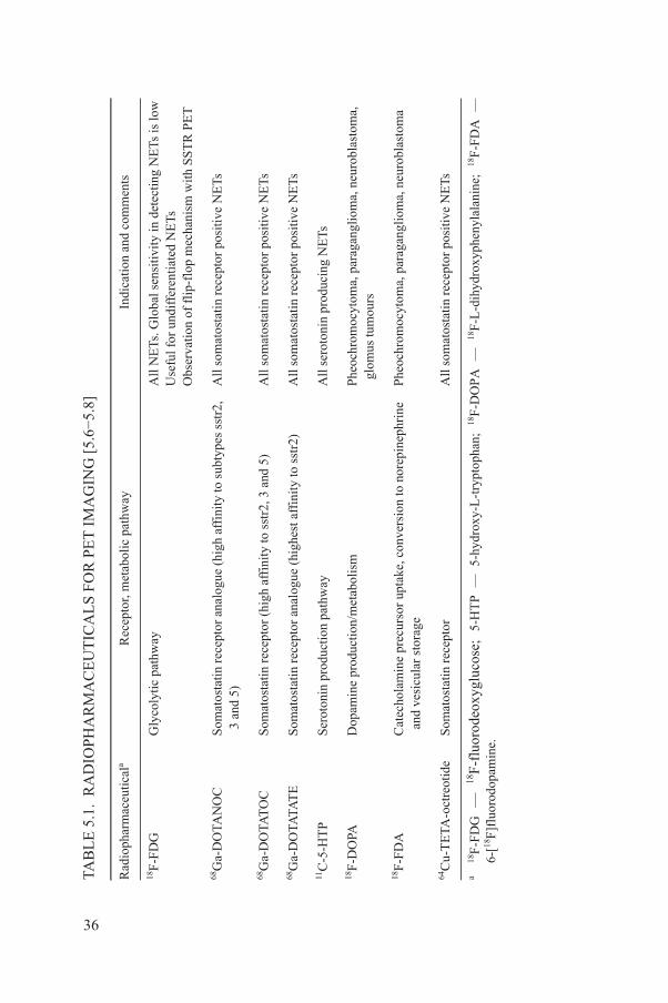
IOP
HA
RM
AC
EU
TIC
AL
S F
OR
PE
T I
MA
GIN
G [
5.6−
5.8]
laR
ecep
tor,
met
abol
ic p
athw
ayIn
dica
tion
and
com
men
ts
Gly
coly
tic p
athw
ayA
ll N
ETs
. Glo
bal s
ensi
tivit
y in
det
ecti
ng N
ETs
is lo
w
Use
ful f
or u
ndif
fere
ntia
ted
NE
Ts
Obs
erva
tion
of f
lip-
flop
mec
hani
sm w
ith S
ST
R P
ET
Som
atos
tati
n re
cept
or a
nalo
gue
(hig
h af
fini
ty to
sub
type
s ss
tr2,
3 a
nd 5
)A
ll s
omat
osta
tin r
ecep
tor
posi
tive
NE
Ts
Som
atos
tati
n re
cept
or (
high
aff
init
y to
sst
r2, 3
and
5)
All
som
atos
tatin
rec
epto
r po
siti
ve N
ETs
Som
atos
tati
n re
cept
or a
nalo
gue
(hig
hest
aff
init
y to
sst
r2)
All
som
atos
tati
n re
cept
or p
osit
ive
NE
Ts
Ser
oton
in p
rodu
ctio
n pa
thw
ayA
ll s
erot
onin
pro
duci
ng N
ETs
Dop
amin
e pr
oduc
tion
/met
abol
ism
Phe
ochr
omoc
ytom
a, p
arag
angl
iom
a, n
euro
blas
tom
a,
glo
mus
tum
ours
Cat
echo
lam
ine
prec
urso
r up
take
, con
vers
ion
to n
orep
inep
hrin
e a
nd v
esic
ular
sto
rage
Phe
ochr
omoc
ytom
a, p
arag
angl
iom
a, n
euro
blas
tom
a
deS
omat
osta
tin r
ecep
tor
All
som
atos
tatin
rec
epto
r po
siti
ve N
ETs
-flu
orod
eoxy
gluc
ose;
5-
HT
P
—
5-hy
drox
y-L
-try
ptop
han;
18
F-D
OP
A
—
18F
-L-d
ihyd
roxy
phen
ylal
anin
e;
18F
-FD
A
—
min
e.
36
TAB
LE
5.1
. R
AD
Rad
ioph
arm
aceu
tica
18F
-FD
G
68G
a-D
OTA
NO
C
68G
a-D
OTA
TO
C
68G
a-D
OTA
TAT
E
11C
-5-H
TP
18F
-DO
PA
18F
-FD
A
64C
u-T
ETA
-oct
reot
i
a 18
F-F
DG
—
18
F6-
[18F
]flu
orod
opa
5.3.2. 18F-FDG
One of the main sources of energy for any tumour is glucose. 18F-2-fluoro-2-deoxyglucose (18F-FDG) targets the glycolytic pathway, the main source of energy. FDG enters the glycolytic pathway like glucose in the cytoplasm, where it is phosphorylated by the enzyme hexokinase to FDG-6-phosphate. The latter molecule does not undergo further metabolization, but becomes trapped and accumulates inside the neoplastic cells. In spite of its wide use in oncology, 18F-FDG PET is frequently negative in patients with well differentiated NETs [5.15, 5.16].
111In-labelled Octreoscan and other, better tracers such as 99mTc-HYNICTOC or 68Ga-DOTA-conjugated peptides (like DOTANOC, DOTATATE and DOTATOC), provide reliable second-line diagnostic tools to improve overall diagnostic accuracy. Despite its low diagnostic sensitivity, 18F-FDG PET was recently demonstrated to have a strong prognostic value. In a prospective study of 98 patients, it was found that a maximum standardized uptake value (SUVmax) of over 9 and a high Karnofsky index (Ki) of 67 were significant predictors of overall survival, while an SUVmax of over 3 was the only predictor of progression-free survival (PFS) [5.17].
5.3.3. 18F-DOPA
NETs are characterized by the production and storage of several biogenic amines. One tracer with a design based on this observation is 18F-labelled L-dihydroxyphenylalanine (18F-DOPA), an aromatic amino acid labelled with 18F that was first used for the assessment of patients with Parkinson’s disease. Because they belong to the APUD cell system, a high proportion of NET cells avidly take up 18F-DOPA and can therefore be visualized by 18F-DOPA-PET scans. Recent studies have demonstrated increased L-DOPA decarboxylase activity in 80% of NETs, and it has been suggested that this could be used as a marker of tumour activity, particularly in so-called carcinoids. Nevertheless, it has been demonstrated that imaging with 68Ga-DOTA-conjugated peptides is superior to that with 18F-DOPA, which could be regarded as a second choice exam. Moreover, imaging with 68Ga peptides can guide therapeutic indications for applying PRRNT [5.8, 5.18−5.20].
37

5.4. OTHER MOLECULAR TARGETS
New tracers are being developed to target additional G-protein coupled receptors, including the glucagon-like peptide-1 receptor (GLP1-R), the gastrin releasing peptide receptor (GRP-R), the melanocortin receptor (McR) and the vasoactive intestinal polypeptide receptor (VIP-R) [5.21−5.24]. Like somatostatin analogues, these peptide ligands must be designed to have improved stability in serum and to accommodate a metal chelator such as DOTA, TETA or ETA.
Receptor antagonists constitute another class of tracers now undergoing development [5.25, 5.26]. Antagonists for the GRP-R and somatostatin receptors have been successfully tested in animal models and are currently in early human trials [5.27].
REFERENCES TO SECTION 5
[5.1] REUBI, J.C., Peptide receptors as molecular targets for cancer diagnosis and therapy, Endocr. Rev. 24 (2003) 389–427.
[5.2] KWEKKEBOOM, D.J., KOOIJ, P.P., BAKKER, W.H., MÄCKE, H.R., KRENNING, E.P., Comparison of 111In-DOTA-Tyr3-octreotide and 111In-DTPA-octreotide in the same patients: biodistribution, kinetics, organ and tumour uptake, J. Nucl. Med. 40 (1999) 762–767.
[5.3] CHITI, A., et al., Comparison of somatostatin receptor imaging, computed tomography and ultrasound in the clinical management of neuroendocrine gastroenteropancreatic tumours, Eur. J. Nucl. Med. 25 (1998) 1396–1403.
[5.4] DROMAIN, C., et al., Detection of liver metastases from endocrine tumours: A prospective comparison of somatostatin receptor scintigraphy, computed tomography, and magnetic resonance imaging, J. Clin. Oncol. 23 (2005) 70–78.
[5.5] DECRISTOFORO, C., et al., 99mTc-EDDA/HYNIC-TOC: A new 99mTc-labelled radiopharmaceutical for imaging somatostatin receptor-positive tumours: First clinical results and intra-patient comparison with 111In-labelled octreotide derivatives, Eur. J. Nucl. Med. 27 (2000) 1318–1325.
[5.6] AMBROSINI, V., TOMASSETTI, P., FRANCHI, R., FANTI, S., Imaging of NETs with PET radiopharmaceuticals, Q. J. Nucl. Med. Mol. Imaging 54 (2010) 16–23.
[5.7] PRASAD, V., AMBROSINI, V., ALAVI, A., BAUM, R.P., PET/CT in neuroendocrine tumours: Evaluation of receptor status and metabolism, PET Clinic. 2 (2007) 351–375.
[5.8] HAUG, A.R., et al., Intraindividual comparison of 68Ga-DOTA-TATE and 18F-DOPA
38
PET in patients with well-differentiated metastatic neuroendocrine tumours, Eur. J. Nucl. Med. 36 (2009) 765–770.
[5.9] MAECKE, H.R., HOFMANN, M., HABERKORN, U., (68)Ga-labeled peptides in tumour imaging, J. Nucl. Med. 46 (2005) 172s–178s.
[5.10] GINJ, M., et al., New pansomatostatin ligands and their chelated versions: Affinity profile, agonist activity, internalization, and tumour targeting, Clin. Cancer Res. 14(2008) 2019–2027.
[5.11] WILD, D., et al., DOTA-NOC, a high-affinity ligand of somatostatin receptor subtypes 2, 3 and 5 for labelling with various radiometals, Eur. J. Nucl. Med. Mol. Imaging 30 (2003) 1338–1347.
[5.12] GABRIEL, M., et al., 68Ga-DOTA-Tyr3-octreotide PET in neuroendocrine tumours: Comparison with somatostatin receptor scintigraphy and CT, J. Nucl. Med. 48 (2007) 508–518.
[5.13] VIRGOLINI, I., et al., Procedure guidelines for PET/CT tumour imaging with 68Ga DOTA-conjugated peptides: 68Ga-DOTA-TOC, 68Ga-DOTA-NOC, 68Ga-DOTA-TATE, Eur. J. Nucl. Med. Mol. Imaging 37 (2010) 2004–2010.
[5.14] AMBROSINI, V., et al., 68Ga-DOTANOC PET/CT clinical impact in patients with neuroendocrine tumours, J. Nucl. Med. 51 (2010) 669–673.
[5.15] SCANGA, D.R., MARTIN, W.H., DELBEKE, D., Value of FDG PET imaging in the management of patients with thyroid, neuroendocrine, and neural crest tumours, Clin. Nucl. Med. 29 (2004) 86–90.,
[5.16] ADAMS, S., et al., Limited value of fluorine-18 fluorodeoxyglucose positron emission tomography for the imaging of neuroendocrine tumours, Eur. J. Nucl. Med. 25 (1998) 79–83.
[5.17] BINDERUP, T., et al., 18F-Fluorodeoxyglucose positron emission tomography predicts survival of patients with neuroendocrine tumours, Clin. Cancer Res. 16 (2010) 978–985.
[5.18] BEUTHIEN-BAUMANN, B., et al., Diagnostic impact of PET with 18F-FDG, 18F-DOPA and 3-O-methyl-6-[18F]fluoro-DOPA in recurrent or metastatic medullary thyroid carcinoma, Eur. J. Nucl. Med. Mol. Imaging 34 (2007) 1604–1609.
[5.19] AMBROSINI, V., et al., Role of 18F-DOPA PET/CT imaging in the management of patients with 111In-pentetreotide negative GEP tumours, Nucl. Med. Commun. 28 (2007) 473–477.
[5.20] AMBROSINI, V., et al., Comparison between 68Ga-DOTA-NOC and 18F-DOPA PET for the detection of gastroenteropancreatic and lung neuro-endocrine tumours, Eur. J. Nucl. Med. Mol. Imaging 35 (2008) 1431–1438.
[5.21] WILD, D., MÄCKE, H., CHRIST, E., GLOOR, B., REUBI, J.C., Glucagon-like peptide 1-receptor scans to localize occult insulinomas, N. Engl. J. Med. 359 (2008) 766–768.
[5.22] CHRIST, E., et al., Glucagon-like peptide-1 receptor imaging for localization of insulinomas, J. Clin. Endocrinol. Metab. 94 (2009) 4398–4405.
[5.23] CANTORIAS, M.V., et al., Development of high-specific-activity (68)Ga-labeled DOTA-rhenium-cyclized alpha-MSH peptide analog to target MC1 receptors overexpressed by melanoma tumours, Nucl. Med. Biol. 36 (2009) 505–513.
[5.24] THAKUR, M.L., et al., PET imaging of oncogene overexpression using 64Cu-vasoactive intestinal peptide (VIP) analog: Comparison with 99mTc-VIP analog,
39
J. Nucl. Med. 45 (2004) 1381–1389.[5.25] GINJ, M., et al., Radiolabeled somatostatin receptor antagonists are preferable to
agonists for in vivo peptide receptor targeting of tumours, Proc. Natl. Acad. Sci. USA 103 (2006) 16436–16441.
[5.26] MANSI, R., et al., Evaluation of a 1,4,7,10-tetraazacyclododecane-1,4,7,10-tetraacetic acid-conjugated bombesin-based radioantagonist for the labeling with single-photon emission computed tomography, positron emission tomography, and therapeutic radionuclides, Clin. Cancer Res. 15 (2009) 5240–5249.
[5.27] WASER, B., TAMMA, M.L., CESCATO, R., MAECKE, H.R., REUBI, J.C., Highly efficient in vivo agonist-induced internalization of sst2 receptors in somatostatin target tissues, J. Nucl. Med. 50 (2009) 936–941.
40
6. OPTIONS FOR THE CARE OF NET PATIENTS
This section discusses the options for the care of patients affected by local, regional or distant NET metastases. NETs of the gastroenteropancreatic system most often metastasize to the liver, so the ultimate goal of therapy is to preserve viable liver tissue for as long as possible. These tumours may remain clinically silent until a significant liver tumour burden has accumulated.
The therapeutic options discussed in this section are not mutually exclusive and for the most part are interchangeable. For patients in the hands of an experienced multidisciplinary team, these options can be associated with a high-benefit to low-risk ratio and extended quality of life.
6.1. THE ROLE OF SURGERY
The optimal management of NETs is early surgical removal prior to vascular or lymphatic invasion, transmural extension and the subsequent development of regional or distant metastases. Unfortunately, a large proportion of NETs patients are diagnosed with metastatic disease, thereby ruling out the radical surgical approach. The decision on surgical intervention is often complex and must take into account various factors such as the patient’s condition, the extent of the disease and the potential risks versus possible benefits of surgical exploration.
If the extent of the tumour(s) is limited or localized, then segmental resection with removal of the regional nodes is beneficial and is indicated. Removal of the primary tumour is indicated to prevent complications such as bleeding or small bowel obstruction, and also to prevent desmoplastic reaction (i.e. tumour associated growth of fibrous or connective tissue), even in advanced disease. The removal of the primary tumour is associated with a positive impact on survival even in the presence of metastases [6.1, 6.2]. Inoperable tumours of the pancreas can be rendered operable by means of repeated cycles of PRRNT (see Figs 7.4 and 7.5).
For tumours arising in the small bowel, resection of the affected segment should include lymph nodes leading up to the trunk of the superior mesenteric
41
artery (SMA). A situation that arises not infrequently in small bowel NETs is that the nodal involvement might extend to the root of the SMA. Here it might be very difficult to remove the higher lymph nodes involved without compromising the arterial blood supply to the entire small bowel. This means that at least ~30 cm of small bowel will have to be removed if there is a single tumour. It should be remembered, however, that many patients have multiple small bowel tumours,
and careful palpation of the entire small bowel should be carried out prior to resection. All identifiable lesions with their corresponding nodes should be resected; on occasion, that may require that more than 100 cm of small bowel be removed. In these cases, it is important that the total small bowel length be carefully measured to ensure sufficient residual absorptive surface, and to preserve the ileo-coecal valve, where possible. Trans-serosal extension of the tumour is a risk factor for disseminated intraperitoneal spread of tumour, so careful examination of the peritoneal lining and intra-abdominal organs should be carried out; if manageable, excision of these peritoneal nodules may be performed.
In cases of non-ruptured appendiceal carcinoid tumours, if the tumours are less than 1 cm in diameter and do not extend trans-serosally, then simple appendectomy is an adequate treatment. If these tumours are greater than 2 cm, then ileocolectomy and node dissection should be performed. Tumours 1–2 cm in size or those with trans-serosal extension are more of a grey area, but ileocolectomy is preferred in these cases as well, especially in young patients in whom missing nodal metastases would inevitably result in liver metastases and shortened survival [6.3, 6.4].
Gastric NETs may develop in response to hypergastrinemia, such as seen in atrophic gastritis and pernicious anaemia (type 1 gastric carcinoids). They may also be seen in patients with coeliac disease. These tumours are often small, with little risk of metastases, and can be excised locally or endoscopically. Type 2 gastric carcinoids occur in patients with MEN-1 due to gastrinoma and chronic hypergastrinemia. The primary goal is to locate the tumour source (either duodenal or pancreatic) and to excise it. In contrast, type 3 gastric NETs are sporadic in nature and occur in the absence of hypergastrinemia; they tend to be larger and more aggressive than the other two types of tumour. Surgical removal according to endocrine surgery standards (including subtotal gastrectomy) is indicated [6.5, 6.6].
Duodenal NETs, if small (<1 cm), are usually removed endoscopically. Larger tumours (>1–2 cm) have a propensity for nodal and distant metastases, and therefore duodenectomy with nodal dissection or, in cases of invasion, duodenal resection are indicated [6.7].
Rectal NETs are also often small (<1 cm) and benign, and are usually removed endoscopically. Larger tumours amenable to surgery will be treated by
42
local excision; wide resection is very rarely indicated in larger or G3 tumours. Colonic NETs are less common but are usually larger, necessitating segmental colonic resection with node dissection [6.8].
Pancreatic NETs located in the body and tail of the pancreas can be resected by distal pancreatectomy, commonly performed with splenectomy. Nodes along the splenic artery should be removed, as should perisplenic and coeliac nodes.
Tumours in the head of the pancreas will, whenever possible, be enucleated; larger tumours generally require pancreaticoduodenectomy, with the removal of portocaval and aortocaval nodes. In the case of a MEN-1 setting, where multiple adenomas (most of which are benign and slow growing) are the rule, not resecting should be considered. Functional tumours, where surgery is indicated, represent an exception [6.9].
The optimal management of a primary tumour with liver metastases, a condition that occurs in approximately 60–80% of cases, frequently poses a dilemma. Because of the relatively indolent course of many NETs, removal of the primary tumour along with debulking of liver metastases has several potential advantages. One is that removal of the primary may prevent additional metastatic seeding. Second, the debulking of liver lesions may improve tumour associated hormone secretory symptoms, as their serum levels can be reduced [6.10, 6.11]. Third, reducing the number of potential sites taking up the radiolabelled somatostatin analogues or other therapies may improve the ‘kill’ in the remaining tumour. Fourth, it appears that patients receiving liver-directed treatment along with removal of the intestinal primary survive longer than those who do not undergo surgery or the removal of the primary tumour alone [6.1, 6.12]. Prospective randomized trials to assess the impact of the removal of the primary tumour and/or metastases are, however, lacking.
There are several options for debulking or removing liver metastases. Resection may be indicated for solitary lesions, or when a few lesions are localized to one lobe of the liver, although some would argue for a more aggressive approach. Surface lesions can be enucleated relatively easily, as the tumours tend to be firm within the relatively softer parenchyma. Furthermore, they often do not recur despite the presence of tumour at their margins. Another effective debulking option is radiofrequency ablation (RFA) if there are many small lesions or several large ones. If the lesions are small and diffuse, then treating just a few lesions will not achieve significant hormonal relief, so that other postoperative strategies should be employed, such as embolization, chemoembolization or radioembolization with 90Y-labelled microspheres [6.13].
Another important adjunct to surgical exploration for NETs is cholecystectomy. Most patients will receive adjuvant somatostatin analogue treatment (such as Sandostatin-LAR or Lanreotide Autogel), often on a monthly basis. This treatment predisposes patients to develop gallstones, and therefore
43
prophylactic cholecystectomy will spare these individuals from biliary colic later in the course of their disease.
In patients presenting with metastatic disease with an unknown primary tumour, the decision to carry out a surgical exploration depends on whether or not there is a reasonable idea of where the primary tumour might be and the medical need or urgency to locate it (e.g. insulinoma).

6.2. THE ROLE OF SOMATOSTATIN ANALOGUES
Octreotide and lanreotide are somatostatin receptor agonists that play an essential role in the disease control of both symptomatic and asymptomatic NETs. These drugs should be considered as first-line therapy.
The first somatostatin receptor agonist to be described was octreotide acetate (SMS 201-995, Sandostatin®) in 1980 by Marbach. It was approved for clinical use in Europe in 1988 and in the USA in 1989. The approved indications for Sandostatin at the time of approval were for the diarrhoea and flushing of carcinoid syndrome and the WDS of VIP secreting tumours.
More recently, lanreotide has been introduced for symptomatic treatment of carcinoid syndrome in Europe, but not yet in the USA. Both octreotide and lanreotide have also been approved for growth hormone secreting pituitary tumours with acromegaly in adults, and gigantism in children and adolescents.
6.3. MOLECULAR BASIS FOR THE ACTION OF SOMATOSTATIN ANALOGUES
More than 90% of well differentiated NETs express sstr2. The rationale for the use of somatostatin receptor agonists or analogues (SSAs) for symptomatic control rests with the fact that virtually all the target cells of the tumour (e.g. secreting endocrine cells, cutaneous vessels) also express sstr2. In addition, symptomatic control can be exerted via the binding of the analogue to sstr5 at high doses of octreotide.
The biological effects of SSAs are probably mediated by a family of G-protein coupled receptors that are expressed in a tissue specific manner. The binding of SSAs to sstr2 and sstr5 exerts inhibitory effects on cellular signal transduction pathways through the inhibition of cyclic adenosine monophosphate (AMP) production. This in turn leads to the inhibition of amine and peptide secretion and cellular proliferation, and a more complex pathway, involving several mechanisms, to the induction of apoptosis [6.14]. Somatostatin analogues are widely used in the USA and Europe for both symptomatic and non-symptomatic NET control.
44
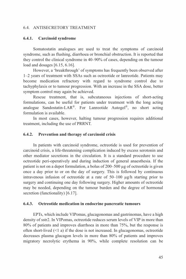
6.4. ANTISECRETORY TREATMENT
6.4.1. Carcinoid syndrome
Somatostatin analogues are used to treat the symptoms of carcinoid syndrome, such as flushing, diarrhoea or bronchial obstruction. It is reported that they control the clinical syndrome in 40–90% of cases, depending on the tumour load and dosages [6.15, 6.16].
However, a ‘breakthrough’ of symptoms has frequently been observed after 1–2 years of treatment with SSAs such as octreotide or lanreotide. Patients may become medication refractory with regard to syndrome control due to tachyphylaxis or to tumour progression. With an increase in the SSA dose, better symptom control may again be achieved.
Rescue treatment, that is, subcutaneous injections of short-acting formulations, can be useful for patients under treatment with the long acting analogue Sandostatin-LAR®. For Lanreotide Autogel®, no short acting formulation is available.
In most cases, however, halting tumour progression requires additional treatment, including the use of PRRNT.
6.4.2. Prevention and therapy of carcinoid crisis
In patients with carcinoid syndrome, octreotide is used for prevention of carcinoid crisis, a life-threatening complication induced by excess serotonin and other mediator secretions in the circulation. It is a standard procedure to use octreotide peri-operatively and during induction of general anaesthesia. If the patient is not on a depot formulation, a bolus of 200–500 µg of octreotide is given once a day prior to or on the day of surgery. This is followed by continuous intravenous infusion of octreotide at a rate of 50–100 µg/h starting prior to surgery and continuing one day following surgery. Higher amounts of octreotide may be needed, depending on the tumour burden and the degree of hormonal secretion (functionality) [6.17].
6.4.3. Octreotide medication in endocrine pancreatic tumours
45
EPTs, which include VIPomas, glucagonomas and gastrinomas, have a high density of sstr2. In VIPomas, octreotide reduces serum levels of VIP in more than 80% of patients and improves diarrhoea in more than 75%, but the response is often short-lived (<1 a) if the dose is not increased. In glucagonomas, octreotide decreases plasma glucagon levels in more than 80% of patients and improves migratory necrolytic erythema in 90%, while complete resolution can be
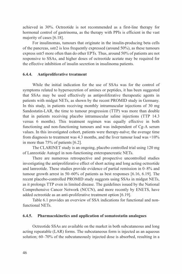
achieved in 30%. Octreotide is not recommended as a first-line therapy for hormonal control of gastrinoma, as the therapy with PPIs is efficient in the vast majority of cases [6.18].
For insulinomas, tumours that originate in the insulin-producing beta cells of the pancreas, sstr2 is less frequently expressed (around 50%), as these tumours express sstr5 more often than do other EPTs. Thus, around 50% of patients are not responsive to SSAs, and higher doses of octreotide acetate may be required for the effective inhibition of insulin secretion in insulinoma patients.
6.4.4. Antiproliferative treatment
While the initial indication for the use of SSAs was for the control of symptoms related to hypersecretion of amines or peptides, it has been suggested that SSAs may be used effectively as antiproliferative therapeutic agents in patients with midgut NETs, as shown by the recent PROMID study in Germany. In this study, in patients receiving monthly intramuscular injections of 30 mg Sandostatin-LAR, the time to tumour progression (TTP) was more than double that in patients receiving placebo intramuscular saline injections (TTP 14.3 versus 6 months). This treatment regimen was equally effective in both functioning and non-functioning tumours and was independent of Cg-A serum values. In this investigated cohort, patients were therapy-naïve; the average time from diagnosis to treatment was 4.3 months, and the liver tumour load was <10% in more than 75% of patients [6.2].
The CLARINET study is an ongoing, placebo controlled trial using 120 mg of Lanreotide Autogel in non-functioning enteropancreatic NETs.
There are numerous retrospective and prospective uncontrolled studies investigating the antiproliferative effect of short acting and long acting octreotide and lanreotide. These studies provide evidence of partial remission in 0–8% and tumour growth arrest in 50–60% of patients as best responses [6.16, 6.19]. The recent placebo-controlled PROMID study suggests using SSAs in midgut NETs, as it prolongs TTP even in limited disease. The guidelines issued by the National Comprehensive Cancer Network (NCCN), and more recently by ENETS, have added octreotide as an anti-proliferative treatment option [6.19].
Table 6.1 provides an overview of SSA indications for functional and non-functional NETs.
46
6.4.5. Pharmacokinetics and application of somatostatin analogues
Octreotide SSAs are available on the market in both subcutaneous and long acting repeatable (LAR) forms. The subcutaneous form is injected as an aqueoussolution; 60–70% of the subcutaneously injected dose is absorbed, resulting in a
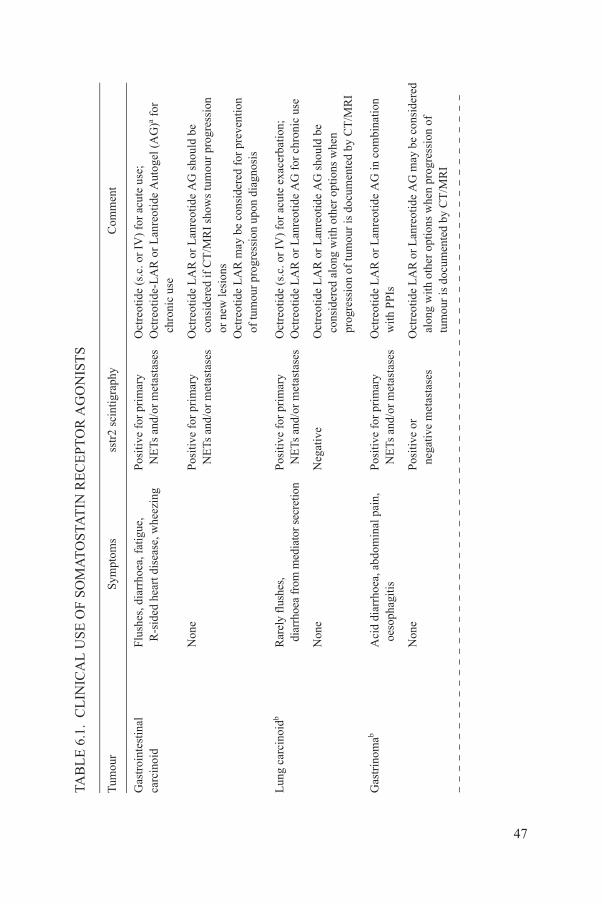
NIC
AL
US
E O
F S
OM
AT
OS
TAT
IN R
EC
EP
TO
R A
GO
NIS
TS
Sym
ptom
sss
tr2
scin
tigr
aphy
Com
men
t
Flu
shes
, dia
rrho
ea, f
atig
ue,
R-s
ided
hea
rt d
isea
se, w
heez
ing
Pos
itiv
e fo
r pr
imar
y N
ETs
and
/or
met
asta
ses
Oct
reot
ide
(s.c
. or
IV)
for
acut
e us
e;
Oct
reot
ide-
LA
R o
r L
anre
otid
e A
utog
el (
AG
)a for
c
hron
ic u
se
Non
eP
osit
ive
for
prim
ary
NE
Ts a
nd/o
r m
etas
tase
sO
ctre
otid
e L
AR
or
Lan
reot
ide
AG
sho
uld
be
con
side
red
if C
T/M
RI
show
s tu
mou
r pr
ogre
ssio
n o
r ne
w le
sion
sO
ctre
otid
e L
AR
may
be
cons
ider
ed f
or p
reve
ntio
n o
f tu
mou
r pr
ogre
ssio
n up
on d
iagn
osis
Rar
ely
flus
hes,
dia
rrho
ea f
rom
med
iato
r se
cret
ion
Pos
itiv
e fo
r pr
imar
y N
ETs
and
/or
met
asta
ses
Oct
reot
ide
(s.c
. or
IV)
for
acut
e ex
acer
bati
on;
Oct
reot
ide
LA
R o
r L
anre
otid
e A
G f
or c
hron
ic u
se
Non
eN
egat
ive
Oct
reot
ide
LA
R o
r L
anre
otid
e A
G s
houl
d be
c
onsi
dere
d al
ong
wit
h ot
her
opti
ons
whe
n p
rogr
essi
on o
f tu
mou
r is
doc
umen
ted
by C
T/M
RI
Aci
d di
arrh
oea,
abd
omin
al p
ain,
oes
opha
giti
sP
osit
ive
for
prim
ary
NE
Ts a
nd/o
r m
etas
tase
sO
ctre
otid
e L
AR
or
Lan
reot
ide
AG
in c
ombi
natio
n w
ith P
PIs
Non
eP
osit
ive
or
neg
ativ
e m
etas
tase
sO
ctre
otid
e L
AR
or
Lan
reot
ide
AG
may
be
cons
ider
ed
alo
ng w
ith
othe
r op
tion
s w
hen
prog
ress
ion
of
tum
our
is d
ocum
ente
d by
CT
/MR
I
47
TAB
LE
6.1
. C
LI
Tum
our
Gas
troi
ntes
tinal
carc
inoi
d
Lun
g ca
rcin
oidb
Gas
trin
omab
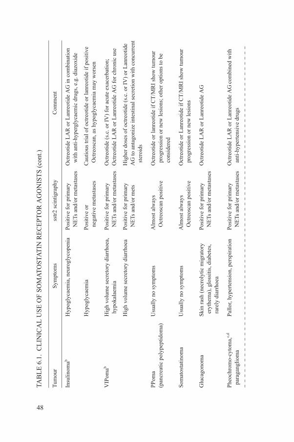
Hyp
ogly
caem
ia, n
euro
glyc
open
iaPo
siti
ve f
or p
rim
ary
NE
Ts a
nd/o
r m
etas
tase
sO
ctre
otid
e L
AR
or
Lan
reot
ide
AG
in c
ombi
natio
n w
ith
anti
-hyp
ergl
ycae
mic
dru
gs, e
.g. d
iazo
xide
Hyp
ogly
caem
iaPo
siti
ve o
r n
egat
ive
met
asta
ses
Cau
tiou
s tr
ial o
f oc
treo
tide
or
lanr
eoti
de if
pos
itiv
e O
ctre
osca
n, a
s hy
pogl
ycae
mia
may
wor
sen
Hig
h vo
lum
e se
cret
ory
diar
rhoe
a,
hyp
okal
aem
iaP
osit
ive
for
prim
ary
NE
Ts a
nd/o
r m
etas
tase
sO
ctre
otid
e (s
.c. o
r IV
) fo
r ac
ute
exac
erba
tion
; O
ctre
otid
e L
AR
or
Lan
reot
ide
AG
for
chr
onic
use
Hig
h vo
lum
e se
cret
ory
diar
rhoe
aPo
siti
ve f
or p
rim
ary
NE
Ts a
nd/o
r m
ets
Hig
her
dose
s of
oct
reot
ide
(s.c
. or
IV)
or L
anre
otid
e A
G to
ant
agon
ize
inte
stin
al s
ecre
tion
wit
h co
ncur
rent
ste
roid
s
idom
a)U
sual
ly n
o sy
mpt
oms
Alm
ost a
lway
s O
ctre
osca
n po
siti
veO
ctre
otid
e or
lanr
eotid
e if
CT
/MR
I sh
ow tu
mou
r p
rogr
essi
on o
r ne
w le
sion
s; o
ther
opt
ions
to b
e c
onsi
dere
d
Usu
ally
no
sym
ptom
sA
lmos
t alw
ays
Oct
reos
can
posi
tive
Oct
reot
ide
or L
anre
otid
e if
CT
/MR
I sh
ow tu
mou
r p
rogr
essi
on o
r ne
w le
sion
s
Skin
ras
h (n
ecro
lyti
c m
igra
tory
ery
them
a), g
loss
itis
, dia
bete
s,
rar
ely
diar
rhoe
a
Pos
itiv
e fo
r pr
imar
y N
ETs
and
/or
met
asta
ses
Oct
reot
ide
LA
R o
r L
anre
otid
e A
G
,c,d
Pal
lor,
hype
rten
sion
, per
spir
atio
nP
osit
ive
for
prim
ary
NE
Ts a
nd/o
r m
etas
tase
sO
ctre
otid
e L
AR
or
Lan
reot
ide
AG
com
bine
d w
ith
ant
i-hy
pert
ensi
ve d
rugs
NIC
AL
US
E O
F S
OM
AT
OS
TAT
IN R
EC
EP
TO
R A
GO
NIS
TS
(co
nt.)
Sym
ptom
sss
tr2
scin
tigr
aphy
Com
men
t
48
Insu
lino
mab
VIP
omab
PP
oma
(pan
crea
tic
poly
pept
Som
atos
tati
nom
a
Glu
cago
nom
a
Phe
ochr
omo-
cyto
ma
par
agan
glio
ma
TAB
LE
6.1
. C
LI
Tum
our
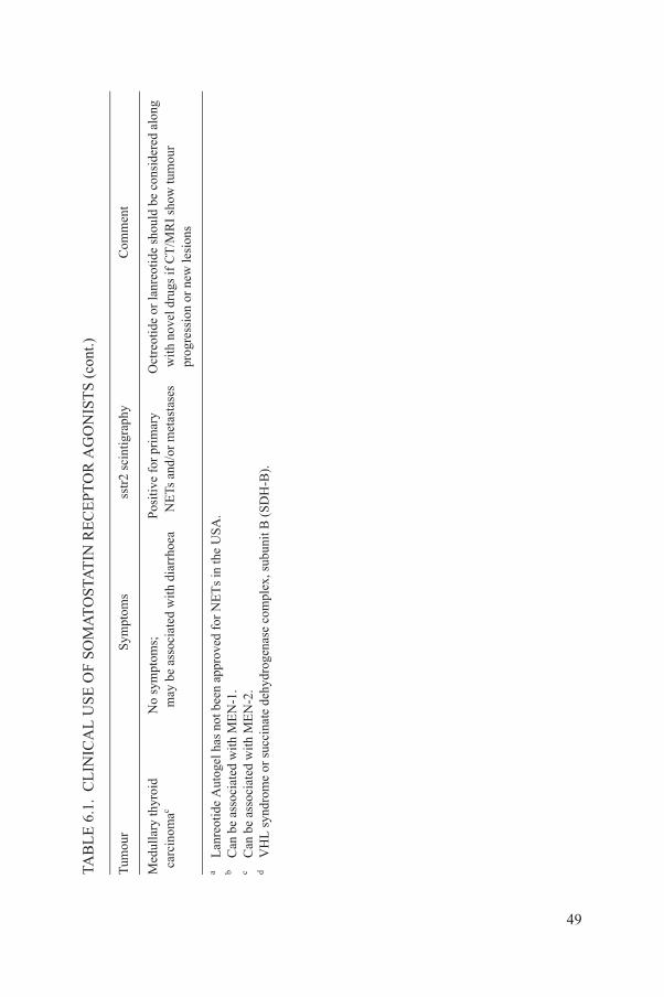
No
sym
ptom
s;
may
be
asso
ciat
ed w
ith
diar
rhoe
aP
osit
ive
for
prim
ary
NE
Ts a
nd/o
r m
etas
tase
sO
ctre
otid
e or
lanr
eoti
de s
houl
d be
con
side
red
alon
g w
ith
nove
l dru
gs if
CT
/MR
I sh
ow tu
mou
r p
rogr
essi
on o
r ne
w le
sion
s
el h
as n
ot b
een
appr
oved
for
NE
Ts
in th
e U
SA
. w
ith
ME
N-1
. w
ith
ME
N-2
.r
succ
inat
e de
hydr
ogen
ase
com
plex
, sub
unit
B (
SD
H-B
).
NIC
AL
US
E O
F S
OM
AT
OS
TAT
IN R
EC
EP
TO
R A
GO
NIS
TS
(co
nt.)
Sym
ptom
sss
tr2
scin
tigr
aphy
Com
men
t
49
Med
ulla
ry th
yroi
d c
arci
nom
ac
aL
anre
otid
e A
utog
bC
an b
e as
soci
ated
c C
an b
e as
soci
ated
dV
HL
syn
drom
e o
TAB
LE
6.1
. C
LI
Tum
our
peak serum concentration after 30 min and reaching a plateau after 60 min. The plasma half-life is approximately 120 min, but only 60 min if it is injected intravenously.
The LAR form of octreotide is available in 10, 20 or 30 mg concentrations as a polymer suspension. This suspension is absorbed at various rates over a period of 28 d. There is a large release into the circulation of octreotide acetate in the first 24 h after the intramuscular injection, followed by a 7–8 d period when very little octreotide is released. The treatment regimen usually requires at least three intramuscular injections at monthly intervals to achieve plasma saturation and a steady state condition level of serum octreotide.
When using octreotide for patients with symptoms of diarrhoea or flushing, it is important to use a subcutaneous octreotide formulation for 8 d before the intramuscular LAR injection, and thereafter to continue with the subcutaneous octreotide acetate for a further 8–10 d to ensure an adequate ‘loading’ serum level. A subcutaneous loading dose of 100–200 μg 3 daily both before and for 7–10 d following the intramuscular injection is recommended. In addition, it is advisable to initiate the subcutaneous form of octreotide at a dose of 100 μg 3daily before putting patients on the LAR form in order to ensure their tolerance to the LAR form.
The pharmokinetics of lanreotide, which is available as a subcutaneous form in Europe, is similar to that of octreotide acetate. The short acting form of lanreotide is not available in Europe; at present, only a lanreotide depot form is available in Europe and the USA. Both the subcutaneous and Autogel forms have absorption rates of 60–70%, similar to that of octreotide acetate. The monthly lanreotide depot is injected into the deep subcutaneous tissue and does not require pre-reconstitution as does Sandostatin-LAR. It is important to remember that for both depot formulations, blood and tissue saturation is only attained three months after starting the LAR therapy form, a fact that necessitates the use of the subcutaneous form during the initial treatment phase.
6.5. SIDE EFFECTS
The most frequent side effects of somatostatin analogues are gastrointestinal, including abdominal discomfort, nausea or diarrhoea that are
50
commonly transient. The risk of developing cholecystolithiasis and gall-bladder ‘sludge’ caused by chronic somatostatin analogue therapy is often precluded by performing a prophylactic cholecystectomy during the first surgical exploration to remove the primary gastrointestinal/pancreatic NET. Cholecystectomy may be required if long term use of the somatostatin congeners is considered, especially when these are effective in controlling symptoms and partially controlling tumour
proliferation. The mechanism of steatorrhoea, or fat malabsorption, is caused by the secretory inhibition of postprandial exocrine pancreatic enzymes. The latter side effect can be well managed by prescribing pancreatic enzyme replacement (pancrease–lipase).
6.6. INTERFERON
For more than 25 years, interferon-alpha (IFN-α) has been used for the treatment of patients with NETs, especially those with carcinoid syndrome, and with somatostatin analogues. IFN-α is considered the main antisecretory drug used in the treatment of these tumours [6.19, 6.20]. Treatment with IFN-α effectively reduces hypersecretion-induced symptoms in patients with carcinoid syndrome, similar to SSAs. Its effect on symptom control is comparable with that of SSAs, although it is not a standard treatment for carcinoid crisis.
In a large study of patients with malignant carcinoids and hepatic metastases, 70% of patients experienced improvements in flushing or diarrhoea, or both, when IFN-α 2b was used at a dose of 5 million units (MU) s.c. 3weekly, or natural IFN-α was used daily. Biochemical response was obtained in 42% of these patients, and significant tumour shrinkage was achieved in 15%. Stabilization of tumour growth is considered an important response and was reported in 39% of patients [6.21]. A biochemical and symptomatic response was reported in up to 50% of patients, whereas partial tumour size responses were demonstrated in 10–15% [6.19, 6.22]. The duration of response was 12–36 months. Interferon is also effective in the treatment of EPTs [6.22].
Side effects of IFN-α are very common, limiting the dose and duration of treatment. Side effects include flu-like symptoms in over 90% of patients, weight loss and fatigue. Major side effects include autoimmune reactions, depression and mental disturbance. Bone marrow toxicity is usually mild, as is hepatotoxicity, which can be managed by dose adjustments.
Pegylated interferon (PEG-IFN) can be considered an alternative treatment to the conventional regimen of INF-α 3 weekly, if the latter is not well tolerated [6.23]. PEG-IFN is at least equally effective compared with the conventional IFN-α regimen. Data are limited, however, and PEG-IFN is not officially registered for the treatment of NETs. Better tolerability of PEG-IFN-α, in contrast
51
with the conventional IFN-α regimen, has been demonstrated in patients with chronic myeloid leukaemia and solid tumours, such as metastatic melanoma and renal cell carcinoma.
In NETs, administration of PEG-IFN-α at dosages of 50–100 µg, once weekly, has been associated with less frequent (24%) and less persistent acute flu-like symptoms than those reported in the literature for non-pegylated IFN-α.
To assess haematological side effects, repeated white and red blood cell counts are recommended. Monitoring of liver enzymes and thyroid stimulating hormone (TSH) levels is required at the beginning of treatment and during follow-up.
6.7. CONCLUDING REMARKS ON THE USE OFSOMATOSTATIN ANALOGUES
In Europe, the use of SSAs combined with IFN-α is the only approved therapy for treating symptomatic NETs. SSAs can be used alongside virtually all other therapeutic options discussed in this section. They are mandatory prior to any invasive intervention at the tumour site, including surgery, local ablation or embolization, and prior to general anaesthesia to prevent carcinoid crisis. Given the knowledge that 87–92% of all NETs express sstr2, patients should always be offered this option alongside other treatments and in the framework of supportive care. Furthermore, the results of a recent placebo controlled study of small intestinal (midgut) NETs highlight the tumour growth inhibitory effect that has been suggested by several prior uncontrolled prospective and retrospective studies. The benefits of the commercially available SSAs far outweigh their risks.
For instructions on the withdrawal of SSA medications prior to the delivery of PRRNT, see Section 7.4.1.
6.8. CHEMOTHERAPY
Systemic chemotherapy is effective in some patients, especially those with PDEC/neuroendocrine carcinoma (grade G3) and progressive NETs of the pancreas. In well differentiated midgut NETs/NET grades G1 or G2, however, response rates to chemotherapy are low (7–20%) and no survival advantages have been demonstrated [6.24, 6.25].
The standard treatment of neuroendocrine carcinoma grade G3 is cisplatin and etoposide. Cisplatin inhibits DNA synthesis; etoposide is a cytostatic drug that arrests the cell cycle in the S and G2 phases, and inhibits mitosis. With this combination, the response rate is 42–67%, but often of short duration
52
(8–9 months) [6.24]. The combination of irinotecan and cisplatin [6.26] or FOLFOX chemotherapy (5-fluorouracil or capecitabine and oxaliplatin) may be an alternative treatment regimen [6.27]. However, the two year survival rate of patients with neuroendocrine carcinoma grade G3 is less than 20%.
PRRNT is rarely a treatment option for neuroendocrine carcinoma grade G3 due to the low expression of somatostatin receptors, but it may be considered
after failure of chemotherapy if somatostatin receptor targeting is sufficiently high, as determined by functional imaging using Octreoscan or 68Ga-DOTATOC PET-CT.
For malignant, inoperable endocrine pancreatic neuro-tumours, the treatment options include somatostatin analogues, systemic chemotherapy and PRRNT. If the tumour is functional, the use of somatostatin analogues is considered first-line therapy.
Streptozotocin (Zanosar®; STZ) based systemic chemotherapy is considered a standard therapy for progressive pancreatic NETs with low or moderate proliferative capacity. Streptozotocin is an alkylating nitrosourea compound and is used in combination with 5-fluorouracil (5-FU) and/or doxorubicin. 5-FU inhibits thymidilate synthase and leads to cellular thymidine depletion and cell death. Doxorubicin binds to DNA and inhibits DNA and RNA synthesis. Combinations of STZ and 5-FU and/or doxorubicin lead to partial remission rates of 35–40% [6.28–6.30]. In earlier trials where clinical criteria for response assessment were used, the reported response rates were higher (up to 69%) [6.30], with an overall response duration of about 1–2 a. Streptozotocin is not widely available, although it has been approved both in the United Sates of America (by the Food and Drug Administration (FDA) in 1976) and in Switzerland. In highly aggressive neuroendocrine carcinomas of the pancreas (proliferation >20%), the ENETS guidelines recommend platin based chemotherapy.
Recent phase II studies have indicated the efficacy of temozolomide based chemotherapy, with either anti-angiogenic drugs or capecitabine, although the numbers of patients included in these trials were low. These initial results, reporting response rates of up to 70%, warrant further investigation in the framework of phase III trials for confirmation [6.31–6.33].
Standards of care for the use of chemotherapy have been defined by ENETS [6.34].
6.9. MOLECULAR TARGETED THERAPIES
6.9.1. Introduction
Cell proliferation and differentiation are regulated by hormones, growth
53
factors and cytokines. These molecules interact with cellular receptors and, via intracellular signalling pathways, with the nucleus of the cell. In cancer cells, key components of these pathways may be altered, overexpressed or mutated. Dysregulation of cell signalling and inhibition of cell proliferation and metastasis are the consequences. The components of these signalling pathways represent potential selective targets for new anticancer therapies. These targets include
ligands, cell membrane receptors, intracellular secondary messengers and transcription factors.
In recent years, molecular targeted therapies have been used in clinical trials of NETs given the expression of various growth factors or receptors and secretion of growth factors. This approach includes neutralization of ligands to growth factor receptors, e.g. bevacizumab, a humanized monoclonal antibody targeting circulating vascular endothelial growth factor (VEGF), and inhibition of growth factor receptors by tyrosine kinase inhibitors, small molecules that inhibit receptor phosphorylation, e.g. gefitinib. Examples of the multispecific inhibitors of signalling of cytoplasmic secondary messengers are imatinib, an inhibitor of the kinase activity of bcr-abl, c-kit and platelet-derived growth factor receptor (PDGFR), and sunitinib, an inhibitor of RET, c-kit, PDGFR and VEGFR. In colon cancer and non-small cell lung cancer (NSCLC), the application of antibodies against circulating growth factors or growth factor receptors represents established therapy.
Novel therapies may be summarized as angiogenesis inhibitors, single or multiple tyrosine kinase inhibitors (small molecules) and somatostatin receptor targeted therapies, like the novel SSA pasireotide (SOM230), a cyclohexapeptide that binds to somatostating reactor subtypes sstr1, 2, 3 and 5. It displays a 30- to 40-fold higher affinity to sstr1 and sstr5 than does Sandostatin.
6.9.2. Anti-angiogenic pharmaceuticals
Angiogenesis inhibitors are currently undergoing clinical evaluation in combination with other drugs for treating gastroenteropancreatic NETs. The most developed drugs in this field are bevacizumab and sunitinib.
In a phase II trial involving 44 patients with advanced carcinoid tumours, the first clinical evidence reported was that bevacizumab in combination with octreotide LAR leads to partial tumour remission in 18% of patients [6.35] and is superior to a treatment with octreotide and PEG-IFN-α. Stable disease was observed in 77% of patients on bevacizumab; however, disease status at entry to the study was known only in a subgroup of patients. The treatment was associated with a significant decrease in tumour blood flow. The toxicity profile was favourable, with hypertension the most frequent adverse event. A large phase III study (SWOG S0518; www.clinicaltrials.gov) is currently recruiting patients to
54
confirm these results.In a phase II study, in combination with temozolomide, a response rate of
24% was achieved [6.36]. Bevacizumab is currently being tested in combination with other drugs such as cytostatics, e.g. with oxaliplatin based chemotherapy (see the National Cancer Institute web site at www.cancer.gov, and the database at www.cinicaltrials.gov).
Sorafenib has been investigated in a phase II trial with 93 patients. The overall response rate was 10% in both carcinoid and islet cell tumours. Minor responses were observed in both patient groups [6.37]. Several drug combinations are under investigation with everolimus, bevacizumab or metronomic chemotherapy.
6.9.2.1. Sunitinib malate
Sunitinib malate is an oral multitargeted tyrosine kinase inhibitor of VEGFR, PDGFR, c-kit, RET and FLT-3, with anti-angiogenic and anti-tumour activities. Sunitinib was first studied in a phase II study of 107 patients (41 carcinoid tumours and 66 EPTs). The response rate was 16.7% in EPTs, and 2.4% in carcinoid tumours. The rates of stable disease were 68% and 83%, respectively [6.38]. A recent international phase III study of sunitinib versus a placebo in patients with progressive well differentiated endocrine pancreatic tumours was stopped early, however, and only 171 of the planned 340 patients were investigated. The primary endpoint of the study was PFS. PFS was superior in the sunitinib arm with 11.1 months compared with 5.5 months in the placebo arm [6.39]. The objective remission rate was less than 10%. The most frequent side effects included diarrhoea (59%), nausea (45%), vomiting (33%), asthenia (33%) and fatigue (32%). Adverse events were rarely grade 3 or 4; the most frequent serious side effects included hypertension (10%) and neutropenia (12%) [6.38].
The drug has recently been approved by the FDA and the European Commission for the treatment of advanced and progressive well-differentiated pancreatic NETs. Sutent has also been approved for this indication in Colombia, the Republic of Korea, the Philippines and Switzerland.
6.9.3. mTOR pathway targeting molecules
The mammalian target of rapamycin (mTOR) inhibitor is structurally related to rapamycin. The protein kinase mTOR exerts a central control function involving multiple signalling pathways in response to growth factors and intracellular signalling by nutrients. The mTOR is involved in the regulation of growth related cellular functions; the best known function is the regulation of
55
translation initiation. Inhibiting the mTOR pathway may reduce cell growth and proliferation and impair the metastatic potential of tumour cells. It also acts on the endothelial cells.
In a recent phase II study, RAD001 (5 or 10 mg po/d) and the long acting somatostatin analogue octreotide LAR (30 mg every 28 d) were given to 60 patients with NETs (30 with carcinoid tumours, 30 with neuroendocrine
tumours of pancreatic origin (PNETs)). The tumour response rate was 22% (with a higher response rate in islet cell tumours than in carcinoid tumours, 17% and 27%, respectively). Stable disease was reported in 70% of patients, although tumour status at entry to the study was not known in all cases [6.40].
A large clinical trial has been initiated to further evaluate the value of everolimus in carcinoid and endocrine pancreatic tumours. More than 1000 patients with NETs have also been included in various other clinical trials with everolimus, e.g. RADIANT-1, -2 and -3 and RAMSETE.
Anti-tumour activity has been confirmed with everolimus in patients with progressive metastatic PNETs after the failure of at least one line of cytotoxic chemotherapy in the RADIANT-1 trial. The 160 patients were divided into two strata, with and without octreotide. The partial remission rates were low, at 9.6% and 4.4%, respectively. However, the rates of disease stabilization were high, at 67.8 and 80%, respectively. The PFS periods were 9.7 and 16.7 months, respectively [6.41].
The efficacy of everolimus has been confirmed in a large, international, placebo controlled trial including 410 patients with progressive PNETs (RADIANT-3) [6.42]. The primary endpoint of this study was PFS. Best supportive care, including the use of SSAs, was allowed in both arms of the study. Compared with the placebo, everolimus significantly reduced the risk of disease progression and led to a prolongation of PFS by 6.4 months (11 months with everolimus versus 4.6 months with the placebo). The objective tumour response was low, and confirmed partial remission was observed in 4.8% of patients. The disease control rate (PR + SD) was high, however, with 77.7% with everolimus compared with 52.7% with placebo plus best supportive care. After 18 months, among the patients treated with everolimus, there were still 34.2% progression-free survivors, compared with 8.9% of the patients in the placebo arm. Most frequent side effects included stomatitis/aptheous stomatitis (64%), rash (49%), diarrhoea (34%) and fatigue (31%). Side effects that merit special consideration are infections (23%) and pulmonary events (lung infiltrates, interstitial pneumonitis) (17%). Side effects were rarely grade 3 or 4; those most frequently reported included stomatitis (7%), anaemia (6%) and hyperglycaemia (5%). Patients with severe or intolerable adverse reactions may require temporary dose reductions to 5 mg/d or dose interruption [6.42].
In May 2011, the FDA approved everolimus (Afinitor®; Novartis
56
Pharmaceuticals) for the treatment of progressive PNETs in patients with unresectable, locally advanced or metastatic disease.
Everolimus was also investigated in a phase III trial with a concomitant stable dose of octreotide compared with a placebo (RADIANT-2) in patients with symptoms associated with carcinoid syndrome. This study was the largest randomized prospective study ever conducted on this type of NET. The primary
endpoint was PFS by central adjudicated review. With daily oral everolimus (10 mg/d) + octreotide LAR 30 mg once every 4 weeks, the PFS was 16.4 months, compared with 11.3 months with a placebo + octreotide LAR 30 mg every 4 weeks (hazard ratio (HR) 0.77). The primary endpoint was, however, narrowly missed (p-value 0.026; pre-specified p-value 0.0246). The results of a local radiological review were consistent with the central analysis, leading to a similar risk reduction (HR 0.78) [6.43]. The FDA considered that the effectiveness of everolimus in the treatment of patients with carcinoid tumours had not been established. Further trials of everolimus with comparable designs and in combination with other drugs in different types of NET are currently ongoing.
In summary, the anti-tumour activity of novel targeted drugs is moderate, with low partial remission rates but high rates of disease stabilization in phase II and III trials. The drugs with the highest evidence of efficacy are sunitinib and everolimus, both of which lead to a prolongation of the PFS in patients with advanced PNETs. For everolimus, there is evidence of its efficacy in treating NETs of other than the pancreas that are associated with carcinoid syndrome.
6.10. LOCOREGIONAL APPROACHES
NETs often metastasize to the liver, which represents a poor prognostic factor. Locoregional approaches or local ablative therapies target predominantly liver metastases, with the goal of achieving local tumour control in order to improve functional syndromes. To this end, different techniques may be applied, depending on individual factors (morphology, size, vascularization, distribution of lesions, etc.), the functional activity of NETs and the availability of local expertise. Most published results have been derived from retrospective analyses performed at individual centres [6.13].
Different technical approaches are utilized for local ablative and locoregional therapies, either to achieve direct tumour destruction or to reduce the vascular flow to the liver lesions (embolization). Older techniques, such as instillation of cytotoxic agents such as ethanol (PEI) or acetic acid (PAI), as well as cryoablation, are difficult to control and have limited value as local ablative therapies for NETs in the liver [6.13, 6.44], and have now been replaced by other
57
techniques.Direct destruction of the tumour by RFA or laser induced interstitial
thermoablation (LITT) is advantageous if the tumour load and the number and size of lesions are limited. These treatment options are feasible in combination with debulking surgery. In these conditions, portal vein embolization may help when combining surgery and local ablative therapies with curative intent. RFA is
effective for relieving symptoms and achieving local tumour control, and is the preferred method of local ablative therapy at most centres, often in combination with surgery. In a recent series of 16 patients treated with RFA and surgery, an overall five year survival of 90% was reported, with 48% being disease-free [6.45]. For RFA used as monotherapy, patients with specific symptoms had significant or complete relief of symptoms in 70% of cases. The duration of symptom control was 11 ± 2.3 months [6.46]. In the choice of treatment options, it is necessary to consider patient morbidity due to RFA, as well as the need for repeated interventions for patients with neuroendocrine liver metastases.
Methods of targeting arterial perfusion of NETs in the liver include transcatheter arterial embolization (TAE), transcatheter arterial chemoembolization (TACE) and selective internal radiotherapy (SIRT). To occlude vessels of NETs, oil-based contrast agents such as lipiodol or beads may be used, both of which may be loaded with a cytotoxic agent such as doxorubicin. Local embolization techniques are especially useful when treating patients with functionally active liver metastases. With TACE, symptomatic response rates of 60–95%, biochemical response rates of 50–90% and radiological response rates of 33–80% have been achieved [6.13, 6.44, 6.47, 6.48], with response durations of between 18 and 24 months. Similar response rates have been achieved with TAE alone [6.44]. In general, the procedure may require repeated interventions.
Coupling beads for embolization with the beta emitter 90Y requires extensive preparation of the arterial liver vessels in order to prevent retrograde perfusion and radiation damage to neighbouring internal organs. In addition, the shunting of liver vessels to the lung has to be estimated before therapy. The response rates at different centres vary [6.49, 6.50], although there have been few prospective studies. In one prospective study involving 34 patients, the objective response rate was 50% [6.49].
In view of the lack of comparative studies of the various techniques used for local ablative and locoregional therapies, the choice of technique will greatly depend on the experience of physicians at the different treatment centres, and on individual criteria such as the number, size, vascularization and distribution of lesions.
In summary, for a single or a few liver lesions, local resection or RFA and/or LITT can be recommended, while in multinodular diseases with higher tumour loads, TACE or TAE are the methods of choice. None of the therapies
58
described above is without risk, and all should be performed only at highly experienced centres. Prospective and comparative studies of these therapies are warranted.
6.11. SUPPORTIVE AND PALLIATIVE CARE
6.11.1. Nutrition
Cachexia is perhaps the leading cause of death in cancer patients [6.51]. This state of inflammation, anorexia and weight loss is accompanied by an outpouring of cytokines, neuroendocrine hormones and catabolic factors that result in a failure of nutrition to engender anabolism [6.52].
In many types of cancer, weight loss is a poor prognostic sign and is associated with reductions in both the length and quality of life [6.52]. This is especially so in NETs, because diarrhoea is a common side effect of NETs that secrete serotonin, substance P or VIP [6.53]. Further, serotonin may induce a cachetic state in addition to diarrhoea [6.54]. An example of the importance of adequate nutrition in the care of patients with NETs is the observation that patients treated with PRRNT experienced an average 20 month survival if diarrhoea was controlled, compared with 11 months for those with continued diarrhoea [6.55].
Nutrition is therefore an essential component of care when delivering PRRNT. The goal must be to control diarrhoea and inflammation, promote weight gain and, most important, induce an anabolic state of tissue repair [6.52]. The data obtained from nutrition questionnaires completed by patients appear to correlate well with measurements of plasma levels of tocopherols, carotenes, trans-fats, omega-3, lipoproteins and cholesterol [6.56].
Teaching points include the following:
— Weight loss before and during cancer treatment is associated with decreased quality of life and decreased survival.
— Nutritional supplements can decrease the inflammatory state and associated cachexia, improve appetite and induce an anabolic state, leading to increased muscle mass and physical activity.
— Data obtained from nutrition questionnaires completed by patients appear to be comparable with measurements of plasma levels of nutrients in determining metabolic state during chemotherapy.
6.11.2. Pain control
59
The quality and severity of pain should be taken seriously, assessed and documented. If possible, the underlying cause of pain, such as obstruction of the gastrointestinal tract, should be treated. Treatment of pain in patients with NETs should follow the general principles of adult and paediatric oncology, and will depend on the expertise available locally. It is therefore advisable to follow local
and current guidelines [6.57]. Effective treatment of NETs may alleviate pain [6.55]. Pancreatic NETs infiltrating the retroperitoneum may be treated by blocking the nerves of the coeliac plexus [6.58]. The treatment options for patients with painful bone metastases are presented in Section 6.11.3.
6.11.3. Evaluation and treatment of bone metastases
Bone metastases are reported in ~15% of patients with NETs. Due to incomplete staging, the frequency of bone metastases from NETs is probably underestimated. With the use of more sensitive imaging techniques, the detection of bone metastases will probably increase. According to a recent study, 68Ga DOTATOC PET seems to be a reliable, novel method for the early detection of bone metastases in patients with NETs [6.59]. Exact assessment of bone metastases is essential, because they may induce pain, neural damage or pathological fractures (although the latter are rare in NETs due to the osteoblastic nature of the majority of bone metastases). Bone metastases of NETs are often invisible on plain X rays but may be picked up by CT or MRI scanning. The detection of asymptomatic bone metastases is further increased by the use of somatostatin receptor scintigraphy. However, 68Ga based somatostatin receptor PET/CT is more sensitive than scintigraphy or CT [6.60]. 18F-fluoride PET/CT may also be used to detect bone metastases in patients with NETs with high sensitivity [6.61].
Treatment of bone metastases of NETs follows the same principles as in other cancers [6.62] and includes application of bisphosphonates as a basic therapy. Since osteonecrosis may occur under this treatment, assessment of the patient’s dental status is required. In the case of vitamin D deficiency, supplementation therapy is mandatory. Painful bone metastases respond to external beam radiation, which may also be indicated to prevent pathological fractures. Other treatment options include PRRNT as for other metastases, or samarium-153 EDTMP (ethylenediamine tetra(methylene phosphonic acid) therapy [6.63].
6.11.4. Family counselling and patient support
Most NETs are sporadic, but a minority may have a hereditary background.
60
According to frequency, hereditary tumour syndromes with development of GEP NETs are (a) MEN syndrome, (b) NF-1, (c) VHL syndrome, and (d) tuberous sclerosis complex [6.64–6.66]. All of these diseases are based on autosomal-dominant inheritance involving tumour suppressor genes.
The loss of heterozygosity (generally, chromosomal loss of the second, non-mutated allele) is the basis for tumour occurrence. Since patients may
present with multiple NETs or disorders, suspicion should always be high, and clinical presentation should be known. In addition, patients may present in their childbearing years and have offspring that may be affected with potentially preventable diseases such as medullary thyroid cancer.
The most frequent syndrome is MEN-1, which is caused by heterozygous mutations in the tumour suppressor menin gene. Patients with MEN-1 initially present with hyperparathyroidism and tumours of the adenohypophysis. NETs of the pancreas occur in patients with MEN-1, with almost 100% penetration with increasing age. Often this involves so-called microadenomas (size <5 mm). If NETs are functionally active, hyperinsulinemic hypoglycaemia is often present, caused by insulinoma. Patients with MEN-1 may frequently develop Zollinger–Ellison syndrome caused by gastrinomas, which are often duodenal, multiple and already metastasized at a size of less than 2 mm. Detection of the primary in the presence of large lymph node metastases poses a particular interdisciplinary challenge, considering the tiny size of these tumours. In parallel, patients with MEN-1 and duodenal gastrinomas develop tumours of the enterochromaffin-like (ECL) cells of the stomach. Hypergastrinemia as a trophic factor, along with the MEN-1 germline mutation that is present in all somatic cells, is the cause of the development of these ECL cell tumours. Multiple endocrine neoplasia-like syndrome has recently been detected, caused by mutations in the p27 gene, and the phenotype resembles MEN-1 [6.64, 6.66].
Mutations in the proto-oncogene RET cause multiple endocrine neoplasia syndrome 2, with its hallmark medullary thyroid cancer, which is associated with pheocheomocytomas (MEN-2A) and also enteral neural hyperplasia visible as neuromas on the tongue (MEN-2B).
Patients with VHL disease present with pheochromocytoma, kidney tumours and PNETs, which may be multiple. Patients with Carney complex, which is caused by mutations in protein kinase A regulatory subunit type 1-alpha (PRKAR1A), present with pigmented skin disorders, cardiac myxomas and endocrine tumours [6.67].
It is possible that a far larger number of GEP NETs have a hereditary background than has been previously assumed. It is known from individual, population based studies that some GEP NETs appear to be clustered in families, although specific factors for this have not been identified. In addition, it is known that some GEP NETs are multiple, like serotonin-producing NETs of the ileum.
61
In patients suspected of harbouring germline mutations, genetic counselling should precede genetic testing.
Patient advocacy groups are active in many countries worldwide. These organizations are important owing to their contribution to the awareness and understanding of NETs, not only for patients but also for physicians treating patients with this rare disease. Patient advocacy groups distribute information and

in some instances also support research and clinical trials. Ideally, patient advocacy groups should be involved in the evaluation of new treatments at an early stage.
REFERENCES TO SECTION 6
[6.1] KERSTRÖM, G., HELLMAN, P., HESSMAN, O., Midgut carcinoid tumours: Surgical treatment and prognosis, Best Pract. Res. Clin. Gastroenterol. 19 (2005) 717–728.
[6.2] RINKE, A., et al., Placebo-controlled, double-blind, prospective, randomized study on the effect of octreotide LAR in the control of tumour growth in patients with metastatic neuroendocrine midgut tumours: A report from the PROMID Study Group, J. Clin. Oncol. 1 27 (2009) 4656–4663.
[6.3] PLÖCKINGER, U., et al., Consensus guidelines for the management of patients with digestive neuroendocrine tumours: Well-differentiated tumour/carcinoma of the appendix and goblet cell carcinoma, Neuroendocrinology 87 (2008) 20–30.
[6.4] RINDI, G., et al., TNM staging of midgut and hindgut (neuro) endocrine tumours: A consensus proposal including a grading system, Virchows Arch. 451 (2007) 757–762.
[6.5] RUSZNIEWSKI, P., et al., Well-differentiated gastric tumours/carcinomas, Neuroendocrinology 84 (2006) 158–164.
[6.6] JENSEN, R.T., et al., Gastrinoma (duodenal and pancreatic), Neuroendocrinology 84(2006) 173–182.
[6.7] JENSEN, R.T., et al., Well-differentiated duodenal tumour/carcinoma (excluding gastrinomas), Neuroendocrinology 84 (2006) 165–172.
[6.8] RAMAGE, J.K., et al., Consensus guidelines for the management of patients with digestive neuroendocrine tumours: Well-differentiated colon and rectum tumour/carcinoma, Neuroendocrinology 87 1 (2008) 31–39.
[6.9] FALCONI, M., et al., Well-differentiated pancreatic nonfunctioning tumours/ carcinoma, Neuroendocrinology 84 3 (2006) 196–211.
[6.10] O’TOOLE, D., et al., Rare functioning pancreatic endocrine tumours, Neuroendocrinology 84 (2006) 189–195.
[6.11] SARMIENTO, J.M., et al., Surgical treatment of neuroendocrine metastases to the liver: A plea for resection to increase survival, J. Am. Coll. Surg. 197 (2003) 29–37.
[6.12] GIVI, B., POMMIER, S.J., THOMPSON, A.K., DIGGS, B.S., POMMIER, R.F., Operative resection of primary carcinoid neoplasms in patients with liver metastases yields significantly better survival, Surgery 140 (2006) 891–897.
[6.13] STEINMÜLLER, T., et al., Consensus guidelines for the management of patients with liver metastases from digestive (neuro)endocrine tumours: Foregut, midgut, hindgut, and unknown primary, Neuroendocrinology 87 (2008) 47–62.
62
[6.14] SCHÖNBRUNN, A., Somatostatin receptors present knowledge and future directions, Ann. Oncol. 10 Suppl. 2 (1999) S17–S21.
[6.15] RUSZNIEWSKI, P., et al., Rapid and sustained relief from the symptoms of carcinoid syndrome: Results from an open 6-month study of the 28-day prolonged-release formulation of lanreotide, Neuroendocrinology 80 (2004) 244–251.

[6.16] MODLIN, I.M., PAVEL, M., KIDD, M., GUSTAFSSON, B.I., Review article: Somatostatin analogues in the treatment of gastroenteropancreatic neuroendocrine (carcinoid) tumours, Aliment. Pharmacol. Ther. 31 (2010) 169–188.
[6.17] ÖBERG, K., et al., Consensus report on the use of somatostatin analogs for the management of neuroendocrine tumours of the gastroenteropancreatic system, Ann. Oncol. 15 (2004) 966–973.
[6.18] METZ, D.C., JENSEN, R.T., Gastrointestinal neuroendocrine tumours: Pancreatic endocrine tumours, Gastroenterology 135 (2008) 1469–1492.
[6.19] ERIKSSON, B., et al., Consensus guidelines for the management of patients with digestive neuroendocrine tumours — Well-differentiated jejunal-ileal tumour/carcinoma, Neuroendocrinology 87 (2008) 8–19.
[6.20] ÖBERG, K., Interferon in the management of neuroendocrine GEP tumours, Digestion 62 Suppl. 1 (2000) 92–97.
[6.21] ÖBERG, K., ERIKSSON, B., The role of interferons in the management of carcinoid tumours, Acta Oncol. 30 (1991) 519–522.
[6.22] ERIKSSON, B., ÖBERG, K., An update of the medical treatment of malignant endocrine pancreatic tumours, Acta Oncol. 32 (1993) 203–208.
[6.23] PAVEL, M.E., BAUM, U., HAHN, E.G., SCHUPPAN, D., LOHMANN, T., Efficacy and tolerability of pegylated interferon alpha in patients with neuroendocrine gastroenteropancreatic carcinomas, J. Interferon Cytokine Res. 26 (2006) 8–13.
[6.24] MOERTEL, C.G., KVOLS, L.K., O’CONNELL, M.J., RUBIN, J., Treatment of neuroendocrine carcinomas with combined etoposide and cisplatin: Evidence of major therapeutic activity in the anaplastic variants of these neoplasms, Cancer 68 (1991) 227–232.
[6.25] MOERTEL, C.G., et al., The management of patients with advanced carcinoid tumours and islet cell carcinomas, Ann. Intern. Med. 120 (1994) 302–309.
[6.26] MANI, M.A., et al., A phase II study of irinotecan and cisplatin for metastatic or unresectable high grade neuroendocrine carcinoma, J. Clin. Oncol. 26 (2008) abstract 15550.
[6.27] BAJETTA, E., et al., Are capecitabine and oxaliplatin (XELOX) suitable treatments for progressing low-grade and high-grade neuroendocrine tumours?, Cancer Chemother. Pharmacol. 59 (2007) 637–642.
[6.28] KOUVARAKI, M.A., et al., Fluorouracil, doxorubicin, and streptozocin in the treatment of patients with locally advanced and metastatic pancreatic endocrine carcinomas, J. Clin. Oncol 22 (2004) 4762–4771. Erratum in: J. Clin. Oncol. 23 (2005) 248.
[6.29] FJALLSKOG, M.L., et al., Treatment with combined streptozotocin and liposomal doxorubicin in metastatic endocrine pancreatic tumours, Neuroendocrinology 88 (2008) 53–58.
[6.30] MOERTEL, C.G., LEFKOPOULO, M., LIPSITZ, S., HAHN, R.G., KLAASSEN, D., Streptozocin-doxorubicin, streptozocin-fluorouracil or chlorozotocin in the treatment of
63
advanced islet-cell carcinoma, N. Engl. J. Med. 326 (1992) 519–523.[6.31] STROSBERG, J.R., et al., First-line chemotherapy with capecitabine and temozolomide
in patients with metastatic pancreatic endocrine carcinomas, Cancer 117 (2011) 268–275.
[6.32] KULKE, M.H., et al., Phase II study of temozolomide and thalidomide in patients with metastatic neuroendocrine tumours, J. Clin. Oncol. 24 (2006) 401–406.

[6.33] KULKE, M.H., et al., O6-methylguanine DNA methyltransferase deficiency and response to temozolomide-based therapy in patients with neuroendocrine tumours, Clin. Cancer Res. 15 (2009) 338–345.
[6.34] ERIKSSON, B., et al., ENETS consensus guidelines for the standards of care in neuroendocrine tumours: Chemotherapy in patients with neuroendocrine tumours, Neuroendocrinology 2 (2009) 214–219.
[6.35] YAO, J., et al., Targeting vascular endothelial growth factor in advanced carcinoid tumour: A random assignment phase II study of depot octreotide with bevacizumab and pegylated interferon alpha-2b, J. Clin. Oncol. 26 (2008) 1316–1323.
[6.36] KULKE, M.H., et al., A phase II study of temozolomide and bevacizumab in patients with advanced neuroendocrine tumours (Proc. 2006 ASCO Ann Mtg, Part I), J. Clin. Oncol. 24 18S (2006) 4044.
[6.37] HOBDAY, T.J., et al., MC044h, a phase II trial of sorafenib in patients (pts) with metastatic neuroendocrine tumours (NET): A phase II consortium (P2C) study, Proc. 2007 ASCO Ann. Mtg, Part I, J. Clin. Oncol. 25 18S (2007) 4504.
[6.38] KULKE, M.H., et al., Activity of sunitinib in patients with advanced neuroendocrine tumours, J. Clin. Oncol. 26 (2008) 3403–3410.
[6.39] RAYMOND, E., et al., Sunitinib malate for the treatment of pancreatic neuroendocrine tumours, N. Engl. J. Med. 364 (2011) 501–513. Erratum in: N. Engl. J. Med. 364 (2011) 1082.
[6.40] YAO, J.C., et al., Efficacy of RAD001 (everolimus) and octreotide LAR in advanced low- to intermediate-grade neuroendocrine tumours: Results of a phase II study, J. Clin. Oncol. 26 (2008) 4311–4318. Erratum in: J. Clin. Oncol. 26 (2008) 5660.
[6.41] YAO, J.C., et al., Daily oral everolimus activity in patients with metastatic pancreatic neuroendocrine tumours after failure of cytotoxic chemotherapy: A phase II trial, J. Clin. Oncol. 28 (2010) 69–76.
[6.42] YAO, J.C., et al., RAD001 in advanced neuroendocrine tumours, third trial (RADIANT-3) study group: Everolimus for advanced pancreatic neuroendocrine tumours, N. Engl. J. Med. 364 (2011) 514–523.
[6.43] PAVEL, M., et al., A randomized, double-blind, placebo-controlled, multicenter phase III trial of everolimus plus octreotide LAR vs placebo plus octreotide LAR in patients with advanced neuroendocrine tumours (NET) (RADIANT-2), Ann. Oncol. 21 S8 (2010) viii4.
[6.44] VOGL, T.J., et al., Liver metastases of neuroendocrine carcinomas: Interventional treatment via transarterial embolization, chemoembolization and thermal ablation, Eur. J. Radiol. 72 (2009) 517–528.
[6.45] ELIAS, D., et al., Combined liver surgery and RFA for patients with gastroenteropancreatic endocrine tumours presenting with more than 15 metastases to the liver, Eur. J. Surg. Oncol. 35 (2009) 1092–1097.
[6.46] MAZZAGLIA, P.J., BERBER, E., MILAS, M., SIPERSTEIN, A.E., Laparoscopic
64
radiofrequency ablation of neuroendocrine liver metastases: A 10-year experience evaluating predictors of survival, Surgery 142 (2007) 10–19.
[6.47] KAMAT, P.P., et al., Hepatic arterial embolization and chemoembolization in the management of patients with large-volume liver metastases, Cardiovasc. Intervent. Radiol. 31 (2008) 299–307.
[6.48] RUSZNIEWSKI, P., MALKA, D., Hepatic arterial chemoembolization in the management of advanced digestive endocrine tumours, Digestion 62 S1 (2000) 79–83.
[6.49] KING, J., et al., Radioembolization with selective internal radiation microspheres for neuroendocrine liver metastases, Cancer 113 (2008) 921–929.
[6.50] KENNEDY, A.S., et al., Radioembolization for unresectable neuroendocrine hepatic metastases using resin 90Y-microspheres: Early results in 148 patients, Am. J. Clin. Oncol. 31 (2008) 271–279.
[6.51] DEWYS, W.D., How should nutrient intake be measured in epidemiologic research?, J. Clin. Epidemiol. 47 (1994) 713–714; discussion 715–717.
[6.52] BARBER, M.D., The pathophysiology and treatment of cancer cachexia, Nutr. Clin. Pract. 17 (2002) 203–209.
[6.53] VINIK, A.I., O’DORISIO, T.M., WOLTERING, E.A., GO, V.L.W., Neuroendocrine Tumours, Interscience Institute, Inglewood, CA (2006).
[6.54] LAVIANO, A., et al., Cracking the riddle of cancer anorexia, Nutrition 12 (1996) 706–710.
[6.55] BUSHNELL, D.L., et al., 90Y-edotreotide for metastatic carcinoid refractory to octreotide, J. Clin. Oncol. 28 (2010) 1652–1659.
[6.56] MEYERHARDT, J.A., et al., Assessment of a dietary questionnaire in cancer patients receiving cytotoxic chemotherapy, J. Clin. Oncol. 23 (2005) 8453–8460.
[6.57] PATRICK, D.L., et al., National Institutes of Health State-of-the-Science Conference Statement: Symptom management in cancer: Pain, depression, and fatigue, J. Natl. Cancer Inst. 5 (2003) 1110–1117.
[6.58] KAUFMAN, M., et al., Efficacy of endoscopic ultrasound-guided celiac plexus block and celiac plexus neurolysis for managing abdominal pain associated with chronic pancreatitis and pancreatic cancer, J. Clin. Gastroenterol. 44 (2010) 127–134.
[6.59] PUTZER, D., et al., Bone metastases in patients with neuroendocrine tumour: 68Ga-DOTA-Tyr3-octreotide PET in comparison to CT and bone scintigraphy, J. Nucl. Med. 50 (2009) 1214–1221.
[6.60] AMBROSINI, V., et al., 68Ga-DOTA-NOC PET/CT in comparison with CT for the detection of bone metastasis in patients with neuroendocrine tumours, Eur. J. Nucl. Med. Mol. Imaging 37 (2010) 722–727.
[6.61] EVEN-SAPIR, E., MISHANI, E., FLUSSER, G., METSER, U., 18F-fluoride positron emission tomography and positron emission tomography/computed tomography, Semin. Nucl. Med. 37 (2007) 462–469.
[6.62] COSTA, L., MAJOR, P.P., Effect of bisphosphonates on pain and quality of life in patients with bone metastases, Nat. Clin. Pract. Oncol. 6 (2009) 163–174.
[6.63] LIN, A., RAY, M.E., Targeted and systemic radiotherapy in the treatment of bone metastasis, Cancer Metastasis Rev. 25 (2006) 669–675.
[6.64] DUERR, E.M., CHUNG, D.C., Molecular genetics of neuroendocrine tumours, Mole Best Pract. Res. Clin. Endocrinol. Metab. 21 (2007) 1–14.
65
[6.65] NORDSTROM-O’BRIEN, M., et al., Genetic analysis of von Hippel–Lindau disease, Hum. Mutat. 31 (2010) 521–537.
[6.66] STARKER, L.F., CARLING, T., Molecular genetic of gastroenteropancreatic neuroendocrine tumours, Curr. Opin. Oncol. 21 (2009) 29–33.
[6.67] HORVATH, A., STRATAKIS, C.A., Carney complex and lentiginosis, Pigment Cell Melanoma Res. 22 (2009) 580–587.
7. PEPTIDE RECEPTOR RADIONUCLIDE THERAPY (PRRNT)
7.1. INTRODUCTION
7.1.1. Historical development
Following the favourable pharmacokinetic and cellular internalization of octreotide labelled with 111In-DTPA, a potent tool was available for the visualization and clinical diagnosis of tissue expressing somatostatin receptors. This radiopharmaceutical was established in clinical routine, introduced as 111In-labelled octreotide (111In-pentetreotide). In 1994 it was approved by the FDA for use in the USA as a scintigraphic agent for patients with NETs.
Once octreotide was radiolabelled for diagnostic imaging in order to localize tumour lesions overexpressing somatostatin receptors [7.1], the next logical step was to develop a PRRNT. Theoretically, PRRNT is used to convey radioactivity inside the tumour cell, owing to the internalization of the somatostatin receptor and radiolabelled analogue complex. The first attempt to perform PRRNT with radiolabelled octreotide began in the 1990s in a multicentre trial using high activities of the diagnostic compound 111In-octreotide. The results obtained, in terms of clinical benefit and overall responses, are due to the Auger and conversion electrons emitted by 111In, which decay in close proximity to the cell nucleus, once that peptide–receptor complex has been internalized.
Despite these premises, partial remissions were exceptional [7.2]. Higher-energy and longer-range emitters such as the pure beta emitter 90Y (Emax 2.27 MeV, Rmax 11 mm, T1/2 64 h) seemed more suitable for therapeutic purposes. Therefore, a new analogue, Tyr3-octreotide, with a similar pattern of affinity for somatostatin receptors, was developed for its high hydrophilicity, simple labelling with 111In and 90Y, and tight binding to the macrocyclic chelator DOTA (1,4,7,10-tetraazacyclododecane-N,Nʹ,Nʹʹ,Nʹʹʹ-tetraacetic acid), to form 90Y-[DOTA]0-Tyr3-octreotide or 90Y-DOTATOC [7.3]. Recently, a new analogue, named octreotate (Tyr3,Thr8-octreotide) with a six- to ninefold higher affinity for sstr2, was synthesized. The chelated analogue [DOTA]0-Tyr3-octreotate, or DOTATATE, can be labelled with the beta–gamma emitter 177
66
Lu (Emax 0.49 MeV, Rmax 2 mm, T1/2 6.7 d) and has been tested in several clinical studies since 2000.
Currently, candidate tumours for PRRNT with radiolabelled somatostatin analogues are basically sstr2-expressing NETs, mainly of the gastroenteropancreatic and bronchial tract, but also pheochromocytomas,
paragangliomas, medullary thyroid carcinomas, and, at least theoretically, any other tumour histological type known and documented as overexpressing sstr2.
The efficacy of PRRNT depends crucially on the radioactive concentration at the tumour site. In this regard, the most important influencing factors are the receptor affinity of the radiopeptide and the receptor density of the tumour [7.4].
7.1.2. Rationale
Neuroendocrine cells are typically regulated by a number of hormones that exert their action via specific receptors on the membrane surface. These receptors are transmembrane-domain G-protein coupled receptors. The most widely exploited and known ligand–receptor system in clinical practice is somatostatin. The rationale for the use of peptide receptor targeted therapy is the presence of a high density of somatostatin receptors expressed on the cell surface of NETs, and the fact that following the binding to the somatostatin-tagged radiopharmaceutical, the complex receptor SSA is internalized by the cell. The sstr2 subtype is the one most frequently expressed on NETs. As previously mentioned, somatostatin analogues such as octreotide and lanreotide currently are mainstays in the treatment of tumour hypersecretion and of primary and metastatic lesion growth.
7.1.3. Peptide affinity and pharmacokinetics
The various octreotide derivatives available possess variable affinity profiles for sstr2, sstr3 and sstr5. Peptides such as DOTATOC and, even more, DOTATATE and DOTANOC possess a high affinity for sstr2, the most widely expressed receptor in NETs (11, 1.5 and 3.3 IC50 nM, respectively; see Table 7.1 [7.5]).
The density of receptors on tumours versus normal organs must be also considered. The higher the density, the greater will be the amount of radiopeptide that can be delivered to the interior of the tumour cells. In clinical practice, the density is evaluated by means of receptor scintigraphy, according to a visual scale named the ‘Rotterdam scale’ (Figs 7.1 and 7.2), where tumours may have an uptake on planar images lower than that of normal liver tissue (grade 1), equal to that of normal liver tissue (grade 2), higher than that of normal liver tissue
67
(grade 3), or higher than those of kidneys and spleen, the ‘hottest’ organs in 111In-octreotide scintigraphy (grade 4). Tumour remission, in fact, is positively correlated with a high uptake in receptor scintigraphy [7.6, 7.7]. Tumours evaluated as grades 2–4 are usually candidates for PRRNT.
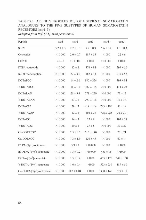
TABLE 7.1. AFFINITY PROFILES (IC50) OF A SERIES OF SOMATOSTATIN ANALOGUES TO THE FIVE SUBTYPES OF HUMAN SOMATOSTATIN RECEPTORS (sstr1–5)(adapted from Ref. [7.5], with permission)
Peptide sstr1 sstr2 sstr3 sstr4 sstr5
SS-28 5.2 ± 0.3 2.7 ± 0.3 7.7 ± 0.9 5.6 ± 0.4 4.0 ± 0.3
Octreotide >10 000 2.0 ± 0.7 187 ± 55 >1000 22 ± 6
CH288 23 ± 2 >10 000 >1000 >10 000 >1000
DTPA-octreotide >10 000 12 ± 2 376 ± 84 >1000 299 ± 50
In-DTPA-octreotide >10 000 22 ± 3.6 182 ± 13 >1000 237 ± 52
DOTATOC >10 000 14 ± 2.6 880 ± 324 >1000 393 ± 84
Y-DOTATOC >10 000 11 ± 1.7 389 ± 135 >10 000 114 ± 29
DOTALAN >10 000 26 ± 3.4 771 ± 229 >10 000 73 ± 12
Y-DOTALAN >10 000 23 ± 5 290 ± 105 >10 000 16 ± 3.4
DOTAVAP >10 000 29 ± 7 419 ± 104 743 ± 190 80 ± 19
Y-DOTAVAP >10 000 12 ± 2 102 ± 25 778 ± 225 20 ± 2.3
DOTAOC >10 000 14 ± 3 27 ± 9 >1000 103 ± 39
Y-DOTAOC >10 000 20 ± 2 27 ± 8 >10 000 57 ± 22
Ga-DOTATOC >10 000 2.5 ± 0.5 613 ± 140 >1000 73 ± 21
Ga-DOTAOC >10 000 7.3 ± 1.9 120 ± 45 >1000 60 ± 14
DTPA-[Tyr3]-octreotate >10 000 3.9 ± 1 >10 000 >1000 >1000
In-DTPA-[Tyr3]-octreotate >10 000 1.3 ± 0.2 >10 000 433 ± 16 >1000
DOTA-[Tyr3]-octreotate >10 000 1.5 ± 0.4 >1000 453 ± 176 547 ± 160
Y-DOTA-[Tyr3]-octreotate >10 000 1.6 ± 0.4 >1000 523 ± 239 187 ± 50
68
Ga-DOTA-[Tyr3]-octreotate >10 000 0.2 ± 0.04 >1000 300 ± 140 377 ± 18
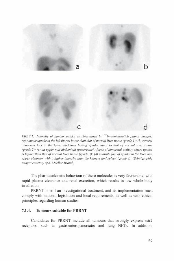
The pharmacokinetic behaviour of these molecules is very favourable, with rapid plasma clearance and renal excretion, which results in low whole-body irradiation.
PRRNT is still an investigational treatment, and its implementation must comply with national legislation and local requirements, as well as with ethical
FIG. 7.1. Intensity of tumour uptake as determined by 111In-pentetreotide planar images: (a) tumour uptake in the left thorax lower than that of normal liver tissue (grade 1); (b) several abnormal foci in the lower abdomen having uptake equal to that of normal liver tissue (grade 2); (c) an upper mid-abdominal (pancreatic?) focus of abnormal activity where uptake is higher than that of normal liver tissue (grade 3); (d) multiple foci of uptake in the liver and upper abdomen with a higher intensity than the kidneys and spleen (grade 4). (Scintigraphic images courtesy of J. Mueller-Brand.)
69
principles regarding human studies.
7.1.4. Tumours suitable for PRRNT
Candidates for PRRNT include all tumours that strongly express sstr2 receptors, such as gastroenteropancreatic and lung NETs. In addition,
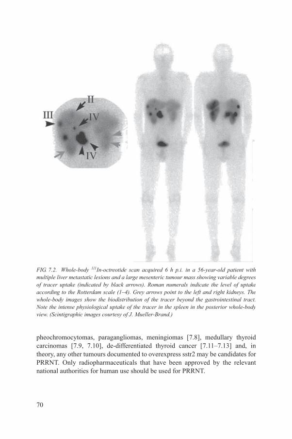
FIG. 7.2. Whole-body 111In-octreotide scan acquired 6 h p.i. in a 56-year-old patient with multiple liver metastatic lesions and a large mesenteric tumour mass showing variable degrees of tracer uptake (indicated by black arrows). Roman numerals indicate the level of uptake according to the Rotterdam scale (1–4). Grey arrows point to the left and right kidneys. The whole-body images show the biodistribution of the tracer beyond the gastrointestinal tract. Note the intense physiological uptake of the tracer in the spleen in the posterior whole-body view. (Scintigraphic images courtesy of J. Mueller-Brand.)
70
pheochromocytomas, paragangliomas, meningiomas [7.8], medullary thyroid carcinomas [7.9, 7.10], de-differentiated thyroid cancer [7.11–7.13] and, in theory, any other tumours documented to overexpress sstr2 may be candidates for PRRNT. Only radiopharmaceuticals that have been approved by the relevant national authorities for human use should be used for PRRNT.
7.1.5. Outcomes: Response, survival and toxicity
7.1.5.1. Response
The evaluation of the response to PRRNT includes assessments of functional and morphological responses, as well as biochemical and symptomatic responses, including an assessment of quality of life. The response is assessed by morphological and functional imaging techniques. Note that post-therapeutic 177Lu-DOTATATE scans provide valuable information on the intensity of uptake and localization of the tracer, and so can be used to assess the response prior to therapy cycles.
The timeline for assessing the response may vary according to clinical needs (aggressiveness and extent of disease). Usually the first follow-up is recommended after 3 months, and subsequent controls should be performed after 3–6 months (see Fig. 7.3). The ENETS guidelines provide a detailed illustration of the standards of care in the follow-up and documentation of patients with NETs [7.14].
PRRNT with somatostatin analogues 90Y-DOTATOC and 177Lu-DOTATATE has been explored in NETs for more than a decade. Current knowledge and clinical studies indicate that it is possible to deliver high absorbed doses to tumours expressing sstr2 receptors, with partial and complete objective responses in up to 30% of patients.
The best objective responses have been reported in GEP NETs, with partial responses ranging from 9% to 29% and complete remission ranging from 2 to 6%. Similar values have been reported for thorax (lung) NETs and neuroectodermic tumours (pheochromocytomas, paragangliomas). Less favourable results have been reported for thymic NETs, medullary thyroid carcinomas and de-differentiated thyroid carcinomas. Encouraging results have been reported for sstr-positive tumours such as meningiomas, medulloblastomas and astrocytomas [7.7–7.11, 7.15–7.17].
Figures 7.3 and 7.4 provide examples of two cases, the first showing a minor response and the second a partial response, as assessed by morphological imaging. Figure 7.5 shows a patient rendered operable after undergoing two cycles of PRRNT using 90Y-DOTATOC.
71
7.1.5.2. Survival
Survival analyses indicate that patients having high somatostatin receptor expression at entry to a study who undergo treatment with 177Lu-DOTATATE or 90Y-DOTATOC have significantly higher objective responses, which translate into significantly longer survival [7.7, 7.18, 7.19]. In addition, biochemical

response has also been shown to be predictive of the overall survival of patients with medullary (calcitonin) and de-differentiated iodide-negative thyroid cancer (thyroglobulin) undergoing treatment with 90Y-DOTATOC [7.9, 7.11].
FIG. 7.3. Example of a minor response to PRRNT. Serial planar scintigraphic images of the abdomen (anterior and posterior views) using 111In-octreotide prior to the first and after the second and third treatment cycles. Note the marked decrease in the intensity, extension and number of metastatic lesions showing tracer uptake in the liver and in a large lymphatic metastasis of the mesenteric root. Minor response images of the abdomen prior to and 3 months after the last treatment cycle, showing reductions of about 20% in the diameter of major liver and mesenteric lesions, from 2 and 2.9 cm (left) to 1.7 and 2.4 cm (right), respectively, following the last treatment cycle. (Scintigraphic and CT images courtesy of A. Belfer.)
72
Symptomatic responses, in particular sustained improvements in diarrhoea after 90Y-DOTATOC, proved to have an impact on PFS [7.20].
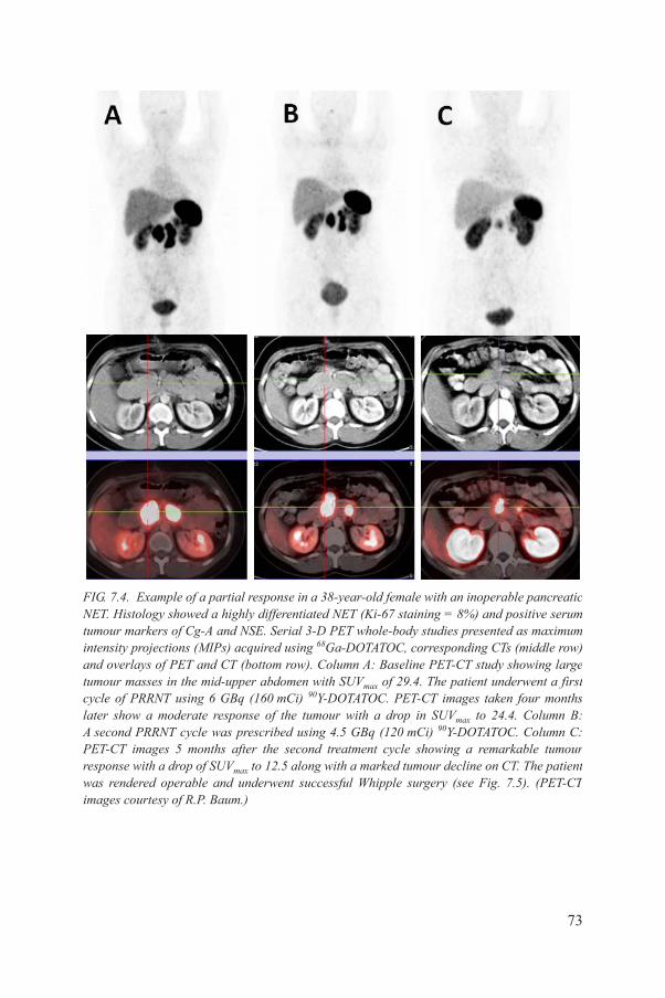
FIG. 7.4. Example of a partial response in a 38-year-old female with an inoperable pancreatic NET. Histology showed a highly differentiated NET (Ki-67 staining = 8%) and positive serum tumour markers of Cg-A and NSE. Serial 3-D PET whole-body studies presented as maximum intensity projections (MIPs) acquired using 68Ga-DOTATOC, corresponding CTs (middle row) and overlays of PET and CT (bottom row). Column A: Baseline PET-CT study showing large tumour masses in the mid-upper abdomen with SUVmax of 29.4. The patient underwent a first cycle of PRRNT using 6 GBq (160 mCi) 90Y-DOTATOC. PET-CT images taken four months later show a moderate response of the tumour with a drop in SUVmax to 24.4. Column B: A second PRRNT cycle was prescribed using 4.5 GBq (120 mCi) 90Y-DOTATOC. Column C: PET-CT images 5 months after the second treatment cycle showing a remarkable tumour response with a drop of SUV to 12.5 along with a marked tumour decline on CT. The patient
73
max
was rendered operable and underwent successful Whipple surgery (see Fig. 7.5). (PET-CT images courtesy of R.P. Baum.)
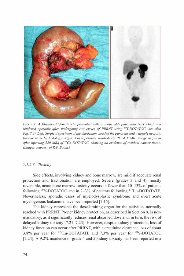
7.1.5.3. Toxicity
Side effects, involving kidney and bone marrow, are mild if adequate renal protection and fractionation are employed. Severe (grades 3 and 4), mostly reversible, acute bone marrow toxicity occurs in fewer than 10–13% of patients following 90Y-DOTATOC and in 2–3% of patients following 177Lu-DOTATATE. Nevertheless, sporadic cases of myelodysplastic syndrome and overt acute myelogenous leukaemia have been reported [7.15].
The kidney represents the dose-limiting organ for the activities normally
FIG. 7.5. A 38-year-old female who presented with an inoperable pancreatic NET which was rendered operable after undergoing two cycles of PRRNT using 90Y-DOTATOC (see also Fig. 7.4). Left: Surgical specimen of the duodenum, head of the pancreas and a largely necrotic tumour mass by histology. Right: Post-operative whole-body PET-CT MIP image acquired after injecting 220 MBq of 68Ga-DOTATOC, showing no evidence of residual cancer tissue. (Images courtesy of R.P. Baum.)
74
reached with PRRNT. Proper kidney protection, as described in Section 9, is now mandatory, as it significantly reduces renal absorbed dose and, in turn, the risk of delayed kidney toxicity [7.21–7.23]. However, despite kidney protection, loss of kidney function can occur after PRRNT, with a creatinine clearance loss of about 3.8% per year for 177Lu-DOTATATE and 7.3% per year for 90Y-DOTATOC [7.24]. A 9.2% incidence of grade 4 and 5 kidney toxicity has been reported in a

series of 1109 patients treated with 90Y-DOTATOC [7.19]. Kidney toxicity after 90Y-DOTATOC has been more frequently observed in patients with risk factors for delayed renal toxicity, such as long-standing and poorly controlled hypertension and diabetes mellitus [7.20].
A transient impairment of fertility was reported in males undergoing PRRNT, due to the irradiation of Sertoli cells, as testified by a transient rise in follicle-stimulating hormone (FSH) and a consensual drop in inhibin-B. Recovery is usually complete within two years following the end of therapy [7.6].
Despite the presence of somatostatin receptors in normal pituitary, thyroid and adrenal glands and Langerhans cells, no significant alteration of endocrine functions has been reported [7.25].
In patients with or without minor metastatic liver involvement, no significant hepatic toxicity has been reported. However, in patients with extensive metastatic liver involvement and impaired liver function, liver toxicity may occur as a serious complication that should be considered, along with pre-existing conditions affecting the liver, when choosing the appropriate radioisotope and therapy dosing. In such cases, 177Lu-labelled peptides are recommended and the administered activity should be reduced accordingly.
7.2. MULTIDISCIPLINARY APPROACHES TO PRRNT
For newly referred NET patients, a tumour board should be established, with a minimum number of specialists including the surgeon, pathologist, oncologist, (interventional) radiologist, nuclear medicine physician, radiotherapist, endocrinologist, gastroenterologist and others who share an interest in NETs. The primary purpose of the NET tumour board is to assist the attending physician in prioritizing elements of the treatment plan, which may include several options and therapeutic modalities at the initiation of care for newly referred patients.
It is understood that the primary caring physician has the privilege of being able to return to the NET tumour board for further consultation or confirmation of the treatment plan, in case of disease progression. In some cases, alternative strategies may be appropriate, perhaps involving additional specialists to deal with special co-morbid conditions.
75
Viable therapeutic approaches are the following:
(a) Surgery with a curative intent should always be performed whenever feasible.
(b) In selected cases, and within a multidisciplinary approach, PRRNT may be beneficial as a neoadjuvant therapy to render the patient accessible to surgery.
(c) For functionally active tumours, cyto-reductive strategies (e.g. surgery, TACE, TAE and RFA) should always be considered, with the aim of alleviating clinical symptoms.
7.3. PATIENT ELIGIBILITY FOR PRRNT
7.3.1. Inclusion criteria
A complete clinical history and informed consent should always be obtained from all patients. The use of a structured clinical history form is advised (see Annex III). When deciding whether to administer PRRNT to a patient, the following information is mandatory:
• Histopathologically proven NET;• High somatostatin receptor expression determined by functional whole-
body imaging (see Section 5) or immunohistochemistry.
The following criteria should also be taken into consideration:
(a) Karnofsky performance status >60 or ECOG <2.(b) Tumour differentiation, preferably in G1–G2.(c) Tumour proliferation rate, preferably with a Ki-67/mitotic index of ≤20%.
The rate of tumour growth, as determined by CT or MRI, may also be considered. Note that, in general, less differentiated tumours showing high proliferation rates are good candidates for chemotherapy.
7.3.1.1. Renal function
The patient’s renal function should be assessed by means of laboratory tests (creatinine and blood urea nitrogen (BUN)), the calculated GFR (e.g. Cockcroft–Gault formula) or nuclear medicine methods (e.g. 99mTc-MAG3,
99m
76
including the determination of the tubular extraction rate (TER), Tc-DTPA GFR or Hippuran effective renal plasma flow (ERPF)).
For 90Y-labelled peptides, an age-adjusted normal renal function is essential. Patients with compromised renal function may still be considered for 177Lu-labelled peptides.
For 177Lu-labelled peptides, a mild to moderate grade of renal impairment can be tolerated (e.g. creatinine ≤1.7 mg/dL). The GFR and TER should be at least 70% of the mean age-adjusted normal values.
7.3.1.2. Haematological status
A non-compromised haematological reserve should be present before PRRNT:
(a) White blood cell count (WBC) >3000/μL, platelet count (PLT) >75 000/μL for 177Lu-DOTATATE;
(b) WBC >3000/μL, PLT >90 000/μL for 90Y-DOTATOC, red blood cell count >3 000 000/μL.
7.3.2. Aggravating conditions (caveats)
The following aggravating conditions, if not treated, can lead to serious organ damage:
(a) Renal outflow obstruction, which could potentially lead to hydronephrosis and ultimately the loss of renal function, should always be ruled out or, if possible, corrected before PRRNT.
(b) Previous myelotoxic chemotherapy and extended radiation fields to the bone marrow (pelvis, spine), especially if performed in the weeks preceding PRRNT, pose an additional risk for post-PRRNT bone marrow failure. In questionable cases of haematological compromise, a bone marrow biopsy might be indicated to discriminate treatable cases, especially for pretreated patients and subsequent PRRNT cycles. Depending on the amount of 90Y-DOTATOC or 177Lu-DOTATATE activity injected, depressed platelet values following prior PRRNT cycle(s) can preclude the timing and dosing of subsequent cycles.
(c) Liver failure should be regarded with caution before considering PRRNT.
7.3.3. Exclusion criteria
77
PRRNT should not be considered for some NET patients, including:
(a) Pregnant women;(b) Breastfeeding mothers (if not discontinued);(c) Patients with severe or acute concomitant illnesses;(d) Patients with severe psychiatric disorders.
7.4. IMPLEMENTING PRRNT
PRRNT can be administered employing fixed-activity treatment cycles or individualized dosing, adjusted on the basis of clinical parameters (e.g. body surface area, haematological or renal function and clinical status), or can be dosimetry based.
Dosimetry based regimens are desirable but are seldom feasible or in routine practice. The fixed or individualized approach is therefore most commonly used.
Different centres have also shown variations in the intervals between treatment cycles, cumulative activities and the radioisotopes used.
Cumulative activities for 90Y-labelled peptides of up to 18 GBq (~500 mCi) have been reported, provided that the renal absorbed dose (or, better, the bioeffective renal dose) threshold was not exceeded.
Reported cumulative activities for 177Lu-labelled peptides were in the range of 22–30 GBq (~600–800 mCi), provided that the renal absorbed dose (or, better, the bioeffective dose) threshold was not exceeded.
7.4.1. Withdrawal of somatostatin analogues
Somatostatin analogues are available as short acting or long acting preparations. These should be discontinued prior to PRRNT, as they may interfere with receptor targeting. The duration of such an interruption, however, depends on the half-life of the analogue used. For long acting release formulations, an interruption of 3–4 weeks is considered adequate, while for short acting formulations, interruptions of at least 24 h are considered good clinical practice. This topic is still a matter of ongoing debate.
7.4.2. Pre-medications for PRRNT
Pre-medications are required to prevent acute adverse effects of the radiation and the amino acid infusion. Recommended medications include:
(a) Serotonin 5-HT3 receptor antagonists, for example granisetron (Kytril), ondansetron (Zofran) or tropisetron (Navoban) given intravenously
78
shortly before the radiopeptide infusion. These may be repeated if required.(b) Corticosteroid, for example dexamethasone, 4 mg or more, given
intravenously shortly before the radiopeptide infusion. This may be repeated if required.
For renal protection, please refer to Section 9.
7.4.3. Treatment regimens using 90Y-DOTATATE/90Y-DOTATOC
For non-compromised patients, the following treatment regimens are in use:
• Administered activity: 3.7 GBq (100 mCi)/m2 body surface;• Number of cycles: 2;• Time interval between cycles: 10–12 weeks.
or• Administered activity: 2.78–4.44 GBq (75–120 mCi);• Number of cycles: 2–4; • Time interval between cycles: 10–12 weeks.
7.4.4. Treatment regimen using 177Lu-DOTATATE/177Lu-DOTATOC
For non-compromised patients, the following treatment regimen is in use:
• Administered activity: 5.55–7.4 GBq (150–200 mCi);• Number of cycles: 3–5;• Time interval between cycles: 10–12 weeks.
7.4.5. Combination 177Lu/90Y-peptide therapy regimens
For non-compromised patients, the following treatment regimen is in use:
• 177Lu administered activity: 5.55–7.4 GBq (150–200 mCi);• 90Y administered activity: 2.5–5.0 GBq (68–135 mCi);• Number of cycles: 2–6;• Time interval between cycles: 10–16 weeks.
Combination therapies with 90Y and 177Lu peptides are being actively investigated and may prove to have additional therapeutic benefits. However, such combination treatments should be performed at centres with sufficient competence and extensive experience with PRRNT. In this case, administered activities should be adjusted on an individual basis. Concurrent therapies administering a cocktail of 177Lu- and 90Y-labelled peptides are also emerging.
79
7.4.6. Additional measures in compromised patients
For compromised patients, the administered activities are usually reduced and individualized, according to clinical/biochemical parameters or dosimetric studies.
In patients with reduced renal function, the following additional interventions are used:
(a) Nephro-urology consultation.(b) Extensive hydration (e.g. 2–3 L of fluid intake, if clinically appropriate)
prior to PRRNT.(c) Diuretics (e.g. furosemide) should be considered in cases of dilated pelvis
and delayed renal urinary drainage. (d) Whenever possible, the patient should be considered for 177Lu based
treatment.
In patients with reduced haematological values, the following additional interventions are used:
(i) Haematologist consultation. (ii) When indicated, packed red blood cells and/or platelet concentrates are to
be given, particularly before PRRNT.(iii) If needed, growth factor treatment with granulocyte-stimulating factors or
erythropoietin (or its derivatives) may be considered not earlier than 10 d after PRRNT.
(iv) In patients with severely compromised bone marrow reserves, as a precautionary measure, peripheral stem cell harvesting could be considered before PRRNT and, if necessary, reinfusion may be performed at an appropriate time after PPRNT.
7.4.7. Special considerations for PRRNT in children
90Y-DOTATOC has been used in children by applying the activities of 1.5–1.85 GBq/m2 per cycle in up to three cycles.
With regard to the use of 177Lu-DOTATATE in children, there is no widespread experience, and activities should be adapted per square metre [7.26].
A more extensive description of the use of PRRNT in paediatric patients is provided in Section 3.
7.4.8. Re-treatment options
80
The decision as to whether to re-treat a patient with PRRNT should be taken within the framework of the tumour board. In patients who previously responded to PRRNT, re-treatment may be considered in cases of well documented disease progression. This new PRRNT course will apply the same eligibility criteria as those previously described for the first radiopeptide treatment.
The re-treatment options include the use of the same or a different radiopeptide. For instance, choosing 177Lu-labelled peptides may be warranted, especially when considering the preservation of kidney function. When designing a re-treatment, cautious consideration should be paid to the possibility of exceeding the renal threshold dose, especially in patients with a good prognosis and long survival expectancy.
REFERENCES TO SECTION 7
[7.1] KRENNING, E.P., et al., Somatostatin receptor scintigraphy with [111In-DTPA-D-Phe1]-octreotide and [123I-Tyr3]-octreotide: The Rotterdam experience with more than 1000 patients, Eur. J. Nucl. Med. 20 (1993) 716–731.
[7.2] VALKEMA, R., et al., Phase I study of peptide receptor radionuclide therapy with [In-DTPA]-octreotide: The Rotterdam experience, Semin. Nucl. Med. 32 (2002) 110–122.
[7.3] DE JONG, M., et al., Pre-clinical comparison of [DTPA0,Tyr3]octreotide and [DOTA0,D-Phe1,Tyr3]octreotide as carriers for somatostatin receptor-targeted scintigraphy and radionuclide therapy, Int. J. Cancer 75 (1998) 406–411.
[7.4] BODEI, L., et al., Peptide receptor therapies in neuroendocrine tumours, J. Endocrinol. Invest. 32 (2009) 360–369.
[7.5] REUBI, J.C., et al., Affinity profiles for human somatostatin receptor subtypes sst1–sst5 of somatostatin radiotracers selected for scintigraphic and radiotherapeutic use, Eur. J. Nucl. Med. 27 (2000) 2732–2782.
[7.6] KWEKKEBOOM, D.J., et al., Radiolabeled somatostatin analog [177Lu-DOTA,Tyr3]octreotate in patients with endocrine gastroenteropancreatic tumours, J. Clin. Oncol. 23 (2005) 2754–2762.
[7.7] KWEKKEBOOM, D.J., et al., Somatostatin receptor-based imaging and therapy of gastroenteropancreatic neuroendocrine tumours, Endocr. Relat. Cancer 17 (2010) R53.
[7.8] BARTOLOMEI, M., et al., Peptide receptor radionuclide therapy with (90)Y-DOTATOC in recurrent meningioma, Eur. J. Nucl. Med. Mol. Imaging 36 (2009) 1407–1416.
[7.9] ITEN, F., et al., Response to [90Yttrium-DOTA]-TOC treatment is associated with long-term survival benefit in metastasized medullary thyroid cancer: A phase II clinical trial, Clin. Cancer Res. 15 (2007) 6696–6702.
[7.10] BODEI, L., et al., Receptor radionuclide therapy with 90Y-DOTATOC in patients with medullary thyroid carcinomas, Cancer Biother. Radiopharm. 19 (2004) 65–71.
[7.11] ITEN, F., et al., [(90)Yttrium-DOTA]-TOC response is associated with survival benefit in iodine-refractory thyroid cancer: Long-term results of a phase 2 clinical trial, Cancer
81
115 (2009) 2052–2062.[7.12] TEUNISSEN, J.J., KWEKKEBOOM, D.J., KOOIJ, P.P., BAKKER, W.H.,
KRENNING, E.P., Peptide receptor radionuclide therapy for non-radioiodine-avid differentiated thyroid carcinoma, J. Nucl. Med. 46 S1 (2005) 107S.
[7.13] GÖRGES, R., et al., Radionuclide-labeled somatostatin analogues for diagnostic and therapeutic purposes in nonmedullary thyroid cancer, Thyroid 11 (2001) 647–659.

[7.14] ARNOLD, R., et al., Mallorca Consensus Conference, European Neuroendocrine Tumour Society: ENETS Consensus Guidelines for the Standards of Care in Neuroendocrine Tumours: Follow-up and documentation, Neuroendocrinology 90(2009) 227–233.
[7.15] KWEKKEBOOM, D.J., et al., Overview of results of peptide receptor radionuclide therapy with 3 radiolabeled somatostatin analogs, J. Nucl. Med. 46 S1 (2005) 62S.
[7.16] VAN ESSEN, M., et al., Peptide receptor radionuclide therapy with 177Lu-octreotate in patients with foregut carcinoid tumours of bronchial, gastric and thymic origin, Eur. J. Nucl. Med. Mol. Imaging 34 (2007) 1219–1227.
[7.17] BEUTLER, D., et al., Three-year recurrence-free survival in a patient with recurrent medulloblastoma after resection, high-dose chemotherapy, and intrathecal yttrium-90-labeled DOTA0-D-Phe1-Tyr3-octreotide radiopeptide brachytherapy, Cancer 103 (2005) 869–873.
[7.18] KWEKKEBOOM, D.J., et al., Treatment with the radiolabeled somatostatin analog [177Lu-DOTA0,Tyr3]octreotate: Toxicity, efficacy, and survival, J. Clin. Oncol. 26 (2008) 2124–2130.
[7.19] IMHOF, A., et al., Response, survival, and long-term toxicity after therapy with the radiolabeled somatostatin analogue [90Y-DOTA]-TOC in metastasized neuroendocrine cancers, J. Clin. Oncol. 29 (2011) 2416–2423.
[7.20] BUSHNELL, D.L., et al., 90Y-Edotreotide for metastatic carcinoid refractory to octreotide, J. Clin. Oncol. 28 (2010) 1652–1659.
[7.21] MOLL, S., et al., A new cause of renal thrombotic microangiopathy: Yttrium 90-DOTATOC internal radiotherapy, Am. J. Kidney Dis. 37 (2001) 847–851.
[7.22] BODEI, L., et al., Long-term evaluation of renal toxicity after peptide receptor radionuclide therapy with 90Y-DOTATOC and 177Lu-DOTATATE: The role of associated risk factors, Eur. J. Nucl. Med. Mol. Imaging 35 (2008) 1847–1856
[7.23] VEGT, E., et al., Renal toxicity of radiolabeled peptides and antibody fragments: Mechanisms and impact on radionuclide therapy, J. Nucl. Med. 51 (2010) 1049–1058.
[7.24] VALKEMA, R., et al., Long-term follow-up of renal function after peptide receptor radiation therapy with 90Y-DOTA0,Tyr3-octreotide and 177Lu-DOTA0, Tyr3-octreotate, J. Nucl. Med. 46 (2005) 83S–91S.
[7.25] TEUNISSEN, J.J., et al., Effects of therapy with [177Lu-DOTA0,Tyr3]octreotate on endocrine function, Eur. J. Nucl. Med. Mol. Imaging 36 (2009) 1758–1766.
[7.26] SCHMIDT, M., BAUM, R.P., SIMON, T., HOWMAN-GILES, R., Therapeutic nuclear medicine in pediatric malignancy, Q. J. Nucl. Med. Mol. Imaging 54 (2010) 411–428.
82

8. EVALUATION OF RESPONSE
The evaluation of treatment response includes consideration of the clinical, biochemical, morphological and functional status, and the well-being of the patient.
For clinical and biochemical evaluations, please refer to Section 2. For evaluating the quality of life, please refer to the quality of life form (QLQ-C30) provided by the European Organisation for Research and Treatment of Cancer (EORTC; http://groups.eortc.be/qol/questionnaires_downloads.htm).
The morphological (or anatomical) response is determined by the acquisition of morphological and/or anatomical sequential imaging studies using ultrasound, contrast enhanced CT or MRI. Criteria for determining the objective response are provided by WHO, SWOG and Response Evaluation Criteria in Solid Tumors (RECIST). For assessing response, CT is the preferred imaging technique, but MR imaging is also of great value, if available. In some cases, CT and MRI can be complementary. In any case, the same imaging technique should be applied to follow individual patients. The interval between the follow-up examinations will depend on the disease duration and tumour biology, but these examinations are usually performed at 3–6 months initially, but may be extended to every 12 months thereafter.
In 1979, WHO attempted to define criteria for assessing the objective response. Later, in 1992, SWOG introduced four criteria for defining the magnitude of the objective response – complete response, partial response, stable disease or progression – based on the change in the tumour load, which is calculated by adding the sum of the products of maximum perpendicular diameters for all assessable lesions [8.1, 8.2].
More recently, in 2010, RECIST introduced criteria 1.0 and 1.1 in an attempt to simplify and standardize the complex SWOG criteria for assessing response. The RECIST require measurement of the longest diameter or the sum of the two longest diameters of a particular lesion(s) [8.3]. This is now the preferred method applied in clinical trials to assess tumour response.
When assessing the growth rate of NETs, in the majority of cases a combination of functional and morphological imaging techniques provides better insight into the true behaviour of the tumour under treatment. Such hybrid
83
imaging approaches may include somatostatin receptor SPECT/CT applying 99mTc or 111In or, if available, somatostatin receptor imaging PET/CT using 68Ga-DOTA peptides. Functional imaging is an invaluable instrument for assessing tumour biology in the course of the disease, as it is capable of predicting the morphological response [8.4]; however, it is not yet accepted as a substitute for the anatomical response for this purpose.

REFERENCES TO SECTION 8
[8.1] WORLD HEALTH ORGANIZATION, WHO Handbook for Reporting Results of Cancer Treatment, WHO, Geneva (1979),http://whqlibdoc.who.int/offset/WHO_OFFSET_48.pdf
[8.2] GREEN, S., WEISS, G.R., Southwest Oncology Group standard response criteria, endpoint, definitions and toxicity criteria, Invest. New Drugs 10 (1992) 239–253.
[8.3] VAN PERSIJN VAN MEERTEN, E.L., GELDERBLOM, H., BLOEM, J.L., RECIST revised: Implications for the radiologist — A review article on the modified RECIST guideline, Eur. Radiol. 20 (2010) 1456–1457.
[8.4] HAUG, A.R., et al., 68Ga-DOTATATE PET/CT for the early prediction of response to somatostatin receptor-mediated radionuclide therapy in patients with well-differentiated neuroendocrine tumours, J. Nucl. Med. 51 (2010) 1349–1356.
84
9. PRACTICAL ASPECTS OF PRRNT
9.1. FACILITIES
The design of facilities and equipment used in PRRNT will depend on relevant national legislation regarding the use of therapeutic radioactive agents.
9.2. ADMINISTRATION OF THERAPEUTIC RADIOPHARMACEUTICALS
During the administration of radiopharmaceuticals, a medical doctor must remain in close proximity. A resuscitation cart, as well as a trained emergency team, must be available. Radiopharmaceuticals should be diluted with saline to a final volume ranging from 10 to 100 mL and administered via an indwelling catheter over a period of 10–30 min, depending on the infusion system used. Radiopeptides may be co-infused with amino acid solutions via a three-way stopcock (‘piggy-back’). The line should be flushed with saline after the completion of the radiopeptide infusion.
PRRNT infusion may provoke and reproduce the syndromes of the respective functional tumours due to the stimulation of tumour receptors and the sudden substantial release of hormones. The clinical manifestations will depend on the specific hormones involved. A number of precautionary measures are therefore recommended. The patient’s blood pressure and pulse should be monitored at short intervals prior to, during and after infusion, giving special attention to symptoms. The required medications should be available and kept in close proximity to the patient in order to allow immediate treatment of acute paraneoplastic symptoms. The group of patients most at risk (i.e. those likely to exacerbate) are those with known functioning NETs that may result in any of the following syndromes: carcinoid syndrome hypotension, hypoglycaemia, hypergastrinemia, hypertension, hypotension, watery diarrhoea, hypokalaemia achlorhydria syndrome or electrolyte imbalance. For a comprehensive classification of adverse effects, please refer to the Common Terminology Criteria for Adverse Events (CTCAE), version 4.0, available at http://evs.nci.nih.gov/ftp1/CTCAE/About.html
85
Patients presenting with urinary incontinence should be catheterized prior to the administration of PRRNT, and the catheter should remain in place for 2 d.
Male patients should consider sperm banking before therapy, and female patients should avoid pregnancy for at least 6 months after treatment.
9.3. RENAL PROTECTION
9.3.1. Physiology of renal irradiation by PRRNT
The kidneys are the critical organs in PRRNT. Proximal tubular reabsorption of the radiopeptide and subsequent retention in the interstitium will result in renal irradiation. Nephrotoxicity is accelerated by risk factors such as pre-existing hypertension or diabetes [9.1]. To counteract and reduce the high risk of retention of radiopeptides in the kidney, positively charged molecules such as L-lysine and/or L-arginine are commonly used competitively to inhibit the proximal tubular reabsorption of the radiopeptide. Extensive experience supports the utilization of positively charged amino acids in both animals and humans. The co-administration of amino acids has been found to result in significant reductions in the renal irradiation absorbed dose ranging from 9 to 53% [9.2]. The renal absorbed dose may be further reduced by up to 39% by extending the infusion time of the amino acid solution to 10 h, and by up to 65% by providing amino acid infusion over two days following radiopeptide administration, thereby more effectively covering the renal elimination phase of the radiopeptide [9.3, 9.4].
Due to the beta emission characteristics of the two radionuclides, the renal absorbed dose is higher for 90Y-labelled peptides than for 177Lu-labelled ones.
Different regimens of amino acid co-infusion have been used by various groups, leading to a mean reduction of 27% in the renal absorbed dose (range 9–53%), without affecting receptor targeting or tumour uptake [9.2]. The use of amino acids, therefore, permits the safe administration of higher activities, leading to the delivery of potentially tumoricidal irradiation doses. Side effects such as nausea, headache and (rarely) vomiting are associated with the amino acid induced metabolic acidosis and occur in the majority of patients [9.5, 9.6].
To exert a protective effect on the kidney, a sufficient mass of positively charged amino acids, at least 25 g as a cumulative amount, should be infused. In this respect, it should be noted that solutions containing only a few grams of lysine or arginine are not effective and this practice should be avoided.
9.3.2. Amino acid protection protocols
86
Lysine and/or arginine should be diluted appropriately in large volumes of normal saline in order to hydrate the patient. Concentrated solutions should be avoided, as they might induce dangerous electrolyte imbalances, leading to severe metabolic acidosis and cardiac arrhythmia. The minimum recommended appropriate dilution is 25 g of amino acid in 1 L of normal saline.
Before proceeding with the amino acid infusion, appropriate measures must be taken to counter nausea and vomiting, including the administration of corticosteroids and antiemetics, as described in detail in Section 7. The amino acid infusion should be started 30–60 min before the administration of the radiopeptide and maintained for more than 4 h thereafter. Several studies have shown that prolonging the infusion over 10 h [9.3] or repeating the short protocol over the following two days allows the achievement of higher renal protection [9.4]. Particular attention should be given to possible electrolyte imbalances, namely hyperkalaemia, hypernatremia and the consequent metabolic acidosis that commonly leads to mild nausea and vomiting. These latter side effects can be managed by hydrating the patient with normal saline and possibly repeating the administration of a corticosteroid or antiemetic.
Four amino acid protection protocols are proposed:
• Single-day 50 g protection protocol. A solution containing a 50 g cocktail of lysine and arginine (25:25 g) diluted in 2 L of normal saline infused over 4 h, starting 30–60 min before PRRNT.
• Three-day 25 g protection protocol. On the first day, a cocktail of 25 g of lysine diluted in 1 L of normal saline infused over 4 h, starting 30–60 min prior to PRRNT. This should be followed by the administration of a 12.5 g of lysine solution in 500 mL of normal saline, over 3 h, twice a day, on the second and third days post-therapy. This protocol is intended to maximize renal protection while minimizing the side effects of the amino acid infusion.
• Three-day 50 g protection protocol. A solution containing a 50 g cocktail of lysine and arginine (25:25 g) diluted in 2 L of normal saline infused over 4 h on the first day, starting 30–60 min before therapy. This is followed by the administration of an additional 12.5 g of lysine diluted in 500 mL of normal saline, infused over 3 h twice a day, on the second and third days post-therapy.
• Single-day 50 g amino acid plust Gelofusine® protocol. A combination of 25 g of lysine plus 25 g of arginine diluted in 2 L of normal saline, infused over 4 h, starting 30–60 min before therapy, and Gelofusine infusion as a bolus of 1 mL/kg body weight (BW) for 10 min before therapy, followed by a Gelofusine infusion at 0.02 mL/kg BW per minute for 3 h after
87
radiopeptide infusion.
Due to reported adverse immunogenic reactions, all patients should be monitored during Gelofusine infusion, including vital signs (blood pressure and pulse) and clinical conditions [9.7].
9.3.3. Gelofusin renal protection protocol
The renal uptake of radiolabelled somatostatin analogues is partly associated with the megalin/cubilin system. Gelofusine is a ready-for-infusion solution containing 4% succinylated (or modified fluid) bovine gelatine, sodium hydroxide and water. It is used as a plasma expander and can be applied to further reduce the kidney absorbed radiation dose by about 45% [9.8]. There have been some safety concerns about the use of Gelofusin due to the relatively high incidence of allergic reactions, although these were mild in most cases [9.9]. The treating physician should be aware of these effects and be prepared to treat them accordingly with antihistamine drugs, corticosteroids or epinephrine.
This regimen has been used in a large series of patients, and no serious kidney toxicity has occurred. When applying the kidney protective regimen, mild to moderate allergic side effects, graded 1 or 2 according to the CTCAE (see Table 9.1), were recorded in 1.4% of cases, and grade 3 common toxicity criteria (CTC) reactions occurred in 0.6%. In addition, there is extensive experience with the administration of succinylated gelatine to patients in acute care or during surgical procedures, and serious side effects are very rare. Severe anaphylactoid reactions were reported in approximately 0.04% of such patients [9.10].
9.3.4. Precautions in special clinical conditions
In patients with severe cardiac insufficiency, volume overload that might lead to acute cardiac insufficiency and decompensation should be avoided. Therefore, formulations with smaller amounts of amino acids and hence lower volumes than those proposed above should be chosen (e.g. 25 g of lysine or arginine diluted in a maximum of 1 L of normal saline). In any case, stringent monitoring with the involvement of the cardiologist is recommended.
In patients with pre-existing nephrolithiasis, forced diuresis might mobilize kidney stones, leading to acute renal colic. These events should be treated accordingly but, if possible, anticipated and avoided by infusing lower volumes.
Phlebitis at the site of injection, associated with the hyperosmolarity of the infused amino acid solution, may occur. This effect can be treated with local vasoprotective creams.
88
9.4. POST-THERAPY IMAGING
When using 177Lu-labelled peptides, whole-body imaging should always be performed following each treatment cycle in order to document the targeting and
distribution of the radiopharmaceutical and to judge the functional response to PRRNT.
9.4.1. 177Lu-DOTATATE
Planar images should be obtained with a double-headed gamma camera, equipped with parallel-hole medium-energy collimators utilizing the higher gamma emission peak at 208 keV with a window width of 15%. A whole-body scanning time of 30 min is recommended, while the spot view scanning time can be limited to 5 min. A reference radioactivity source containing approximately 200 µCi in a ~20 mL vial, should be prepared on the day of injection, placed alongside the patient’s head and scanned simultaneously.
Regions of interest (ROIs) in the kidneys and, if possible, in a measurable tumour site, should be drawn, and CT based volumetry followed by dosimetric calculations should be performed.
After the first treatment, whole-body images or spot views of the upper abdomen and all involved sites should be acquired at three time points, preferably on days 1, 4 and 7 post-injection.
SPECT images can be obtained on any day for better topographic comparison with CT/MR images.
For subsequent treatments, whole-body or spot views of the upper abdomen and other sites of interest should be acquired. SPECT is optional at two time points after treatment, preferably on days 1 and 4 post-injection.
9.4.2. 90Y-DOTATOC
Using 90Y-labelled peptides, bremsstrahlung imaging is performed to assess the distribution of radioactivity, although the quality of such images is rather poor. Yttrium is a rare-earth metal that is chemically similar to the lanthanides. The radionuclide 90Y emits beta particles with a mean electron energy of 0.935 MeV and a maximum energy of 2.3 MeV, and a half-life of 64.1 h. The absence of gamma emissions from 90Y does not permit direct imaging useful for diagnostic and dosimetry purposes, although poor-quality images can be obtained from the bremsstrahlung (braking radiation) of beta particles.
For diagnostic and dosimetric purposes, 111In has been introduced in clinical
89
practice as a substitute tracer because of its similar chemical properties. Alternatively, when available, the positron emitter isotope 86Y has been used for PET imaging and dosimetry for research purposes.
However, pure bremsstrahlung whole-body imaging is usually performed at 24 h after 90Y-DOTATOC injection in order to evaluate the distribution of radioactivity throughout the body. Quantification of the radionuclide content in
organs and tumours is usually rough, and its accuracy depends on the corrections for photon attenuation and collimator response. Nevertheless, these images can be used to obtain a rough estimation of the biodistribution of radioactivity.
As a clinical compromise between sensitivity and resolution, gamma cameras equipped with a 3/8ʺ (0.95 cm) NaI(Tl) crystal and medium-energy general-purpose collimators with a wide energy window (25–285 KeV) are generally accepted [9.11].
To exclude the X ray characteristic emission, gamma cameras equipped with a 1 inch (2.54 cm) NaI(Tl) crystal and high-energy general-purpose collimators can be used with a 60% energy window centred at 150 keV with a range of 105–195 keV [9.12].
9.5. POST-THERAPY MONITORING AND MANAGEMENT
Following PRRNT, the evaluation of renal function is extremely important, as the kidney is the critical organ. The follow-up should include at least an evaluation of serum creatinine levels and a determination of creatinine clearance. In patients with pre-existing risk factors for delayed renal toxicity (the high risk group), in particular, those with long-standing and poorly controlled hypertension and diabetes mellitus, a single kidney or previously known insults to the kidneys (particularly nephrotoxic chemotherapy), more precise methods for assessing renal function are recommended. These may include measurements of the GFR using 99mTc-DTPA or 51Cr-EDTA, or measurements the clearance rate of 99mTc-MAG3.
Tables 9.1 and 9.2 summarize the toxicity/adverse events criteria for grading bone marrow and kidney function recommended by the US National Cancer Institute (NCI).
9.5.1. Between cycles
A complete blood cell count should be determined every 2−4 weeks, or more frequently if clinically required. The results of renal and liver function tests should be available before confirming subsequent cycles.
Following careful clinical evaluations, patients whose blood cell count
90
values are below the limits indicated for the first PRRNT cycle should receive a lower activity and/or the next PRRNT cycle should be postponed. In severe cases, interruption of PRRNT should be considered.
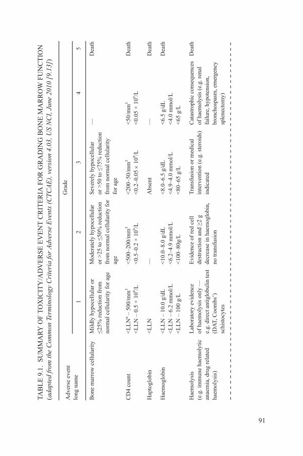
MA
RY
OF
TO
XIC
ITY
/AD
VE
RS
E E
VE
NT
CR
ITE
RIA
FO
R G
RA
DIN
G B
ON
E M
AR
RO
W F
UN
CT
ION
Com
mon
Ter
min
olog
y C
rite
ria
for
Adv
erse
Eve
nts
(CT
CA
E),
ver
sion
4.0
3, U
S N
CI,
Jun
e 20
10 [
9.13
])
Gra
de
12
34
5
rity
Mil
dly
hypo
cell
ular
or
≤25%
red
ucti
on f
rom
norm
al c
ellu
lari
ty f
or a
ge
Mod
erat
ely
hypo
cell
ular
or >
25 to
≤50
% r
educ
tion
from
nor
mal
cel
lula
rity
for
age
Sev
erel
y hy
poce
llul
ar
or >
50 to
≤75
% r
educ
tion
from
nor
mal
cel
lula
rity
for
age
—D
eath
<L
LN
a – 5
00/m
m3
<L
LN
– 0
.5 ×
109 /L
<50
0–20
0/m
m3
<0.
5–0.
2 ×
109 /L
<20
0–50
/mm
3
<0.
2–0.
05
109 /L
<50
/mm
3
<0.
05 ×
109 /L
Dea
th
<L
LN
—A
bsen
t—
Dea
th
<L
LN
– 1
0.0
g/dL
<
LL
N –
6.2
mm
ol/L
<L
LN
– 1
00 g
/L
<10
.0–8
.0 g
/dL
<6.
2–4.
9 m
mol
/L
<10
0–80
g/L
<8.
0–6.
5 g/
dL
<4.
9–4.
0 m
mol
/L
<80
–65
g/L
<6.
5 g/
dL
<4.
0 m
mol
/L
<65
g/L
Dea
th
lyti
cd
Lab
orat
ory
evid
ence
of h
aem
olys
is o
nly
—
e.g.
dir
ect a
ntig
lobu
lin
test
(DA
T, C
oom
bs’)
sc
hist
ocyt
es
Evi
denc
e of
red
cel
lde
stru
ctio
n an
d ≥2
g
decr
ease
in h
aem
oglo
bin,
no tr
ansf
usio
n
Tra
nsfu
sion
or
med
ical
inte
rven
tion
(e.
g. s
tero
ids)
indi
cate
d
Cat
astr
ophi
c co
nseq
uenc
es
of h
aem
olys
is (
e.g.
ren
al
failu
re, h
ypot
ensi
on,
bron
chos
pasm
, em
erge
ncy
sple
nect
omy)
Dea
th
91
TAB
LE
9.1
. S
UM
(ada
pted
from
the
Adv
erse
eve
nt
long
nam
e
Bon
e m
arro
w c
ellu
la
CD
4 co
unt
Hap
togl
obin
Hae
mog
lobi
n
Hae
mol
ysis
(e.g
. im
mun
e ha
emo
anae
mia
, dru
g re
late
haem
olys
is)

—A
sym
ptom
atic
iron
over
load
, int
erve
ntio
nno
t ind
icat
ed
Iron
ove
rloa
d,
inte
rven
tion
indi
cate
dO
rgan
impa
irm
ent
(e.g
. end
ocri
nopa
thy,
card
iopa
thy)
Dea
th
<L
LN
– 3
000/
mm
3
<L
LN
– 3
.0 ×
109 /L
<30
00–2
000/
mm
3 <
3.0–
2.0
× 1
09 /L<
2000
–100
0/ m
m3
<2.
0–1.
0 ×
109 /L
<10
00/m
m3
<1.
0 ×
109 /L
Dea
th
<L
LN
– 8
00/m
m3
<L
LN
– 0
.8
109 /L
<80
0–50
0/m
m3
<0.
8–0.
5 ×
109 /L
<50
0–20
0 m
m3
<0.
5–0.
2 ×
109 /L
<20
0/m
m3
<0.
2 ×
109 /L
Dea
th
——
Abn
orm
al m
arro
w
cyto
gene
tics
(mar
row
bla
sts ≤5
%)
RA
EB
b or
RA
EB
-T
(mar
row
bla
sts
>5%
)D
eath
cyte
s<
LL
N –
150
0/m
m3
<L
LN
– 1
.5 ×
109 /L
<15
00–1
000/
mm
3
<1.
5–1.
0 ×
109 /L
<10
00–5
00/m
m3
<1.
0–0.
5 ×
109 /L
<50
0/m
m3
<0.
5 ×
109 /L
Dea
th
<L
LN
– 7
5 00
0/m
m3
<L
LN
– 7
5.0
× 1
09 /L<
75 0
00–5
0 00
0/m
m3
<75
.0–5
0.0
× 1
09 /L<
50 0
00–2
5 00
0/m
m3
<50
.0–2
5.0
× 1
09 /L<
25 0
00/m
m3
<25
.0 ×
109 /L
Dea
th
MA
RY
OF
TO
XIC
ITY
/AD
VE
RS
E E
VE
NT
CR
ITE
RIA
FO
R G
RA
DIN
G B
ON
E M
AR
RO
W F
UN
CT
ION
Com
mon
Ter
min
olog
y C
rite
ria
for
Adv
erse
Eve
nts
(CT
CA
E),
ver
sion
4.0
3, U
S N
CI,
Jun
e 20
10 [
9.13
]) (
cont
.)
Gra
de
12
34
5
92
Iron
ove
rloa
d
Leu
kocy
tes
(tot
al w
hite
blo
od
cell
cou
nt)
Lym
phop
enia
Mye
lody
spla
sia
Neu
trop
hils
/gra
nulo
(AN
C/A
GC
c )
Pla
tele
ts
TAB
LE
9.1
. S
UM
(ada
pted
from
the
Adv
erse
eve
nt
long
nam
e

Inci
dent
al f
indi
ngs
(e.g
. How
ell–
Joll
y bo
dies
)P
roph
ylac
tic
anti
biot
ics
indi
cate
d—
Lif
e-th
reat
enin
gco
nseq
uenc
esD
eath
M
ild
Mod
erat
eS
ever
eL
ife-
thre
aten
ing;
disa
blin
gD
eath
it of
nor
mal
val
ues.
tory
ana
emia
wit
h ex
cess
bla
sts.
solu
te n
eutr
ophi
l cou
nt/a
bsol
ute
gran
uloc
yte
coun
t.
MA
RY
OF
TO
XIC
ITY
/AD
VE
RS
E E
VE
NT
CR
ITE
RIA
FO
R G
RA
DIN
G B
ON
E M
AR
RO
W F
UN
CT
ION
Com
mon
Ter
min
olog
y C
rite
ria
for
Adv
erse
Eve
nts
(CT
CA
E),
ver
sion
4.0
3, U
S N
CI,
Jun
e 20
10 [
9.13
]) (
cont
.)
Gra
de
12
34
5
93
Spl
enic
fun
ctio
n
Blo
od/b
one
mar
row
Oth
er
aL
LN
— lo
wer
lim
bR
AE
B —
Ref
rac
cA
NC
/AG
C —
ab
TAB
LE
9.1
. S
UM
(ada
pted
from
the
Adv
erse
eve
nt
long
nam
e
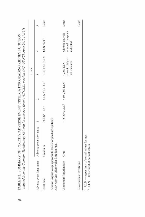
MA
RY
OF
TO
XIC
ITY
/AD
VE
RS
E E
VE
NT
CR
ITE
RIA
FO
R G
RA
DIN
G K
IDN
EY
FU
NC
TIO
N C
omm
on T
erm
inol
ogy
Cri
teri
a fo
r A
dver
se E
vent
s (C
TC
AE
), v
ersi
on 4
.03,
US
NC
I, J
une
2010
[9.
13])
Gra
de
nam
eA
dver
se e
vent
sho
rt n
ame
12
34
5
Cre
atin
ine
>U
LN
a –
1.5
×
UL
N >
1.5–
3.0
×
UL
N >
3.0–
6.0
×U
LN
>6.
0 ×
Dea
th
ge a
ppro
pria
te le
vels
for
pae
diat
ric
patie
nts.
erul
ar f
iltr
atio
n ra
te.
rat
eG
FR
<75
–50%
LL
Nb
<50
–25%
LL
N<
25%
LL
N,
chro
nic
dial
ysis
not i
ndic
ated
Chr
onic
dia
lysi
s
or r
enal
tran
spla
nt
indi
cate
d
Dea
th
inin
eD
eath
it o
f no
rmal
val
ues
for
age.
it of
nor
mal
val
ues.
94
TAB
LE
9.2
. S
UM
(ada
pted
from
the
Adv
erse
eve
nt lo
ng
Cre
atin
ine
Rem
ark:
Adj
ust t
o a
Als
o co
nsid
er: G
lom
Glo
mer
ular
fil
trat
ion
Als
o co
nsid
er: C
reat
a U
LN
— u
pper
lim
bL
LN
— lo
wer
lim
9.5.2. Intermediate and long term follow-up
A complete blood cell count, and renal and liver function should be determined every 8−12 weeks for the first 12 months following PRRNT, and twice a year thereafter, if clinically indicated.
REFERENCES TO SECTION 9
[9.1] BODEI, L., et al., Long-term evaluation of renal toxicity after peptide receptor radionuclide therapy with 90Y-DOTATOC and 177Lu-DOTATATE: The role of associated risk factors, Eur. J. Nucl. Med. Mol. Imaging 35 (2008) 1847–1856.
[9.2] DE JONG, M., KRENNING, E.P., New advances in peptide receptor radionuclide therapy, J. Nucl. Med. 43 (2002) 617–620.
[9.3] JAMAR, F., et al., (86YDOTA0)-DPhe1-Tyr3-octreotide (SMT487) — A phase 1 clinical study: Pharmacokinetics, biodistribution and renal protective effect of different regimens of amino acid co-infusion, Eur. J. Nucl. Med. Mol. Imaging 30 (2003) 510–518.
[9.4] BODEI, L., et al., Receptor radionuclide therapy with 90Y-[DOTA]0-Tyr3-octreotide (90Y-DOTATOC) in neuroendocrine tumours, Eur. J. Nucl. Med. Mol. Imaging 31(2004) 1038–1046.
[9.5] BODEI, L., et al., Receptor-mediated radionuclide therapy with 90Y-DOTATOC in association with amino acid infusion: A phase I study, Eur. J. Nucl. Med. Mol. Imaging 30 (2003) 207–216.
[9.6] ROLLEMAN, E.J., VALKEMA, R., DE JONG, M., KOOIJ, P.P., KRENNING, E.P., Safe and effective inhibition of renal uptake of radiolabelled octreotide by a combination of lysine and arginine, Eur. J. Nucl. Med. Mol. Imaging 30 (2003) 9–15.
[9.7] WATKINS, J., Reactions to gelatin plasma expanders, Lancet 30 (1994) 328–329.[9.8] ROLLEMAN, E.J., et al., Kidney protection during peptide receptor radionuclide
therapy with somatostatin analogues, Eur. J. Nucl. Med. Mol. Imaging 37 (2010) 1018–1031.
[9.9] BARRON, M.E., WILKES, M.M., NAVICKIS, R.J., A systematic review of the comparative safety of colloids, Arch. Surg. 139 (2004) 552–563.
[9.10] WEHRMANN, C., SENFTLEBEN, S., ZACHERT, C., MÜLLER, D., BAUM, R.P., Results of individual patient dosimetry in peptide receptor radionuclide therapy with 177Lu DOTA-TATE and 177Lu DOTA-NOC, Cancer Biother. Radiopharm. 22 (2007) 406–416.
[9.11] SHEN, S., DENARDO, G., YUAN, A., DENARDO, D., DENARDO, S., Planar gamma camera imaging and quantitation of yttrium-90 bremsstrahlung, J. Nucl. Med. 35 (1994)
95
1381.[9.12] MINARIK, D., LJUNGBERG, M., SEGARS, P., SJÖGREEN GLEISNER, K.,
Evaluation of quantitative planar 90Y bremsstrahlung whole-body imaging, Phys. Med. Biol. 54 (2009) 5873–5883.

[9.13] US NATIONAL CANCER INSTITUTE, Common Terminology Criteria for Adverse Events (CTCAE), version 4.03, June 2010,http://evs.nci.nih.gov/ftp1/CTCAE/About.html
96
10. DOSIMETRY
10.1. INTRODUCTION
Following PRRNT, patient specific dosimetry should be performed whenever possible to allow an assessment of the organ absorbed dose and possibly to predict the risk of delayed renal toxicity. Different dosimetry methods may be applied depending on their purpose and the availability of resources. Input data include blood and urine samples, and scintigraphic images are to be scheduled up to 3 d post injection. Planar images are useful for deriving biokinetics over time, while SPECT and SPECT/CT fused images, although more time consuming, permit detailed insights into intra-organ activity distribution. The medical internal radiation dse (MIRD) scheme represents the reference formalism for internal dosimetry. Dedicated software (OLINDA/EXM) has been used to derive mean absorbed dose estimates for 177Lu and 90Y peptides (see Annex I).
10.2. BIOLOGICALLY EFFECTIVE DOSE CONCEPT
Kidney radiation toxicity is typically evident several months after irradiation due to the slow repair characteristics of renal cells. According to studies of renal toxicity derived from external radiotherapy, the accepted renal tolerated dose is in the range of 23–25 Gy. As stated by the National Council on Radiation Protection and Measurements (NCRP), a dose of 23 Gy to the kidney will cause detrimental deterministic effects in 5% of patients within five years [10.1, 10.2]. Nevertheless, clinical experience and dosimetric studies clearly indicate that this renal dose threshold does not accurately correlate with the renal toxicity observed in patients undergoing PRRNT [10.3].
PRRNT is a form of continuous radiation delivery with a decreasing dose rate over time. The irradiation produces both lethal and sub-lethal damage that can be repaired during the irradiation itself, but the differential between the creation of new damage and its repair depends on the specific dose rate at any particular time and on the repair capability (T½ rep) of the tissue. In PRRNT, low
97
dose rates will spare normal tissue more than the tumour, and this may allow benefits similar to fractionation in external radiotherapy [10.4].
The linear quadratic model can be used to mathematically interpret this differential sparing. The biologically effective dose (BED) concept is used to quantify the biological effects induced by different patterns of radiation delivery. This model has recently been revised for radionuclide therapy and applied in
particular to PRRNT, with the aim of enhancing the dose–response correlation [10.5]. Focusing on the concerns about the kidney, the BED has proven to be a reliable predictor of renal toxicity, and thus helpful in the implementation of individual treatment planning [10.6].
REFERENCES TO SECTION 10
[10.1] NATIONAL COUNCIL ON RADIATION PROTECTION AND MEASUREMENTS, Misadministration of Radioactive Material in Medicine — Scientific Background, NCRP, Bethesda, MD (1991) 27.
[10.2] CASSADY, J.R., Clinical radiation nephropathy, Int. J. Radiat. Oncol. Biol. Phys. 31(1995) 1249–1256.
[10.3] BARONE, R., et al., Patient-specific dosimetry in predicting renal toxicity with (90)Y-DOTATOC: relevance of kidney volume and dose rate in finding a dose–effect relationship, J. Nucl. Med. 46 (2005) 99S–106S.
[10.4] DALE, R., CARABE-FERNANDEZ, A., The radiobiology of conventional radiotherapy and its application to radionuclide therapy, Cancer Biother. Radiopharm. 20 (2005) 47–51.
[10.5] DALE, R., Use of the linear-quadratic radiobiological model for quantifying kidney response in targeted radiotherapy, Cancer Biother. Radiopharm. 19 (2004) 363–370.
[10.6] BODEI, L., et al., Long-term evaluation of renal toxicity after peptide receptor radionuclide therapy with 90Y-DOTATOC and 177Lu-DOTATATE: The role of associated risk factors, Eur. J. Nucl. Med. Mol. Imaging 35 (2008) 1847–1856.
98

Annex I
DOSIMETRIC METHODS
Dosimetry of normal organs and malignant lesions represents a fundamental aid in the planning of PRRNT. The aim is to deliver the maximum radiation dose to the tumour while remaining within the therapeutic window with regard to the dose delivered to normal organs, particularly the kidneys, the dose-limiting organs and bone marrow.
Dose estimates in organs are generally calculated using the medical internal radiation dose (MIRD) scheme, with the basic formula
D = Ã × S = A0 × τ × S
where à is the integral activity in the organ, A0 is the initial activity in the organ, τ is the residence time, corresponding to the total number of decays (ND) occurring in the organ, and S is a factor that depends on the properties of the radionuclide and the target. Once the integral activities in organs of interest are determined by numerical or compartmental models [I–1, I–2], absorbed dose calculations are generally performed using dedicated software (OLINDA/EXM, RADAR) that consider as inputs the residence time τ or ND [I–3, I–4].
The typical kinetics of radiopeptides, namely, the very fast blood clearance and renal elimination, determine the information required to obtain the integral activities in organs and the tumour, which include a whole dataset of scintigraphic images and blood and urine collections (Table I–1). Once the rough data are analysed, the activity in normal and tumour tissues has to be converted into time–activity curves, and the absorbed doses finally estimated (Table I–2).
Specifically, the essential data required are blood samples (preferably three or four samples taken within the following time intervals: 0–1 h, 1–5 h, 5–24 h and 24–72 h p.i.), a complete urine collection within preselected time intervals, and scintigraphic images (anterior and posterior whole-body and SPECT acquisitions). Although, in principle, planar views are not ideal for dosimetry, the availability of 5–7 whole-body serial images (acquired 2–3 h and up to almost 3d p.i.) might offer complete and satisfactory information on the biodistribution and its variation over time [I–5, I–6].
99
The calibration of the system can be performed by means of absolute or relative calibration methods. The former method uses a known source to obtain a factor that converts the counts per unit of time into activity, while the latter normalizes the counts per unit of time of the first whole-body image to 100% of the activity.
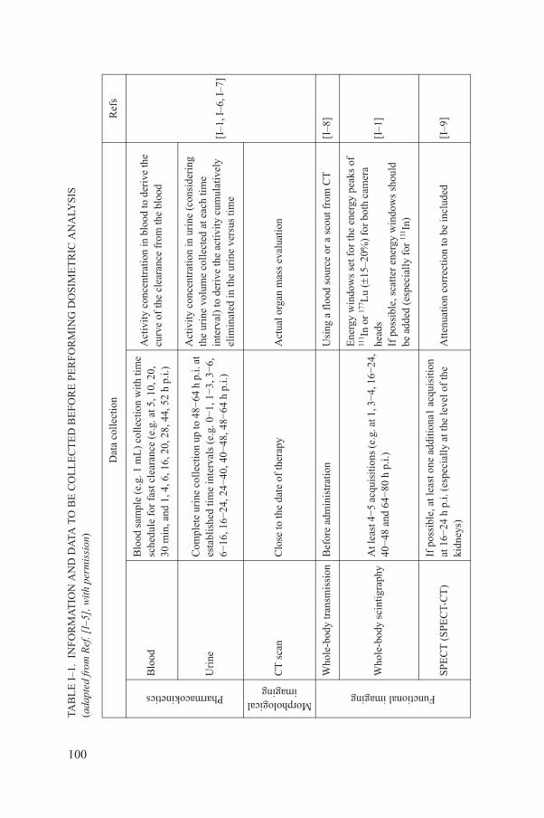
MA
TIO
N A
ND
DA
TA T
O B
E C
OL
LE
CT
ED
BE
FO
RE
PE
RF
OR
MIN
G D
OS
IME
TR
IC A
NA
LYS
IS
I–5]
, wit
h pe
rmis
sion
)
Dat
a co
llec
tion
Ref
s
Blo
od s
ampl
e (e
.g. 1
mL
) co
llec
tion
wit
h ti
me
sche
dule
for
fas
t cle
aran
ce (
e.g.
at 5
, 10,
20,
30 m
in, a
nd 1
, 4, 6
, 16,
20,
28,
44,
52
h p.
i.)
Act
ivit
y co
ncen
trat
ion
in b
lood
to d
eriv
e th
e cu
rve
of th
e cl
eara
nce
from
the
bloo
d
[I–1
, I–6
, I–7
]
Com
plet
e ur
ine
coll
ecti
on u
p to
48−
64 h
p.i.
at
esta
blis
hed
time
inte
rval
s (e
.g. 0−
1, 1−
3, 3−
6,
6−16
, 16−
24, 2
4−40
, 40−
48, 4
8−64
h p
.i.)
Act
ivit
y co
ncen
trat
ion
in u
rine
(co
nsid
erin
g th
e ur
ine
volu
me
coll
ecte
d at
eac
h ti
me
inte
rval
) to
der
ive
the
acti
vity
cum
ulat
ivel
y el
imin
ated
in th
e ur
ine
vers
us ti
me
Clo
se to
the
date
of
ther
apy
Act
ual o
rgan
mas
s ev
alua
tion
y tr
ansm
issi
onB
efor
e ad
min
istr
atio
nU
sing
a f
lood
sou
rce
or a
sco
ut f
rom
CT
[I–8
]
y sc
inti
grap
hyA
t lea
st 4−
5 ac
quis
itio
ns (e
.g. a
t 1, 3−
4, 1
6−24
,40−
48 a
nd 6
4−80
h p
.i.)
Ene
rgy
win
dow
s se
t for
the
ener
gy p
eaks
of
111 In
or
177 L
u (
15–2
0%)
for
both
cam
era
head
sIf
pos
sibl
e, s
catt
er e
nerg
y w
indo
ws
shou
ld
be a
dded
(es
peci
ally
for
111 In
)
[I–1
]
PEC
T-C
T)
If p
ossi
ble,
at l
east
one
add
itio
na1
acqu
isit
ion
at 1
6−24
h p
.i. (
espe
cial
ly a
t the
leve
l of
the
kidn
eys)
Att
enua
tion
cor
rect
ion
to b
e in
clud
ed[I
–9]
100
TAB
LE
I–1
. IN
FO
R(a
dapt
ed fr
om R
ef. [
Pharmacokinetics
Blo
od
Uri
ne
Morphologicalimaging
CT
sca
n
Functional imaging
Who
le-b
od
Who
le-b
od
SPE
CT
(S
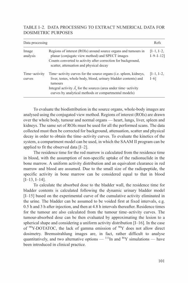
To evaluate the biodistribution in the source organs, whole-body images are analysed using the conjugated view method. Regions of interest (ROIs) are drawn over the whole body, tumour and normal organs — heart, lungs, liver, spleen and kidneys. The same set of ROIs must be used for all the performed scans. The data collected must then be corrected for background, attenuation, scatter and physical decay in order to obtain the time–activity curves. To evaluate the kinetics of the system, a compartment model can be used, in which the SAAM II program can be applied to fit the observed data [I–2].
The residence time for the red marrow is calculated from the residence time in blood, with the assumption of non-specific uptake of the radionuclide in the bone marrow. A uniform activity distribution and an equivalent clearance in red marrow and blood are assumed. Due to the small size of the radiopeptide, the specific activity in bone marrow can be considered equal to that in blood [I–13, I–14].
To calculate the absorbed dose to the bladder wall, the residence time for bladder contents is calculated following the dynamic urinary bladder model [I–15] based on the experimental curve of the cumulative activity eliminated in the urine. The bladder can be assumed to be voided first at fixed intervals, e.g. 0.5 h and 3 h after injection, and then at 4.8 h intervals thereafter. Residence times for the tumour are also calculated from the tumour time–activity curves. The
TABLE I–2. DATA PROCESSING TO EXTRACT NUMERICAL DATA FOR DOSIMETRIC PURPOSES
Data processing Refs
Image analysis
Regions of interest (ROIs) around source organs and tumours in planar (conjugate view method) and SPECT imagesCounts converted to activity after correction for background, scatter, attenuation and physical decay
[I–1, I–2,I–9–I–12]
Time−activitycurves
Time−activity curves for the source organs (i.e. spleen, kidneys, liver, testes, whole body, blood, urinary bladder contents) and tumoursIntegral activity Ãs for the sources (area under time−activity curves by analytical methods or compartmental models)
[I–1, I–2, I–6]
101
tumour-absorbed dose can be then evaluated by approximating the lesion to a spherical shape and considering a uniform activity distribution [I–16]. In the case of 90Y-DOTATOC, the lack of gamma emission of 90Y does not allow direct dosimetry. Bremsstrahlung images are, in fact, rather difficult to analyse quantitatively, and two alternative options — 111In and 86Y simulations — have been introduced in clinical practice.

For dosimetric purposes, 111In-DOTATOC has been introduced in clinical practice as a substitute tracer because its chemical properties are similar to those of 90Y-DOTATOC. Since the physical half-life of 111In is almost identical to that of 90Y, and is also compatible with the kinetics of peptides, the diagnostic activities usually administered (185 MBq) allow the collection of serial images (planar and SPECT) over a suitable period of time (usually 3–4 d).
An alternative solution, at least theoretically, is the use of DOTATOC labelled with the positron emitter isotope 86Y, the identical chemical element of the therapeutic counterpart. The DOTATOC peptide labelled with 86Y totally preserves the chemical nature of 90Y derivatives and offers good spatial resolution due to the possibility of PET imaging. Nevertheless, limitations do indeed exist, mainly related to the short time interval available for data collection (<24–40 h) due to the short physical half-life of 86Y of 14.7 h and its low positron abundance, high cost and limited availability.
Despite the advantages and drawbacks of the two methods mentioned above, the extrapolated absorbed doses are reasonably similar.
It should be noted that neither 111In-pentetreotide scintigraphy nor 68Ga-DOTATOC PET are suitable for a correct dosimetric analysis; the former because of its different kinetics and receptor affinity properties, and the latter because of the too-short physical half-life (68 min) of 68Ga compared with the biological half-life of DOTATOC, and also its possibly different chemical properties and behaviour compared with the 90Y counterpart.
Recently, a new potential for 90Y imaging has emerged, pointing out the possibility of omitting simulations by 111In or 86Y imaging [I–10, I–17, I–18].
In the case of 177Lu-DOTATATE, the gamma photons emitted by 177Lu are suitable for scintigraphy, enabling both imaging and dosimetry with the same compound. Since the treatment is repeated in multiple cycles, dosimetry is usually performed directly during the first courses of therapy, after the administration of therapeutic activities of 177Lu-DOTATATE.
REFERENCES TO ANNEX I
[I–1] SIEGEL, J.A., et al., Techniques for quantitative radiopharmaceutical biodistribution data acquisition and analysis for use in human radiation dose estimates, MIRD Pamphlet No. 16, J. Nucl. Med. 40 (1999) S37–S61.
102
[I–2] FOSTER, D., BARRET, P., “Developing and testing integrated multicompartment models to describe a single-input multiple-output study using the SAAM II software system”, in Proc. 6th Int. Radiopharmaceutical Dosimetry Symp., Oak Ridge Institute for Science and Education, Oak Ridge, TN (1998).
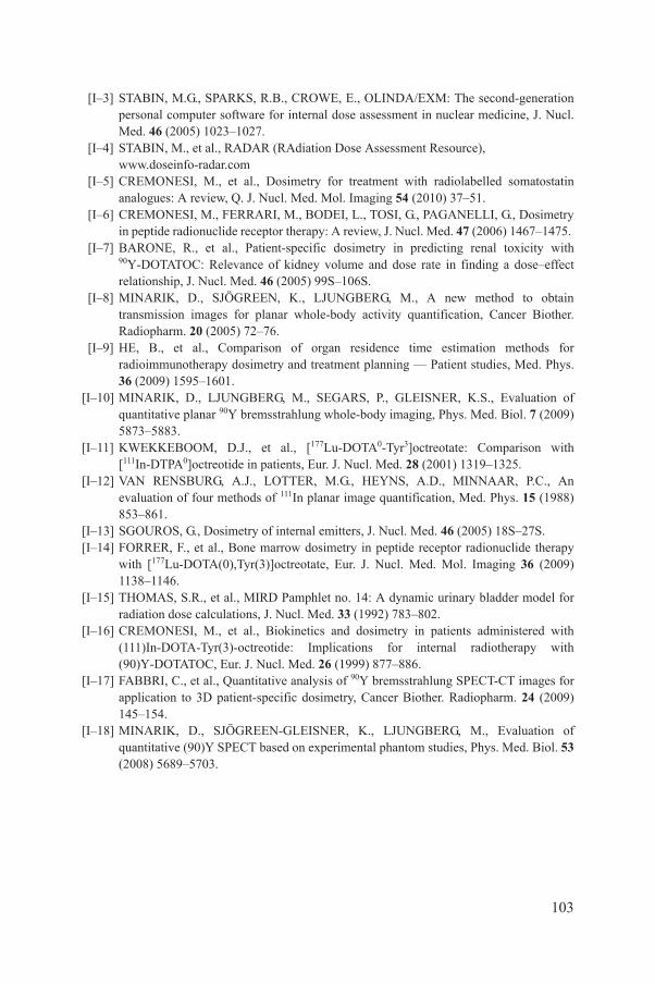
[I–3] STABIN, M.G., SPARKS, R.B., CROWE, E., OLINDA/EXM: The second-generation personal computer software for internal dose assessment in nuclear medicine, J. Nucl. Med. 46 (2005) 1023–1027.
[I–4] STABIN, M., et al., RADAR (RAdiation Dose Assessment Resource),www.doseinfo-radar.com
[I–5] CREMONESI, M., et al., Dosimetry for treatment with radiolabelled somatostatin analogues: A review, Q. J. Nucl. Med. Mol. Imaging 54 (2010) 37–51.
[I–6] CREMONESI, M., FERRARI, M., BODEI, L., TOSI, G., PAGANELLI, G., Dosimetry in peptide radionuclide receptor therapy: A review, J. Nucl. Med. 47 (2006) 1467–1475.
[I–7] BARONE, R., et al., Patient-specific dosimetry in predicting renal toxicity with 90Y-DOTATOC: Relevance of kidney volume and dose rate in finding a dose–effect relationship, J. Nucl. Med. 46 (2005) 99S–106S.
[I–8] MINARIK, D., SJÖGREEN, K., LJUNGBERG, M., A new method to obtain transmission images for planar whole-body activity quantification, Cancer Biother. Radiopharm. 20 (2005) 72–76.
[I–9] HE, B., et al., Comparison of organ residence time estimation methods for radioimmunotherapy dosimetry and treatment planning — Patient studies, Med. Phys. 36 (2009) 1595–1601.
[I–10] MINARIK, D., LJUNGBERG, M., SEGARS, P., GLEISNER, K.S., Evaluation of quantitative planar 90Y bremsstrahlung whole-body imaging, Phys. Med. Biol. 7 (2009) 5873–5883.
[I–11] KWEKKEBOOM, D.J., et al., [177Lu-DOTA0-Tyr3]octreotate: Comparison with [111In-DTPA0]octreotide in patients, Eur. J. Nucl. Med. 28 (2001) 1319–1325.
[I–12] VAN RENSBURG, A.J., LOTTER, M.G., HEYNS, A.D., MINNAAR, P.C., An evaluation of four methods of 111In planar image quantification, Med. Phys. 15 (1988) 853–861.
[I–13] SGOUROS, G., Dosimetry of internal emitters, J. Nucl. Med. 46 (2005) 18S–27S.[I–14] FORRER, F., et al., Bone marrow dosimetry in peptide receptor radionuclide therapy
with [177Lu-DOTA(0),Tyr(3)]octreotate, Eur. J. Nucl. Med. Mol. Imaging 36 (2009) 1138–1146.
[I–15] THOMAS, S.R., et al., MIRD Pamphlet no. 14: A dynamic urinary bladder model for radiation dose calculations, J. Nucl. Med. 33 (1992) 783–802.
[I–16] CREMONESI, M., et al., Biokinetics and dosimetry in patients administered with (111)In-DOTA-Tyr(3)-octreotide: Implications for internal radiotherapy with (90)Y-DOTATOC, Eur. J. Nucl. Med. 26 (1999) 877–886.
[I–17] FABBRI, C., et al., Quantitative analysis of 90Y bremsstrahlung SPECT-CT images for application to 3D patient-specific dosimetry, Cancer Biother. Radiopharm. 24 (2009) 145–154.
[I–18] MINARIK, D., SJÖGREEN-GLEISNER, K., LJUNGBERG, M., Evaluation of quantitative (90)Y SPECT based on experimental phantom studies, Phys. Med. Biol. 53
103
(2008) 5689–5703.

Annex II
KARNOFSKY PERFORMANCE SCALE
The following table is based on information available at: http://www.cibmtr.org/DataManagement/TrainingReference/Manuals/DataManagement/Documents/appendix-l.pdf
General status Ki (%) Specific criteria
Able to carry on normal activityNo special care needed
100 Normal general status, no complaints, no evidence of disease
90 Able to carry on normal activity, minor signs or symptoms of disease
80 Normal activity with effort, some signs or symptoms of disease
Unable to workAble to live at home and care for most personal needsVarying amount of assistance needed
70 Able to care for self, unable to carry on normal activity or to work
60 Requires occasional assistance from others and frequent medical care
50 Requires considerable assistance from others and frequent medical care.
Unable to care for selfRequires institutional or hospital care or equivalentDisease may be rapidly progressing
40 Disabled, requires special care and assistance
30 Severely disabled, hospitalization indicated, death not imminent
20 Very sick, hospitalization necessary, active supportive treatment necessary
Terminal states10 Moribund
0 Dead
104
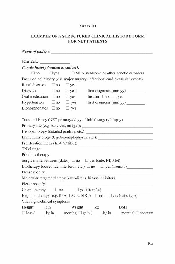
Annex III
EXAMPLE OF A STRUCTURED CLINICAL HISTORY FORM FOR NET PATIENTS
Name of patient: __________________________________________________
Visit date: _______________
Family history (related to cancer):no yes MEN syndrome or other genetic disorders
Past medical history (e.g. major surgery, infections, cardiovascular events)
Renal diseases no yes
Diabetes no yes first diagnosis (mm yy) _________
Oral medication no yes Insulin no yes
Hypertension no yes first diagnosis (mm yy) _________
Biphosphonates no yes
Tumour history (NET primary/dd yy of initial surgery/biopsy)
Primary site (e.g. pancreas, midgut): ___________________________________
Histopathology (detailed grading, etc.): _________________________________
Immunohistology (Cg-A/synaptophysin, etc.): ___________________________
Proliferation index (Ki-67/MiB1): _____________________________________
TNM stage
Previous therapy
Surgical interventions (dates) no yes (date, PT, Met)
Biotherapy (octreotide, interferon etc.) no yes (from/to)_____________
Please specify _____________________________________________________
Molecular targeted therapy (everolimus, kinase inhibitors)
Please specify _____________________________________________________
Chemotherapy no yes (from/to) _________________________
Regional therapy (e.g. RFA, TACE, SIRT) no yes (date, type)
Vital signs/clinical symptoms
105
Height _____ cm Weight_____ kg BMI ________
loss (_____ kg in ____ months) gain (_____ kg in ____ months) constant
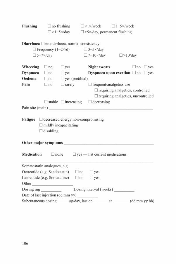
Flushing no flushing <1×/week 1−5×/week
>1−5×/day >5×/day, permanent flushing
Diarrhoea no diarrhoea, normal consistency
Frequency (1−2×/d) 3−5×/day
5−7×/day 7−10×/day >10/day
Wheezing no yes Night sweats no yes
Dyspnoea no yes Dyspnoea upon exertion no yes
Oedema no yes (pretibial)
Pain no rarely frequent/analgetics use
requiring analgetics, controlled
requiring analgetics, uncontrolled
stable increasing decreasing
Pain site (main) ___________________________________________________
Fatigue decreased energy non-compromising
mildly incapacitating
disabling
Other major symptoms ____________________________________________
Medication none yes — list current medications
________________________________________________________________
Somatostatin analogues, e.g.
Octreotide (e.g. Sandostatin) no yes
Lanreotide (e.g. Somatuline) no yes
Other ____________________
Dosing mg __________ Dosing interval (weeks) __________
Date of last injection (dd mm yy) __________
Subcutaneous dosing _____ µg/day, last on _______ at ________ (dd mm yy hh)
106
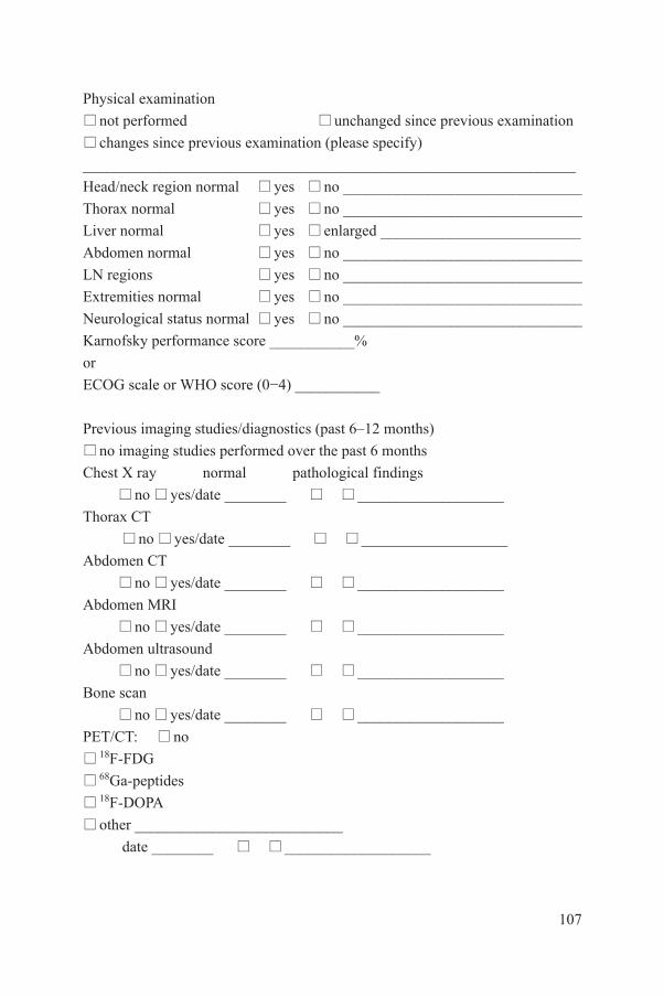
Physical examination
not performed unchanged since previous examination
changes since previous examination (please specify)
________________________________________________________________
Head/neck region normal yes no _______________________________
Thorax normal yes no _______________________________
Liver normal yes enlarged __________________________
Abdomen normal yes no _______________________________
LN regions yes no _______________________________
Extremities normal yes no _______________________________
Neurological status normal yes no _______________________________
Karnofsky performance score ___________%orECOG scale or WHO score (0−4) ___________
Previous imaging studies/diagnostics (past 6–12 months)
no imaging studies performed over the past 6 months
Chest X ray normal pathological findings
no yes/date ________ ___________________
Thorax CT
no yes/date ________ ___________________
Abdomen CT
no yes/date ________ ___________________
Abdomen MRI
no yes/date ________ ___________________
Abdomen ultrasound
no yes/date ________ ___________________
Bone scan
no yes/date ________ ___________________
PET/CT: no
18F-FDG
107
68Ga-peptides
18F-DOPA
other ___________________________
date ________ ___________________
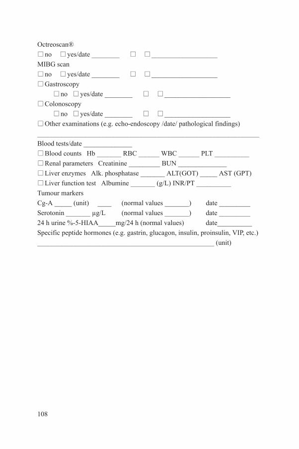
Octreoscan®
no yes/date ________ ___________________
MIBG scan
no yes/date ________ ___________________
Gastroscopy
no yes/date ________ ___________________
Colonoscopy
no yes/date ________ ___________________
Other examinations (e.g. echo-endoscopy /date/ pathological findings)________________________________________________________________
Blood tests/date ______________
Blood counts Hb _______ RBC ______ WBC ______ PLT __________
Renal parameters Creatinine _________ BUN ______________
Liver enzymes Alk. phosphatase _______ ALT(GOT) _____ AST (GPT)
Liver function test Albumine _______ (g/L) INR/PT __________
Tumour markers
Cg-A _____ (unit) ____ (normal values _______) date _________
Serotonin _______ µg/L (normal values _______) date _________
24 h urine %-5-HIAA_____mg/24 h (normal values) date__________
Specific peptide hormones (e.g. gastrin, glucagon, insulin, proinsulin, VIP, etc.)
___________________________________________________ (unit)
108
Annex IV
INFORMED CONSENT
No patient may be treated using PRRNT without her/his informed consent. This consent must be given voluntarily, in writing, prior to the delivery of PRRNT for each individual cycle. The treatment protocol, along with a copy of the signed informed consent form, must then be approved by the medical ethics committee in charge.
Prior to implementing PRRNT, the nuclear medicine physician (or legally authorized representative) should explain to the patient the nature of the therapy, its purpose, the procedures involved, the expected duration of treatment, the potential risks and benefits involved, as well as any known side effects, discomfort or complications that may arise.
Patients must be informed of their right to withdraw their consent at any time, and thereby discontinue the treatment, and that this action will not affect their right to receive qualitative medical treatment or result in any other disadvantages.
The informed consent form should be written in clear and simple language that non-specialists will be able to understand. Patients should be given the opportunity to read the form carefully and consider its contents prior to signing. Pending questions should always be answered in advance by the responsible physician. An oral explanation by the physician, although important, is not a substitute for the written form.
If the patient is unable to provide written consent, his/her oral consent is acceptable as long as this is witnessed by one or more individuals who sign a statement explaining why the patient is unable to sign the consent form.
The informed consent form should comply with all relevant regulatory requirements appropriate to the institution, government or country, and the delivery of treatment and the follow-up treatment received by the patient should comply with the Helsinki Declaration on Ethical Principles for Medical Research Involving Human Subjects (www.wma.net/en/30publications/10policies/b3/).
ELEMENTS OF INFORMED CONSENT
109
To assist and guide professionals considering using PRRNT at their facility, the key elements that must be included in an informed consent form are listed here:
— A statement that PRRNT is the treatment option chosen by the patient’s treating physician in agreement with the nuclear medicine team and/or a decision (recommendation) by the multidisciplinary team;
— The rationale for the choice of this particular treatment option;— An explanation of the indications for PRRNT tailored to the patient’s
disease and clinical condition;— An explanation of the purposes of the particular treatment procedure/
radiopharmaceutical; — Details of any possible short term side effects or discomfort related to the
administration of the radiopeptide, and/or of any kidney protection drug used, and their probability of occurrence, including the source of the data;
— Details of any possible medium and long term complications, and their probability of occurrence, including the source of the data;
— A discussion of appropriate treatments to be used to counter the side effects that might occur following PRRNT;
— Details of the expected results/benefits of PRRNT;— A description of the expected duration of the response to PRRNT;— Details of the number of PRRNT treatment cycles and the duration of
intervals between cycles;— A description of the treatment protocol for each individual cycle;— Details of the expected total duration of the treatment;— Circumstances that might lead to the interruption or discontinuation of the
treatment, by either the physician or the patient;— A description of recommended post-therapy treatment and a list of
laboratory tests to be performed following each treatment cycle;— A description of laboratory tests and imaging procedures to be performed
(recommended) after the completion of the last cycle;— The names and contact information of individual(s) to whom the patient
may address questions regarding PRRNT;— Where appropriate, an acknowledgment that the treatment may involve
currently unknown risks to the patient or to the foetus of a patient who becomes pregnant during/after the treatment.
SAMPLE CONSENT FORM
110
The following sample consent form includes information that should be provided in writing or communicated orally in simple and comprehensible language to the patient and/or their guardian(s).

Note that this sample consent form is intended to be used only for reference, as local requirements, regulations or procedures may vary among institutions. Hence the final consent form should express those particular local requirements.
111
Informed Consent and Authorization Formfor patients with neuroendocrine tumours receivingpeptide receptor radionuclide therapy (PRRNT)
[Name and address of hospital]
[Date]
Dear Madam/Sir,
Your physician/multidisciplinary team has suggested that you undergo peptide receptor radionuclide therapy (PRRNT) and may have informed you in broad terms what this treatment entails. To help you decide whether you wish to undergo this treatment, in addition to conversations with your physician, you will receive a detailed printed information sheet for you to read at your leisure and to discuss its contents with others. If you require additional information, please contact any of the doctors listed at the end of this form.
YOUR CURRENT MEDICAL SITUATION
From prior discussions, it is clear that you suffer from a malignant tumour that cannot be cured by surgery and/or that has not responded to previous medical treatments. One of the novel forms of treatment for your disease is termed peptide receptor radionuclide therapy (PRRNT).
Recently you had a scan in the department of nuclear medicine. The scan determined that the malignant tumour cells are capable of binding a carrier peptide that is linked to a radioactive substance. Because of this, the disease can be irradiated from within. The intention of this treatment is to reach all malignant cells in your body without affecting any normal tissue.
AIMS OF THE TREATMENT
112
The aims of this treatment in patients with neuroendocrine tumours are:
— To reduce/abolish the signs/symptoms caused by the disease;— To accomplish tumour shrinkage by reducing the size and the number of
tumour sites;
— To enable the use of a combination of complementary forms of treatment such as embolization or further surgical resection.
TREATMENT PRINCIPLES (HOW THE TREATMENT WORKS)
Peptide receptor radionuclide therapy is a treatment procedure delivered by nuclear medicine to treat somatostatin receptor-positive malignant tumours. For PRRNT, a short range beta emitting radioisotope, either yttrium-90 (Y-90) or lutetium-177 (Lu-177), is used to irradiate the tumour. For this purpose, the radioisotope is attached to a molecule (peptide) similar to the one used in your recent diagnostic nuclear medicine investigation (scintigraphy), which showed that PRRNT treatment can be effective in your case. It confirmed that the therapeutic (the radiopeptide) binds effectively to the malignant cells to allow their irradiation from within.
Much experience has been acquired with this type of treatment for patients with neuroendocrine tumours in recent years. Since the mid-1990s, many patients have been treated in institutions worldwide. The published results to date are encouraging. Sustained relief from symptoms and no further growth of the tumour (stabilization of disease progression) have been reported in over 70% of patients, and reductions in the size of the cancer (tumour shrinkage) in up to 30%.
PROCEDURE (HOW THE TREATMENT IS ADMINISTERED)
On the day of the therapy, following your admission to the nuclear medicine ward, an intravenous line will be placed in your forearm, and an anti-emetic to prevent vomiting will be administered. Your vital signs will be monitored by measuring blood pressure, pulse rate and your general well-being. You will receive two additional infusions. The first contains saline (salt solution) to make sure that you have sufficient fluids in your body to produce a lot of urine. The second infusion contains a mixture of amino acids that will protect your kidneys and minimize their radiation by the radiopharmaceutical. The amino acid infusion will continue for 4–5 hours. The administration of the radioactive peptide will begin 30 minutes after the start of the amino acid infusion and will last for
113
5−30 minutes. In the days following the treatment, part of the radioactivity will be
expelled in your urine. The level of radioactivity in your body will be monitored until it has fallen to safe levels and you can be discharged. In most cases, patients are able to return home within 24−48 hours.
During the first week after therapy, you will be required to return to the medical centre for several measurements using a SPECT camera to determine the distribution of the radiopeptide in your body. This will allow your physician to assess the irradiation of the kidneys and of the cancer tissue. The latter procedure is termed ‘dosimetry’.
Following the first cycle and subsequent PRRNT treatment cycles, your blood will be monitored for changes in white blood cell and blood platelet counts and kidney function. These blood tests should be performed four weeks after each treatment cycle and two to four weeks before the next cycle.
POTENTIAL RISKS
PRRNT does have some temporary side effects. The most frequent side effects are mild reductions in the number of white blood cells and blood platelets. A decrease in the white blood cell count may result in infections. Blood platelets assist in the clotting of blood, so a decrease in platelet concentration may increase the risk of internal bleeding. In a few cases the platelet count may fall to dangerous levels, which may require treatment and could lead to the postponement of the next treatment cycle.
An increased frequency of infections or internal bleeding has not been observed in patients receiving PRRNT. Other, more common side effects include fatigue, nausea (30%) and vomiting (15%), usually on the first day. About 65% of patients receiving PRRNT experience temporary hair loss (not baldness), but the hair will resume growing when the treatment is concluded.
Over the longer term, a few patients may experience more serious side effects.
Myelodysplastic syndrome (MDS), a pre-stage of leukaemia, has occurred in three patients out of 500 receiving this treatment.
In two patients, a serious deterioration of kidney function occurred. In one patient, renal insufficiency developed one year after the last treatment. This patient had already had periods of unexplained decreased kidney function in the year preceding the therapy. It is therefore not certain whether the therapy was the cause of the further deterioration of the kidney function. In the other patient, a serious deterioration of kidney function occurred more than three years after the
114
last therapy. In part this was also caused by the medication the patient was taking.Finally, in three patients with very extensive, diffuse liver metastases, a
deterioration of liver function occurred in the weeks following PRRNT therapy. In two patients this was temporary, whereas the other patient died shortly afterwards.
KNOWN SIDE EFFECTS AND DISCOMFORTS OF PRRNT
PRRNT has a number of known side effects, which vary from person to person. Many of these will disappear after a short time, although others may be serious with longer lasting and/or permanent effects on the body, and could indirectly be fatal.
During the therapy cycles, you may experience some or all of the side effects listed below. If you experience any unexplained signs or symptoms, please contact your physician to discuss your condition based on your individual risk profile.
Short term (commonly observed) side effects
(1) Nausea, vomiting, diarrhoea and indigestion may occur during or immediately after therapy. These symptoms tend to disappear following the cessation of the amino acid infusion.
(2) Allergic reactions (itchy rash, anaphylaxis) may occur following the amino acid infusion with a plasma expander such as gelatin (Gelafusin®) during therapy.
(3) If your cancer is functionally active, you may experience a so-called carcinoid crisis during or following therapy due to the sudden release of hormones from the cancer cells into the bloodstream. This can cause circulatory and breathing difficulties, accompanied by headache and other neurological symptoms. If this occurs, it will be treated vigorously, and may sometimes require short acting somatostatin medication.
Medium term (commonly observed) side effects
(1) Temporary decrease in the counts of red blood cells (erythrocytes) and white blood cells (leucocytes/ lymphocytes).
(2) Temporary decrease in blood platelets (thrombocytes).
Long term (rarely observed) side effects
(1) Repeated administration of PRRNT may result in serious deterioration of
115
renal function in less than 1% of patients.(2) Myelodysplastic syndrome (MDS) has been observed in less than 1% of
patients and may be associated with prior chemotherapy.(3) Other unknown deleterious effects may occur that have not been observed
to date.

PREGNANCY AND BREASTFEEDING
Medical facilities are forbidden by law to administer PRRNT treatment to pregnant women. Women of reproductive age should take precautions to avoid becoming pregnant during up to six months after the last treatment. If you wish, your attending physician can provide advice on suitable methods of contraception. If, despite all precautions, you do become pregnant during the treatment period, please contact your attending physician immediately. The potential hazard of this treatment to your unborn child is not known. Women with babies should refrain from breastfeeding during the entire period of therapy.
POST-THERAPY MONITORING
To evaluate the results and the effects of the treatment, additional investigations and tests are necessary. The results of the treatment are evaluated by means of blood tests, urine tests, radiological procedures (usually CT or MRI scans) and additional tests, if necessary. Following the last therapy cycle, your attending physician will continue to monitor the course of your disease. In this respect, it is important that you report any side effects and their severity to your attending physician.
PRIVACY AND CONFIDENTIALITY DECLARATION
All the data and outcomes related to your treatment will treated as confidential by the physicians and staff responsible for this therapy. Information about your treatment can only be consulted by the treating team or by a committee that oversees ethical conduct of care. Your data will be handled in accordance with all laws and regulations regarding personal data protection and privacy relevant to the institution or country (if applicable).
HOSPITAL NAME/CLINICIAN NAME would store the data related to your treatment in order to perform further evaluations of the treatment, or for publication purposes in the future. Please indicate below whether you agree or do not agree to such use of the data. We will of course respect your wishes.
116
I agree/I do NOT agree (delete as applicable) that my data should be used in further evaluations or publications.
Signed____________________________________ Date: ____________

Refusal and discontinuation during the treatment
You may discontinue your treatment at any time without further explanation. You are free to withdraw your consent by informing your physician. Your decision not to continue will not affect your further medical management.
Further information/inquiries
If you have any questions about the treatment, before, during or after the cycles, please contact your attending physician or any other physician at the department of nuclear medicine listed below:
Name of institution:
Dept of Nuclear Medicine: Phone:
Responsible physicians:
NamePhone: Email:
NamePhone: Email:
Please sign and date the attached consent form in the presence of the physician responsible for the therapy.
AUTHORIZATION FORM TO RECEIVE TREATMENT with [name of the radiopharmaceutical]
I confirm that I have read and understand the information for patients. I have had the opportunity to ask additional questions, which have been answered to my satisfaction. I have had sufficient time to think about the proposed treatment. I know that my decision is voluntary and that I can withdraw my consent at any time without further explanation.
117
I hereby give my permission to inform my general practitioner and/or attending physician about my decision to receive treatment with PRRNT.
I hereby give my permission to use the data for the purposes described above and agreed upon.
I hereby give my permission to be treated with PRRNT according to the information provided above.
Patient’s name (print) and signature: Date:
_____________________________ _____________________
Responsible physician’s name and signature: Date:
_____________________________ _____________________
118

ACRONYMS AND ABBREVIATIONS
5-FU 5-fluorouracil 5-HIAA 5-hydroxyindoleacetic acid 5-HTP 5- hydroxy-L-tryptophan99mTc-MAG3 99mTc-mercapto-acetylglycyl-glycyl-glycine ACTH adrenocorticotropic hormoneAGC absolute granulocyte countAMP adenosine monophosphateANC absolute neutrophil count APUD amin precursor uptake and decarboxylation BED biologically effective doseBMI body mass indexBq becquerel (one nuclear disintegration per second)BUN blood urea nitrogenCg-A chromogranin ACEA carcino-embryonic antigenCi CurieCLARINET study of Lanreotide Autogel in non-functioning entero-
pancreatic endocrine tumours CR complete responseCT computed tomographyCTC common toxicity criteriaCTCAE Common Terminology Criteria for Adverse Events DAT direct antiglobulin test DOTA Ga-68 68Ga-labelled 1,4,7,10-tetraazacyclododecane-N,N',N,N'-
tetraacetic acid used with PETDOTATOC DOTA-d-Phe(1)-Tyr(3)-octreotideDOTANOC Somatostatin analogue 1-NaI3-octreotide (NOC) attached to
68Ga-DOTA has high affinity to sstr subtypes 1, 2 and 5 DTPA diethylene triamine pentaacetic acidECOG Eastern Cooperative Oncology Group (USA)EDTA ethylene diamine tetraacetic acid EDTMP ethylenediamine tetra(methylene phosphonic acid)ECL enterochromaffin-like (cell)
119
ELISA enzyme linked immunosorbent assayENETS European Neuroendocrine Tumour SocietyEORTC European Organisation for Research and Treatment of
Cancer ERPF effective renal plasma flowEPT endocrine pancreatic tumour

FDA Food and Drug Administration (USA)F-FDA 6-[18F]fluorodopamineF-DOPA 18F-L-dihydroxyphenylalanineFNA fine needle aspirationFOLFOX folinic acid, fluorouracil and oxaliplatin FSH follicle-stimulating hormone GEP gastroenteropancreatic GFR glomerular filtration rateGHRH growth hormone releasing hormoneGLP1-R glucagon-like peptide–1 receptor GRP-R gastrin releasing peptide receptor Gy gray (1 J of ionizing radiation absorbed by 1 kg of tissue)HPF high-power field magnificationHPLC high performance liquid chromatographyHR hazard ratio HVA homovanillic acidIC50 half maximal inhibitory concentration i.m. intramuscularINF-α interferon-alphaIV intravenous Ki-67 immunohistochemical staining to assess cellular
proliferation using Ki-67 monoclonal antibodyLAR long acting repeatable/releaseLITT laser induced interstitial thermoablation LLN lower limit of normal values MAG3 mercaptoacetyltriglycineMcR melanocortin receptor MDS myelodysplastic syndrome MEN-1/-2 multiple endocrine neoplasia syndrome types 1 and 2MIBG metaiodobenzylguanidine MIP maximum intensity projection MIRD Medical Internal Radiation DoseMR minor responseMRI magnetic resonance imaging MTC medullary thyroid cancer/carcinoma
120
MU million unitsNANETS North American Neuroendocrine Tumour SocietyNCCN National Comprehensive Cancer NetworkNCI National Cancer Institute (USA)NCRP National Council on Radiation Protection and MeasurementsND number of decays

NET neuroendocrine tumourNF-1 neurofibromatosis type 1 NME necrolytic migratory erythema NSCLC non-small cell lung cancer NSE neuron specific enolase Octreotide an octapeptide that mimics the action of the naturally
occurring somatostatinOLINDA/EXM Organ Level Internal Dose Assessment/Exponential
Modeling PAI percutaneous acetic acid injectionPD progressive disease PDEC poorly differentiated endocrine carcinoma PDGFR platelet-derived growth factor receptor PEG- IFN pegylated interferon PEI percutaneous ethanol injection.PET positron emission tomographyPET/CT positron emission tomography/computed tomographyPFS progression-free survival pH potential of hydrogenp.i. post injectionPLT platelet countPNET pancreatic neuroendocrine tumourpo per os (by mouth)PP pancreatic polypeptidePPI proton pump inhibitorPPoma pancreatic polypeptidomaPR partial response PRKAR1A protein kinase A regulatory subunit type 1-alpha PROMID Placebo-controlled, double-blind, prospective Randomized
study of the effect of Octreotide LAR in the control of tumour growth in patients with metastatic neuroendocrine Midgut tumours
PRRNT peptide receptor radionuclide therapyPTH parathyroid hormone RAEB refractory anaemia with excess blasts
121
RADAR Radiation Dose Assessment ResourceRAMSETE RAD001 in Advanced Metastatic Silent Neuroendocrine
Trial in EuropeRECIST Response Evaluation Criteria in Solid TumorsRFA radiofrequency ablationRIA radioimmunoassay
ROI region of interest SAAM Simulation, Analysis and Modeling software s.c. subcutaneousSCLC small cell lung cancer SD stable diseaseSDH-B, -D succinate dehydrogenase complex, subunit B, subunit D SDHD succinate dehydrogenase deficiency syndrome SEER Surveillance, Epidemiology and End Results (database)SIRT selective internal radiotherapy Sm samariumSMA superior mesenteric artery Somatostatin growth-hormone-inhibiting hormone, interacts with cell
signalling via the G-protein coupled somatostatin receptor(s)
SPECT single photon emission computed tomographySPECT/CT single photon emission computed tomography/computed
tomographySSA somatostatin receptor agonist/analogue sstr somatostatin receptor subtypes 1, 2, 3, 4 and 5 SUV standardized uptake valueSv sievertTACE transcatheter arterial/transarterial chemoembolization TAE transcatheter arterial/transarterial embolization TER tubular extraction rate TNM tumour-node metastasisTSH thyroid stimulating hormoneTTP time to tumour progression UICC Union for International Cancer Control ULN upper limit of normal values VEGF vascular endothelial growth factor VHL Von Hippel–Lindau syndromeVIP vasoactive intestinal peptide VIPoma vasoactive intestinal peptide secreting tumour VIP-R vasoactive intestinal polypeptide receptor VMA vanillylmandelic acid
122
WBC white blood cell countWDEC well differentiated endocrine carcinomaWDET well differentiated endocrine tumourWDS watery diarrhoea syndromeWHO World Health Organization
CONTRIBUTORS TO DRAFTING AND REVIEW
Bal, C.S. All India Institute of Medical Sciences, India
Baum, R.P. Zentralklinik Bad Berka, Germany
Belfer, A. Hospital Israelita Albert Einstein, Brazil
Bodei, L. European Institute of Oncology, Italy
Cremonesi, M. European Institute of Oncology, Italy
Howe, J.R. University of Iowa, United States of America
Hörsch, D. Zentralklinik Bad Berka, Germany
Mueller-Brand, J. Universitätsspital Basel, Switzerland
O’Dorisio, M.S. University of Iowa, United States of America
O’Dorisio, T.M. University of Iowa, United States of America
Pavel, M.E. Charité Universitätsmedizin Berlin, Germany
Walter, M. Universitätsspital Basel, Switzerland
Zaknun, J.J. International Atomic Energy Agency
Consultants Meetings
Vienna, Austria: 7–11 December 2009, 15–19 March 2010, 23–27 May 2011
123

.
@������
��������� ���������������� ��������������������������������� �������������������������� ������������ ���������
����������������������������������� �������������������������������� ����� !"����������
������ �#��������� ��� ��$�����%&'�(� �������)�����*�+!,�*�-.-/��
+�������0�1%.�-�2/.3�4444�5�6�70�1%.�-�2/.3�44''��
���0����$��8������ ��������5�(���� �0�� �099���������� ��������
!� " �#:�������;��������$��������)��/3/��<=..23�<��������
+�������0�1-/�/�>-'�&-�3'�5�6�70�1-/�/�>-'�3'�&.��
���0��������������8������������5�(���� �0�� �099��������=��=���������
$���%�<������������ ����&>3.�6������<$��� � ��/33��;�������*#�/343%=&-&%��� ���
+�������0�.='33='%>=-&>4�5�6�70�.='33='%>=-&>3��
���0���� ��������8�����������5�(���� �0�� �099���������������
)������������?�!�������; ����.=>-%2�!��� ���)����" �����"� �����@.:�2:-��
+�������0�1%.-�4&>�/%%>�5�6�70�1%.-�4&>�4%%3��
���0���������� 8����������������5�(���� �0�� �099��������������������
$& ������������ ������!�����0�!���������������?������� ���!������ ����+����� ��� �� ������"��<�7�/.3-��<���?�
$'�$&���(�! $ ������!A�� �)�"���@�����$��-&4��.'3�/.�������2��
+�������0�1&/3�/%%3-�>-%&�5�6�70�1&/3�/'&'/�.%&%��
���0������8��������B�5�(���� �0�� �099������������B�
) � ��%��� �������@������������"�<"C�./'�D@������� ��.E��6��=33.3.�,������
+�������0�1->'�2�./.�&.�5�6�70�1->'�2�./.�&&>3��
���0���� ���8��� �����������5�(���� �0�� �099������� �����������
)���$�6���=�� ��>������:����������"��<�7�/>��6=4>2/.������!���7�.2��
+�������0�1--�.�&/�3.�&2�&2�5�6�70�1--�.�&/�3.�23�23��
���0������� 8������ ����5�(���� �0�� �099����������� ����
;�$����� � ��.&>�����������$?����2&/-%�!������!���7��
+�������0�1�--�.�&4�&3�%4�3/�5�6�7�1--�.�&4�&3�%4�3/��
���0��������$�����8�$��������5�(���� �0�� �099�����$��������
"��#��*��"=F���?��F�� ����=�����F���?��G��,�����,��?�� ���.3��#=>-..-�<�����
+�������0�1�&2�//'�2&�23�/3�5�6�70�1&2�//'�2&�23�/3����1&2�//'�2&�23�///��
���0���� ���?8���=$���?����5�(���� �0�� �099�������=$���?����
&��"��*;��� �����; ����<��������� ����"��<�7�./%��,=.%>%�<������ ��
+�������0�1-%�.�/>4�4444�5�6�70�1-%�.�/>4�4&4/�5����0������8��� ��������
�% ��������������G������.� �6�����#������,������.>��:�����,������*��?��<������� � ���*�����&33�33.���
+�������0�12.�//�//%.42/%9/4�5�6�70�12.�//�//%.42/'��
���0�����8$�������5�(���� �0�� �099��������������������
<�������/94/����������!������#���..3332��
+�������0�12.�..�/-/%'4'%��12.�..�/-/>4/%&�5�6�70�12.�..�/-/'.-.>��
���0�������8$����� �
�� *;������ ��� H���#� ��;������<����I���"�J��F��!������%���=/3.&%�*����
+�������0�1-2�3/�&'�2>�&>�>/����&'�2>�&>�%/�5�6�70�1-2�3/�&'�2>�&>�&'��
���0����8�������������5�(��� �0�����������������
JAPANMaruzen Company Ltd, 1-9-18, Kaigan, Minato-ku, Tokyo, 105-0022
Telephone: +81 3 6367 6079 • Fax: +81 3 6367 6207
Email: [email protected] • Web site: http://www.maruzen.co.jp
REPUBLIC OF KOREAKINS Inc., Information Business Dept. Samho Bldg. 2nd Floor, 275-1 Yang Jae-dong SeoCho-G, Seoul 137-130
Telephone: +02 589 1740 • Fax: +02 589 1746 • Web site: http://www.kins.re.kr
NETHERLANDSDe Lindeboom Internationale Publicaties B.V., M.A. de Ruyterstraat 20A, NL-7482 BZ Haaksbergen
Telephone: +31 (0) 53 5740004 • Fax: +31 (0) 53 5729296
Email: [email protected] • Web site: http://www.delindeboom.com
Martinus Nijhoff International, Koraalrood 50, P.O. Box 1853, 2700 CZ Zoetermeer
Telephone: +31 793 684 400 • Fax: +31 793 615 698
Email: [email protected] • Web site: http://www.nijhoff.nl
Swets and Zeitlinger b.v., P.O. Box 830, 2160 SZ Lisse
Telephone: +31 252 435 111 • Fax: +31 252 415 888
Email: [email protected] • Web site: http://www.swets.nl
NEW ZEALANDDA Information Services, 648 Whitehorse Road, MITCHAM 3132, Australia
Telephone: +61 3 9210 7777 • Fax: +61 3 9210 7788
Email: [email protected] • Web site: http://www.dadirect.com.au
SLOVENIACankarjeva Zalozba d.d., Kopitarjeva 2, SI-1512 Ljubljana
Telephone: +386 1 432 31 44 • Fax: +386 1 230 14 35
Email: [email protected] • Web site: http://www.cankarjeva-z.si/uvoz
SPAINDíaz de Santos, S.A., c/ Juan Bravo, 3A, E-28006 Madrid
Telephone: +34 91 781 94 80 • Fax: +34 91 575 55 63
Email: [email protected], [email protected], [email protected], [email protected]
Web site: http://www.diazdesantos.es
UNITED KINGDOMThe Stationery Office Ltd, International Sales Agency, PO Box 29, Norwich, NR3 1 GN
Telephone (orders): +44 870 600 5552 • (enquiries): +44 207 873 8372 • Fax: +44 207 873 8203
Email (orders): [email protected] • (enquiries): [email protected] • Web site: http://www.tso.co.uk
On-line orders
DELTA Int. Book Wholesalers Ltd., 39 Alexandra Road, Addlestone, Surrey, KT15 2PQ
Email: [email protected] • Web site: http://www.profbooks.com
Books on the Environment
Earthprint Ltd., P.O. Box 119, Stevenage SG1 4TP
Telephone: +44 1438748111 • Fax: +44 1438748844
Email: [email protected] • Web site: http://www.earthprint.com
UNITED NATIONSDept. I004, Room DC2-0853, First Avenue at 46th Street, New York, N.Y. 10017, USA
(UN) Telephone: +800 253-9646 or +212 963-8302 • Fax: +212 963-3489
Email: [email protected] • Web site: http://www.un.org
UNITED STATES OF AMERICABernan Associates, 4501 Forbes Blvd., Suite 200, Lanham, MD 20706-4346
Telephone: 1-800-865-3457 • Fax: 1-800-865-3450
Email: [email protected] · Web site: http://www.bernan.com
Renouf Publishing Company Ltd., 812 Proctor Ave., Ogdensburg, NY, 13669
Telephone: +888 551 7470 (toll-free) • Fax: +888 568 8546 (toll-free)
Email: [email protected] • Web site: http://www.renoufbooks.com
Orders and requests for information may also be addressed directly to:
Marketing and Sales Unit, International Atomic Energy Agency
Vienna International Centre, PO Box 100, 1400 Vienna, Austria
Telephone: +43 1 2600 22529 (or 22530) • Fax: +43 1 2600 29302
Email: [email protected] • Web site: http://www.iaea.org/books
12-0
9541
RELATED PUBLICATIONS
www.iaea.org/books
Nuclear MediciNe resources MaNualSTI/PUB/1198 (532 pp.; 2006)ISBN 92–0–107504–9 Price: €65.00
Quality MaNageMeNt audits iN Nuclear MediciNe PracticesSTI/PUB/1371 (57 pp.; 2008)ISBN 978–92–0–112108–0 Price: €25.00
a guide to cliNical Pet iN oNcology: iMProviNg cliNical MaNageMeNt of caNcer PatieNtsiaea tecdoc series No. 1605IAEA-TECDOC-1605 (55 pp.; 2008)ISBN 978–92–0–110608–7 Price: €15.00
criteria for PalliatioN of BoNe Metastases – cliNical aPPlicatioNsiaea tecdoc series No. 1549IAEA-TECDOC CD-1549 (55 pp.; 2007)ISBN 92–0–104507–7 Price: €15.00
Nuclear MediciNe iN thyroid caNcer MaNageMeNt: a Practical aPProachiaea tecdoc series No. 1608IAEA TECDOC-1608 (273 pp.; 2009)ISBN 978–92–0–113108–9 Price: €15.00
release of PatieNts after radioNuclide theraPysafety reports series No. 63STI/PUB/1417 (77 pp.; 2009)ISBN 978–92–0–108909–0 Price: €28.00
Quality assuraNce for radioactive MeasureMeNt iN Nuclear MediciNetechnical reports series No. 454STI/DOC/010/454 (81 pp.; 2006)ISBN 92–0–105306–1 Price: €42.00
IAEA HUMAN HEALTH SERIES PUBLICATIONS
The mandate of the IAEA human health programme originates from Article II of its Statute, which states that the “Agency shall seek to accelerate and enlarge the contribution of atomic energy to peace, health and prosperity throughout the world”. The main objective of the human health programme is to enhance the capabilities of IAEA Member States in addressing issues related to the prevention, diagnosis and treatment of health problems through the development and application of nuclear techniques, within a framework of quality assurance.
Publications in the IAEA Human Health Series provide information in the areas of: radiation medicine, including diagnostic radiology, diagnostic and therapeutic nuclear medicine, and radiation therapy; dosimetry and medical radiation physics; and stable isotope techniques and other nuclear applications in nutrition. The publications have a broad readership and are aimed at medical practitioners, researchers and other professionals. International experts assist the IAEA Secretariat in drafting and reviewing these publications. Some of the publications in this series may also be endorsed or co-sponsored by international organizations and professional societies active in the relevant fields. There are two categories of publications in this series:
IAEA HUMAN HEALTH SERIESPublications in this category present analyses or provide information of an
advisory nature, for example guidelines, codes and standards of practice, and quality assurance manuals. Monographs and high level educational material, such as graduate texts, are also published in this series.
IAEA HUMAN HEALTH REPORTSHuman Health Reports complement information published in the IAEA Human
Health Series in areas of radiation medicine, dosimetry and medical radiation physics, and nutrition. These publications include reports of technical meetings, the results of IAEA coordinated research projects, interim reports on IAEA projects, and educational material compiled for IAEA training courses dealing with human health related subjects. In some cases, these reports may provide supporting material relating to publications issued in the IAEA Human Health Series.
All of these publications can be downloaded cost free from the IAEA web site:http://www.iaea.org/Publications/index.html
Further information is available from:Marketing and Sales UnitInternational Atomic Energy AgencyVienna International CentrePO Box 1001400 Vienna, Austria
Readers are invited to provide their impressions on these publications. Information may be provided via the IAEA web site, by mail at the address given above, or by email to:
INTERNATIONAL ATOMIC ENERGY AGENCYVIENNA
ISBN 978–92–0–129210–0ISSN 2075–3772
Practical G
uidance on Peptide R
eceptor Radionuclide Therapy (P
RR
NT) for N
euroendocrine Tumours
This publication provides comprehensive, multidisciplinary guidance on the use of peptide receptor radionuclide therapy (PRRNT) in the treatment of patients with neuroendocrine tumours (NETs) and gastroenteropancreatic cancers, taking into account the recent international classifi cations of NETs. It provides comprehensive protocols for employing 90Y or 177Lu tagged somatostatin receptor targeting peptides as well as clinically assessed protocols for renal protection. It provides comprehensive, evidence based clinical guidelines, with input from experienced and renowned medical professionals in the fi eld. The various sections of the book cover clinical presentation, patient eligibility criteria and means of assessing the effectiveness of therapy using molecular and morphological medical imaging techniques.
Practical G
uidance on Peptide R
eceptor Radionuclide Therapy (P
RR
NT) for N
euroendocrine Tumours
IAEA HUMAN HEALTH SERIES IAEA HUMAN HEALTH SERIESNo. 20
IAEA HUMAN HEALTH SERIES No. 20
Practical Guidance on Peptide Receptor
Radionuclide Therapy (PRRNT) for
Neuroendocrine Tumours
IAEA HUMAN HEALTH SERIES