I o' E •- I - Defense Technical Information Center / ABSTRACT (continued) used in an intermediate...
25
Transcript of I o' E •- I - Defense Technical Information Center / ABSTRACT (continued) used in an intermediate...


• • —^—•
•
I o' E •- I tt 1-
It I« I I L
Z5
2.2
2.0
1.25 11.4
1.8
1.6
L
MICROCOPY RESOLUTION TEST CHART
NATIONAL BUREAU OF STANDARDS 1963-A

00
U _
HUMAN PLATELET SENESCENCE STUDY
Final Summary Report
AD
CO
Simon Karpatkin, M.D.
March 1980
Supported by
US ARMY MEDICAL RESEARCH AND DEVELOPMENT COMMAND Fort Detrick, Frederick, Maryland 21701
Contract No. DADA17-68-C-8163
New York University Medical Center 550 First Avenue
New York, New York 10016 ^ELECTt^ %v MAR 7 1984
.
A
O CJ>
Approved for public release; distribution unlimited
The findings in this report are not to be construed as an official Department of the Army position unless so designated by other authorized documents.
• • —•
84 03 06 152
•MMBMHk

$KJ S*
SECURITY CLASSIFICATION OF THIS PAGE (>>'•" Data Bnfrod)
REPORT DOCUMENTATION PAGE 1. REPORT NUMBER 2. OQVT ACCESSION N
*. TITLE fand Subtltl»)
HUMAN PLATELET SENESCENCE STUDY
7. AUTHOR!»
Simon Karpatkin, M.D.
9. PERFORMING ORGANIZATION NAME AND ADDRESS
New York University Medical Center 550 First Avenue New York. NY 100)6 It. CONTROLLING OFFICE NAME AND ADDRESS
U.S Army Medical Research and Development Cornman* Fort Detrick, Frederick. Maryland 21701
I«. MONITORING AGENCY NAME ft AOORESSf" dltUrmnl /ram Controlling OHlco)
READ INSTRUCTIONS BEFORE COMPLETING FORM
3. RECIPIENT'S CATALOG NUMBER
S. TYPE OF REPORT ft PERIOD COVERED
Final Summary Report Lulls lififl - SgatiHBhi 1322
6. PERFORMING ORG. REPORT NUMBER
8. CONTRACT OR GRANT NUMBER(a)
DADA 17-68-C-8163
10. PROGRAM ELEMENT. PROJECT, TASK AREA 4 WORK UNIT NUMBERS
62 7 72A.3S162 772A815.00.026
12. REPORT DATE
March 1980 13. NUMBER OF PAGES
22 1S. SECURITY CLASS. fo( ihim report.)
Unclassified Report
is«, OECLASSIFICATION/DOWNGRADING SCHEDULE
16. DISTRIBUTION STATEMENT (ot thtm Report)
Approved for Public Release Distribution Unlimited
17. DISTRIBUTION STATEMENT (of ihm mbmtrmct Mlmd In Slock 20, It different from Report)
I«. SUPPLEMENTARY NOTES
It. KEY WORDS (Contlnum on rereree old» II neceeeary and Idontltr by block number;
crossed immunoelectrophoresis of platelet membranes platelet membrane antigens co1 eh i c i ne cAMP phosphodiesterase platelet aggregation
platelet microtubules platelet
JO. ABSTW ACT (Quirl i «a reeare» eM> It IIMIIIT —I tdomHlr »r block tnmvmm)
A technique was developed for the identification of platelet membrane This involves agar electrophoresis in one direction, followed by rotation slide 90° and electrophoresis into agar containing anti-piatelet membrane body. Nineteen different platelet membrane antigens (immunoprecipitates) detected. These include: Albumin, fibrinogen and an antigen absent in p from patients with Glanzmann's thrombasthenia. The relative cell surface tigens can be detected by adsorbtion studies with intact platelets. The of carbohydrate can be detected with suitable lectins, such as concanaval
DO i JMTTI 1473 ed-no» or • HOv •» is OMOCKTK
SECURITY CLASSIFICATION OF THIS P»ttt (Wmmr, Dmtm Entered)
•>-.,:!
••V**-
pa_
/
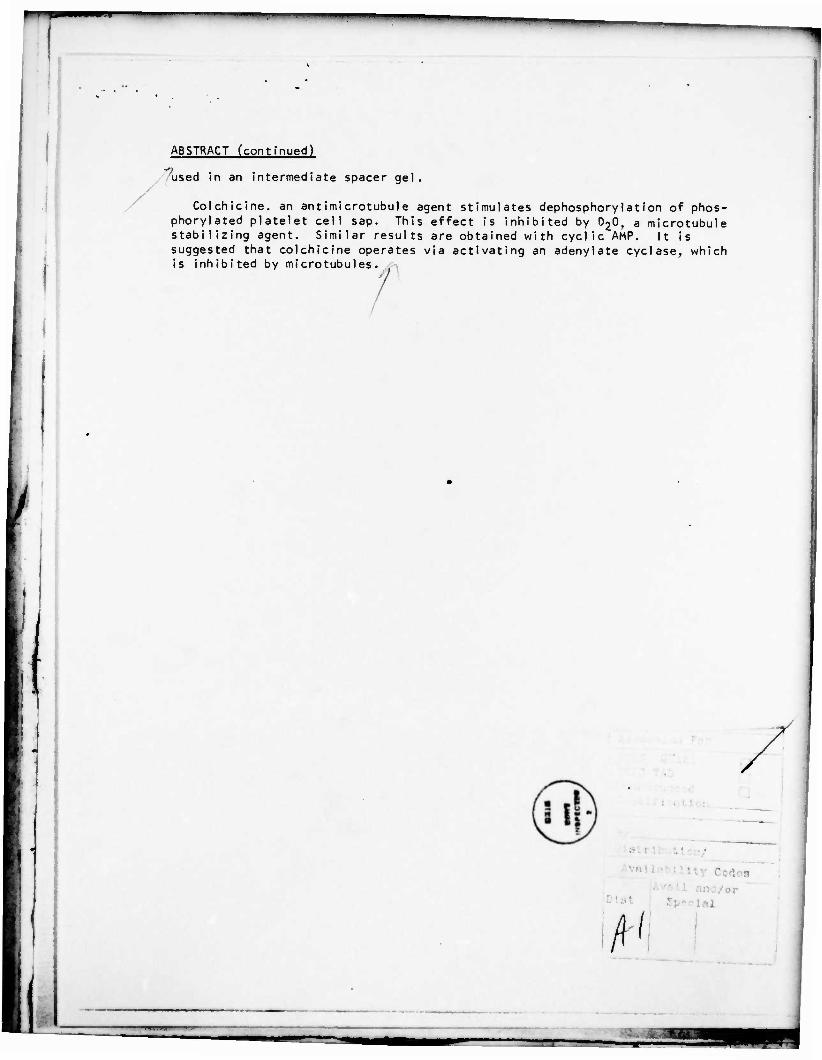
ABSTRACT (continued)
used in an intermediate spacer gel.
Colchicine. an antimicrotubule agent stimulates dephosphorylation of phos- phorylated platelet cell sap. This effect is inhibited by 0,0, a microtubule stabilizing agent. Similar results are obtained with cyclic AMP. It is suggested that colchicine operates via activating an adenylate cyclase. which is inhibited by microtubules.
J
C I s
•
4-1 fi
rrr

9
ABSTRACT
A technique was developed for the identification of platelet membrane antigens. This involves agar electrophoresis in one direction, followed by rotation of the slide 90° and electrophoresis into agar containing anti-platelet membrane antibody. Nineteen different platelet membrane antigens (immunoprecipitates) can be detected. These include: albumin, fibrinogen and an antigen absent in platelets from patients with Glanzmann's thrombasthenia. The relative cell surface antigens can be detected by absorption studies with intact platelets. The presence of carbohydrate can be detected with suitable lectins, such as concanavalin A used in an intermediate spacer gel.
Colchicine, an antimicrotubule agent stimulates dephosphorylation of phospho- rylated platelet cell sap. This effect is inhibited by D20, a microtubule stabilizing agent. Similar results are obtained with cyclic AMP. It is sug- gested that colchicine operates via activating an adenylate cyclase, which is inhibited by microtubules.
_

1
FOREWORD
In conducting ehe research described in chis report, the investigator(s) adhered to the "Guide for the Care and Use of Laboratory Animals," prepared by the Committee on Care and Use of Laboratory Animals of the Institute of Laboratory Animal Resources, National Research Council (DHEW Publication No. (NIH) 78-23, Revised 1978).
For the protection of human subjects the investigator(s) have adhered to policies of applicable Federal Law 45CFR46.
1 •2=3

Simon Kji'pütki n, M.U, DADA 17-68-C-8163
For the past year our laboratory has been engaged in two areas of platelet (and megathrombocyte) research with regard to: heterogeneity and senescence as related to microtubules, protein phosphorylation, platelet aggregation and secretion, and platelet membrane topography.
1. Crossed Immunoelectrophoresis of Platelet Membranes.
2. Requirement of Micro tubules for Platelet Aggregation and Secretion; Association of Phosphodiesterase Activity wi th Platelet Tubulin.
I

TZ
Simon Korpatkin, M.D. DADA 17-68-C-8163
SUMMARY OF PROJECTS
i
I. Crossed Immunoelectrophoresis of Platelet Membranes. Early studies on platelet membranes, employing SOS-PAGE, revealed the
presence of three major glycoproteins, GPI, GPII, and GPIII, with apparent molecular weights of 155,000, 135,000 and 103,000 (1-3). In 197^ Nurden and Caen, employing SDS-PAGE and PAS carbohydrate staining, demonstrated the absence of GPII and the diminished presence of GPIII in 3 patients with Glanzmann's thrombasthenia (3). This was confirmed by Phillips et al (4) who noted a dimished content of GPII rather than its absence. In 1975 Nurden and Caen also demonstrated absent-to-trace amounts of GPI in patients with Bernard-Soulier disease (5) and suggested that GPI was the recep- tor for Von Willebrand factor. Nurden and Caen also demonstrated that GPI was rich in sialic acid (5) and probably responsible for the platelets' surface charge. More refined techniques for demonstrating platelet membrane glycopeptides have subsequently demonstrated the presence of at least 6 to 7 surface glycopeptides (6): GPIs, GPIb, GPIc (non-PAS positive), GPI la, GPIlb, GPIII and GPIV (87,000 m.w., also called GPI I lb) The defect in Glanzmann's thrombasthenia has now been designated as diminished GPIlb and GPI I la; the defect in Bernard-Soulier as diminished GPIs and GPIb. In 1978 Kunicki and Aster (7) presented evidence for the association of the platelet antigen PLAI with GPIlb or GPII la, since platelets from patients with Glanzmann's thrombas- thenia do not release prelabelled Cr51 following addition of potent anti-PLAI antibody, These workers have recently presented evidence that PLAI resides on GPI I la (8). Aster and co-workers later demonstrated that platelets from patients with Bernard-Sou!ier disease do not release prelabelled Cr51 following the addition of Ag-Ab complexes, suggesting the presence of the platelet Fc receptor on GPI (9). Similar conclusions were drawn by Nachman and co-workers (10) who noted competition between Fc binding of non-specific IgG and Von Willebrand factor. Jamieson and co-workers have purified soluble GPIs, which they have termed 'glycocaliein' (11), because of its origin in the platelet glycocalyx. They have suggested that GPIs is associated with the thrombin receptor since purified GPIs binds thrombin and inhibits thrombin-induced platelet aggregation; and platelets from patients with Bernard-Soulier disease aggregate poorl'. with low concentrations of thrombin (11-14).
We have recently acquired a very sensitive technique for the measurement of platelet membrane constituents, which does not completely denature the membrane constituents, and which is 10-100 times more sensitive than SOS-PAGE. This is the technique of crossed-immunoelectrophoresis of plasma membranes, recently developed for the study of bacterial membranes by Or. Salton (17), which gives a two-dimensional display of rockets or peaks of immunoprecipitates. The technology involves solubili- zation of platelet membranes in 1% Triton X-100 and electrophoresis in one direction on a 1% agarose slide in tris-barbital buffer, pH 8.6 at room temperature at 150 volts for 2 to 3 hours. Most of the agarose is then removed and a strip of the re- maining electrophoresed constituents retained. Fresh agar, containing purified rabbit antibody against human platelet membranes, is then poured onto the slide and allowed to harden. The slide is rotated 90° and the antigens then electrophoresed into the agar containing the antibody at 55 volts for 16 hours. The agar is then pressed free

Simon Korpatkin, M.D. DADA 17-68-C-8163
1
I
I*
of excess fluid and washed in 0.1M NaCl with 0.25% Cocmassie blue; and destained advantages of this technique are: 1) the peak areas of individual immunoprecipita ratios: 2) high sensitivity of detection over 10 antigens); 3) ability to measure certain enzymes to their oxidation-reduc other enzymes which can be measured by o phosphatase, acid phosphatase, chymotryp carbohydrate lectins such as concanavali dimension (instead of the antibody) to d
>:o remove excess antibody; dried and stained with 5% HAc-i+5% ethanol (Figure la-d). The technique is essentially quantitative, the
tes being proportional to the antigen/antibody (e.g., on immunoelectrophoresis we can detect platelet membrane enzyme activity - by coupling tion reactions and nitro-blue tetrazolium dyes: ther techniques (catalase, lipase, alkaline sin, trypsin, esterases (17)); M use of n A or wheat germ agglutinin in the second etect specific carbohydrate constituents.
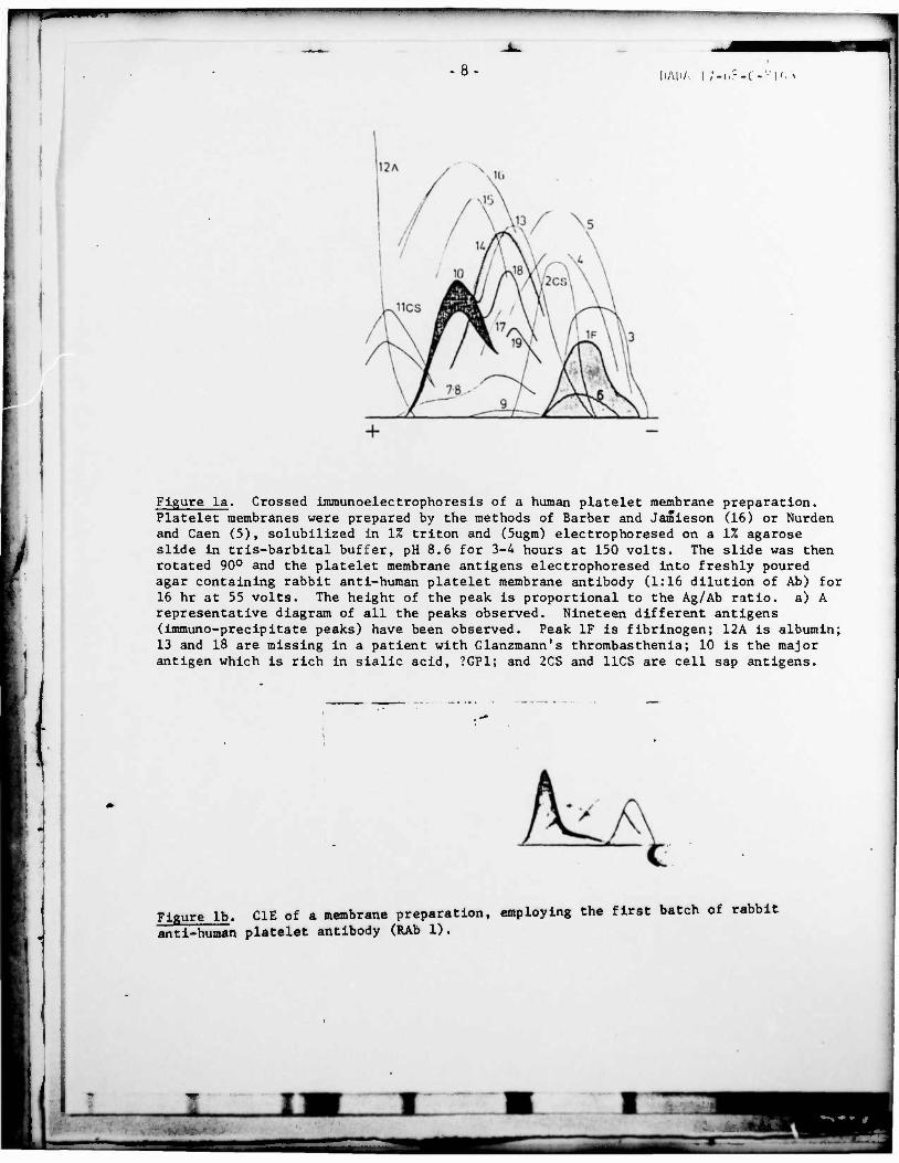
We have developed this procedure with human platelet membrane antigens and can detect at least 19 different platelet membrane immunoprecipitates (Figure 1). One has been identified as fibrinogen, by crossed immunoelectrophorcsis of fibrinogen alone, as well as fibrinogen mixed with the solubilized platelet membrane. Another has been identified as albumin, by similar methods. A third antigen has been identi- fied as a 'Glanzmann's antigen(s) since it is missing on platelets from a patient' with Glanzmann's thrombasthenia. A fourth antigen is sensitive to neuraminidase and chymotrypsin treatment, and is probably glycopeptide 1.
The rabbit anti-human platelet membrane antibody can be completely adsorbed with 1 x 10⁹ platelets (Figure 2a-k). Note the increase in the height of the immuno- precipitins with increasing adsorption of antibody following exposure to increasing numbers of platelets. The antibody cannot be adsorbed with a comparable volume of red blood cells, granulocytes or lymphocytes, demonstrating platelet specificity. We have identified at least k of these 19 antigens. Fibrinogen has been identified bv electrophoresis of f'orinogen alone, membrane constituents alone, and fibrinogen plus membrane constituents (Figure 3a-c). Note the increased height of the fibrinogen pea' Similar results were obtained with albumin (Figure ^a-c). A third antigen has been identified as a 'Glanzmann's antigen(s) since it appears to be missing on platelets from a patient with this disease (Figure 5fl-c) . Note the retention of the IF fibrino- gen peak, suggesting its relative absence on the patient's platelets. A fourth antic,«. is sensitive.to neuraminidase and chymotrypsin treatment, and is probably glycopeotidc (Figure 6a,b). Note shift of 10 toward the anode with development of the right side of its peak; disappearance of 13 and )8; and apparent increase in height of 10 and IF. Figure la, b demonstrates the effect of concanavalin A ligand. Those immunoprecipitates reacting with the ligand are pulled down into the spacer containing the ligand. Anti- gens 10, 13, 18, and IF have been pulled down into the con A lectin, whereas 2CS, 3, 5 (barely visible) and 16 are not affected.
2. Requirement of Microtubules for Platelet Aggregation and Secretion: Association of Phosphodiesterase Activity with Platelet Tubulin. In our previous Army Contract Report (19) we presented data on the
effect of colchicine, O2O, and cyclic AMP on platelet aggregation and protein phosphor/- lation: Platelets were incubated in the presence and absence of colchicine (10~°M), 020 (60-100%), lOuM dibutyrl cycl i.c AMP (db cAMP), 1UM PGE] or lOmM NaF; prior to platelet aggregation induced by Ca ionophore A23817 (lug/ml). Protein phosphory1 at ion was measured by incubation with lmC JZP0^/ml. Cells were washed, sonicated, and the reduced cell sap applied to 10% SOS PAGE; gels were monitored for radioactivity and m.w. Colchicine completely inhibited the 2° wave of platelet aggregation (release) as well as phosphorylation of proteins 10-100,000 m.w. -*H-colchicine (10~°M) was shown to specifically bind to tubulin by sephacryl gel filtration of cell sap (single peak, 110,000 m.w.) and 10% PAGE of isolated peak which coelectrophoresed with purified brain tubulin. O2O completely overcame the colchicine inhibition of aggregation and
Simon Kurudtkin, M. DADA I7-68-C-8I63
phosphory1 at ion. Ob cAMP also completely inhibited platelet aggregation; and db cAMP as well as PGE) and NaF markedly inhibited protein phosphory1 ation; however, O2O did not reverse cAMP-inhibition of phosphorylat ion. These data indicate that unperturbed microtubules are necessary for protein phosphory1 at ion and the 2° wave of platelet aggregation (release).
These data have been extended: colchicine and cyclic AMP both have now been shown to stimulate dephosphorylation or phosphory1ated cell sap. Colchicine was shown to operate via elevation of cyclic AMP levels. Both colchicine and cyclic AMP have also been shown to inhibit the platelet release reaction. D2O overcomes the inhibitory effect of colchicine.
I ib i torv Effect of Cyclic AMP on Phosphory1 a ted Plotclet Cell Sap. Because inh
agents were added 15 minutes prior to the addition of J/:P0/( iv> the i nc.tib.'U i on *• and were therefore inhibiting incorporation of -*'f,Üit into platelet cell sap, we to determine what effect elevated cyclic AMP levels would have on platelet prote which hod already been phosphoryI a ted. Accordingly, washed platelets were prelo with ^'-POi, for 30 minutes and then exposed to agents which would elevate intr-^co c,c:ic AMP levels. These agents served to dephosphory 1 ate the incorporated "''-PC platelet cell sap. Such was the case with 10 mM NaF, Figure 8a (representative experiments); 0.1 mM dibutyrl cyclic AMP, Figure 8b; or 10 uM prostaglandin E|, Figure? 8c (representative of 4 experiments).
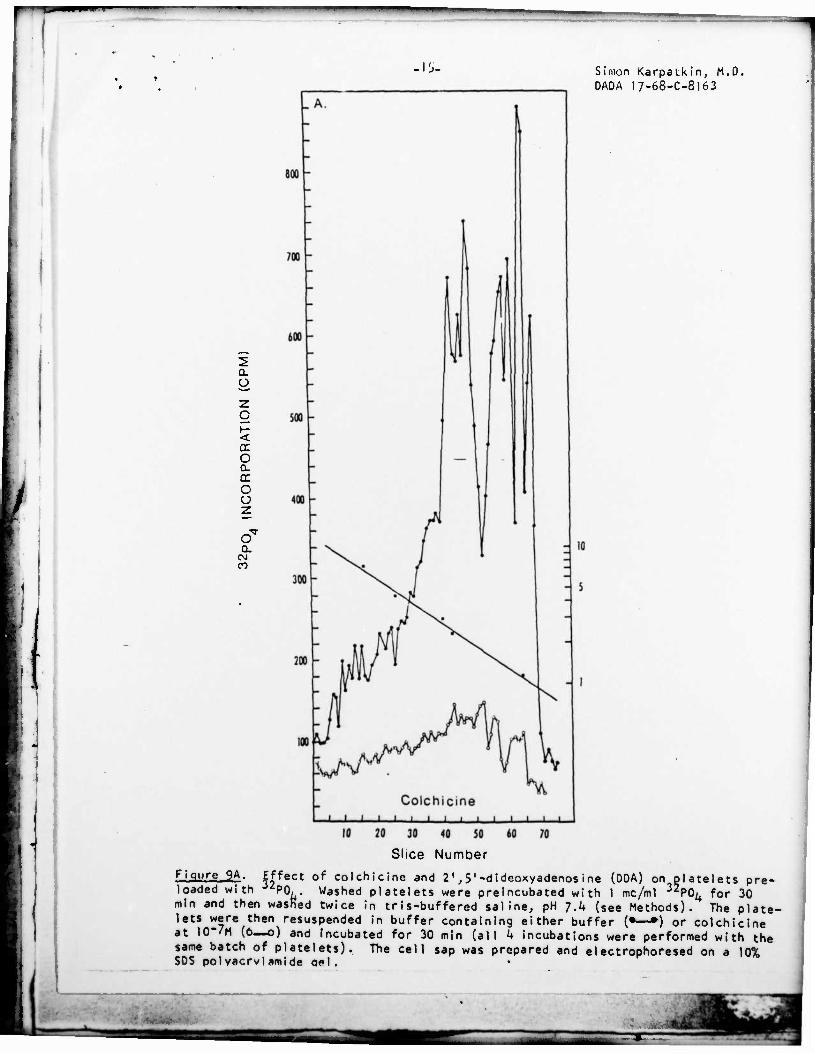
Effect of Colchicine and 2',5'-draeoxyadenosine (PDA) on Phosphory1ateo PIatelet Cel1 Sap. Because colchicine appeared to have the same effect a with respect to inhibition of platelet aggregation and inhibition of inco
P0;, into platelet cell sap, experiments were designed to determine whet was acLing via cyclic AMP. Washed platelets were preloaded with J^PC^ fo and then exposed to colchicine with or without 0.1 mM 2',5'-dideoxyaaenos potent inhibitor of adenylate cyclase (19)- Figure 9a demonstrates that cine also served to dephosphoryI ate the incorporated P0i+ of platelet :e teins (representative of 9 experiments), like cyclic AMP (see Figure 8). effect by itself on phosphory1ated platelet cell sap (Figure 9b^ out comp vented the de'phosphorylation effect of colchicine (Figure §c> representat periments). Similar effects of O.lmM DDA were obtained when platelets we with 10 mM NaF (data not shown).
Effect of Colchicine on IntracelIu1ar Cyclic AMP Levels. The next experimt" l was designed to determine directly whether 10"7M colchicine had any effect on plateU*-
intracellular cyclic AMP levels. Washed platelets (7.5 volumes %) were incubated at 37°C with and without 10~'M colchicine and O2O for 15 minutes in the presence of '; aminophyl1ine and then treated with 6% TCA to extract cyclic AMP which was assayed by a competitive protein binding technique (20). Cyclic AMP levels in the absence of colchicine, 640 picomoles/m! platelet extract, increased to 875 picomoles/ml (2 experi- ments). O2O (100%) prevented the rise in cyclic AMP levels which were 515 picomoles ml in the absence of colchicine and 545 picomoles/ml in its presence (2 experiments).
Effect of 0?0 and Colchicine on Platelet Aggregation and Release of Serotonin Induced by Ca-H- lonophore A23187. D2O, an agent known to stabilize micro- tubules in other tissues, shortened the Ca++ ionophore-induced lag period by 43% and increased the velocity of platelet aggregation by 52% i 14% (SEM) at a D2O concentra- tion of 60% (average of 7 experiments). The combination of 40% O2O and colchicine, or D2O and vinblastinc resulted in partial correction of the alkaloid-induced inhibi- tion of platelet aggregation velocity and prolongation of the lag period. For example,
s c rpo i— _
r 3 i ne 10" I I DC
let i ve e
elected ins aded !lular 4 Of of 3
yclic AMP ration of ;,•.!;-• '
J "i.^ici (00A), 1
7M colchi- sao orc- - had eIy pre- of 5 ex-
i ncubated
Simon Kji'pjtkirij M.D. DADA 17-68-C-8163
,1 J
at 40% D 0, colchicine inhibited the HÖH control by 33%, whereas colchicine and D2O inhibited the D20 control by 18%; vinblastine inhibited the HOH control by k$% whereas vinblastine and D2O inhibited the D2O control by 23%.
Table I demonstrates the effect of colchicine and D2O on the platelet release reaction. Thus, ^C-serotonin release was inhibited by colchicine at 0.2- 500uM, following aggregation by Ca++ ionophore at 5-10uM. D2C [kk%) partially to completely prevented the colchicine-induced inhibition of -'C-serotonin release.
These data therefore indicate that unperturbed microtubules are necessary for protein phosphorylation, the secondary wave of platelet aggregation and the plate- let release reaction. The data strongly suggest that intact microtubules serve to inhibit adenylate cyclase activity.
Evidence for the Association of cAMP Phosphodiesterase Activity with Platelet Microtubules. Our studies with intact platelets strongly suggest inhibi- tion of adenylate cyclase activity by umperturbed platelet microtubules, since col- chicine binds to platelet microtubules; colchicine operates like cyclic AMP; colchicine raises intracel1ular cyclic AMP levels; the effects of colchicine on platelet aggrega- tion, secretion and phosphorylation can be reversed with D2O, a stabilizer of micro- tubules; and colchicine's effect on phosphorylation can be reversed by 2',5'-dideoxy- adenosine, an inhibitor of adenylate cyclase activity. We therefore propose that platelet tubulin may act as a phosphodiesterase or be closely associated with a pro- tein with phosphodiesterase activity.
Preliminary studies suggest that this may be the case. We have partially purified platelet tubulin by modifications of the methods of Shelanski (21) and Steiner (22). This preparation contains phosphodiesterase activity.
We have assayed cAMP phosphodiesterase by measuring the release of Pi employing the phosphomolybdate reaction. A 350-fold purification preparation requires Mg++ and gives a Km of 77 uM for cyclic AMP. It does not react with 2 mM cyclic GMP and is strongly inhibited by 5 uM NaF (50% inhibition at 0.1 mM cAMP); as well as heavy metals 'in the mM range: Zn++>Cu++>Fe++>Mn-H-, This cAMP phosphodiesterase appears to be unique in that it is not inhibited by 10 mM caffeine, theobromine, theophy11 ine, methyl isobutyl xanthine, or xanthine, whereas it is inhibited (50% inhibition) by 35 uM hypoxanthine, 150 uMpapavarine and 250 uM aminophy11ine.
r

-1-*
REFERENCES
Simon Karpatkin, M.D. DADA 17-68-C-8163
I
2. 3. 4.
5. 6. 7. 8. 9. 10. 1 1 . 12. 13. \k. 15- 16.
17- 18.
19- 20.
21 . 22. 23. 24.
25- 26. 27. 28. 29- 30. 31 . 32. 33. 3k.
35. 36. 37. 38. 39- 40.
Nachman, R.L. and Ferris, B. 1972 Phillips, D.R. 1972. Biochemistry Nurden, A.T. and Coon, J.P Phi I 1ips, O.R., Jenkins, C Nature 257:599- Nurden, A.T. and Caen, J.P Phi 11ips, D.R. and Agin, P Kunicki, T.J. and Aster, R
T.J. and Aster, R T.J., Johnson, M
. Biol. Chem. 247:4468. 1:4582.
1974. Brit. J. Haemat. 28:253. P., luscher, E.F. and Larrieu, M. J. 1975.
Kun i cki, Kun icki, Moore, A. Okumura, Okumura, Jami eson, Ganguly, P. Karpatkin, S Barber, A.J. Owen, P. and • Carpatkin, S Zenser, T.V. Zur ia, R.B.,
Karpatkin, S. Karpatkin, S. Karpatkin, S. Poulos, T.L. Bennett, J.S. Greenberg, J,
.P.
.H.
.H, ,M.
1978. J. Brit. J.
and Aster, nvest. 62:
Proc
H.N.
975. Nature 255:720. 1977- J. Biol. Chem.
Cl in, Haemat R.H.
, Ross, G.D. and Nachman, R.L. 1978. T. and Jamieson, G.A. 1976. J. Biol. T. and Jamieson, G.A. 1976. Thromb. G.A. and Okumura, T. 1978. J. Clin
1977- Brit. J. Haemat. 37:47. 1978. Brit. J. Haemat. 39:457.
and Jamieson, G.A. 1970. J. Biol. Chem. Salton, M.R.J. 1977- J. Bact. 132:974. , Wittels, E., Menche, D. and Israel, A. 1976. Proc. Soc Exp. Biol. Med. 152:126.
Weissmann, G., Hoffstein, S., Kammerman, S. J. Clin. Invest. 53:297- Shelanski, M.L., Gaskin, F. and Cantor, C.R. 1973. Proc Ikeda, Y. and Steine,', M. 1979- J. Biol. Chem. 254:66. Marchalonis, J.J. 1969- Biochem. J. 113:299.
and Strick, N. 1972. J. Clin. Invest. 51 1967- J. Bio). Chem. 242:3525. 1968. J. Biol. Chem. 243-3841.
and Price, P.A. 1974. J. Biol. Chem. 249:1453. , Colman, R.F. and Colman, R.W. 1978. J. Biol. Chem P., Rand, M.L. and Packham, M.A. 1977- Fed. Proc. 36:453
Eldik, L.J. Van and Watterson, D.M. 197S- Fed. Proc. 38:1953a. Murphy. D.S., Valee, R.B. and Borisy. G.G. 1977. Biochem. 12:2598. Watanäbe, K. and West, W.L. 1979- Fed. Proc. 38 Karpatkin, S. 1973. Brit. J. Haemat. 39:459- Shulman, N.R., Watkins, S.P. Jr., Itscovitz, S.B Trans. Assoc. Am. Phys. 81:302.
1969. J. Clin. Invest. 48:1083. 1978. Blood 5':207.
252:2121 . Invest. 61:1225 . (in press) 1978. J. Clin. J. Clin, Invest.
. Chem. 251:5944. Res. 8:701.
. Invest. 61:861.
245:6357-
1979- Fed
and Tai,
. Nat. Acad
62:716. 053.
Sei
38:2992a.
1974.
. 70: •
1235.
253:7346.
194a.
and Students, A.B. 1968.
Karpatkin, S Karpatkin, S Houston, I.I Odell, J., Lee, Y.C. Wilson, L. 1970. Biochem. 9:4999- Rubin, R.W. and Weiss, G.D. 1975- Corash, L., Shafer, B. and Perlow,
and Hines, R.H. 1974. J. Mol. Biol. 87:14'.
J. Cel1. Biol. 64:42. M. 1978. Blood 52:726.
Ui }
Simon Karpatkin, M.D. DADA I7-68-C-8I63
RECENT WORK SUPPORTED BY ARMY CONTRACT
!. Freedman, M.L. and Korpatkin, S. Heterogeneity of rabbit platelets. IV. Thrombo
cytosis with absolute megathrombocytosis in phenylhydrazine-induced hemolytic anemia in rabbits. Thromb. Diath. Haemorr. 33:335, 1975.
2. Khan, I., Zucker-Franklin, D. and Karpatkin, S. Microthrombocytosis and platelet fragmentation associated with idiopathic/autoimmune thrombocytopenic purpura.
Srit. J. Haemat. 31:449, 1975-
3. Freedman, M.L. and Karpatkin, S. Heterogeneity of rabbit platelets. V. Preferer tial splenic sequestration of megathrombocyte'. Brit. J. Haemat. 31:255, 1975-
4. Weintraub, A.W., Khan, I., Karpatkin, S. Evidence for a splenic release factor of platelets in chronic bloodless plasma of rabbits. Brit. J. Haenat. 34:421, ,1976.
5. Freedman, M.L., Altszuler, N. and Karpatkin, S. Presence of a non-splenic platelet pool. Blood 50:4)9, 1977-
6. Karpatkin, S. Heterogeneity of Human Platelets. VI. Correlation of platelet function with platelet volume. Blood 51:307, 1978.
7. Friedhoff, A.J., Miller, J.C. and Karpatkin, S. Heterogeneity of human platelets. VII. Platelet Monamine Oxidase Activity in Normals and Patients with autoimmune thrombocytopenic purpura and reactive thrombocytosis; its relationship to platelet protein density. Blood 51:317, 1378.
8. Karpatkin, S. Heterogeneity of rabbit platelets. VI. Further resolution of changes in platelet density, volume and radioactivity following cohort labelling with Se75_selenomethionine. Brit. J. Haemat. 39:459, 1978.
9. Karpatkin, S. and Freedman, M.L. Differentiation of thrombocytopenia of hyper- splenism" from increased peripheral destruction by platelet volume distribution. Ann. Intl. Med. 89:200, 1978.
10. Menche, D.S. and Karpatkin, S. Platelets and microtubules. I. Effect of cr';hi- cine, D2O and cyclic AMP on platelet aggregation and release induced by ca urn ionophore A23187 (submitted for publication).
11. Wittels, E., Israel, A. and Karpatkin, S. Platelets and microtubuies. II. Effect of colchicine, D2O and cyclic AMP on phosphory1 at ion and dephosphorylation of platelet cell sap proteins (submitted for publication).
12. Shulman, S. and Karpatkin, S. Crossed immunoelectrophoresis of human platelet membranes. I. Detection of 19 different antigens, (manuscript in preparation).
HH
1 " —WWWW^^^ ii — • •'
-8 Ii/Mi/. | ;.,,-.(-' |r, ,
Figure la. Crossed immunoelectrophoresis of a human platelet membrane preparation. Platelet membranes were prepared by the methods of Barber and Jamieson (16) or Nurden and Caen (5), solubilized in 1% triton and (5ugm) electrophoresed on a 1% agarose slide in tris-barbital buffer, pH 8.6 for 3-4 hours at 150 volts. The slide was then rotated 90° and the platelet membrane antigens electrophoresed into freshly poured agar containing rabbit anti-human platelet membrane antibody (1:16 dilution of Ab) for 16 hr at 55 volts. The height of the peak is proportional to the Ag/Ab ratio, a) A representative diagram of all the peaks observed. Nineteen different antigens (immuno-precipitate peaks) have been observed. Peak IF is fibrinogen; 12A is albumin; 13 and 18 are missing in a patient with Glanzmann's thrombasthenia; 10 is the major antigen which is rich in sialic acid, ?GP1; and 2CS and 11CS are cell sap antigens.
J
Figure lb. C1E of a membrane preparation, employing the first batch of rabbit
anti-human platelet antibody (RAb 1).
_-
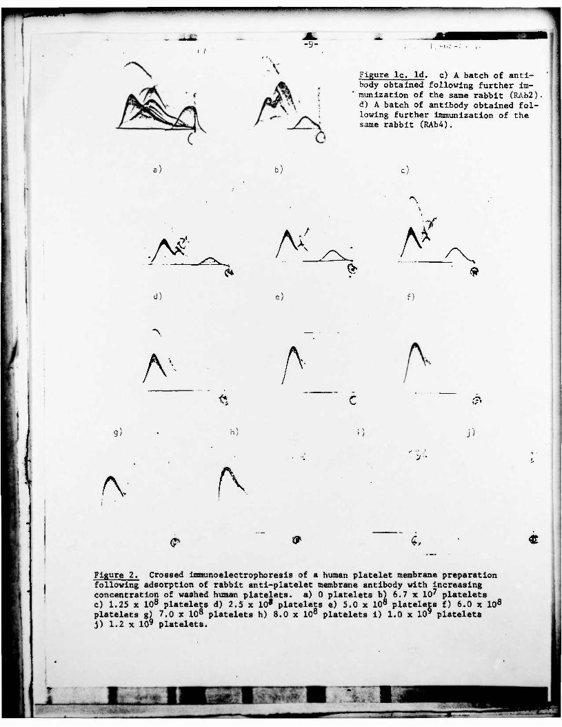
I / -9-
Figure lc. Id. c) A batch of anti- body obtained following further im- munization of the same rabbit (RAb2). d) A batch of antibody obtained fol- lowing further immunization of the same rabbit (RAb4).
a)
"\
.A* £ A< &
A % /
& 9
d) c) f)
"\
A"
9) h) 0 J)
I i\ A. >••
<P S,
Figure 2. Crossed immunoelectrophoresis of a human platelet membrane preparation following adsorption of rabbit anti-platelet membrane antibody with increasing concentration of washed human platelets, a) 0 platelets b) 6.7 x 10⁷ platelets c) 1.25 x 10⁸ platelets d) 2.5 x 10* platelets e) 5.0 x 10⁸ platelets f) 6.0 x 10⁸
platelets g) 7.0 x 10⁸ platelets h) 8.0 x 10⁸ platelets i) 1.0 x 10' platelets J) 1.2 x 10⁹ platelets.
r JL
=E

T5
•j) b)
•10-
c) liAO/i I/-(.n-('-,':|i, I
A. •.•*%w,.'rS£ÄKi*' *5r v
F i cjre 3 . Crossed imrounoel ectrophores i s of a) human fibrinogen (0.12 agm) ; b) platelet membrane preparation; c) fibrinogen plus platelet membrane preparation, This CIE was run for a shorter time interval in the first dimension.
.) b) c)
'--&
Fjoure U• Crossed immunoelectrophoresis of a) human albumin lO.l .:gm): b) p1äte 1 • ••nenbrane prepared en; c) albumin plus platelet membrane preparation.
.-,) b) c)
T ^* »<r-
;^\
/v c.
\r
F i q u r c 5 • Crossed immunoeIectrophoresis of a human platelet membrane with rabbit anti-pI a to Ict membrane antibody adsorbed with washed platelets from a patient with
anzmann's thrcmbasthenia. a) unabsorb.id antibody: b) antibodv adsorbed with C 1 adsorbed wi
0- platelets from a patient with Glanzmann's thromba s thc-n i a; c) antibody th x 10- platelets from a normal subject.
in» •-,• .>• •* ». • *' -ii*.
I li iT

• II' iihiiA i /-(,;',-1 -;;i u i
I
•0
\
A
r* >v
c
i0
AA c
.1)
Figure 6. Crossed immunoelectrophoresis of a human platelet membrane preparation before and after treatment of the original washed platelets with neuraminidase• a) Washed platelets (8 x 10⁸) treated with buffer: b) Washed platelets treated with neuraminidase, 2 units/ml. for one hour at 37°C. C1E of a human platelet membrane preparation before and after treatment of original washed platelets with a-chymotrypsin: c) 8 x 10^ washed platelets treated with buffer: d) Washed platelets treated with lmg/ml chymotryps in for 20 min at 22°C. (Hazy peaks in background are artifacts ?due to fast running proteins in first dimension which enter buffer and cross-react with antibody in second dimension.
a)
^^
b)
mv
Q> K®
1
:
[ j 'ii""1, 7 • Crossed immunoelectrophoresis of a human platelet membrane preparation In the pretence of <•> spacer gel (inserted with ihe second dimension <0 containing ofjnrosc -iliwv*: i>) containing coneanaval in A {?\0 f/g/ml) in agnrosc. Mute ontic,ens pullcil down into l^e lectin. (Similar artifacts noted, as in Figure 6).
f.)

.1?. Simon Kdrpätkiiij M.D. OAOA 17-68-C-3163
700 -
600 -
2
u
< oc O
cc O ü
o CM CO
500 -
400 -
V \
I 10 20 30 40 50 60 70
SI ice Number
set of NaF, dibutyrl cyclic AMP, and prostaglandin E| on platelets ^P0r. Washed platelets were preincubated with I mc/ml *P0r for
Figure 8A. Effec. preloaded with JiP0^. Washed platelets were preincubated with I mc/mi 30 minutes and then washed twice in tris-buffered saline, pH 7A (see Methods) . The platelets were then resuspended in buffer containing either buffer (* H or 10 mM NaF (o—o), and incubated for 30 min (all *+ incubations were performed with rh« «am* batch of platelets). The cell sao was prepared and electroohoresed on a 10t SDS polyacrylamide gel.

wmm
I L Simon Korpotkin, M.D. DADA 17-68-C-8163
400 -
300
200
100
B.
- 10
J L
0.1 mM CAMP
' ' ' L J I I L
10 20 30 40 50 60
SI ice Number
70
Figure 8B. Effect of NaF, dibutyrt cyclic AMP, and prostaglandin E| on platelets preloaded with ^PO^. Washed platelets were preincibated with l mc/ml P0i+ for 30 minutes and then washed twice in tris-buffered saline, pH 7.^ (see Methods). The platelets were then resuspended in buffer containing 0.1 mM dibutyrl cyclic AMP (•—• ), and incubated for 30 min (all k incubations were performed with the same batch of platelets). The cell sap was prepared and electrophoresed on a 10% SDS polyacrylamide gel.

"
.IM. Simon Karpatkin, M.D. DADA I7-63-C-8I63
1 400
200
J I I L J 1 I L '''''
10 m o c p* > 33
m ö
o
T
10 20 30 40 50 60 70
Slice Number
Figure 8C. Effect of NaF, dibutyrl cyclic AMP, and prosgalandin E ] on plate I lets preloaded with -^PO^. Washed platelets were preincubated with l mc/ml "izP0/+ for 30 minutes and then washed twice in tris-buffered saline, pH 7.k (see Methods). The platelets were then resuspended in buffer containing 10 urn prostagland in E] (•—•) and incubated for 30 min (all k incubations were performed with the same batch of platelets). The cell sap was prepared and electrophoresed on a 10% SDS polyacrylamide gel.
i
r
-15-
2 Q- O
z c
o Q- ac O o z
o CL
(N CD
It •>
,
Simon Karpatkin, M.O. DADA 17-68-C-8I63
Figure 9A• latelets pre- PO^ for 30
70 JO 40 SO
Slice Number
Effect of colchicine and 2',5'-dideoxyadenosine (DDA) on loaded with PO, . Washed platelets were preincubated with 1 mc/ml 3 . min and then washed twice in tris-buffered saline, pH 7.k (see Methods). The plate lets were then resuspended in buffer containing either buffer (•—») or colchicine at 10"/M (6—o) and incubated for 30 min (all k incubations were performed with the same batch of platelets). The cell sap was prepared and electrophoresed on a 10% SOS pol vacrvl amide o«*l .

rr . if.. Simon Kürpatkin, M.O.
DADA I7-68-C-8I63 1
20 30 <0 SO
Slice Number «0 70
Figure 9B. Effect of colchicine and 2',5'-dideoxyadenosine (DDA) on platelets pre- loaded wi th 32 PO, Washed platelets were preincubated with 1 mc/ml 'P0Y for 30 min and then washed twice in tris-buffered saline, pH 7-*+ (see Methods)." The plate- lets were then rcsuspended in buffer containing 0.1 mM dideoxyadenosine and incubated for 30 min (all *i incubations were performed with the same batch of platelets). The ' cell sap was prepared and electrophoresed on a 10% SDS polyacrylamide gel.
• —

r ""-
- 11
1
i 1
•
1 r. *
i
I
Simon Korpcilkin, M.O. DAOA 17-68-C-8163
m o c
ID IT
o CO
' I
20 30 40 SO 60 Slice Number
Figure 9C Effect of colchicine and 21,5'-dideoxyadenosine (DOA) on platelets pre loaded with •^2P0i+. Washed platelets were preincubated with 1 mc/ml -*2po^ for 30 min and then washed twice in tris-buffered saline, pH 7-^ (see Methods). Th? plate- lets were then resuspended in buffer containing colchicine at 10"'M plus dideoxyade- nosine, and incubated for 30 min (all k incubations were performed with the same batch of platelets). The cell sap was prepared and electrophoresed on a 10% SOS polyacrylamide gel.