I. Cell Theory II. Studying cells III. Prokaryotic vs ... 120 Lectures/NVC... · 1 Chapter 6 - The...
34
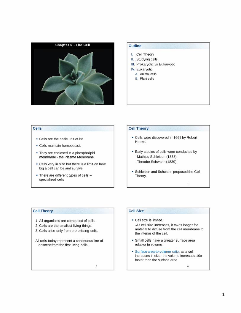
1 Chapter 6 - The Cell Outline I. Cell Theory II. Studying cells III. Prokaryotic vs Eukaryotic IV. Eukaryotic A. Animal cells B. Plant cells Cells Cells are the basic unit of life Cells maintain homeostasis They are enclosed in a phospholipid membrane - the Plasma Membrane Cells vary in size but there is a limit on how big a cell can be and survive There are different types of cells – specialized cells 4 Cell Theory Cells were discovered in 1665 by Robert Hooke. Early studies of cells were conducted by - Mathias Schleiden (1838) - Theodor Schwann (1839) Schleiden and Schwann proposed the Cell Theory. Cell Theory 1. All organisms are composed of cells. 2. Cells are the smallest living things. 3. Cells arise only from pre-existing cells. All cells today represent a continuous line of descent from the first living cells. 5 6 Cell Size Cell size is limited. -As cell size increases, it takes longer for material to diffuse from the cell membrane to the interior of the cell. Small cells have a greater surface area relative to volume Surface area-to-volume ratio: as a cell increases in size, the volume increases 10x faster than the surface area
Transcript of I. Cell Theory II. Studying cells III. Prokaryotic vs ... 120 Lectures/NVC... · 1 Chapter 6 - The...
1
Chapter 6 - The Cell Outline
I. Cell TheoryII. Studying cellsIII. Prokaryotic vs EukaryoticIV. Eukaryotic
A. Animal cellsB. Plant cells
Cells
Cells are the basic unit of life
Cells maintain homeostasis
They are enclosed in a phospholipid membrane - the Plasma Membrane
Cells vary in size but there is a limit on how big a cell can be and survive
There are different types of cells –specialized cells
4
Cell Theory
Cells were discovered in 1665 by Robert Hooke.
Early studies of cells were conducted by- Mathias Schleiden (1838)- Theodor Schwann (1839)
Schleiden and Schwann proposed the Cell Theory.
Cell Theory
1. All organisms are composed of cells.2. Cells are the smallest living things.3. Cells arise only from pre-existing cells.
All cells today represent a continuous line of descent from the first living cells.
5 6
Cell Size
Cell size is limited.-As cell size increases, it takes longer for material to diffuse from the cell membrane to the interior of the cell.
Small cells have a greater surface area relative to volume
Surface area-to-volume ratio: as a cell increases in size, the volume increases 10x faster than the surface area
2
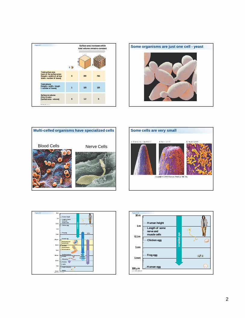
Surface area increases whiletotal volume remains constant
Total surface area[sum of the surface areas(height width) of all boxsides number of boxes]
Total volume[height width length number of boxes]
Surface-to-volume(S-to-V) ratio[surface area volume]
1
5
6 150 750
1
1251251
1.26 6
Figure 6.7 Some organisms are just one cell - yeast
Multi-celled organisms have specialized cells
Blood Cells Nerve Cells
Some cells are very small
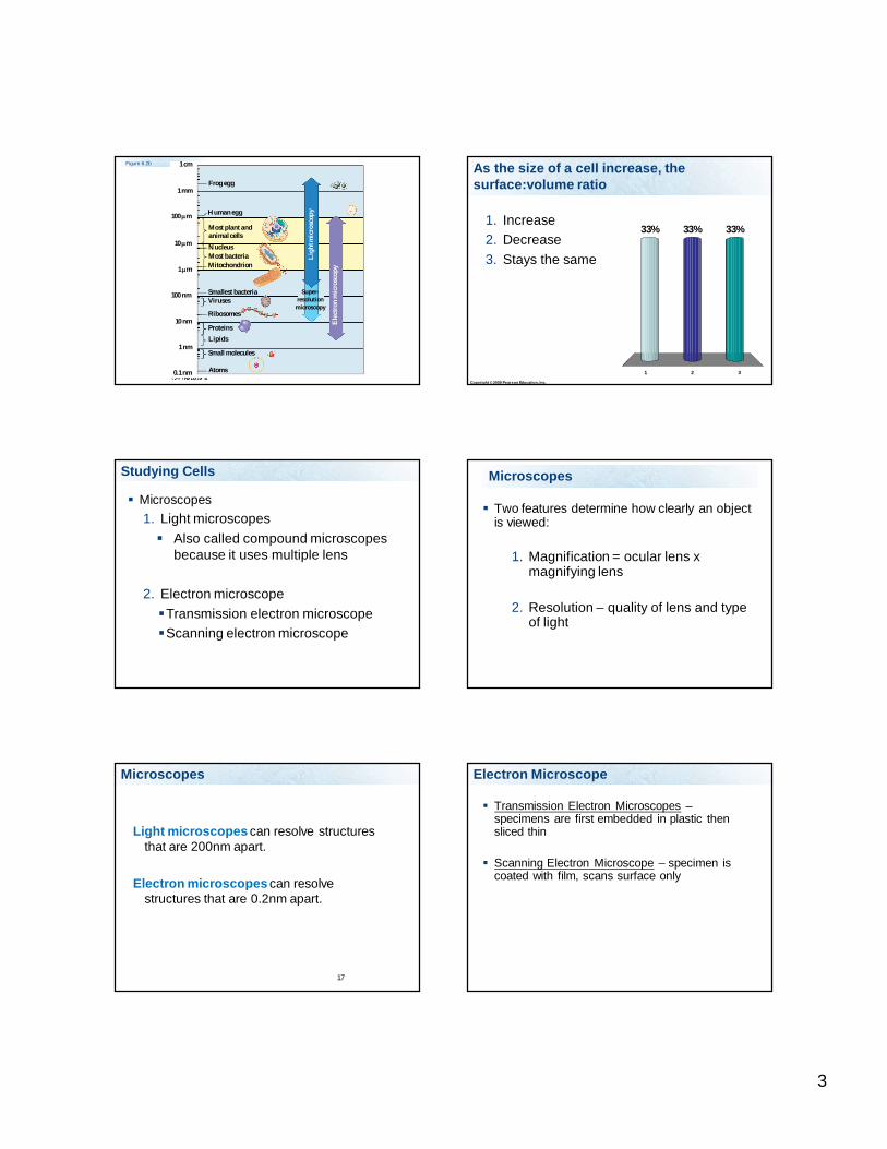
Figure 6.2 10 m
1 m
0.1 m
1 cm
1 mm
100 m
10 m
1 m
100 nm
10 nm
1 nm
0.1 nm Atoms
Small molecules
Lipids
Proteins
Ribosomes
VirusesSmallest bacteria
MitochondrionMost bacteriaNucleus
Most plant andanimal cells
Human egg
Frog egg
Chicken egg
Length of somenerve andmuscle cells
Human height
Una
ided
eye
Ligh
t mic
rosc
opy
Ele
ctro
n m
icro
scop
y
Super-resolution
microscopy
10 m
1 m
0.1 m
1 cm
1 mm
100 m Human egg
Frog egg
Chicken egg
Length of somenerve andmuscle cells
Human height
Una
ided
eye
Figure 6.2a
3
Figure 6.2b
1 mm
100 m
10 m
1 m
100 nm
10 nm
1 nm
0.1 nm Atoms
Small molecules
LipidsProteins
Ribosomes
VirusesSmallest bacteria
MitochondrionMost bacteriaNucleus
Most plant andanimal cells
Human egg
Ligh
t mic
rosc
opy
Ele
ctro
n m
icro
scop
y
Super-resolution
microscopy
1 cm
Frog egg
Copyright © 2009 Pearson Education, Inc.
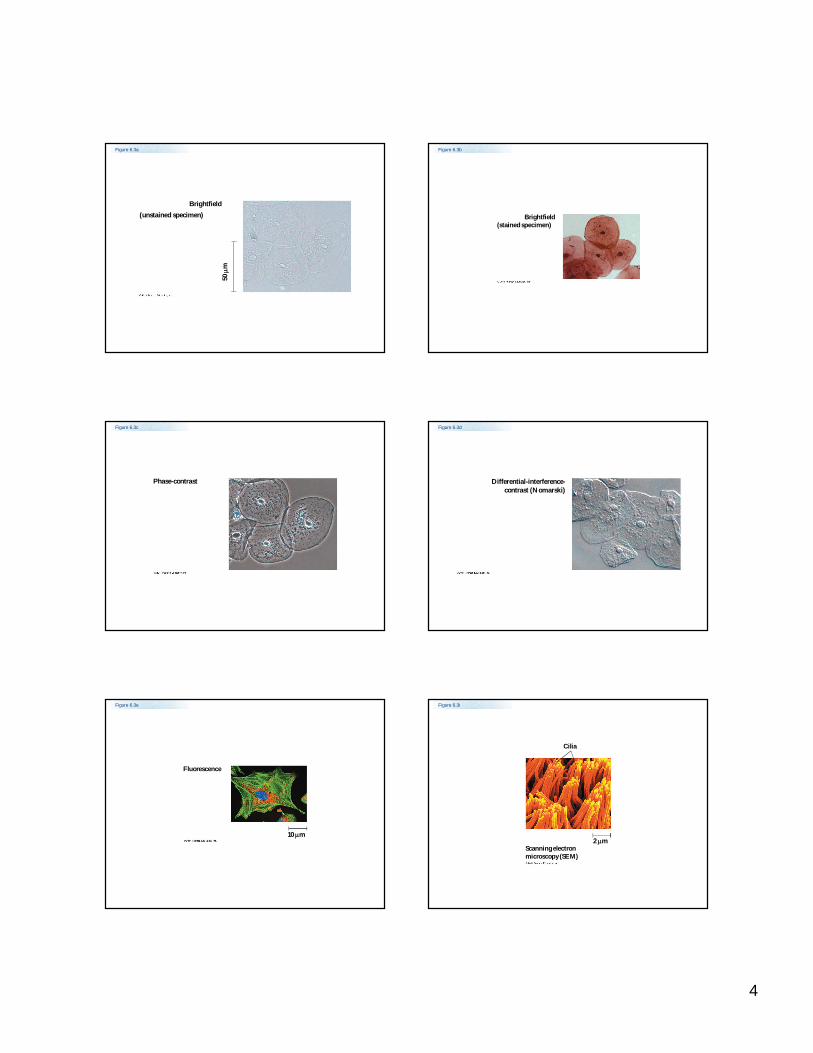
As the size of a cell increase, the surface:volume ratio
1 2 3
33% 33%33%1. Increase2. Decrease3. Stays the same
Studying Cells
Microscopes1. Light microscopes Also called compound microscopes
because it uses multiple lens
2. Electron microscope Transmission electron microscopeScanning electron microscope
Microscopes
Two features determine how clearly an object is viewed:
1. Magnification = ocular lens x magnifying lens
2. Resolution – quality of lens and type of light
17
Microscopes
Light microscopes can resolve structures that are 200nm apart.
Electron microscopes can resolve structures that are 0.2nm apart.
Electron Microscope
Transmission Electron Microscopes –specimens are first embedded in plastic then sliced thin
Scanning Electron Microscope – specimen is coated with film, scans surface only
4
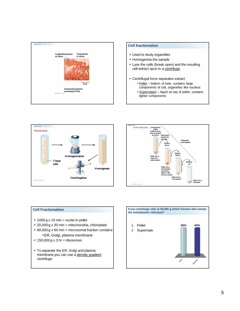
Figure 6.3a
(unstained specimen)50
m
Brightfield
Figure 6.3b
Brightfield(stained specimen)
Figure 6.3c
Phase-contrast
Figure 6.3d
Differential-interference-contrast (Nomarski)
Figure 6.3e
Fluorescence
10 m
Figure 6.3i
Cilia
2 mScanning electronmicroscopy (SEM)
5
Figure 6.3j
Longitudinal sectionof cilium
Cross sectionof cilium
2 m
Transmission electronmicroscopy (TEM)
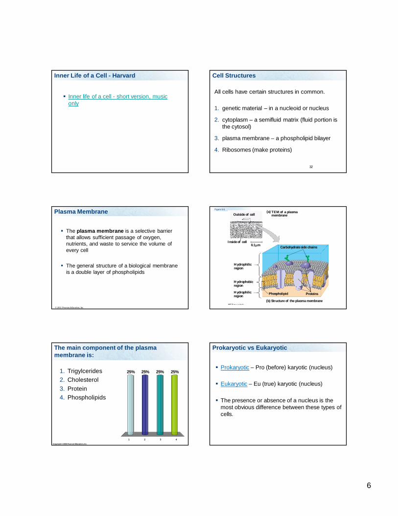
Cell fractionation
Used to study organelles Homogenize the sample Lyse the cells (break open) and the resulting
cell extract spun in a centrifuge
Centrifugal force separates extractPellet – bottom of tube, contains large
components of cell, organelles like nucleusSupernatant – liquid on top of pellet, contains
lighter components
Figure 6.4a
TECHNIQUE
Homogenization
Tissuecells
Homogenate
Centrifugation
Differentialcentrifugation
Centrifuged at1,000 g
(1,000 times theforce of gravity)
for 10 min Supernatantpoured intonext tube
20 min
60 minPellet rich innuclei andcellular debris
3 hr
Pellet rich inmitochondria(and chloro-plasts if cellsare from a plant)
Pellet rich in“microsomes”
Pellet rich inribosomes
20,000 g
80,000 g
150,000 g
TECHNIQUE (cont.)Figure 6.4b
Cell Fractionation
1000 g x 10 min = nuclei in pellet 20,000 g x 30 min = mitochondria, chloroplast 80,000 g x 60 min = microsomal fraction contains:
ER, Golgi, plasma membrane 150,000 g x 3 hr = ribosomes
To separate the ER, Golgi and plasma membrane you can use a density gradientcentrifuge
If you centrifuge cells at 80,000 g which fraction will contain the endoplasmic reticulum?
1. Pellet2. Supernate
Pellet
Supernate
50%50%
6
Inner Life of a Cell - Harvard
Inner life of a cell - short version, music only
32
Cell Structures
All cells have certain structures in common.
1. genetic material – in a nucleoid or nucleus
2. cytoplasm – a semifluid matrix (fluid portion is the cytosol)
3. plasma membrane – a phospholipid bilayer
4. Ribosomes (make proteins)
The plasma membrane is a selective barrier that allows sufficient passage of oxygen, nutrients, and waste to service the volume of every cell
The general structure of a biological membrane is a double layer of phospholipids
© 2011 Pearson Education, Inc.
Plasma Membrane Figure 6.6
Outside of cell
Inside of cell0.1 m
(a) TEM of a plasmamembrane
Hydrophilicregion
Hydrophobicregion
Hydrophilicregion
Carbohydrate side chains
ProteinsPhospholipid
(b) Structure of the plasma membrane
Copyright © 2009 Pearson Education, Inc.
The main component of the plasma membrane is:
1 2 3 4
25% 25%25%25%1. Trigylcerides2. Cholesterol3. Protein4. Phospholipids
Prokaryotic vs Eukaryotic
Prokaryotic – Pro (before) karyotic (nucleus)
Eukaryotic – Eu (true) karyotic (nucleus)
The presence or absence of a nucleus is the most obvious difference between these types of cells.
7
Eukaryotic cells have internal membranes that compartmentalize their functions
The basic structural and functional unit of every organism is one of two types of cells: prokaryotic or eukaryotic
Only organisms of the domains Bacteria and Archaea consist of prokaryotic cells
Protists, fungi, animals, and plants all consist of eukaryotic cells
© 2011 Pearson Education, Inc.
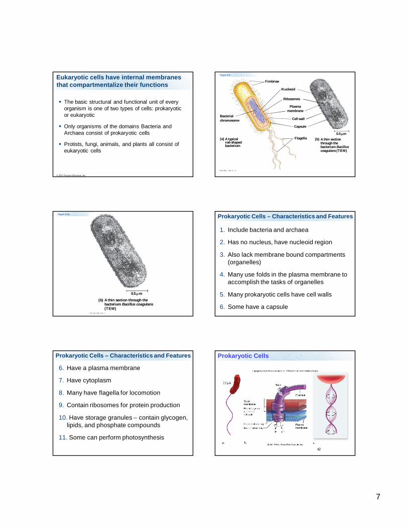
Fimbriae
Bacterialchromosome
A typicalrod-shapedbacterium
(a)
Nucleoid
Ribosomes
Plasmamembrane
Cell wall
Capsule
Flagella A thin sectionthrough thebacterium Bacilluscoagulans (TEM)
(b)0.5 m
Figure 6.5
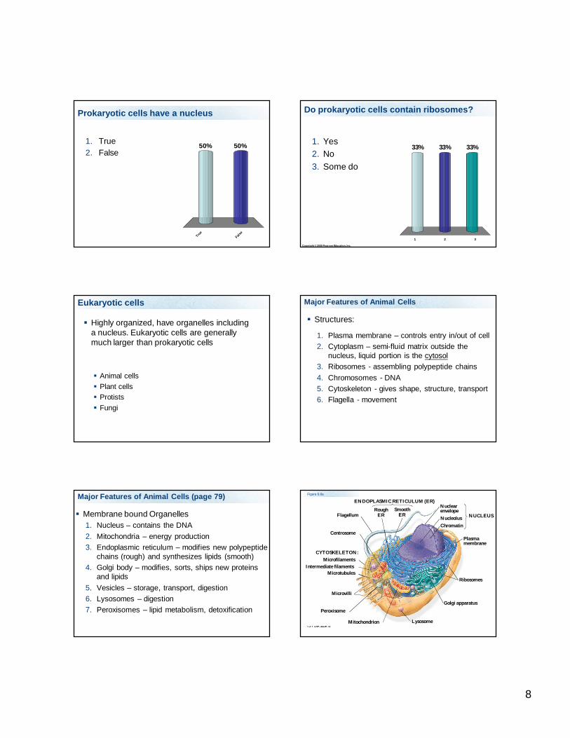
Figure 6.5a
A thin section through thebacterium Bacillus coagulans(TEM)
(b)
0.5 m
Prokaryotic Cells – Characteristics and Features
1. Include bacteria and archaea
2. Has no nucleus, have nucleoid region
3. Also lack membrane bound compartments (organelles)
4. Many use folds in the plasma membrane to accomplish the tasks of organelles
5. Many prokaryotic cells have cell walls
6. Some have a capsule
Prokaryotic Cells – Characteristics and Features
6. Have a plasma membrane
7. Have cytoplasm
8. Many have flagella for locomotion
9. Contain ribosomes for protein production
10. Have storage granules – contain glycogen, lipids, and phosphate compounds
11. Some can perform photosynthesis
42
Prokaryotic Cells
8
Prokaryotic cells have a nucleus
1. True2. False
True
False
50%50%
Copyright © 2009 Pearson Education, Inc.
Do prokaryotic cells contain ribosomes?
1 2 3
33% 33%33%1. Yes2. No3. Some do
Eukaryotic cells
Highly organized, have organelles including a nucleus. Eukaryotic cells are generally much larger than prokaryotic cells
Animal cells Plant cells Protists Fungi
Major Features of Animal Cells
Structures:
1. Plasma membrane – controls entry in/out of cell2. Cytoplasm – semi-fluid matrix outside the
nucleus, liquid portion is the cytosol3. Ribosomes - assembling polypeptide chains4. Chromosomes - DNA5. Cytoskeleton - gives shape, structure, transport6. Flagella - movement
Major Features of Animal Cells (page 79)
Membrane bound Organelles 1. Nucleus – contains the DNA 2. Mitochondria – energy production 3. Endoplasmic reticulum – modifies new polypeptide
chains (rough) and synthesizes lipids (smooth) 4. Golgi body – modifies, sorts, ships new proteins
and lipids5. Vesicles – storage, transport, digestion6. Lysosomes – digestion7. Peroxisomes – lipid metabolism, detoxification
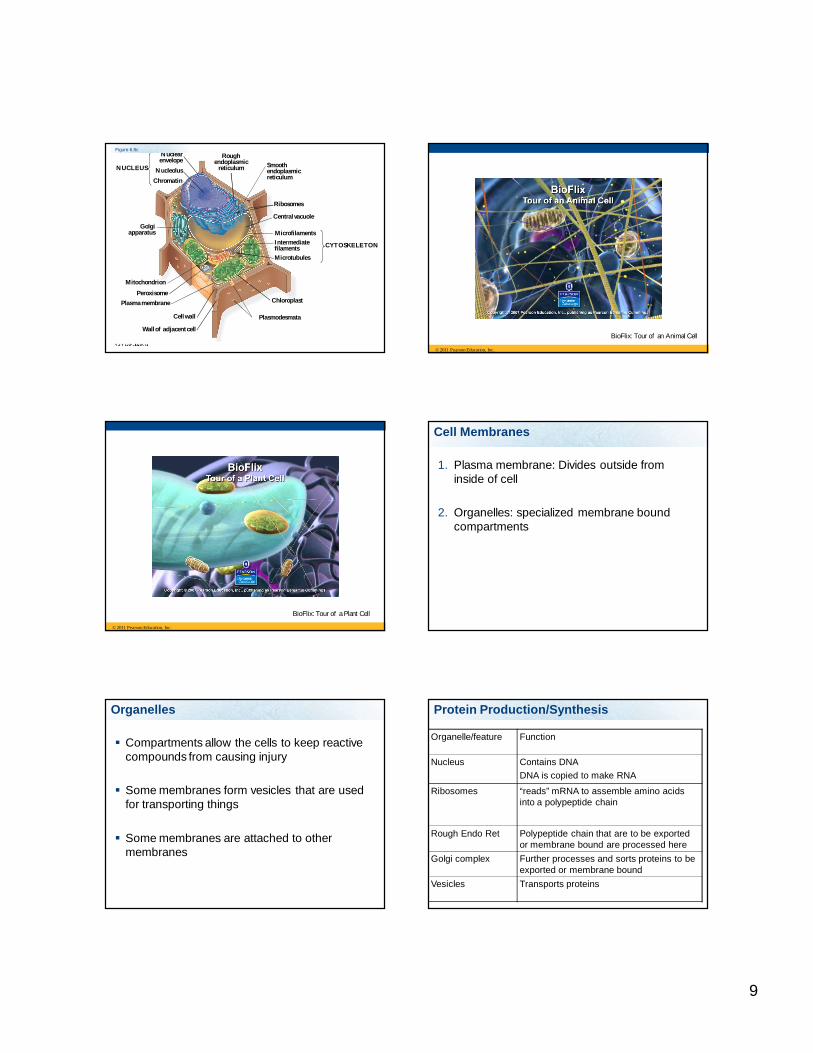
Figure 6.8a
ENDOPLASMIC RETICULUM (ER)
RoughER
SmoothER
NuclearenvelopeNucleolusChromatin
Plasmamembrane
Ribosomes
Golgi apparatus
LysosomeMitochondrion
Peroxisome
Microvilli
MicrotubulesIntermediate filaments
Microfilaments
Centrosome
CYTOSKELETON:
Flagellum NUCLEUS
9
NUCLEUS
Nuclearenvelope
NucleolusChromatin
Golgiapparatus
Mitochondrion
PeroxisomePlasma membrane
Cell wall
Wall of adjacent cell
Plasmodesmata
Chloroplast
Microtubules
Intermediatefilaments
Microfilaments
CYTOSKELETON
Central vacuole
Ribosomes
Smoothendoplasmicreticulum
Roughendoplasmic
reticulum
Figure 6.8c
© 2011 Pearson Education, Inc.
BioFlix: Tour of an Animal Cell
© 2011 Pearson Education, Inc.
BioFlix: Tour of a Plant Cell
Cell Membranes
1. Plasma membrane: Divides outside from inside of cell
2. Organelles: specialized membrane bound compartments
Organelles
Compartments allow the cells to keep reactive compounds from causing injury
Some membranes form vesicles that are used for transporting things
Some membranes are attached to other membranes
Organelle/feature Function
Nucleus Contains DNADNA is copied to make RNA
Ribosomes “reads” mRNA to assemble amino acids into a polypeptide chain
Rough Endo Ret Polypeptide chain that are to be exported or membrane bound are processed here
Golgi complex Further processes and sorts proteins to be exported or membrane bound
Vesicles Transports proteins
Protein Production/Synthesis
10
The eukaryotic cell’s genetic instructions are housed in the nucleus and carried out by the ribosomes
The nucleus contains most of the DNA in a eukaryotic cell
Ribosomes use the information from the DNA to make proteins
© 2011 Pearson Education, Inc.
Nucleus
Nucleus protects DNA
Separates DNA from rest of cell
Place where DNA replicates itself
Place where DNA is copied to make RNA
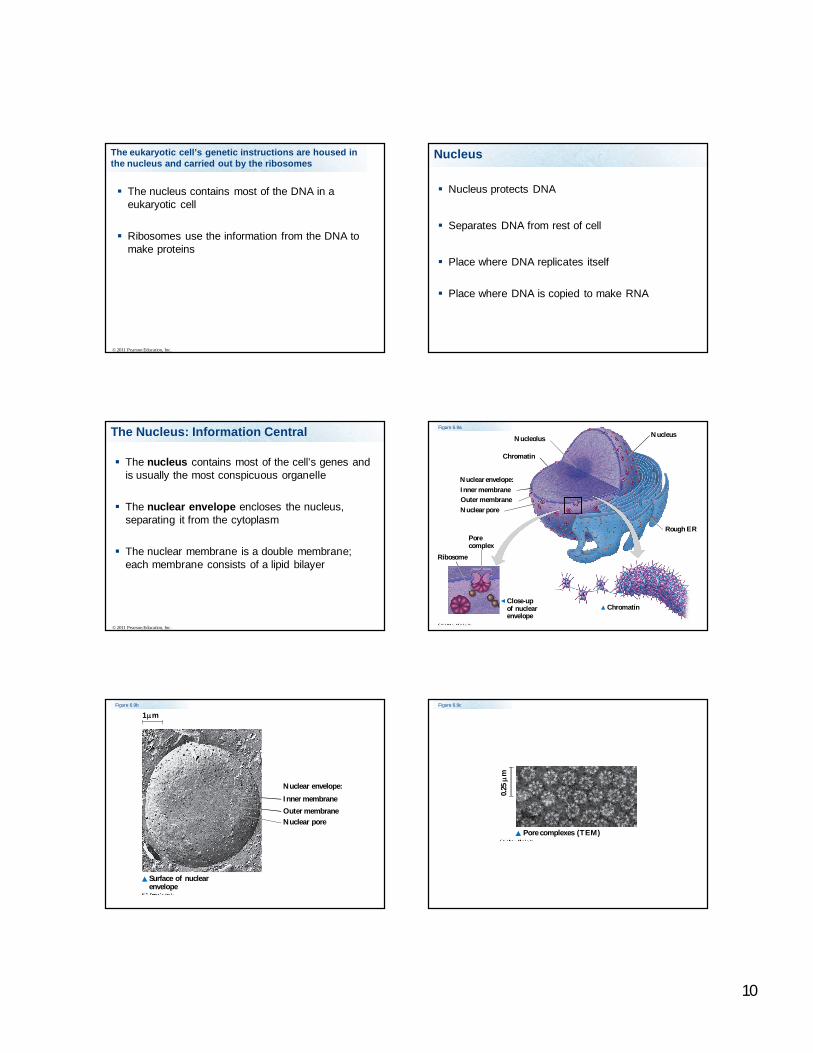
The Nucleus: Information Central
The nucleus contains most of the cell’s genes and is usually the most conspicuous organelle
The nuclear envelope encloses the nucleus, separating it from the cytoplasm
The nuclear membrane is a double membrane; each membrane consists of a lipid bilayer
© 2011 Pearson Education, Inc.
Nucleus
Rough ER
Nucleolus
Chromatin
Nuclear envelope:Inner membraneOuter membraneNuclear pore
Chromatin
Ribosome
Porecomplex
Close-upof nuclearenvelope
Figure 6.9a
Figure 6.9b
Nuclear envelope:
Inner membraneOuter membraneNuclear pore
Surface of nuclearenvelope
1 mFigure 6.9c
Pore complexes (TEM)
0.25
m
11
Pores regulate the entry and exit of molecules from the nucleus
The shape of the nucleus is maintained by the nuclear lamina, which is composed of protein
© 2011 Pearson Education, Inc.
In the nucleus, DNA is organized into discrete units called chromosomes
Each chromosome is composed of a single DNA molecule associated with proteins
The DNA and proteins = histones are together called chromatin
Chromatin condenses to form discrete chromosomes as a cell prepares to divide
The nucleolus is located within the nucleus and is the site of ribosomal RNA (rRNA) synthesis
© 2011 Pearson Education, Inc.
Parts of the Nucleus
2. Nucleolus – dense area in the nucleus is where rRNA are produced and ribosomesare assembled
3. Nucleoplasm – area within nucleus
Ribosomes: Protein Factories
Ribosomes are particles made of ribosomal RNA and protein
Ribosomes carry out protein synthesis in two locations In the cytosol (free ribosomes) On the outside of the endoplasmic reticulum or the
nuclear envelope (bound ribosomes)
© 2011 Pearson Education, Inc.
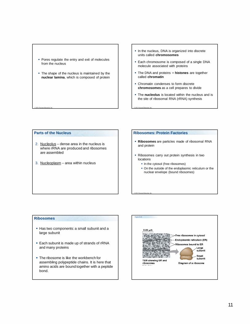
Ribosomes
Has two components: a small subunit and a large subunit
Each subunit is made up of strands of rRNA and many proteins
The ribosome is like the workbench for assembling polypeptide chains. It is here that amino acids are bound together with a peptide bond.
Figure 6.10
0.25 m
Free ribosomes in cytosol
Endoplasmic reticulum (ER)
Ribosomes bound to ERLargesubunit
Smallsubunit
Diagram of a ribosomeTEM showing ER andribosomes
12
The endomembrane system regulates protein traffic and performs metabolic functions in the cell
Components of the endomembrane system Nuclear envelope Endoplasmic reticulum Golgi apparatus Lysosomes Vacuoles Plasma membrane
These components are either continuous or connected via transfer by vesicles
© 2011 Pearson Education, Inc.
The Endoplasmic Reticulum: Biosynthetic Factory
The endoplasmic reticulum (ER) accounts for more than half of the total membrane in many eukaryotic cells
The ER membrane is continuous with the nuclear envelope
There are two distinct regions of ER Smooth ER, which lacks ribosomes Rough ER, surface is studded with ribosomes
© 2011 Pearson Education, Inc.
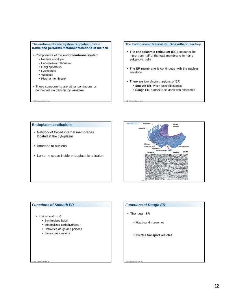
Endoplasmic reticulum
Network of folded internal membranes located in the cytoplasm
Attached to nucleus
Lumen = space inside endoplasmic reticulum
Figure 6.11 Smooth ER
Rough ER
ER lumen
CisternaeRibosomes
Smooth ERTransport vesicle
Transitional ER
Rough ER 200 nm
Nuclearenvelope
Functions of Smooth ER
The smooth ER Synthesizes lipids Metabolizes carbohydrates Detoxifies drugs and poisons Stores calcium ions
© 2011 Pearson Education, Inc.
Functions of Rough ER
The rough ER
Has bound ribosomes
Creates transport vesicles
© 2011 Pearson Education, Inc.
13
Rough Endoplasmic Reticulum (RER)
Ribosomes that are producing polypeptide chains for export or to be embedded in membranes dock with the surface of the RER
The growing polypeptide chain enters the lumen of the RER
Functions of Rough Endoplasmic Reticulum (RER)
In the RER the polypetide chain is folded
Enzymes called molecular chaperones aid in the folding of the polypeptide chains into proteins
Some of the polypeptide chains may get modified here “tagged” with carbohydrate chain = glycoproteins
The polypeptide chains/proteins are put into transport vesicles
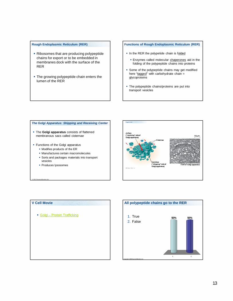
The Golgi apparatus consists of flattened membranous sacs called cisternae
Functions of the Golgi apparatus Modifies products of the ER Manufactures certain macromolecules Sorts and packages materials into transport
vesicles Produces lysosomes
The Golgi Apparatus: Shipping and Receiving Center
© 2011 Pearson Education, Inc.
Figure 6.12
cis face(“receiving” side ofGolgi apparatus)
trans face(“shipping” side ofGolgi apparatus)
0.1 m
TEM of Golgi apparatus
Cisternae
V Cell Movie
Golgi – Protein Trafficking
Copyright © 2009 Pearson Education, Inc.
All polypeptide chains go to the RER
1 2
50%50%1. True2. False
14
Protein Production - Overview1. DNA in the nucleus are the instructions for making
protein
2. A copy of the DNA is made = mRNA
3. mRNA leaves the nucleus
4. mRNA docks with a ribosome to assemble a chain of amino acids.
5. tRNA brings amino acids to ribosomes
6. At the ribosome the amino acids are linked together with a peptide bond to form a polypeptide chain
Protein Production Cont
7. Ribosome with the growing polypeptide chain docks with the rough endoplasmic reticulum if the protein is to be exported or embedded in a membrane
8. The polypeptide chain enters the lumen of the RER where they are folded and may get a carbohydrate “tag” attached to it
9. The RER buds off a transport vesicles that can carry the newly formed proteins to the golgi
Protein Production Cont
10. The golgi processes, sorts, packages proteins and lipids from the RER and SER
11. Proteins that are exported are shipped in transport vesicles to the plasma membrane
12. Proteins may be put into lysosomes
13. Proteins that are membrane bound are embedded in the transport vesicles membrane
Cytosolic proteins
Proteins that are not shipped out of cell are made on free floating ribosomes
Chaperone proteins fold the proteins in the cytosol
Lysosomes
Produced by the Golgi
Lysosomes are small membrane bound sacs that contain digestive enzymes. The pH is relatively acidic (pH 5) in the lysosomes.
Because the lysosomes are acidic and contain digestive enzymes, their contents must be kept separate from the rest of the cell
Lysosomes
1. Contain strong acids and enzymes
2. Engulf molecules and digest them or
3. Fuse with other organelles and vesicles to destroy them
4. Can fuse with plasma membrane to expel waste
5. Destroy bacteria
15
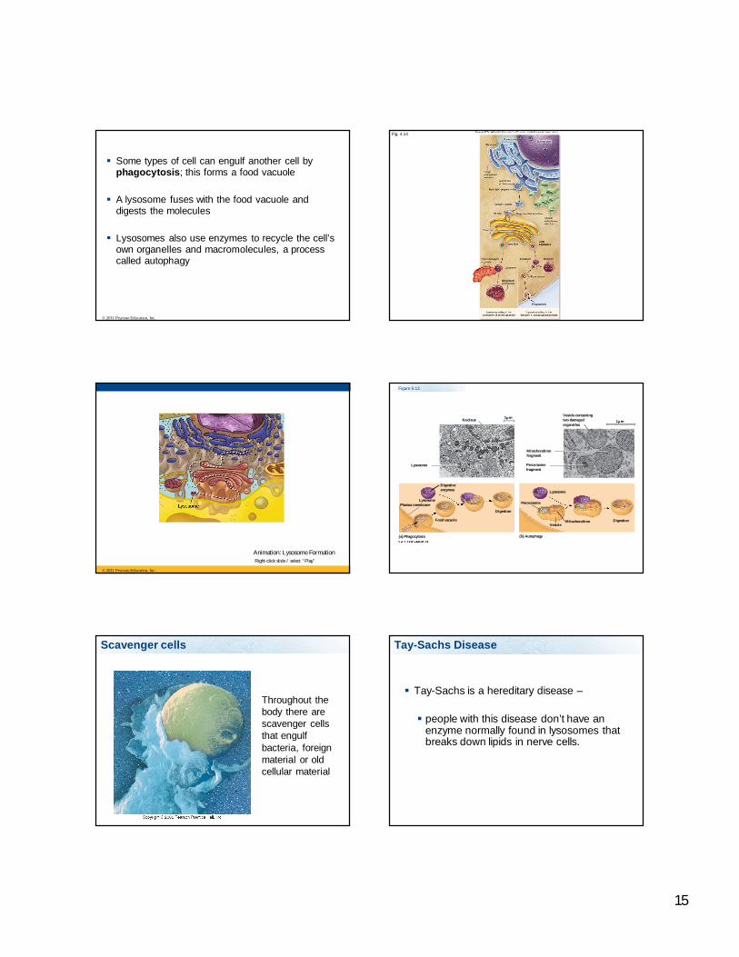
Some types of cell can engulf another cell by phagocytosis; this forms a food vacuole
A lysosome fuses with the food vacuole and digests the molecules
Lysosomes also use enzymes to recycle the cell’s own organelles and macromolecules, a process called autophagy
© 2011 Pearson Education, Inc.
Fig. 4.14
© 2011 Pearson Education, Inc.
Animation: Lysosome FormationRight-click slide / select “Play”
Figure 6.13
Nucleus
Lysosome
1 m
Digestiveenzymes
Digestion
Food vacuole
LysosomePlasma membrane
(a) Phagocytosis
Vesicle containingtwo damagedorganelles
1 m
Mitochondrionfragment
Peroxisomefragment
(b) Autophagy
Peroxisome
VesicleMitochondrion
Lysosome
Digestion
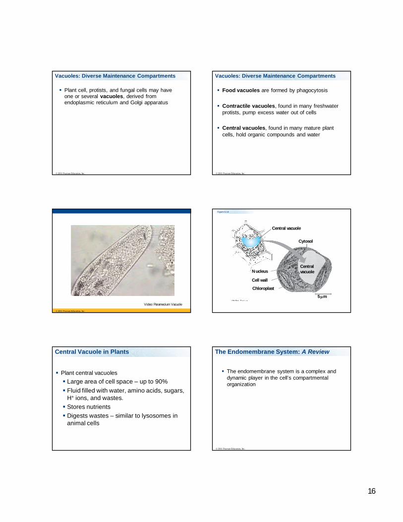
Scavenger cells
Throughout the body there are scavenger cells that engulf bacteria, foreign material or old cellular material
Tay-Sachs Disease
Tay-Sachs is a hereditary disease –
people with this disease don’t have an enzyme normally found in lysosomes that breaks down lipids in nerve cells.
16
Vacuoles: Diverse Maintenance Compartments
Plant cell, protists, and fungal cells may have one or several vacuoles, derived from endoplasmic reticulum and Golgi apparatus
© 2011 Pearson Education, Inc.
Food vacuoles are formed by phagocytosis
Contractile vacuoles, found in many freshwater protists, pump excess water out of cells
Central vacuoles, found in many mature plant cells, hold organic compounds and water
© 2011 Pearson Education, Inc.
Vacuoles: Diverse Maintenance Compartments
© 2011 Pearson Education, Inc.
Video: Paramecium Vacuole
Figure 6.14
Central vacuole
Cytosol
Nucleus
Cell wall
Chloroplast
Centralvacuole
5 m
Central Vacuole in Plants
Plant central vacuoles Large area of cell space – up to 90% Fluid filled with water, amino acids, sugars,
H+ ions, and wastes. Stores nutrients Digests wastes – similar to lysosomes in
animal cells
The Endomembrane System: A Review
The endomembrane system is a complex and dynamic player in the cell’s compartmental organization
© 2011 Pearson Education, Inc.
17
Figure 6.15-1
Smooth ER
Nucleus
Rough ER
Plasmamembrane
Figure 6.15-2
Smooth ER
Nucleus
Rough ER
Plasmamembrane
cis Golgi
trans Golgi
Figure 6.15-3
Smooth ER
Nucleus
Rough ER
Plasmamembrane
cis Golgi
trans Golgi
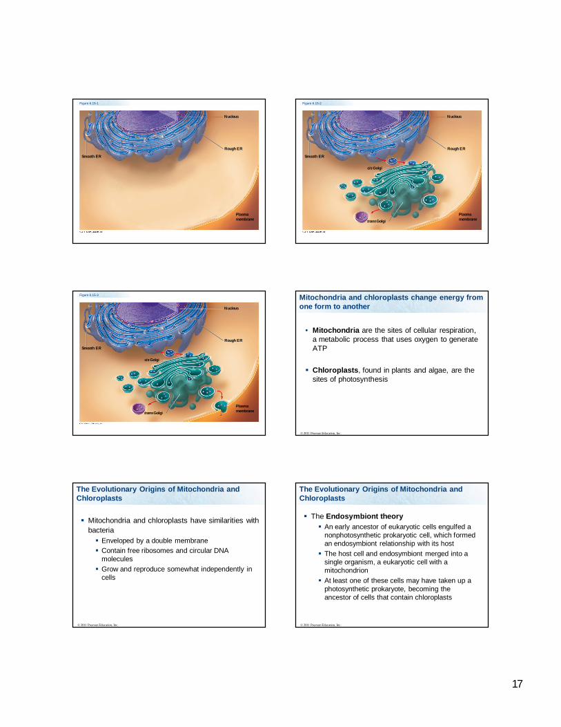
Mitochondria and chloroplasts change energy from one form to another
• Mitochondria are the sites of cellular respiration, a metabolic process that uses oxygen to generate ATP
Chloroplasts, found in plants and algae, are the sites of photosynthesis
© 2011 Pearson Education, Inc.
Mitochondria and chloroplasts have similarities with bacteria Enveloped by a double membrane Contain free ribosomes and circular DNA
molecules Grow and reproduce somewhat independently in
cells
© 2011 Pearson Education, Inc.
The Evolutionary Origins of Mitochondria and Chloroplasts
The Endosymbiont theory An early ancestor of eukaryotic cells engulfed a
nonphotosynthetic prokaryotic cell, which formed an endosymbiont relationship with its host The host cell and endosymbiont merged into a
single organism, a eukaryotic cell with a mitochondrion At least one of these cells may have taken up a
photosynthetic prokaryote, becoming the ancestor of cells that contain chloroplasts
© 2011 Pearson Education, Inc.
The Evolutionary Origins of Mitochondria and Chloroplasts
18
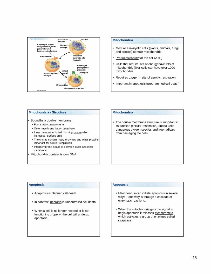
NucleusEndoplasmicreticulum
Nuclear envelope
Ancestor ofeukaryotic cells(host cell)
Engulfing of oxygen-using nonphotosyntheticprokaryote, whichbecomes a mitochondrion
Mitochondrion
Nonphotosyntheticeukaryote
Mitochondrion
At leastone cell
Photosynthetic eukaryote
Engulfing ofphotosyntheticprokaryote
Chloroplast
Figure 6.16 Mitochondria
Most all Eukaryotic cells (plants, animals, fungi and protists) contain mitochondria
Produces energy for the cell (ATP)
Cells that require lots of energy have lots of mitochondria (liver cells can have over 1000 mitochondria
Requires oxygen = site of aerobic respiration
Important in apoptosis (programmed cell death)
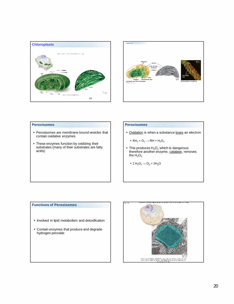
Mitochondria - Structure
Bound by a double membrane Forms two compartments Outer membrane faces cytoplasm Inner membrane folded forming cristae which
increases surface area The cristae contain many enzymes and other proteins
important for cellular respiration Intermembrane space is between outer and inner
membrane Mitochondria contain its own DNA
Mitochondria
The double membrane structure is important in its function (cellular respiration) and to keep dangerous oxygen species and free radicals from damaging the cells.
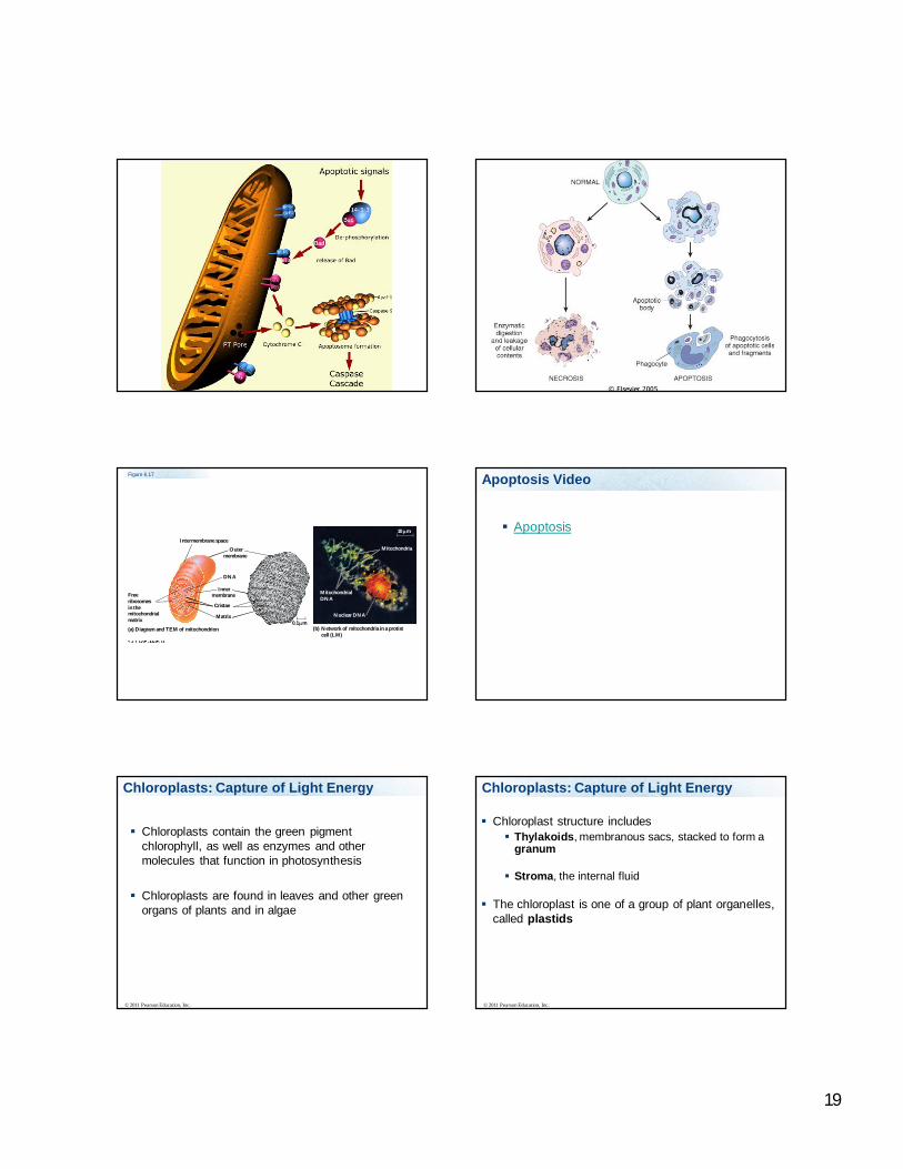
Apoptosis
Apoptosis is planned cell death
In contrast, necrosis is uncontrolled cell death
When a cell is no longer needed or is not functioning properly, the cell will undergo apoptosis.
Apoptosis
Mitochondria can initiate apoptosis in several ways – one way is through a cascade of enzymatic reactions.
When the mitochondria gets the signal to begin apoptosis it releases cytochrome c, which activates a group of enzymes called caspases
19
Figure 6.17
Intermembrane space
Outermembrane
DNA
Innermembrane
Cristae
Matrix
Freeribosomesin themitochondrialmatrix
(a) Diagram and TEM of mitochondrion (b) Network of mitochondria in a protistcell (LM)
0.1 m
MitochondrialDNA
Nuclear DNA
Mitochondria
10 m
Apoptosis Video
Apoptosis
Chloroplasts: Capture of Light Energy
Chloroplasts contain the green pigment chlorophyll, as well as enzymes and other molecules that function in photosynthesis
Chloroplasts are found in leaves and other green organs of plants and in algae
© 2011 Pearson Education, Inc.
Chloroplast structure includes Thylakoids, membranous sacs, stacked to form a
granum
Stroma, the internal fluid
The chloroplast is one of a group of plant organelles, called plastids
© 2011 Pearson Education, Inc.
Chloroplasts: Capture of Light Energy
20
115
Chloroplasts Figure 6.18
RibosomesStroma
Inner and outermembranes
Granum
1 mIntermembrane spaceThylakoid(a) Diagram and TEM of chloroplast (b) Chloroplasts in an algal cell
Chloroplasts(red)
50 m
DNA
Peroxisomes
Peroxisomes are membrane bound vesicles that contain oxidative enzymes
These enzymes function by oxidizing their substrates (many of their substrates are fatty acids)
Peroxisomes
Oxidation is when a substance loses an electron
RH2 + O2 → RH + H2O2
This produces H2O2 which is dangerous therefore another enzyme, catalase, removes the H2O2
2 H2O2 → O2 + 2H2O
Functions of Peroxisomes
Involved in lipid metabolism and detoxification
Contain enzymes that produce and degrade hydrogen peroxide
Fig. 4.15
21
What organelle produces energy (ATP)?
1 2 3 4 5
20% 20% 20%20%20%1. Ribosomes2. Golgi complex3. Mitochondria4. SER5. Lysosomes
Where are polypeptide chains assembled?
1 2 3 4
25% 25%25%25%1. Ribosomes2. Golgi complex3. SER4. RER
Where are lipids synthesized?
1 2 3 4 5
20% 20% 20%20%20%1. Ribosomes2. Golgi complex3. Peroxisomes4. SER5. Lysosomes
These are membrane bound sacs with digestive enzymes
1 2 3 4 5
20% 20% 20%20%20%1. Ribosomes2. Golgi complex3. Mitochondria4. SER5. Lysosomes
Inner Life of a Cell - Harvard
Inner Life of a Cell Narrated, long version
Cytoskeleton
Interconnected system of fibers and lattices
Gives cells their organization, shape, ability to move, transport things in cell, important in cell division
Some permanent others only present when needed Microtubules Microfilaments Intermediate filaments
22
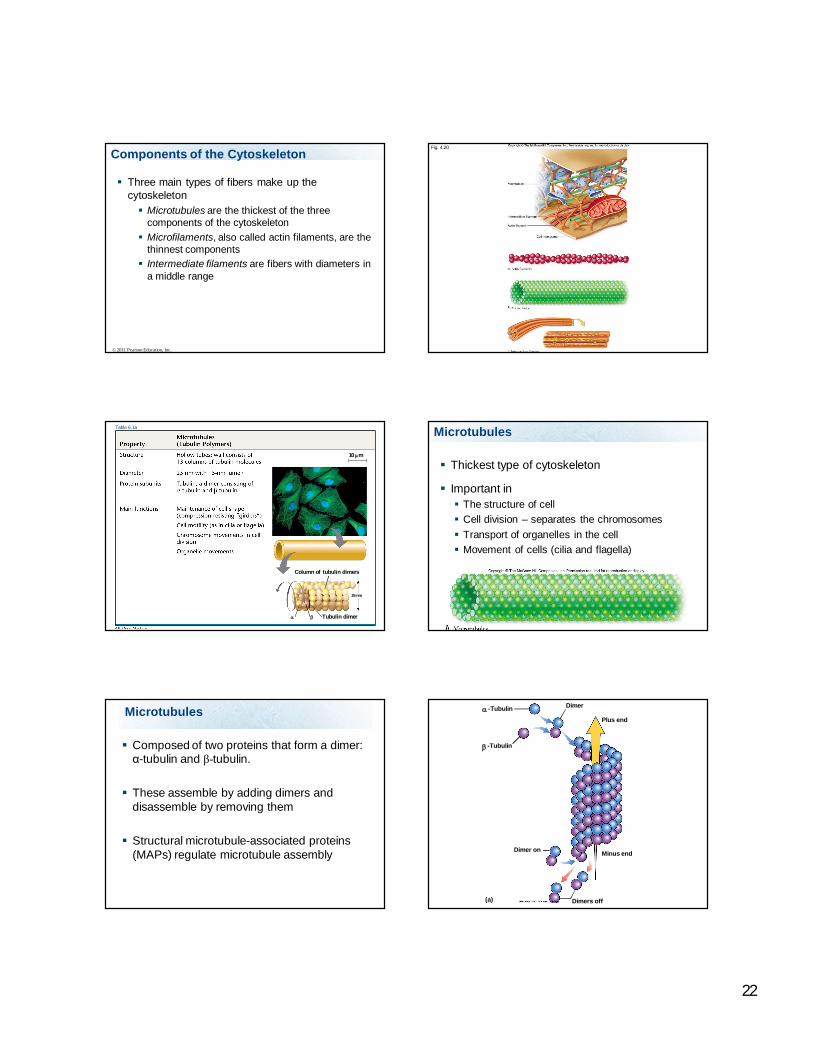
Components of the Cytoskeleton
Three main types of fibers make up the cytoskeleton Microtubules are the thickest of the three
components of the cytoskeleton Microfilaments, also called actin filaments, are the
thinnest components Intermediate filaments are fibers with diameters in
a middle range
© 2011 Pearson Education, Inc.
Fig. 4.20
Tubulin dimer
25 nm
Column of tubulin dimers
10 m
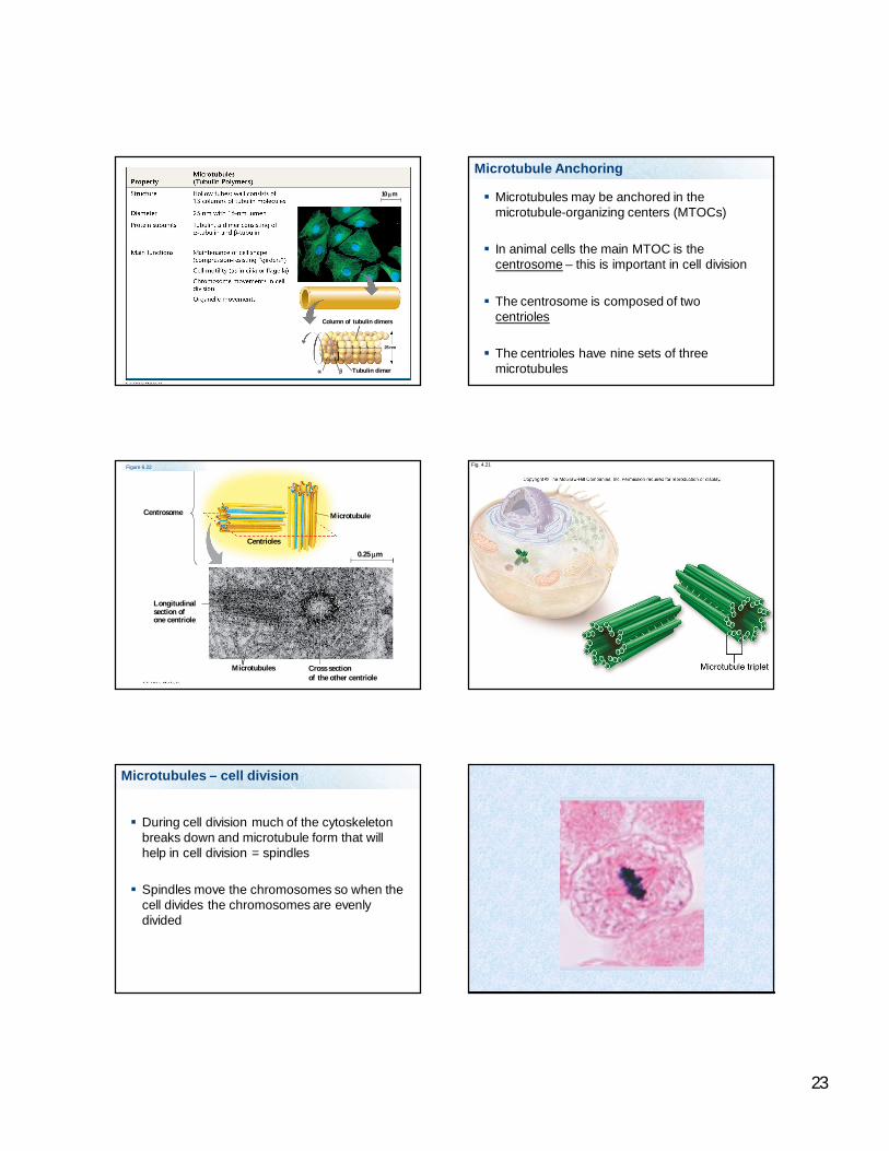
Table 6.1a Microtubules
Thickest type of cytoskeleton
Important in The structure of cell Cell division – separates the chromosomes Transport of organelles in the cell Movement of cells (cilia and flagella)
Microtubules
Composed of two proteins that form a dimer: α-tubulin and β-tubulin.
These assemble by adding dimers and disassemble by removing them
Structural microtubule-associated proteins (MAPs) regulate microtubule assembly
-Tubulin
-Tubulin
Dimer
Plus end
Minus endDimer on
Dimers off
(a)
23
Tubulin dimer
25 nm
Column of tubulin dimers
10 m
Microtubule Anchoring
Microtubules may be anchored in the microtubule-organizing centers (MTOCs)
In animal cells the main MTOC is the centrosome – this is important in cell division
The centrosome is composed of two centrioles
The centrioles have nine sets of three microtubules
Centrosome
Longitudinalsection ofone centriole
Centrioles
Microtubule
0.25 m
Microtubules Cross sectionof the other centriole
Figure 6.22 Fig. 4.21
Microtubules – cell division
During cell division much of the cytoskeleton breaks down and microtubule form that will help in cell division = spindles
Spindles move the chromosomes so when the cell divides the chromosomes are evenly divided

24
Microtubule – transport of organelles
Microtubule can be used to transport vesicles and other structures.
The microtubule is stationary, transport proteins (kinesin and dynein) move the item.
Kinesin moves items in one direction (+) and Dynein moves items in the opposite direction (-)
Fig. 4.22
Cilia and flagella
Microtubules important in movement of the cell
Project from cell surface
Flagella are long microtubules
Cilia are short microtubules
Flagella are found on sperm and many one celled organisms
Cilia found on many one celled organisms and on cells that line passageways in multi-celled organisms
142
Cilia and Flagella
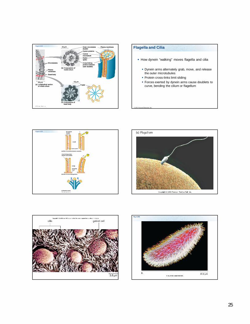
Flagella and Cilia
Both flagella and cilia have nine pairs of microtubules in an outer ring and a pair in the center (9 + 2).
Anchored by a basal body
A motor protein called dynein, which drives the bending movements of a cilium or flagellum
© 2011 Pearson Education, Inc.
Animation: Cilia and FlagellaRight-click slide / select “Play”
25
Microtubules
Plasmamembrane
Basal body
Longitudinal sectionof motile cilium
(a)0.5 m 0.1 m
0.1 m
(b) Cross section ofmotile cilium
Outer microtubuledoubletDynein proteins
CentralmicrotubuleRadialspoke
Cross-linkingproteins betweenouter doublets
Plasma membrane
Triplet
(c) Cross section ofbasal body
Figure 6.24
How dynein “walking” moves flagella and cilia
Dynein arms alternately grab, move, and release the outer microtubules Protein cross-links limit sliding Forces exerted by dynein arms cause doublets to
curve, bending the cilium or flagellum
© 2011 Pearson Education, Inc.
Flagella and Cilia
Figure 6.25 Microtubuledoublets
Dynein protein
ATP
(a) Effect of unrestrained dynein movement
Cross-linking proteinsbetween outer doublets ATP
Anchoragein cell
(b) Effect of cross-linking proteins
(c) Wavelike motion
1
2
3
Fig. 4.24b
26
Direction of swimming
(b) Motion of cilia
Direction of organism’s movement
Power stroke Recovery stroke
(a) Motion of flagella 5 m
15 m
Figure 6.23 Cilia
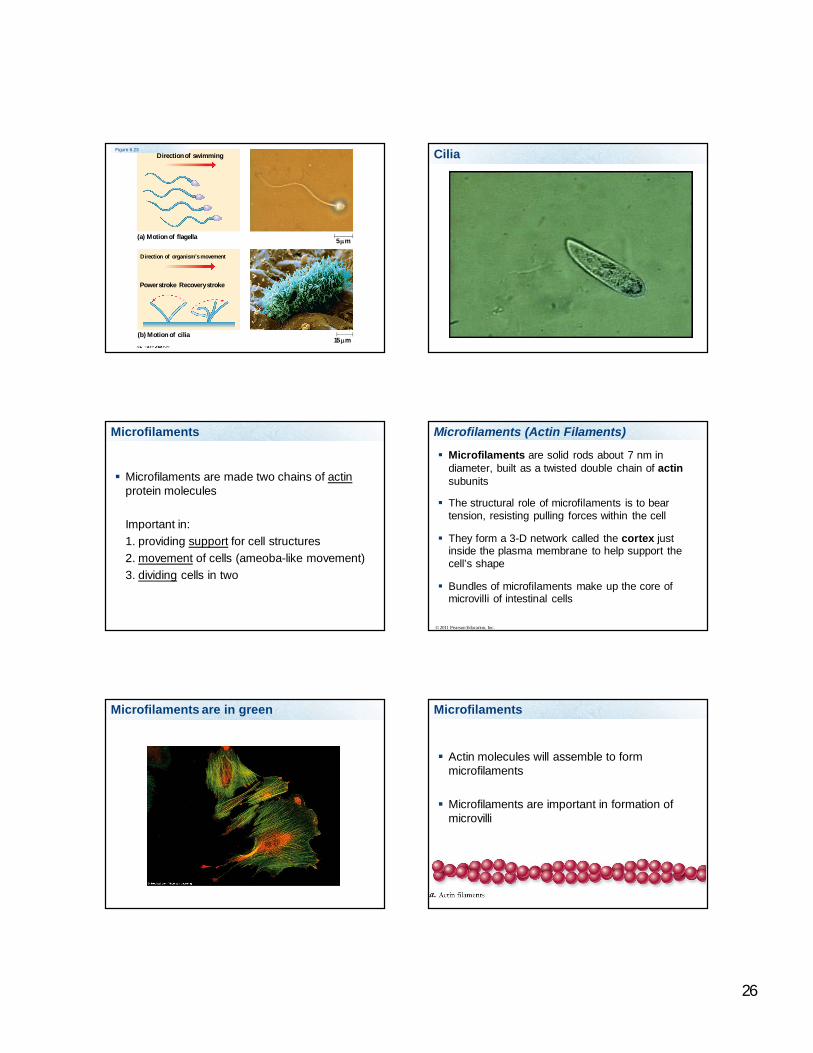
Microfilaments
Microfilaments are made two chains of actinprotein molecules
Important in:1. providing support for cell structures2. movement of cells (ameoba-like movement)3. dividing cells in two
Microfilaments (Actin Filaments)
Microfilaments are solid rods about 7 nm in diameter, built as a twisted double chain of actinsubunits
The structural role of microfilaments is to bear tension, resisting pulling forces within the cell
They form a 3-D network called the cortex just inside the plasma membrane to help support the cell’s shape
Bundles of microfilaments make up the core of microvilli of intestinal cells
© 2011 Pearson Education, Inc.
Microfilaments are in green Microfilaments
Actin molecules will assemble to form microfilaments
Microfilaments are important in formation of microvilli
27
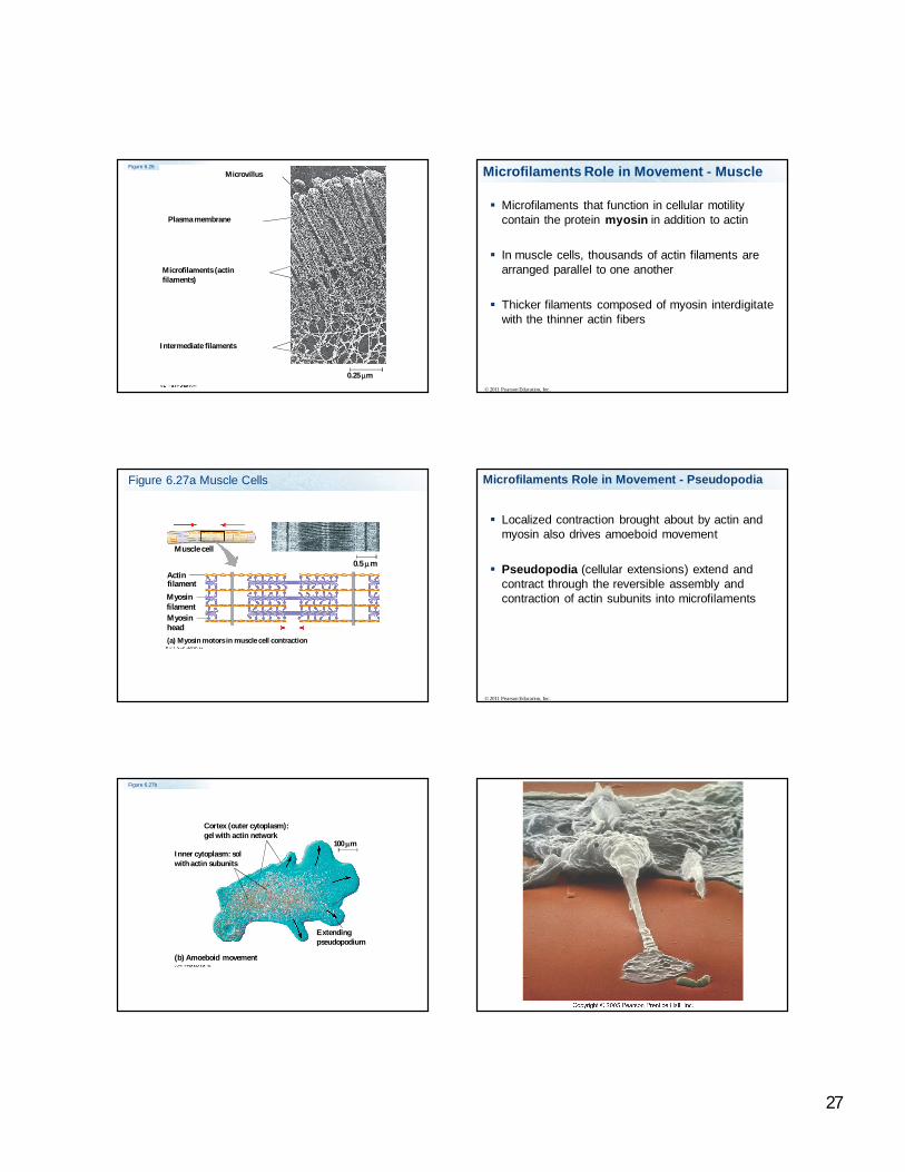
Figure 6.26Microvillus
Plasma membrane
Microfilaments (actinfilaments)
Intermediate filaments
0.25 m
Microfilaments that function in cellular motility contain the protein myosin in addition to actin
In muscle cells, thousands of actin filaments are arranged parallel to one another
Thicker filaments composed of myosin interdigitatewith the thinner actin fibers
© 2011 Pearson Education, Inc.
Microfilaments Role in Movement - Muscle
Figure 6.27a Muscle Cells
Muscle cell
ActinfilamentMyosin
Myosinfilament
(a) Myosin motors in muscle cell contraction
0.5 m
head
Localized contraction brought about by actin and myosin also drives amoeboid movement
Pseudopodia (cellular extensions) extend and contract through the reversible assembly and contraction of actin subunits into microfilaments
© 2011 Pearson Education, Inc.
Microfilaments Role in Movement - Pseudopodia
Figure 6.27b
100 m
Cortex (outer cytoplasm):gel with actin network
Inner cytoplasm: solwith actin subunits
(b) Amoeboid movement
Extendingpseudopodium
28
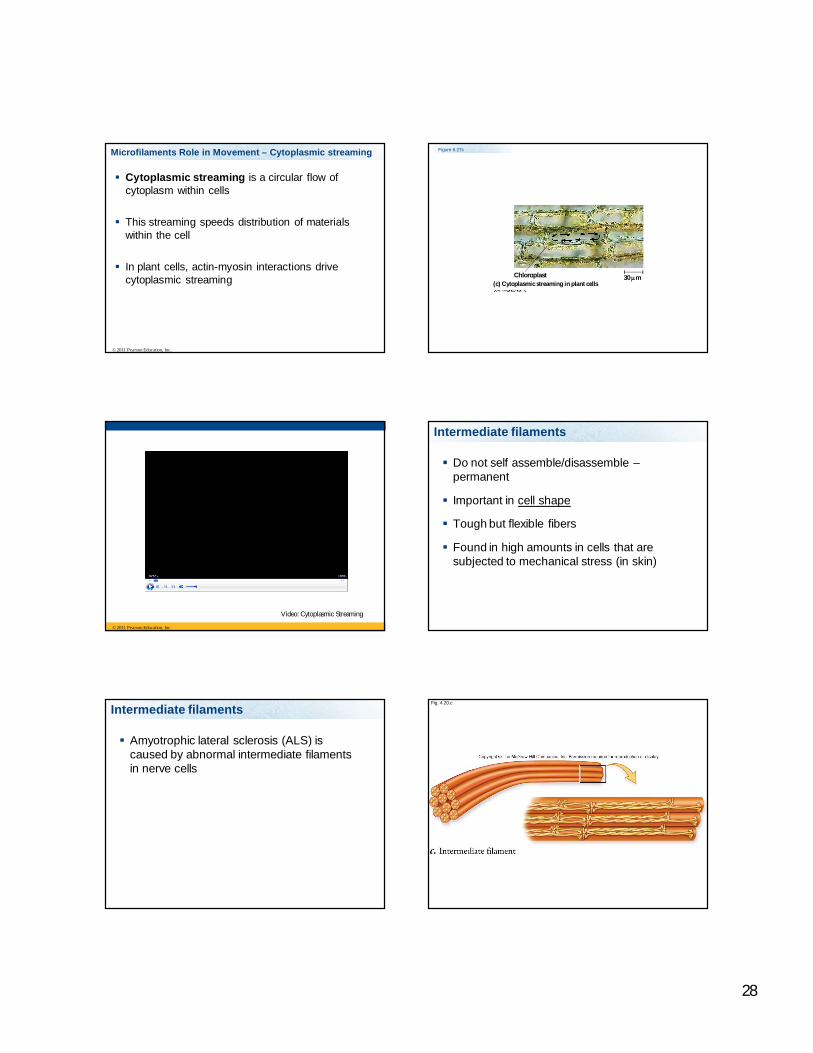
Cytoplasmic streaming is a circular flow of cytoplasm within cells
This streaming speeds distribution of materials within the cell
In plant cells, actin-myosin interactions drive cytoplasmic streaming
© 2011 Pearson Education, Inc.
Microfilaments Role in Movement – Cytoplasmic streaming Figure 6.27c
30 m(c) Cytoplasmic streaming in plant cells
Chloroplast
© 2011 Pearson Education, Inc.
Video: Cytoplasmic Streaming
Intermediate filaments
Do not self assemble/disassemble –permanent
Important in cell shape
Tough but flexible fibers
Found in high amounts in cells that are subjected to mechanical stress (in skin)
Intermediate filaments
Amyotrophic lateral sclerosis (ALS) is caused by abnormal intermediate filaments in nerve cells
Fig. 4.20.c

29
Cilia and flagella are composed of this type of cytoskeleton
1. Microtubules2. Microfilaments3. Intermediate
filaments
Micr
otubule
s M
icrofi
lamen
ts In
termed
iate f
ilamen
ts
33% 33%33%
Microfilaments are composed of:
1 2 3 4
25% 25%25%25%1. Kinesin2. Actin3. Tubulin4. Dynein
Copyright © 2009 Pearson Education, Inc.
This type of cytoskeleton is more permanent
1 2 3
33% 33%33%1. Microtubules2. Microfilaments3. Intermediate
Fibers
Extracellular components and connections between cells help coordinate cellular activities
Most cells synthesize and secrete materials that are external to the plasma membrane
These extracellular structures include Cell walls of plants The extracellular matrix (ECM) of animal cells Intercellular junctions
© 2011 Pearson Education, Inc.
Cell Walls of Plants
The cell wall is an extracellular structure that distinguishes plant cells from animal cells
Prokaryotes, fungi, and some protists also have cell walls
The cell wall protects the plant cell, maintains its shape, and prevents excessive uptake of water
Plant cell walls are made of cellulose fibers embedded in other polysaccharides and protein
© 2011 Pearson Education, Inc.
Cell Wall
Composed of :
1. Cellulose2. Lignins3. Sticky polysaccharides4. Glycoproteins
30
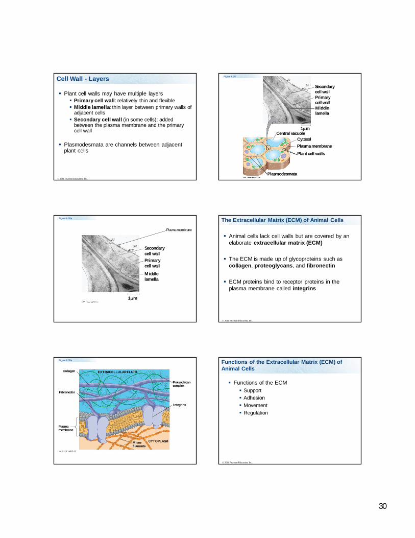
Plant cell walls may have multiple layers Primary cell wall: relatively thin and flexible Middle lamella: thin layer between primary walls of
adjacent cells Secondary cell wall (in some cells): added
between the plasma membrane and the primary cell wall
Plasmodesmata are channels between adjacent plant cells
© 2011 Pearson Education, Inc.
Cell Wall - LayersSecondarycell wallPrimarycell wallMiddlelamella
Central vacuoleCytosolPlasma membrane
Plant cell walls
Plasmodesmata
1 m
Figure 6.28
Figure 6.28a
Secondarycell wallPrimarycell wall
Middlelamella
1 m
Plasma membrane
The Extracellular Matrix (ECM) of Animal Cells
Animal cells lack cell walls but are covered by an elaborate extracellular matrix (ECM)
The ECM is made up of glycoproteins such as collagen, proteoglycans, and fibronectin
ECM proteins bind to receptor proteins in the plasma membrane called integrins
© 2011 Pearson Education, Inc.
Figure 6.30a
EXTRACELLULAR FLUIDCollagen
Fibronectin
Plasmamembrane
Micro-filaments
CYTOPLASM
Integrins
Proteoglycancomplex
Functions of the ECM Support Adhesion Movement Regulation
© 2011 Pearson Education, Inc.
Functions of the Extracellular Matrix (ECM) of Animal Cells
31
Cell Junctions
Neighboring cells in tissues, organs, or organ systems often adhere, interact, and communicate through direct physical contact
Intercellular junctions facilitate this contact
There are several types of intercellular junctions Plasmodesmata Tight junctions Desmosomes Gap junctions
© 2011 Pearson Education, Inc.
Plasmodesmata in Plant Cells
Plasmodesmata are channels that perforate plant cell walls
Through plasmodesmata, water and small solutes (and sometimes proteins and RNA) can pass from cell to cell
© 2011 Pearson Education, Inc.
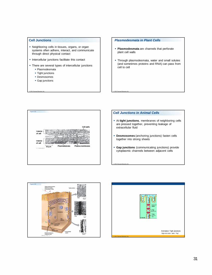
Figure 6.31
Interiorof cell
Interiorof cell
0.5 m Plasmodesmata Plasma membranes
Cell walls
Cell Junctions in Animal Cells
At tight junctions, membranes of neighboring cells are pressed together, preventing leakage of extracellular fluid
Desmosomes (anchoring junctions) fasten cells together into strong sheets
Gap junctions (communicating junctions) provide cytoplasmic channels between adjacent cells
© 2011 Pearson Education, Inc.
Figure 6.32
Tight junctions preventfluid from movingacross a layer of cells
Tight junction
Tight junction
TEM0.5 m
TEM1 m
TE
M
0.1 m
ExtracellularmatrixPlasma membranes
of adjacent cells
Spacebetween cells
Ions or smallmolecules
Desmosome
Intermediatefilaments
Gapjunction
© 2011 Pearson Education, Inc.
Animation: Tight JunctionsRight-click slide / select “Play”
32
Tight junctions
Tight junctions: prevent substances from leaking across tissues
Found in high concentration between cells: Lining the intestine Lining capillaries in the brain
© 2011 Pearson Education, Inc.
Animation: Desmosomes Right-click slide / select “Play”
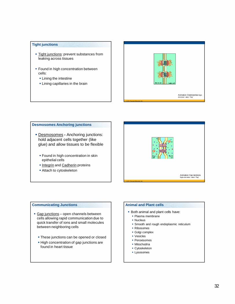
Desmosomes Anchoring junctions
Desmosomes - Anchoring junctions: hold adjacent cells together (like glue) and allow tissues to be flexible
Found in high concentration in skin epithelial cells Integrin and Cadherin proteins Attach to cytoskeleton
© 2011 Pearson Education, Inc.
Animation: Gap JunctionsRight-click slide / select “Play”
Communicating Junctions
Gap junctions – open channels between cells allowing rapid communication due to quick transfer of ions and small molecules between neighboring cells
These junctions can be opened or closed High concentration of gap junctions are
found in heart tissue
Animal and Plant cells
Both animal and plant cells have: Plasma membrane Nucleus Smooth and rough endoplasmic reticulum Ribosomes Golgi complex Vesicles Peroxisomes Mitochodria Cytoskeleton Lysosomes
33
Plant Cells
Main Plant cell components not present in animal cells Cell wall Vacuoles (usually central) Plastids including Chloroplast Glyoxysomes
Not present in plant cells: centrioles
Found only in Animal Cells
Animal cells have centrioles, plants have a different type of MTOCs
Glyoxysomes
Plants also have a type of organelle called glyoxysomes
Glyoxysomes contain enzymes that convert stored fatty acids to sugar that is used for energy, this is especially important in germinating seedlings.
Animal cells do not have these type of peroxisomes (we can’t convert fatty acids to sugar)
Plastids
Chromoplasts – contain pigments, give plant color, attract pollinators
Amyloplasts – store starch
Chlorplasts – contain a green pigment, chlorophyll and carotenoids – yellow and orange pigments. Site of photosynthesis.
Plant cells do not contain:
1 2 3 4
25% 25%25%25%1. Mitochondria2. Centrioles3. Nucleus4. Ribosomes
Important Concepts
Know the vocabulary in the lecture
Be able to describe cell theory and the properties of cells and structures common to all cells.
Know how cells are studied, the types of microscopy, the centrifugation techniques. How would you separate the organelles of the cell?
What are the main difference between prokaryotic cells and eukaryotic cells, and examples of each type?
34
Important Concepts
What are the features and characteristics of prokaryotic cells?
What is apoptosis and what organelle is an important player in the process? What molecules are released and activated during apoptosis?
What are the major features of eukaryotic cells, their location, features, structure, and their function – both plants and animals?
Important Concepts
Describe the steps needed to make a protein and ship it out of the cells, or embed the protein in a membrane, or have the protein stay in the cytosol.
What is the cause of Tay Sachs disease?
What are the differences between plant and animal cells?
Be able to describe the endosymbiont theory
Important Concepts
What are the types of cytoskeleton – what are their functions, structure, what proteins are they composed of, and how they are assembled?
How are microtubules assembled, anchored, how do they transport items and what proteins are used by microtubles to transport items.
What is the cause of ALS?
What are cell walls composed of, what are the layers?
Know the types of cell junctions, their functions and locations where there are found in high concentration
Know the role and components of extracellular matrix
Important Concepts