Hypocrea/Trichoderma (Ascomycota, Hypocreales - CBS
36
CHAVERRI & SAMUELS 1 Hypocrea/Trichoderma (Ascomycota, Hypocreales, Hypocreaceae): species with green ascospores Priscila Chaverri 1 * and Gary J. Samuels 2 1 The Pennsylvania State University, Department of Plant Pathology, Buckhout Laboratory, University Park, Pennsylvania 16802, U.S.A. and 2 United States Department of Agriculture, Agricultural Research Service, Systematic Botany and My- cology Laboratory, Rm. 304, B-011A, 10300 Baltimore Avenue, Beltsville, Maryland 20705, U.S.A. Abstract: The systematics of species of Hypocrea with green ascospores and their Trichoderma anamorphs is presented. Multiple phenotypic characters were analysed, including teleomorph and anamorph, as well as col- ony morphology and growth rates at various temperatures. In addition, phylogenetic analyses of two genes, the RNA polymerase II subunit (RPB2) and translation elongation factor 1-alpha (EF-1α), were performed. These analyses revealed that species of Hypocrea with green ascospores and Trichoderma anamorphs are derived from within Hypocrea but do not form a monophyletic group. Therefore, Creopus and Chromocrea, genera formerly segregated from Hypocrea only based on their coloured ascospores, are considered synonyms of Hy- pocrea. The present study showed that phenotypic characters alone are generally not helpful in understanding phylogenetic relationships in this group of organisms, because teleomorph characters are generally highly con- served and anamorph characters tend to be morphologically divergent within monophyletic lineages or clades. The species concept used here for Hypocrea/Trichoderma is based on a combination of phenotypic and geno- typic characteristics. In this study 40 species of Hypocrea/Trichoderma having green ascospores are described and illustrated. Dichotomous keys to the species are given. The following species are treated (names in bold are new species or new combinations): H. albocornea, H. atrogelatinosa, H. aureoviridis/T. aureoviride, H. can- dida/T. candidum, H. catoptron/T. catoptron, H. centristerilis, H. ceracea/T. ceraceum, H. ceramica/T. ce- ramicum, H. chlorospora/T. chlorosporum, H. chromosperma/T. chromospermum, H. cinnamomea/T. cin- namomeum, H. clusiae, H. cornea, H. costaricensis, H. crassa/T. crassum, H. cremea/T. cremeum, H. cu- neispora/T. cuneisporum, H. estonica/T. estonicum, H. gelatinosa/T. gelatinosum, H. gyrosa, H. lixii/T. har- zianum, H. macrospora, H. melanomagna/T. melanomagnum, H. nigrovirens/T. nigrovirens, H. phyllostachy- dis/T. phyllostachydis, H. rugulosa, H. sinuosa/T. sinuosum, H. spinulosa, H. straminea/T. stramineum, H. strictipilosa/T. strictipile, H. substipitata, H. sulawesensis, H. surrotunda/T. surrotundum, H. tawa/T. tawa, H. thailandica/T. thailandicum, H. thelephoricola/T. thelephoricola, H. tuberosa, H. velenovskyi, H. virens/T. virens, and H. virescentiflava. The following species are excluded: H. andinogelatinosa, H. dacrymycella, H. dichromospora, H. palmicola, H. pseudogelatinosa, H. subalbocornea, H. subatrogelatinosa, H. tropicosinen- sis, H. viscidula, H. viridis, and Chromocrea leucostroma. Key words: anamorph-teleomorph connection, monograph, phylogeny, RNA Polymerase gene, RPB2, species concept, systematics, Translation Elongation Factor 1-α gene. * To whom correspondence should be addressed. Current address: United States Department of Agriculture, Agricultural Research Ser- vice, Systematic Botany and Mycology Laboratory, Rm. 304, B-011A, 10300 Baltimore Avenue, Beltsville, Maryland 20705, U.S.A. Email: [email protected].
Transcript of Hypocrea/Trichoderma (Ascomycota, Hypocreales - CBS

CHAVERRI & SAMUELS
1
Hypocrea/Trichoderma (Ascomycota, Hypocreales, Hypocreaceae): species with green ascospores
Priscila Chaverri1* and Gary J. Samuels2
1The Pennsylvania State University, Department of Plant Pathology, Buckhout Laboratory, University Park, Pennsylvania 16802, U.S.A. and 2United States Department of Agriculture, Agricultural Research Service, Systematic Botany and My-cology Laboratory, Rm. 304, B-011A, 10300 Baltimore Avenue, Beltsville, Maryland 20705, U.S.A. Abstract: The systematics of species of Hypocrea with green ascospores and their Trichoderma anamorphs is presented. Multiple phenotypic characters were analysed, including teleomorph and anamorph, as well as col-ony morphology and growth rates at various temperatures. In addition, phylogenetic analyses of two genes, the RNA polymerase II subunit (RPB2) and translation elongation factor 1-alpha (EF-1α), were performed. These analyses revealed that species of Hypocrea with green ascospores and Trichoderma anamorphs are derived from within Hypocrea but do not form a monophyletic group. Therefore, Creopus and Chromocrea, genera formerly segregated from Hypocrea only based on their coloured ascospores, are considered synonyms of Hy-pocrea. The present study showed that phenotypic characters alone are generally not helpful in understanding phylogenetic relationships in this group of organisms, because teleomorph characters are generally highly con-served and anamorph characters tend to be morphologically divergent within monophyletic lineages or clades. The species concept used here for Hypocrea/Trichoderma is based on a combination of phenotypic and geno-typic characteristics. In this study 40 species of Hypocrea/Trichoderma having green ascospores are described and illustrated. Dichotomous keys to the species are given. The following species are treated (names in bold are new species or new combinations): H. albocornea, H. atrogelatinosa, H. aureoviridis/T. aureoviride, H. can-dida/T. candidum, H. catoptron/T. catoptron, H. centristerilis, H. ceracea/T. ceraceum, H. ceramica/T. ce-ramicum, H. chlorospora/T. chlorosporum, H. chromosperma/T. chromospermum, H. cinnamomea/T. cin-namomeum, H. clusiae, H. cornea, H. costaricensis, H. crassa/T. crassum, H. cremea/T. cremeum, H. cu-neispora/T. cuneisporum, H. estonica/T. estonicum, H. gelatinosa/T. gelatinosum, H. gyrosa, H. lixii/T. har-zianum, H. macrospora, H. melanomagna/T. melanomagnum, H. nigrovirens/T. nigrovirens, H. phyllostachy-dis/T. phyllostachydis, H. rugulosa, H. sinuosa/T. sinuosum, H. spinulosa, H. straminea/T. stramineum, H. strictipilosa/T. strictipile, H. substipitata, H. sulawesensis, H. surrotunda/T. surrotundum, H. tawa/T. tawa, H. thailandica/T. thailandicum, H. thelephoricola/T. thelephoricola, H. tuberosa, H. velenovskyi, H. virens/T. virens, and H. virescentiflava. The following species are excluded: H. andinogelatinosa, H. dacrymycella, H. dichromospora, H. palmicola, H. pseudogelatinosa, H. subalbocornea, H. subatrogelatinosa, H. tropicosinen-sis, H. viscidula, H. viridis, and Chromocrea leucostroma.
Key words: anamorph-teleomorph connection, monograph, phylogeny, RNA Polymerase gene, RPB2, species concept, systematics, Translation Elongation Factor 1-α gene.
* To whom correspondence should be addressed. Current address: United States Department of Agriculture, Agricultural Research Ser-vice, Systematic Botany and Mycology Laboratory, Rm. 304, B-011A, 10300 Baltimore Avenue, Beltsville, Maryland 20705, U.S.A. Email: [email protected].

HYPOCREA/TRICHODERMA WITH GREEN ASCOSPORES
2
INTRODUCTION Members of the Hypocreales (Fungi, Ascomycota) are common in all types of moist forests (for general ref-erences see Rossman et al. 1999). These fungi are eas-ily recognized by their brightly coloured fructifica-tions. The most conspicuous members, species of Hy-pomyces (Fr.) Tul. (Hypocreaceae), parasitize mush-rooms and bracket fungi. Another commonly encoun-tered genus is Hypocrea Fr. (Hypocreaceae). Species of Hypocrea and its anamorph Trichoderma Pers. are commonly encountered in humid tropical or subtropi-cal forests although they also occur in arid, temperate or boreal forests, even in the most extreme north and south latitudes. The Hypocrea teleomorph can be found on wood, on other members of the Ascomycota, on resupinate basidiomycetes, or on perennial bracket fungi in varying stages of decay, and less commonly on herbaceous substrata. The Hypocrea form typically presents itself as a cushion-shaped, brightly or lightly coloured, fleshy stroma that is no more than 5 mm in diameter, although stromata of some species may be several centimeters in extent and may even be club-shaped or turbinate. All investigated Trichoderma spe-cies are intimately related to Hypocrea, and, increas-ingly, named Trichoderma species are being shown to be the anamorphs of Hypocrea species (Kuhls et al. 1996, Samuels et al. 1998, Chaverri et al. 2001a). The Hypocrea form is rarely seen in culture. On the other hand, it is in the Trichoderma state that members of this genus are recovered in ecological investigations or cultivated in connection with commercial applications. The Trichoderma forms are commonly isolated from soils but also sporulate on moist wood, mushrooms, or bracket fungi in forests, where they are easily recog-nized by their masses of conidia that are usually green, but less commonly white or yellow. They are also found in diverse habitats: for example, they may be found in water-damaged buildings, or as endophytes within the trunks of asymptomatic tropical forest trees (Evans et al. 2003). In addition, they may be isolated as etiologic agents of opportunistic infection in im-munocompromised humans (Kuhls et al. 1999). The fact that many Trichoderma species have demon-strated antifungal or plant-growth-stimulating activi-ties has led to their exploitation as biological control agents, and in this connection some isolates are used in commercially available applications. More than 200 species of Hypocrea have been de-scribed, yet the genus has never been monographed. Recent systematic research suggests that it is not pos-sible to identify a Hypocrea species unless its Tricho-derma anamorph is known (Samuels et al. 1998, Chaverri et al. 2003a, Lu et al. 2003). Although only about 50 species of Trichoderma have been described, exploration of new geographical locations and eco-logical niches has revealed many additional unde-
scribed species. Because the anamorph is the form most commonly connected to information on the eco-nomic and ecological significance of these organisms, it is essential to clarify the biology of Trichoderma and Hypocrea in a unitary way. Holomorphs must be studied in order to effectively determine both life cy-cles and species concepts. The genus Hypocrea (anamorph Trichoderma) Hypocrea was first described by Elias Fries in 1825, based on Sphaeria rufa Pers. : Fr., a species with hya-line ascospores. Currently, the type species of the ge-nus is represented by Hypocrea rufa (Pers. : Fr.) Fr. Hypocrea is generally characterized by having perithecia embedded in fleshy stromata formed by pseudoparenchymatous tissue or highly compacted hyphae. Hypocrea has eight 1-septate ascospores that disarticulate at the septum early in their development, producing 16 part-ascospores in each ascus. Even though the disarticulation of ascospores is one of the distinguishing characters of the genus, other genera such as Aphysiostroma, Arachnocrea, Dialhypocrea, Podostroma, Protocrea, Pseudohypocrea, and Sporo-phagomyces also have disarticulating ascospores (Rossman et al. 1999). Recent studies using DNA se-quence data suggest that Podostroma (H.L. Chamber-lain, pers. comm.) and Protocrea (B.E. Overton, pers. comm.) are congeneric with Hypocrea. Podostroma was defined to include species with upright, stipitate stromata. In several studies, similarities were noted between Podostroma and Hypocrea, which differ only in the stalked stroma of the former (Boedijn 1934, Doi 1966, Rossman et al. 1999). Other characteristics of Podostroma, such as morphology of the stromatal tis-sue, perithecia, and anamorphs and ecology, are indis-tinguishable from corresponding features of Hypocrea. Aphysiostroma, Arachnocrea, Dialhypocrea, Pseudo-hypocrea, and Sporophagomyces, which are similar to Hypocrea in the disarticulation of their ascospores, have distinct teleomorph and anamorph morphology. Most notably, ascospores of Arachnocrea, Pseudohy-pocrea, and Sporophagomyces are biconical. Arach-nocrea and Sporophagomyces are mycoparasitic. Re-cent molecular-phylogenetic studies show that Aphysiostroma and Arachnocrea are basal to Hy-pocrea s. str. (Põldmaa et al. 1999, Zhang & Black-well 2002). Hypocrea pallida Ellis & Everh., a para-site of members of the Aphyllophorales sensu lato, was placed in Hypocrea because of its disarticulating, 2-celled ascospores and perithecia embedded in a pseudostroma or subiculum. However, H. pallida has a gliocladium-like anamorph that is similar to G. peni-cillioides Corda, the type species of the genus Glio-cladium and the anamorph of Sphaerostilbella aureonitens (Tul.) Seifert et al.

CHAVERRI & SAMUELS
3
Table 1. Species published in Chromocrea and Creopus.
Hypocrea Fr. Creopus Link Chromocrea Seaver H. aureoviridis Plowr. & Cooke 1880 Ch. aureoviridis (Plowr. & Cooke ) Petch
1938 H. ceramica Ellis & Everh. 1892 Ch. ceramica (Ellis & Everh.) Seaver 1910 H. cupularis (Fr.) Sacc. 1883 Ch. cupularis (Fr.) Petch 1938 H. gelatinosa (Tode : Fr.)Fr. 1849 Cr. gelatinosus (Tode : Fr.) Link 1833 Ch. gelatinosa (Tode:Fr.) Seaver 1910 Ch. marathwadi Tilak & Gaikwad 1978 H. nigricans (Imai) Yoshim. Doi 1972 Ch. nigricans Imai 1935 H. palmicola Berk. & Cooke 1873 Cr. palmicola (Berk. & Cooke) Boedjin
1951
H. spinulosa Fuckel 1869 Cr. spinulosus (Fuckel) Moravec 1956 Ch. spinulosa (Fuckel) Petch 1950 Ch. substipitata Seaver 1910 Cr. velenovskyi Z. Moravec 1956
Molecular data show that H. pallida is more closely related to Hypomyces and Sphaerostilbella than to Hypocrea (Põldmaa et al. 1999). Ascospore pigmentation has been used to charac-terize genera in the Hypocreales. Two genera, Cre-opus Link and Chromocrea Seaver, were segregated from Hypocrea because of their green ascospores. In 1791, Tode described Sphaeria gelatinosa Tode, a green-spored species that was placed in Hypocrea by Fries (1849) as H. gelatinosa (Tode : Fr.) Fr. Link (1833) based his description of the genus Creopus on S. gelatinosa. Even though Link did not explicitly state that Creopus was segregated based on green ascospores, most of the species subsequently placed in the genus have been those possessing this feature. A total of four species have been included in Creopus (Table 1). Later, Seaver (1910) proposed Chro-mocrea for species of Hypocrea with green asco-spores; he designated C. gelatinosa as the type, thus making Chromocrea a later, homotypic synonym of Creopus. Eight species have been included in Chro-mocrea (see Table 1). Sarawakus Lloyd is the only other hypocrea-like genus that has green ascospores. In the Hypocreales, apart from Hypocrea, only mem-bers of the genus Viridispora Samuels & Rossman (Nectriaceae) and some soon-to-be-published species of Neonectria (Samuels pers. comm.) have green as-cospores. The first ‘modern’ treatment of Hypocrea was published by Dingley (1952, 1957). She described the anatomy of the stroma and also undertook cultural studies to link species to their anamorphs. Several new species of Hypocrea and their anamorphs were described from New Zealand (Dingley 1952, 1957). Dingley identified all of the Hypocrea anamorphs as being typical of T. viride Pers. : Fr., despite differ-ences among them that were clearly visible in her illustrations. Developing on the works of Webster (1964), Webster & Rifai (1968) and Rifai & Webster (1966a, b), Rifai (1969) published a critical re-examination of Trichoderma in which he included
nine aggregate species. Since that time, Doi has pro-duced a floristic monograph of Hypocrea and its rela-tives in Japan (Doi 1969, 1972) that is the largest work on Hypocrea ever published. In it, the ana-morphs of many species of Hypocrea were described and illustrated. Doi and collaborators described many new species of Hypocrea and their anamorphs based on isolates collected in Japan, New Guinea, and South America (Doi 1966, 1968, 1969, 1971, 1972, 1973a, b, 1975, 1976, 1978, Doi & Doi 1980, Doi & Yamatoya 1989). Doi proposed an infrageneric taxonomy for Hy-pocrea based in large part on anatomy of the stroma, and to a lesser extent on anamorphs and ascospore pigmentation. Based mainly on teleomorph charac-ters, Doi described two subgenera: H. subg. Hetero-crea and H. subg. Hypocrea. Within H. subg. Hy-pocrea, two sections were defined: H. sect. Homalocrea and H. sect. Hypocrea. Doi (1972) rec-ognized H. subsect. Creopus within H. sect. Hy-pocrea, in which he included mostly Hypocrea spe-cies with green ascospores. Within this subsection, he distinguished three “artificial groups” of species, viz. AUREOVIRIDIS, CORNEA and GELATINOSA, based on types of stroma and conidiophore branch-ing. The AUREOVIRIDIS group included species with friable stromata, generally without perithecial protuberances and with trichoderma- or pachy-basium-like (Bissett 1991a, b) anamorphs. The COR-NEA group was characterized by coriaceous stromata and verticillium-like anamorphs. The GELATINOSA group was characterized by friable stromata, generally with protuberances and gliocladium-like anamorphs. Recent studies based on classical and molecular techniques, have shown that Hypocrea species with trichoderma-, pachybasium-, gliocla-dium-, or verticillium-like anamorphs do not form monophyletic groups (Kindermann et al. 1998, Kull-nig-Gradinger et al. 2002). Considerable evolutionary convergence occurs in the phenotype of the teleo-morph, and this phylogenetic study does not support

HYPOCREA/TRICHODERMA WITH GREEN ASCOSPORES
4
Doi’s subdivisions of Hypocrea (Lieckfeldt et al. 1998a, Chaverri et al. 2000, Kullnig-Gradinger et al. 2002). Most Hypocrea species were described in the 19th and early 20th centuries, often based on collections made by travelers, missionaries, pathologists or oth-ers in exotic lands and sent to experts such as M.J. Berkeley in England, P. Hennings in Germany, and J.B. Ellis in the United States. As was typical at the time, these newly described species often were pub-lished in larger works that included a wide diversity of fungi. None of the descriptions were illuminating, comprising at best a few lines of descriptive text and a note about the substratum. The better descriptions included ascospore measurements, but no effort was made to link collections to anamorphs or to describe stromatal anatomy. The systematic use of teleomorph anatomy by first Dingley and then Doi represented a significant move beyond these brief descriptions. The teleomorph is, after all, the element upon which the system for classifying Ascomycota is based, and it is considered to be the ‘name-bearing’ part of the holo-morph (i.e. teleomorph + anamorph) under Article 59 of the International Code of Botanical Nomenclature. However, with increasing efforts to link teleomorphs and anamorphs in the Ascomycota, including Hy-pocrea and Trichoderma morphs, and with the intro-duction of molecular phylogenetics into Hypocrea taxonomy, it has become apparent that the Hypocrea stroma possesses a highly conserved structure and is of limited use in species-level classification. For ex-ample, Samuels et al. (1998), in their monograph of the Hypocrea schweinitzii (Fr.) Sacc. Complex, showed that teleomorphs of H. schweinitzii sensu lato were almost indistinguishable from each other. Dif-ferences among species were seen, however, in ana-morphs, growth characteristics and DNA sequences. In cases involving closely related species, significant differences are more likely to be visible in the mor-phology of the Trichoderma anamorphs than in the anatomy or morphology of the teleomorph. In general, Trichoderma species have hyaline phi-alides formed on exposed fertile branches or ‘co-nidiophores’, and conidia that are generally smooth, rarely ornamented, typically ellipsoidal to nearly ob-long, rarely globose, and mostly green or hyaline, rarely yellow. If chlamydospores are formed, they are typically globose to subglobose and are formed within or at the tips of hyphae. In T. stromaticum Samuels et al., chlamydospores are formed that con-sist of balls of cells; typical chlamydospores are also formed (Samuels et al. 2000). Trichoderma virens (Miller et al.) Arx is characterized in part by the great abundance of chlamydospores that form in pure cul-ture (Chaverri et al. 2001a). Persoon (1794) originally described Trichoderma as a Gasteromycete. Only one of the small number of
species originally placed in the genus, namely T. viride, remains there today. Later, the Tulasne broth-ers (Tulasne & Tulasne 1860) and Brefeld (1891) proved the link between T. viride and H. rufa. The Tulasne brothers illustrated H. rufa and T. viride as one species, following hyphae from the Hypocrea stroma to the Trichoderma conidiophores. Brefeld isolated single ascospores of H. rufa and obtained T. viride in culture. Bisby (1939) could not distinguish the anamorph and teleomorph of H. rufa from those of H. gelatinosa, stating that H. gelatinosa “is only a growth form or mature condition of H. rufa.” Bisby reduced Trichoderma to a single species, T. viride. Trichoderma has been linked only to sexual stages in Hypocrea, and to some species of Podostroma and Sarawakus. The anamorph of the type species of Po-dostroma, P. alutaceum (Pers. : Fr.) Atk., is verticil-lium-like and not typical of Trichoderma, although some unidentified species of the genus have typical Trichoderma anamorphs. No anamorph has been linked to the type species of Sarawakus, S. lycoga-loides (Berk. & Broome) Lloyd. Samuels & Rossman (1992) transferred two species from Hypocrea to Sa-rawakus because of their formation of eight unicellu-lar ascospores in each ascus. These species have Trichoderma anamorphs but, apart from the unicellu-lar ascospores, they are not morphologically consis-tent with S. lycogaloides. With few exceptions, e.g. H. pallida, H. lutea (Tode) Petch, and H. sulphurea (Schwein.) Sacc., Hypocrea species have anamorphs that are morphologically typical of Trichoderma. Rifai (1969) divided Trichoderma species into nine “species aggregates,” namely, T. aureoviride Rifai, T. hamatum (Bonord.) Bain., T. harzianum Ri-fai, T. koningii Oudem., T. longibrachiatum Rifai. T. piluliferum Rifai , T. polysporum (Link : Fr.) Rifai, T. pseudokoningii Rifai, and T. viride. Each species ag-gregate was regarded as possibly comprising more than one morphologically cryptic species. Rifai con-cluded that anamorph characters alone might not pro-vide a useful taxonomy of Trichoderma. Bissett (1991a) discussed the implications of bas-ing a classification of Trichoderma on Rifai’s species aggregates. Bissett stated that five of Rifai’s aggre-gates (i.e. T. harzianum, T. longibrachiatum, T. pilu-liferum, T. polysporum, and T. pseudokoningii) were apparently narrowly defined species, whereas the re-maining aggregates could be interpreted as having relatively large numbers of species. For example, he suggested that more than 20 distinct species could be assigned to each of the morphological species T. ha-matum, T. koningii, and T. viride (Bissett 1984, 1991a, b, c, 1992). Bissett (1984, 1991a, b, c, 1992) recognized five sections: T. sect. Hypocreanum Bis-sett, T. sect. Longibrachiatum Bissett, and T. sect. Pachybasium (Sacc.) Bissett; T. sect. Saturnisporium Yoshim. Doi et al., and T. sect. Trichoderma. The

CHAVERRI & SAMUELS
5
sections were distinguished by differences in co-nidiophore branching patterns, phialides, and conidia. Trichoderma sect. Trichoderma (type species: T. viride) is distinguished by producing anamorphs that have narrow and flexuous conidiophores and branches with 2–3 phialides per verticil. The phi-alides are generally lageniform and are attached to the conidiophore at wide angles, and the conidia are smooth or ornamented. Anamorphs with T. sect. Trichoderma morphology are often referred in the literature as trichoderma-like. Bissett (1991a) placed T. koningii and the Trichoderma states corresponding to Hypocrea rufa/Trichoderma viride, H. aureoviri-dis/T. aureoviride and H. atroviridis/T. atroviride in sect. Trichoderma. Several studies have shown that some of the species included by Bissett in T. sect. Pachybasium, including the neotype of T. hamatum, cluster with species in T. sect. Trichoderma (Kindermann et al. 1998, Dodd et al. 2000, Lieckfeldt et al. 2001, Dodd et al. 2002, Kullnig-Gradinger et al. 2002). These same studies have also shown that H. aureoviridis/T. aureoviride and H. rufa/T. viride are not closely related. Trichoderma sect. Longibrachiatum is distin-guished by having aggregated conidiophores forming weakly developed pustules. The conidiophores con-sist of long primary branches and short, unbranched secondary branches. The phialides are solitary, rarely in verticils, and they produce ellipsoidal to oblong, green conidia. One characteristic of the group is the formation of ‘intercalary phialides’ (i.e. short, spur-like phialidic openings that form below the septum delimiting a terminal phialide). These are also found in some other groups but are not nearly as common or as conspicuous as in sect. Longibrachiatum. The H. schweinitzii Complex, which coincides with species producing anamorphs in T. sect. Longibrachiatum, was shown to be a monophyletic group based on morphological, cultural and molecular sequence data (Kuhls et al. 1997, Samuels et al. 1998). Species such as H. jecorina/T. reesei and H. schweinitzii/T. citri-noviride are examples of taxa within this complex. All known species in the H. schweinitzii Complex have colourless ascospores. Trichoderma sect. Satur-nisporium was originally distinguished from other sections by its tuberculate conidia; however, molecu-lar sequence data showed that it was nested within the H. schweinitzii Complex (Kuhls et al. 1997, Samuels et al. 1998). Trichoderma sect. Hypocreanum is characterized by having verticillium- and acremonium-like ana-morphs. Bissett (1991a) considered the anamorph of H. lactea (Fr. : Fr.) Fr. to be representative of T. sect. Hypocreanum. Preliminary data show that species closely related to H. lactea, such as H. citrina (Tode : Fr.) Fr., H. pulvinata Fuckel, and H. sulphurea (Schwein.) Sacc., all have anamorphs with T. sect.
Hypocreanum morphology (B.E. Overton, unpubl. data). This type of morphology, however, has been shown to be polyphyletically distributed. Hypocrea citrina was studied in detail by Canham (1969). Hy-pocrea tawa Dingley, T. hamatum, T. pubescens Bis-sett, H. poronioidea A. Möller, and H. strictipilosa/T. strictipile, which are not related to H. lactea, also have acremonium- or verticillium-like anamorphs and synanamorphs (Samuels & Lodge 1996, Kullnig-Gradinger et al. 2002, Chaverri et al. 2003a). Verti-cillium Nees s. str. is characterized by primary branches rebranching once or a few times and then terminating in verticils of long, subulate phialides. The verticillium-like anamorphs of Hy-pocrea/Trichoderma are distinguished from true Ver-ticillium by the presence of a dendritic conidiophore branching pattern, which generally includes secon-dary branches. Verticillium luteoalbum (Link : Fr.) Subram., the type species of the genus, does not be-long in the Hypocreales, but is rather closely related to Glomerella Spauld. & H. Schrenk (Zare et al. 2000). Trichoderma sect. Pachybasium, in its morpho-logical sense, so far contains the anamorphs of the majority of the described Hypocrea/Trichoderma species. Species in this section often produce com-pact conidiogenous pustules with branching generally in a pyramidal pattern, with or without fertile or ster-ile conidiophore elongations, and with phialides typi-cally short and wide, generally produced in crowded clusters of 2–7. Saccardo (1885) erected the genus Pachybasium to accommodate T. hamatum and T. hamatum var. candidum Sacc., a species with white conidia ( a synonym of T. hamatum var. candidum = T. polysporum). Bissett (1991a) expanded the concept of Pachybasium to include species with similar co-nidiophores and phialides, and elevated the genus Pachybasium to a section within Trichoderma. Bis-sett (1991b) also included species consisting of or possessing gliocladium-like anamorphs, e.g. T. virens, T. flavofuscum Bissett, H. gelatinosa, and H. lutea (Tode) Petch, in his concept of T. sect. Pachy-basium. Trichoderma sect. Pachybasium has since been shown to be polyphyletic (Kindermann et al. 1998, Lieckfeldt & Seifert 2000, Kullnig-Gradinger et al. 2002). Kindermann et al. (1998) divided T. sect. Pachybasium into two distinct phylogenetic groups termed “A” and “B”; further studies support this divi-sion (Kullnig-Gradinger et al. 2002, Chaverri et al. 2003a). Group A included species, such as T. atro-viride P. Karsten, T. hamatum (the type species of T. sect. Pachybasium), T. koningii, T. minutisporum Bissett, T. piluliferum Webster & Rifai in Rifai, T. polysporum (Link : Fr.) Rifai, T. pubescens Bissett, T. strigosum Bissett, and T. viride; and group B in-cluded T. crassum Bissett, T. fertile Bissett, T. flavo-fuscum, T. harzianum Rifai, T. longipile Bissett, H.

HYPOCREA/TRICHODERMA WITH GREEN ASCOSPORES
6
semiorbis (Berk.) Berk., T. spirale Bissett, H. stric-tipilosa/T. strictipile, T. tomentosum Bissett, and H. virens/T. virens. Section Pachybasium is to be re-duced to a descriptive term, but should not be used as a formal section. One of the main characteristics of Trichoderma sect. Pachybasium is that phialides are clustered in heads, often arising from a broad cell. Bissett (1991b) included species in the section that had previously been placed in Gliocladium (e.g. T. virens, T. flavo-fuscum) or that, based on classical morphological cri-teria, could have been included in Gliocladium (e.g. the anamorph of H. gelatinosa). Seifert (1985) and other authors (Rehner & Samuels 1994) found that Hypocrea species with gliocladium-like anamorphs are phylogenetically distinct from the type of Glio-cladium. Closer examination of their morphology shows that all of them can be readily excluded from Gliocladium, but not all of them are phylogenetically consistent with Trichoderma. DNA sequence analy-ses indicates that T. virens (= Gliocladium virens) is a species of Trichoderma; it is the anamorph of H. virens Chaverri & Samuels (Chaverri et al. 2001a) and is closely related to T. harzianum (Chaverri et al. 2003a). The Gliocladium viride Matr. anamorph of H. lutea is phylogenetically a member of Tricho-derma but its gliocladium-like conidiophore is atypi-cal of the genus. The anamorphs of H. pallida and related species (Doi & Yamatoya 1989) are virtually indistinguishable from Gliocladium penicillioides, the type species of Gliocladium and the anamorph of Sphaerostilbella aureonitens. Sequence analysis ex-cludes H. pallida from Hypocrea and places it closer to Hypomyces and Sphaerostilbella than it is to H. rufa (Rehner & Samuels 1994, Põldmaa et al. 1999). In addition to H. virens/T. virens, H. gelatinosa, and H. lutea/G. viride, a number of unrelated Hypocrea species have gliocladium-like anamorphs, including H. psychrophila Müller et al., H. luteovirens Yoshim. Doi, H. argillacea Phill. & Plowr., and T. crassum Bissett, among others. The introduction of DNA sequencing and cladistic analysis of DNA sequences in the early 1990’s opened a new era for fungal systematics by providing a new set of independently derived data that could be analyzed in tandem with more classically derived data. The upshot of the molecular revolution has been the ability to understand interrelationships among taxa at all levels. The first DNA-based phylogenetic studies that included genera of the Hypocreales ana-lyzed the relationship among different ascomycetes based on ribosomal DNA sequence data (Spatafora & Blackwell 1993, Rehner & Samuels 1994, 1995). As a result of these studies, it was shown that there were four monophyletic clades in the Hypocreales: Bionec-tria, Nectria, Claviceps and Hypocrea, now known as the families Bionectriaceae, Nectriaceae, Clavicipi-
taceae and Hypocreaceae, respectively (Rossman et al. 1999). Within this group, only the Hypocrea and Bionectria clades were well supported statistically. The Hypocrea clade included Hypocrea, Hypomyces and Sphaerostilbella. As mentioned above, more re-cent molecular studies have shown that the order Hy-pocreales could be defined to include the clades Bionectria (Bionectriaceae), Claviceps (Clavicipita-ceae), Hypocrea (Hypocreaceae), Nectria (Nectri-aceae), and Niesslia (Niessliaceae). Certain subgroups within the Hypocreales have been studied in detail in recent years. Lieckfeldt et al. (1998a) reviewed the progress in molecular systemat-ics of the Hypocreales, with emphasis on Hypocrea and Trichoderma. Lieckfeldt & Seifert (2000) sum-marized the value of ITS sequences in the taxonomy of the Hypocreales. Põldmaa et al. (1999) studied phylogenetic relationships in Hypomyces and allied genera and Põldmaa (2000) evaluated the generic delimitation of fungicolous Hypocreaceae. Phyloge-netic relationships of species of Hy-pocrea/Trichoderma have also been studied. The first accounts were gene genealogies based on a single gene region, viz. nuclear ribosomal internal tran-scribed spacers (ITS) DNA. For example, Lieckfeldt et al. (1998b) and later Dodd et al. (2000) studied the relationship of several species of Hy-pocrea/Trichoderma. Kuhls et al. (1997) studied the phylogenetic relationships of species in T. sect. Longibrachiatum. Samuels et al. (1998) monogra-phed the H. schweinitzii Complex, which included T. sect. Longibrachiatum, and also presented a phylog-eny. Kindermann et al. (1998) analyzed the phylog-eny of species in T. sect Pachybasium. Lieckfeldt et al. (1999) used ITS and morphological data to distin-guish the new species T. asperellum Samuels et al. from T. viride, and to show the close relationship be-tween these species. Samuels et al. (2000) described a new species of Trichoderma used in biocontrol, T. stromaticum, and presented its phylogenetic relation-ship to other biocontrol species (T. harzianum and T. virens). Lieckfeldt et al. (2001) studied the phyloge-netic position and morphological characterization of H. aureoviridis/T. aureoviride. As the development of analytical techniques for phylogenetic sequence analysis progressed, new methods to recognize species based on multiple genes emerged. Lieckfeldt et al. (2000) used the endochiti-nase gene (ECH42) to show phylogenetic relation-ships, mostly of species in T. sect. Trichoderma. Samuels et al. (2002) used ITS rDNA and translation elongation factor 1-alpha (EF-1α) to study the sys-tematics of two very closely related species, T. har-zianum and T. aggressivum Samuels & W. Gams. Dodd et al. (2002) studied the phylogenetic relation-ship of two morphologically similar species, H. pa-tella Cooke & Peck and H. neorufa Pat., using ITS
CHAVERRI & SAMUELS
7
rDNA and EF-1α. Kullnig-Gradinger et al. (2002) presented a multigene phylogeny of many species of Hypocrea/Trichoderma using 28S and ITS rDNA, small subunit (SSU) mitochondrial (mt) ribosomal DNA (rDNA), EF-1α, and ECH42. Analyses of phenotype including anamorphs, teleomorphs, and growth rates, in combination with molecular sequence data, has contributed to our un-derstanding of the evolution of Hy-pocrea/Trichoderma and has helped to show link be-tween particular anamorphs and teleomorphs. Samuels and collaborators included multiple morpho-logical characters, growth data and DNA analyses in their monograph of the H. schweinitzii Complex (Samuels et al. 1998). Similar combination studies were performed in the study of the “green mold” of commercial mushrooms, T. aggressivum, and its rela-tionship to T. harzianum (Samuels et al. 2002); in distinguishing morphologically similar species (Lieckfeldt et al. 1999, Samuels et al. 1999, Dodd et al. 2002); and in showing the relationships of species in T. sect. Pachybasium with conidiophore elonga-tions (Chaverri et al. 2003a). These studies have added more than 20 newly described species of Hy-pocrea and Trichoderma. In addition to showing phylogenetic relationships among species, studies in which phenotype and DNA analyses were combined have tended to confirm the monophyly of species distinguished by Bissett (1984, 1991a, b, c, 1992) on morphological grounds. In some instances, however, even those species have been shown to comprise more than one species. Lieckfeldt et al. (1999) and Samuels et al. (1999) demonstrated that the type species of Trichoderma, T. viride, comprised at least two species, one of which was described as T. asperellum. Trichoderma viride is mainly found in the northern hemisphere, whereas T. asperellum is ubiquitous in both mesic and xeric soil habitats in regions as diverse as Russia, Central Africa and Saudi Arabia. The residual T.viride is still paraphyletic. The current morphological species con-cept of T. koningii is also polyphyletic, comprising at least four phylogenetic species (G. J. Samuels, un-publ. data). Both T. viride and T. koningii may be conceived of as species complexes, as discussed be-low. On the other hand, the cosmopolitan species H. atroviridis/T. atroviride is monophyletic (Dodd et al. 2003). Surprisingly, T. harzianum, a ubiquitous species that has been thought to be paraphyletic (e.g. Grondona et al. 1997) because of its seemingly great phenotypic and genotypic variability, has been shown to be a monophyletic ‘species complex,’ that is, a species that comprises multiple phenotypically indis-tinguishable evolutionary lines. High infraspecific variation has been found in H. lixii/T. harzianum in studies using multiple morphological, physiological
and molecular characters (Grondona et al. 1997, Hermosa et al. 2000, Lee & Hseu 2002, Samuels et al. 2002, Chaverri et al. 2003b). Multigene phyloge-netic analyses have revealed that species complexes in Trichoderma are common. Besides H. lixii/T. har-zianum, the aforementioned H. rufa/T. viride and H. koningii/T. koningii groups may also be thought of as representing species complexes. Samuels et al. are in the process of revealing multiple species within the H. rufa/T. viride and H. koningii/T. koningii com-plexes (G. J. Samuels unpubl.). In addition, a degree of morphological variation has been encountered in specimens and isolates of Hypocrea cf. chlorospora B. & M.A. Curtis. In the present study, the close phy-logenetic and morphological relationships of these specimens and isolates are examined to determine if they constitute a species complex. In order to develop a natural taxonomic system for ascomycetous fungi that reflects the full biology (e.g. the life cycles) of these organisms, the delineation of anamorph-teleomorph relationships is a major goal of fungal systematics. The combination of phenotypic and DNA sequence data has also been proven useful for making such connections in Hypocrea/Tricho-derma. The first anamorph-teleomorph connection made using DNA analyses was made by Kuhls et al. (1996), who showed that the asexual industrial fun-gus Trichoderma reesei E.G. Simmons was the ana-morph of Hypocrea jecorina Berk. & Broome. Samuels et al. (1998), in their monograph of the H. schweinitzii Complex, showed the connection of H. pseudokoningii and T. pseudokoningii, H. schweinitzii and T. citrinoviride, and H. orientale and T. cf. longibrachiatum. Additional links of Hypocrea species to named Trichoderma species are presented in Table 2. In the present paper the previously un-known teleomorph of T. crassum is described. In ad-dition, anamorphs of species of Hypocrea with green ascospores are named and described. The objectives of the present study were: 1) to describe or re-describe species of Hypocrea with green ascospores; 2) to determine if species of Hy-pocrea with green ascospores form a monophyletic group; 3) to determine and describe monophyletic groups that exclusively contain species with green ascospores; and 4) to describe Hypocrea species complexes that include at least some species with green ascospores (e.g. H. cf. chlorospora and H. lixii/T. harzianum complex). To accomplish these objectives, multiple phenotypic characters were evaluated, and phylogenetic analyes were performed based on sequences of two genes, the RNA poly-merase II subunit (RPB2) and EF-1α. Descriptions and illustrations of 40 species of Hypocrea and their known Trichoderma anamorphs are presented as well as keys to these species.

HYPOCREA/TRICHODERMA WITH GREEN ASCOSPORES
8
Table 2. Anamorph-teleomorph connections to named species of Hypocrea/Trichoderma.*
Teleomorph Anamorph Source H. atroviridis T. atroviride Dodd et al. 2003 H. aureoviridis T. aureoviride Lieckfeldt et al. 2001 H. jecorina T. reesei Kuhls et al. 1996 H. koningii T. koningii Lieckfeldt et al. 1998b H. lixii T. harzianum Chaverri & Samuels
2002; Chaverri et al. 2003b
H. lutea Gliocladium viride
Domsch et al. 1980
H. minutispora T. minutisporum Lu et al. 2003 H. pachy-basioides
T. polysporum Doi 1972; Bissett 1991b; Lu et al. 2003
H. pilulifera T. piluliferum Rifai 1969 H. pseudokon-ingii
T. pseudokon-ingii
Samuels et al. 1998
H. rufa T. viride Tulasne & Tulasne 1860
H. schweinitzii T. citrinoviride Samuels et al. 1998 H. strictipilosa T. strictipile Chaverri et al. 2003a H. virens T. virens Chaverri et al. 2001a
* Species newly described in this study are not included in the table. Ecology and economic importance of Hy-pocrea/Trichoderma Species of Hypocrea/Trichoderma typically grow on decaying woody substrata but also on other fungi, including ascomycetes and basidiomycetes, and rarely on herbaceous tissues. Trichoderma is univer-sally found as a soil fungus. There is usually no apparent host specialization (Rossman et al. 1999). There are, however, some ex-ceptions to this trend. Hypocrea pulvinata Fuckel occurs only on aphyllophoraceous basidiomycetes, e.g. Tyromyces, Fomitopsis, and Piptoporus, while H. latizonata Peck is restricted to Cyathus, H. avellanea Carey & Rogerson to Gymnopus subnudus, H. pallida to members of the Aphyllophorales sensu lato and H. spinulosa Fuckel to grasses growing in alpine and boreal regions. Hypocrea psychrophila has only been found growing on Rhododendron ferrugineum and Vaccinium myrtillus in the Swiss Alps (Müller et al. 1972). Trichoderma stromaticum Samuels et al. and its Hypocrea teleomorph are only known from spe-cies of cocoa (genus Theobroma, family Sterculi-aceae) and are often associated with tissue infected with the basidiomycetous pathogen Crinipellis perni-ciosa. Trichoderma aggressivum Samuels et al. has so far been found only on Agaricus bisporus in com-mercial mushroom production. Because Hypocrea stromata are most often found on decaying wood that is inhabited by other fungi, it is not possible to tell whether the Hypocrea is obtaining its nutrients at the expense of another fungus or the decaying wood or both. Some species show biogeographical specializa-tion. Hypocrea jecorina/Trichoderma reesei is known
only from the equatorial zone, while H. semiorbis (Berk.) Berk. is known only from New Zealand and Australia and H. aureoviridis/T. aureoviride, H. pilu-lifera/T. piluliferum, H. placentula W.B. Grove are found only in northern Europe and the United King-dom. Trichoderma stromaticum naturally occurs only in the Amazon basin (Samuels et al. 2000) and H. nigrovirens Chaverri, Samuels & E.L. Stewart has been found only in Costa Rica (Chaverri et al. 2001b). Hypocrea minutispora/T. minutisporum is known only from north temperate collections and H. pachybasioides/T. polysporum is cosmopolitan at north and south temperate latitudes but has never been found in tropical regions (“tropical” defined as occurring between the Tropics of Cancer and Capri-corn) (Lu et al. 2003). Hypocrea/Trichoderma species are economically important because of the effect that some of them have on disease-causing fungi. This antifungal activ-ity has been extensively documented and suggestions have been made that the biocontrol effects exerted by these fungi may be based on one or more of three possible factors, namely antibiosis (Howell & Sti-panovic 1983, 1991, Lumsden et al. 1992, Ghisalberti & Rowland 1993, Sawa et al. 1994, Elad 2000a, Kex-iang et al. 2002), mycoparasitism (Elad 2000a, Mishra et al. 2000, Viterbo et al. 2002), and nutrient competition (Simon & Sivasithamparam 1989). The volume edited by Harman & Kubicek (1998) contains several papers that include comprehensive informa-tion about Hypocrea/Trichoderma enzymes, biologi-cal control and commercial applications. Green-ascospore-producing species of Hypocrea/Tricho-derma that have known antifungal activities are H. virens/T. virens and H. lixii/T. harzianum. In addi-tion, T. harzianum and T. virens are the active ingre-dients in the commercial preparations, TRICHO-DEX and SoilGard , respectively, used in biologi-cal control of various fungal diseases (Ricard 1981, Harman 1990, Lumsden et al. 1993, Harman & Lo 1996). Trichoderma harzianum has been found to be ef-fective against many phytopathogens, and is consid-ered a generalist biocontrol agent. For example, it is effective against Rhizoctonia damping-off in radish, corn, soybean, cucumber, and sugar beet (Lewis & Papavizas 1980, Kommedahl et al. 1981, Lifshitz et al. 1985, Dutta & Das 1999, Mishra et al. 2000), Sclerotium rolfsii plant wilt (El-Katatny et al. 2000, Mishra et al. 2000), grey-mold diseases caused by Botrytis cinerea (Harman et al. 1995, Elad & Kapat 1999, Elad 2000b, Kovach et al. 2000, Hjeljord et al. 2001), take-all disease in wheat (Ghisalberti & Siva-sithamparam 1991), Botryosphaeria berengeriana f. sp. piricola apple ring-rot (Kexiang et al. 2002), Ar-millaria spp. root-rot (Otieno et al. 2003a, b), Ven-turia inaequalis apple scab (Bolar et al. 2000, Otieno

CHAVERRI & SAMUELS
9
et al. 2003a, b), Fusarium udum wilt of pigeon pea (Prasad et al. 2002), and cacao witches’ broom caused by Crinipellis perniciosa (de Azevedo et al. 2000, De Marco et al. 2000). Trichoderma harzianum has also been useful in the control of root diseases caused by nematodes, e.g. strawberry black root-rot caused by Pratylenchus penetrans and Rhizoctonia fragariae (LaMondia & Cowles 2002) and root knot caused by Meloidogyne javanica (Sharon et al. 2001). It has also been found to enhance plant growth and disease resistance upon plants (Inbar et al. 1994, Gromovich et al. 1998, Yedidia et al. 2001). Trichoderma virens has also been an effective biocontrol agent against many plant pathogens. For example, it is useful against Phytophthora eryth-roseptica pink rot of potato and root and stem rot of tomato (Etebarian et al. 2000), damping-off diseases of various types of seedlings (Roberts & Lumsden 1990, Howell & Stipanovic 1991, Lumsden et al. 1996, Harris & Lumsden 1997), black-pod of cacao caused by Phytophthora palmivora (Krauss & Sober-anis 2001, 2002), gladiolus corm rot and wilt caused by Fusarium oxysporum f. sp. gladioli (Mishra et al. 2000), Sclerotinia sclerotiorum infection of beans (Huang et al. 2000), and the root-knot nematode Meloidogyne incognita on bell pepper (Meyer et al. 2001). Trichoderma virens was reported as having growth-promoting activities when it was cultivated with rice seedlings (Mishra et al. 2000). The my-cotoxins gliotoxin, viridin, and gliovirin have been linked to the antifungal capabilities of T. virens (Weindling & Emerson 1936, Weindling 1937, 1941, Howell & Stipanovic 1983, DiPietro et al. 1993). Trichoderma flavofuscum, which we consider a syno-nym of T. virens, also produces these mycotoxins (Avent et al. 1993). Gliotoxin is also formed by As-pergillus fumigatus Fresen. (Nieminen et al. 2002, Wenehed et al. 2003). Peptaibol metabolites pro-duced by some Trichoderma species have also been linked to antifungal action (Schirmbock et al. 1994, Oh et al. 2002). Other secondary metabolites, includ-ing mycotoxins, produced by Hypocrea/Trichoderma were listed by Sivasithamparam & Ghisalberti (1998). In addition to the use of Hypocrea/Trichoderma species in the biological control of plant pathogenic fungi, many species have the ability to break down cellulosic materials through the production of cellu-lases. This ability has led to the commercial exploita-tion of some Hypocrea/Trichoderma species in pro-duction of enzymes used in manufacture of clothes-washing detergent, animal feed and fuel (Reese & Mandels 1989, Kubicek et al. 1990, Nsereko et al. 2002). One unusual application of an unidentified species of Trichoderma is in the bioconversion or biodegradation of domestic wastewater sludge into compost (Molla et al. 2002). Properties of other spe-
cies that might be useful in bioremediation have also been investigated. Several studies have shown the potential of T. harzianum to bioremediate soils con-taminated with pesticides such as pentachlorophenol and endosulfan (Katayama & Matsumura 1993, Rigot & Matsumura 2002). With regard to biosafety concerns, Hypocrea/ Trichoderma species are not known to cause disease in healthy humans; however, there are numerous re-ports documenting pathogenicity in humans with immunocompromising conditions (Loeppke et al. 1983, Jacobs et al. 1992, Kuhls et al. 1999). One case has been reported in which T. harzianum caused a fatal disseminated infection in a renal transplant pa-tient (Guarro et al. 1999). MATERIALS AND METHODS Isolates and herbarium specimens The isolates and specimens investigated are listed with the description of each species. Isolates used for DNA analyses are listed in Table 3 with their corre-sponding GenBank accession numbers. The majority of isolates were obtained from fresh collections of Hypocrea, but others were obtained from Centraalbu-reau voor Schimmelcultures, Utrecht, The Nether-lands (CBS) and from Agriculture and Agri-Food Canada, Eastern Cereals and Oilseeds Research Cen-tre, Ottawa, Canada (DAOM). Representative isolates have been deposited in American Type Culture Col-lection, Manassas, VA, USA (ATCC) as well as CBS and DAOM. Single-ascospore isolations from fresh collections of Hypocrea were made on cornmeal dex-trose medium (CMD), consisting of Difco cornmeal agar (Difco Laboratories, Detroit, MI, USA) + 2% dextrose + 1% antibiotic solution (0.2% Sigma [Sigma-Aldrich Corp., St. Louis, MO, USA] Strep-tomycin Sulfate + 0.2% Sigma Neomycin Sulfate + distilled water). A micromanipulator was used to ob-tain single propagules for inoculation. The cultures obtained are maintained at the U.S. National Fungus Collection (BPI) on Difco cornmeal agar (CMA) slants at 8 ºC and in liquid nitrogen in cryovials with 10% glycerol. The majority of herbarium specimens examined are kept in BPI, but others, such as type specimens, were obtained from William and Lynda Steere Her-barium (NY), the Herbarium of the Royal Botanic Gardens, Kew (K), the New Zealand Fungal Herbar-ium (PDD), the Farlow Reference Library and Her-barium of Cryptogamic Botany (FH), the Museum of Evolution, Botany Section, Uppsala University (UPS), the University of Sheffield Botany Depart-ment Herbarium (SHD), the Herbarium of Mycology and Plant Pathology Department, Agharkar Research Institute (AMH), the Department of Botany Herbar-

HYPOCREA/TRICHODERMA WITH GREEN ASCOSPORES
10
ium of INBio, the National Biodiversity Institute of Costa Rica (INB), the Herbarium Conservatoire & Jardin botaniques de la Ville de Genève (G), and oth-
ers. The frequently cited collectors G.J. Samuels and C.T. Rogerson are abbreviated as G.J.S. and C.T.R.
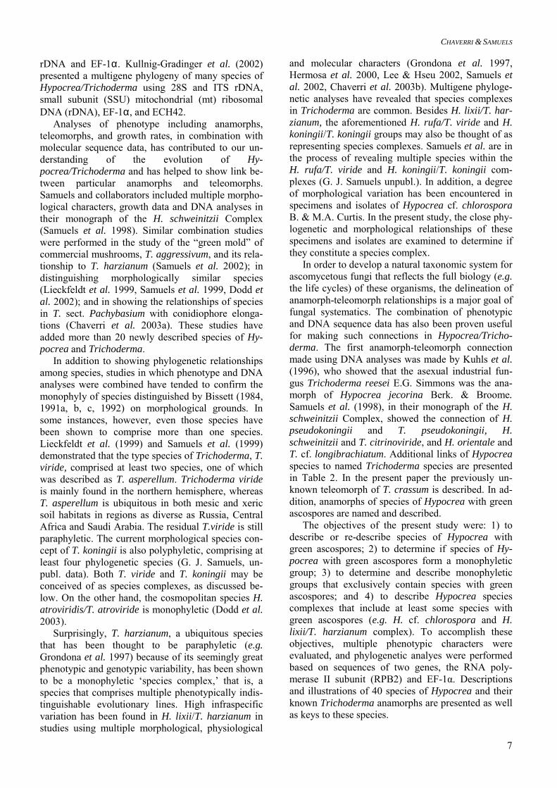
Table 3. List of isolates studied, geographic origin and Genbank accession numbers.
GenBank numbers Species Isolate Geographic origin EF-1α RPB2 Hypocrea aureoviridis CBS 245.63* U.K. AF534575 AF545509 Hypocrea aureoviridis G.J.S. 98-23 (= IMI 355906) U.K. AY391961 AY391898 Hypocrea avellanea C.T.R. 77-155 Pennsylvania, U.S.A. AY225857 AF545562 Hypocrea candida P.C. 59* (= CBS 114249) Costa Rica AY391962 AY391899 Hypocrea catoptron G.J.S. 02-76 (= CBS 114232) Sri Lanka AY391963 AY391900 Hypocrea ceracea G.J.S. 88-28 New York, U.S.A. AY391964 AY391901 Hypocrea ceracea G.J.S. 89-136 North Carolina, U.S.A. AY391965 AY391902 Hypocrea ceracea G.J.S. 95-159* (= CBS 114245) New York, U.S.A AF534603 AF545508 Hypocrea ceramica G.J.S. 88-70* (= CBS 114576) North Carolina, U.S.A. AF534593 AF545510 Hypocrea chlorospora G.J.S. 88-33* (= CBS 114231) Connecticut, U.S.A. AY391966 AY391903 Hypocrea chlorospora G.J.S. 91-150 Maryland, U.S.A. AY391967 AY391904 Hypocrea chlorospora G.J.S. 95-88 Puerto Rico AY391905 Hypocrea chlorospora G.J.S. 98-1 Costa Rica AY391968 AY391906 Hypocrea chlorospora P.C. 4 Pennsylvania, U.S.A. AF534578 AF545515 Hypocrea chromosperma G.J.S. 90-58 New York, U.S.A. AY391969 AY391908 Hypocrea chromosperma G.J.S. 90-59 New York, U.S.A. AY391970 AY391909 Hypocrea chromosperma G.J.S. 91-128 Maryland, U.S.A. AY391971 AY391910 Hypocrea chromosperma G.J.S. 92-88 Maryland, U.S.A. AY391972 AY391911 Hypocrea chromosperma G.J.S. 94-67 Maryland, U.S.A. AY391973 AY391912 Hypocrea chromosperma G.J.S. 94-68* (= CBS 114577) Maryland, U.S.A. AY391974 AY391913 Hypocrea chromosperma G.J.S. 95-196 Indiana, U.S.A. AY391975 AY391914 Hypocrea chromosperma G.J.S. 98-73 New Jersey, U.S.A. AY391976 AY391915 Hypocrea cinnamomea G.J.S. 96-128 Taiwan AY391977 AY391916 Hypocrea cinnamomea G.J.S. 96-165 Taiwan AY391917 Hypocrea cinnamomea G.J.S. 97-230* (= CBS 114235) Missouri, U.S.A. AY391918 Hypocrea cinnamomea G.J.S. 97-233 Missouri, U.S.A. AY391978 AY391919 Hypocrea cinnamomea G.J.S. 97-237 Missouri, U.S.A. AY391979 AY391920 Hypocrea citrina CBS 894.85 Belgium AY225856 AF545561 Hypocrea costaricensis P.C. 21 Costa Rica AY391980 AY391921 Hypocrea crassa G.J.S. 01-227* (= CBS 114230) Thailand AY481587 Hypocrea cremea G.J.S. 02-52 New Zealand AY391981 AY391922 Hypocrea cremea G.J.S. 91-125* (= CBS 111146) New York, U.S.A. AF534598 AF545511 Hypocrea cuneispora G.J.S. 91-93* (= CBS 111148) Virginia, U.S.A. AF534600 AF545512 Hypocrea estonica G.J.S. 96-129* (= CBS 111147) Estonia AF534604 AF545514 Hypocrea gelatinosa G.J.S. 88-17 France AF534579 AF545516 Hypocrea gelatinosa G.J.S. 93-10 France AY391982 AY391923 Hypocrea gelatinosa G.J.S. 98-184* (= CBS 114246) Austria AY391983 AY391924 Hypocrea lixii G.J.S. 90-22 Wisconsin, U.S.A. AY391984 AY391925 Hypocrea lutea G.J.S. 89-129 New York, U.S.A. AF534581 AF545517 Hypocrea megalocitrina B.E.O. 00-09 North Carolina, U.S.A. AY225855 AF545563 Hypocrea melanomagna G.J.S. 99-153* (= CBS 114236) Australia AY391985 AY391926 Hypocrea minutispora CBS 901.72 AY392009 AY481588 Hypocrea nigrovirens G.J.S. 99-64* (= CBS 114330) Costa Rica AF534582 AF545518 Hypocrea pachybasioides G.J.S. 90-4 Mississippi, U.S.A. AY392010 AY481589

CHAVERRI & SAMUELS
11
Hypocrea pezizoides G.J.S. 01-231 Thailand AY225859 AF545564 Hypocrea phyllostachydis G.J.S. 92-123* (= CBS 114071) France AF534576 AF545513 Hypocrea phyllostachydis G.J.S. 92-81 France AY391986 AY391927 Hypocrea pilulifera CBS 814.68* U.K. AF534583 AF545519 Hypocrea psychrophila Hy 8 Switzerland AF534584 AF545520 Hypocrea pulvinata G.J.S. 98-104 Germany AY225861 AF545559 Hypocrea rufa G.J.S. 89-127 North Carolina, U.S.A. AF534585 AF545521 Hypocrea semiorbis DAOM 167636 New Zealand AF545568 AF545522 Hypocrea sinuosa G.J.S. 90-117 North Carolina, U.S.A. AY391987 AY391928 Hypocrea sinuosa G.J.S. 90-131 AY391929 Hypocrea sinuosa G.J.S. 90-38 Guyana AY391988 AY391930 Hypocrea sinuosa G.J.S. 90-41 Connecticut, U.S.A. AY391989 AY391931 Hypocrea sinuosa G.J.S. 90-88 North Carolina, U.S.A. AY391990 AY391932 Hypocrea sinuosa G.J.S. 91-72 Virginia, U.S.A. AY391933 Hypocrea sinuosa G.J.S. 92-79 New York, U.S.A. AY391991 AY391934 Hypocrea sinuosa G.J.S. 95-147 New York, U.S.A. AY391992 AY391935 Hypocrea sinuosa G.J.S. 95-203 Puerto Rico AY391993 AY391936 Hypocrea sinuosa G.J.S. 97-205 France AY391994 AY391937 Hypocrea sinuosa G.J.S. 95-206 Puerto Rico AY391938 Hypocrea sinuosa G.J.S. 95-209 Puerto Rico AY391939 Hypocrea sinuosa G.J.S. 97-221 U.S.A. AY391995 AY391940 Hypocrea sinuosa G.J.S. 98-163 France AY391996 AY391941 Hypocrea sinuosa P.C. 8* (= CBS 114247) New York, U.S.A. AY391997 AY391942 Hypocrea sinuosa P.C. 9 New York, U.S.A. AY391998 AY391943 Hypocrea sp. (= Podostroma) G.J.S. 95-28 Puerto Rico AY392008 AY391960 Hypocrea spinulosa G.J.S. 99-25 Unknown AY391944 Hypocrea straminea G.J.S. 02-84* (= CBS 114248) Sri Lanka AY391999 AY391945 Hypocrea strictipilosa C.T.R. 77-149 New York, U.S.A. AF534589 AF545523 Hypocrea strictipilosa C.T.R. 78-201 Denmark AF534590 AF545524 Hypocrea strictipilosa G.J.S. 00-170 Russia AF534591 AF545525 Hypocrea strictipilosa G.J.S. 00-171 Russia AF534592 AF545526 Hypocrea strictipilosa G.J.S. 89-114 Maryland, U.S.A. AF534595 AF545527 Hypocrea strictipilosa G.J.S. 89-115 Maryland, U.S.A. AF534596 AF545528 Hypocrea strictipilosa G.J.S. 90-64 New York, U.S.A. AF534597 AF545529 Hypocrea strictipilosa G.J.S. 91-126 New York, U.S.A. AF534599 AF545530 Hypocrea strictipilosa G.J.S. 93-33 France AY391946 Hypocrea strictipilosa G.J.S. 93-57 Maryland, U.S.A. AY392000 AY391947 Hypocrea strictipilosa G.J.S. 94-114 Estonia AY391948 Hypocrea strictipilosa G.J.S. 94-97 France AF534601 AF545531 Hypocrea strictipilosa G.J.S. 95-163 Austria AY392001 AY391949 Hypocrea strictipilosa G.J.S. 96-130 Estonia AF534605 AF545532 Hypocrea strictipilosa G.J.S. 96-162 Indiana, U.S.A. AY391950 Hypocrea strictipilosa G.J.S. 96-189 Indiana, U.S.A. AF534606 AF545533 Hypocrea strictipilosa G.J.S. 96-190 Indiana, U.S.A. AF534607 AF545534 Hypocrea strictipilosa G.J.S. 97-196 Japan AY391951 Hypocrea strictipilosa G.J.S. 97-236 Missouri, U.S.A. AY391952 Hypocrea strictipilosa G.J.S. 98-110 Germany AF534608 AF545535 Hypocrea strictipilosa G.J.S. 98-113 Germany AF534609 AF545536 Hypocrea strictipilosa G.J.S. 98-117 Germany AF534610 AF545537 Hypocrea strictipilosa G.J.S. 98-91 Pennsylvania, U.S.A. AF534612 AF545538
HYPOCREA/TRICHODERMA WITH GREEN ASCOSPORES
12
Hypocrea sulawesensis G.J.S. 85-228* Indonesia AY392002 AY391954 Hypocrea sulphurea G.J.S. 95-190 Indiana, U.S.A. AY225858 AF545560 Hypocrea surrotunda G.J.S. 88-73* (= CBS 111145) Connecticut, U.S.A. AF534594 AF545540 Hypocrea tawa G.J.S. 02-79 Sri Lanka AY392003 AY391955 Hypocrea tawa G.J.S. 97-174* (= CBS 114233) Thailand AY392004 AY391956 Hypocrea thailandica G.J.S. 97-61* (= CBS 114234) Thailand AY392005 AY391957 Hypocrea thelephoricola G.J.S. 95-135* (= CBS 114237) Maryland, U.S.A. AY392006 AY391958 Hypocrea virescentiflava P.C. 278 Costa Rica AY392007 AY391959 Hypomyces stephanomatis G.J.S. 88-50 North Carolina, U.S.A. AF534632 AF545566 Nectria cinnabarina G.J.S. 91-111 Virginia, U.S.A. AF534633 AF545567 Trichoderma aggressivum CBS 100525 United Kingdom AF534614 AF545541 Trichoderma citrinoviride G.J.S. 01-364 New York, U.S.A. AY225860 AF545565 Trichoderma crassum DAOM 164916* (= CBS
336.93) Quebec, Canada AF534615 AF545542
Trichoderma crassum G.J.S. 95-157 New York, U.S.A. AF534602 AF545543 Trichoderma fasciculatum DAOM 167646* (= CBS
118.72) The Netherlands AF534616 AF545544
Trichoderma fertile DAOM 167070 Canada AF534617 AF545545 Trichoderma fertile DAOM 167161* (= CBS
339.93) Canada AF534618 AF545546
Trichoderma flavofuscum DAOM 167652* (= CBS 248.59)
Georgia, U.S.A. AF534619 AF545547
Trichoderma hamatum DAOM 167057* (= CBS 102160)
Canada AF534620 AF545548
Trichoderma harzianum CBS 226.95* England AF534621 AF545549 Trichoderma longipile DAOM 177227* (= CBS
340.93) Canada AF534622 AF545550
Trichoderma oblongisporum DAOM 167085 (= CBS 344.98) Canada AF534623 AF545551 Trichoderma pubescens DAOM 166162* (= CBS
345.93) North Carolina, U.S.A. AF534624 AF545552
Trichoderma spirale DAOM 183974* (= CBS 346.93)
Thailand AF534626 AF545553
Trichoderma strictipile DAOM 167072 Canada AF534627 AF545554 Trichoderma strictipile DAOM 172827* (= CBS
347.93) Quebec, Canada AF534628 AF545555
Trichoderma strigosum DAOM 166121* (= CBS 348.93)
North Carolina, U.S.A. AF534629 AF545556
Trichoderma stromaticum P.C. 209 Brazil AF534613 AF545539 Trichoderma tomentosum DAOM 178713A* (= CBS
349.93) Ontario, Canada AF534630 AF545557
Trichoderma virens Gli 39* (= CBS 249.59) U.S.A. AF534631 AF545558
* Ex-type culture.
Morphological characterization Morphological observations of the anamorph were based on cultures grown on CMD in vented plastic Petri plates 9 cm in diam in an incubator at 20 °C, with 12 h fluorescent light and 12 h darkness. Obser-vations were made and digital images taken within approximately one week or when the first green co-nidia were formed. The following standard characters were noted and measured: type of anamorph (pachy-basium-, verticillium-, gliocladium-, or trichoderma-like; see Fig. 1); presence or absence of synana-morphs; width of phialide base, phialide width at the widest point, phialide length and length/width ratio (L/W); width of the cell from which phialides arise (= metula, subtending hypha); conidial colour, length, width, L/W, and ornamentation; presence or absence
of chlamydospores, and chlamydospore width. Meas-urements of continuous characters such as length were made using the beta 4.0.2 version of Scion Image software (Scion Corporation, Frederick, MD, USA). Colony appearance was described from CMD at 20 °C and potato-dextrose-agar (PDA, Difco) at 25 °C, and included observations on the formation, distribution and shape of tufts or pustules. The presence of chla-mydospores was recorded by examining the reverse of a colony grown on CMD for ca. one week at 20 °C under 12 h darkness and 12 h cool white fluorescent light with 40 × objective of a compound microscope. Colour terminology is from Kornerup & Wanscher (1978). Herbarium specimens of Hypocrea were rehy-drated briefly in 3% KOH. Rehydrated stromata were

CHAVERRI & SAMUELS
13
supported by Tissue-Tek O.C.T. Compound 4583 (Miles Inc., Elkhart, IN, U.S.A.) and sectioned at a thickness of ca. 15 µm with a freezing microtome. The following teleomorph characters were evalu-ated: diameter, height, colour, shape and surface tex-ture of the stroma; perithecial shape, length and width; reaction to 3% KOH, colour and width of the perithe-cial wall; ostiolar canal length; colour and 3% KOH reaction of the outer region of the stroma; shape, length and wall thickness of cells of the outer, middle (situated immediately below the outer region) and in-ner region (situated below perithecia) of the stroma; ascus length and width; distal and proximal part-ascospore length and width. Confidence intervals (α = 0.05), minimum and maximum values for the anamorph and teleomorph morphological characters measured were calculated using Systat 8.0 (SPSS, Inc., Chicago, IL, U.S.A.).
Fig. 1. Selected examples of conidiophore types of the Trichoderma anamorphs of Hypocrea species with green ascospores. *Trichoderma asperellum, which belongs in T. sect. Trichoderma s. str., does not have a known teleo-morph. All known teleomorphs in T. sect. Trichoderma s. str. have teleomorphs with hyaline ascospores. Growth and colony characterization Growth rate and optimum temperature for growth were determined following the protocol of Samuels et al. (2002) on PDA and “Synthetischer nährstoffarmer” agar (synthetic nutrient-poor agar, SNA, Nirenberg 1976). Each 9-cm Petri plate contained 20 mL of freshly made media. Five-mm plugs taken from the edge of actively growing colonies were placed 1 cm from the edge of the plate. Colony radius was meas-ured at 24, 48, and 72 h at 15, 20, 25, 30, and 35 °C.
Each growth-rate experiment was repeated three times and the results were averaged for each isolate. The time of first appearance of green conidia, the presence of diffusing pigment in the agar, and the colony ap-pearance and odour were also noted. DNA extraction, polymerase chain reaction (PCR) and sequencing To obtain fresh mycelia for DNA extraction, the iso-lates were grown in Difco potato dextrose broth in a 5 cm-diam Petri plate for 3–5 d. The mycelial mat was dried using clean, absorbent paper towels. The entire dried mycelial mat was then placed in a 1.5-mL Ep-pendorf tube for immediate DNA extraction. Extrac-tion of genomic DNA was done using the Puregene™ Genomic DNA Isolation Kit (Gentra Systems, Min-neapolis, MN, U.S.A.). The gene regions studied were RPB2 and EF-1α. Primers were fRPB2-5F (5’- GA(T/C)GA(T/C)(A/C)G(A/T)GATCA(T/C)T T (T/C)GG-3’), and fRPB2-7cR (5’-CCCAT (A/G)GCTTG(T/C)TT(A/G)CCCAT-3’) (Liu et al. 1999) for the former region and EF1-983F (5’-GC(C/T)CC(C/T)GG(A/C/T)CA(C/T)GGTGA(C/T)TT(C/T)AT-3’) (Carbone & Kohn 1999) and EF1-2218R(5’-ATAC(A/G)TG (A/G)GC(A/G)AC (A/G) GT(C/T)TG-3’) (S. A. Rehner, pers. comm.) for the latter. PCR reactions were set-up using the following ingredients for each 50 µL reaction: 5 µL of Perkin-Elmer 10 × Buffer with MgCl2 (Applied Biosystems, Branchburg, NJ, U.S.A.), 10 µL of 1 mM dNTPs, 2.5 µL of 10 µM forward primer, 2.5 µL of 10 µM reverse primer, 1 µL of Sigma dimethyl sulfoxide (DMSO), 0.5 µL of Perkin-Elmer AmpliTaq Gold Taq Poly-merase, a maximum of 25 ng/µL of genomic DNA, and double-distilled water to complete a total of 50 µL per reaction. The PCR reactions were placed in a Bio-Rad iCycler thermocycler (Bio-Rad Laboratories, Hercules, CA, U.S.A.) under the following tempera-ture-cycling parameters: Step 1) 10 min at 95 ºC; step 2) 40 cycles of 30 sec at 94 ºC, followed by 30 sec at 50 ºC for RPB2 or 55 ºC for EF-1α, followed by 1 min at 72 ºC; and step 3) 10 min at 72 ºC. The resulting products were purified with the QIAquick PCR Puri-fication Kit (Qiagen, Inc., Valencia, CA, U.S.A.) and QIAquick Gel Extraction Kit, when more than one band was amplified. Sequencing was performed at the DNA Sequencing Facility (Center for Agricultural Biotechnology, University of Maryland, College Park, MD, U.S.A.) using Perkin-Elmer Big Dye terminators with dITP (Applied Biosystems) and an Applied Bio-systems DNA sequencer model 3100. Sequences were edited and assembled using Sequencher 4.1 (Gene Codes, Madison, WI, U.S.A.). Clustal X 1.81 (Thompson et al. 1997) was used to align the se-quences, and then the alignment was refined by hand. The sequences were deposited in GenBank, and

HYPOCREA/TRICHODERMA WITH GREEN ASCOSPORES
14
aligned to a previously submitted alignment in Tree-Base (SN 1244) (Chaverri et al. 2003a). Analyses of molecular data Neighbor-Joining (NJ) and Bayesian analyses were performed for selected sequences of each taxon. The outgroup taxa were Nectria cinnabarina and Hypomy-ces stephanomatis. The NJ trees were constructed us-ing PAUP b8 version (Swofford 1999) using the Ki-mura 2-parameter model. MrBayes (Huelsenbeck 2000) was used to reconstruct phylogenetic trees using the Bayesian approach (Mau et al. 1999, Rannala & Yang 1996). MrBayes was run for 350,000 genera-tions for each gene separately. Consensus trees were calculated using the 50% majority rule option in PAUP*. Bootstrap analyses were replicated 1000 times. A Shimodaira-Hasegawa (S–H) test (Shimodaira & Hasegawa 1999) was performed to test the significance of an alternative phylogenetic hy-pothesis; this analysis was done for each gene. The alternative hypothesis was a tree topology constructed by constraining species with green ascospores to be monophyletic. The likelihood of the resulting con-strained tree was compared to that of the uncon-strained. The models of DNA substitution were calcu-lated using Modeltest 3.0 (Posada & Crandall 1998) and the results were the following: For RPB2 the Symmetrical model was used, with equal base fre-quencies, substitution model R(a) [A-C] = 1.2779, R(b) [A-G] = 4.4312, R(c) [A-T] = 1.0949, R(d) [C-G] = 0.6059, R(e) [C-T] = 7.6123, R(f) [G-T] = 1.000, proportion of invariable sites = 0.4203, and a gamma distribution shape parameter = 0.7863; and for EF-1α the Tamura-Nei model was used, with variable base frequencies (freqA = 0.2077, freqC = 03427, freqG = 0.2292, freqT = 0.2205), equal transversion frequen-cies, substitution model R(a) [A-C] = 1.0000, R(b) [A-G] = 2.2176, R(c) [A-T] = 1.000, R(d) [C-G] = 1.000, R(e) [C-T] = 6.9226, R(f) [G-T] = 1.000, proportion of invariable sites = 0.5754, and a gamma distribution shape parameter = 0.6746. The Incongruence Length Difference Test or the Partition Homogeneity Test (PHT) in PAUP* was used to test the congruence among data sets (Cunningham 1997). For this test, parsimony-uninformative characters were excluded, gaps were treated as missing, and 500 repetitions were per-formed. A maximum of 100 trees was saved to con-serve computer memory. RASA (Lyons-Weiler et al. 1996) Web Tool (http://bioinformatics.uml.edu/ RASA.shtml) was used to detect potential problems with long-branch attraction (LBA). Taxa showing such effects were excluded from the analysis.
RESULTS Molecular analyses Table 4 shows the results of EF-1α and RPB2 se-quence analyses. In general, RPB2 had more polymor-phic sites than EF-1α, with 34% versus 21% for the latter. Most of the polymorphisms in RPB2 and EF-1α were found in the third codon position. The amount of homoplasy (i.e. parallel, convergent, reversed, or/and superimposed changes) found in the sequence data for both genes was high (ca. 0.7 homoplasy index) (Table 4). Whelan et al. (2001) stated that as homoplasy lev-els increase the likelihood of finding the correct evolu-tionary tree using Maximum Parsimony is progres-sively reduced. Therefore, we used NJ and Bayesian analyses. Based on sequence analyses we distinguished sev-eral groups. Most species with green ascospores clus-ter in a large group, which we have designated GE-LATINOSA. Even if some hyaline-spored species are included, the grouping is not supported by bootstrap in the NJ EF-1α tree (Fig. 2). The GELATINOSA group is also not present in the EF-1α Bayesian tree (Fig. 3).
Table 4. Statistics of EF-1α and RPB2 sequence analyses.
Locus EF-1α RPB2 Combined Total number of bp included
698 903 1601
Informative polymor-phic sites (%)
143 (21)
304 (34)
443 (28)
Unique polymorphisms 58 66 110 Consistency index 0.32 0.33 0.32 Homoplasy index 0.68 0.67 0.68

CHAVERRI & SAMUELS
15
Neighbor-Joining EF-1α
Anamorph type♣ gliocladium-like
♦ trichoderma-like
♥ verticillium-like
♠ pachybasium-like
♦♠ trichoderma- to pachybasium-like
♣♥ gliocladium- to verticillium-like
♠♣ pachybasium-to gliocladium-like
Other or unknown
H. aureoviridis CBS 245.63 ♦H. chromosperma G.J.S. 94-67 ♣♥
H. candida P.C. 59 ♦H. ceracea G.J.S. 95-159 ♠♣
T. tomentosum DAOM178713A ♠H. cinnamomea G.J.S. 97-237 ♠H. straminea G.J.S. 02-84 ♠
H. catoptron G.J.S. 02-76 ♠H. tawa G.J.S. 97-174 ♥T. aggressivum CBS 1005.25 ♠
H. lixii G.J.S. 90-22 ♠H. thailandica G.J.S. 97-61 ♠
H. virescentiflava P.C. 278 T. crassum DAOM 164916 ♣♠
T. flavofuscum DAOM 167652 ♣T. virens Gli 39 ♣
H. ceramica G.J.S. 88-70 ♠H. estonica G.J.S. 96-129 ♠
H. gelatinosa G.J.S. 88-17 ♣H. cuneispora G.J.S. 91-93 ♠
T. longipile DAOM177227 ♠T. strictipile DAOM172827 ♠H. cremea G.J.S. 91-125 ♦♠H. surrotunda G.J.S. 88-73 ♦♠
H. chlorospora G.J.S. 98-1 ♦♠H. thelephoricola G.J.S. 95-135 ♣
H. sinuosa G.J.S. 98-163 ♦♠H. costaricensis P.C. 21 ♣♥
H. phyllostachydis G.J.S. 92-123 ♠T. polysporum G.J.S. 90-4 ♠
T. spirale DAOM 183974 ♠T. stromaticum P.C. 209 ♠
H. nigrovirens G.J.S. 99-64 ♣H. sulawesensis G.J.S. 85-228 ♣♥
H. melanomagna G.J.S. 99-153 ♣H. lutea G.J.S. 89-129 ♣
T. citrinoviride G.J.S. 01-364 H. avellanea C.T.R. 77-155 ♥
H. pezizoides G.J.S. 01-231 ♥H. rufa G.J.S. 89-127 ♦
T. strigosum DAOM 166121 ♠T. hamatum DAOM 167057 ♠T. pubescens DAOM 166162 ♠
H. citrina CBS 894.85 ♥H. pulvinata G.J.S. 98-104 ♥
H. sulphurea G.J.S. 95-190 ♥H. pilulifera CBS 814.68 ♠
“Podostroma” sp. G.J.S. 95-28 T. minutisporum CBS 901.72 ♠
H. semiorbis DAOM 167636 ♠T. fertile DAOM 167161 ♠T. oblongisporum DAOM 167085 ♠
H. megalocitrina B.E.O. 00-09 ♣ ♥H. psychrophila Hy 8 ♣ ♥
Nec. cinnabarina G.J.S. 91-111Hy. stephanomatis G.J.S. 88-50
0.01 substitutions/site
65
7553
9691
100
90
10099
10060
72
70
100
53
9354
88
8999
10098
7998
72
89
CATOPTRON/LIXII
VIRESCENTIFLAVA
CERAMICA
VIRENS
STRICTIPILOSA
CHLOROSPORA
POLYSPORUM
LUTEA
RUFA
CITRINA
PILULIFERA
SEMIORBIS
MEGALOCITRINA
GELATINOSA
AscosporesAscosporesGreenHyaline
Unknown
Hypocrea
Neighbor-Joining EF-1α
Anamorph type♣ gliocladium-like
♦ trichoderma-like
♥ verticillium-like
♠ pachybasium-like
♦♠ trichoderma- to pachybasium-like
♣♥ gliocladium- to verticillium-like
♠♣ pachybasium-to gliocladium-like
Other or unknown
H. aureoviridis CBS 245.63 ♦H. chromosperma G.J.S. 94-67 ♣♥
H. candida P.C. 59 ♦H. ceracea G.J.S. 95-159 ♠♣
T. tomentosum DAOM178713A ♠H. cinnamomea G.J.S. 97-237 ♠H. straminea G.J.S. 02-84 ♠
H. catoptron G.J.S. 02-76 ♠H. tawa G.J.S. 97-174 ♥T. aggressivum CBS 1005.25 ♠
H. lixii G.J.S. 90-22 ♠H. thailandica G.J.S. 97-61 ♠
H. virescentiflava P.C. 278 T. crassum DAOM 164916 ♣♠
T. flavofuscum DAOM 167652 ♣T. virens Gli 39 ♣
H. ceramica G.J.S. 88-70 ♠H. estonica G.J.S. 96-129 ♠
H. gelatinosa G.J.S. 88-17 ♣H. cuneispora G.J.S. 91-93 ♠
T. longipile DAOM177227 ♠T. strictipile DAOM172827 ♠H. cremea G.J.S. 91-125 ♦♠H. surrotunda G.J.S. 88-73 ♦♠
H. chlorospora G.J.S. 98-1 ♦♠H. thelephoricola G.J.S. 95-135 ♣
H. sinuosa G.J.S. 98-163 ♦♠H. costaricensis P.C. 21 ♣♥
H. phyllostachydis G.J.S. 92-123 ♠T. polysporum G.J.S. 90-4 ♠
T. spirale DAOM 183974 ♠T. stromaticum P.C. 209 ♠
H. nigrovirens G.J.S. 99-64 ♣H. sulawesensis G.J.S. 85-228 ♣♥
H. melanomagna G.J.S. 99-153 ♣H. lutea G.J.S. 89-129 ♣
T. citrinoviride G.J.S. 01-364 H. avellanea C.T.R. 77-155 ♥
H. pezizoides G.J.S. 01-231 ♥H. rufa G.J.S. 89-127 ♦
T. strigosum DAOM 166121 ♠T. hamatum DAOM 167057 ♠T. pubescens DAOM 166162 ♠
H. citrina CBS 894.85 ♥H. pulvinata G.J.S. 98-104 ♥
H. sulphurea G.J.S. 95-190 ♥H. pilulifera CBS 814.68 ♠
“Podostroma” sp. G.J.S. 95-28 T. minutisporum CBS 901.72 ♠
H. semiorbis DAOM 167636 ♠T. fertile DAOM 167161 ♠T. oblongisporum DAOM 167085 ♠
H. megalocitrina B.E.O. 00-09 ♣ ♥H. psychrophila Hy 8 ♣ ♥
Nec. cinnabarina G.J.S. 91-111Hy. stephanomatis G.J.S. 88-50
0.01 substitutions/site
65
7553
9691
100
90
10099
10060
72
70
100
53
9354
88
8999
10098
7998
72
89
CATOPTRON/LIXII
CATOPTRON/LIXII
VIRESCENTIFLAVAVIRESCENTIFLAVA
CERAMICACERAMICA
VIRENSVIRENS
STRICTIPILOSASTRICTIPILOSA
CHLOROSPORACHLOROSPORA
POLYSPORUMPOLYSPORUM
LUTEALUTEA
RUFARUFA
CITRINACITRINA
PILULIFERAPILULIFERA
SEMIORBISSEMIORBIS
MEGALOCITRINAMEGALOCITRINA
GELATINOSA
AscosporesAscosporesGreenHyaline
Unknown
Hypocrea
Fig. 2. EF-1α Neighbor-Joining tree. Bootstrap values > 50 % are shown at branches. Ascospore colour and conidiophore type are indicated.

HYPOCREA/TRICHODERMA WITH GREEN ASCOSPORES
16
Within this group, two species, H. pachybasioides (anamorph T. polysporum) and Hypocrea stromatica Bezerra et al. (anamorph T. stromaticum) have hyaline ascospores. The EF-1α NJ tree (Fig. 2) shows several subgroups with bootstrap values > 50% containing Hypocrea/Trichoderma species with green ascospores. We have designated these subgroups CATOPTRON, LIXII, VIRESCENTIFLAVA, VIRENS, CE-RAMICA, STRICTIPILOSA, CHLOROSPORA, and LUTEA. The LUTEA subgroup is the only one to in-clude a species with hyaline ascospores, H. lutea. The same subgroups are also observed in the Bayesian analysis tree (Fig. 3), with the exception of VIRENS, the constituent elements of which, T. crassum and T. virens/T. flavofuscum, are seen as separate groups. In Fig. 2, CATOPTRON includes H. ceracea/T. tomento-sum, H. cinnamomea/H. straminea groups, and H. catoptron/T. catoptron. LIXII includes H. tawa/T. ag-gressivum group, and H. lixii. VIRESCENTIFLAVA contains H. thailandica/T. thailandicum and H. vires-centiflava. CERAMICA subgroup is formed by H. ceramica/T. ceramicum and H. estonica/T. estonicum. STRICTIPILOSA includes H. cuneispora/T. longipile group, and H. strictipilosa/T. strictipile. The close re-lationship of species in STRICTIPILOSA was also shown in Chaverri et al. (2003a). Hypocrea cremea, H. surrotunda, H. chlorospora, H. thelephoricola, and H. sinuosa comprise CHLOROSPORA. LUTEA in-cludes H. lutea and H. melanomagna. The relationship of Hypocrea gelatinosa, the type of Creopus and Chromocrea, to other species is not supported in the NJ EF-1α tree. It is, however, closely related to STRICTIPILOSA in the EF-1α Bayesian tree (89% probability). The H. spinulosa sequence (isolate G.J.S. 99-25) evidenced a long-branch attraction when ana-lyzed using the RASA tool. This sequence was there-fore removed from the analyses. The RPB2 NJ tree (Fig. 4) exhibits higher boot-strap support for terminal and some internal nodes than is seen in the other analyses. Most of the groups present in the EF-1α trees are also present in the RPB2 NJ and Bayesian trees (Figs. 4, 5). The only exception is the CATOPTRON subgroup, which is paraphyletic with LIXII nested within it. CATOPTRON and LIXII form a monophyletic group, hereafter designated CATOPTRON/LIXII, in the RPB2 trees. All other groups and subgroups of species of Hy-pocrea/Trichoderma with green ascospores are sup-ported by bootstrap values > 80 %. The RPB2 trees show a close relationship of CHLOROSPORA to H. aureoviridis, H. candida, H. spinulosa, and the VIRESCENTIFLAVA subgroup. In addition RPB2 trees show that the GELATINOSA group containing most of the species with green ascospores is supported by a 93% bootstrap value in NJ (Fig. 4) and 99 % probability in the Bayesian tree (Fig. 5). In the Bayes-ian tree, T. polysporum is the only species in GE-
LATINOSA that has a known teleomorph with hyaline ascospores. In the RPB2 trees, H. gelatinosa appears to be closely related to H. chromosperma. The Partition Homogeneity Test (PHT) of the combined EF-1α and RPB2 sequences resulted in sig-nificant incongruence between data sets (p-value = 0.002). Most of the incongruences were encountered in the internal nodes; the combined NJ tree showed higher support for internal and terminal nodes than the individual data sets alone. Even though some authors have recommended not to combine partitions when the data sets are incongruent, we have combined the RPB2 and EF-1α sequence data following the precedents discussed in Cunningham (1997), Barker & Lutzoni (2002) and Chaverri et al. (2003a, b). Figure 6 shows a NJ tree with the combined sequence data. In Fig. 6 CHLOROSPORA, VIRESCENTIFLAVA, LIXII, VIRENS, CERAMICA, and STRICTIPILOSA sub-groups are supported by bootstrap values higher than 95%. CATOPTRON, as in previous analyses of indi-vidual genes, is not monophyletic but it forms the mo-nophyletic CATOPTRON/LIXII subgroup in combi-nation with LIXII. The individual and combined trees also show that anamorphic characters, such as type of anamorph and conidial colour are not monophyletic. Species with pachybasium-, gliocladium-, trichoderma-, or verticil-lium-like anamorphs do not form clades (Figs. 2–6). Most pachybasium-like anamorphs are in “Pachy-basium A” and “Pachybasium B” (Kindermann et al. 1998); as was discussed in Chaverri et al. (2003a). Most species with hyaline conidia are in the basal groups such as CITRINA and PILULIFERA (Figs. 2–6).

CHAVERRI & SAMUELS
17
Bayesian EF-1α
H. aureoviridis CBS 245.63H. chromosperma G.J.S. 94-67H. candida P.C. 59H. ceramica G.J.S. 88-70H. estonica G.J.S. 96-129H. melanomagna G.J.S. 99-153H. lutea G.J.S. 89-129H. citrina CBS 894.85H. pulvinata G.J.S. 98-104H. sulphurea G.J.S. 95-190T. citrinoviride G.J.S. 01-364H. nigrovirens G.J.S. 99-64H. avellanea C.T.R. 77-155H. pezizoides G.J.S. 01-231H. rufa G.J.S. 89-127T. hamatum DAOM 167057T. pubescens DAOM 166162T. strigosum DAOM 166121H. pilulifera CBS 814.68“Podostroma” sp. G.J.S. 95-28T. minutisporum CBS 90-172H. phyllostachydis G.J.S. 92-123T. stromaticum P.C. 209T. polysporum G.J.S. 90-4T. spirale DAOM 183974H. ceracea G.J.S. 95-159T tomentosum DAOM 178713AH. cinnamomea G.J.S. 97-237H. straminea G.J.S. 02-84H. catoptron G.J.S. 02-76H. tawa G.J.S. 97-174T. aggressivum CBS 100525H. lixii G.J.S. 90-22H. sulawesensis G.J.S. 85-228T. flavofuscum DAOM 167652T. virens Gli 39T. crassum DAOM 164916H. cuneispora G.J.S. 91-93T. longipile DAOM 177227T. strictipile DAOM 172827H. gelatinosa G.J.S. 88-17H. cremea G.J.S. 91-125H. surrotunda G.J.S. 88-73H. thelephoricola G.J.S. 95-135H. costaricensis P.C. 21H. sinuosa G.J.S. 98-163H. chlorospora G.J.S. 98-1H. thailandica G.J.S. 97-61H. virescentiflava P.C. 278H. megalocitrina BEO 00-09H. psychrophila Hy 8H. semiorbis DAOM 167636T. fertile DAOM 167161T. oblongisporum DAOM 167085Nec. cinnabarina G.J.S. 91-111Hy. stephanomatis G.J.S. 88-50
100
92
78
92
9292
95
77
92
91
100
9696
67
51
77
86 96
10093
88
72
74
74
74
69
7691
96
100100
95
8969
100
10051
9661
100
100
89
92100
CATOPTRON/LIXII
VIRESCENTIFLAVA
CERAMICA
VIRENS
STRICTIPILOSA
CHLOROSPORA
POLYSPORUM
LUTEA
CITRINA
PILULIFERA
SEMIORBIS
MEGALOCITRINA
RUFA
Hypocrea
AscosporesGreenHyaline
Unknown
Bayesian EF-1α
H. aureoviridis CBS 245.63H. chromosperma G.J.S. 94-67H. candida P.C. 59H. ceramica G.J.S. 88-70H. estonica G.J.S. 96-129H. melanomagna G.J.S. 99-153H. lutea G.J.S. 89-129H. citrina CBS 894.85H. pulvinata G.J.S. 98-104H. sulphurea G.J.S. 95-190T. citrinoviride G.J.S. 01-364H. nigrovirens G.J.S. 99-64H. avellanea C.T.R. 77-155H. pezizoides G.J.S. 01-231H. rufa G.J.S. 89-127T. hamatum DAOM 167057T. pubescens DAOM 166162T. strigosum DAOM 166121H. pilulifera CBS 814.68“Podostroma” sp. G.J.S. 95-28T. minutisporum CBS 90-172H. phyllostachydis G.J.S. 92-123T. stromaticum P.C. 209T. polysporum G.J.S. 90-4T. spirale DAOM 183974H. ceracea G.J.S. 95-159T tomentosum DAOM 178713AH. cinnamomea G.J.S. 97-237H. straminea G.J.S. 02-84H. catoptron G.J.S. 02-76H. tawa G.J.S. 97-174T. aggressivum CBS 100525H. lixii G.J.S. 90-22H. sulawesensis G.J.S. 85-228T. flavofuscum DAOM 167652T. virens Gli 39T. crassum DAOM 164916H. cuneispora G.J.S. 91-93T. longipile DAOM 177227T. strictipile DAOM 172827H. gelatinosa G.J.S. 88-17H. cremea G.J.S. 91-125H. surrotunda G.J.S. 88-73H. thelephoricola G.J.S. 95-135H. costaricensis P.C. 21H. sinuosa G.J.S. 98-163H. chlorospora G.J.S. 98-1H. thailandica G.J.S. 97-61H. virescentiflava P.C. 278H. megalocitrina BEO 00-09H. psychrophila Hy 8H. semiorbis DAOM 167636T. fertile DAOM 167161T. oblongisporum DAOM 167085Nec. cinnabarina G.J.S. 91-111Hy. stephanomatis G.J.S. 88-50
100
92
78
92
9292
95
77
92
91
100
9696
67
51
77
86 96
10093
88
72
74
74
74
69
7691
96
100100
95
8969
100
10051
9661
100
100
89
92100
CATOPTRON/LIXII
VIRESCENTIFLAVA
CERAMICACERAMICA
VIRENS
STRICTIPILOSA
CHLOROSPORA
POLYSPORUMPOLYSPORUM
LUTEALUTEA
CITRINA
PILULIFERA
SEMIORBIS
MEGALOCITRINA
RUFA
Hypocrea
AscosporesGreenHyaline
Unknown
Fig. 3. EF-1α Bayesian tree. Probabilities (%) are shown at branches. Ascospore colour is indicated.

HYPOCREA/TRICHODERMA WITH GREEN ASCOSPORES
18
H. aureoviridis CBS 245.63 ♦H. candida P.C. 59 ♦
H. cremea G.J.S. 91-125 ♦♠H. surrotunda G.J.S. 88-73 ♦♠H. sinuosa G.J.S. 98-163 ♦♠H. chlorospora G.J.S. 98-1 ♦♠
H. thelephoricola G.J.S. 95-135 ♣H. costaricensis P.C. 21 ♣♥
H. spinulosa G.J.S. 99-25 H. thailandica G.J.S. 97-61 ♠H. virescentiflava P.C. 278
H. ceracea G.J.S. 95-159 ♠♣T. tomentosum DAOM 178713A ♠H. tawa G.J.S. 97-174 ♥T. aggressivum CBS 1005.25 ♠H. lixii G.J.S. 90-22 ♠
H. cinnamomea G.J.S. 97-237 ♠H. straminea G.J.S. 02-84 ♠
H. catoptron G.J.S. 02-76 ♠T. crassum DAOM 164916 ♠ ♣
T. flavofuscum DAOM 167652 ♣T. virens Gli 39 ♣
H. ceramica G.J.S. 88-70 ♠H. estonica G.J.S. 96-129 ♠
H. semiorbis DAOM 167636 ♠T. fertile DAOM 167161 ♠
T. oblongisporum DAOM 167085 ♠T. stromaticum P.C. 209 ♠
H. cuneispora G.J.S. 91-93 ♠T. longipile DAOM 177227 ♠
T. strictipile DAOM 172827 ♠H. phyllostachydis G.J.S. 92-123 ♠T. polysporum G.J.S. 90-4 ♠T. spirale DAOM 183974 ♠H. gelatinosa G.J.S. 88-17 ♣
H. chromosperma G.J.S. 94-67H. nigrovirens G.J.S. 99-64 ♣
H. sulawesensis G.J.S. 85-228 ♣♥H. melanomagna G.J.S. 99-153 ♣H. lutea G.J.S. 89-129 ♣H. citrina CBS 894.85 ♥
H. pulvinata G.J.S. 98-104 ♥H. sulphurea G.J.S. 95-190 ♥
H. megalocitrina B.E.O. 00-09 ♣ ♥H. psychrophila Hy 8 ♣ ♥H. avellanea C.T.R. 77-155 ♥
H. pezizoides G.J.S. 01-231 ♥H. rufa G.J.S. 89-127 ♦
T. strigosum DAOM 166121 ♠T. hamatum DAOM 167057 ♠T. pubescens DAOM 166162 ♠
H. pilulifera CBS 814.68 ♠“Podostroma” sp. G.J.S. 95-28
T. minutisporum CBS 901.72 ♠T. citrinoviride G.J.S. 01-364
Nec. cinnabarina G.J.S. 91-111Hy. stephanomatis G.J.S. 88-50
0.01 substitutions/site
Neighbor-JoiningRPB2 100
70
57
100100
837786
100
100
97 9391
99
100100
100
10081
93
10099
100
93
100
60
100100
98
78
9995
95
95
99100
50
75
CATOPTRON/LIXII
VIRESCENTIFLAVA
CERAMICA
VIRENS
STRICTIPILOSA
CHLOROSPORA
POLYSPORUM
LUTEA
CITRINA
PILULIFERA
SEMIORBIS
MEGALOCITRINA
RUFA
Anamorph type♣ gliocladium-like
♦ trichoderma-like
♥ verticillium-like
♠ pachybasium-like
♦♠ trichoderma- to pachybasium-like
♣♥ gliocladium- to verticillium-like
♠♣ pachybasium-to gliocladium-like
Other or unknown
Hypocrea
GELATINOSA
AscosporesGreenHyaline
Unknown
H. aureoviridis CBS 245.63 ♦H. candida P.C. 59 ♦
H. cremea G.J.S. 91-125 ♦♠H. surrotunda G.J.S. 88-73 ♦♠H. sinuosa G.J.S. 98-163 ♦♠H. chlorospora G.J.S. 98-1 ♦♠
H. thelephoricola G.J.S. 95-135 ♣H. costaricensis P.C. 21 ♣♥
H. spinulosa G.J.S. 99-25 H. thailandica G.J.S. 97-61 ♠H. virescentiflava P.C. 278
H. ceracea G.J.S. 95-159 ♠♣T. tomentosum DAOM 178713A ♠H. tawa G.J.S. 97-174 ♥T. aggressivum CBS 1005.25 ♠H. lixii G.J.S. 90-22 ♠
H. cinnamomea G.J.S. 97-237 ♠H. straminea G.J.S. 02-84 ♠
H. catoptron G.J.S. 02-76 ♠T. crassum DAOM 164916 ♠ ♣
T. flavofuscum DAOM 167652 ♣T. virens Gli 39 ♣
H. ceramica G.J.S. 88-70 ♠H. estonica G.J.S. 96-129 ♠
H. semiorbis DAOM 167636 ♠T. fertile DAOM 167161 ♠
T. oblongisporum DAOM 167085 ♠T. stromaticum P.C. 209 ♠
H. cuneispora G.J.S. 91-93 ♠T. longipile DAOM 177227 ♠
T. strictipile DAOM 172827 ♠H. phyllostachydis G.J.S. 92-123 ♠T. polysporum G.J.S. 90-4 ♠T. spirale DAOM 183974 ♠H. gelatinosa G.J.S. 88-17 ♣
H. chromosperma G.J.S. 94-67H. nigrovirens G.J.S. 99-64 ♣
H. sulawesensis G.J.S. 85-228 ♣♥H. melanomagna G.J.S. 99-153 ♣H. lutea G.J.S. 89-129 ♣H. citrina CBS 894.85 ♥
H. pulvinata G.J.S. 98-104 ♥H. sulphurea G.J.S. 95-190 ♥
H. megalocitrina B.E.O. 00-09 ♣ ♥H. psychrophila Hy 8 ♣ ♥H. avellanea C.T.R. 77-155 ♥
H. pezizoides G.J.S. 01-231 ♥H. rufa G.J.S. 89-127 ♦
T. strigosum DAOM 166121 ♠T. hamatum DAOM 167057 ♠T. pubescens DAOM 166162 ♠
H. pilulifera CBS 814.68 ♠“Podostroma” sp. G.J.S. 95-28
T. minutisporum CBS 901.72 ♠T. citrinoviride G.J.S. 01-364
Nec. cinnabarina G.J.S. 91-111Hy. stephanomatis G.J.S. 88-50
0.01 substitutions/site
Neighbor-JoiningRPB2 100
70
57
100100
837786
100
100
97 9391
99
100100
100
10081
93
10099
100
93
100
60
100100
98
78
9995
95
95
99100
50
75
CATOPTRON/LIXII
CATOPTRON/LIXII
VIRESCENTIFLAVAVIRESCENTIFLAVA
CERAMICACERAMICA
VIRENSVIRENS
STRICTIPILOSASTRICTIPILOSA
CHLOROSPORACHLOROSPORA
POLYSPORUMPOLYSPORUM
LUTEALUTEA
CITRINACITRINA
PILULIFERAPILULIFERA
SEMIORBISSEMIORBIS
MEGALOCITRINAMEGALOCITRINA
RUFARUFA
Anamorph type♣ gliocladium-like
♦ trichoderma-like
♥ verticillium-like
♠ pachybasium-like
♦♠ trichoderma- to pachybasium-like
♣♥ gliocladium- to verticillium-like
♠♣ pachybasium-to gliocladium-like
Other or unknown
Hypocrea
GELATINOSA
AscosporesGreenHyaline
Unknown
Fig. 4. RPB2 Neighbor-Joining tree. Bootstrap values > 50% are shown at branches. Ascospore colour and conidiophore type are indicated.

CHAVERRI & SAMUELS
19
H. thelephoricola G.J.S. 95-135
H. phyllostachydis G.J.S. 92-123H. sulawesensis G.J.S. 85-228
T. tomentosum DAOM178713A
T. aggressivum CBS 1005.25
H. cinnamomea G.J.S. 97237
T. flavofuscum DAOM 167652
H. chromosperma G.J.S. 94-67
T. citrinoviride G.J.S. 01-364
H. semiorbis DAOM 167636
T. oblongisporum DAOM 167085
T. pubescens DAOM 166162“Podostroma” sp. G.J.S. 95-28T. minutisporum CBS 901.72
H. megalocitrina BEO 00-09
H. melanomagna G.J.S. 99-153
Nec. cinnabarina G.J.S. 91-111Hy. stephanomatis G.J.S. 88-50
Bayesian RPB2
CATOPTRON/LIXII
CERAMICA
VIRENS
CHLOROSPORA
POLYSPORUM
LUTEA
SEMIORBIS
H. aureoviridis CBS 245.63H. candida P.C. 59H. cremea G.J.S. 91-125H. surrotunda G.J.S. 88-73H. sinuosa G.J.S. 98-163
H. chlorospora G.J.S. 98-1H. costaricensis P.C. 21H. spinulosa G.J.S. 99-25H. thailandica G.J.S. 97-61H. virescentiflava P.C. 278H. ceramica G.J.S. 88-70H. estonica G.J.S. 96-129H. cuneispora G.J.S. 91-93T. longipile DAOM 177227
T. strictipile DAOM 172827T. polysporum G.J.S. 90-4T. spirale DAOM183974H. ceracea G.J.S. 95-159
H. tawa G.J.S. 97-174
H. lixii G.J.S. 90-22
H. straminea G.J.S. 02-84H. catoptron G.J.S. 02-76T. crassum DAOM 164916
T. virens Gli 39H. gelatinosa G.J.S. 88-17
H. nigrovirens G.J.S. 99-64
T. stromaticum P.C. 209
T. fertile DAOM 167161
H. avellanea C.T.R. 77-155H. pezizoides G.J.S. 01-231H. rufa G.J.S. 89-127T. strigosum DAOM 166121T. hamatum DAOM 167057
H. pilulifera CBS 814.68H. citrina CBS 894.85H. pulvinata G.J.S. 98-104H. sulphurea G.J.S. 95-190
H. psychrophila Hy 8
H. lutea G.J.S. 89-129
99
99
99
99
99
54
54
99
99
100
100100
71100
100
100
54
65
100
63
100
99
100
100
9298
100100
100
100100
100
98
100100
100
100
100
84
10075
100
99
100
100100
100
100
100
VIRESCENTIFLAVA
STRICTIPILOSA
CITRINA
PILULIFERA
MEGALOCITRINA
RUFA
STRICTIPILE
Hypocrea
GELATINOSA
AscosporesGreenHyaline
Unknown
H. thelephoricola G.J.S. 95-135
H. phyllostachydis G.J.S. 92-123H. sulawesensis G.J.S. 85-228
T. tomentosum DAOM178713A
T. aggressivum CBS 1005.25
H. cinnamomea G.J.S. 97237
T. flavofuscum DAOM 167652
H. chromosperma G.J.S. 94-67
T. citrinoviride G.J.S. 01-364
H. semiorbis DAOM 167636
T. oblongisporum DAOM 167085
T. pubescens DAOM 166162“Podostroma” sp. G.J.S. 95-28T. minutisporum CBS 901.72
H. megalocitrina BEO 00-09
H. melanomagna G.J.S. 99-153
Nec. cinnabarina G.J.S. 91-111Hy. stephanomatis G.J.S. 88-50
Bayesian RPB2
CATOPTRON/LIXII
CERAMICA
VIRENSVIRENS
CHLOROSPORACHLOROSPORA
POLYSPORUMPOLYSPORUM
LUTEALUTEA
SEMIORBISSEMIORBIS
H. aureoviridis CBS 245.63H. candida P.C. 59H. cremea G.J.S. 91-125H. surrotunda G.J.S. 88-73H. sinuosa G.J.S. 98-163
H. chlorospora G.J.S. 98-1H. costaricensis P.C. 21H. spinulosa G.J.S. 99-25H. thailandica G.J.S. 97-61H. virescentiflava P.C. 278H. ceramica G.J.S. 88-70H. estonica G.J.S. 96-129H. cuneispora G.J.S. 91-93T. longipile DAOM 177227
T. strictipile DAOM 172827T. polysporum G.J.S. 90-4T. spirale DAOM183974H. ceracea G.J.S. 95-159
H. tawa G.J.S. 97-174
H. lixii G.J.S. 90-22
H. straminea G.J.S. 02-84H. catoptron G.J.S. 02-76T. crassum DAOM 164916
T. virens Gli 39H. gelatinosa G.J.S. 88-17
H. nigrovirens G.J.S. 99-64
T. stromaticum P.C. 209
T. fertile DAOM 167161
H. avellanea C.T.R. 77-155H. pezizoides G.J.S. 01-231H. rufa G.J.S. 89-127T. strigosum DAOM 166121T. hamatum DAOM 167057
H. pilulifera CBS 814.68H. citrina CBS 894.85H. pulvinata G.J.S. 98-104H. sulphurea G.J.S. 95-190
H. psychrophila Hy 8
H. lutea G.J.S. 89-129
99
99
99
99
99
54
54
99
99
100
100100
71100
100
100
54
65
100
63
100
99
100
100
9298
100100
100
100100
100
98
100100
100
100
100
84
10075
100
99
100
100100
100
100
100
VIRESCENTIFLAVAVIRESCENTIFLAVA
STRICTIPILOSA
CITRINACITRINA
PILULIFERAPILULIFERA
MEGALOCITRINAMEGALOCITRINA
RUFARUFA
STRICTIPILE
Hypocrea
GELATINOSA
AscosporesGreenHyaline
Unknown
Fig. 5. RPB2 Bayesian tree. Probabilities (%) are shown at branches. Ascospore colour is indicated.

HYPOCREA/TRICHODERMA WITH GREEN ASCOSPORES
20
Neighbor-JoiningEF-1α + RPB2 H. aureoviridis CBS 245.63 ♦
H. candida P.C. 59 ♦H. cremea G.J.S. 91-125 ♦ ♠H. surrotunda G.J.S. 88-73 ♦ ♠H. sinuosa G.J.S. 98-163 ♦ ♠
H. chlorospora G.J.S. 98-1 ♦ ♠H. thelephoricola G.J.S. 95-135 ♣H. costaricensis P.C. 21 ♣♥H. thailandica G.J.S. 97-61 ♠H. virescentiflava P.C. 278
H. gelatinosa G.J.S. 88-17 ♣H. chromosperma G.J.S. 94-67 ♣♥
H. nigrovirens G.J.S. 99-64 ♣H. sulawesensis G.J.S. 85-228 ♣♥
H. ceracea G.J.S. 95-159 ♠ ♣T. tomentosum DAOM 178713A ♠
H. tawa G.J.S. 97-174 ♥T. aggressivum CBS 1005.25 ♠
H. lixii G.J.S. 90-22 ♠H. cinnamomea G.J.S. 97-237 ♠H. straminea G.J.S. 02-84 ♠
H. catoptron G.J.S. 02-76 ♠T. crassum DAOM 164916 ♠ ♣
T. flavofuscum DAOM 167652 ♣T. virens Gli 39 ♣
H. ceramica G.J.S. 88-70 ♠H. estonica G.J.S. 96-129 ♠
H. cuneispora G.J.S. 91-93 ♠T. longipile DAOM 177227 ♠
T. strictipile DAOM 172827 ♠H. phyllostachydis G.J.S. 92-123 ♠
T. polysporum G.J.S. 90-4 ♠T. spirale DAOM 183974 ♠T. stromaticum P.C. 209 ♠H. semiorbis DAOM 167636 ♠
T. fertile DAOM 167161 ♠T. oblongisporum DAOM 167085 ♠
H. avellanea C.T.R. 77-155 ♥H. pezizoides G.J.S. 01-231 ♥
H. rufa G.J.S. 89-127 ♦T. strigosum DAOM 166121 ♠
T. hamatum DAOM 167057 ♠T. pubescens DAOM 166162 ♠
H. pilulifera CBS 814.68 ♠“Podostroma” sp. G.J.S. 95-28
T. minutisporum CBS 901.72 ♠H. citrina CBS 894.85 ♥
H. pulvinata G.J.S. 98-104 ♥H. sulphurea G.J.S. 95-190 ♥
H. megalocitrina B.E.O. 00-09 ♣ ♥H. psychrophila Hy 8 ♣ ♥
H. melanomagna G.J.S. 99-153 ♣H. lutea G.J.S. 89-129 ♣
T. citrinoviride G.J.S. 01-364 Nec. cinnabarina G.J.S. 91-111
Hy. stephanomatis G.J.S. 88-500.01 substitutions/site
100
71
100
96 100100
78
93
100
999999
100
100100
76
100
88
100
97100
86
94
100100
75
70
100100
96
99
100100
100100
10055
99
Anamorph type♣ gliocladium-like
♦ trichoderma-like
♥ verticillium-like
♠ pachybasium-like
♦♠ trichoderma- to pachybasium-like
♣♥ gliocladium- to verticillium-like
♠♣ pachybasium-to gliocladium-like
Other or unknown
CATOPTRON/LIXII
VIRESCENTIFLAVA
CERAMICA
VIRENS
STRICTIPILOSA
CHLOROSPORA
POLYSPORUM
LUTEA
CITRINA
PILULIFERA
SEMIORBIS
MEGALOCITRINA
RUFA
Hypocrea
GELATINOSA
AscosporesGreenHyaline
Unknown
Neighbor-JoiningEF-1α + RPB2 H. aureoviridis CBS 245.63 ♦
H. candida P.C. 59 ♦H. cremea G.J.S. 91-125 ♦ ♠H. surrotunda G.J.S. 88-73 ♦ ♠H. sinuosa G.J.S. 98-163 ♦ ♠
H. chlorospora G.J.S. 98-1 ♦ ♠H. thelephoricola G.J.S. 95-135 ♣H. costaricensis P.C. 21 ♣♥H. thailandica G.J.S. 97-61 ♠H. virescentiflava P.C. 278
H. gelatinosa G.J.S. 88-17 ♣H. chromosperma G.J.S. 94-67 ♣♥
H. nigrovirens G.J.S. 99-64 ♣H. sulawesensis G.J.S. 85-228 ♣♥
H. ceracea G.J.S. 95-159 ♠ ♣T. tomentosum DAOM 178713A ♠
H. tawa G.J.S. 97-174 ♥T. aggressivum CBS 1005.25 ♠
H. lixii G.J.S. 90-22 ♠H. cinnamomea G.J.S. 97-237 ♠H. straminea G.J.S. 02-84 ♠
H. catoptron G.J.S. 02-76 ♠T. crassum DAOM 164916 ♠ ♣
T. flavofuscum DAOM 167652 ♣T. virens Gli 39 ♣
H. ceramica G.J.S. 88-70 ♠H. estonica G.J.S. 96-129 ♠
H. cuneispora G.J.S. 91-93 ♠T. longipile DAOM 177227 ♠
T. strictipile DAOM 172827 ♠H. phyllostachydis G.J.S. 92-123 ♠
T. polysporum G.J.S. 90-4 ♠T. spirale DAOM 183974 ♠T. stromaticum P.C. 209 ♠H. semiorbis DAOM 167636 ♠
T. fertile DAOM 167161 ♠T. oblongisporum DAOM 167085 ♠
H. avellanea C.T.R. 77-155 ♥H. pezizoides G.J.S. 01-231 ♥
H. rufa G.J.S. 89-127 ♦T. strigosum DAOM 166121 ♠
T. hamatum DAOM 167057 ♠T. pubescens DAOM 166162 ♠
H. pilulifera CBS 814.68 ♠“Podostroma” sp. G.J.S. 95-28
T. minutisporum CBS 901.72 ♠H. citrina CBS 894.85 ♥
H. pulvinata G.J.S. 98-104 ♥H. sulphurea G.J.S. 95-190 ♥
H. megalocitrina B.E.O. 00-09 ♣ ♥H. psychrophila Hy 8 ♣ ♥
H. melanomagna G.J.S. 99-153 ♣H. lutea G.J.S. 89-129 ♣
T. citrinoviride G.J.S. 01-364 Nec. cinnabarina G.J.S. 91-111
Hy. stephanomatis G.J.S. 88-500.01 substitutions/site
100
71
100
96 100100
78
93
100
999999
100
100100
76
100
88
100
97100
86
94
100100
75
70
100100
96
99
100100
100100
10055
99
Anamorph type♣ gliocladium-like
♦ trichoderma-like
♥ verticillium-like
♠ pachybasium-like
♦♠ trichoderma- to pachybasium-like
♣♥ gliocladium- to verticillium-like
♠♣ pachybasium-to gliocladium-like
Other or unknown
CATOPTRON/LIXII
CATOPTRON/LIXII
VIRESCENTIFLAVAVIRESCENTIFLAVA
CERAMICACERAMICA
VIRENSVIRENS
STRICTIPILOSASTRICTIPILOSA
CHLOROSPORACHLOROSPORA
POLYSPORUMPOLYSPORUM
LUTEALUTEA
CITRINACITRINA
PILULIFERAPILULIFERA
SEMIORBISSEMIORBIS
MEGALOCITRINAMEGALOCITRINA
RUFARUFA
Hypocrea
GELATINOSA
AscosporesGreenHyaline
Unknown
Fig. 6. Combined EF-1α and RPB2 Neighbor-Joining tree. Bootstrap values > 50% are shown at branches. Ascospore colour and conidio-phore type are indicated.

CHAVERRI & SAMUELS
21
Table 5. Shimoidara-Hasegawa test results a.
Hy-pothe-
sis
Lo-cus
#Trees Length –ln L best
–ln L worst
pb
Uncon-strained
EF-1α
7 800 4809.755
4813.431
RPB2
20 1866 9221.287
9233.213
Green asco-spores con-
strained
EF-1α
RPB2
6
40
837
1976
4914.980
9494.127
4925.583
9514.472
0.00c
0.00c
a Taxa with unknown teleomorphs were excluded from the analyses. b Significantly less likely at p = 0.05, thus, null hypothesis that monophyly of the particular group is not an equally likely explanation of the data. c Indicates signifi-cantly worst explanations of the data.
As mentioned above, S–H analysis was performed to test the hypothesis that species with green asco-spores formed a monophyletic group. RPB2 and EF-1α sequences, separately, of species with green asco-spores were constrained into monophyly and phyloge-netic trees were constructed that were then compared to the unconstrained trees. The results showed that the hypothesis of monophyly of species with green asco-spores was not an equally likely explanation of the data when compared to the unconstrained trees, thus allowing rejection of the hypothesis of monophyly (Table 5). CHLOROSPORA is a subgroup of phenotypically and genotypically closely related species (i.e. H. cre-mea, H. surrotunda, H. chlorospora, and H. sinuosa). Morphologically, the teleomorphs and anamorphs are almost indistinguishable. For these reasons, analyses of multiple isolates of species of CHLOROSPORA from different geographical origins were analyzed to distinguish among closely related species. Figure 7 shows three NJ gene trees of EF-1α, RPB2, and com-bined sequence data from several isolates of H. chlo-rospora, H. costaricensis, H. cremea, H. sinuosa and H. surrotunda. Hypocrea candida, H. thailandica and H. aureoviridis are included as outgroup species. In both RPB2 and EF-1α gene trees, isolates of H. chlo-rospora, H. cremea, H. surrotunda, and H. sinuosa each form monophyletic groups supported by boot-strap values > 75%. Morphological characters support the individual monophyletic species groups. Hypocrea costaricensis is basal to H. cremea, H. surrotunda, H. sinuosa, H. chlorospora, and H. thelephoricola. This relationship, however, is not observed in the large EF-1α trees (Figs. 2 and 3).
Morphological analyses of Hypocrea species with green ascospores Stroma anatomy. The stromatal size of the species treated in this study ranges from 0.5–15.0 mm diam. Six out of the 40 species described (i.e. H. albocornea, H. cornea, H. melanomagna, H. substipitata, H. su-lawesensis, and H. velenovskyi) have large stromata that range from 2–15 mm diam. The smallest stromata are those of H. clusiae, H. estonica, H. nigrovirens, and H. thelephoricola, which range between 0.4 and 0.7 mm in diam. The size of the stroma is generally correlated with the number of perithecia per stroma. The colours of the stromata of the species studied are in shades of white, yellow, orange, brown, green, or black. Yellowish colours are most common, being found in 22 species; these species do not form a mo-nophyletic group (see Figs. 2–6). A few species, i.e. H. cornea, H. lixii, H. melanomagna, H. nigrovirens, H. rugulosa, and H. tawa, have dark brown or dark green to almost black stromata; however, only H. lixii and H. tawa are phylogenetically closely related to one an-other (see Figs. 2–6). The shapes of the stromata are somewhat conserved. Most stromata are pulvinate with a broad or narrow base, but a few are more or less disciform, peltate or almost cylindrical. Hypocrea al-bocornea has disciform stromata, H. cornea is some-what peltate, and H. substipitata is short-cylindrical and seated on a thick, effused subiculum. The surface of the stroma can be shiny or dull/matt, opaque or semitransparent (waxy), glabrous or scurfy, and to-mentose or roughened. Most species of Hypocrea have a stromatal surface that is glabrous and opaque with slight perithecial protuberances; however, there are many exceptions. For example, the stromata of H. ceracea, H. chlorospora, H. cremea, and H. gelati-nosa, among others, have shiny, waxy, somewhat transparent stromata. Hypocrea sulawesensis, H. mac-rospora, and H. substipitata have a whitish tomentum on the surface of the stroma. Hypocrea nigrovirens and H. tuberosa are rendered conspicuously tubercu-late by their perithecial protuberances. Hypocrea rugu-losa is deeply furrowed or wrinkled. The reaction of stromatal tissue to 3% KOH is variable among species and it is not indicative of phylogenetic relationships at the infraspecific level. However, it can be useful at the species level in combination with other characters. Stromata of some species do not change in colour in KOH, while in others there is a weak or strong change. For example the stromata of H. clusiae, H. cuneispora, H. gyrosa, H. straminea, and H. thailandica change from yellowish to dark brown or reddish brown when KOH is added. Most other species either have no reac-tion or have a slight change to light brownish. The tissue structure of the stroma surface is highly con-served among species of Hypocrea with green asco-spores.

HYPOCREA/TRICHODERMA WITH GREEN ASCOSPORES
22
a) NJ EF-1α
H. thailandica G.J.S. 97-61 (Thailand)H. aureoviridis CBS 245.63 (U.K.)
H. candida P.C. 59 (Costa Rica)P.C. 4 (PA, U.S.A.)
G.J.S. 91-150 (MD, U.S.A.)G.J.S. 88-33 (CO, U.S.A.)G.J.S. 98-1 (Costa Rica)
H. surrotunda G.J.S. 88-73 (CO, U.S.A.)G.J.S. 02-52 (New Zealand)
G.J.S. 91-125 (NY, U.S.A.)H. thelephoricola G.J.S. 95-135 (MD, U.S.A.)
G.J.S. 90-117 (NC, U.S.A.)G.J.S. 97-205 (France)P.C. 8 (NY, U.S.A.)G.J.S. 90-41 (CO, U.S.A.)
G.J.S. 90-38 (Guyana)P.C. 9 (NY, U.S.A.)G.J.S. 92-79 (NY, U.S.A.)G.J.S. 98-163 (France)G.J.S. 95-147 (NY, U.S.A.)G.J.S. 97-221 (VA, U.S.A.)G.J.S. 90-88 (NC, U.S.A.)G.J.S. 95-203 (Puerto Rico)
H. costaricensis P.C. 21 (Costa Rica)H. virescentiflava P.C. 278 (Costa Rica)0.005 substitutions/site
100
99
87
8276
73
59
83
S
Ch
Cr
H. aureoviridis CBS 245.63 (U.K.)G.J.S. 91-125 (NY, U.S.A.)G.J.S. 02-52 (New Zealand)
H. surrotunda G.J.S. 88-73 (CO, U.S.A.)G.J.S. 90-117 (NC, U.S.A.)G.J.S. 97-221 (VA, U.S.A.)G.J.S. 90-131 (NC, U.S.A.)P.C. 8 (NY, U.S.A.)G.J.S. 92-79 (NY, U.S.A.)G.J.S. 90-88 (NC, U.S.A.)G.J.S. 90-38 (Guyana)G.J.S. 90-41 (CO, U.S.A.)G.J.S. 95-147 (NY, U.S.A.)G.J.S. 97-205 (France)G.J.S. 98-163 (France)P.C. 9 (NY, U.S.A.)G.J.S. 91-72 (VA, U.S.A.)G.J.S. 95-203 (Puerto Rico)G.J.S. 95-209 (Puerto Rico)G.J.S. 95-206 (Puerto Rico)G.J.S. 88-33 (CO, U.S.A.)G.J.S. 91-150 (MD, U.S.A.)G.J.S. 95-88 (Puerto Rico)P.C. 4 (PA, U.S.A.)G.J.S. 98-1 (Costa Rica)H. thelephoricola G.J.S. 95-135 (MD, U.S.A.)
H. costaricensis P.C. 21 (Costa Rica)H. thailandica G.J.S. 97-61 (Thailand)H. virescentiflava P.C. 278 (Costa Rica)
H. candida P.C. 59 (Costa Rica)0.005 substitutions/site
b) NJ RPB2
99
100
100
100
61
77
95
59
96
82
100
S
Ch
Cr
H. aureoviridis CBS 245.63 (U.K.)H. candida P.C. 59 (Costa Rica)
H. costaricensis P.C. 21 (Costa Rica)G.J.S. 02-52 (New Zealand)
G.J.S. 91-125 (NY, U.S.A.)H. surrotunda G.J.S. 88-73 (CO, U.S.A.)
G.J.S. 90-117 (NC, U.S.A.)G.J.S. 92-79 (NY, U.S.A.)P.C. 8 (NY, U.S.A.)G.J.S. 90-88 (NC, U.S.A.)G.J.S. 97-221 (VA, U.S.A.)
G.J.S. 90-41 (CO, U.S.A.)G.J.S. 90-38 (Guyana)G.J.S. 95-203 (Puerto Rico)G.J.S. 95-147 (NY, U.S.A.)G.J.S. 97-205 (France)G.J.S. 98-163 (France)P.C. 9 (NY, U.S.A.)
G.J.S. 88-33 (CO, U.S.A.)G.J.S. 91-150 (MD, U.S.A.)
P.C. 4 (PA, U.S.A.)G.J.S. 98-1 (Costa Rica)
H. thelephoricola G.J.S. 95-135 (MD, U.S.A.)H. thailandica G.J.S. 97-61 (Thailand)H. virescentiflava P.C. 278 (Costa Rica)
0.005 substitutions/site
100
100
9696
100
100
55
96100
100
Cr
S
Ch
c) NJ EF-1α + RPB2
100
a) NJ EF-1α
H. thailandica G.J.S. 97-61 (Thailand)H. aureoviridis CBS 245.63 (U.K.)
H. candida P.C. 59 (Costa Rica)P.C. 4 (PA, U.S.A.)
G.J.S. 91-150 (MD, U.S.A.)G.J.S. 88-33 (CO, U.S.A.)G.J.S. 98-1 (Costa Rica)
H. surrotunda G.J.S. 88-73 (CO, U.S.A.)G.J.S. 02-52 (New Zealand)
G.J.S. 91-125 (NY, U.S.A.)H. thelephoricola G.J.S. 95-135 (MD, U.S.A.)
G.J.S. 90-117 (NC, U.S.A.)G.J.S. 97-205 (France)P.C. 8 (NY, U.S.A.)G.J.S. 90-41 (CO, U.S.A.)
G.J.S. 90-38 (Guyana)P.C. 9 (NY, U.S.A.)G.J.S. 92-79 (NY, U.S.A.)G.J.S. 98-163 (France)G.J.S. 95-147 (NY, U.S.A.)G.J.S. 97-221 (VA, U.S.A.)G.J.S. 90-88 (NC, U.S.A.)G.J.S. 95-203 (Puerto Rico)
H. costaricensis P.C. 21 (Costa Rica)H. virescentiflava P.C. 278 (Costa Rica)0.005 substitutions/site
100
99
87
8276
73
59
83
S
ChCh
CrCr
H. aureoviridis CBS 245.63 (U.K.)G.J.S. 91-125 (NY, U.S.A.)G.J.S. 02-52 (New Zealand)
H. surrotunda G.J.S. 88-73 (CO, U.S.A.)G.J.S. 90-117 (NC, U.S.A.)G.J.S. 97-221 (VA, U.S.A.)G.J.S. 90-131 (NC, U.S.A.)P.C. 8 (NY, U.S.A.)G.J.S. 92-79 (NY, U.S.A.)G.J.S. 90-88 (NC, U.S.A.)G.J.S. 90-38 (Guyana)G.J.S. 90-41 (CO, U.S.A.)G.J.S. 95-147 (NY, U.S.A.)G.J.S. 97-205 (France)G.J.S. 98-163 (France)P.C. 9 (NY, U.S.A.)G.J.S. 91-72 (VA, U.S.A.)G.J.S. 95-203 (Puerto Rico)G.J.S. 95-209 (Puerto Rico)G.J.S. 95-206 (Puerto Rico)G.J.S. 88-33 (CO, U.S.A.)G.J.S. 91-150 (MD, U.S.A.)G.J.S. 95-88 (Puerto Rico)P.C. 4 (PA, U.S.A.)G.J.S. 98-1 (Costa Rica)H. thelephoricola G.J.S. 95-135 (MD, U.S.A.)
H. costaricensis P.C. 21 (Costa Rica)H. thailandica G.J.S. 97-61 (Thailand)H. virescentiflava P.C. 278 (Costa Rica)
H. candida P.C. 59 (Costa Rica)0.005 substitutions/site
b) NJ RPB2
99
100
100
100
61
77
95
59
96
82
100
SS
ChCh
Cr
H. aureoviridis CBS 245.63 (U.K.)H. candida P.C. 59 (Costa Rica)
H. costaricensis P.C. 21 (Costa Rica)G.J.S. 02-52 (New Zealand)
G.J.S. 91-125 (NY, U.S.A.)H. surrotunda G.J.S. 88-73 (CO, U.S.A.)
G.J.S. 90-117 (NC, U.S.A.)G.J.S. 92-79 (NY, U.S.A.)P.C. 8 (NY, U.S.A.)G.J.S. 90-88 (NC, U.S.A.)G.J.S. 97-221 (VA, U.S.A.)
G.J.S. 90-41 (CO, U.S.A.)G.J.S. 90-38 (Guyana)G.J.S. 95-203 (Puerto Rico)G.J.S. 95-147 (NY, U.S.A.)G.J.S. 97-205 (France)G.J.S. 98-163 (France)P.C. 9 (NY, U.S.A.)
G.J.S. 88-33 (CO, U.S.A.)G.J.S. 91-150 (MD, U.S.A.)
P.C. 4 (PA, U.S.A.)G.J.S. 98-1 (Costa Rica)
H. thelephoricola G.J.S. 95-135 (MD, U.S.A.)H. thailandica G.J.S. 97-61 (Thailand)H. virescentiflava P.C. 278 (Costa Rica)
0.005 substitutions/site
100
100
9696
100
100
55
96100
100
Cr
S
Ch
c) NJ EF-1α + RPB2
100
Fig. 7. Neighbor-Joining trees of species in the CHLOROSPORA group. a) EF-1α; b) RPB2; c) Combined EF-1α and RPB2. — Hypocrea aureoviridis, H. candida, H. thailandica, and H. virescentiflava are outgroup taxa. — Ch = H. chlorospora isolates; Cr = H. cremea iso-lates; S = H. sinuosa isolates. The thicker branch in each tree represents the CHLOROSPORA subgroup. Bootstrap values > 50% are shown at branches.

CHAVERRI & SAMUELS
23
All species studied have the tissue type called textura angularis. Some differences are found in the thickness of the cell walls; for example, most species with dark-pigmented stromata, such as H. lixii, H. tawa, and H. melanomagna, have somewhat thickened cell walls. Hypocrea cornea has extremely thick cell walls (7.0–8.0 µm thick). Most other species have cell walls that range between ca. 0.5 and 1.5 µm thick. The tissue below the outermost layer of the stroma and be-tween the perithecia is generally of textura angularis, textura epidermoidea or a condition intergrading be-tween these tissue types. It is hyaline and possesses thin-walled cells. The stromatal inner tissue below perithecia is also generally of textura angularis to textura epidermoidea with thin-walled hyaline cells, though there are some exceptions. In H. macrospora, H. melanomagna, and H. sulawesensis, this layer is of textura epidermoidea intergrading with textura intri-cata. In this layer, H. cornea has extremely thick-walled cells 11.5–14.0 µm thick, making the stroma hard or coriaceous. Perithecia of H. spinulosa are par-tially immersed in a stroma formed by very loose in-terwoven hyphae. Perithecium anatomy. Perithecia of Hypocrea, in lon-gitudinal section, are generally elliptical to subglo-bose, sometimes globose, with walls ca. 10–20 µm thick, composed of 3–4 layers of highly compacted thin-walled cells. The walls of the perithecia of some species sometimes change colour from hyaline to light brownish when KOH is added. Asci and ascospores. The asci of Hypocrea species with green ascospores are always cylindrical, with the tip thickened and with a inconspicuous ring. Asco-spores are uniseriate. The length and width of the as-cus is directly correlated to the size of the ascospores. The part-ascospores have a conserved morphology, in the vast majority of species being spinulose to warted, generally dimorphic, and 4–6 µm diam. Asco-spores of some species, such as H. macrospora and H. tuberosa, have large warts. Others, such as H. mela-nomagna, have small warts and are almost smooth. Most part-ascospores are dimorphic, the distal part being generally globose to subglobose and the proxi-mal part wedge-shaped, ellipsoidal, or subcylindrical. A few species have monomorphic globose part-ascospores. Hypocrea aureoviridis, H. candida, H. chlorospora, H. macrospora, H. sinuosa, H. virens, H. virescentiflava have this character. Most of these spe-cies are phylogenetically closely related (Figs. 2–7), with the exception of H. macrospora (molecular data not available) and H. virens. The colour of the part-ascospores does not vary greatly. For the most part, the species studied have part-ascospores ca. 4–6 µm diam; this is true of 68% of the species studied. Hy-pocrea aureoviridis, H. candida, H. clusiae, H. cor-
nea, H. phyllostachydis, H. sulawesensis, and H. thelephoricola have small part-ascospores 2.5–4.0 µm in diam. Hypocrea albocornea, H. centristerilis, H. cuneispora, H. velenovskyi, and H. virescentiflava, have large part-ascospores 6.5–8.5 µm in diam. Hy-pocrea macrospora and H. tuberosa have extremely large part-ascospores 10.0–13.5 µm in diam. A high degree of variation was encountered in the morphology of anamorphs of the species studied. This finding was in contrast to the conserved mor-phology of the corresponding teleomorphs. Anamorph morphology was useful in distinguishing species, al-though, in many cases, there was variation within one species (e.g. H. lixii/T. harzianum). Colony. The extent of variation in appearance of colo-nies on CMD at 20 ºC after 1–2 week of growth is significant at the species level. The majority of the species studied grow as flat colonies with moderately to highly aggregated conidiophores, forming compact to loose pustules or tufts. Some species form a verticil-lium- or gliocladium-like mononematous synana-morph in greater or lesser abundance in the aerial my-celium along with the pustulate anamorph (see also Chaverri et al. 2003a). Species with this character in-clude H. catoptron, H. cinnamomea, H. crassa, H. cremea, H. cuneispora, H. estonica, H. surrotunda, H. strictipilosa, and H. straminea. In less common cases, the colonies form only verticillium- or gliocladium-like conidiophores, which may be scarce or abundant. This character is typical of H. costaricensis, H. gelati-nosa, H. melanomagna, H. virens, H. nigrovirens, H. chromosperma, H. tawa, and H. thelephoricola. Colo-nies on PDA at 25 ºC after 1–2 weeks of growth are also highly variable. Pustules are generally not ob-served when isolates are grown on PDA, though H. aureoviridis, H. ceramica, H. chlorospora, H. phyl-lostachydis, H. sinuosa, and H. straminea are excep-tions to this generality. No distinctive odour was no-ticed in any of the species studied. Production of dif-fusing pigment was not commonly observed, but when such a pigment was present, it was often incon-sistently formed within individual species. Hypocrea aureoviridis, H. ceracea, H. costaricensis, H. lixii, and H. virens produced yellow to brownish diffusing pig-ments. Hypocrea aureoviridis produced yellow granu-lar inclusions in the hyphae on malt extract agar. The highest level of variability was found within H. lixii/T. harzianum, which produced pigments ranging from pale yellowish, to brownish or reddish brown. Conidiophores. Conidiophore morphology is variable, sometimes even within individual species. The major-ity of the Trichoderma anamorphs of Hypocrea spe-cies with green ascospores have pachybasium-like morphology with irregular branching patterns. The pachybasium-like conidiophore typically includes an

HYPOCREA/TRICHODERMA WITH GREEN ASCOSPORES
24
identifiable main axis; often the main axis terminates in a sterile or terminally fertile extension. Fertile branches arise near the base of the extension or along the length of the main axis in cases when no exten-sions are formed. The branches tend to be paired or in whorls, but unilateral branching is also common. The fertile branches often rebranch and the secondary branches are paired or unpaired. Branches often are not uniformly spaced and the archetypical ‘pyramidal’ construction that is central to the traditional form-concept of Trichoderma is not apparent. Each branch terminates in one, a few, or numerous phialides. Sometimes the phialides are in whorls of 3–4 with each phialide disposed at or near a 90° angle to its nearest neighbours. Other species have more typical trichoderma-like conidiophores. These also branch irregularly, generally once. Branches are attached at wide angles, and are paired or single, with 2–3 phi-alides per whorl. This type of structure is found, for example, in H. aureoviridis, H. candida, and H. cre-mea. In some cases, the conidiophores have a mor-phology that is intermediate between trichoderma- and pachybasium-like. Examples are H. chlorospora, H. sinuosa, and H. thailandica. Verticillium- and gliocla-dium-like conidiophores are less common than pustu-late conidiophores. Hypocrea costaricensis and H. tawa have verticillium-like conidiophores; and H. crassa, H. gelatinosa, H. melanomagna, H. nigro-virens, H. thelephoricola, and H. virens have gliocla-dium-like conidiophores. Verticillium-like anamorphs are characterized by mononematous conidiophores with few branches that arise at relatively narrow an-gles to the conidiophore axis. There are 1–3 phialides per metula. Phialides are slender, and conidia are green. Gliocladium-like conidiophores have branches arising at narrow angles, bending towards the tip of the conidiophore. Phialides are crowded and arise at narrow angles, giving rise to wet, slimy masses of green conidia. Mononematous anamorphs can be formed on the agar or on aerial hyphae. Some pustule-forming species produce synanamorphs that are ver-ticillium-like and that are formed in the aerial hyphae or within the pustule. This character is found in H. catoptron, H. cinnamomea, H. cremea, H. cuneispora, H. estonica, H. straminea, H. strictipilosa, H. surro-tunda, and H. thailandica. Synanamorphs may also be gliocladium-like, as in H. crassa. Phialides. Phialides are generally ampulliform or la-geniform, straight but sometimes hooked, as seen in H. chlorospora, H. cinnamomea, H. costaricensis, H. sinuosa, and H. thailandica. Rarely, they are twisted, as is seen in H. chromosperma. Phialides of some spe-cies are longer and more slender than is typical; how-ever, such phialides are usually found only in the ver-ticillium- or gliocladium-like anamorphs. Phialides of most species measure 5.5–8.0 × 3–4 µm. Phialides of
Hypocrea aureoviridis, H. candida, H. cinnamomea, H. cremea, H. gelatinosa, H. melanomagna, H. nigro-virens, H. sulawesensis, H. tawa, H. thelephoricola, and H. virens measure 8.0–15 × 3–4 µm. Hypocrea costaricensis and H. estonica have extremely long (ca. 15–25 µm) and H. straminea has very short phialides (4.7–5.0 µm). Short and wide phialides have an L/W of 1.5–2.0, as in H. catoptron, H. clusiae, H. cuneis-pora, H. strictipilosa, and H. straminea; long and slender phialides have a L/W of 4.5–8.0, as in H. aureoviridis, H. costaricensis, H. estonica, H. mela-nomagna, H. tawa, and H. thelephoricola. Conidia. The conidia of the species of Hy-pocrea/Trichoderma studied here are green and smooth, except in the case of T. flavofuscum, which has yellowish conidia. Conidia of most species are 3.0–4.5 × 3.0–4.0 µm, but in some species they are smaller or larger. Hypocrea costaricensis, H. cuneis-pora, H. sinuosa, H. strictipilosa, H. surrotunda, H. tawa, and H. virens have 4.5–6.0 µm long conidia and in H. nigrovirens they are 6.0–6.5 µm long. Hypocrea catoptron, H. ceramica, H. clusiae, H. lixii, H. mela-nomagna, H. phyllostachydis, H. straminea, H. su-lawesensis, and H. thelephoricola have 1.5–3.0 µm wide conidia. Most of the species have subglobose to ellipsoidal conidia (L/W 1.2–1.4); some species, viz. H. candida, H. chlorospora, H. cremea, H. lixii, and H. thailandica, have almost globose conidia (L/W 1.1–1.2) while other species, viz. H. catoptron, H. co-staricensis, H. cuneispora, H. chromosperma, H. straminea, H. tawa, and H. thelephoricola, have o-blong conidia (L/W 1.4–1.8). Chlamydospores. Observed in some species and iso-lates. In H. costaricensis, H. cuneispora, H. lixii, H. sinuosa, and H. virens. Hypocrea costaricensis, H. cuneispora, and H. virens chlamydospores are abun-dant in the aerial and the submerged mycelium in young and old colonies. When they occur, are gener-ally globose to subglobose, thick-walled, hyaline, and terminal or intercalary. Chlamydospores of Hypocrea costaricensis sometimes exhibit a faint brownish colouration. Growth rate. There is a wide range of growth rates among the species studied. The optimum for growth of all species on PDA and SNA is between 25 ºC and 30 ºC and few grow at 35 ºC. The rate of growth can be useful in distinguishing species. For example, T. ag-gressivum and T. harzianum (teleomorph H. lixii) can be distinguished by the ability of T. harzianum to grow at 35 ºC. On the other hand, T. virens (teleo-morph H. virens) and T. flavofuscum have indistin-guishable growth curves, as previously shown by Chaverri et al. (2003a: Fig. 1). Some species such as H. costaricensis, H. cremea, H. straminea, and H.

CHAVERRI & SAMUELS
25
strictipilosa show moderate growth at 35 ºC, giving rise to colonies 2–10 mm in radius after 3 d. Other species, namely, H. lixii, H. melanomagna, H. nigro-virens, and H. virens, grow rapidly at 35 ºC, giving rise to colonies 10–40 mm radius after 3 d. Although in most species there is little difference in the growth rates seen at 25 and 30 ºC, H. aureoviridis and H. es-tonica have a sharp drop in growth rate from 25 to 30 ºC. The growth rate on PDA is generally faster than that seen on SNA, except in H. virens/T. virens and T. flavofuscum (see also Chaverri et al. 2003a). In gen-eral, H. aureoviridis, H. cinnamomea, H. costaricen-sis, and H. thelephoricola have slow growth, with col-ony radii at 25 ºC after 3 d < 20 mm. Hypocrea catop-tron, H. chlorospora, H. ceramica, H. cremea, H. cu-neispora, H. lixii, H. melanomagna, H. nigrovirens, H. sinuosa, H. straminea, H. strictipilosa, and H. virens grow rapidly, with colony radii at 25 ºC after 3 d > 35 mm. Species in the CHLOROSPORA subgroup are similar in teleomorphic and anamorphic morphology. Table 6 shows a summary of the diagnostic teleo-morph and anamorph features of the CHLORO-SPORA subgroup. A detailed description of each spe-cies is given in the taxonomy section. Significant dif-ferences in colony appearance and growth rates among species in the CHLOROSPORA subgroup were found; these are shown in Table 6 and Fig. 8. Figure 8 shows the growth curves of species in the CHLOROSPORA group. Hypocrea chlorospora, H. cremea, and H. sinuosa all have rapid growth. Substrata. Hypocrea species with green ascospores form teleomorphic structures on decaying woody sub-strata (with bark or decorticated), on ascomycetes or basidiomycetes, and rarely on leaves or on herbaceous monocotyledonous plants. Hypocrea atrogelatinosa, H. catoptron, H. ceramica, H. estonica, H. lixii, H. strictipilosa, and H. thelephoricola grow on basidio-mycetes (Aphyllophorales s. l.); none have been found on basidiomata of Agaricales s. l. Hypocrea chro-mosperma and H. strictipilosa grow on black pyreno-mycetes. Hypocrea clusiae was found on leaves of Clusia sp. H. phyllostachydis grows on decaying culms of the bamboo Phyllostachys bambusoidea, H. virescentiflava was described from culms of an uni-dentified bamboo, and H. spinulosa was described from alpine/boreal grasses. The rest of the species studied grow on bark or decorticated wood. In the ma-jority of the cases, the substrata we examined were blackened, indicating the possibility that these Hy-pocrea species were growing on dematiaceous myce-lia of unidentified ascomycetes. In other cases longi-tudinal sections of the stroma revealed that it was seated on dematiaceous mycelia. Hypocrea chloro-spora, H. cremea, and H. sinuosa grow on well-rotten, often wet decorticated wood. The well-rotten wood
resembled wood affected by soft-rot, which is some-times caused by Agaricales s. l. DISCUSSION Generic concept and the GELATINOSA group As mentioned above, Creopus and Chromocrea, both based on H. gelatinosa, have been segregated from Hypocrea because of their formation of green asco-spores. Based on common anamorph and teleomorph characters, this distinction has been generally aban-doned in recent decades. Preliminary DNA and mor-phological evidence suggested that a segregation of Creopus and Chromocrea is not justified (Chaverri et al. 1999). In the present study, based on RPB2 and EF-1α gene genealogies, it could conclusively be demonstrated that all Hypocrea species with green ascospores were derived from within the genus Hy-pocrea. The Shimoidara-Hasegawa test proved that species with green ascospores should not be segre-gated from Hypocrea. It was also found that most spe-cies with green ascospores, except H. melanomagna, were in a derived group (GELATINOSA), which is supported by high bootstrap values in the RPB2 and combined RPB2 and EF-1α genealogies (Figs. 2–6). However, the limits of GELATINOSA are not well resolved because the position of SEMIORBIS is un-clear. EF-1α trees show that SEMIORBIS is outside GELATINOSA, while RPB2 trees and EF-1α/RPB2 combined, show SEMIORBIS nested within GE-LATINOSA. Hypocrea melanomagna occupies a po-sition that is basal yet outside the GELATINOSA group. In this group, no teleomorphic characters other than ascospore colour are significantly divergent from the typical concept of Hypocrea. In addition, the ana-morphs of Hypocrea species with green ascospores fit the concept of Trichoderma, as is the case with the anamorphs of other Hypocrea species. Therefore, we conclude that Creopus and Chromocrea are synonyms of Hypocrea. Evolution of teleomorph characters Most species in the Hypocreales have hyaline asco-spores. Genera that are probably ancestral to Hypocrea (e.g. Arachnocrea, Aphysiostroma, Hypomyces, Sphaerostilbella) also have hyaline ascospores. Fig-ures 2–6 show that the majority of the basal subgroups of Hypocrea (MEGALOCITRINA, SEMIORBIS, PILULIFERA, CITRINA, and RUFA) have hyaline ascospores, except H. melanomagna, which groups with H. lutea in a basal position; the more derived species composing the GELATINOSA group have green ascospores. Kullnig-Gradinger et al. (2002) pre-sented a multi-gene study of the phylogeny of Tricho-derma. They included mostly species with teleo-morphs producing hyaline ascospores. Species with
HYPOCREA/TRICHODERMA WITH GREEN ASCOSPORES
26
green ascospores included in their study were T. virens, H. tawa, T. harzianum, T. strictipile, and H. aureoviridis. In their phylogenetic trees, species with green ascospores show significantly different phy-logenetic relationships to species with hyaline asco-spores than those presented in our study. However, these results could have been affected by the inappro-priate selection of H. aureoviridis as outgroup. With the inclusion of additional closely related species, H. aureoviridis is seen to be derived rather than basal, and closely related to the CHLOROSPORA subgroup (Figs. 2–6). Stromatal anatomy is highly conserved in Hy-pocrea. The stroma of most species is pulvinate to dis-coidal and has a differentiated, pigmented surface re-gion that is underlain by somewhat loosely intertwined hyphae or pseudoparenchymatous tissue. The anatomy of the surface region is more or less variable according to groups of species. There is a tendency for stromata of more basal species to be effused, sometimes indefi-nite in extent, and thus suggestive of genera outside of Hypocrea such as Hypomyces and Aphysiostroma. However other species that are also basal, such as H. semiorbis, H. lutea and H. rufa, have pulvinate to dis-coidal stromata. Interestingly, the immature stroma of H. rufa and its relatives is semi-effused when young, most often becoming pulvinate as it matures. Other examples of this kind of stroma are in the CITRINA subgroup, H. megalocitrina, and H. psychrophila (Canham 1969, Doi 1972, Müller et al. 1972, respec-tively). However, subgroups such as SEMIORBIS and LUTEA, which are also basal, have pulvinate stromata composed of tissue types of textura angularis to tex-tura epidermoidea. Hypocrea spinulosa has a stroma formed of very loose interwoven hyphae. Its relation-ship to other species with similar teleomorphic struc-tures cannot yet be clarified because in the EF-1α trees its sequence presented a long-branch attraction. RPB2 and EF-1α sequencing shows that the anatomy of the stroma is a polyphyletic character. Other teleomorphic characters, such as specific KOH reactions, stromatal surface types, stromatal size features, and stromatal colours, have also arisen and been lost multiple times in the evolution of Hypocrea/Trichoderma. These characters are not useful to recognize groups at the infraspecific level, but are, in many cases, useful to distinguish individual species. In conclusion, similar-ity in teleomorphic characters is not indicative of close phylogenetic relationships. Doi (1972) developed a classification system for Hypocrea based mainly on characteristics of the teleomorph; more specifically, stromatal tissue micro-anatomy was emphasized. Based on the results of the present study, we conclude that Doi’s H. subsection Creopus is an artificial group. Doi included all species with green ascospores in this group and a few species with hyaline ascospores, such as H. lutea. We found
that species with green ascospores do not form a mo-nophyletic group and that H. lutea is a basal species, not closely related to H. gelatinosa, the type species of H. subsect. Creopus (Link) Yoshim. Doi. Doi also used stromatal tissue characteristics to segregate subsect. Creopus. He stated that members of this group had textura angularis surface tissue com-posed of cells that generally were thick-walled and pigmented. We found that other species that are not in subsect. Creopus, such as H. pachybasioides, also have stromata composed of textura angularis tissue. In addition, several of the species with green ascospores, e.g. H. chlorospora, H. cremea, and H. costaricensis, lack pigmented or thick-walled stromatal surface cells. Evolution of anamorph characters Most anamorphs in Hypocrea are readily recognizable as being Trichoderma. The Trichoderma anamorph is unique in the ascomycetes while verticillium- and gliocladium-like morphologies (more specifically the characteristic conidiophore branching patterns seen in these morphs) have occured many times in the asco-mycetes. These relatively simple anamorphs are found in the more basal groups of Hypocrea, as well as in genera such as Hypomyces s. l., Arachnocrea, and Aphysiostroma that are hypothetically ancestral to Hy-pocrea s. l. A higher degree of complexity in conidio-phore branching is seen in more derived groups; how-ever, there are exceptions. For example, the basal groups RUFA and PILU-LIFERA have complex trichoderma- and pachy-basium-like anamorphs. The most basally situated subgroups, that is, MEGALOCITRINA, LUTEA, and CITRINA, have acremonium-, gliocladium- or verti-cillium-like conidiophores. Anamorphs of hypotheti-cally ancestral and closely related genera, viz. Aphysiostroma, Arachnocrea, Sporophagomyces, and Sphaerostilbella, also have gliocladium- or verticil-lium-like conidiophores. On the other hand, the more derived species comprised within the GELATINOSA group have more “complex,” pachybasium- or tricho-derma-like conidiophore morphology. RPB2 and EF-1α gene phylogenies suggest that “simpler” conidio-phore morphology, as seen in gliocladium- and verti-cillium-like conidiophores, is probably an ancestral character, and that more “complex” conidiophore morphology is derived from within Hy-pocrea/Trichoderma. In addition, conidia produced from gliocladium- and verticillium-like conidiophores are held in wet, slimy heads, while conidia formed from pachybasium- and trichoderma-like conidio-phores usually have dry conidial masses. Conidial masses of some isolates of T. harzianum, which is pachybasium-like, may appear to be slimy. Conidial colour was found to be a character that has converged multiple times in Hy-pocrea/Trichoderma. The majority of species have

CHAVERRI & SAMUELS
27
green conidia; however, a few species, such as T. polysporum and members of the CITRINA, PILULIF-ERA, and MEGALOCITRINA subgroups, have col-ourless conidia. Species with colourless conidia are found in the basal subgroups, as well as in most other genera in the Hypocreales, while species with green conidia are derived from within Hy-pocrea/Trichoderma. Species with green conidia are also found in certain basal subgroups such as RUFA and LUTEA. None of the anamorphs of Hypocrea species with green ascospores has hyaline conidia. Obviously, the production of green melanin-type pig-ments is a widespread capacity in Hypocrea, that mostly is activated in the conidia, sometimes also in the ascospores. Subgroups of species with green ascospores There are several subgroups that contain mostly spe-cies with green ascospores. They are: CATOP-TRON/LIXII, VIRESCENTIFLAVA, VIRENS, CE-RAMICA, STRICTIPILOSA, CHLOROSPORA, and LUTEA. Of these subgroups, LUTEA is the only one that contains a species with hyaline ascospores, namely, H. lutea. Groups of species with hyaline asco-spores fall outside of the scope of this study and are not discussed in detail. However, as mentioned before, most species/groups with hyaline ascospores are in the basal groups, as seen in Figs. 2–6. Hypocrea aureoviridis/H. candida subgroup. Hy-pocrea aureoviridis and H. candida form a mono-phyletic group in the RPB2 and combined trees (Figs. 4–6) supported by bootstrap values of 100%. These two species are also morphologically very similar. The anamorphs have trichoderma-like conidiophores with simple branching and few phialides. The conidia are green, small and almost globose; the ascospores are green and globose to subglobose, with nearly smooth surfaces ornamented with very small warts. Differ-ences between the species can be found in the colour of the stromata – yellowish in H. candida and some-what orange-brown in H. aureoviridis – as well as in the sizes of the phialides and conidia, the tendency to form pustules or not on CMD and PDA, and the geo-graphic distribution of the species. (H. aureoviridis occurs in Western Europe and H. candida in Costa Rica.) CATOPTRON/LIXII subgroup. The CATOP-TRON/LIXII subgroup includes H. ceracea, T. tomen-tosum, H. cinnamomea, H. straminea, H. catoptron, H. tawa, H. lixii, and T. aggressivum. This group is mo-nophyletic in the RPB2, EF-1α and combined trees (Figs. 2–6) but it is weakly supported in the EF-1α trees. Most of the teleomorphic species in this group
have darkly pigmented stromata, except H. straminea and H. catoptron in which stromata are greyish yel-low. In addition, species in this group have pachy-basium-like anamorphs and green ascospores with the exception of H. tawa, which has a verticillium-like anamorph. In T. harzianum and T. aggressivum co-nidiophore morphology is intermediate between pachybasium- and trichoderma-like. Species in this group are also fungicolous, except T. tomentosum of which the original description mentions the substratum Ulmus sp. bark (Bissett 1991b). As detailed in the In-troduction above, Hypocrea lixii/T. harzianum is an important biocontrol agent of fungal plant pathogens and T. aggressivum is the agent that causes the “green mold” disease of the mycelium of commercially grown mushrooms. VIRESCENTIFLAVA subgroup. The VIRESCENTI-FLAVA subgroup consists of H. virescentiflava and H. thailandica. The close relationship between these two species is strongly supported in the RPB2 and EF- 1α trees. However, no significant morphological char-acters were found to be consistent in both species. Hy-pocrea virescentiflava grows on bamboo and H. thai-landica on decorticated wood. CHLOROSPORA subgroup. The CHLOROSPORA subgroup consists of H. cremea, H. surrotunda, H. chlorospora s. str., and H. sinuosa; these species are morphologically almost indistinguishable in their teleomorphs. This group of species was hypothesized to be a species complex. All members have pale yel-low, semi-translucent stromata, and globose to sub-globose, green ascospores. The anamorphs of H. sinu-osa and H. chlorospora are very similar to one an-other, but small differences can be found in sizes of conidia, phialides and ascospores, as well as in growth rate (Table 6, Fig. 8). Hypocrea chlorospora, H. cre-mea, H. surrotunda, and H. sinuosa grow on well-rotten, decorticated wood, while H. thelephoricola grows on the hymenium of a member of the Thele-phoraceae. It is possible that the species recorded from wood could also be fungicolous because myce-lium of other fungi can be observed in longitudinal sections of the stromata of some species. CERAMICA subgroup. The CERAMICA subgroup consists of H. ceramica and H. estonica. Chaverri et al. (2003a) mentioned that these two species were similar in the small size of their conidia. Both species possess pachybasium-like anamorphs with conidio-phore elongations. The teleomorphs are very distinct: the stroma of H. ceramica is reddish brown and that of H. estonica is greyish yellow.

HYPOCREA/TRICHODERMA WITH GREEN ASCOSPORES
28
STRICTIPILOSA subgroup. This subgroup is formed by H. strictipilosa/T. strictipile, H. cuneispora and T. longipile. The phylogenetic relationship of these spe-cies was already shown in Chaverri et al. (2003a). Hy-pocrea strictipilosa and H. cuneispora have relatively large green ascospores and stromata in shades of yel-low. The anamorphs of the three species are pachy-basium-like, with ellipsoidal to oblong green conidia and with conidiophore elongations that are generally fertile. Chaverri et al. (2003a) also demonstrated that T. fasciculatum is a synonym of T. strictipile. VIRENS subgroup. The VIRENS subgroup includes H. virens/T. virens (= T. flavofuscum) and H. crassa/T. crassum. The general characteristics of this group are fast growth rate, gliocladium-like anamorphs, and yel-lowish stromata. Hypocrea virens/T. virens is distin-guished from H. crassa/T. crassum by its faster growth at 25–30 ºC, as well as by its ability to grow at a moderate rate at 35 ºC and by its abundant chlamy-dospore production. Trichoderma flavofuscum has the same growth characteristics as H. virens/T. virens, including pro-duction of abundant chlamydospores and of a gliocla-dium-like anamorph. It was originally distinguished from T. virens by its yellowish conidia. Trichoderma virens and T. flavofuscum are the only species or iso-lates of Trichoderma known to produce the mycotox-ins gliotoxin and viridin (Brian & Hemming 1945, Brian & McGowan 1945, Webster 1964, Avent et al. 1993). In addition, the isolates of H. virens/T. virens and T. flavofuscum grow faster on SNA than on PDA, grow moderately fast at 35 ºC, and have identical RPB2 and EF-1α sequences (see Chaverri et al. 2003a, Fig. 1). Therefore, T. flavofuscum is considered a
synonym of T. virens LUTEA subgroup. The anamorph of H. melanomagna is almost indistinguishable from Gliocladium viride (teleomorph H. lutea). In addition, RPB2 and EF-1α sequences are almost identical. The readily perceptible difference between the two species lies in the teleo-morph. Hypocrea lutea has small, pale yellow stro-mata and hyaline ascospores, while H. melanomagna has large, dark brown stromata and green ascospores. It is possible that H. melanomagna is a form of H. lutea; however, more specimens and cultures of H. melanomagna are needed to test this hypothesis. Therefore, in this paper, the new species H. mela-nomagna is retained as separate. Ecology Hypocrea lixii/T. harzianum and H. virens/T. virens are the only species with green ascospores that have clearly proven antifungal activities useable in biocon-trol. However, because many species are fungicolous, there is potential for some of these species also to be effective against phytopathogens. In particular, species other than H. lixii/T. harzianum in the CATOP-TRON/LIXII subgroup and other than H. virens/T. virens in the VIRENS subgroup would seem to be good candidates for further study. These two sub-groups are phylogenetically related (Figs. 2–6). The close phylogenetic relationship of T. virens and T. harzianum was also discussed in Samuels et al. (2000). Trichoderma stromaticum, a species with a teleomorph producing hyaline ascospores, viz. Hy-pocrea stromatica (Bezerra et al. 2003), is nested within the GELATINOSA group; it has biocontrol activities against cacao witches’ broom caused by
H. virescentiflava (h)H. thelephoricola (g)H. surrotunda (f)H. sinuosa (e)H. cremea (d)H. costaricensis (c)H. chlorospora (b)H. candida (a)
SPECIES
15 20 25 30 35Temp (°C)
0
25
50
75PDA
15 20 25 30 35Temp (°C)
SNA
mm
aa
b
b
cc
d
de
e
ff
gg
hh
H. virescentiflava (h)H. thelephoricola (g)H. surrotunda (f)H. sinuosa (e)H. cremea (d)H. costaricensis (c)H. chlorospora (b)H. candida (a)
SPECIES
15 20 25 30 35Temp (°C)
0
25
50
75PDA
15 20 25 30 35Temp (°C)
SNA
mm
aa
b
b
cc
d
de
e
ff
gg
hh
Fig. 8. Colony radii after 3 d on PDA and SNA at 15, 20, 25, 30, and 35 ºC, of species in the CHLOROSPORA subgroup.

CHAVERRI & SAMUELS
29
Crinipellis perniciosa (Samuels et al. 2000). It is, in fact, a direct parasite of C. perniciosa mycelium (J. de Sousa, pers. comm.) Most species of Trichoderma disperse readily in water, but there is a tendency for conidia of species that form pustules, such as H. lixii/T. harzianum, H. chlorospora and H. phyllostachydis, not to possess this capability. The formation of pustulate conidio-phores is often associated with the formation of pro-truding, sterile hairs or conidiophores. These could be adaptations to dispersal by animals such as arthropods or nematodes. It was also observed that the pustules are often easily dislodged from the agar surface, so that the entire pustule/conidiophore could be dis-persed as a unit. The conidia produced in slimy masses on gliocla-dium- and verticillium-like Hypocrea anamorphs dis-perse quickly in KOH or water. Gliocladium- and ver-ticillium-like morphologies have convergently evolved or been lost multiple times in the evolution of Hy-pocrea/Trichoderma and it can be hypothesized that they represent an adaptation facilitating dispersal by water or by insects. In the latter case, efficacy would be conferred by the slimy, sticky nature of the conidial masses. Species complexes in Hypocrea/Trichoderma Species complexes may be common in Hy-pocrea/Trichoderma. In many cases, even though “phylogenetic species” are observed, no discrete char-acter or set of characters was found that could be em-ployed to create a morphologically based taxonomy. This problem affects, for example, the H. lixii/T. har-zianum species complex (Chaverri et al. 2003b). Nu-merous studies have been published in which the tax-onomy of fungal groups has been revised based pri-marily on results of phylogenetic DNA sequence analysis. In most cases, phenotypic characters to dis-tinguish the “phylogenetic species” were found and the species were described based on these characters. Examples of this process include studies on T. aggres-sivum (Samuels et al. 2002), Cryptococcus neofor-mans var. grubyi (Franzot et al. 1999), Gibberella fu-jikoroi species complex (Nirenberg & O'Donnell 1998, O'Donnell et al. 1998), and Fusarium hostae (Geiser et al. 2001). In the present study, the CHLO-ROSPORA subgroup was initially considered a spe-cies complex. The included species are phylogeneti-cally and morphologically very similar. However, within this group we found that H. cremea, H. surro-tunda, H. sinuosa, H. chlorospora, and H. thelephori-cola each form a monophyletic group supported by high bootstrap values in both EF-1α and RPB2 studies (Figs. 2–7). We also found discrete anamorphic and colonial morphology characters and growth rate dif-ferences that can be used to distinguish them (see Ta-ble 6, Fig. 8). On the other hand, the RPB2 and EF-1α
sequences obtained within species such as H. virens/T. virens (Chaverri et al. 2001a), H. chromosperma, H. strictipilosa/T. strictipile (Chaverri et al. 2003a), and H. cinnamomea show less variable or almost identical RPB2 and EF-1α sequences within species (data not shown; but sequences are deposited in GenBank). Samuels and collaborators are in the process of per-forming parallel studies on the H. koningii/T. koningii and H. rufa/T. viride species complexes, teleomorphs of which have hyaline ascospores. Genetics of Hypocrea species with green ascospores and associated Trichoderma species The production of fertile stromata in artificial condi-tions in Hypocrea/Trichoderma is rare. Among the few species that have been found to produce stromata and ascospores in culture are H. citrina, H. citrina var. americana Canham, H. psychrophila, and H. pulvi-nata, which have hyaline ascospores (Canham 1969), and the green-ascospored H. cuneispora (Chaverri et al. 2003a), H. nigrovirens (Chaverri et al. 2001b), and H. spinulosa (Mathieson 1952). Because these cultures were isolated from single ascospores, it is not known if the formation of fertile stromata is a result of homothallism or mating-type switching; a phenome-non that has been reported for H. spinulosa. Mathieson (1952) and Perkins (1987) stated that sexual reproduction in H. spinulosa was under the control of one locus with two alleles, but half of the progeny appeared to be self-sterile. This could be a result of mating-type switching, i.e. of a unidirectional shift yielding the compatible mating type in some my-celial nuclei. Chaverri et al. (2003b) did preliminary mating compatibility tests in the H. lixii/T. harzianum species complex. They found that mycelium of all the isolates in this complex intermingled when crossed or selfed, but only one combination produced stromata with fertile ascospores. Towards a species concept in Hypocrea/ Tricho-derma One of the purposes of systematics is to identify hier-archical taxonomic levels (e.g. species) that reflect the evolution of the organisms. Over the course of scien-tific history, taxonomists and evolutionists have at-tempted to define species concepts using several ap-proaches. The best-known types of species concepts include the morphological (Hawksworth et al. 1995, John & Maggs 1997), Biological (Mayr 1940), phy-logenetic (Cracraft 1983), and evolutionary species concepts (Simpson 1951, 1961, Wiley 1978). These will be abbreviated here as MSC, BSC, PSC, and ESC, respectively.

HYP
OC
REA/
TRIC
HO
DER
MA
WIT
H G
REE
N A
SCO
SPO
RES
Tab
le 6
. D
iagn
ostic
phe
noty
pic
char
acte
rs in
the
CH
LOR
OSP
OR
A su
bgro
up.
Cha
ract
er
chlo
rosp
ora
crem
ea
sinu
osa
surr
otun
da
thel
epho
rico
la
cost
aric
ensi
s Su
bstra
tum
W
ell-r
otte
n de
corti
-ca
ted
woo
d D
ecor
ticat
ed w
ood
Wel
l-rot
ten
deco
rti-
cate
d w
ood
Dec
ortic
ated
woo
d B
asid
iom
ycet
e D
ecor
ticat
ed w
ood
Part-
asco
spor
es
S
hape
m
onom
orph
ic
dim
orph
ic
mon
omor
phic
di
mor
phic
di
mor
phic
di
mor
phic
D
ista
l (µm
) 5.
0–5.
2 ×
4.8–
5.2
5.5–
6.0
× 5.
2–5.
5 5.
5–5.
7 ×
5.0–
5.5
5.0–
5.5
× 5.
0–5.
5 3.
5–3.
7 ×
3.5–
3.7
5.5–
5.7
× 5.
5–5.
7 Pr
oxim
al (µ
m)
5.0–
5.3
× 4.
5–4.
7 5.
7–6.
2 ×
4.7–
5.2
5.5–
5.7
× 4.
7–5.
0 4.
7–5.
2 ×
4.5–
4.7
3.8–
4.0
× 3.
0–3.
2 5.
5–6.
0 ×
5.2–
5.7
Form
atio
n of
pus
tule
s on
C
MD
+
+ +
+ -
- PD
A
+ -
+ -
- -
Con
idio
phor
e ty
pe
trich
oder
ma-
pach
ybas
ium
-like
tri
chod
erm
a-lik
e tri
chod
erm
a-lik
e pa
chyb
asiu
m-li
ke
glio
clad
ium
-like
ve
rtici
llium
-like
Phia
lides
shap
e ho
oked
st
raig
ht
hook
ed
stra
ight
st
raig
ht
Si
ze (µ
m)
7.7–
8.0
× 4.
0–4.
2 10
.5–1
2.7
× 2.
7–3.
2 7.
5–8.
0 ×
3.5–
3.7
8.5–
9.5
× 3.
7–4.
0 12
.0–1
3.5
× 2.
5–2.
7 16
.0–2
5.5
× 2.
7–4.
0 L/
W
1.9–
2.0
3.5–
4.2
2.0–
2.2
2.2–
2.6
4.6–
5.3
5.5–
7.2
Con
idia
Si
ze (µ
m)
4.0–
4.3
× 3.
5–3.
8 4.
0–4.
5 ×
3.5–
3.7
4.5–
4.7
× 3.
5–3.
7 4.
5–5.
0 ×
3.7–
4.0
4.0–
4.5
× 2.
8–3.
0 5.
2–6.
0 ×
3.2–
4.0
L/W
1.
1–1.
2 1.
1–1.
2 1.
2–1.
3 1.
2–1.
3 1.
4–1.
6 1.
5–1.
8 Fo
rmat
ion
of
chla
my-
dosp
ores
-
- +
- -
++
Col
ony
radi
us
(mm
) on
PD
A a
fter 3
d a
t:
15 °C
12
–20
14–1
9 4–
18
9–14
0–
2 5–
7 20
°C
32–4
2 47
–50
26–4
3 18
–20
2–9
11–1
3 25
°C
51–6
0 63
–72
41–5
8 26
–32
6–16
16
–17
30 °C
52
–62
70–7
4 45
–60
20–2
2 13
–15
15–1
8 35
°C
1–3
4–5
0–3
0 0–
1 7–
8 C
olon
y ra
dius
(m
m)
on
SNA
afte
r 3 d
at:
15 °C
12
–16
12–2
1 3–
12
2–7
1 4–
6 20
°C
27–3
7 27
–48
21–2
8 6–
14
3–4
9–12
25
°C
40–4
6 42
–55
29–3
6 12
–24
4–10
11
–13
30 °C
43
–47
55–6
3 29
–39
19–2
6 4–
7 11
–15
35 °C
1–
2 2–
7 0–
2 0
0 7–
8
Chaverri & Samuels
31
The most extensively used concept is the MSC, which has been described as requiring a discrete character or group of characters that can be used to distinguish a species. Generally, this concept is not very effective for most groups of fungi because mod-ern studies have shown that most morphological char-acters are a result of convergent evolution. For example, description of species of Hypocrea based only on teleomorph characters is ineffective because the morphology of the stroma and ascospores is highly conserved, that is, there is little variation in the stroma colour and shape, and ascospore size and shape. For example, in the CHLOROSPORA sub-group, H. strictipilosa, H. catoptron, H. straminea, H. cuneispora, H. chromosperma all have yellowish stromata but are not phylogenetically closely related. Trichoderma species can be used to illustrate another problem with morphological species concepts, in that they show a high level of phenotypic heterogeneity in which quantifiable characters are lacking, leading to a morphological continuum within which it is difficult to circumscribe species. A dramatic example is seen in the H. lixii/T. harzianum species complex. The BSC recognizes a species as a group of inter-breeding populations that are reproductively isolated from other groups. This approach has been useful for taxa that can produce fruiting bodies in the laboratory as a result of mating tests, such as Neurospora spe-cies (Shear & Dodge 1927) and basidiomycetes such as Armillaria, Pleurotus, and Omphalotus (Petersen & Hughes 1999). As mentioned above, Chaverri et al. (2003b) crossed several isolates of H. lixii/T. har-zianum to see if phylogenetic lineages correlated with biological groups and found that the crosses did not produce fertile fruiting bodies, with one exception. The only successful mating involved isolates of the same phylogenetic lineage. Unfortunately, mating tests in most Ascomycetes, including some species of Hypocrea/Trichoderma, generally have not been suc-cessful. Therefore, the BSC is generally not a viable option to distinguish species for most ascomycetes. The PSC was defined as the “...smallest diagnos-able cluster [clade] of individual organisms within which there is a pattern of ancestry and descent. ” (Cracraft 1983). Several mycological studies have proposed revised taxonomies based on the PSC, as delimited using molecular sequence data. This has been done, for example, within the Gibberella fuji-koroi species complex (Nirenberg & O'Donnell 1998, O'Donnell et al. 1998) and within the Aspergillus fla-vus complex (Geiser et al. 2000). In many cases where such studies were done, even though clades
were found, no diagnosable phenotypic characters were found to correlate with these clades, as is seen in H. lixii/T. harzianum (Chaverri et al. 2003b). An estimated 25 % of the published studies on fungal molecular systematics/phylogenetics have not pro-posed a formal taxonomy, in part and perhaps in re-sponse to this phenotypic species diagnosis problem (Hibbett & Donoghue 1998). Many of these studies were based on a single gene; however, the value of this approach has been cast into doubt (O'Donnell & Cigelnik 1997, O'Donnell et al. 1998, Lieckfeldt & Seifert 2000, Taylor et al. 2000). Nowadays, many molecular phylogenetic studies are basing their taxo-nomic proposals on analyses of multiple genes, as suggested by Taylor et al. (2000) in his advocacy of “genealogical concordance phylogenetic species rec-ognition” (GCPSR). The ESC was reviewed by Mayden (1997) and presented as the only species concept that was pri-marily theoretical. The ESC was described as defin-ing “...a single lineage of ancestor-descendent popu-lations which maintains its identity from such other lineages and which has its own evolutionary tenden-cies and historical fate” (Wiley 1978). Mayden gave preference to this concept, even though it entailed no particular recognition criteria. Several recent studies on the systematics of Hy-pocrea/Trichoderma have proposed a taxonomy based on the combination of phenotypic and geno-typic characters. As mentioned before, this approach was used in studies on the H. schweinitzii Complex (Samuels et al. 1998) and on the “green mold” of commercial mushrooms (Samuels et al. 2002), as well as in the distinction of certain morphologically similar species (Lieckfeldt et al. 1999, Samuels et al. 1999, Dodd et al. 2002, Chaverri et al. 2003a). We are trying to develop a species concept of Hy-pocrea/Trichoderma (based on the results presented in this study) as a combination of the GCPSR and MSC, which can be defined as the smallest diagnos-able phylogenetic lineage (clade) that can be distin-guished by discrete phenotypic character or charac-ters. This species concept attempts to reflect true evo-lutionary relationships and make species diagnosis practical to naturalists, biologists, or mycologists who do not have access to molecular phylogenetic meth-odologies. There are still many more species of Hy-pocrea/Trichoderma to be found and described, espe-cially in unexplored places and niches. This study strives to be a significant contribution in the effort to monograph the “phylogenetic genus” Hy-pocrea/Trichoderma.

Hypocrea/Trichoderma with green ascospores
32
DICHOTOMOUS KEYS Key to Hypocrea species with green ascospores based on teleomorphs1 1. Stromata formed of loosely interwoven hyphae, readily forming in culture; no conidiophores observed in culture ......................................................................................... H. spinulosa (28) Stromata formed of pseudoparenchymatous tissue, perithecia rarely formed in culture; rarely on herbaceous substrata ......................................................................................................................... 2 2. Part-ascospores on average ≥ 8.5 µm diam ..................................................................................................... 3 Part-ascospores on average < 8.5 µm diam . .................................................................................................... 4 3. Stromata pulvinate, with whitish tomentum on the surface, 1.0–2.5 mm diam; distal part-ascospores 11.4–13.0 × 11.0–12.5 µm, proximal part-ascospores 11.5–13.5 × 10.8–13.2 µm ..................................................................................................................... H. macrospora (22) Stromata tuberculate, hirsute, 0.6–0.9 mm diam; distal part-ascospores 10.2–11.5 × 9.5–10.5 µm, proximal part-ascospores 9.5–10.7 × 8.5–9.0 µm ......................... H. tuberosa (37) 4. Distal part-ascospore on average > 7.0 µm long .............................................................................................. 5 Distal part-ascospore on average < 7.0 µm long ............................................................................................. 8 5. Stromata light orange, KOH–; ascospores dimorphic, distal part subglobose to
ellipsoidal, (6.0–)7.0–8.2(–9.0) × (5.5–)6.5–7.0(–8.5) µm; proximal part wedge-shaped to ellipsoidal, (6.2–)7.0–8.0(–10.0–) × (5.0–)5.7–7.0(–8.5) µm; known only from Czech Republic ..................................................................................... H. velenovskyi (38)
Stromata and ascospores not as above; known from Asia or Neotropics ........................................................ 6 6. Stromata greyish-yellow to yellow-orange, KOH+; ascospores monomorphic,
globose to subglobose, distal part-ascospores 7.0–7.5 × 6.5–7.0 µm, proximal part-ascospores 6.7–7.5 × 6.2–6.7 µm; Neotropical ..................................................... H. virescentiflava (40)
Stromata and ascospores not as above; Asia .................................................................................................... 7 7. Stromata pale greyish yellow, 1–2 mm diam, KOH+; distal part-ascospores 7.3–8.0 × 5.8–6.0 µm, proximal part-ascospores 7.1–7.6 × 5.5–6.0 µm ............................ H. centristerilis (6) Stromata greyish yellow to greyish orange, 2–12 mm diam, KOH–; distal part-ascospores 7.2–8.2 × 6.2–7.0 µm, proximal part-ascospores 7.5–8.5 × 5.7–6.5 ........... H. albocornea (9) 8. Distal part-ascospores 6.5–7.0 µm long, proximal part-ascospore 7.0–7.7 µm long; stromata brownish-orange, KOH+ ............................................................................................. H. cuneispora/ T. cuneisporum (17) Distal and proximal part-ascospores on average <6.5 µm long . ...................................................................... 9 9. Part-ascospores monomorphic, almost globose; stromata in various shades of yellow ................................. 10 Part-ascospores dimorphic, globose, subglobose, wedge-shaped, or subcylindrical; stromata of various colours ............................................................................................................................ 14 10. Part-ascospores on average 3.3–4.0 µm diam ............................................................................................... 11 Part-ascospores on average 4.5–5.7 µm diam ................................................................................................ 12 11. Stromata yellow to brownish orange, 1.5–2.5 mm diam, somewhat flattened; ascospores slightly warted, part-ascospores 3.5–4.0 µm diam; known only from Northern Europe ....................................................................................................................................... H. aureoviridis/
T. aureoviride (3) Stromata pale greyish yellow, 0.7–1.0 mm diam, pulvinate; ascospores warted, part-ascospores 3.3–3.5 µm diam; known only from Costa Rica ......................... H. candida/T. candidum (4)
1 Values given represent 95% confidence intervals

Chaverri & Samuels
33
12. Stromata yellowish, opaque, KOH+; anamorph gliocladium-like ............................... H. virens/T. virens (39) Stromata yellowish, waxy, somewhat transparent, KOH–; anamorph not gliocladium-like, with sinuous conidiophores and branches ......................................................................... 13 13. Stromata generally gregarious, with a constricted base; distal part-ascospores 5.5–5.7 × 5.0–5.5 µm, proximal part-ascospores 5.5–5.7 × 4.7–5.0 µm; conidiophores and branches narrow, phialide L/W 2.0–2.2, conidia length 4.5–4.7 µm, conidia L/W 1.2–1.3; colony radius on SNA after 3 d at 25 ºC 29–36 mm, at 30 ºC 29–39 mm H. sinuosa/T. sinuosum (27) Stroma generally not gregarious, not constricted at the base; distal part-asco- spores 5.0–5.2 × 4.8–5.2 µm, proximal part-ascospores 5.0–5.3 × 4.5–4.7 µm; conidiophores and branches somewhat wide, phialide L/W 1.9–2.0, conidial length 4.0–4.3 µm, conidial L/W 1.1–1.2; colony radius on SNA after 3 d at 25 ºC 40–46 mm, at 30 ºC 43–47 mm .................................................................................................H. chlorospora/ T. chlorosporum (9) 14. Distal part-ascospores on average < 4.0 µm diam ......................................................................................... 15 Distal part-ascospores on average > 4.0 µm diam ......................................................................................... 18 15. On decaying leaves of Clusia sp.; stromata KOH+; distal part-ascospores 2.8–3.0 × 2.5–2.7 µm.................................................................................................................................. H. clusiae (12) Not on leaves; stromata KOH–; distal part-ascospores 2.5–3.7 µm diam ..................................................... 16 16. Stromata dark brown almost black, 6–15 mm diam, surface glabrous, coriaceous, tissue composed of very thick-walled cells; distal part-ascospores 3.5–3.7 × 3.0–3.1 µm .................. H. cornea (13) Stromata whitish to pale yellow, 0.6–7.8 mm diam, surface smooth or tomentose, tissue not coriaceous ................................................................................................................................................ 17 17. Stromata white, 1.8–7.8 mm diam, surface tomentose, distal part-ascospores 2.5–3.3 µm diam; distinct macroconidia present; on wood; known from Indonesia ........ H. sulawesensis (32) Stromata pale yellow, 0.6–0.8 mm diam, surface hirsute, distal part-ascospores 3.5–3.7 µm diam; anamorph gliocladium-like, not forming macroconidia; on hymenium of Thelephoraceae; known from U.S.A. ............................................................ H. thelephoricola/ T. thelephoricola (36) 18. Stromata very dark brown or green, often appearing black ........................................................................... 19 Stromata in pale shades of yellow, brown and orange ................................................................................... 21 19. Stromata 2.5–5.0 mm diam; distal part-ascospores 5.0–5.0 × 4.5–4.8 µm; anamorph gliocladium-like (similar to G. viride) .................................................................................. H. melanomagna/ T. melanomagnum (23) Stromata on average < 1.5 mm diam ............................................................................................................. 20 20. Stromata 1.0–1.5 mm diam, surface smooth; distal part-ascospores 4.3–4.4 × 3.9–4.0 µm; anamorph pachybasium- to trichoderma-like, conidia subglobose ...... H. lixii/T. harzianum (21) Stromata 0.5–1.0 mm diam, surface tuberculate; distal part-ascospores 6.0–6.5 × 5.5–6.0 µm; anamorph gliocladium-like .................................................................................... H. nigrovirens/ T. nigrovirens (24) 21. Stromata yellow to yellow-orange, cylindrical, surface flat to somewhat concave, seated on a thick subiculum ................................................................................................ H. substipitata (31) Stromata yellow to brown, not on a thick subiculum and not cylindrical ...................................................... 22 22. Stromata in shades of brown (but not appearing black) ................................................................................. 23 Stromata in shades of yellow ......................................................................................................................... 30 23. Stromata reddish brown, deeply furrowed or wrinkled; distal part-ascospores 5.2–5.5 × 4.7–5.0 µm, proximal part-ascospores 5.0–6.0 × 4.5–4.7 µm; known only from India and Sri Lanka ........................................................................................................ H. rugulosa (26) Stromata not deeply furrowed or wrinkled; not known from Sri Lanka or India ........................................... 24 24. Distal part-ascospores on average 5.3–6.5 long ............................................................................................. 25 Distal part-ascospores on average < 5.3 µm long .......................................................................................... 26

Hypocrea/Trichoderma with green ascospores
34
25. Stromata KOH+, brownish orange; distal part-ascospores 5.8–6.5 × 4.0–5.0 µm; anamorph pachybasium-like ............................................................................................ H. atrogelatinosa (2) Stromata KOH–, brown, violet-brown, or dark reddish brown; distal part-asco- spores 5.3–5.5 × 4.5–5.0 µm; anamorph verticillium-like ................................................ H. tawa/T. tawa (34) 26. Stromata brownish orange, 0.5–0.7 mm diam, waxy, somewhat transparent; distal
part-ascospores 4.3–4.5 x 4.0–4.2 µm, proximal part-ascospores 4.8–5.2 × 3.5–3.7 µm; anamorph gliocladium-like, phialides 10.5–11.5 × 3.3–3.5 µm (L/W 3.1–3.5), conidia conspicuously held in drops of clear green liquid .......................................................... H. gelatinosa/ T. gelatinosum (19)
Stromata characters not in above combination; conidia apparently held in dry heads .................................. 27 27. Stromata reddish brown (brick red), opaque, 0.8–1.7 mm diam; distal part-ascospores 3.9–4.7 × 3.4–4.2 µm, proximal part-ascospores 4.2–5.2 × 3.1–3.7 µm; anamorph pachybasium-like, conidia 3.3–3.7 × 2.7–3.0 (L/W 1.1–1.3) ........................... H. ceramica/T. ceramicum (8) Stromata and anamorph characters not in above combination ....................................................................... 28 28. Stromata brown to light brown, KOH–, opaque; phialides 9.0–10.3 × 3.7–4.0 µm,
conidia 4.2–4.5 × 3.3–3.5 µm ................................................................................................. H. cinnamomea/ T. cinnamomeum (11)
Stromata brownish orange or pale to dark reddish brown, KOH+, waxy, semitransparent; phialides 6.5–8.0 µm long,conidia 3.0–3.7 × 2.3–3.2 µm .................................................. 29 29. Distal part-ascospores 4.5–4.7 × 4.0–4.2 µm, proximal part-ascospores 4.8–5.0 × 3.5–3.7 µm; anamorph pachybasium- to gliocladium-like, conidia 3.4–3.7 × 3.0–3.2 µm; known only from U.S.A. ........................................................................................ H. ceracea/T. ceraceum (7) Distal part-ascospores 3.5–4.0 × 3.5–4.0 µm, proximal part-ascospores 4.0 × 3.0–3.5 µm; anamorph pachybasium-like, conidia 3.0 × 2.3–2.5 µm; known only from France ................................................................................................................................. H. phyllostachydis/ T. phyllostachydis (25) 30. Stromata KOH+ ............................................................................................................................................. 31 Stromata KOH– ............................................................................................................................................. 36 31. Distal part-ascospores on average < 4.5 µm long, proximal part-ascospores 4.2–4.7 ×
3.5–3.8 µm ................................................................................................................................................... 32 Distal part-ascospores on average ≥ 4.5 µm long, proximal part-ascospores on average > 4.5 µm diam .................................................................................................................................. 33 32. Stromata generally gregarious, pale yellow, 0.9–1.2 mm diam; distal part-asco- spores 4.2–4.3 × 4.0–4.2 µm, proximal part-ascospores 4.2–4.5 × 3.6–3.8 µm; anamorph with few phialides formed; phialides hooked or twisted; conidia pale green; known only from U.S.A. ...................................................................................................... H. chromosperma/ T. chromospermum (10) Stromata not gregarious, yellowish brown, 1.4–2.1 mm diam; distal part-ascospores 4.0–4.3 × 3.8–4.0 µm, proximal part-ascospores 4.5–4.7 × 3.5–3.7 µm; anamorph with pachybasium- and gliocladium-like branches, abundant phialides; phialides straight, conidia green; known only from Thailand ................................................................... H. thailandica/ T. thailandicum (35) 33. Distal part-ascospores 4.5–5.0 × 3.7–4.0 µm, proximal part-ascospores 4.5–5.3 × 3.2–3.7 µm ..................................................................................................................................................... 34 Distal part-ascospores 5.0–5.5 µm diam, proximal part-ascospores 5.0–6.2 × 4.2–5.2 µm ..................................................................................................................................................... 35 34. Stromata 1–4 mm diam, greyish yellow to greyish orange; known only from Brazil ................ H. gyrosa (20) Stromata 0.1–1.0 mm diam, pale yellow to greyish yellow; known only from Sri Lanka ................................................................................................................ H. straminea/T. stramineum (29) 35. Phialides 14.7–18.2 × 2.5–3.0 µm (L/W 5.4–7.9); conidia 4.0–4.2 × 3.2–3.5 µm H. estonica/T. estonicum (18) Phialides 7.2–7.7 × 4.0–4.2 µm (L/W 1.8–2.0); conidia 4.5–4.7 × 3.5–3.7 µm H. strictipilosa/T. strictipile (30)

Chaverri & Samuels
35
36. Anamorph verticillium-like; phialides 16.0–25.5 × 2.7–4.0 µm (L/W 5.5–7.2) ............... H. costaricensis (14) Anamorph trichoderma- or pachybasium-like; phialides 5.5–13.0 × 2.7–4.2 µm (L/W 1.4–4.2) ................................................................................................................................................ 37 37. Phialides 5.5–7.2 × 3.2–4.2 µm (L/W 1.4–2.2); conidia 3.5–4.0 × 2.3–2.7 µm (L/W 1.4–1.6) ..................................................................................................... H. catoptron/T. catoptron (5) Phialides 8.5–13.0 × 2.7–4.0 µm (L/W 2.2–4.2); conidia 4.0–5.0 × 3.5–4.0 µm (L/W 1.1–1.3) ................................................................................................................................................ 38 38. Stromata 0.9–1.2 mm diam; distal part-ascospores 5.5–6.0 × 5.2–5.5 µm, proximal part-ascospores 5.7–6.2 × 4.7–5.2 µm; phialides 10.5–12.7 × 2.7–3.2 µm (L/W 3.5–4.2); conidia 4.0–4.5 × 3.5–3.7 µm (L/W 1.1–1.2) ................................ H. cremea/T. cremeum (6) Stromata 1.5–2.6 µm; distal part-ascospores 5.0–5.5 × 5.0–5.5 µm, proximal part-ascospores 4.7–5.2 × 4.5–4.7 µm; phialides 8.5–9.5 × 3.7–4.0 µm (L/W 2.2–2.6); conidia 4.5–5.0 × 3.7–4.0 µm (L/W 1.2–1.3) ............................................................................ H. surrotunda/ T. surrotundum (3)
Key to Hypocrea species with green ascospores based on anamorph characters 1. Conidia on average > 5 µm long, green ........................................................................................................... 2 Conidia on average < 5 µm long, green ........................................................................................................... 6 2. Anamorphs gliocladium-like; conidia 6.0–6.5 × 4.5–5.0 µm .......................................................................... 3 Anamorphs verticillium- or pachybasium-like; conidia 5.0–6.0 × 3.2–4.0 µm ............................................... 4 3. Conidiophores generally arising from the surface of the agar; phialides 13.5–15.5 × 3.5–4.0 µm (L/W 3.5–4.0); stromata black, strongly tuberculate ............... H. nigrovirens/T. nigrovirens (24) Conidiophores arising from aerial hyphae; phialides 13.5–15.7 × 4.3–4.6 (L/W 3.1–3.6); conidia 6.0–6.5 × 4.7–5.0 µm (L/W 1.3); pustulate anamorph sometimes formed; phialides from pustulate anamorph 4.4–9.5 × 3.0–4.2 µm; conidia from pustulate anamorph 3.7–5.3 × 2.6–3.7 µm; stromata yellowish, pulvinate, not tuberculate ......................................................................................................... H. crassa/T. crassum (15) 4. Anamorph pachybasium-like, conidiophore elongations present, fertile or not,
verticillium-like synanamorph rarely present; phialides 6.0–7.2 × 3.7–4.0 µm ....................... H. cuneispora/ T. cuneisporum (17)
Anamorphs verticillium-like, conidiophore elongations absent; phialides 14–26 × 2.7–4.0 µm........................................................................................................................................................ 5 5. Phialides 16.0–25.5 × 2.7–4.0 µm (L/W 5.5–7.2); conidia 5.2–6.0 × 3.2–4.0 µm
(L/W 1.5–1.8); abundant chlamydospores formed; stromata pale yellow ....................... H. costaricensis (14) Phialides 14.0–16.0 × 3.0–3.2 µm (L/W 4.7–5.5); conidia 5.2–6.0 × 3.5–3.7 µm (L/W 1.4–1.6); no chlamydospores formed; stromata brown, brown-violet or orange-brown .................................................................................................................... H. tawa/T. tawa (34) 6. Anamorph forming micro- and macro-conidia; micro-conidia green, aseptate,
ellipsoidal; macro-conidia hyaline, subulate, with a rounded, somewhat thick-walled basal cell, often multiseptate ........................................................................................... H. sulawesensis (32)
Anamorphs not forming macro-conidia ........................................................................................................... 7 7. Anamorphs gliocladium-like ........................................................................................................................... 8 Anamorphs trichoderma-, pachybasium-, or verticillium-like ....................................................................... 11 8. Conidiophores formed in irregular fascicles throughout the plate; phialides 10.5–11.5 × 3.3–3.5 µm (L/W 3.1–3.5); conidia 4.0–4.3 × 3.3–3.5 µm (L/W 1.2–1.3), held in drops of clear green liquid .................................................................................................................... H. gelatinosa/ T. gelatinosum (19) Conidiophores not in fascicles, rather arising singly from the agar or from aerial hyphae; phialides on average >11.5 µm long .................................................................................................. 9

Hypocrea/Trichoderma with green ascospores
36
9. Conidiophores generally arising from aerial hyphae; phialides 8.8–9.2 × 4.0–4.2 µm (L/W 2.2–2.3); conidia 4.5–4.7 × 3.8–4.0 µm (L/W 1.2), held in drops of clear green liquid; abundant chlamydospores formed .................................................................. H. virens/T. virens (39)
Phialide L/W > 4.5; conidia 3.2–3.7 × 2.5–3.2 µm ....................................................................................... 10 10. Discrete conidiophore, branching 3–4 times (penicillus ter- to quaterverticillate);
phialides 11.5–13.0 × 1.7–2.0 µm (L/W 5.8–6.8); conidia 3.3–3.7 × 2.5–2.7 µm (L/W 1.3–1.4); anamorph similar to Gliocladium viride; stromata black, 2.5–5.0 mm diam; on decorticated wood ................................................................................................ H. melanomagna/ T. melanomagnum (23)
More or less discrete conidiophores, branching 2–3 times (penicillus bi- to terverticillate); phialides 12.0–13.5 × 2.5–2.7 µm (L/W 4.6–5.3); conidia 4.0–4.5 × 2.8–3.0 µm (L/W 1.4–1.6); stromata yellowish, 0.6–0.8 mm diam; on hymenium of Thelephoraceae .................................................................................................................... H. thelephoricola/ T. thelephoricola (36) 11. Anamorph odd gliocladium-like; conidiophores with verrucose base, generally with very few phialides formed; phialides lageniform, more often hooked or twisted, 6.9–14.5 × 2.5–3.5 µm (L/W 2.5–4.8); conidia pale green, 4.0–6.5 × 3.7–3.8 µm (L/W 1.4–1.7); found in Eastern U.S.A. .............................................................................. H. chromosperma/ T. chromospermum (10) Anamorph characteristics not in the above combination .............................................................................. 12 12. Pachybasium-like anamorphs with conidial L/W > 1.4 on average ............................................................... 13 Various types of anamorphs with conidial L/W < 1.4 on average ................................................................ 15 13. Conidia 3.0–3.2 × 2.0–2.2 µm (L/W 1.4–1.5); sinuous conidiophore elongations present; verticillium-like synanamorph present; phialides 4.7–5.0 × 3.0–3.2 µm; known only from Sri Lanka ............................................................................................. H. straminea/T. stramineum (29) Conidia on average > 3.5 µm long and >2.5 µm wide ................................................................................... 14 14. Gliocladium-like synanamorph formed abundantly in aerial hyphae; phialides from pustulate anamorph 4.4–9.5 × 3.0–4.2 µm; conidia 3.7–5.3 × 2.6–3.7 µm (L/W 1.4); phialides from gliocladium-like anamorph 13.5–15.7 × 4.3–4.6 µm; conidia 5.9–6.4 × 4.7–4.9 µm ............................................................................................... H. crassa/T. crassum (15) Verticillium-like synanamorph formed in areas of effuse conidiation; phialides of pachybasium-like anamorph 5.5–7.2 × 3.2–4.2 µm; conidia 3.5–4.0 × 2.3–2.7 µm (L/W 1.4–1.6) ..................................................................................................... H. catoptron/T. catoptron (5) 15. Phialides on average > 8.5 µm long ............................................................................................................... 16 Phialides on average < 8.5 µm long ............................................................................................................... 21 16. Phialides on average <15 µm long ................................................................................................................. 17 Phialides 14.7–18.2 × 2.5–3.0 µm (L/W 5.4–7.9); conidia 4.0–4.2 × 3.2–3.5 µm (L/W 1.2–1.3); stromata pale yellow to greyish yellow, KOH+ ........................ H. estonica/T. estonicum (18) 17. Anamorph pachybasium-like; phialide L/W < 2.9 on average ...................................................................... 18 Anamorph trichoderma-like; phialide L/W > 2.9 on average ........................................................................ 19 18. Phialides 9.0–10.3 × 3.7–4.0 µm (L/W 2.4–2.8), conidia 4.2–4.5 × 3.3–3.5 µm (L/W 1.2–1.3); colony radius on PDA at 25 ºC after 3 d 2–6 mm; stromata brown to light brown .............................................................................................................................. H. cinnamomea/ T. cinnamomeum (11) Phialides 8.5–9.5 × 3.7–4.0 µm (L/W 2.2–2.6); conidia 4.5–5.0 × 3.7–4.0 µm (L/W 1.2–1.3); colony radius on PDA at 25 ºC after 3 d 26–32 mm; stromata pale yellow ........................................................................................................................................ H. surrotunda/ T. surrotundum (33) 19. Phialides 10.5–12.7 × 2.7–3.2 µm; conidia 4.0–4.5 × 3.5–3.7 µm (L/W 1.1–1.2); colony radius on PDA at 25 ºC after 3 d 63–72 mm; stromata pale yellow ......... H. cremea/T. cremeum (16) Conidia on average <4.0 µm long and <3.5 µm wide; colony radius on PDA at 25 ºC after 3 d on average < 35 mm; stromata in shades of yellow or brownish orange ............................... 20