Hyperbaric Oxygen Therapy and SPECT Brain Imaging in the ... · Hyperbaric Oxygen Therapy and SPECT...
19
LHE May 02, 2002 Copyright Retained Paul G. Harch, M.D. 2002 Dr. Paul Harch, President 504-348-1660 www.hyperbarics.org Ken Locklear, Executive Director 561-640-4546 www.hbomedtoday.com/dbsearch.html Presentation prepared by Anita Poole CPS/CAP LHE May 02, 2002 Copyright Retained Paul G. Harch, M.D. 2002 • Refined low-pressure Hyperbaric Oxygen Therapy (HBOT) protocols for neurological injuries. Hyperbaric Oxygen Therapy and SPECT Brain Imaging in the Treatment of Chronic Brain Injury • Presentation includes selective sampling of nearly 400 chronic brain injuries over the past 12 years. • Original case was demented diver with residual brain decompression sickness 7 months after injury. • Diver achieved clinical, psychometric, & functional brain imaging improvement. • After demonstrating effectiveness in humans, a 1996 and 2001 animal study demonstrated the first ever improvement in chronic brain injury. • Treatment protocol developed by Dr. Harch now being used in research/clinical practices by multiple centers throughout the United States and internationally. • World-wide experience has demonstrated the generic rehabilitative potential of low pressure HBOT in chronic brain and neurological injury. • Discovery then generalized to patients with: Paul G. Harch, M.D. Clinical Assistant Professor L.S.U. School of Medicine * Chronic traumatic brain injury. * Alzheimer’s Disease. * Cerebral palsy. * Autism. * Chronic carbon monoxide poisoning. * Substance abuse. * Toxic brain injury. * 30 other neurological conditions. * Chronic stroke.
Transcript of Hyperbaric Oxygen Therapy and SPECT Brain Imaging in the ... · Hyperbaric Oxygen Therapy and SPECT...

1
LHE May 02, 2002Copyright RetainedPaul G. Harch, M.D. 2002
Dr. Paul Harch, President 504-348-1660 www.hyperbarics.orgKen Locklear, Executive Director 561-640-4546 www.hbomedtoday.com/dbsearch.html
Presentation prepared by Anita Poole CPS/CAP
LHE May 02, 2002Copyright RetainedPaul G. Harch, M.D. 2002
• Refined low-pressure Hyperbaric Oxygen Therapy (HBOT) protocols for neurological injuries.
Hyperbaric Oxygen Therapy and SPECT Brain Imaging in the Treatment of Chronic Brain Injury
• Presentation includes selective sampling of nearly 400 chronic brain injuries overthe past 12 years.
• Original case was demented diver with residual brain decompression sickness 7 monthsafter injury.
• Diver achieved clinical, psychometric, & functional brain imaging improvement.
• After demonstrating effectiveness in humans, a 1996 and 2001 animal study demonstratedthe first ever improvement in chronic brain injury.
• Treatment protocol developed by Dr. Harch now being used in research/clinical practicesby multiple centers throughout the United States and internationally.
• World-wide experience has demonstrated the generic rehabilitative potential of lowpressure HBOT in chronic brain and neurological injury.
• Discovery then generalized to patients with:
Paul G. Harch, M.D.Clinical Assistant Professor L.S.U. School of Medicine
* Chronic traumatic brain injury. * Alzheimer’s Disease. * Cerebral palsy. * Autism.* Chronic carbon monoxide poisoning. * Substance abuse.* Toxic brain injury. * 30 other neurological conditions.* Chronic stroke.
2
LHE May 02, 2002Copyright RetainedPaul G. Harch, M.D. 2002
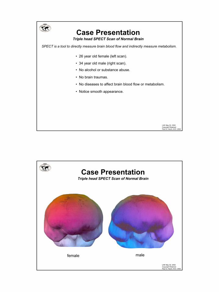
• 26 year old female (left scan).
Case PresentationTriple head SPECT Scan of Normal Brain
• No alcohol or substance abuse.
• No brain traumas.
• No diseases to affect brain blood flow or metabolism.
• Notice smooth appearance.
SPECT is a tool to directly measure brain blood flow and indirectly measure metabolism.
• 34 year old male (right scan).
LHE May 02, 2002Copyright RetainedPaul G. Harch, M.D. 2002
Case PresentationTriple head SPECT Scan of Normal Brain
female male
3
LHE May 02, 2002Copyright RetainedPaul G. Harch, M.D. 2002
• 19 year old male; college freshman. Ejected from motor vehicle at 65 mph in 1991(1st HBOT began 19 hours post accident).
• Impacted left frontal/parietal skull region.• Within ½ hour Glascow coma scale was 6-7, ventilator dependent.
1st scan: SPECT image 1 month after accident shows significant injury to left frontal area and contra coup injury to right parietal/occipital with luxury perfusion.
Case PresentationAcute & Chronic Treatment of Traumatic Brain Injury and Coma
• After initial treatments patient became conversant & independently ambulatory with slight spasticity.
• CT revealed diffuse edema, midline shift, petechial hemorrhages, subarachnoid hemorrhage, small subdural hematoma, basilar skull fracture.
• Scan shows very large defects in brain flood flow.
• Within 8 weeks of accident patient went from ventilator to walking and talking.
LHE May 02, 2002Copyright RetainedPaul G. Harch, M.D. 2002
2nd scan: 1 year later after 188 HBOT treatments.
• Treatments discontinued when patient enrolled in remedial college courses.
• In Jan. 2001, 12 years post injury, patient called physician to tell him of 2nd
promotion at the bank. Patient active, functional, and employed.
• Current cost of 188 HBOT treatments at $150-$200 each:
Case PresentationAcute & Chronic Treatment of Traumatic Brain Injury and Coma (continued)
• Noticeable improvement in cognition (40% gain written computation math).
$28,200-$37,600
• Patient verbalized insight to condition and that he could no longer aspire to be a surgeon.
• Balance and gait improvement from 3-wheel trike to 2-wheel bike.
• Patient referred to as “Lazarus” by his doctor.
$28,200-$37,600• Note: treatment during immediate acute phase of injury could cost more.
• Improved perfusion in ischemic penumbral areas of right-sided lesions.

4
LHE May 02, 2002Copyright RetainedPaul G. Harch, M.D. 2002
Scan #1 Scan #2
Case PresentationAcute & Chronic Treatment of Traumatic Brain Injury and Coma
19 y. male
LHE May 02, 2002Copyright RetainedPaul G. Harch, M.D. 2002
See previous page for case history on this patient.

5
LHE May 02, 2002Copyright RetainedPaul G. Harch, M.D. 2002
• 23 year old male, Navy Medical Corpsman, graduated second in class.
Case PresentationTraumatic Brain Injury and Substance Abuse
• Complaints of migraines, short & long-term memory loss, speech problems, seriousalcohol abuse. Treated at Bethesda and Walter Reed Brain Injury Center.
• 5 TBIs; (4 w/loss of consciousness) the 3rd resulted in largest loss of memory and started migraines.
1st scan: Extensive frontal lobe injury & top scan shows extensive tissue damage.
• Jan. 2001: 40 HBOT treatments over 4 weeks.
2nd scan: Extensive improvement in blood flow to frontal & parietal lobes.
• Dramatic cessation of migraine headaches. • Cessation of marijuana use & dramatic reduction in alcohol use.
• 1 month after treatments: Return of memory, tremendous improvement in speech, cognition, math skills, and energy levels.
• 8 months after treatment: Married & remains functional to date; patient to return for additional HBOTs.
• Unable to remember anatomy and physiology during rehab training; 14 jobs in 2 years.
• Discharged from Navy with 60% VA disability.
LHE May 02, 2002Copyright RetainedPaul G. Harch, M.D. 2002
Case PresentationTraumatic Brain Injury and Substance Abuse
23 y. male
Scan #1
Scan #2

6
LHE May 02, 2002Copyright RetainedPaul G. Harch, M.D. 2002
• 23 year old female.
Case PresentationTraumatic Brain Injury
• 5 ½ years post motor vehicle accident; major TBI & ventilation for 3 weeks.
• Complaints of aphasia, left body weakness with spasticity, cognitive problems w/generalized decrease in intellectual capacity, emotional instability, moodswings & temper tantrums post injury.
• Prior to HBOT: Innumerable therapies with no resolution of problems.
1st scan: Marked reduction in flow to right temporal lobe.Marked decrease in parasagittal regions of brain. Surface texture very coarse (heterogeneous blood flow).
2nd scan: Shows greatly improved brain blood flow.• Improvement in left body paresis/imbalance.• Temper tantrums/mood swings noticeably decreased.• Ability to read/write substantially improved.• Marked improvement in physical endurance.
• 80 HBOT treatments.
LHE May 02, 2002Copyright RetainedPaul G. Harch, M.D. 2002
Case PresentationTraumatic Brain Injury
23 y. female
Scan #1
Scan #2
7
LHE May 02, 2002Copyright RetainedPaul G. Harch, M.D. 2002
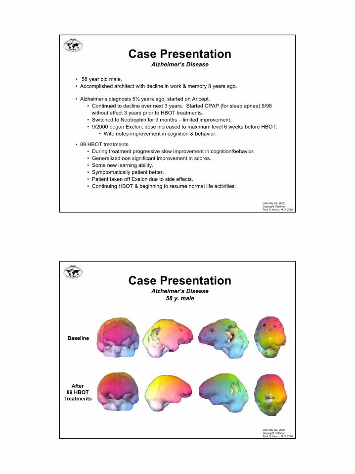
Case PresentationAlzheimer’s Disease
• 58 year old male.• Accomplished architect with decline in work & memory 8 years ago.
• Alzheimer’s diagnosis 5½ years ago; started on Aricept.• Continued to decline over next 3 years. Started CPAP (for sleep apnea) 9/98
without effect 3 years prior to HBOT treatments.• Switched to Neotrophin for 9 months – limited improvement.• 9/2000 began Exelon; dose increased to maximum level 6 weeks before HBOT.
• Wife notes improvement in cognition & behavior.
• 89 HBOT treatments.• During treatment progressive slow improvement in cognition/behavior.• Generalized non significant improvement in scores.• Some new learning ability.• Symptomatically patient better.• Patient taken off Exelon due to side effects.• Continuing HBOT & beginning to resume normal life activities.
LHE May 02, 2002Copyright RetainedPaul G. Harch, M.D. 2002
Case PresentationAlzheimer’s Disease
58 y. male
Baseline
After89 HBOT
Treatments

8
LHE May 02, 2002Copyright RetainedPaul G. Harch, M.D. 2002
Case PresentationPhysical Abuse
• 21 year old female, gang raped and beaten extensively at age 12.
• Significant cognitive deficit though apparently normal motor coordination.• Severe difficulties with sleep.
• 60 HBOT treatments.
• Improved cognitive function enables patient to operate in a higher capacityin a mentally demanding job.
1st scan: SPECT shows significant frontal lobe injury with severe frontal lobe tissue damage.
2nd scan: SPECT shows greatly increased blood flow to frontal lobemanifested by thickening & filling in on scan.
• Patient promoted 6 months post treatment.• Sleep difficulties improved.
LHE May 02, 2002Copyright RetainedPaul G. Harch, M.D. 2002
Case PresentationPhysical Abuse
21 y. female
Scan #1 Scan #2

9
LHE May 02, 2002Copyright RetainedPaul G. Harch, M.D. 2002
• 44 year old male. Mentally retarded from likely combination of delivery-inducedtrauma and TBI at 2 weeks old.
• Unable to read or spell more than a few words. No abstract reasoning ability. Difficulty understanding concepts, i.e., food will spoil if left out.
• Seizures bi-weekly with constant tremors; on medication. Worked 2 days/week at Goodwill doing menial tasks.
• 40 HBOT treatments.
1st scan: SPECT shows reduction in blood flow to frontal lobe (extensive frontallobe damage).
2nd scan: SPECT shows a marked increase in blood flow in the frontal lobe, manifest by closure of fissures.
• Work attendance from 2 days to 4 days per week. • Seizure rate fell to 1 per month on medication. Noticeable reduction in motor tremors.• Presently learning to read at a Kindergarten level. • Able to understand abstract concepts better, i.e., that food spoils. • Able to do more complex work tasks. Increased rational behavior.
Case PresentationMental Retardation
LHE May 02, 2002Copyright RetainedPaul G. Harch, M.D. 2002
Scan #1 Scan #2
Case PresentationMental Retardation
44 y. male

10
LHE May 02, 2002Copyright RetainedPaul G. Harch, M.D. 2002
• 8 year old boy.
Case PresentationCerebral Palsy
• Complicated, difficult delivery resulted in ischemic hypoxic injury to brain.
1st scan: Prominent abnormalities in temporal lobes, especially on left side.
2nd scan: Generalized improvement in blood flow with greater amount of yellow on the slices on far right of picture.
• Patient’s mother reported:• Some improvement in inappropriate behavior.• Less leg dragging. • Markedly improved ability to have bowel movements with no incontinence. • Muscle tone, attention, concentration improvements.
• Improved flow to both temporal lobes.
LHE May 02, 2002Copyright RetainedPaul G. Harch, M.D. 2002
Case PresentationCerebral Palsy
8 y. male
Scan #1 Scan #2 #1 #2

11
LHE May 02, 2002Copyright RetainedPaul G. Harch, M.D. 2002
• 60 year old male. 2 years post stroke.
Case PresentationStroke
• History of multiple white matter strokes.
• Complaints of body weakness, intractable dizziness, difficulty swallowing, speech problems.
1st scan: Surface of brain with coarse texture & markedly decreasedblood flow in right temporal lobe.
• 80 HBOT treatments.
2nd scan: Improvement in coarse texture & greatly increased blood flow.
• Dizziness reduced to point that patient could:• Leave house. • Walk without cane. • Stopped using left knee brace.• Speech & swallowing improved.• Overall mood improved.
LHE May 02, 2002Copyright RetainedPaul G. Harch, M.D. 2002
Case PresentationStroke
60 y. male
Scan #1 Scan #2

12
LHE May 02, 2002Copyright RetainedPaul G. Harch, M.D. 2002
• 68 year old male. 1 month post embolic stroke right middle cerebral artery.
Case PresentationAlcoholism and Stroke
• Chronic heavy drinker. Developed atrial fibrillation, likely secondary to the alcohol.
• Severe weakness left side of body; profound left-sided neglect.• Noticeable cognitive deterioration (power of attorney to son).
1st scan: Multiple areas of decreased perfusion in frontal & temporal lobes.
• 1 month of HBOT treatments:
2nd scan: Improved flow to all lobes. Global smoothing of brain surface.
• Slightly coarse appearance to brain surface. Note large gap on left side of each image denoting right hemispheric stroke.
• Dramatic improvement in cognition, speed of thinking, decrease in neglect.• Markedly improved motor function on left side of body.• Rescinded power of attorney & resumed active pursuit of business affairs.
LHE May 02, 2002Copyright RetainedPaul G. Harch, M.D. 2002
Scan #1 Scan #2
Case PresentationAlcoholism and Stroke
68 y. male
13
LHE May 02, 2002Copyright RetainedPaul G. Harch, M.D. 2002
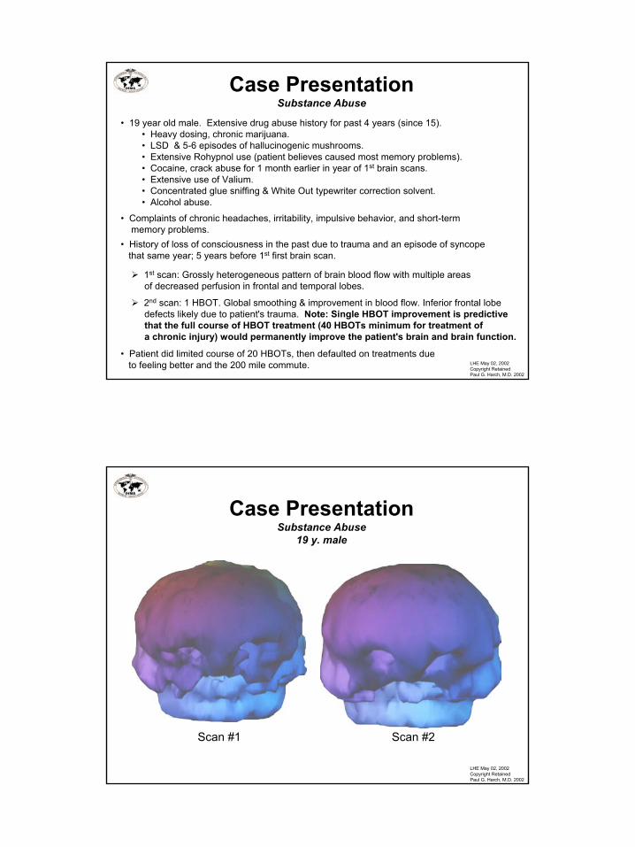
• 19 year old male. Extensive drug abuse history for past 4 years (since 15).• Heavy dosing, chronic marijuana.• LSD & 5-6 episodes of hallucinogenic mushrooms.• Extensive Rohypnol use (patient believes caused most memory problems).• Cocaine, crack abuse for 1 month earlier in year of 1st brain scans.• Extensive use of Valium.• Concentrated glue sniffing & White Out typewriter correction solvent.• Alcohol abuse.
Case PresentationSubstance Abuse
• Complaints of chronic headaches, irritability, impulsive behavior, and short-termmemory problems.
• History of loss of consciousness in the past due to trauma and an episode of syncope that same year; 5 years before 1st first brain scan.
1st scan: Grossly heterogeneous pattern of brain blood flow with multiple areas of decreased perfusion in frontal and temporal lobes.
2nd scan: 1 HBOT. Global smoothing & improvement in blood flow. Inferior frontal lobe defects likely due to patient's trauma. Note: Single HBOT improvement is predictive that the full course of HBOT treatment (40 HBOTs minimum for treatment of a chronic injury) would permanently improve the patient's brain and brain function.
• Patient did limited course of 20 HBOTs, then defaulted on treatments dueto feeling better and the 200 mile commute.
LHE May 02, 2002Copyright RetainedPaul G. Harch, M.D. 2002
Scan #1 Scan #2
Case PresentationSubstance Abuse
19 y. male

14
LHE May 02, 2002Copyright RetainedPaul G. Harch, M.D. 2002
• 51 year old female.
Case PresentationCarbon Monoxide Poisoning
• Exposed to home gasoline powered generator in poorly ventilated area.
• Complaints of headaches, trouble thinking, walking, talking, functioning, increasingly somnolent & confused. Diagnosed with altered mental status.
• At 36 hours blood carboxyhemoglobin elevated at 2.2% (upper limits ofnormal, 1.5%).
1st scan: Pattern very coarse with multiple significant deficits in blood flow.
• 1 HBOT 68 hours after admission at 2 atms for 35 mins (patient claustrophobic).
2nd scan: 3 hours later• Dramatic improvement in brain blood flow.• Smoothing of the overall pattern.• Noticeable improvement in mental and cognitive status.
LHE May 02, 2002Copyright RetainedPaul G. Harch, M.D. 2002
Scan #1
Scan #2
Case PresentationCarbon Monoxide Poisoning
51 y. female
top view
15
LHE May 02, 2002Copyright RetainedPaul G. Harch, M.D. 2002
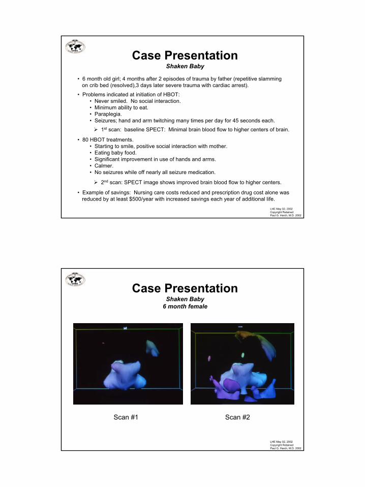
Case PresentationShaken Baby
• 6 month old girl; 4 months after 2 episodes of trauma by father (repetitive slammingon crib bed (resolved),3 days later severe trauma with cardiac arrest).
• Problems indicated at initiation of HBOT:• Never smiled. No social interaction.• Minimum ability to eat.• Paraplegia.• Seizures; hand and arm twitching many times per day for 45 seconds each.
1st scan: baseline SPECT: Minimal brain blood flow to higher centers of brain.
• 80 HBOT treatments.• Starting to smile, positive social interaction with mother.• Eating baby food.• Significant improvement in use of hands and arms.• Calmer.• No seizures while off nearly all seizure medication.
2nd scan: SPECT image shows improved brain blood flow to higher centers.
• Example of savings: Nursing care costs reduced and prescription drug cost alone was reduced by at least $500/year with increased savings each year of additional life.
LHE May 02, 2002Copyright RetainedPaul G. Harch, M.D. 2002
Case PresentationShaken Baby
6 month female
Scan #1 Scan #2

16
LHE May 02, 2002Copyright RetainedPaul G. Harch, M.D. 2002
• 29 year old female; self-inflicted .38 caliber hollow point to right temple.
Case PresentationGun Shot Wound to Brain
1st scan: baseline SPECT: Marked diffuse decrease in blood flow with worstarea along path of bullet.
• 80 HBOT treatments.• Physiatrist reports: Generalized decrease in spasticity, increase in left hand grip,
and movement in knees.• Patient reports:
• Increased trunk and extremity motor function.• Marked decrease in insomnia.• Natural bowel movements without constipation.• Decreased headaches.
2nd scan: Generalized increase in brain blood flow with pattern of diffuse smoothing.
• 6 years post trauma:• Severe spasticity arms and legs.• Paraplegia with severe weakness in arms.• Poor trunk control.• Insomnia secondary to muscle spasms.• Intractable constipation.• Headaches.
LHE May 02, 2002Copyright RetainedPaul G. Harch, M.D. 2002
Case PresentationGun Shot Wound to Brain
29 y. female
Scan #1 Scan #2right view
17
LHE May 02, 2002Copyright RetainedPaul G. Harch, M.D. 2002
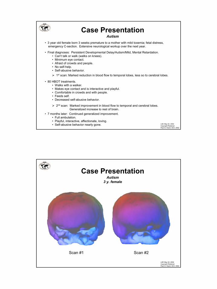
Case PresentationAutism
• 7 months later: Continued generalized improvement.• Full ambulation.• Playful, interactive, affectionate, loving.• Self-abusive behavior nearly gone.
• 3 year old female born 3 weeks premature to a mother with mild toxemia; fetal distress, emergency C-section. Extensive neurological workup over the next year.
• Final diagnoses: Persistent Developmental Delay/Autism/Mild, Mental Retardation.• Can't talk or walk (walks on knees).• Minimum eye contact.• Afraid of crowds and people.• No self-help.• Self-abusive behavior.
1st scan: Marked reduction in blood flow to temporal lobes, less so to cerebral lobes.
• 80 HBOT treatments.• Walks with a walker.• Makes eye contact and is interactive and playful.• Comfortable in crowds and with people.• Feeds self.• Decreased self-abusive behavior.
2nd scan: Marked improvement in blood flow to temporal and cerebral lobes.Generalized increase to rest of brain.
LHE May 02, 2002Copyright RetainedPaul G. Harch, M.D. 2002
Case PresentationAutism
3 y. female
Scan #1 Scan #2

18
LHE May 02, 2002Copyright RetainedPaul G. Harch, M.D. 2002
Case PresentationTraumatic Brain Injury from Child Abuse
• 48 year old male. 45 years post injury; extensive physical damage to right parietal bone at age 3.
• 63 HBOT treatments.
• Apparently normal motor coordination. • Difficulty with higher math and remembering names. • Dreamed in black and white.
• Improved motor coordination, math skills, and name memory.• Now dreams in color.
1st scan: Before
2nd scan: After
LHE May 02, 2002Copyright RetainedPaul G. Harch, M.D. 2002
Case PresentationTraumatic Brain Injury from Child Abuse
48 y. male
Scan #1 Scan #2

19
LHE May 02, 2002Copyright RetainedPaul G. Harch, M.D. 2002
Dr. Harch’s cost-effective, refined low-pressure Hyperbaric Oxygen Therapy
protocols have improved the quality of life for 1,000’s of brain and neurologically injured
patients. Treatment is here today!
Dr. Harch’s success has been replicated by physicians in multiple treatment centers
throughout the United States and many other countries around the world.
For a treatment center near you please visitwww.hbomedtoday.com/dbsearch.html
or call Ken Locklear, Executive Director of IHMA, at 561-640-4546.
For further technical information please visitwww.hyperbarics.org
or call Dr. Paul Harch, President of IHMA, at 504-348-1660.
Presentation prepared by Anita Poole CPS/CAP