Hydrops fetalis
57
PROF. M.C.BANSAL . MBBS. MS . MICOG. FICOG. FOUNDR PRINCIPAL & CONTROLLER; JHALAWAR MEDICAL COLLEGE AND HOSPITAL, JHALWAR. EX . PRINCIPAL & CONTROLLER; MAHATMA GANDHI MEDICLA COLLEGE AND HOSPITAL, SITAPURA , JAIPUR. HYDROPS FETALIS
-
Upload
drmcbansal -
Category
Education
-
view
2.884 -
download
11
description
Transcript of Hydrops fetalis
PROF. M.C.BANSAL . MBBS. MS . MICOG. FICOG.
FOUNDR PRINCIPAL & CONTROLLER; JHALAWAR MEDICAL COLLEGE AND HOSPITAL, JHALWAR.
EX . PRINCIPAL & CONTROLLER; MAHATMA GANDHI MEDICLA COLLEGE AND HOSPITAL,
SITAPURA , JAIPUR.
HYDROPS FETALIS
Hydrops Fetalis
It is defined as generalized fetal edema or anasarca , which can be detected by an Antenatal USG.
It may be manifestation of a variety of underlying disorders.
The condition is subdivided in to (a) Immune hydrops . ( b) non immune hydrops. immune variety is more common in developing countries
as Rh grouping is not an universal phenomenon . Non immune variety is more common in the developed
countries; ratio being reported to be 9: 1 of non immune to immune cases.
Rh –Ve persions ---India 5- 10 % 0f population –it is least in Japan < 2% and 15 % in Europe and USA.
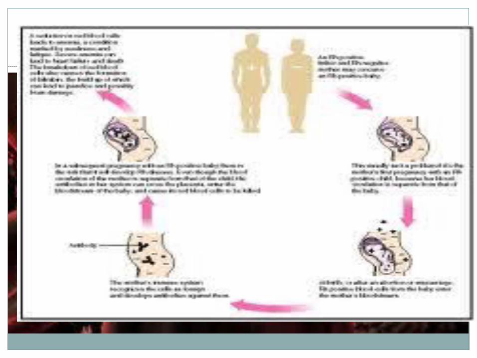
Development of Rh- D antibodies
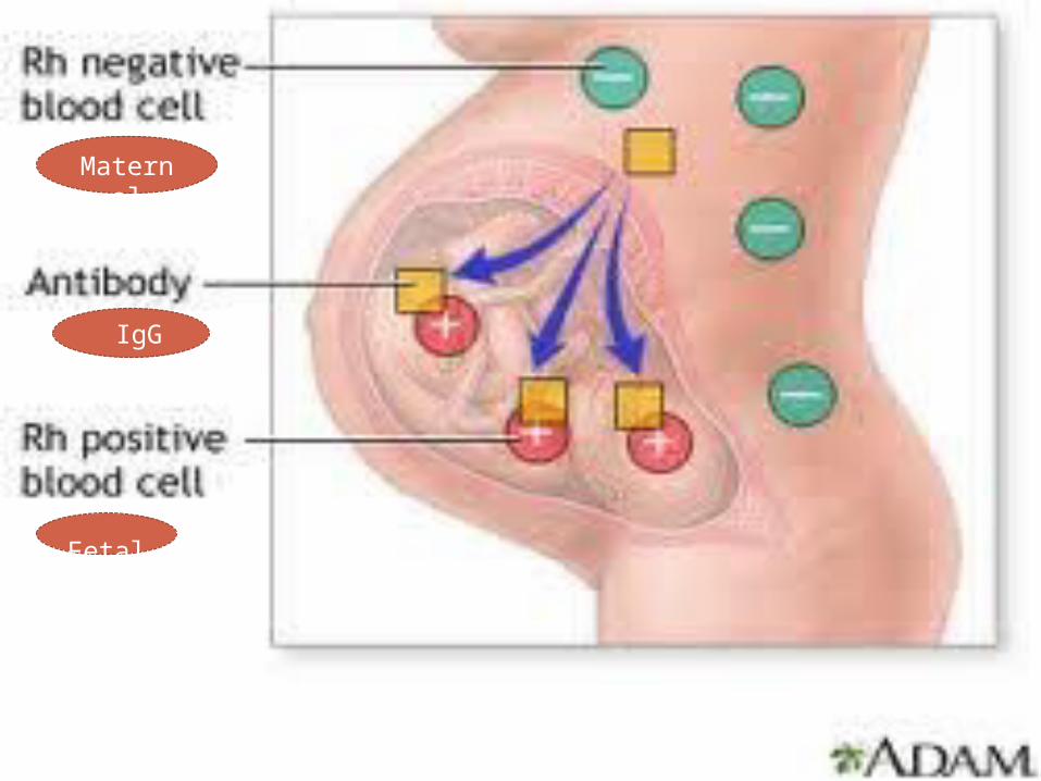
When Rh +Ve RBCs enters the Rh-Ve mother’s blood circulation; they stimulate the maternal immune response producing RBC antigen –specific antibodies –isoimmunization (sensitization ) .
After few weeks , IgM antibodies ( Saline antibodies ) are produced. IgM do not cross the placenta barrier Hence can not
cause fetal damage . On subsequent exposure of fetal Rh D antigen , her previously
primed B cells act swiftly to produce IgG ( albumin antibodies ) which can cross placenta ---and bounds to antigen of fetal RBCs
causing sequestration and destruction of fetal RBCs. Antibodies are formed in 16% of the mothers significantly after
6months of larger amount of fetomaternal bleeding and once formed remain in circulation through the life. And each
subsequent pregnancy will have booster effect especially when fetus is Rh +Ve fetus is in utero.
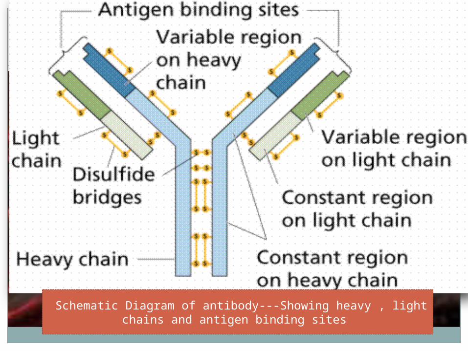
Schematic Diagram of antibody---Showing heavy , light chains and antigen binding sites
Matern
al
IgG
Fetal

Immune Hydrops
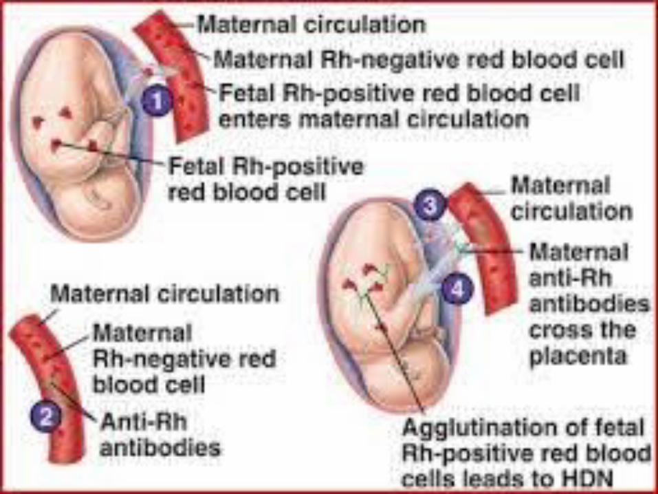
The commonest cause of immune hydrops fetalis is pregnancy in Rh negetive women with a Rh positive fetus .
Rh negetive mother who is already sensitized by either by a previous pregnancy or blood transfusion, is capable of producing Immunogloblin G.
These Immunoglobin G (IgG) antibodies cross the placenta and destroy (haemolysed ) the Rh positive RBCs of fetus.
This phenomenon was reported by Levine in 1941.
Immune Hydrops Fetalis-------
First pregnancy proceeds normally as Rh -ve mother is usually not sensitized.
Feto -placental maternal transfusion occurs at the time of placental separation and the Rh +Ve fetal RBCs sensitize t6he maternal immune system.
Other procedures during pregnancy (even in first pregnancy ) can sensitize the mother are feto- maternal blood transfusion--- occuring in amnioscentesis , threatened abortion ,Ectopic pregnancy, MTP,D&E, abruptio placenta, APH , External podalic version, IPV, LSCS, MRP, Chorionic Villus Sampling , PIH , etc.
Immune Hydrops----
In subsequent pregnancy the fetal RBCs entering in maternal circulation may be destroyed /or some of them may have booster effect for maternal sensitization.
Maternal Immunoglobins G cross the placenta and destroy fetal RBCs .
In mild cases, the fetus has haemolytic disease resulting in anaemia with mild icterus.
In severe cases fetus develops hydrops fetalis. If IgG titers are high the disease process may start
even in early period of gestation --- resulting in IUFD.
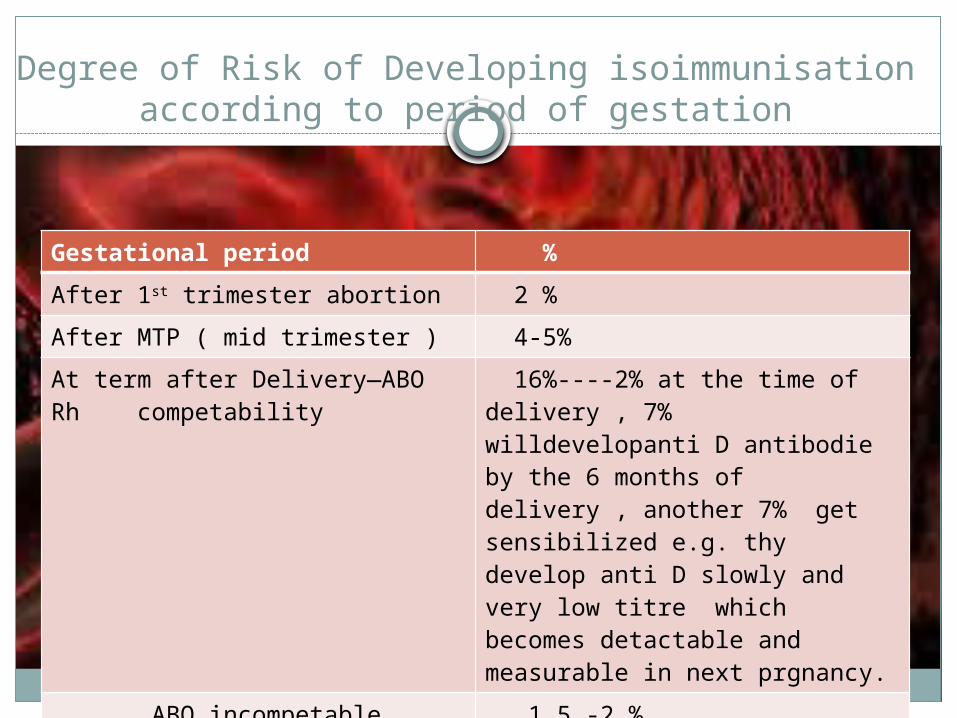
Degree of Risk of Developing isoimmunisation according to period of gestation
Gestational period %
After 1st trimester abortion 2 %
After MTP ( mid trimester ) 4-5%
At term after Delivery—ABO Rh competability
16%----2% at the time of delivery , 7% willdevelopanti D antibodie by the 6 months of delivery , another 7% get sensibilized e.g. thy develop anti D slowly and very low titre which becomes detactable and measurable in next prgnancy.
ABO incompetable 1.5 -2 %
ABO incompatibility confer substantial protection against developing Rh (D) issoimmunisation

Why all babies are not affected in Rh –Ve mother
Volume of fetomaternal trnsfusion may be too low to sensitise.
Immunological non responders ---pregnancy induced suppression of immune system –30%.
ABO Major group incompetability. Rh set up of the fetal blood may change the
Rh –antigenicity ( stimulus )
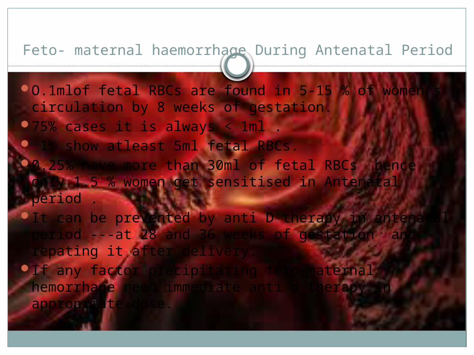
Feto- maternal haemorrhage During Antenatal Period
O.1mlof fetal RBCs are found in 5-15 % of women’s circulation by 8 weeks of gestation.
75% cases it is always < 1ml . 1% show atleast 5ml fetal RBCs. 0.25% have more than 30ml of fetal RBCs hence
only 1.5 % women get sensitised in Antenatal period .
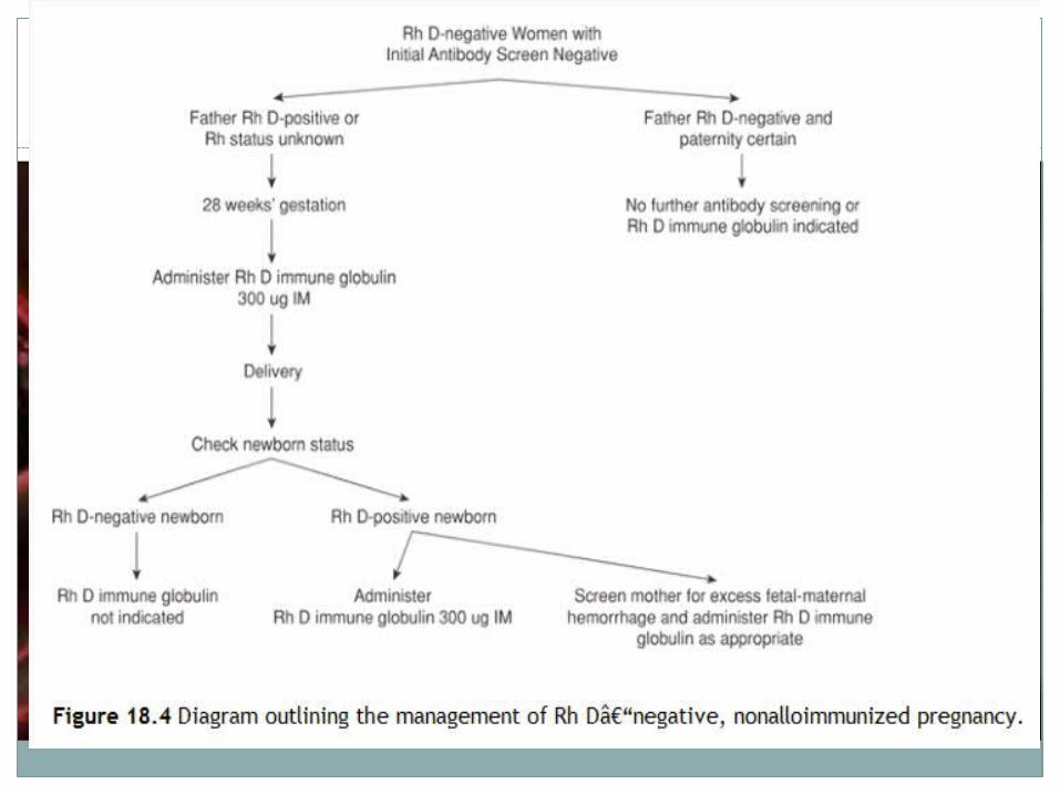
It can be prevented by anti D therapy in antenatal period ---at 28 and 36 weeks of gestation and repating it after delivery.
If any factor precipitating feto-maternal hemorrhage need immediate anti d therapy in appropriate dose.

Prophylaxis-----
Universal ABO Rh grouping of all girls / expactant mother.
Premarrital councellig regarding The Rh factor in both life partners --- advising marriage of identical Rh factor.
Rh Immunoglobulin (Rh-IgG) therapy within 24-48 hrs to all Rh –Ve women after ectopic/MTP/ abortion/ delivery /amnioscentasis ,Version , APH bleeding and at 28th and 36th week of gestation in primigravida too..
This therapy was introduced in1966 and its world wide acceptance has remarkably reduced the incidence of immune hydrops in developed countries , but it is still common type of hydrops in developing world.

Hydrops fetalis
Occasionally . Immune hydrops may be due to other erythrocyte antigen apart from Rh D antigen.
These include –major A.B.O. incopetability and minor blood group antigens ---C D E, Kell , Jk, S, c , and Duffy.
If issoimmunization occurs , then the titers of antibodies are monitored at appropriate intervals depending on the levels and rapidity with which they are changing ( either every 2 or 4 weeks).
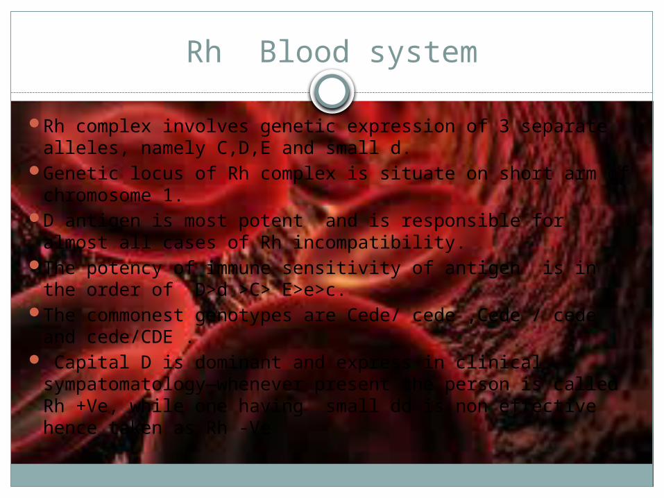
Rh Blood system
Rh complex involves genetic expression of 3 separate alleles, namely C,D,E and small d.
Genetic locus of Rh complex is situate on short arm of chromosome 1.
D antigen is most potent and is responsible for almost all cases of Rh incompatibility.
The potency of immune sensitivity of antigen is in the order of D>d >C> E>e>c.
The commonest genotypes are Cede/ cede ,Cede / cede and cede/CDE .
Capital D is dominant and express in clinical sympatomatology—whenever present the person is called Rh +Ve, while one having small dd is non effective hence taken as Rh -Ve

Homozygous / Heterozygous Rh- Grouping.
Homozygous Rh + Ve = DD ( D on both sets of antigen--- mother and father both Have contributed D &D.
Homologous Rh –ve = dd eg. Each of both small d parents have contributed set of small d( Maternal d and paternal d)
Heterozygous means---Rh +ve when Dd( one logus has D and another logus has d) eg. D has come from father / mother and d has come from Mother / father – both parents have contributed different D and d.
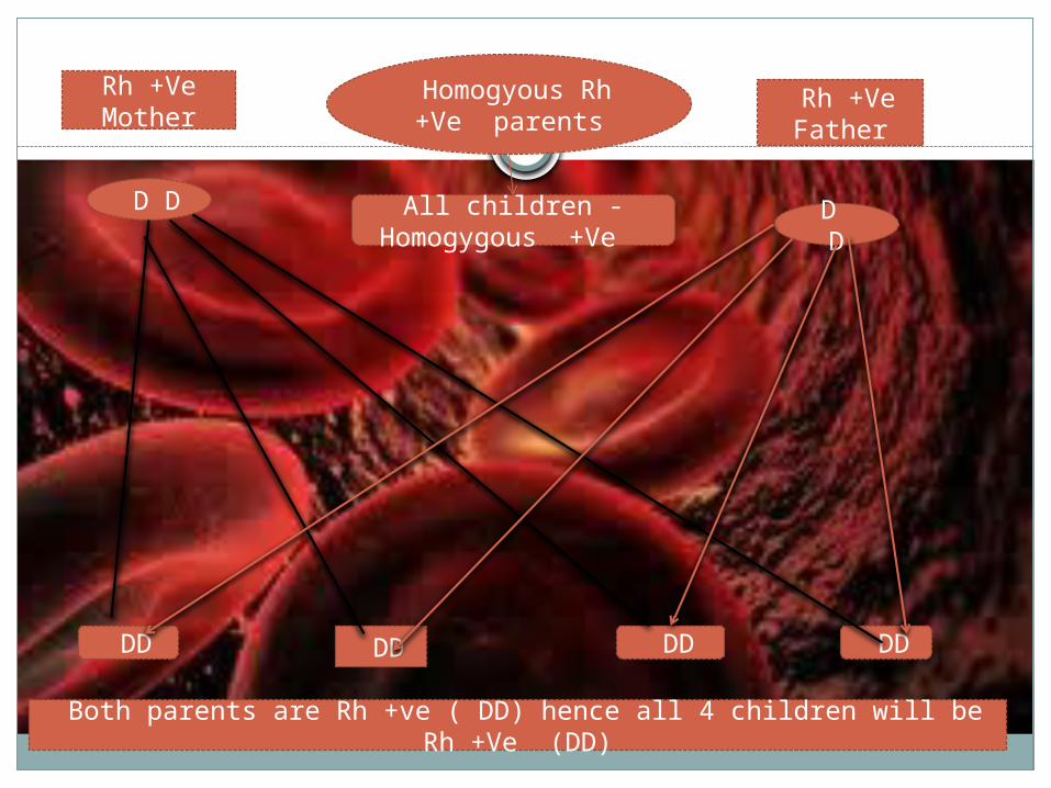
Rh +Ve Mother
Rh +Ve Father
D D D D
DD DD DD DD
Both parents are Rh +ve ( DD) hence all 4 children will be Rh +Ve (DD)
Homogyous Rh +Ve parents
All children - Homogygous +Ve
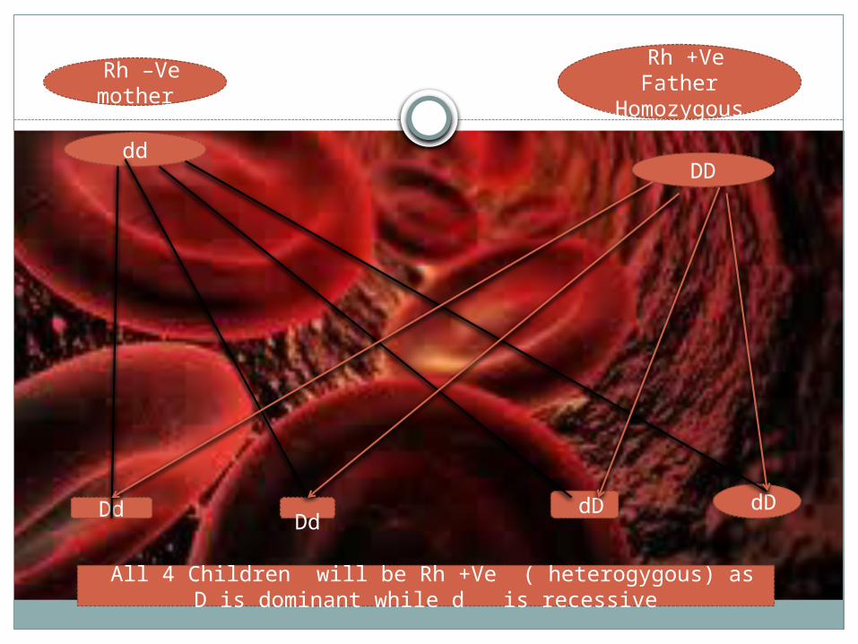
Rh –Ve mother
Rh +Ve Father
Homozygous
ddDD
Dd
Dd dD dD
All 4 Children will be Rh +Ve ( heterogygous) as D is dominant while d is recessive
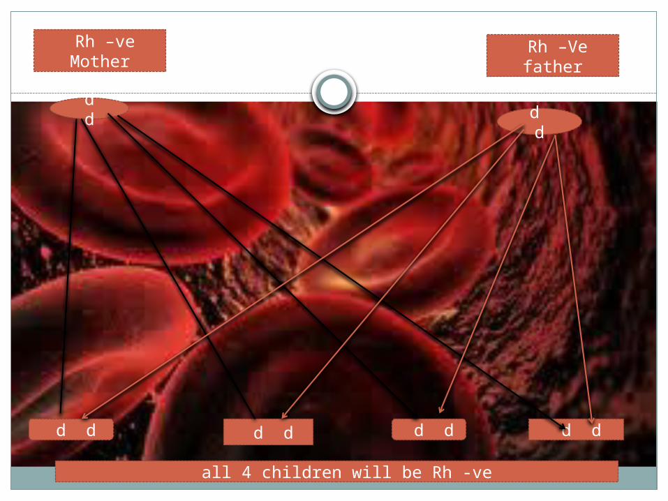
Rh –ve Mother
Rh –Ve father
d d d d
d d d d d d d d
all 4 children will be Rh -ve
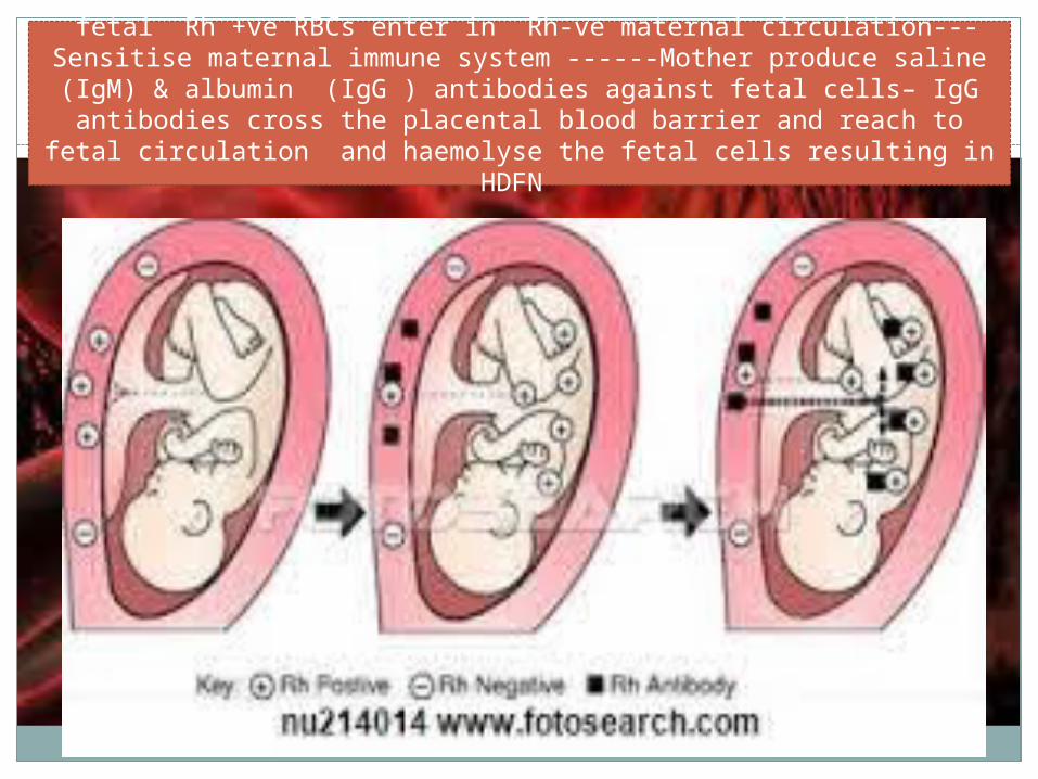
fetal Rh +ve RBCs enter in Rh-ve maternal circulation---Sensitise maternal immune system ------Mother produce saline (IgM) & albumin (IgG ) antibodies against fetal cells– IgG antibodies cross the placental
blood barrier and reach to fetal circulation and haemolyse the fetal cells resulting in HDFN
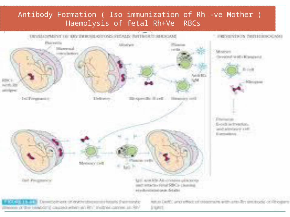
Antibody Formation ( Iso immunization of Rh -ve Mother ) Haemolysis of fetal Rh+Ve RBCs
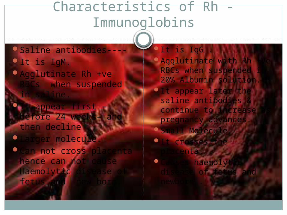
Characteristics of Rh -Immunoglobins
Saline antibodies----It is IgM.Agglutinate Rh +ve
RBCs when suspended in saline.
It appear first –before 24 weeks– and then decline
Larger molecule.Can not cross placenta
hence can not cause Haemolytic disease of fetus and new born.
It is IgG .Agglutinate with Rh +Ve
RBCs when suspended in 20% Albumin solution.
It appear later the saline antibodies & continue to increase as pregnancy advances.
Small Molecule.It crosses the placenta.Causes haemolytic
disease of fetus and newborn .
Manifestation Of HDFN ( Erythroblastosis Fetalis
Depending upon the degree of fetal RBCs haemolysis ---It may be - 1. Hydrops Fetalis ----Fetus is severely affected .. 2. Icterus gravidorum neonatorum -----Relatively less affected. 3.Congenital Anaemia ---of the newborn ; mildly affected . Baby develops anaemia ; jaundice is not so evident or mild , prognosis is good
1. S e v e r e f o r m o f R h – H D F N.2. S e v e r a n a e m i a , g e n e r a l i s e d e d e m a , a s c i t e s , a n d h y d r o t h o r a x .3. H i g h c a r d i a c o u t p u t .4. L i v e r d a m a g e d u e t o f e t a l a n o x a e m i a a n d a c i d o s i s . 5. L i v e r b e c o m e t h e s i t e o f h e m o p o e s i s t o c o m p e n s a t e a n e m i a
d u e t o h a e m o l y s i s . 6. L i v e r f a i l s t o p r o d u c e p r o t e i n s f r o m a m i n o a c i d s - - -
h y p o p r o t e i n a e m i a ; r e s p o n s i b l e f o r g e n e r a l i s e d e d e m a .7. E n l a r g e m e n t o f p l a c e n t a d u e t o h y p e r p l a s i a t o m e e t t h e
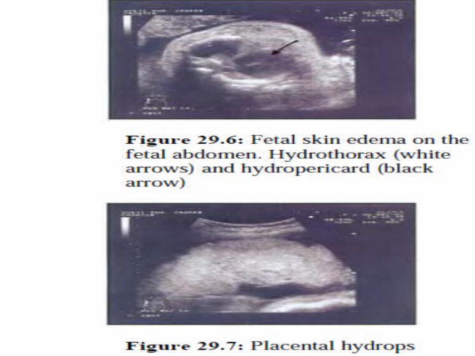
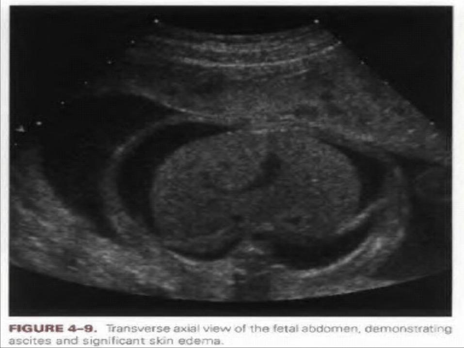
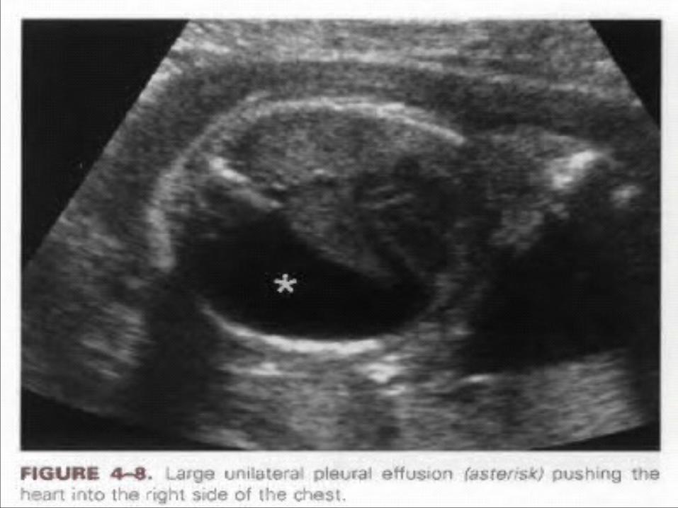
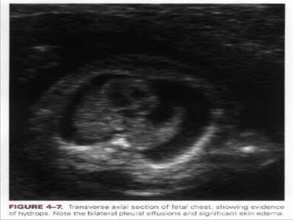
d e m a n d o f o 2 t r a n s fu s i o n t o f e t u s .8. U SG - - - s c a l p e d e m a , s k i n e d e m a , p e r i c a r d i a l , p l e u r a l e ff u s i o n
a n d a s c i t e s .9. X- r a y — b u d d h a p o s i t i o n _ _ h a l l o a r o u n d s c a l p — s c a l p
e d e m a . L i m b s a p a r t - - - d u e t o a b d o m i n a l d i s t e n s i o n .10. O u t c o m e u s u a l l y I U F D , s o m e t i m e l i v e b i r t h b u t i m m e d i a t e
n e w b o r n d e a t h .
Hydrops Fetalis
Lesser form of haemolyt ic d isease than hydrops feta l is .Newborn develops jaundice wi th in 24 hours a f ter b ir th . In uterus , most of the unconjugated b i l i rubin is washed away through the p lacenta and thus no development of feta l jaundice in utero. Af ter de l ivery excess of b i l i rubin is not excreted due to fa i lure of a large amount of b i l i rubin load by the l iver of neonate . When serum bi l i rubin leve l r ise above 20mg % i t crosses the b lood bra in barr ier to depos i t in basa l gangl ia resul t ing in kernicterus . This baby needs exchange transfus ion.
Icterus Gravis Neonatorum
Blood in v ials are sent to lab –(a) 3ml oxalate / EDTA blood-- for Hb% and
peripheral smear. - - -Haemolysed RBCs, ghost cel ls (Maternal cel ls )
(b) 2ml clotted blood in another v ial - - - for ABO Rh grouping , direct coomb’s test and serum bi lrubin
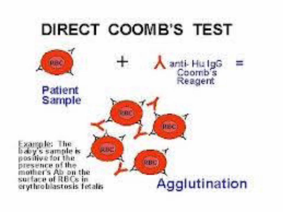
level .DIRECT COOMB’S TEST i t is test to know , is baby
is aff ected by Rh –haemolyt ic disease or not .I f direct coomb’s test is +ve . i t means baby is aff ected and special management is to be done
acoording to serum bi l irubin level . As mother is already sensit ized there is no role of
prophylact ic anti D therapy . I f direct coomb’s test is –ve - - -newborn is not
aff ected by Rh – iso immunizationi
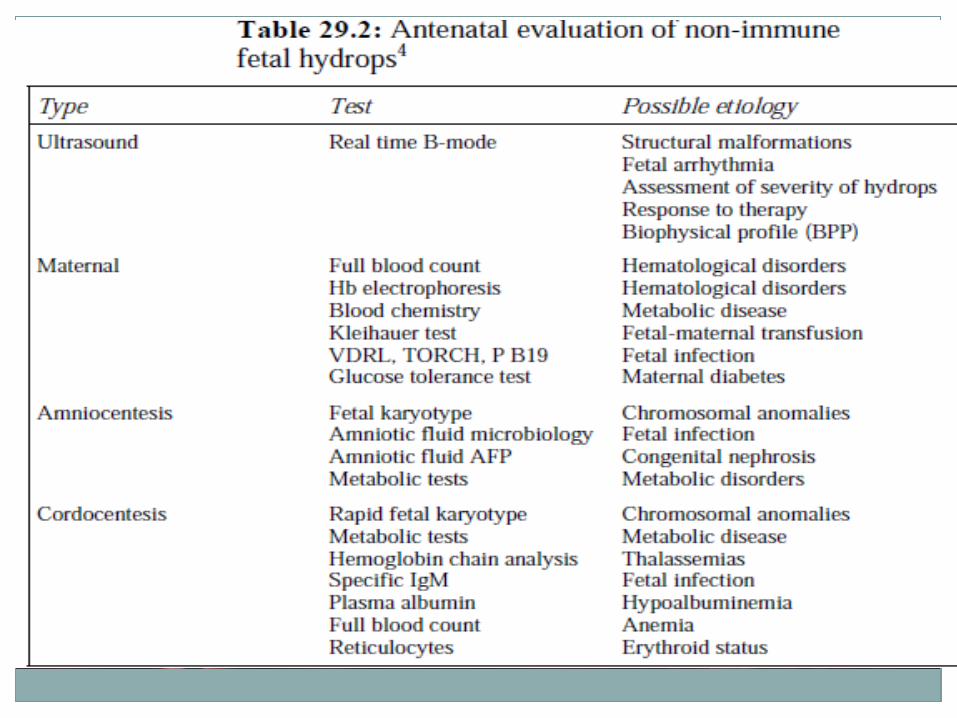
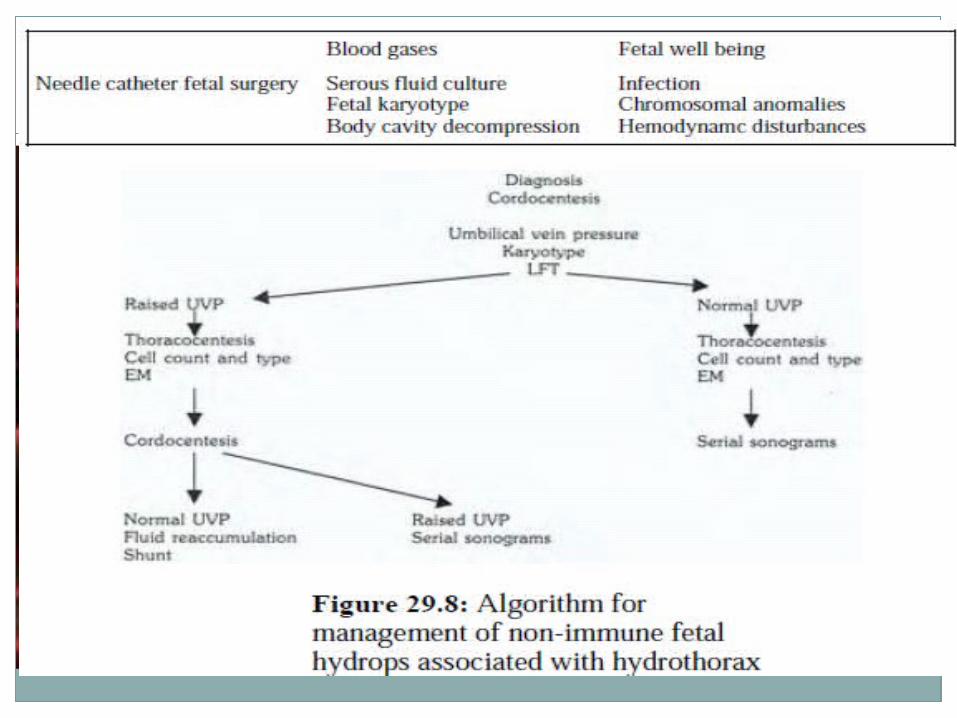
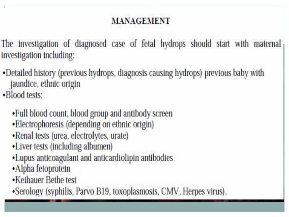
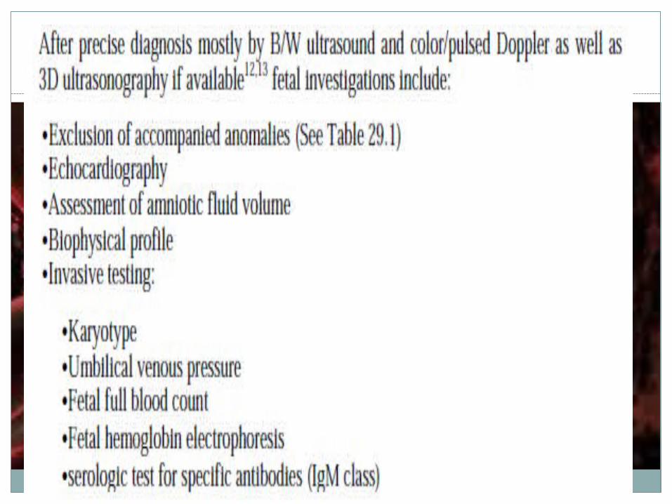
Investigations
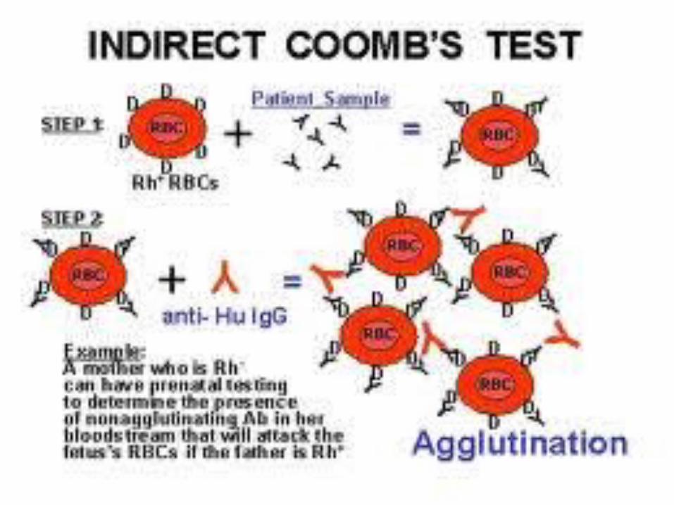
(a)INDIRECT COOMB’S TEST RBC’s of specifi c or mixed antigen ( KNOWN) are mixed with
serum of patient and anti human antiglobul in is added .
I f agglutination of RBC’s occur ; i t means antibodies against Rh factor are present in
patient ’s blood. Antibodies present in patient ’s serum get attached
to RBCs and agglutination occurs.
Titer 1: 16 means agglutination occurs up to 16 t imes di lut ion of patients serum .
(b)DIRECT COOMB’S TEST Anti human anti globul in are added direct ly to the rocs of baby.
Agglutination means anti bodies are bound to the rocs of baby.
Investigations
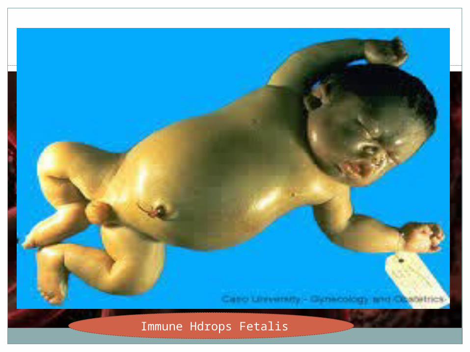
Immune Hdrops Fetalis
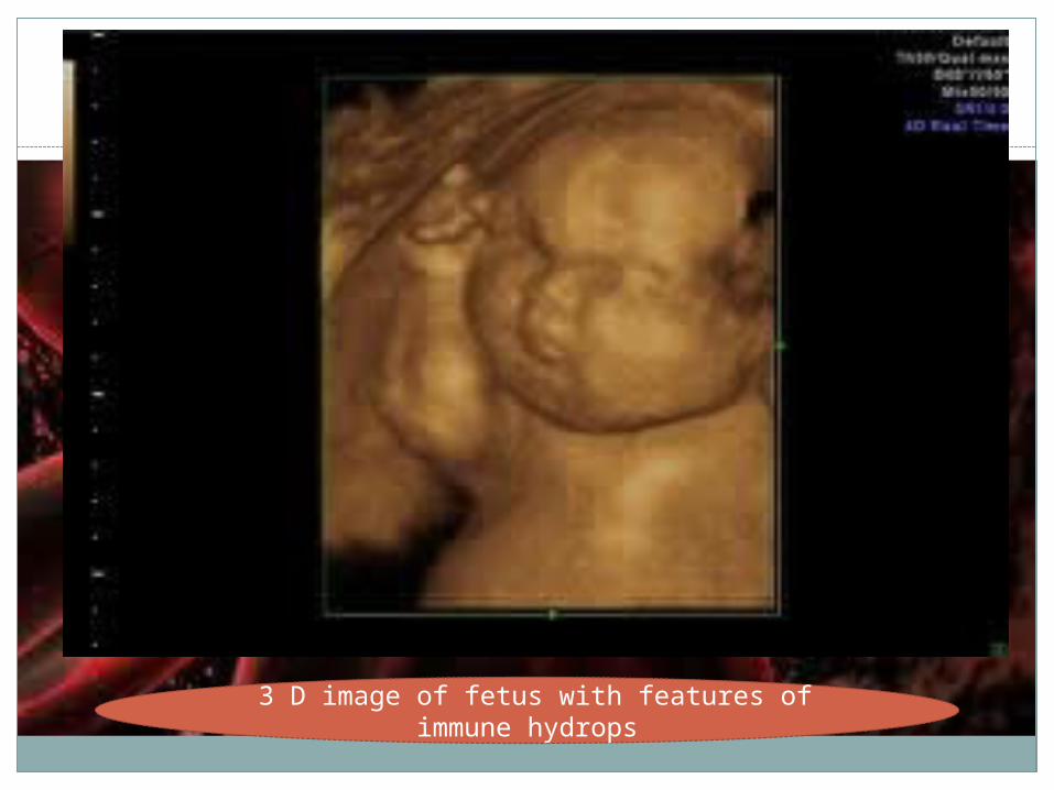
3 D image of fetus with features of immune hydrops
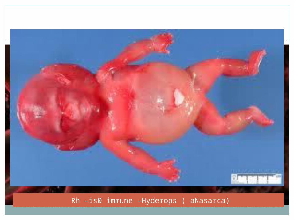
Rh –is0 immune –Hyderops ( aNasarca)
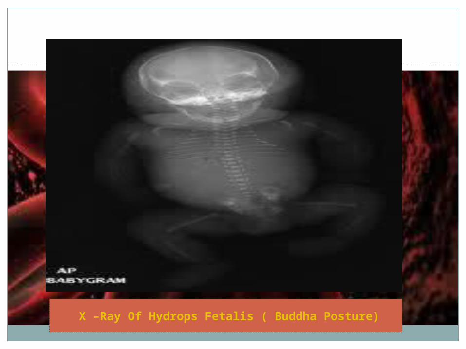
X –Ray Of Hydrops Fetalis ( Buddha Posture)
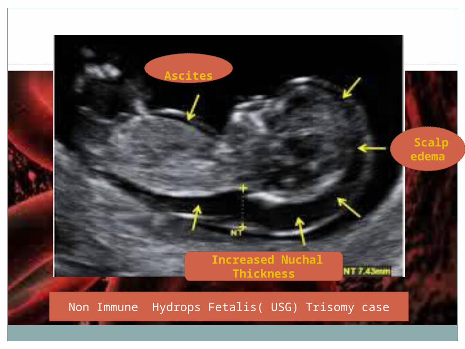
Non Immune Hydrops Fetalis( USG) Trisomy case
Ascites
Scalp edema
Increased Nuchal Thickness
Routine antenatal Rh anti therapy is recommended to Rh negetive women married to
Rh+ve partner.300 ug , im in deltoid muscle at 28 weeks and
36 weeks of gestation.100 ug , im is given after any bleeding in 1 s t
trimester.Higher dose to be given after any precipitating
factor or procedure l ikely to cause feto placental hemorrhage.
300ug anti D is suff icient to neutralize 15 ml of have fetal cell / 30ml of whole blood.
Immunoprophylaxis
KLEIHAUER- BETKE TEST - Based on acid elution technique. Fetal and maternal rocs have diff erent response to KOH solution.Maternal cel ls (adult Hb ) get eluded leaving behind only cel l membrane and hence appear as swol len round large “GHOST CELLS” against normal fetal cel ls whose Hb remain unaltered hence look as red refract i le round cel ls .
I f in 50 low power fi elds of maternal peripheral blood 80 fetal RBC’s are found- i t is est imated that
4ml of fetomaternal hemorrhage has occurred. For 1ml of fetal blood 10ug of Rh anti D is needed. Thus 300ug anti D wi l l be suff icient for 30 ml of fetal blood which has entered the maternal c irculat ion.
Measurement of fetomaternal Haemorrhage
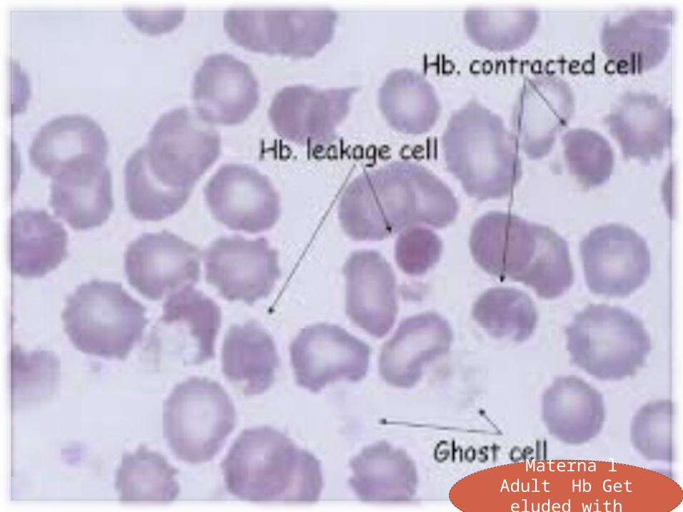
Materna l Adult Hb Get eluded
with KOH
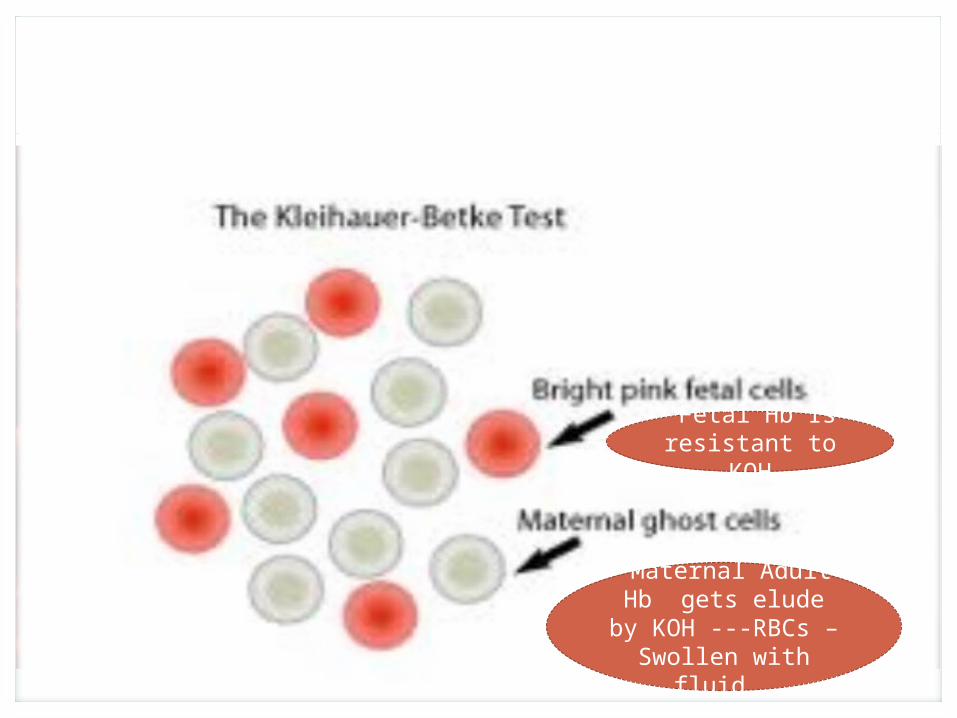
Fetal Hb Is resistant to
KOH
Maternal Adult Hb gets elude by
KOH ---RBCs –Swollen with fluid
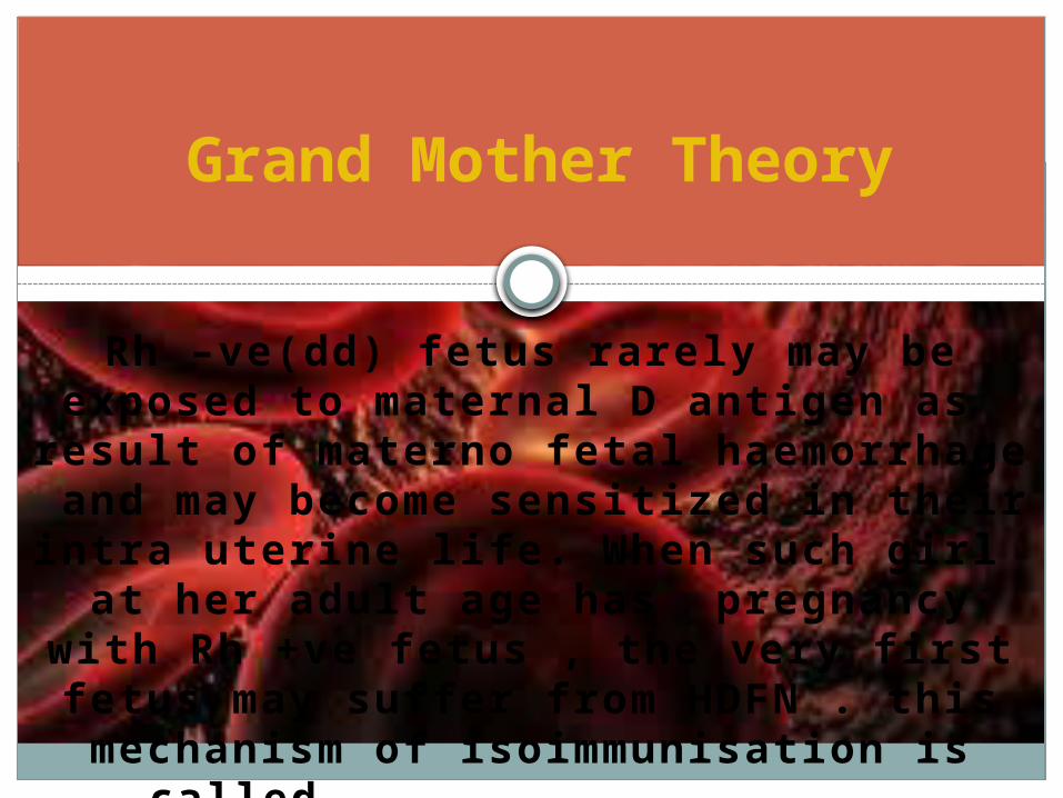
Rh –ve(dd) fetus rarely may be exposed to maternal D antigen as
result of materno fetal haemorrhage and may become sensitized in their
intra uterine life. When such girl at her adult age has pregnancy with Rh
+ve fetus , the very fi rst fetus may suff er from HDFN . this mechanism of isoimmunisation is called GRAND
MOTHER THEORY.
Grand Mother Theory
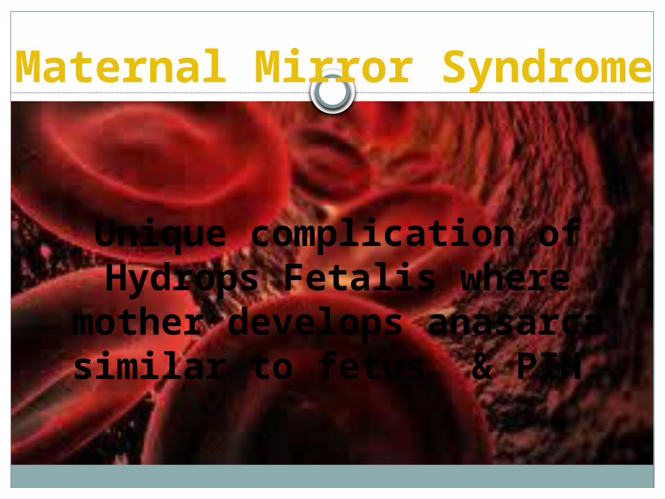
Unique complication of Hydrops Fetalis where
mother develops anasarca similar to fetus & PIH.
Maternal Mirror Syndrome
In a previously sensitized / sensibilized women the antibody
titer may rise to high level in subsequent pregnancy.
This may occur in d-negative fetus also.
Amnestic Response
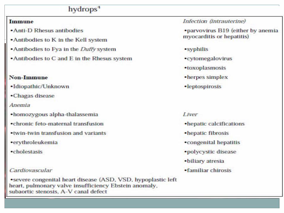

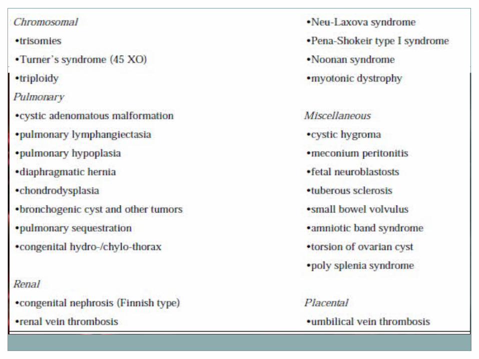
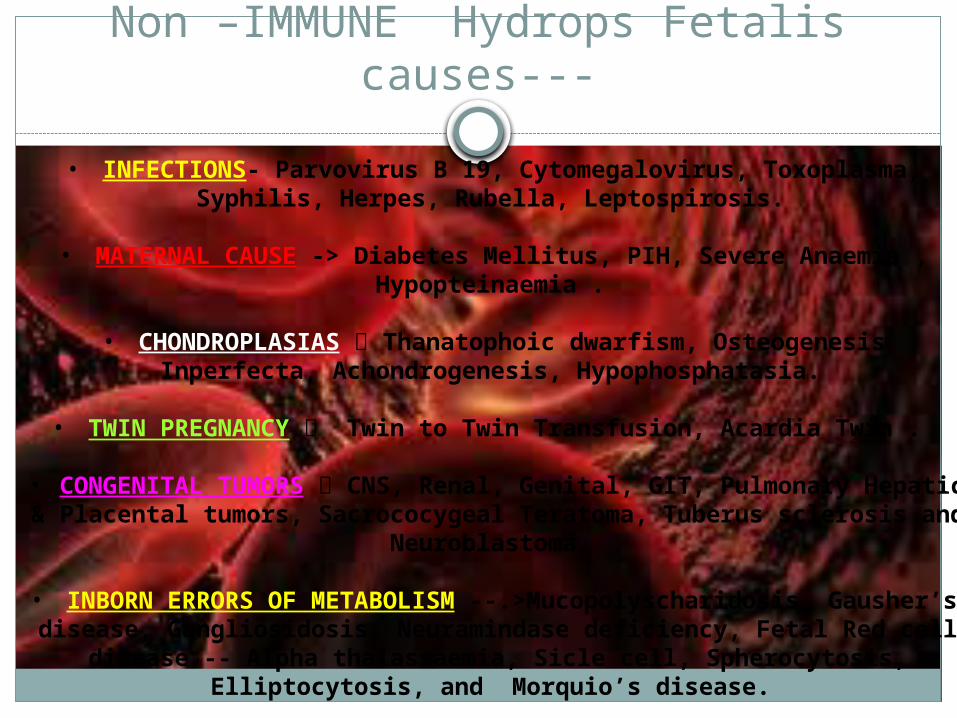
Non –IMMUNE Hydrops Fetalis causes---
• INFECTIONS- Parvovirus B 19, Cytomegalovirus, Toxoplasma, Syphilis, Herpes, Rubella, Leptospirosis.
• MATERNAL CAUSE -> Diabetes Mellitus, PIH, Severe Anaemia , Hypopteinaemia .
• CHONDROPLASIAS Thanatophoic dwarfism, Osteogenesis Inperfecta, Achondrogenesis, Hypophosphatasia.
• TWIN PREGNANCY Twin to Twin Transfusion, Acardia Twin .
• CONGENITAL TUMORS CNS, Renal, Genital, GIT, Pulmonary Hepatic & Placental tumors, Sacrococygeal Teratoma, Tuberus
sclerosis and Neuroblastoma.
• INBORN ERRORS OF METABOLISM --.>Mucopolyscharidosis, Gausher’s disease, Gangliosidosis, Neuramindase deficiency, Fetal Red cell disease -- Alpha thalassaemia, Sicle cell, Spherocytosis,
Elliptocytosis, and Morquio’s disease.
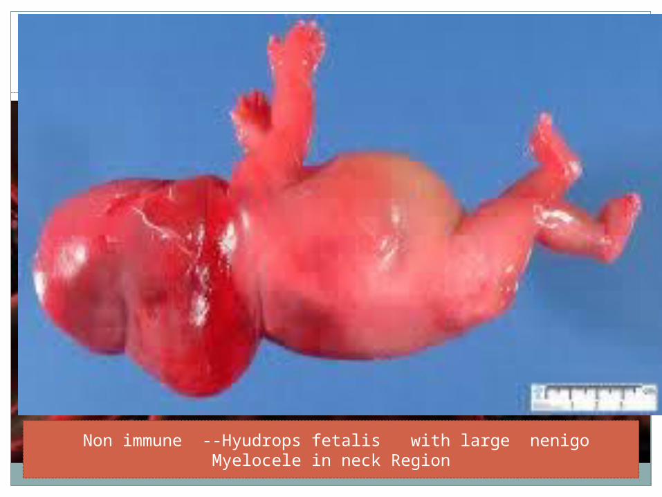
Non immune --Hyudrops fetalis with large nenigo Myelocele in neck Region