Hydrogen peroxide induces DNA single- and double...
12
Hydrogen peroxide induces DNA single- and double-strand breaks in thyroid cells and is therefore a potential mutagen for this organ Natacha Driessens 1 , Soetkin Versteyhe 1 , Chiraz Ghaddhab 1 , Agne ` s Burniat 1 , Xavier De Deken 1 , Jacqueline Van Sande 1 , Jacques-Emile Dumont 1 , Franc ¸oise Miot 1 and Bernard Corvilain 1,2 1 Institut de Recherche Interdisciplinaire en Biologie Humaine et Mole ´ culaire and 2 Department of Endocrinology of Erasme Hospital, Faculty of Medicine, Universite ´ Libre de Bruxelles, Bat C Local C4.145, Campus Erasme, 808, Route de Lennik, B-1070 Brussels, Belgium (Correspondence should be addressed to N Driessens; Email: [email protected]) Abstract DNA double-strand breaks (DSBs) are considered as one of the primary causes of cancer but their induction by hydrogen peroxide (H 2 O 2 ) is still controversial. In this work, we studied whether the high levels of H 2 O 2 produced in the thyroid to oxidize iodide could induce DNA modifications. Scores of DNA damage, in terms of strand breaks, were obtained by comet assay (alkaline condition for single-strand breaks (SSBs) and neutral condition for DSBs). We demonstrated that in a rat thyroid cell line (PCCl3), non-lethal concentrations of H 2 O 2 (0.1–0.5 mmol/l) as well as irradiation (1–10 Gy) provoked a large number of SSBs (w2–3 times control DNA damage values) but also high levels of DSBs (1.2–2.3 times control DNA damage values). We confirmed the generation of DSBs in this cell line and also in human thyroid in primary culture and in pig thyroid slices by measuring phosphorylation of histone H2AX. L-Buthionine-sulfoximine, an agent that depletes cells of glutathione, decreased the threshold to observe H 2 O 2 -induced DNA damage. Moreover, we showed that DNA breaks induced by H 2 O 2 were more slowly repaired than those induced by irradiation. In conclusion, H 2 O 2 causes SSBs and DSBs in thyroid cells. DSBs are produced in amounts comparable with those observed after irradiation but with a slower repair. These data support the hypothesis that the generation of H 2 O 2 in thyroid could also play a role in mutagenesis particularly in the case of antioxidant defense deficiency. Endocrine-Related Cancer (2009) 16 845–856 Introduction Thyroid nodules are common and constitute an important clinical problem. They may occur in up to 50% of a population above 60 years old (Ross 2002). Among these nodules, 5% are cancerous, mainly papillary cancers (PTC). While the prevalence of clinically significant PTC remains relatively low, the prevalence of papillary microcarcinoma is clearly higher and reaches 13% in some series of autopsies (Nasir et al. 2000). There is no clear explanation for this high frequency of thyroid tumors. Iodine defici- ency increases the prevalence of hot nodules and multinodular goiters but does not modify the global incidence of thyroid cancers even though it raises the relative proportion of follicular carcinoma (Krohn & Paschke 2002). Irradiation is the only environmental risk factor clearly implicated in thyroid cancer pathogenesis (Ron et al. 1995). The initial event in the majority of PTC consists of an activation of the RAS/RAF/MEK/MAP kinase pathway, either directly by mutation of BRAF and more rarely of RAS, or indirectly by constitutive activation of tyrosine kinase receptors resulting from chromosomal rearrangements (RET/PTC, TRK; Lacroix et al. 2005). After irradiation, carcinogenic processes are mainly attributed to the formation of Endocrine-Related Cancer (2009) 16 845–856 Endocrine-Related Cancer (2009) 16 845–856 1351–0088/09/016–845 q 2009 Society for Endocrinology Printed in Great Britain DOI: 10.1677/ERC-09-0020 Online version via http://www.endocrinology-journals.org
Transcript of Hydrogen peroxide induces DNA single- and double...

Endocrine-Related Cancer (2009) 16 845–856
Hydrogen peroxide induces DNAsingle- and double-strand breaks inthyroid cells and is therefore a potentialmutagen for this organ
Natacha Driessens1, Soetkin Versteyhe1, Chiraz Ghaddhab1, Agnes Burniat1,Xavier De Deken1, Jacqueline Van Sande1, Jacques-Emile Dumont1,Francoise Miot1 and Bernard Corvilain1,2
1Institut de Recherche Interdisciplinaire en Biologie Humaine et Moleculaire and 2Department of Endocrinology of Erasme Hospital,
Faculty of Medicine, Universite Libre de Bruxelles, Bat C Local C4.145, Campus Erasme, 808, Route de Lennik, B-1070 Brussels,
Belgium
(Correspondence should be addressed to N Driessens; Email: [email protected])
Abstract
DNA double-strand breaks (DSBs) are considered as one of the primary causes of cancer but theirinduction by hydrogen peroxide (H2O2) is still controversial. In this work, we studied whether thehigh levels of H2O2 produced in the thyroid to oxidize iodide could induce DNA modifications.Scores of DNA damage, in terms of strand breaks, were obtained by comet assay (alkalinecondition for single-strand breaks (SSBs) and neutral condition for DSBs). We demonstrated thatin a rat thyroid cell line (PCCl3), non-lethal concentrations of H2O2 (0.1–0.5 mmol/l) as well asirradiation (1–10 Gy) provoked a large number of SSBs (w2–3 times control DNA damage values)but also high levels of DSBs (1.2–2.3 times control DNA damage values). We confirmed thegeneration of DSBs in this cell line and also in human thyroid in primary culture and in pig thyroidslices by measuring phosphorylation of histone H2AX. L-Buthionine-sulfoximine, an agent thatdepletes cells of glutathione, decreased the threshold to observe H2O2-induced DNA damage.Moreover, we showed that DNA breaks induced by H2O2 were more slowly repaired than thoseinduced by irradiation. In conclusion, H2O2 causes SSBs and DSBs in thyroid cells. DSBs areproduced in amounts comparable with those observed after irradiation but with a slower repair.These data support the hypothesis that the generation of H2O2 in thyroid could also play a role inmutagenesis particularly in the case of antioxidant defense deficiency.
Endocrine-Related Cancer (2009) 16 845–856
Introduction
Thyroid nodules are common and constitute an
important clinical problem. They may occur in up to
50% of a population above 60 years old (Ross 2002).
Among these nodules, 5% are cancerous, mainly
papillary cancers (PTC). While the prevalence of
clinically significant PTC remains relatively low, the
prevalence of papillary microcarcinoma is clearly
higher and reaches 13% in some series of autopsies
(Nasir et al. 2000). There is no clear explanation for
this high frequency of thyroid tumors. Iodine defici-
ency increases the prevalence of hot nodules and
multinodular goiters but does not modify the global
Endocrine-Related Cancer (2009) 16 845–856
1351–0088/09/016–845 q 2009 Society for Endocrinology Printed in Great
incidence of thyroid cancers even though it raises the
relative proportion of follicular carcinoma (Krohn &
Paschke 2002). Irradiation is the only environmental
risk factor clearly implicated in thyroid cancer
pathogenesis (Ron et al. 1995).
The initial event in the majority of PTC consists of
an activation of the RAS/RAF/MEK/MAP kinase
pathway, either directly by mutation of BRAF and
more rarely of RAS, or indirectly by constitutive
activation of tyrosine kinase receptors resulting from
chromosomal rearrangements (RET/PTC, TRK;
Lacroix et al. 2005). After irradiation, carcinogenic
processes are mainly attributed to the formation of
Britain
DOI: 10.1677/ERC-09-0020
Online version via http://www.endocrinology-journals.org

N Driessens et al.: H2O2 provokes DNA breaks in thyroid cells
DNA double-strand breaks (DSBs; Sarasin et al. 1999).
This damage results from a direct effect of irradiation
but also from the generation of reactive oxygen species
(ROS) formed during water radiolysis (Mikkelsen &
Wardman 2003). However, the precise molecular
mechanisms of most radiation-induced cancers are
largely unknown. Nevertheless, irradiation is certainly
not responsible for the majority of thyroid tumors.
Therefore, since 1990, we hypothesized that the
elevated frequency of thyroid tumors (benign or
malignant) could be partially explained by the
prominent mutagenic environment present in the
thyroid, resulting from its metabolism producing
large amounts of hydrogen peroxide (H2O2; Song
et al. 2007). Indeed, in thyroid, H2O2 generated by the
DUOX enzymes (Dupuy et al. 1999, De Deken et al.
2000) is required and is the limiting co-factor for
iodide oxidation by thyroperoxidase (TPO) and thyroid
hormone synthesis (Nunez & Pommier 1982). Quan-
titatively, a stimulated thyrocyte generates almost as
much H2O2 as a stimulated leukocyte (Corvilain et al.
1994). However, while the leukocyte dies soon after
activation, the thyrocyte life is much longer (maximum
7 divisions during adulthood; Coclet et al. 1989)
allowing mutations to accumulate. H2O2 could facili-
tate a mutagenic process and lead to tumorigenesis by
altering the DNA (oxidation of bases, DNA single-
strand breaks (SSBs) and DSBs). H2O2 could also
enhance cell proliferation through various mechanisms
(Stone & Yang 2006). Arguments to support the
involvement of H2O2 in mutagenesis and etiopatho-
genesis of thyroid nodules were recently reviewed by
our group and others (Maier et al. 2006, Song et al.
2007). The existence of a more mutagenic environment
in the thyroid than other organs are suggested: 1) the
spontaneous mutation rate is around 10 times higher in
the thyroid than in the liver, 2) the comet assay detects
increased levels of oxidized pyrimidine and purine in
thyroid compared with other organs, and 3) immuno-
histochemistry methods reveal higher levels of
8-oxoguanine in the thyroid (Maier et al. 2006).
A gene expression signature reflecting the differences
in cellular response to g-radiation and H2O2 could
distinguish radiation-induced (from the Chernobyl
Tissue Bank) and spontaneous PTCs (from French
patients with no history of radiation exposure; Detours
et al. 2007). These data reinforce our hypothesis
suggesting that in thyroid, H2O2 could provoke DNA
damage and mutations. Nevertheless, harmful effects
caused by H2O2 are tightly controlled in thyroid,
thanks to the restricted apical localization of its
production and the presence of various intracellular
H2O2 detoxifying enzymes, like seleno-dependent
846
glutathione peroxidase (GPx; Kohrle et al. 2005). In
epidemiological studies, it has been shown that the
modification of antioxidant capacities in the diet can
modulate cancer risk (Bertram et al. 1987, Ip et al.
1994, Vogt et al. 2003). Antioxidant status of cells may
also play a role in the mutagenic potency of irradiation
and oxidative stress (Kim et al. 2000, Neumann et al.
2003, Wang et al. 2003).
While carcinogenic DNA DSBs are clearly induced
by irradiation, their induction by H2O2 is still
controversial. Therefore, we analyzed in this work
the capacity of H2O2 to generate DNA SSBs and
especially DSBs and compared this with the effects
observed with a well-known carcinogenic factor
(irradiation) to determine if H2O2, like irradiation,
could therefore be implicated in the pathogenesis of
thyroid tumors.
Materials and methods
Cell lines and culture conditions
PCCl3 cells, a rat thyroid cell line, were cultured
as previously described (Rigutto et al. 2007). Non-
transformed rat fibroblasts F208 were grown in
DMEM with 10% v/v fetal bovine serum, 1% v/v
sodium pyruvate, 100 U/ml penicillin, 100 mg/ml
streptomycin, and 2.5 mg/ml fungizone. Pig thyroids
were obtained from freshly killed animals in accord-
ance to ethical guidelines. Thyroids were cut into thin
slices (0.3 mm) of w50 mg wet weight. Slices were
preincubated at 37 8C for 1 h in 2 ml Krebs-Ringer–
Hepes (KRH) medium and then transferred to fresh
KRH before treatment (Corvilain et al. 2000). Primary
cultures of human thyroid cells were prepared and
incubated as previously described; the experiments
were performed, four days after seeding the thyroid
follicles, on differentiated thyrocytes (Roger et al.
1988). Human thyroid tissues were obtained from
patients undergoing thyroidectomy for multinodular
goiter with approval from the institutional ethic
committee.
Cell treatment
Irradiation
Cells were exposed to a single dose of g-irradiationfrom a Cesium137 source at a dose rate of 2.03 Gy/min
at room temperature. To determine the initial DNA
damage, cells were placed on ice immediately after
treatment. For the kinetic experiments cells were
incubated at 37 8C for different periods of time after
irradiation.
www.endocrinology-journals.org

Endocrine-Related Cancer (2009) 16 845–856
Hydrogen peroxide
H2O2 (Merck) dilutions were prepared in culture
medium immediately before use. The H2O2 solutions
were diluted in the medium of dishes containing cells
in culture or thyroid slices.
H2O2 generating system
A relatively stable production of H2O2 was obtained in
the cell culture medium using proline (Sigma–Aldrich)
in conjunction with 5 mU/ml D-amino-oxidase (DAO
from Sigma–Aldrich).
DL-Buthionine-[S,R]-sulfoximine treatment
Cells were preincubated overnight with 10 mmol/l
L-buthionine-sulfoximine (BSO; Sigma–Aldrich)
before H2O2 or irradiation treatment.
Cell survival test
The cytotoxic potential of the different treatments
was evaluated by the 3-(4,5-dimethylthiazol-2-yl)-
5-(3-carboxymethoxyphenyl)-2-(4-sulfophenyl)-2H-
tetrazolium (MTS) cell proliferation assay. This
colorimetric method measures the formation of a
soluble formazan product that is directly proportional
to the number of living cells in culture. After different
intervals of time after treatment, cells were incubated
with a MTS/phenazine methosulfate solution for 1–3 h
in the dark at 37 8C and in a 5% CO2 atmosphere as
described in the Promega (Promega) technical bulletin
no. 169. The soluble formazan product has an
absorbance maximum at 490–500 nm and was
recorded using an ELISA plate reader. Viability was
calculated as a percentage of the control. Triton 0.5%
v/v was used as a positive control for cell death.
H2O2 measurement
H2O2 was measured in the cell culture medium
by a sensitive fluorimetric assay based on the
H2O2-dependent oxidation of homovanillic acid
(3-methoxy-4-hydroxyphenylacetic acid, HVA;
Sigma–Aldrich) to a highly fluorescent dimer (2,2 0-
dihydroxy-3,3 0-dimethoxydiphenyl-5,5 0-diacetic acid)
by HRP (Benard & Brault 1971).
Iodide organification (protein bound iodide)
Pig thyroid slices were preincubated for 30 min in KRH
buffer containing 0.5 g/l BSA supplemented or not with
0.5 mmol/l H2O2. The slices were then incubated in
fresh medium supplemented with KI (10K5 mol/l) and125I (1 mCi/ml) for 30 min. Methimazole (1 mmol/l),
that blocks iodide organification, added during pre-
incubation and incubation, was used to estimate the
www.endocrinology-journals.org
background in the assay. Iodide organification was
measured in basal conditions and in slices stimulated
by TSH (10 mU/ml) and ionomycin (2 mmol/l). The
slices were homogenized in a methimazole solution
(1 mmol/l). Proteins were precipitated with 10% v/v
trichloroacetic acid and counted. Iodide bound to
proteins is expressed as picomoles of iodide organified
per 100 mg wet weight tissue/30 min.
Comet assays
The comet assay was adapted from Singh et al. (1988)
and Olive et al. (1990) as previously described (Chico
Galdo et al. 2006). This assay is based on the
separation from supercoiled DNA of DNA loops
containing strand breaks (SSBs and DSBs) that
become free to migrate out of the nucleus towards
the anode during an alkaline electrophoresis in a
solution of 0.3 M NaOH, 1 mM EDTA, pHO13. To
detect DSBs more specifically, the comet assay was
adapted to neutral electrophoresis in a buffer of
300 mM sodium acetate, 100 mM Tris–HCl, pH 8.3 as
previously described (Wojewodzka et al. 2002).
DNA images were captured after staining with
ethidium bromide (20 mg/ml) with a Zeiss Axioplan
2 imaging microscope with a 40! objective lens. For
quantification, the comets were classified into different
categories. We subdivided cell DNA damage into five
stages (1–5) for the alkaline assay (Collins 2004) and
four stages (2–5) for the neutral assay according to the
length and the intensity of the comet tail as illustrated
in Fig. 1A. Stage 1 (no tail) and stage 2 (halo around
the nucleus) corresponded to cells without a significant
number of DNA strand breaks. Stages 3–5 corre-
sponded to a gradual increase in DNA damage. We
measured the comet score in 200 randomly selected
cells per slide. Results were, first, expressed as the
percentage of each stage of comets per slide. In a
second step, a comet score was calculated, following a
modification of Collins’ method, as the sum of the
percentage of each comet stage n (from 1 to 5)
multiplied by nK1 (Collins 2004). For this calculation,
all the negative comets (stages 1 and 2) were
considered as stage 2. The scores were expressed in
arbitrary units on a scale from 100 (all the comets are in
stage 2) to 400 (all the comets are in stage 5). While the
scale of the comet assay was the same in alkaline
(reflecting SSBs and DSBs) and neutral condition
(more specific for DSBs), the observed score remained
semi-quantitative and therefore the values could not be
compared to extrapolate the number of SSBs.
Comet immunoassays were performed as previously
described (Wojewodzka et al. 2002).
847

Figure 1 (A) Classification of the comets. Comet categories aredefined by the size of the head (nucleus) and the length andintensity of the tail. Five stages are defined in alkaline conditions(upper panel) reflecting global DNA damage (SSBs and DSBs)and four stages in neutral conditions (lower panel) reflectingessentially DSBs. Stage 1, normal nucleus; stage 2, haloaround the nucleus; stage 3–5, gradual increase in the lengthand intensity of the comet tail evolving in parallel with adecrease in the nuclear DNA content. (B) Comet assaysperformed on PCCl3 cells after incubation with 1 mM H2O2.Alkaline assays showed comets in stage 5 after labeling withethidium bromide and an Ab ssDNA. In neutral assays, the tailof the comet was not labeled by the Ab ssDNA, even thoughalmost all comets were in stage 3 after staining with ethidiumbromide. This means that in the neutral condition, the DNAcontent in the tail results mainly from DSBs. Ab ssDNA,antibody against SSBs; SSBs, single-strand breaks; and DSBs,double-strand breaks.
Table 1 Score of DNA damage measured by comet assay in
PCCl3 cells immediately after irradiation (A) and 15 min after
addition of various H2O2 concentrations (B). DNA damage was
evaluated in alkaline conditions (SSBsCDSBs) and in neutral
conditions (DSBs). Data are expressed as mean (arbitrary units
on a scale from 100 to 400)GS.E.M.; (n), number of
measurements. Statistical analyses were made by comparison
of control values and values obtained after treatments
Alkaline Neutral
(A) Irradiation
Ctl 120.4G2.1 (32) 113.2G1.9 (26)
1–2 Gy 165.3G15.4 (6)* 201.0G6.1 (6)*
4–5 Gy 202.1G15.8 (10)* 244.5G30.0 (2)*
10 Gy 283.0G6.4 (21)* 253.5G8.6 (24)*
(B) H2O2 (mmol/l)
0 120.1G2.2 (38) 110.5G1.1 (33)
0.01 125.7G5.7 (9) 105.8G1.9 (6)
0.05 128.1G7.2 (10) 132.8G11.2 (4)*
0.1 245.0G11.7 (19)* 215.6G13.4 (14)*
0.2 321.8G45.2 (6)* 241.9G23.2 (6)*
0.5 400.0G0.0 (4)* 225.4G42.3 (4)*
1 391.3G8.1 (15)* 204.5G9.7 (19)*
*P!0.001. SSBs, single-strand breaks; DSBs, double-strandbreaks.
N Driessens et al.: H2O2 provokes DNA breaks in thyroid cells
Western blotting and immunocytochemistry
Two or four mg of histone extracts obtained after lysis
in buffer (150 mmol/l NaCl, 1 mmol/l EDTA,
20 mmol/l Tris–HCl (pH 8), 0.5% v/v NP40) were
separated by 15% SDS/PAGE and transferred to
nitrocellulose. Immune complexes were detected with
HRP-coupled anti-rabbit or anti-mouse IgG antibodies
according to the ECL method (NEN Life science
product) as previously described (De Deken et al.
2000). Some nitrocellulose membranes were analyzed
by the Odyssey infrared imaging system (LI-COR,
Biosciences, Erembodegem, Belgium) using fluor-
escent secondary antibodies (IRDye 680 Goat Anti-
Mouse and IRDye 800 Goat Anti-Rabbit from LI-COR)
as previously described (Picariello et al. 2006).
For immunocytochemistry, treated cells were
washed with cold Tris 0.05 mol/l, NaCl 0.15 mol/l
pH 7.4 (TBS) and fixed in 95% v/v ethanol – 5% v/v
acetic acid. Cells were treated with 3% v/v horse
848
serum/TBS before incubation with primary antibodies
against phosphorylated Ser139 of histone H2AX
(gH2AX) and total histone H2AX (Upstate Cell
Signaling Solutions) (Bioconnect, TE Huissen,
The Netherlands) at a 1:500 dilution in blocking
buffer for 1 h at room temperature. After washing with
TBS, cells were incubated with 1:400 Cy3-conjugated
donkey anti-mouse secondary antibody (Jackson
Immuno Research, Suffolk, UK) for 1 h at room
temperature in the dark. Cells were washed with TBS
and counterstained with bis-benzimide. Observations
were performed with a Zeiss Axioplan 2 imaging
microscope with a 40! objective lens.
Statistical analysis
Non-parametric unpaired tests were performed with
the use of GraphPad Prism Software (San Diego,
CA, USA).
Results
Assessment of DNA damage after irradiation in a
thyroid cell line
We analyzed DNA damage (SSBs and DSBs) by comet
assay immediately after g-irradiation. Irradiation from
1 to 10 Gy of PCCl3 cells induced a dose-dependent
increase in the comet score in alkaline condition (SSBs
and DSBs) as well as in neutral condition (DSBs). This
damage reached a significant score after irradiation of
www.endocrinology-journals.org

Table 2 Cell survival was evaluated by (A) MTS assay in PCCl3 cells and (B) human thyroid in primary culture for 2, 24, and 48 h after
addition of various H2O2 concentrations and after different doses of irradiation. Triton 0.5% v/v was used as a positive control for cell
death. Viability was calculated as a percentage of the control. Data are expressed as meanGS.E.M.; (n), number of measurements
2 h 24 h 48 h
(A) H2O2 (mmol/l)
0 100.0G1.7 (9) 100.0G1.0 (9) 100.0G2.2 (9)
0.1 101.4G1.5 (9) 101.9G0.6 (9) 99.5G2.2 (9)
0.2 100.9G1.1 (9) 100.2G0.8 (9) 95.3G1.3 (9)
0.5 101.1G1.1 (9) 101.0G1.2 (9) 101.9G1.9 (9)
1 77.4G2.3 (9)† 87.4G2.0 (9)† 94.9G1.5 (9)
10 14.2G2.2 (9)† 0.9G0.2 (9)† 0.8G0.2 (9)
Irradiation (Gy)
1 116.7G1.8 (9) 115.8G1.9 (9) 108.0G1.6 (9)
10 125.1G2.1 (9) 112.7G1.7 (9) 104.2G2.4 (9)
Triton 0.5% v/v 0.8G0.1 (9)† 0.7G0.1 (9)† 0.5G0.2 (9)†
(B) H2O2 (mmol/l)
0 100.0G1.0 (9) 100.0G0.5 (9) 100.0G1.2 (6)
0.1 102.2G1.7 (9) 102.2G1.4 (9) 99.8G1.3 (6)
0.25 100.3G0.9 (9) 99.5G1.5 (9) 101.0G2.7 (6)
0.5 102.9G2.6 (9) 101.6G2.5 (9) 99.2G2.5 (6)
1 104.2G2.4 (9) 100.2G2.8 (9) 101.5G2.1 (6)
10 41.8G8.4 (9)† 53.8G5.2 (9)† 59.3G14.3 (6)*
100 2.0G1.2 (6)† 2.6G1.0 (6)† 1.2G0.6 (6)†
Irradiation (Gy)
1 106.4G1.0 (9) 101.7G1.2 (9) 102.3G1.7 (6)
5 112.6G3.3 (9) 105.7G3.1 (9) 105.4G2.0 (6)
10 104.9G2.3 (9) 104.5G2.8 (9) 104.9G1.1 (6)
50 106.1G4.3 (9) 101.1G2.5 (9) 104.2G1.7 (6)
Triton 0.5% v/v 1.2G0.3 (9)† 1.2G0.6 (6)† 0.9G0.4 (6)†
*P!0.05; †P!0.001.
Endocrine-Related Cancer (2009) 16 845–856
1–2 Gy (Table 1). No significant cell death was
observed by the MTS assay after an irradiation of
10 Gy (Table 2). We confirmed the presence of DSBs
by analysis of phosphorylation of histone variant
H2AX on serine 139 by western blotting. Histone
H2AX phosphorylation, that reflects the presence of
DSBs, increased in a dose-dependent way from 1 to
10 Gy (Fig. 2A). These results were corroborated by an
immunocytochemistry method that detects foci of
phosphorylated histone H2AX using the same antibody
as in western blotting. A similar relationship was
observed between the dose of g-irradiation and the
number of foci (pink spots in the nucleus) per cell as
shown in Fig. 2B.
Assessment of DNA damage after exposure to
H2O2 in thyroid and non-thyroid cell lines
As H2O2 is rapidly degraded when added to the
cells, we first estimated how long H2O2 was present
in the incubation medium of the cells in the
described experiments. After addition of H2O2, the
remaining quantities in the medium were measured at
www.endocrinology-journals.org
different time intervals in comparison with the same
concentration of H2O2 added to medium without cells.
0.1 mmol/l H2O2 added to PCCl3 cells disappeared
rapidly; 23 and 1.6% remained after 15 min and 1 h
respectively.
DNA damage in PCCl3 cells was quantified by
comet assay 15 min after exposure to H2O2. In alkaline
conditions, no DNA damage was observed up to
0.05 mmol/l H2O2 but the scores increased abruptly
and significantly above the control values at
0.1 mmol/l H2O2 and reached a maximum value
(400) between 0.2 and 0.5 mmol/l H2O2 (Table 1)
corresponding to the upper limit of the assay. The
scores obtained in neutral conditions were significantly
increased in cells exposed to a concentration of
0.05 mmol/l H2O2 compared to non-treated cells
(Table 1). These scores reached a plateau at a H2O2
concentration of 0.1 mmol/l. We also examined the
phosphorylation of histone H2AX after H2O2 treat-
ments (Fig. 2C). One hour after the treatment, we
observed a concentration-dependent increase of
histone H2AX phosphorylation from 0.1 up till
1 mmol/l H2O2. Immunocytochemistry confirmed the
849

Figure 2 DNA DSBs evaluation using phosphorylation of histone H2AX in PCCl3 cells an hour after irradiation (A and B) or additionof H2O2 in the medium (C and D). Phosphorylated (gH2AX) and total histones H2AX were detected with specific antibodies.(A) Western blot showing a dose-dependent effect of irradiation on H2AX phosphorylation. (B) Immunocytochemistry detectingphosphorylated histone H2AX foci (pink spots). (C) Western blot showing dose-dependent effect of H2O2 on H2AX phosphorylation.(D) Immunocytochemistry detecting phosphorylated histone H2AX foci (pink spots). DSBs, double-strand breaks.
N Driessens et al.: H2O2 provokes DNA breaks in thyroid cells
presence of a rising number of phosphorylated histone
H2AX foci, reflecting the presence of DNA DSBs, in
cells treated with 0.1 and 1 mmol/l of H2O2 (Fig. 2D).
H2O2 concentrations equal to or below 0.5 mmol/l did
not induce significant cell death in contrast to
10 mmol/l H2O2 that provoked a massive cell death
(Table 2). A viability of 77.4% in comparison with the
control cells was observed after 2 h treatment with
1 mmol/l H2O2 (Table 2).
Western blotting (g-H2AX) was also used to
evaluate DNA damage in a non-thyroid rat cell line,
the F208 fibroblast cell line. We observed the presence
of DSBs by the detection of significant levels of
phosphorylated H2AX with 0.5–1 mmol/l H2O2 (data
not shown).
Assessment of DNA damage after exposure to
H2O2 in pig thyroid slices and in human thyroid
primary culture cells
DSBs were detected 1 h after 1 and 10 Gy irradiation in
pig thyroid slices by measuring H2AX phosphorylation
levels; 3.9G1.0-fold (meanGS.E.M.; PZ0.0063) and
32.0G5.0-fold (meanGS.E.M.; P!0.0001) in compari-
son with the control for 1 and 10 Gy irradiation
respectively. We also evaluated the induction of
DSBs after incubation with different concentrations
850
of H2O2: 0.1, 0.5, 1, and 10 mmol/l. One hour after
treatment, a significant induction of phosphorylated
H2AX was observed from 0.5 to 10 mmol/l H2O2;
2.3G0.5-fold (meanGS.E.M.; PZ0.01), 4.9G1.1-fold
(meanGS.E.M.; PZ0.0056) and 33.1G2.9-fold
(meanGS.E.M.; P!0.0001) in comparison with the
control for 0.5, 1 and 10 mmol/l H2O2 respectively.
Cell survival was indirectly evaluated by the capacity
of the pig thyroid slices to organify iodide after
treatments with H2O2. In the basal condition,
0.5 mmol/l H2O2 did not change the capacity of pig
thyroid slices to organify iodide (protein bound iodide
(PB125I) measurements); 97.8G11.2% (meanGS.E.M.)
of the control was measured after H2O2 treatment. In
slices stimulated with 10 mU/ml TSH or 2 mMionomycin (that stimulate H2O2 production), H2O2
did not modify extensively the iodide organifying
function; 76.5G5.2% of the control and 92.0G11.4%
of the control were measured respectively.
In human thyroid in primary culture, the phos-
phorylation of histone H2AX was increased following
a dose-dependent curve between 1 and 10 Gy (data not
shown). No significant cell death was observed by the
MTS assay 24 and 48 h after an irradiation of 1 to
50 Gy (Table 2). Human thyroid primary culture cells
displayed phosphorylation of histone H2AX one hour
www.endocrinology-journals.org

Figure 3 DNA DSBs evaluation by phosphorylation of histoneH2AX in human thyrocytes in primary culture an hour after H2O2
addition or irradiation. Phosphorylated and total histones H2AXwere detected by western blotting with specific antibodies.DSBs, double-strand breaks. This western blot is representa-tive of six independent cell cultures.
Endocrine-Related Cancer (2009) 16 845–856
after treatment with H2O2 from 0.2 to 0.5 mmol/l in a
dose-dependent way; 1.9G0.3 fold (meanGS.E.M.;
PZ0.0199) and 8.2G3.5 fold (meanGS.E.M.;
PZ0.0161) in comparison to the control for 0.2 and
0.5 mmol/l H2O2 respectively. One representative
experiment is shown in Fig. 3. By MTS assay, we
measured that H2O2 concentrations equal to or below
1 mmol/l did not induce significant cell death 24 and
48 h after treatment in contrast to 100 mmol/l H2O2
that provoked a massive cell death already 2 h after
treatment (Table 2).
Figure 4 (A) Effect of H2O2 and irradiation on comet formationin PCCl3 cells with or without overnight preincubation with10 mM BSO. Comet assays were performed in alkalinecondition. Comparisons were made between scores obtainedwith and without BSO. Data are expressed as meanGS.E.M.of three experiments in duplicate. NS, non-significant;*P!0.05; **P!0.01; ***P!0.001; BSO, L-buthionine-sulfoximine. (B) Effect of a combined treatment by a H2O2-generating system and irradiation on comet formation inPCCl3 cells. The H2O2-generating system constituted DAOand various concentrations of proline. Cells were preincu-bated during 1 h with the H2O2-generating system beforebeing irradiated. Induced DNA damage was measuredimmediately after irradiation by alkaline comet assay. Dataare expressed as meanGS.E.M. of three experiments induplicate. Statistical significance was calculated andillustrated after comparing 1) control score values and valuesobtained with various concentrations of proline and 2) scorevalues obtained with 50 mM proline, 4 Gy irradiation andcombined H2O2/irradiation treatment. DAO, D-amino-oxidase;*P!0.05; **P!0.01; ***P!0.001.
Effect of depletion of glutathione on DNA damage
induced by H2O2 and irradiation in PCCl3 cells
BSO irreversibly inhibits g-glutamylcysteine synthe-
tase (g-GCS) leading to a decrease of glutathione
(GSH) concentrations in the cells. GSH is a cofactor
for selenium (Se) dependent GPx involved in the
detoxification of most cellular H2O2. Comet assays in
alkaline conditions were performed on PCCl3 cells
preincubated overnight or not with 10 mmol/l BSO
before H2O2 or irradiation treatment (Fig. 4A). BSO by
itself did not have any effect on DNA integrity.
Preincubation with 10 mmol/l BSO rendered cells
more sensitive to H2O2 in terms of DNA breaks:
0.05 mmol/l H2O2 provoked more DNA damage in
BSO preincubated cells than in corresponding control
cells (score of 188G6 in BSO treated cells compared
with 126G6 in cells not preincubated with BSO;
P!0.0001). BSO also increased the DNA damage
observed at 0.1 mmol/l H2O2 (P!0.0001). Significant
DNA damage was induced after 2 and 4 Gy irradiation
but contrary to what we observed after H2O2
treatments, BSO did not increase the DNA damage
of irradiated cells (Fig. 4A).
H2O2 concentrations measured in the incubation
medium increased linearly with the dose of irradiation
(2–20 Gy) from 1.0 to 10 mmol/l (data not shown).
These concentrations of H2O2 are largely below those
needed to obtain significant DNA damage.
www.endocrinology-journals.org 851
N Driessens et al.: H2O2 provokes DNA breaks in thyroid cells
852
DNA damage in PCCl3 cells after a combined
treatment with H2O2 and irradiation
To evaluate DNA damage induced by a combined
H2O2 and irradiation treatment, we measured radi-
ation-induced DNA damage on cells preincubated with
a H2O2-generating system constituted by proline and
DAO. This H2O2-generating system produced in 1 h
from 0.06 to 0.1 mmol/l H2O2 with 10 to 100 mmol/l
proline (data not shown).
H2O2 generated by using 50 and 100 mmol/l proline
during 1 h induced DNA damage with a score
evaluated in alkaline conditions at 282G18 and
356G7 respectively. When cells preincubated with
DAO/proline at 50 mmol/l during 1 h were irradiated
with 4 Gy, the damage score recorded immediately
after irradiation nearly reached the sum of the score
observed after DAO/proline (50 mmol/l) treatment
alone and after irradiation alone (Fig. 4B). This
additive effect was not observed with 100 mmol/l
proline due to the saturation of the assay (Fig. 4B).
Repair of DNA damage in PCCl3 cells
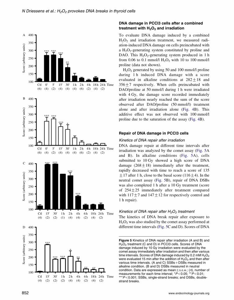
Kinetics of DNA repair after irradiation
DNA damage repair at different time intervals after
irradiation was analyzed by the comet assay (Fig. 5A
and B). In alkaline conditions (Fig. 5A), cells
submitted to 10 Gy showed a high score of DNA
damage (268G18) immediately after the treatment,
rapidly decreased with time to reach a score of 135
G17 after 1 h, close to the basal score (116G4). In the
neutral comet assay (Fig. 5B), repair of DNA DSBs
was also completed 1 h after a 10 Gy treatment (score
of 254G25 immediately after treatment compared
with 117G7 and 147G12 for respectively control and
1 h repair).
Kinetics of DNA repair after H2O2 treatment
The kinetics of DNA break repair after exposure to
H2O2 was also studied by the comet assay performed at
different time intervals (Fig. 5C and D). Scores of DNA
Figure 5 Kinetics of DNA repair after irradiation (A and B) andH2O2 treatment (C and D) in PCCl3 cells. Scores of DNAdamage induced by 10 Gy irradiation were evaluated by thecomet assay immediately after irradiation and then after varioustime intervals. Scores of DNA damage induced by 0.2 mM H2O2
were evaluated 15 min after the addition of H2O2 and then aftervarious time intervals. (A and C) SSBsCDSBs measured inalkaline condition. (B and D) DSBs measured in neutralcondition. Data are expressed as meanGS.E.M.; (n), number ofmeasurements for each time interval; *P!0.05; **P!0.01;***P!0.001; SSBs, single-strand breaks; and DSBs, double-strand breaks.
www.endocrinology-journals.org
Endocrine-Related Cancer (2009) 16 845–856
damage were maximal 15 min after exposure to
0.2 mmol/l H2O2. In alkaline conditions, the assay
was almost saturated (score around 400). Comet scores
decreased byw50% after 4 h and reached nearly basal
values 6 h after treatment (score 128G4; Fig. 5C). The
repair of DSBs (comet in neutral conditions) was
slower than in irradiated cells; comet scores above
control values were still observed 24 h after H2O2
treatment (104G2 for the control and 124G1 after
24 h repair; Fig. 5D).
Discussion
The thyroid produces large amounts of H2O2 that
constitute a potentially mutagenic environment
(Bjorkman & Ekholm 1988). The aim of this study
was to compare, in a rat thyroid cell line (PCCl3) and in
more physiological models (human thyroid primary
culture cells and pig thyroid slices), DNA damage
induced by a well-known carcinogenic factor (irradi-
ation) with that obtained by a putative carcinogenic
agent (H2O2).
The physiological levels of H2O2 in cells vary from
0.001 mmol/l to a maximum of 0.7 mmol/l (Song et al.
2007) but no data are available for the thyrocyte. As
the apparent Km of TPO for H2O2 is w300 mmol/l,
it has been hypothesized that H2O2 reaches especially
high concentrations but in a restricted place at the
periphery of the thyrocyte, in the postulated thyroxi-
some (Song et al. 2007). In thyroid, H2O2 is produced
outside the thyrocyte at its apical pole by DUOX and
is used by TPO located in the vicinity of DUOX to
oxidize iodide. Therefore, our experimental model in
which H2O2 is added in the incubation medium
mimics the in vivo extracellular production. The range
of H2O2 concentrations used in this study is
comparable to those probably needed to oxidize
iodide. It is difficult to estimate H2O2 concentrations
achieved in the limited space of the follicular lumen
in vivo but with the generation of 10 nmol/100 mg per
h they could easily reach the micromolar level
(Corvilain et al. 2000).
In our experiments, extracellular H2O2 is rapidly
reduced and has nearly disappeared after 15 min. At the
concentrations used in this study, H2O2was not lethal as
confirmed by viability tests. Apoptosis has not been
specifically evaluated in this work but previous studies
performed on thyroid cells failed to detect apoptosis for
similar doses of irradiation and showed apoptosis only
in a very small number of cells when treated with
similar concentrations of H2O2 (Yang et al. 1997, Riou
et al. 1999). These previous observations, along with
www.endocrinology-journals.org
the absence of mortality in H2O2 or irradiation treated
cells exclude that apoptosis may significantly contrib-
ute to the measured comet scores.
Comet assays were used to evaluate DNA strand
breaks in individual cells. We studied the effects of
H2O2 and irradiation on generation of SSBs and DSBs.
Interestingly, thyroid cancers occur at doses as low as
0.1 Gy with a linear dose–response curve and the
related risk increases by 10 times at 1 Gy (Ron et al.
1995). In our different thyroid models, we observed a
significant number of SSBs and DSBs after an
irradiation of 1 Gy or more. No differences in
sensitivity to irradiation were observed between
PCCl3 cells, pig thyroid slices and human thyroid
primary culture cells. High levels of DSBs formation
were confirmed by estimation of phosphorylation of
histone H2AX by western blotting and by immuno-
cytochemistry. Experiments on non-transformed rat
fibroblasts (F208) showed a significant production of
DSBs with 1 Gy. These results are in keeping with
previous data showing that the formation of w35
DSBs per gray, per cell, and per cell cycle is a constant
(Rogakou et al. 1998, Wojewodzka et al. 2002,
Takahashi et al. 2005).
DSBs provoked by H2O2 are considered to be rare
events: 1 DSBs for w2000 SSBs (Bradley et al.
1979, Takahashi et al. 2005). However, Bradley &
Kohn (1979) showed that in mouse leukemia L1210
cells, H2O2 induced DSBs with a ratio of DSBs to
SSBs comparable with that caused by X-rays.
Takahashi & Ohnishi (2005) reviewed one study
demonstrating formation of DSBsand histone H2AX
phosphorylation by immunocytochemistry in normal
human fibroblasts exposed to 0.1 mmol/l H2O2 for 2 h
(Takahashi et al. 2005).
DSBs are considered to be more carcinogenic than
SSBs. We demonstrated in PCCl3 cells that high but
non-lethal concentrations of H2O2 provoke a large
number of SSBs but also as many DSBs as irradiation.
In the presence of large amounts of H2O2-induced
SSBs, some apparent DSBs could be due to closely
spaced SSBs (Bradley et al. 1979). Therefore, we
confirmed the presence of real DSBs detected by neutral
comet assays by highlighting phosphorylated histone
H2AX. In this test, PCCl3 cells demonstrated the same
apparent damage caused by H2O2 (0.05–0.1 mmol/l) as
by irradiation. Data obtained on human thyroid
primary culture cells and on pig thyroid slices showed
a threshold of respectively 0.2 and 0.5 mmol/l H2O2
for the appearance of DSBs. We also observed a
significant number of DSBs after a 0.5 mmol/l H2O2
treatment of a non-thyroid cell line (F208, a non-
transformed rat fibroblast cell line) meaning that many
853

N Driessens et al.: H2O2 provokes DNA breaks in thyroid cells
if not all mammalian cells are sensitive to such
concentrations of H2O2. Thus, our work is clearly
demonstrated by two different methods an induction of
DSBs by H2O2 in a thyroid cell line and, more akin to
the in vivo situation, in human thyroid primary cultures
and pig thyroid slices. The demonstration of H2O2-
induced DNA damage does not necessarily imply a
mutagenic role but it can be extrapolated. A difficulty
for this extrapolation is that levels of DNA damage
acutely achieved in vitro must be compared with lower
levels accumulated over years. However, several
arguments support such an extrapolation: 1) H2O2, as
the well accepted mutagen X-ray, induces DNA DSBs,
2) thyroid in which oxidative DNA damage has been
demonstrated in vivo displays a higher level of
mutations than liver (Maier et al. 2006), and 3) low
levels of Se in serum (i.e. presumably lower activity of
Se dependent GPx) constitute a risk factor promoting
thyroid cancer development (Duntas 2006).
Comparison of the apparent sensitivity of our
different models to extra-cellular H2O2 is probably
worthless as it may reflect differences in cell membrane
permeability and in antioxidant capacities of the cells.
When H2O2 is applied to the exterior of cultured cells,
the intracellular concentrations are estimated to be
w10-fold lower than the extra-cellular concentrations
(Song et al. 2007). In thyroid, under physiological
conditions, a part of the H2O2 not used for thyroglo-
bulin iodination may diffuse into the cells where it is
degraded by very efficient antioxidant enzymes like Se
dependent GPx. BSO decreases intracellular GSH and
therefore the activity of Se dependent GPx. In PCCl3
cells exposed to H2O2, the presence of BSO decreased
the concentration of H2O2 needed to observe DNA
strand breaks to 0.05 mmol/l without affecting the
damage induced by irradiation. BSO alone in absence
of externally added H2O2 did not increase the level of
DNA strand breaks probably because basal H2O2
production in PCCl3 cells is not sufficient to induce
DNA damage (De Deken et al. 2002). Thyroid
destruction in myxoedematous endemic cretinism has
been related to impaired H2O2 degradation in
stimulated but Se deficient thyroids (Contempre et al.
2004). Therefore, we may extrapolate that in vivo the
potential DNA damaging effect of H2O2 will increase
in case of deficient antioxidant defense.
Because radiation increases 8-oxoguanine modifi-
cations, it was suggested that both radiation and
endogenous oxidative stress could synergistically
lead to the initiation of thyroid cancer (Riou et al.
1998). We looked therefore for a possible synergic
effect of a combined treatment of irradiation and H2O2
on DNA damage. The observed effects of irradiation on
854
cells preincubated with a H2O2-generating system were
additive with no synergy whatever the conditions used.
The very low H2O2 concentrations measured after
irradiation of the culture medium as well as the absence
of a potentiating effect of BSO suggest that in PCCl3
cells, H2O2 produced through the radiolysis of water is
not the main mechanism involved in DNA damage
following irradiation.
As it is well known that DNA repair deficiencies are
strongly associated with high cancer risk in humans,
we compared the kinetics of repair of DNA breaks
induced by irradiation and H2O2. DNA damage
induced by a 10 Gy irradiation and measured by the
comet assay in PCCl3 cells was completely repaired
after 1 h. The kinetics of repair was clearly made slow
for a similar amount of DSBs induced by H2O2. This
observation is probably related to different parameters:
1) the very high quantity of SSBs produced by H2O2
could saturate the repair systems. 2) H2O2 may induce
DNA damage, but also have direct inhibitory effects on
DNA repair. H2O2 at 0.1 mmol/l can inactivate the
human DNA mismatch repair system (Chang et al.
2002) and inhibit the repair of certain types of DNA
lesions through redox control of ADP ribosylation and
unscheduled DNA synthesis (Pero et al. 1990). 3) In
any case, the delay in H2O2-induced damage repair is
not due to the persistence of H2O2 in the medium as we
demonstrated that in our experimental conditions,
H2O2 rapidly (15 min) disappeared from the medium.
In conclusion, H2O2 produces DNA damage in the
thyroid. Concentrations of H2O2 that cause significant
DNA damage are not lethal for the cells and do not
modify cell functioning. These observations reinforce
the hypothesis that H2O2 is a potential carcinogenic
agent in the thyroid. H2O2 induces SSBs but also more
mutagenic DSBs in amounts comparable with what is
obtained with irradiation. The low repair efficiency of
DNA DSBs induced by H2O2 strengthens the possible
role of H2O2, generated in the thyroid to oxidize iodide,
in thyroid tumorigenesis. Therefore, chronic endogen-
ous exposure of thyroid cells to H2O2 could be a key to
explain the high frequency of thyroid tumors and
thyroid microcarcinoma, particularly in case of anti-
oxidant defense deficiency as demonstrated by the
increase of damage observed in the presence of BSO
and suggested in epidemiological studies in case of Se
deficiency (Duntas 2006, Kaprara & Krassas 2006).
Declaration of interest
The authors declare that there is no conflict of interest that
could be perceived as prejudicing the impartiality of the
research reported.
www.endocrinology-journals.org
Endocrine-Related Cancer (2009) 16 845–856
Funding
This work was supported by Fonds Erasme pour la Recherche
Medicale, Televie, Fonds de la Recherche Scientifique
Medicale (FRSM), Actions de Recherches Concertees de la
Communaute Francaise de Belgique (ARC), European Union
Contract FP6-36495 (GENRISK-T) and the ICT Impulse
program 2006, Brussels Capital Region, Belgium (In Silico
Project). XDD: postdoctoral researcher at the Fonds National
de la Recherche Scientifique (FRS-FNRS).
Acknowledgements
We thank Claude Massart, Bernadette Bournonville, and
Chantal Degraef for their excellent technical assistance.
References
Benard B & Brault J 1971 Production of peroxide in the
thyroid. L’Union Medicale du Canada 100 701–705.
Bertram JS, Kolonel LN & Meyskens FL Jr 1987 Rationale
and strategies for chemoprevention of cancer in humans.
Cancer Research 47 3012–3031.
Bjorkman U & Ekholm R 1988 Accelerated exocytosis and
H2O2 generation in isolated thyroid follicles enhance
protein iodination. Endocrinology 122 488–494.
Bradley MO & Kohn KW 1979 X-ray induced DNA double
strand break production and repair in mammalian cells as
measured by neutral filter elution. Nucleic Acids Research
7 793–804.
Chang CL, Marra G, Chauhan DP, Ha HT, Chang DK,
Ricciardiello L, Randolph A, Carethers JM & Boland CR
2002 Oxidative stress inactivates the human DNA
mismatch repair system. American Journal of Physiology.
Cell Physiology 283 C148–C154.
Chico Galdo V, Massart C, Jin L, Vanvooren V,
Caillet-Fauquet P, Andry G, Lothaire P, Dequanter D,
Friedman M & Van Sande J 2006 Acrylamide, an
in vivo thyroid carcinogenic agent, induces DNA
damage in rat thyroid cell lines and primary cultures.
Molecular and Cellular Endocrinology 257–258 6–14.
Coclet J, Foureau F, Ketelbant P, Galand P & Dumont JE
1989 Cell population kinetics in dog and human adult
thyroid. Clinical Endocrinology 31 655–665.
Collins AR 2004 The comet assay for DNA damage and
repair: principles, applications, and limitations.Molecular
Biotechnology 26 249–261.
Contempre B, de Escobar GM, Denef JF, Dumont JE &
Many MC 2004 Thiocyanate induces cell necrosis and
fibrosis in selenium- and iodine-deficient rat thyroids: a
potential experimental model for myxedematous endemic
cretinism in central Africa. Endocrinology 145 994–1002.
Corvilain B, Laurent E, Lecomte M, Vansande J & Dumont
JE 1994 Role of the cyclic adenosine 3 0,5 0-mono-
phosphate and the phosphatidylinositol-Ca2C cascades in
mediating the effects of thyrotropin and iodide on
www.endocrinology-journals.org
hormone synthesis and secretion in human thyroid slices.
Journal of Clinical Endocrinology and Metabolism 79
152–159.
Corvilain B, Collyn L, van Sande J & Dumont JE 2000
Stimulation by iodide of H2O2 generation in thyroid slices
from several species. American Journal of Physiology.
Endocrinology and Metabolism 278 E692–E699.
De Deken X, Wang D, Many MC, Costagliola S, Libert F,
Vassart G, Dumont JE & Miot F 2000 Cloning of two
human thyroid cDNAs encoding new members of the
NADPH oxidase family. Journal of Biological Chemistry
275 23227–23233.
De Deken X, Wang D, Dumont JE & Miot F 2002
Characterization of ThOX proteins as components of the
thyroid H2O2-generating system. Experimental Cell
Research 273 187–196.
Detours V, Delys L, Libert F, Weiss SD, Bogdanova T,
Dumont JE, Franc B, Thomas G & Maenhaut C 2007
Genome-wide gene expression profiling suggests distinct
radiation susceptibilities in sporadic and post-Chernobyl
papillary thyroid cancers. British Journal of Cancer 97
818–825.
Duntas LH 2006 The role of selenium in thyroid auto-
immunity and cancer. Thyroid 16 455–460.
Dupuy C, Ohayon R, Valent A, Noel-Hudson MS, Deme D&
Virion A 1999 Purification of a novel flavoprotein
involved in the thyroid NADPH oxidase. Cloning of
the porcine and human cdnas. Journal of Biological
Chemistry 274 37265–37269.
Ip C, Lisk DJ & Scimeca JA 1994 Potential of food
modification in cancer prevention. Cancer Research 54
1957s–1959s.
Kaprara A & Krassas GE 2006 Selenium and thyroidal
function; the role of immunoassays. Hellenic Journal of
Nuclear Medicine 9 195–203.
Kim H, Lee TH, Park ES, Suh JM, Park SJ, Chung HK,
Kwon OY, Kim YK, Ro HK & Shong M 2000 Role of
peroxiredoxins in regulating intracellular hydrogen
peroxide and hydrogen peroxide-induced apoptosis in
thyroid cells. Journal of Biological Chemistry 275
18266–18270.
Kohrle J, Jakob F, Contempre B & Dumont JE 2005
Selenium, the thyroid, and the endocrine system.
Endocrine Reviews 26 944–984.
Krohn K & Paschke R 2002 Somatic mutations in thyroid
nodular disease. Molecular Genetics and Metabolism 75
202–208.
Lacroix L, Soria JC, Bidart JM & Schlumberger M 2005
Oncogenes and thyroid tumors. Bulletin du Cancer 92
37–43.
Maier J, van Steeg H, van Oostrom C, Karger S, Paschke R &
Krohn K 2006 Deoxyribonucleic acid damage and
spontaneous mutagenesis in the thyroid gland of rats and
mice. Endocrinology 147 3391–3397.
Mikkelsen RB & Wardman P 2003 Biological chemistry of
reactive oxygen and nitrogen and radiation-induced signal
transduction mechanisms. Oncogene 22 5734–5754.
855

N Driessens et al.: H2O2 provokes DNA breaks in thyroid cells
Nasir A, Chaudhry AZ, Gillespie J & Kaiser HE 2000
Papillary microcarcinoma of the thyroid: a clinico-
pathologic and prognostic review. In Vivo 14 367–376.
Neumann CA, Krause DS, Carman CV, Das S, Dubey DP,
Abraham JL, Bronson RT, Fujiwara Y, Orkin SH & Van
Etten RA 2003 Essential role for the peroxiredoxin Prdx1
in erythrocyte antioxidant defence and tumour suppres-
sion. Nature 424 561–565.
Nunez J & Pommier J 1982 Formation of thyroid hormones.
Vitamins and Hormones 39 175–229.
Olive PL, Banath JP & Durand RE 1990 Heterogeneity in
radiation-induced DNA damage and repair in tumor and
normal cells measured using the ‘comet’ assay. Radiation
Research 122 86–94.
Pero RW, Anderson MW, Doyle GA, Anna CH, Romagna F,
Markowitz M & Bryngelsson C 1990 Oxidative stress
induces DNA damage and inhibits the repair of DNA
lesions induced by N-acetoxy-2-acetylaminofluorene in
human peripheral mononuclear leukocytes. Cancer
Research 50 4619–4625.
Picariello L, Sala SC, Martineti V, Gozzini A, Aragona P,
Tognarini I, Paglierani M, Nesi G, Brandi ML & Tonelli F
2006 A comparison of methods for the analysis of low
abundance proteins in desmoid tumor cells. Analytical
Biochemistry 354 205–212.
Rigutto S, Hoste C, Dumont JE, Corvilain B, Miot F &
De Deken X 2007 Duox1 is the main source of hydrogen
peroxide in the rat thyroid cell line PCCl3. Experimental
Cell Research 313 3892–3901.
Riou C, Remy C, Rabilloud R, Rousset B & Fonlupt P 1998
H2O2 induces apoptosis of pig thyrocytes in culture.
Journal of Endocrinology 156 315–322.
Riou C, Tonoli H, Bernier-Valentin F, Rabilloud R,
Fonlupt P & Rousset B 1999 Susceptibility of differ-
entiated thyrocytes in primary culture to undergo
apoptosis after exposure to hydrogen peroxide: relation
with the level of expression of apoptosis regulatory
proteins, Bcl-2 and Bax. Endocrinology 140 1990–1997.
Rogakou EP, Pilch DR, Orr AH, Ivanova VS & Bonner WM
1998 DNA double-stranded breaks induce histone H2AX
phosphorylation on serine 139. Journal of Biological
Chemistry 273 5858–5868.
Roger P, Taton M, Van Sande J & Dumont JE 1988
Mitogenic effects of thyrotropin and adenosine 3 0,5 0-
monophosphate in differentiated normal human thyroid
cells in vitro. Journal of Clinical Endocrinology and
Metabolism 66 1158–1165.
856
Ron E, Lubin JH, Shore RE, Mabuchi K, Modan B,
Pottern LM, Schneider AB, Tucker MA & Boice JD Jr
1995 Thyroid cancer after exposure to external radiation:
a pooled analysis of seven studies. Radiation Research
141 259–277.
Ross DS 2002 Nonpalpable thyroid nodules – managing an
epidemic. Journal of Clinical Endocrinology and
Metabolism 87 1938–1940.
Sarasin A, Bounacer A, Lepage F, Schlumberger M &
Suarez HG 1999 Mechanisms of mutagenesis in
mammalian cells. Application to human thyroid tumours.
Comptes Rendus de l’Academie des Sciences. Serie III
322 143–149.
Singh NP, McCoy MT, Tice RR & Schneider EL 1988 A
simple technique for quantitation of low levels of DNA
damage in individual cells. Experimental Cell Research
175 184–191.
Song Y, Driessens N, Costa M, De Deken X, Detours V,
Corvilain B, Maenhaut C, Miot F, van Sande J, Many
M-C et al. 2007 Roles of hydrogen peroxide in thyroid
physiology and disease. Journal of Clinical Endo-
crinology and Metabolism 92 3764–3773.
Stone JR & Yang S 2006 Hydrogen peroxide: a signaling
messenger. Antioxidants & Redox Signaling 8 243–270.
Takahashi A & Ohnishi T 2005 Does gH2AX foci formation
depend on the presence of DNA double strand breaks?
Cancer Letters 229 171–179.
Vogt TM, Ziegler RG, Graubard BI, Swanson CA,
GreenbergRS, Schoenberg JB, SwansonGM,Hayes RB&
Mayne ST 2003 Serum selenium and risk of prostate
cancer in U.S. blacks and whites. International
Journal of Cancer 103 664–670.
Wang X, Phelan SA, Forsman-Semb K, Taylor EF, Petros C,
Brown A, Lerner CP & Paigen B 2003 Mice with targeted
mutation of peroxiredoxin 6 develop normally but are
susceptible to oxidative stress. Journal of Biological
Chemistry 278 25179–25190.
Wojewodzka M, Buraczewska I & Kruszewski M 2002
A modified neutral comet assay: elimination of lysis at
high temperature and validation of the assay with
anti-single-stranded DNA antibody. Mutation Research
518 9–20.
Yang T, Namba H, Hara T, Takmura N, Nagayama Y,
Fukata S, Ishikawa N, Kuma K, Ito K & Yamashita S
1997 p53 induced by ionizing radiation mediates DNA
end-jointing activity, but not apoptosis of thyroid cells.
Oncogene 14 1511–1519.
www.endocrinology-journals.org