Hydrogen peroxide in inducible plant stress responses
64
Hydrogen peroxide in inducible plant stress responses Riikka Pellinen Institute of Biotechnology and Department of Biosciences, Division of Genetics University of Helsinki Finland Academic dissertation To be presented for public criticism, with permission of the Faculty of Science, University of Helsinki, in the auditorium 1041 of the Biocenter, Viikinkaari 5 on December 7 th , 2001, at 12 o’clock noon Helsinki 2001
Transcript of Hydrogen peroxide in inducible plant stress responses

Hydrogen peroxide in inducible plant stress responses
Riikka Pellinen
Institute of Biotechnology and
Department of Biosciences, Division of Genetics University of Helsinki
Finland
Academic dissertation To be presented for public criticism, with permission of the Faculty of Science,
University of Helsinki, in the auditorium 1041 of the Biocenter, Viikinkaari 5 on December 7th, 2001, at 12 o’clock noon
Helsinki 2001

Supervisors: Professor Jaakko Kangasjärvi Institute of Biotechnology University of Helsinki, Finland Professor Tapio Palva Institute of Biotechnology University of Helsinki, Finland Reviewers: Docent Elina Oksanen Department of Ecology and Environmental Science University of Kuopio, Finland Docent Kurt Fagerstedt Department of Biosciences,
Division of Plant Physiology University of Helsinki, Finland
Opponent: Dr. Christian Langebartels
Institute of Biochemical Plant Pathology GSF National Research Center for Environment and Health Germany
ISSN 1239-9469 ISBN 952-91-4117-3 (nid.) ISBN952-10-0216-6 (PDF)

Table of contents
Table of contents......................................................................................................................3
Abbreviations...........................................................................................................................5
Original publications .............................................................................................................6
Summary ...................................................................................................................................7
1 Introduction......................................................................................................................8
1.1 Reactive oxygen species (ROS) ............................................................................8
1.2 Plants and the environment................................................................................10 1.2.1 Environmental stress.....................................................................................10
1.2.1.1 Ozone (O3)...................................................................................................12 1.2.1.2 Light.............................................................................................................13 1.2.1.3 Plant -pathogen interactions ....................................................................14
1.3 Defence responses ................................................................................................16 1.3.1 Antioxidant enzymes ....................................................................................16
1.3.1.1 Superoxide dismutase (SOD) ...................................................................16 1.3.1.2 Peroxidase ...................................................................................................17 1.3.1.3 Glutathione reductase (GR)......................................................................18 1.3.1.4 Catalase (CAT) ...........................................................................................19 1.3.1.5 Glutathione-S-transferase (GST)..............................................................19
1.3.2 Phenylpropanoid synthesis..........................................................................20 1.3.3 Polyamines......................................................................................................21 1.3.4 Lipids ...............................................................................................................21 1.3.5 Signalling in stress responses.......................................................................21
1.3.5.1 Salicylic acid (SA) ......................................................................................22 1.3.5.2 Ethylene.......................................................................................................23
1.4 Programmed cell death (PCD)............................................................................23
1.5 Gene expression ....................................................................................................24 1.5.1 Stress-induced genes .....................................................................................24
1.5.1.1 Phenylalanine ammonia-lyase (Pal)........................................................25 1.5.1.2 Chalcone synthase (Chs)...........................................................................26 1.5.1.3 Pathogenesis related protein 10 (Ypr10) ................................................26 1.5.1.4 Mitochondrial phosphate translocator (Mpt1) ......................................28
1.5.2 Transcriptional regulation of stress related genes ....................................29
2 Aims of the present study .............................................................................................31
3 Materials and methods .................................................................................................32

3.1 Plant material.........................................................................................................32
3.2 Growth conditions................................................................................................32
3.3 Stress treatments ...................................................................................................32
3.4 Histology ................................................................................................................32
3.5 Enzyme activities ..................................................................................................32
3.6 Polyamine concentrations ...................................................................................33
3.7 Isolation of genes..................................................................................................33
3.8 Northern analysis .................................................................................................33
3.9 Lipid analysis ........................................................................................................33
3.10 Secondary metabolite analysis...........................................................................33
3.11 Inhibitor treatments .............................................................................................33
3.12 Sequence analysis.................................................................................................33
3.13 DNA fragmentation .............................................................................................33
3.14 Western analysis ...................................................................................................33
4 Results and discussion..................................................................................................34
4.1 Abiotic and biotic stresses induce H2O2 accumulation .................................34 4.1.1 H2O2 localisation ............................................................................................34 4.1.2 Origin of the H2O2..........................................................................................34 4.1.3 Intracellular H2O2 accumulation..................................................................35 4.1.4 Markers of ROS accumulation .....................................................................37
4.2 Stress-induced H2O2 accumulation and cell death are closely linked........37 4.2.1 Tissue damage................................................................................................37 4.2.2 Role of mitochondria in the process of cell death .....................................39 4.2.3 Markers of PCD..............................................................................................40 4.2.4 H2O2 is a crucial component of the cell death process ..............................40
4.3 Stress-induced gene expression can be mediated by H2O2 ..........................41 4.3.1 Isolation of genes ...........................................................................................41 4.3.2 Organisation of genes ...................................................................................41 4.3.3 Gene expression .............................................................................................41 4.3.4 Promoter analysis ..........................................................................................44
4.4 ROS independent stress responses ...................................................................45
4.5 Concluding remarks.............................................................................................46
5 Acknowledgements........................................................................................................49
6 References........................................................................................................................51

Abbreviations ABA abscisic acid HRGP hydroxyproline-rich glycopro-
tein AOX alternative oxidase JA jasmonic acid APX ascorbate peroxidase MJ methyl jasmonate ARE antioxidant responsive element MPT1 mitochondrial phosphate
translocator1 CAT catalase MRE metal responsive element Cat1AS CAT1 antisense mRNA messenger RNA cDNA complementary DNA MTF-1 metal response element bind-
ing transcription factor 1 CHS chalcone synthase NahG bacterial gene encoding salicy-
late hydroxylase CHX cycloheximide PAL phenylalanine ammonia-lyase DAB 3’3- diaminobezidine PAO polyamine oxidase DAO diamine oxidase PCD programmed cell death DDRT-PCR differential display reverse
transcriptase- PCR PCR polymerase chain reaction
DPI diphenylene iodonium PiC phosphate carrier ECM extracellular matrix POX peroxidase EC-SOD extracellular superoxide dis-
mutase PR10 pathogenesis related protein 10
ERELEE4 ethylene responsive element, Lycopersicon esculentum E4
PR- protein pathogenesis related protein
ERF ethylene responsive element binding factor
Q quinacrine
EST expressed sequence tag RbcS Rubisco small subunit G glucose rbohA respiratory burst oxidase ho-
molog A GO glucose oxidase rcd1 radical induced cell death 1 GPX glutathione peroxidase ROS reactive oxygen species GR glutathione reductase SA salicylic acid GSH glutathione SAR systemic acquired resistance GSSG glutathione disulphide SOD superoxide dismutase GST glutathione-S-transferase TEM transmission electron micros-
copy HL high light TMV tobacco mosaic virus HPLC high performance liquid chro-
matography UV ultraviolet
HR hypersensitive response VOC volatile organic compound WRKY
protein DNA- binding protein contain-ing WRKYGQC -amino acid sequence in the N- terminus

Original publications This thesis is based on the following publications, which will be referred to in
the text with their Roman numerals. Additional unpublished data will also be pre-sented in the text.
I Tuomainen J, Pellinen R, Roy S, Kiiskinen M, Eloranta T, Karjalainen R and Kangasjärvi J. 1996. Ozone affects birch (Betula pendula Roth) phenylpropanoid, polyamine and active oxygen detoxifying pathways at biochemical and gene-expression level. J.Plant Physiol. 148, 179-188.
II Pellinen R, Palva T and Kangasjärvi J. 1999. Subcellular localization of ozone-induced hydrogen peroxide production in birch (Betula pendula) leaf cells. Plant J. 20(3), 349-356. (Short communication).
III Pellinen R, Korhonen M, Kiiskinen M, Utriainen M, Overmyer K, Lapinjoki S, Palva ET and Kangasjärvi J. H2O2 activates cell death and defense gene-expression in birch (Betula pendula) (Manuscript).
IV Dat JF, Pellinen R, Beeckman T, Kangasjärvi J, Inzé D and Van Breusegem F. H2O2 primes an active cell death process in tobacco (Manuscript).
V Wulff A, Anttonen S, Pellinen R, Savonen E-M, Sutinen M-L, Heller W, San-dermann Jr H and Kangasjärvi J. 1999. Birch (Betula pendula Roth) responses to high UV-B radiation. Boreal Env. Res. 4, 77-88.

- 7 -
Summary Plant inducible defence responses during stress were studied in the commer-
cially important forest tree species Silver birch (Betula pendula Roth). A model system, catalase1 antisense tobacco (Nicotiana tabacum)(Cat1AS, deficient in catalase activity), was used to reveal further mechanisms underlying oxidative stress -induced cell death process. Numerous environmental factors affect the productivity of plants dur-ing their life span. Some of these are stress inducing and they may be biotic (such as pathogens) or abiotic (such as air pollutants or irradiation). Many biotic and abiotic stresses have in common the ability to induce the accumulation of apoplastic and intracellular reactive oxygen species (ROS) in plant tissues. These ROS have numer-ous tasks in inducible plant defence responses. Both in Silver birch and tobacco a ROS species, H2O2, is being actively produced in response to stress. In Silver birch the apoplastic H2O2 is produced by the plasma membrane NADPH oxidase together with cell wall peroxidases. Similarly in Cat1AS tobacco, abiotic stress induces NADPH oxidase dependent H2O2 accumulation. In both species stress-induced H2O2 accumulation precedes cell death, correlating spatially with lesion formation. H2O2 alone is sufficient to induce cell death, which appears to be an active process, and has features resembling programmed cell death (PCD). In both plant systems various morphological features of PCD occur as a response to oxidative stress. In addition to the ability of H2O2 to induce cell death, it can induce the expression of numerous stress- related genes. These stress -inducible genes share some common regulatory features in their promoter sequences. Stress causing agents that do not involve H2O2 production induce a distinct set of stress-inducible genes, differing from that induced by oxidative stresses. H2O2 is therefore a crucial component in signalling oxidative stress related responses and PCD in Silver birch and tobacco.

- 8 -
1 Introduction
1.1 Reactive oxygen species (ROS) Reactive oxygen species (ROS) include H2O2, .O2-, hydroxyl radicals (.OH) and
singlet oxygen. They are all formed during normal cellular metabolism, but under stress conditions their formation is accelerated (Noctor and Foyer, 1998).
1O2
3O2 O2-
O H2
H O2 2 OH H O2
hν
e- e-e-e-
2H+ H+ H+
Figure 1. Formation of .O2-, H2O2 and .OH.
Generation of .O2-, that exists in equilibrium with its protonated form hydroper-
oxyl radical (.O2H), requires energy (Figure 1). At physiological pH, .O2- is relatively non-toxic against cellular macromolecules and in aqueous solutions it disproportion-ates to H2O2 and O2 either spontaneously or by the action of superoxide dismutase (SOD) (Wojtaszek, 1997). During .O2- disproportionation H2O2 is always formed. H2O2 is a relatively stable form of ROS and it is electrically neutral. It has an ability to pass through membranes, and therefore it reaches cellular components distant from its site of synthesis. It may be destroyed by catalases (CAT) or peroxidases. H2O2 can also be produced by peroxidases as shown in Figure 2 (Wojtaszek, 1997).
.RH + O2 → .O2- + RH Peroxidase + .O2- → Compound III Compound III + RH2 → Peroxidase (Fe3+) + .RH 2 .O2- + 2 H ↔ H2O2 + O2 RH2 + .O2- → .RH + H2O
Figure 2. H2O2 generation by peroxidases. RH and .RH represent organic hydrocarbons and their respective radicals.
.OH, produced in the Haber-Weiss reaction (Figure 3), is the most harmful of
ROS forms in the plant tissue (Wojtaszek, 1997).
H2O2 + .O2- → .OH + OH- + O2
Figure 3. Haber-Weiss reaction.
Under normal cellular conditions Haber-Weiss reaction proceeds very slowly and only very low amounts of .OH are formed. It is however, formed in significant

- 9 -
amounts in the Fenton reaction (Figure 4) with transition metals such as Cu+ and Fe2+ and a subsequent reaction with .O2- (Wojtaszek, 1997).
H2O2 + Fe2+ (Cu+) → Fe3+ (Cu2+) + .OH + OH- .O2 + Fe3+ (Cu2+) → Fe2+ (Cu+) + O2
Figure 4. The Fenton reaction.
Since transition metals act as catalysts in the formation of .OH, their subcellular
localization also determines the location of .OH synthesis. .OH can initiate radical chain reactions and it is believed to be the main ROS responsible for alterations in cellular macromolecules and organellar damage (Wojtaszek, 1997). Hydroxyl radicals and singlet oxygen are extremely toxic to living cells and therefore must be efficiently removed. H2O2 and .O2-, however, are formed continuously in the cells where they are involved in many biological processes. H2O2 and .O2- toxicity results mainly from their ability to initiate reactions leading to the formation of more harmful ROS spe-cies (Noctor and Foyer, 1998).
ROS can be produced also enzymatically within the plant tissues. At the mo-ment the two most intensively studied enzyme systems involved in apoplastic ROS production conferring to the oxidative burst are the NADPH oxidase complex and the pH dependent cell wall peroxidases. A germin/oxalate oxidase system is also able to produce H2O2 in response to pathogen challenge (Wojtaszek, 1997) and both dia-mine and polyamine oxidases (DAO and PAO, respectively) produce H2O2 as a re-sponse to external stimuli (Smith, T.A., 1985).
Desikan et al. (1996) isolated protein components from Arabidopsis extracts that share immunological properties with the mammalian NADPH oxidase complex. Al-so, Keller et al. (1998) isolated an Arabidopsis rbohA (respiratory burst oxidase ho-molog A) gene, that has pronounced similarity to one of the subunits of the neutrophil respiratory burst NADPH oxidase. In addition, plant cell lines were found to express proteins resembling those of the mammalian NADPH oxidase complex in response to elicitation (Dwyer et al., 1996). Plant NADPH oxidase homolog rbohA also contains Ca2+ binding domains similar to the ones in their mammalian counterparts. Unlike mammalians, no cytosolic components of the enzyme complex were recognised (Kel-ler et al., 1998).
According to data obtained from plant oxidative burst studies and mammalian NADPH oxidase complex, the oxidative burst reaction begins with the recognition of the elicitor molecule by a corresponding receptor molecule that lies on the plasma membrane. Receptors isolated in plants so far are still putative and only partially characterized (Wojtaszek, 1997). Components of the signalling pathway downstream of the receptor include at least GTP binding proteins, ion channels, protein kinases (Rajasekhar et al., 1999), phosphatases, phospholipases A and C, and cyclic AMP. Finally, NADPH oxidase is activated, .O2- produced and dismutated to H2O2 (Wo-jtaszek, 1997). Dismutation of .O2- to H2O2 in the apoplast is thought to occur via ex-

- 10 -
tracellular SOD (EC-SOD). Involvement of the EC-SOD is mainly based on inhibitor studies (Bestwick et al., 1997; Jabs et al., 1997), but EC-SOD activity has been deter-mined (Desikan et al., 1996) and the gene has actually been isolated in Scots pine (Streller and Wingsle, 1994).
In the model where oxidative burst is created by pH dependent cell wall per-oxidases, signalling components are less important while pre-existing components present in the extracellular matrix (ECM) are of utmost importance. In this model an elicitor is recognized by a receptor molecule, which leads to the activation of ion channels (Wojtaszek, 1997). Ion fluxes in turn cause transient alcalinization of the ECM, leading to the activation of pH dependent cell wall peroxidases, and finally H2O2 is formed as shown in Figure 2.
1.2 Plants and the environment Plants can survive even in the most extreme environmental conditions, but also
in areas where growing conditions are relatively good, environmental factors can affect photosynthesis and hence plant productivity is rarely optimal. Environmental changes force plants to adapt to surrounding conditions on a daily, or even an hourly basis (Etherington, 1988). Industrialisation has made the environment even more complex for plant survival by adding numerous air pollutants into the atmosphere (Oleksyn and Innes, 2000). Some of the most tragic air pollution problems among forest ecosystems have taken place in the boreal and temperate climate zones (Lut-termann and Freedman, 2000). The most harmful air pollutants affecting forests are ozone (O3), sulphur dioxide (SO2), hydrogen sulphide (H2S), nitrous oxides (NOx), ammonia (NH3) and fluorides (especially HF). These pollutants may cause visible symptoms as well as numerous physiological level alterations (Luttermann and Freedman, 2000).
In Finland 66% of the land is covered by forest. The most important forest tree is Scots pine (Pinus sylvestris), which takes up 45% of the area, while Norway spruce (Picea abies) has the second place. The deciduous trees Silver and Downy birch (Betula pendula and Betula pubescens), many willow (Salix sp.) and oak (Quercus robur) are less important. However, the importance of birch is increasing in Finland and in Sweden where it is used for the reforestation of farmland and to increase the productivity of coniferous forests (Selldén et al., 1997). The importance of deciduous trees also in-creases as the climate change is predicted to favour their growth rather than the growth of coniferous species (Kellomäki et al., 1996).
1.2.1 Environmental stress Defining stress in plants is almost impossible, since no fixed stress points can be
set. Factors that can cause stress to plants can be classified to seven main classes, rep-resented in Table 1 (Elstner and Osswald, 1994). In a broad sense, plant stress can be defined as "any unfavourable condition or substance that affects or blocks a plant's

- 11 -
metabolism, growth, or development" (Lichtenthaler, 1996). In this case however, it is important to differentiate between low stress responses, that can partially be over-come by acclimation and repair, from strong or chronic stress effects, which may cause irreversible damage and cell death (Lichtenthaler, 1996). Table 1. Stress causing agents in plants Light High intensity
Low intensity Radiation UV (ultraviolet)
γ α β X- ray
Temperature High temperature Low temperature -Freezing -Chilling
Hydration Drought Flooding
Chemical factors Salts Heavy metals pH Air pollutants -O3 -SO2 -H2S -NOx -NH3 -HF
Mechanical factors Wind Lightning Fire Snow Cutting, biting Pressing
Biological influence Flowering Fruit ripening Insects Infections Allelopathy factors Competition
In many of the stresses mentioned in Table 1, ROS are involved as central sig-
nalling components (Noctor and Foyer, 1998). Of these ROS related stresses, O3, light, pathogen infections and mechanical stress will be described in more detail in the fol-lowing chapters. UV-B irradiation will be discussed also since ROS are involved in UV-B induced stress responses in some cases (Green and Fluhr, 1995).

- 12 -
1.2.1.1 Ozone (O3) Stratospheric O3 filters the deleterious UV-B irradiation before it reaches earth
(Attridge, 1990b). In the troposphere, however, O3 is a secondary air pollutant, formed through the photo-oxidation of volatile organic compounds (VOCs) in the presence of NOx. NO and hydrocarbons are produced as by -products in combustion processes in traffic and industry (Luttermann and Freedman, 2000). Although the overall reactions in the formation of photochemical “smog” are well known, many features in the formation of O3 still remain unclear (Rao et al., 2000). O3 is considered the most harmful of all photochemical air pollutants because of the small difference between ambient and toxic concentrations (Laurila, 1995).
Phytotoxic O3 concentrations occur in Fennoscandia regularly. 100 nL L-1 is con-sidered as a harmful level for vegetation (The Finnish Meteorological Institute, http://interim.fmi.fi/o3tietoa.html), but the proposed long term and short term critical levels for O3 are preferably expressed as cumulative exposure over the threshold concentration of 40 nL L-1. This exposure index is referred to as the AOT40 (accumu-lated exposure over a threshold of 40 nL L-1). The AOT40 is calculated as the sum of the differences between the hourly O3 concentrations in nL L-1 and 40 nL L-1 for each hour when the concentration exceeds 40 nL L –1 (Kärenlampi and Skärby, 1996). The highest O3 concentrations in Finland in the year 2000 varied between 50-75 nL L-1. The maximum value measured so far has been 95 nL L-1 in 1996 in Evo (The Finnish Meteorological Institute, http://interim.fmi.fi/o3tietoa.html). O3 risk is highest in the southern coastal part of the country in comparison to the inland sites due to higher background O3 concentrations (Pääkkönen et al., 1997). O3 concentrations vary dur-ing the day (Sanz and Millán, 2000), and depend on the season, the highest concen-trations occurring in the summer especially when the weather is hot and dry (de Leeuw and van Zantvoort, 1997; Luttermann and Freedman, 2000).
Forest vegetation suffers from O3 depending on the concentration and time of exposure. Acute injury occurs when plants are subjected to high concentrations (200-300 nL L-1) for short periods of time (2-4 h). Sensitive plant species show acute symp-toms already when exposed to O3 concentrations as low as 80 nL L-1 (Luttermann and Freedman, 2000), and e.g. in birch, O3 sensitivity varies with age (Pääkkönen et al., 1995b). Transport of O3 from urban sources to forested areas can lead to extended periods of exposure to moderate O3 levels (Luttermann and Freedman, 2000). O3 is considered likely to be the primary cause of forest decline in Europe (Schmieden and Wild, 1995), although it has more recently appeared that the link between O3 and for-est damage in Europe cannot be unequivocally drawn (Skärby et al., 1998; Matyssek and Innes, 1999). It appears that O3 affects forests together with CO2 and nitrogen depositions and that the major target of O3 on mature tree fitness is resource alloca-tion rather than growth (Matyssek and Innes, 1999).
Concern of O3 toxicity partly arises from the fact that it can be transported over long distances from heavily polluted areas to forested areas (Luttermann and Freed-man, 2000). O3 injury was first described in 1940’s in California, USA, and in the fif-ties they were referred to as “smog markings”. In Europe, first O3 caused damage to

- 13 -
vegetation was recorded in the 1970’s (Davison and Barnes, 1998). In the case of O3, high acute peak concentrations generally lead to cell death and therefore to visible symptoms (Heath and Taylor, 1997). At the ultrastructural level, morphological changes in Silver birch, which is an O3 sensitive tree, are first detected in the mesophyll cells around stomatal cavities (i.e. where O3 enters the leaf) as exudates on the cell walls. It continues with the disinte-gration of cytoplasm and cell collapse. In the end the epidermal cells also collapse (Günthardt-Goerg et al., 1993). In detailed transmission electron microscopy (TEM) studies it has been shown that the number of irregularly shaped and more electron dense chloroplasts increases (Pääkkönen et al., 1995a; Selldén et al., 1997), thylakoids become dilated and distorted and the granulation in the stroma increases (Pääkkönen et al., 1995a, 1997), mitochondria become disintegrated, the amount of cytoplasmic lipids increases (Pääkkönen et al., 1996) and the amount of chloroplastic starch de-creases (Pääkkönen et al., 1995b). Also alterations in the morphology of Golgi bodies, ER, and nuclear envelopes in O3 treated spinach have been detected (Miyake et al., 1984).
Mechanisms leading to O3 symptoms are only partly known. O3 can damage cu-ticles, but hardly any O3 penetrates the leaves through it. Instead, O3 enters leaves through stomata and passes into the intercellular space (Guderian, 1985). Therefore, any factor affecting the stomata (e.g. drought), will subsequently affect the intracellu-lar O3 concentration (Sanz and Millán, 2000). The most critical events take place im-mediately as O3 enters the aqueous apoplastic space, where it degrades rapidly form-ing ROS such as H2O2, hydroxyl radicals (.OH), and superoxide (.O2-). Due to this high reactivity of O3 in aqueous environment containing lipids (such as the apoplast), and its short calculated half-life within plant tissue (70 *10-9 s), it is very unlikely that any O3 as such would penetrate cells. In addition to these degradation products, plant cells begin to produce ROS rapidly in response to O3 treatment (Heath and Tay-lor, 1997; Schraudner et al., 1998; Rao and Davis, 1999; Overmyer et al., 2000).
1.2.1.2 Light 41% of solar radiation is visible light, 50% infra-red light and the remaining 9%
consists of x-rays and gamma rays as well as UV irradiation. The light received by a plant leaf varies with latitude, season, time of day, aspect, leaf inclination and cloud cover. Of the light that actually reaches the plant, a portion is reflected, absorbed or transmitted (Attridge, 1990b). Light absorption occurs by various light-absorbing pigments including photosynthetic pigments (chlorophylls and carotenoids), as well as the phytochrome (Attridge, 1990a) and UV-A (320-400 nm) and -B (280-320 nm) photoreceptors (Batschauer, 1999).
Light is indispensable for plant growth, photosynthesis and development. In addition to growth, many physiological processes are light dependent. These include germination, inhibition of hypocotyl growth, chloroplast differentiation, plant green-ing and expression of numerous genes (Beligni and Lamattina, 2000). No matter how

- 14 -
crucial and vital light is for plant development and productivity, it can also be a strong stress causing factor when received in excess. Excess white light leads to photooxidative stress (Karpinski et al., 1999), which damages the photosynthetic ap-paratus. During photooxidative stress H2O2 is formed in the chloroplasts (Foyer, 1997). Excess light is able to cause necrosis, as well as morphological changes, includ-ing the deformation of chloroplasts with pockets of cytoplasm, thylakoid swelling, and increases in the number of plastoglobuli (Cushman et al., 1995).
Stratospheric O3 depletion has lead to a remarkable increase in the amount of UV-B irradiation on earth affecting both animals and plants (Rousseaux et al., 1999). No visible injury is detected in broadleaf trees under naturally occurring UV-B doses (Dillenburg et al., 1995; Sullivan et al., 1996; Zeuthen et al., 1997). However, in Arabi-dopsis thaliana leaf yellowing occurs as a response to UV-B irradiation (Lois, 1994). UV-B irradiation affects the leaf anatomical features differently in conifers and broadleaf species. Conifers seem to be more tolerant to the irradiation and in broad-leaf species tissue thickening in the palisade parenchyma layer is the most prominent anatomical response to UV-B irradiation (Nagel et al., 1998).
In animals UV-B can induce carcinogenesis (Davies, 1995), and in both animals and plants DNA damage by the formation of covalent pyrimidine dimers (Rousseaux et al., 1999). The primary defence mechanism in plants against UV-B irradiation is the production of pigments, flavonoids, which absorb harmful UV wavelengths (Loge-mann et al., 2000), and therefore protect the plant DNA from damage (Kootstra, 1994). ROS production in plant cells also takes place in UV-B treated plants (Green and Fluhr, 1995).
1.2.1.3 Plant -pathogen interactions Plants are attacked by a wide array of microorganisms during their life span, in-
cluding fungi, bacteria, viruses and nematodes. Plants have the ability to protect themselves from microorganisms by both pre-existing and inducible defence re-sponses. Pre-existing defence mechanisms include structural barriers and stored an-timicrobial compounds (Hutcheson, 1998). Primary induced responses take place in the cells in direct contact with the pathogen, and lead to cell death. Secondary re-sponses are induced in the cells surrounding the infection site and the third class of inducible responses consists of the systemic acquired resistance (SAR) that is induced in the whole plant (Hutcheson, 1998). Microbial infection leads to a disease when the microorganism both overcomes the pre-existing defences and avoids to induce the active defence responses (Hutcheson, 1998). In plant- pathogen interaction, the win-ner is either the pathogen, if it can proliferate fast enough in the plant tissue, or the plant, if it can respond fast enough with correct defence responses (Dong, 1998).
Pathogen infiltration has been shown to cause formation of necrotic lesions in plant leaves within 30 hours of injection (Levine et al., 1996; Alvarez et al., 1998). These necrotic lesions bear great resemblance to that seen in O3- injury. In soybean (Glycine max) suspension cultures infected with pathogen, profound morphological

- 15 -
changes occur. These alterations include plasma membrane blebbing, cell shrinkage, and condensation of the cytoplasm and nucleus (Levine et al., 1996).
One of the defence responses taking place during plant-pathogen interactions is cell death that can be seen as restricted necrotic lesions at the infection site, distinct from the surrounding healthy tissue, known as the hypersensitive response (HR). Albeit some host tissue is damaged in the process, this localised cell death effectively restricts the spread of pathogens within the tissue (Tenhaken et al., 1995). Salicylic acid (SA) accumulates during HR, and the highest concentrations are found just around lesions (Enyedi et al., 1992). HR is considered nowadays as a form of pro-grammed cell death (PCD), known as apoptosis in mammalian tissues (Alvarez et al., 1998).
An integral part of HR is the oxidative burst producing ROS. The oxidative burst can be defined as a rapid production of high levels of ROS in response to exter-nal stimuli (Wojtaszek, 1997). The transient ROS accumulation during oxidative burst is very rapid; in suspension cultured plant cells it begins as quickly as 1-2 min after the addition of an elicitor and in plant segments or intact plants ROS can be detected 2-12 hours after the elicitation (Wojtaszek, 1997). This rapid ROS accumulation is proposed to be non-specific (Draper, 1997). Upon recognition of the invading patho-gen, a second burst of ROS accumulation takes place, and this sustained accumula-tion leads to the formation of HR (Draper, 1997). In addition to the local pathogen induced ROS accumulation, minor oxidative bursts can appear in distant locations from the original infection site. These minor oxidative bursts also lead to HR, only the lesion size is greatly diminished (Alvarez et al., 1998).
In mammals macrophages kill invading bacteria by phagocytosis. During this process oxygen is consumed and the reaction is called the respiratory burst. The .O2- accumulating during the respiratory burst is produced by the NADPH oxidase en-zyme complex, and it is a major feature in antibacterial activity of phagocytes (Wo-jtaszek, 1997). The major difference between plant and animal systems in the oxida-tive burst is that in phagocytosis only the pathogen is killed, whereas in HR the plant tissue surrounding infection site is also destroyed (Bolwell et al., 1995). Mechanical stress on plants may also lead to ROS production within tissues (Orozco-Cardenas and Ryan, 1999). This mechanical stress can be caused by insect feeding or by some other destructive event on the plant tissue (Maleck and Dietrich, 1999). Wounding causes bruising in potato (Solanum tuberosum) tubers (Partington et al., 1999), and in barley leaves it leads to necrotic lesions surrounded by chlorotic halos at the wound sites (Ledford and Richardson, 1994).
ROS from the oxidative burst have many important functions in the process of the HR and in other defence responses. They also drive the cross linking of cell wall polymers, trigger localized cell death and act as signalling components leading to other cellular responses such as the induction of transcription of stress related genes (Levine et al., 1994). In addition to pathogens, O3 (Sharma and Davis, 1997), high light (HL) (Foyer, 1997), UV-B (Green and Fluhr, 1995) and wounding (Orozco-Cardenas and Ryan, 1999) are able to cause ROS production in plant tissues. Thus the
- 16 -
oxidative burst can be stimulated by many agents and it may lead to coordinated defence responses that are strikingly similar for various stresses (Sandermann, 1998).
1.3 Defence responses Plants respond to stresses by exclusion, tolerance, compensation and/or repair
(Heath and Taylor, 1997). Concentration and duration of, exposure to the stress factor as well as environmental conditions and developmental and metabolic state of the plants affect the response (Guzy and Heath, 1993).
1.3.1 Antioxidant enzymes Antioxidant enzymes either catalyse reactions where an antioxidant molecule(s)
is able to quench ROS without being transformed into a destructive radical itself or to process ROS directly. Each of the antioxidant enzymes comprise of several isoforms (Noctor and Foyer, 1998). Although they are often induced in similar stress situa-tions, their responses may be differential (Adám et al., 1995). The total foliar activities of these enzymes may be misleading, since the compartments’ activities are the ones that count in the local scavenging reactions. Since ROS are also considered important signalling molecules in plant inducible defence responses, the role for antioxidant enzymes may not be to control ROS level only but also to modulate gene expression through the generation of appropriate signal molecules and the destruction of un-necessary signal molecules (Noctor and Foyer, 1998).
1.3.1.1 Superoxide dismutase (SOD) SOD dismutates .O2- to H2O2 (Noctor and Foyer, 1998), therefore converting one
harmful oxidant to a less harmful one. Three classes of SOD activities are recognized and they differ by their metal cofactors, which are Cu, Mn or Fe and Zn. Subcellular localization of the isoenzymes is known, MnSOD being mitochondrial, FeSOD plas-tidic and CuZnSOD plastidic and cytosolic (Kliebenstein et al., 1998). EC-SOD in Scots pine is CuZnSOD and it has four isoforms (Streller and Wingsle, 1994).
Total or isoform specific enzyme activities and changes in mRNA levels for different SODs in different stress responses vary remarkably as shown in Table 2. This is rather natural considering the numerous SOD isoforms and distinct com-partmentalisation (Kliebenstein et al., 1998). In any case, transgenic plants over- ex-pressing SOD have been shown to be more stress tolerant against oxidative stress (Pitcher and Zilinskas, 1996; Noctor and Foyer, 1998; McKersie et al., 2000), although in some cases no changes in damage formation are detected (Allen, 1995). Both chloroplastic and cytoplasmic targeting of the transgene result in increased stress tolerance (Pitcher and Zilinskas, 1996; McKersie et al., 2000).

- 17 -
Table 2. Changes in the activities or mRNA levels of different SOD isoforms upon various stress treatments. Cyt = cytosolic and Chl= chloroplastic, - = no change, ↓ = down -regulation, ↑ = up -regulation. Stress factor MnSOD FeSOD Cyt CuZnSOD Chl CuZnSOD O3
- (Willekens et al., 1994a; Klie-benstein et al., 1998)
↓ (Willekens et al., 1994a; Conk-lin and Last, 1995)
↑ (Kliebenstein et al., 1998; Overmy-er et al., 2000)
↓ (Kliebenstein et al., 1998; Overmy-er et al., 2000)
Pathogen
↓ (Adám et al., 1995)
↑ (Adám et al., 1995)
Light
- (Kliebenstein et al., 1998)
↑ (Kliebenstein et al., 1998)
↑ (Kliebenstein et al., 1998)
↑ (Kliebenstein et al., 1998)
UV-B irradia-tion
↑ (Kliebenstein et al., 1998)
- (Kliebenstein et al., 1998)
↑ (Kliebenstein et al., 1998)
↑ (Kliebenstein et al., 1998)
1.3.1.2 Peroxidase Peroxidases scavenge H2O2 by reducing it to H2O in the presence of a reductant.
They are found throughout the cell and they have higher affinity to H2O2 than CAT. In plant cells, the most important reducing substrate for peroxidase is ascorbate (Noctor and Foyer, 1998).
Peroxidases (either ascorbate peroxidase [APX], glutathione peroxidase [GPX] or total peroxidase) are up -regulated either at mRNA, protein or enzyme activity level under numerous stresses as shown in Table 3.
- 18 -
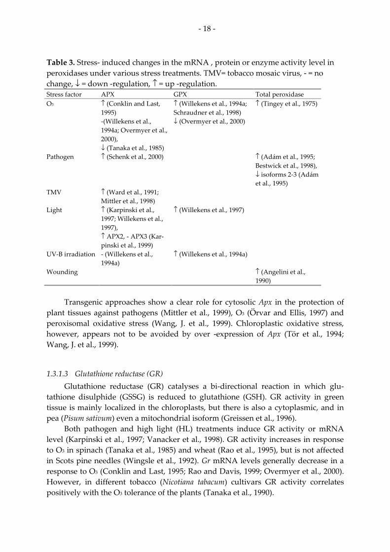
Table 3. Stress- induced changes in the mRNA , protein or enzyme activity level in peroxidases under various stress treatments. TMV= tobacco mosaic virus, - = no change, ↓ = down -regulation, ↑ = up -regulation. Stress factor APX GPX Total peroxidase O3
↑ (Conklin and Last, 1995) -(Willekens et al., 1994a; Overmyer et al., 2000), ↓ (Tanaka et al., 1985)
↑ (Willekens et al., 1994a; Schraudner et al., 1998) ↓ (Overmyer et al., 2000)
↑ (Tingey et al., 1975)
Pathogen
↑ (Schenk et al., 2000) ↑ (Adám et al., 1995; Bestwick et al., 1998), ↓ isoforms 2-3 (Adám et al., 1995)
TMV
↑ (Ward et al., 1991; Mittler et al., 1998)
Light
↑ (Karpinski et al., 1997; Willekens et al., 1997), ↑ APX2, - APX3 (Kar-pinski et al., 1999)
↑ (Willekens et al., 1997)
UV-B irradiation
- (Willekens et al., 1994a)
↑ (Willekens et al., 1994a)
Wounding ↑ (Angelini et al., 1990)
Transgenic approaches show a clear role for cytosolic Apx in the protection of
plant tissues against pathogens (Mittler et al., 1999), O3 (Örvar and Ellis, 1997) and peroxisomal oxidative stress (Wang, J. et al., 1999). Chloroplastic oxidative stress, however, appears not to be avoided by over -expression of Apx (Tör et al., 1994; Wang, J. et al., 1999).
1.3.1.3 Glutathione reductase (GR) Glutathione reductase (GR) catalyses a bi-directional reaction in which glu-
tathione disulphide (GSSG) is reduced to glutathione (GSH). GR activity in green tissue is mainly localized in the chloroplasts, but there is also a cytoplasmic, and in pea (Pisum sativum) even a mitochondrial isoform (Greissen et al., 1996).
Both pathogen and high light (HL) treatments induce GR activity or mRNA level (Karpinski et al., 1997; Vanacker et al., 1998). GR activity increases in response to O3 in spinach (Tanaka et al., 1985) and wheat (Rao et al., 1995), but is not affected in Scots pine needles (Wingsle et al., 1992). Gr mRNA levels generally decrease in a response to O3 (Conklin and Last, 1995; Rao and Davis, 1999; Overmyer et al., 2000). However, in different tobacco (Nicotiana tabacum) cultivars GR activity correlates positively with the O3 tolerance of the plants (Tanaka et al., 1990).

- 19 -
Gr over -expression leads to increased resistance to oxidative stress (Aono et al., 1991; Foyer et al., 1995), but to gain more O3 tolerant plants over -expression is re-quired both in chloroplasts and mitochondria (Broadbent et al., 1995).
1.3.1.4 Catalase (CAT) Catalases (CAT) convert H2O2 to water and molecular oxygen (Noctor and
Foyer, 1998). Cat genes exist in three isoforms (Cat1-3), which have different func-tions and locations in plant tissues. Cat1 is peroxisomal and involved in scavenging photorespiratory H2O2, Cat2 is preferentially expressed in the vascular tissue, while Cat3 has a role in glyoxysomal processes (Willekens et al., 1994b). H2O2 itself can cause a decrease in the transcript levels of these isoforms in low concentrations, but their expression is induced at high concentrations (Polidoros and Scandalios, 1999).
CAT enzyme activity decreased in O3 treated spinach (Tanaka et al., 1985), but increased in soybean more than 10 -fold (Tingey et al., 1975). However, when two different Cat family members were studied at the transcript level, it was clear that mRNA levels of Arabidopsis Cat1 and Cat3 were increased at least two fold in both WT and rcd1 (radical induced cell death1, a mutant exhibiting O3 and .O2- inducible le-sion formation) (Overmyer et al., 2000). Three tobacco Cat genes responded very dif-ferently to O3 exposure: Cat1 was first down -regulated, rising little at the end of the experiment. Cat2 was not affected, whereas Cat3 transcript level clearly increased (Willekens et al., 1994a). Maize (Zea mays) Cat1, 2 and 3 genes were induced also by H2O2 treatment (Polidoros and Scandalios, 1999). Increase in CAT activity upon pathogen invasion appeared to be cultivar dependent (Vanacker et al., 1998, 2000) and it also varied depending on plant species (Adám et al., 1995; Schenk et al., 2000).
Role of CAT in oxidative stress has been extensively studied with catalase an-tisense tobacco (Cat1AS) plants, which retain only 10% of wild type catalase activity. This deficiency leads to accumulation of H2O2 in the tissue under excess light causing photo-oxidative stress, and subsequent white necrotic lesions (Chamnongpol et al., 1996, 1998; Willekens et al., 1997). These plants undergo severe cellular damage un-der HL conditions, and it has therefore been postulated that CAT is a cellular sink for H2O2 produced during photo-oxidative stress (Willekens et al., 1997). In HL treated Arabidopsis, Cat1 transcript level has been shown to increase 2.5 fold (Karpinski et al., 1997). UV-B irradiation of tobacco leaves caused inducion of Cat2 and 3, while sup-pressing Cat1 (Willekens et al., 1994a). Similarly to the Cat1AS plants, which were hypersensitive to pathogen treatment (Mittler et al., 1999), over -expression of to-bacco Cat2 in potato lead to enhanced disease resistance in these transgenic plants (Yu et al., 1999).
1.3.1.5 Glutathione-S-transferase (GST) Glutathione-S-transferases (GST) are a family of enzymes that catalyse the con-
jugation of glutathione via the sulfydryl group to a variety of electophilic centers of

- 20 -
hydrophobic compounds. This reaction makes the compound in question more hy-drophilic and enables its transport to vacuoles or the apoplast. Phenolic oxidants and anthocyanins are among GST substrates. GSTs are also responsible for the detoxifica-tion of highly reactive lipid peroxidation products, generated from oxidative stress damaged membranes (Polidoros and Scandalios, 1999). O3 (Sharma and Davis, 1994; Overmyer et al., 2000), pathogens (Dudler et al., 1991; Schenk et al., 2000), wounding (Reymond et al., 2000), H2O2 (Levine et al., 1994; Chen et al., 1996; Polidoros and Scandalios, 1999), and .O2- (Jabs et al., 1996) are able to increase the Gst transcript level.
1.3.2 Phenylpropanoid synthesis Accumulation of phenylpropanoid compounds is detected in numerous plant
species as a response to different stresses. Phenylpropanoid biosynthetic pathway leads to the synthesis of a vast array of biologically active secondary metabolites (Hahlbrock and Scheel, 1989), such as SA, phytoalexins (Smith, C.J., 1996), stilbenes, UV absorbing flavonoids and isoflavonoids and structural molecules such as lignin as well as anthocyanin pigments (Hahlbrock and Scheel, 1989) (Figure 5).
PhenylalaninePAL
trans- cinnamic acid para-coumarylic acid
Ferulic acidSinapic acid
Salicylic acid
CHS
StilbenesFlavonesFlavonolsAnthocyanins
Lignin
4-coumaroyl-CoA
STS
Figure 5. Phenylpropanoid biosynthesis.
Phenylpropanoid biosynthesis starts from the deamination of phenylalanine by phenylalanine ammonia-lyase (PAL). PAL controls the flux of carbon into the pathway, and therefore efficiency of the whole pathway (Hahlbrock and Scheel, 1989). From 4-coumaryoyl-CoA the pathway branches to the flavonoid and antho-cyanin biosynthetic pathway. This pathway is in turn controlled by chalcone syn-thase (CHS) (Dooner et al., 1991). Flavonoids are synthesised as a response to various environmental stimuli. Most importantly they protect plant tissues from harmful UV-B irradiation by absorbing light in the UV region, and therefore prevent the UV-B induced DNA damage (Kootstra, 1994).
Cell walls are strengthened at the pathogen penetration sites by incorporation and oxidative cross-linking of proteins and various phenolic subunits (Grant and Mansfield, 1999). Fortifying the plant cell wall gives various advantages to the plant.

- 21 -
It prevents leakage of cytoplasmic contents and creates an excellent barrier. Lignin precursor molecules and free radicals formed in the cross-linking reactions may as such disrupt pathogen membranes or inactivate bacterial enzymes and toxins (Hammond-Kosack and Jones, 1996). A localized oxidative burst is often detected during cell wall fortification, but the accumulation of ROS is much below the level seen during the HR (Grant and Mansfield, 1999). Rapid oxidative cross-linking of basic hydroxyproline-rich glycoproteins (HRGPs) with pathogenesis related-proteins (PR-proteins) may be one of the earliest defence responses linked to the oxidative burst (Bradley et al., 1992).
1.3.3 Polyamines The plant polyamines consist of diamine putrescine, and polyamines spermine
and spermidine (Langebartels et al., 1991). Polyamines have crucial roles in numer-ous aspects of plant life, including cell division, macromolecule synthesis, senes-cence, and stress responses (Angelini et al., 1990). For example, in stress situations polyamines may have a role in preventing O3 injury by chelating metal ions that cata-lyse peroxidation reactions leading to lipid peroxidation and changes in plasma membrane permeability, both known consequences of O3 exposure (Kangasjärvi et al., 1994). The ability of polyamines to prevent O3 injury is likely to be due to mem-brane stabilization and scavenging of oxygen radicals (Evans and Malmberg, 1989; Rowland-Bamford et al., 1989). Radical scavenging ability of polyamines is due to their phenolic hydroxy groups (Bors et al., 1989). Pathogen infection can also affect plant polyamine levels and these alterations are able to modulate the pathogen de-fence response (Yamakawa et al., 1998).
1.3.4 Lipids In plant cells, lipids act as major components of the biological membranes, en-
ergy reserves, precursors for waxes, cutin and suberin, and signal transduction chain components (Miguel et al., 2000). For example, the lipid derived signal molecule jas-monic acid (JA) has been implicated of being responsible for wounding induced gene expression in plants (Martín, M. et al., 1999). Also, inositol lipid turnover may trans-duce the elicitor induction of Pal via protein kinases (Kamada and Muto, 1994a).
Lipid peroxidation is detected in plants undergoing HR, and it can be triggered by adding ROS generating systems into plants (Rogers et al., 1988). O3 causes drastic changes in the lipid compositions which may result from these peroxidation events (Sakaki et al., 1994).
1.3.5 Signalling in stress responses Signalling takes place in plant development as well as in sensing environmental
stimuli. Signalling events include recognition of the stimuli and subsequent intracel-
- 22 -
lular events. ROS may also act as signalling molecules (Noctor and Foyer, 1998). The most studied signalling molecules in plant stress responses include SA, JA, ethylene, and abscisic acid (ABA). SA and ethylene and their role in mediating oxidative stress responses will be discussed in more detail in the following chapters.
1.3.5.1 Salicylic acid (SA) SA is derived from the phenylpropanoid biosynthetic pathway (Figure 5) (Ward
et al., 1991). It plays a central role in defence against pathogen invasion, especially when resistance is achieved via HR. In the early phase of pathogen invasion, .O2- and H2O2 are produced locally, and at the same time local induction of defence related genes takes place (Draper, 1997). Some of these genes encode enzymes of the phenylpropanoid biosynthetic pathway, like Pal, which catalyses production of SA precursors (Mauch-Mani and Slusarenko, 1996; Draper, 1997; Smith-Becker et al., 1998). Also the enzyme responsible for SA synthesis from benzoic acid has been shown to be induced by H2O2 (León et al., 1995). Draper (1997) has proposed a model where initial pathogen infection would lead to rapid and short lived production of H2O2. This H2O2 would activate SA synthesis, leading to a potentiating phase where SA would in turn induce secondary prolonged H2O2 production at the same time with actual pathogen recognition. Like in the first phase, H2O2 would again induce SA synthesis, that would now lead to HR, cell death and resistance as well as to de-fence gene induction (also induced by H2O2 from the second phase) and SAR (Draper, 1997). SA enhances ROS accumulation and cell death in numerous plant species (Kauss and Jeblick, 1995; León et al., 1995; Shirasu et al., 1997; Kawano et al., 1998; Hückelhoven et al., 1999; Asai et al., 2000) and it seems that SA potentiates the HR signalling pathway (Alvarez, 2000).
O3 induced HR- like symptoms and antioxidant defence responses were greatly diminished in NahG plants (Arabidopsis plants expressing bacterial salicylate hy-droxylase, therefore deficient in SA) in comparison to WT, suggesting that SA could potentiate responses conferring to plant’s O3- sensitivity (Rao and Davis, 1999). Po-tentiation was also important in hybrid poplar where O3- sensitive clone seemed to be insensitive to SA and therefore unable to produce a full defence response (Koch et al., 2000).
SA also induces binding activity of a stress-inducible transcription factor that binds to a region found in more than 30 different stress responsive genes (Golds-brough et al., 1993). WRKY DNA-binding proteins (recognize elicitor responsive elements in parsley [Petroselinum crispum] PR1 promoter, and contain WRKYGQC -amino acid consensus sequence in their N- termini) may also regulate gene expres-sion during pathogen and SA induced defence responses (Yang et al., 1999), with numerous other proteins binding to SA- responsive elements (Hennig et al., 1993; Guevara-García et al., 1998; Zhou et al., 2000).
- 23 -
1.3.5.2 Ethylene Ethylene is a gaseous plant hormone and its concentration increases shortly af-
ter exposure to various stresses. In O3 stress the amount of this stress ethylene corre-lates with the visible injury formation (Langebartels et al., 1991, 2000; Telewski, 1992; Wellburn and Wellburn, 1996). It has been suggested that a plant’s capability to pro-duce stress ethylene would be the key determinant of its O3 sensitivity (Kangasjärvi et al., 1994). In an O3 sensitive Arabidopsis mutant rcd1, O3 induced .O2- production is ethylene dependent. Ethylene therefore promotes the ROS triggered lesion formation in O3 treated Arabidopsis (Overmyer et al., 2000). Ethylene may also modulate elicitor induced cell death (Asai et al., 2000).
Ethylene can regulate gene expression through various ethylene responsive elements. One of these, the GCC -box may mediate either negative or positive stress responses (Fujimoto et al., 2000). Another ethylene responsive cis-element, ERELEE4 (ethylene responsive element in Lycopersicon esculentum ethylene responsive gene E4), has been shown to regulate senescence related gene expression (Itzhaki et al., 1994).
1.4 Programmed cell death (PCD) HR cell death in plants has been shown to be genetically programmed resem-
bling the apoptosis in mammals (Sasabe et al., 2000). The function of mammalian apoptosis is to selectively eliminate certain cells during development. This is done in order to maintain developmental balance and as a response to stress. Cells that are no longer needed or are damaged, will be self-destructed (Gilchrist, 1998). In plants, all the criteria defining mammalian apoptosis can never be met due to structural dif-ferences between animal and plant cells, and therefore it is here referred to as pro-grammed cell death (PCD).
Apoptosis needs to be separated from necrotic cell death that occurs when cells are exposed to poisons, severe cold or heat, or traumatic injury leading to membrane and organellar damage. In contrast, apoptosis requires active cellular participation (Gilchrist, 1998). Animal cells undergoing apoptosis exhibit cell shrinkage, loss of contact to other cells, fragmentation of nuclear DNA (Gilchrist, 1998), activation of Ca2+ dependent endonuclease, nuclear deformations, formation of numerous micro-nuclei, and plasma membrane blebbing (Falcieri et al., 1994). Later, cells form apop-totic bodies that protect the adjacent cells from possibly toxic materials that could otherwise leak out, and are taken up by neighbouring cells and degraded within minutes to hours (Gilchrist, 1998).
The sequence of events leading to the onset of apoptosis in mammalians in-cludes numerous steps. Cytochrome c release from mitochondria is activated through a voltage dependent anion channel, and when dATP is present, released cy-tochrome c can activate caspase family proteins. Activation of initiator caspases trig-gers a protease cascade that amplifies and executes the cell death signal. Loss of cyto-chrome c from the mitochondria may lead to ROS formation, which again might ac-tivate caspases and subsequently, apoptosis (Lam et al., 1999). Caspases are a family

- 24 -
of cysteine-dependent, aspartate-directed proteases which have crucial roles in the initiation and execution of mammalian apoptosis (Earnshaw et al., 1999).
In plant development PCD takes place at least during death of petals after fer-tilization, diploid parthenogenesis, development of tracheary elements, leaf senes-cence and in barley aleurone layer during germination (Gilchrist, 1998). PCD is also a central part of life cycle of perennial plants. Creeping perennials, such as clover, pro-liferate at apices and push out to new areas, while older parts of the plant age and die. A similar phenomenon takes place in trees, where dead material does not de-compose, but remains in the wood (Thomas et al., 2000). Related to senescence, pre-mature cell death was induced in transgenic tobacco plants with altered phenylpro-panoid pathway regulation, suggesting a role for phenylpropanoid compounds in the regulation of plant PCD (Tamagnone et al., 1998).
Some of the features of mammalian apoptosis can be detected in plants also: DNA fragmentation can be induced by pathogens (Mittler et al., 1997; Gilchrist, 1998; Asai et al., 2000; Koch et al., 2000; Sasabe et al., 2000), by increasing intracellular [Ca2+], H2O2, SA (O'Brien et al., 1998), and O3 (Koch et al., 2000), during germination (Gilchrist, 1998), during senescence (O'Brien et al., 1998), and in root caps (Gilchrist, 1998). Other cellular features of plant PCD include vacuolisation of the cells (Mittler et al., 1997; Fath et al., 2000), loss of plasma membrane integrity (Fath et al., 2000) and increase in monomeric chloroplast DNA (Mittler et al., 1997). HR, which is a form of PCD in plants, differs from the developmentally triggered PCD by associated in-duced defence responses. Reactions involved in HR include ion fluxes, generation of ROS, protein synthesis, intact actin cytoskeleton, SA, protein kinases and an oxida-tive burst (Heath, M.C., 2000; Sasabe et al., 2000).
It has been suggested that the mitochondrial alternative oxidase (Aox) could be a plant specific cell death regulator by controlling the mitochondrial ROS production through the alternative respiration pathway (Lam and del Pozo, 2000). ROS are probable regulators of PCD in many plant systems (Jabs et al., 1996; Alvarez et al., 1998; Desikan et al., 1998; Mittler et al., 1998, 1999; Hückelhoven et al., 1999; Rao et al., 2000) and they affect plant gene expression (Levine et al., 1994; Desikan et al., 1998; Mittler et al., 1998).
1.5 Gene expression
1.5.1 Stress-induced genes Distinct sets of genes are induced upon various stresses or other stimuli in
plants. The most studied groups of genes related to stress responses include genes encoding PR-proteins (Warner et al., 1992), phenylpropanoid pathway enzymes (Kangasjärvi et al., 1994), ethylene biosynthetic enzymes, antioxidant enzymes, lipid metabolism enzymes (Kangasjärvi et al., 1994) and genes involved in the regulation of mitochondrial responses, such as Aox (Murphy et al., 1999). Three genes, phenyla-lanine ammonia lyase (Pal), chalcone synthase (Chs) and pathogenesis related protein

- 25 -
10 (Ypr10), often studied in relation to plant stress responses, and one gene that was first isolated in plants as an O3- inducible gene, the mitochondrial phosphate translo-cator 1 (Mpt1), will be described in the following chapters. Pal, Chs and Ypr10 are good markers for stress and ROS related gene expression and Mpt1 opens up a new line in the study of the oxidative stress related processes in plants.
1.5.1.1 Phenylalanine ammonia-lyase (Pal) Due to phenylpropanoid compounds’ vast array of defence related functions in
plants, PAL enzyme activity and gene -expression in stress situations has been inten-sively studied. PAL activity confers to the production of various phenylpropanoid compounds for plant protection, but it’s main function in defence is to produce pre-cursors for SA, signal molecule derived from a branch of the phenylpropanoid path-way (Figure 5), synthesis (Mauch-Mani and Slusarenko, 1996).
PAL level is increased either at the enzyme activity or mRNA level by numer-ous environmental cues presented in Table 4. Table 4. PAL induction at the enzyme activity or mRNA level upon various stimuli. TMV = tobacco mosaic virus, SA= salicylic acid, JA= jasmonic acid. External stimulus Reference(s) O3
Rosemann et al., 1991; Eckey-Kaltenbach et al., 1994; Sharma and Davis, 1994; Sharma et al., 1996; Pääkkönen et al., 1998; Riehl Koch et al., 1998
Pathogen
Cui et al., 1996; Mur et al., 1996; Rajasekhar et al., 1999; Blilou et al., 2000
Elicitor (fungal or bac-terial)
Edwards et al., 1985; Liang et al., 1989; Lois et al., 1989; Gowri et al., 1991; Kamada and Muto, 1994a, b; Marinelli et al., 1994; Pellegrini et al., 1994; Baillieul et al., 1995; Desikan et al., 1998; Sasabe et al., 2000
TMV
Pellegrini et al., 1994
Wounding
Liang et al., 1989; Lois et al., 1989; Pellegrini et al., 1994; Mur et al., 1996
UV-B irradiation
Kuhn et al., 1984; Lois et al., 1989; Kalbin et al., 1997; Logemann et al., 2000
Light
Liang et al., 1989; Asai et al., 2000
Ethylene
Ecker and Davis, 1987
JA
Gundlach et al., 1992
Reduced glutathione
Wingate et al., 1988
SA Blilou et al., 2000
Role of SA in PAL induction is considered to be the potentiation of the response
induced by some other cue (Mur et al., 1996; Shirasu et al., 1997). The role of ROS in mediating Pal expression has been studied in various plant systems with differing
- 26 -
results. H2O2 seems not to be involved in Pal induction in soybean (Levine et al., 1994) or tobacco suspension cell lines (Dorey et al., 1999; Sasabe et al., 2000), although it is clearly demonstrated that H2O2 alone is able to increasce Pal as well as Gst mRNA level in Arabidopsis suspension cell cultures (Desikan et al., 1998).
1.5.1.2 Chalcone synthase (Chs) Chs expression is remarkably sensitive to UV- and blue light (Strid et al., 1994;
Kalbin et al., 1997; Logemann et al., 2000; Loyall et al., 2000). Increase in the fluence rate of white light increases Chs mRNA level in Arabidopsis, as does blue light also. In some cases red light may also affect Chs transcript level (Jackson and Jenkins, 1995; Frohnmeyer et al., 1998).
In addition to UV-B, Chs up -regulation occurs as a response to O3 (Rosemann et al., 1991), fungal elicitor (Loake et al., 1991; Kato et al., 1995), pathogens (Cui et al., 1996) and wounding (Creelman et al., 1992). Of the known signaling molecules, Chs is induced by methyl jasmonate (MJ) (Creelman et al., 1992), and by the reduced form of glutathione (Wingate et al., 1988; Loyall et al., 2000). Glutathione and the oxidative status of the cells are possibly involved in the transcriptional regulation of UV-B in-duced Chs expression (Loyall et al., 2000).
1.5.1.3 Pathogenesis related protein 10 (Ypr10) PR-proteins are defined as proteins that are induced by pathogen invasion or by
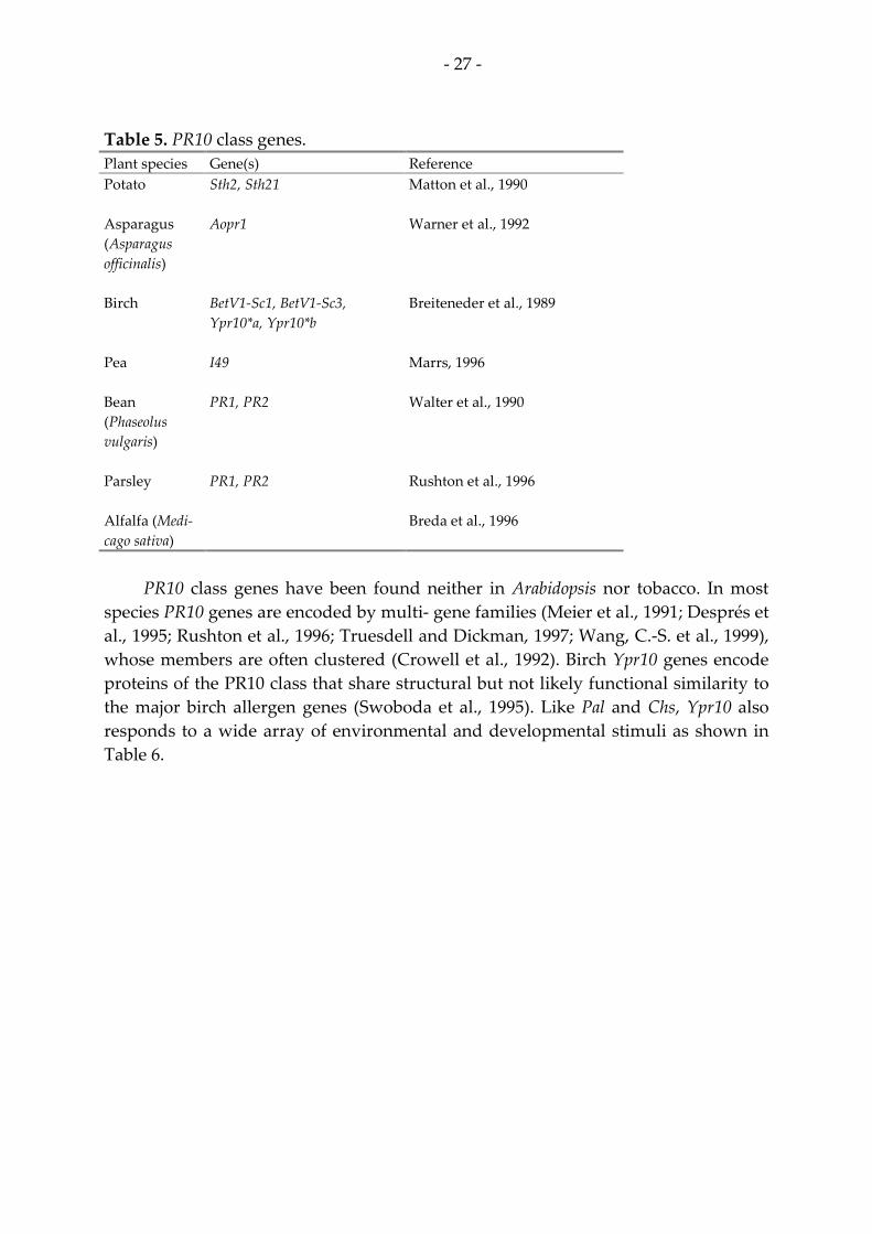
resembling situations. PR genes that have conserved sequences, but are not function-ally designated, are called Ypr-genes (van Loon et al., 1994). PR10 (pathogenesis re-lated protein 10) class proteins are classified as parsley PR1 -like proteins (van Loon et al., 1994). Genes encoding PR10 class proteins have been isolated so far in several plant species shown in Table 5.
- 27 -
Table 5. PR10 class genes. Plant species Gene(s) Reference Potato
Sth2, Sth21 Matton et al., 1990
Asparagus (Asparagus officinalis)
Aopr1 Warner et al., 1992
Birch
BetV1-Sc1, BetV1-Sc3, Ypr10*a, Ypr10*b
Breiteneder et al., 1989
Pea
I49 Marrs, 1996
Bean (Phaseolus vulgaris)
PR1, PR2 Walter et al., 1990
Parsley
PR1, PR2 Rushton et al., 1996
Alfalfa (Medi-cago sativa)
Breda et al., 1996
PR10 class genes have been found neither in Arabidopsis nor tobacco. In most
species PR10 genes are encoded by multi- gene families (Meier et al., 1991; Després et al., 1995; Rushton et al., 1996; Truesdell and Dickman, 1997; Wang, C.-S. et al., 1999), whose members are often clustered (Crowell et al., 1992). Birch Ypr10 genes encode proteins of the PR10 class that share structural but not likely functional similarity to the major birch allergen genes (Swoboda et al., 1995). Like Pal and Chs, Ypr10 also responds to a wide array of environmental and developmental stimuli as shown in Table 6.

- 28 -
Table 6. Changes in Ypr10 enzyme or mRNA levels upon external stimuli. SA= sali-cylic acid, ABA= abscisic acid, JA= jasmonic acid. Stimulus Reference(s) Senescence
Crowell et al., 1992; Valjakka et al., 1999
Copper (Cu2+)
Utriainen et al., 1998
O3
Pääkkönen et al., 1998
Pathogen
Crowell et al., 1992; Warner et al., 1992, 1993; Constabel and Brisson, 1995; Després et al., 1995; Midoh and Iwata, 1996; Trues-dell and Dickman, 1997
H2O2
Crowell et al., 1992; Bi et al., 1995
JA
Moons et al., 1997
ABA
Moons et al., 1997; Wang, C.-S. et al., 1999
Salt stress
Moons et al., 1997
Methyl viologen
Crowell et al., 1992
SA Crowell et al., 1992
Different Ypr10 genes seem to respond to wounding differently, some are
clearly up regulated (Crowell et al., 1992; Warner et al., 1992, 1993; Constabel and Brisson, 1995; Després et al., 1995; Truesdell and Dickman, 1997), while others are not affected (Midoh and Iwata, 1996). In birch Ypr10 genes, differences are found even between different family members (Poupard et al., 1998).
1.5.1.4 Mitochondrial phosphate translocator (Mpt1) The first plant mitochondrial phosphate translocator 1 (Mpt1) was isolated in
birch as an O3-inducible transcript by DDRT-PCR (Kiiskinen et al., 1997). Mpt1 is a transmembrane protein on the inner mitochondrial membrane and it transports inor-ganic phosphate (Pi) into the mitochondrial matrix and carries most of the Pi required for ATP synthesis (Capobianco et al., 1991; Palmieri et al., 1993; Stappen and Kramer, 1994). Since ADP is phosphorylated to ATP by oxidative phosphorylation in mito-chondria, availability of Pi within mitochondria is essential for the energy metabo-lism of the cell (Wohlrab, 1986). A single copy gene encodes Mpt1 in birch. It shares considerable homology with phosphate carriers (Pic) isolated from mammalians and yeast (Kiiskinen et al., 1997).
So far Mpt1 homologs have been isolated from soybean cDNA libraries made of drought stressed plants [BG043543], apical shoots [AW099962], roots [AW102173]

- 29 -
and immature seed coats [AW186456]. It has also been found from potato tuber [BF153103], moss (Physcomitrella patens) [AW476902], and six day cotton (Gossypium hirsutum) fiber cDNA library [AI726701], as well as in the Arabidopsis genome project [AB007650].
1.5.2 Transcriptional regulation of stress related genes In eukaryotes transcription is usually regulated by combinatorial control, mean-
ing that several different proteins must bind DNA in a coordinated manner to achieve appropriate expression of the gene in question. Transcriptional regulation therefore consists of transcription factor(s) (trans- acting factor) and the correspond-ing binding site in the DNA (cis-element) (Wolberger, 1999). Not much is known about the transcriptional regulation of stress related plant genes. Some regulatory elements and trans- acting factors are, however, known and may be of importance in future work considering regulation of plant gene expression.
A class of transcription factors called WRKY have been isolated at least in to-bacco (Yang et al., 1999) and parsley (Eulgem et al., 1999). They are induced by to-bacco mosaic virus (TMV), SA (Yang et al., 1999), fungal elicitors (Eulgem et al., 1999), and wounding (Hara et al., 2000) via phosphorylation dependent activation mechanism (Yang et al., 1999). Binding sites for WRKY are found for example in the promoters of genes encoding tobacco basic chitinase (Yang et al., 1999) and parsley PR1 (Eulgem et al., 1999). Organization of WRKY binding sites within promoter con-fers to efficient activity (Eulgem et al., 1999). In a massive gene -expression study on Arabidopsis SAR responses, 20 stress responsive genes clustered with PR1. Common for the promoters of the genes in this cluster was the existence of at least three WRKY binding sites on each of them (Maleck et al., 2000). Similarly, in an Arabidopsis mutant exhibiting constitutive SAR, mRNA levels of 16 cDNAs were elevated and their promoters carried WRKY binding sites (Petersen et al., 2000).
Numerous eucaryotic transcription factors respond to cellular redox state. These factors include AP-1 and 2, NF-κB, MTF-1 (metal response element binding transcrip-tion factor 1), and MAF (an oncogene family). Also cis -elements responsive to ROS, such as the antioxidant responsive element (ARE), have been described (Dalton et al., 1999; Delaunay et al., 2000). However, regulation of gene expression via these regula-tors does not only occur through ROS, for they respond to, or are fine tuned by, other cellular signals as well (Dalton et al., 1999). ARE was first described in the promoter of the rat Gst gene. ARE is responsive to H2O2 and phenolic antioxidants undergoing redox cycling. ARE may therefore be part of the signal transduction pathway leading to oxidative stress responses in eucaryotic cells (Rushmore et al., 1991). ARE motif was found from the promoters of three maize Cat genes, and it seems to be involved in the expression of Cat1 during senescence related oxidative stress (Polidoros and Scandalios, 1999).
Mammalian metallothionein genes are regulated at the transcriptional level as a response to heavy metals through metal responsive elements (MREs) that occur in

- 30 -
the promoter as multiple copies (Koizumi et al., 1999). In mice with knocked out CuZnSOD, metallothionein expression was markedly induced, and the transcrip-tional activation occurred through MREs (Ghoshal et al., 1999), therefore the gene responded to the changed cellular redox balance.
More than 100 plant transcripts are regulated by light of different qualities. Numerous light responsive cis- elements are found in the promoters of many genes. Still, the assembly of elements varies greatly: Neither can any single element be found in all light regulated promoters nor do any single element confer to light in-duced expression all by itself (Terzaghi and Cashmore, 1995). Several light and UV-B responsive elements have been described in Chs promoters (Batschauer et al., 1996; Schäfer et al., 1997).
Ethylene responsive element binding factors (ERFs) are specific to plants. ERFs bind DNA motifs known as the GCC-box. Different ERF family proteins can either up- or down- regulate transcription by binding to the same GCC-box, and their re-sponsive genes are differentially regulated by ethylene and abiotic stress conditions (Fujimoto et al., 2000). ERELEE4 binding site is also responsive to ethylene, but has been described to be related to the senescence process (Itzhaki et al., 1994). There are also numerous other stress, or signalling molecule responsive, regions in the promot-ers of different stress related genes (Dron et al., 1988; Goldsbrough et al., 1993; Rouster et al., 1997; Chen and Singh, 1999).
No clear, single stress related factors affecting plant transcription are known, al-though some of the primary defence responses are shared between stresses. It ap-pears that the stress induced gene expression in plants is regulated through a com-plex network of transcription factors and their combinations binding to diverse mix-ture of stress related cis -elements in the promoter sequences. Nevertheless, some regulatory elements are recognised as responsible for certain responses, and the pic-ture is getting clearer with the results of various EST and genome projects as well as with the transgenic approaches with deleted promoters.
- 31 -
2 Aims of the present study The aim of this study was to reveal molecular stress responses occurring in the
Silver birch, which is a common and commercially important deciduous tree species, covering large and ecologically diverse areas across the northern hemisphere. To re-veal common factors underlying stress induced cell death, a model plant (Cat1AS tobacco) was used. The studies I-V were carried out to
1. Find possible common factors between biotic and abiotic stresses. 2. Reveal a possible stress-induced oxidative burst and its origin. 3. Study the relationships between the oxidative burst and stress induced
gene -expression and cell death.

- 32 -
3 Materials and methods
3.1 Plant material In birch (Betula pendula) experiments, either seed derived (II, III and V) or tissue
culture derived material (I, additional UV-B irradiation data, clone 36) was used. In the tobacco study, transgenic lines carrying antisense Cat1 gene (Cat1AS) and wild type SR1 Nicotiana tabacum plants (L.) were used (IV).
3.2 Growth conditions Birch saplings were grown either in controlled climate chambers (I, II, III) or in
greenhouse (III, V) under conditions simulating the weather in Finland in June (light: dark [22:2] photoperiod with daytime temperature 19°C and night temperature 12°C and relative humidity 55: 80% [day: night]). Tobacco plants were grown in low light conditions (80 µmol m-2 s-1, 400-700 nm) with 14 h light/10 h dark at 25°C and 70% relative humidity. Mature pre-flowering plants were used.
3.3 Stress treatments O3 and wounding treatments were performed in computer controlled climate
chambers (I, II, III). UV-B irradiations were either made in greenhouse (V) or con-trolled climate chambers (Chs, Pal and Ypr10 expression studies in Figure 10), where birch saplings were exposed to 0 kJ m-2 (control) or 4 kJ m -2 of 300 nm (Caldwell) UV-B radiation for five hours. Philips TL 40/12 lamps covered with acetate filter to re-move UV-C were used as the source of UV-B radiation. UV-B level was measured at plant level with Macam SR9010-PC spectroradiometer. Light level was 270-400 µMol m-2 sec-1. Pathogen and in planta H2O2 production (glucose/glucose oxidase [G/GO]) treatments were performed in greenhouse (III). Tobacco plants were subjected to high light (HL)(1000 µmol m-2 s-1) in a Fitotron chamber (IV).
3.4 Histology Cell viability was studied in O3 exposed birch by Evans Blue staining (I) and HL
treated tobacco by Trypan Blue staining (IV). Light microscopy samples were col-lected from UV-B treated birch (V) and HL treated tobacco (IV).
Transmission electron microscopy (TEM) was subjected to samples from UV-B and O3 treated birch (including CeCl3 staining for subcellular H2O2 localisation)(II) and HL treated tobacco (IV).
H2O2 localisation with 3’3- diaminobenzidine (DAB) at the leaf level was made from O3, pathogen, wound and G/GO treated birch (III) and HL treated tobacco (IV).
3.5 Enzyme activities Enzyme activities of peroxidase (POX), GR and SOD were measured spectro-
fotometrically from O3 treated birch leaves (I).

- 33 -
3.6 Polyamine concentrations Concentrations of putrescine, spermidine and spermine were measured from O3
exposed birch leaves by HPLC (High Performance Liquid Chromatogarphy) (I).
3.7 Isolation of genes For obtaining gene specific probes or genomic clones, PCR with degenerate or
specific primers (I, III) and cDNA and genomic library screens were performed (III).
3.8 Northern analysis RNA was extracted and subjected to northern analysis with different probes.
Treatments that were studied by northern method include O3 (I, III), UV-B (V), pathogen (III), wounding (III), G/GO (III) and HL (IV). In situ hybridisations were made from UV-B treated birch leaves (V).
3.9 Lipid analysis Membrane lipids and lipid peroxidation products were analysed from UV-B
treated birch by gas chromatography (V).
3.10 Secondary metabolite analysis Secondary metabolites were extracted and analysed by HPLC from UV-B ex-
posed birch (V).
3.11 Inhibitor treatments To unravel the enzyme complexes responsible for H2O2 production, effect of
various inhibitors on the subcellular H2O2 accumulation in O3 treated birch (II) and on the H2O2 accumulation in general in HL treated tobacco (IV) were studied. Also the involvement of various signalling intermediates in HL treated tobacco, were studied with the help of inhibitory chemicals (IV).
3.12 Sequence analysis Sequence analysis was performed with GCG (Genetics Computer Group) soft-
ware (I, III), and promoter elements were scanned with the plant cis -acting element database (III).
3.13 DNA fragmentation DNA fragmentation was analysed from HL treated tobacco protoplasts by flow
cytometry (IV).
3.14 Western analysis Proteins were extracted from HL treated tobacco and subjected to western
analysis with different antibodies (IV).
- 34 -
4 Results and discussion
4.1 Abiotic and biotic stresses induce H2O2 accumulation As shown in various studies on plant stress physiology, accumulation of ROS as
one of the earliest events is common to both biotic and abiotic stress –induced de-fence responses (Lamb and Dixon, 1997; Bolwell, 1999). In most cases ROS detected are either H2O2 or .O2- (Jabs et al., 1996; Schraudner et al., 1998). Both in birch and to-bacco H2O2 was detected after stress treatments. Increase in the amount of H2O2 after stress treatments results either from increased H2O2 production or decreased detoxi-fication or both. With the methods used in this study, it is impossible to distinguish which events are responsible for the increased amount of H2O2. Thus, the term “H2O2 accumulation” is used to describe the changes observed in the quantity of H2O2 in the experimental material.
4.1.1 H2O2 localisation In birch .O2- accumulation was not detected after O3 or pathogen treatments
(data not shown), presumably due to its rapid dismutation to slightly less harmful form of ROS, H2O2. H2O2 accumulation was indeed detected in birch leaves after O3, pathogen, wounding and G/GO treatment (III). Timing of the H2O2 accumulation coincided with lesion development, and H2O2 accumulation was most prominent surrounding the lesions, either in a halo- or spot-like manner (III). The O3 induced H2O2 accumulation was also studied in more detail and at the subcellular level this H2O2 was detected first at the cell walls of palisade parenchyma cells (2h), and later both in the cell walls and on the plasma membranes at the same location (10h) (II).
In HL treated Cat1AS tobacco, H2O2 was detected in cells along leaf veins in patches (IV). Wild type SR1 tobacco cells also accumulated H2O2 as a response to HL, but the accumulation was much weaker and uniform than in the transgenic line (IV). H2O2 accumulation therefore appears to be a general stress response occurring upon various stresses both in birch and in tobacco.
4.1.2 Origin of the H2O2 The origin of the H2O2 burst in stressed birch and tobacco was studied with in-
hibitor treatments (II, IV). Diphenylene iodine (DPI), an inhibitor of flavin containing oxidases, was used to block the activity of the plasma membrane NADPH oxidase (II, IV). DPI can inhibit also other oxidases, but here only its effect on the apoplastic H2O2 production was studied, and low enough concentrations were used to avoid inhibi-tion of peroxidases (Bolwell et al., 1998). Both in birch (II) and tobacco (IV), DPI was sufficient to partly block either the O3 or HL induced H2O2 accumulation, clearly in-dicating a role for a plasma membrane oxidase in the initial oxidative burst upon
- 35 -
stress. The NADPH oxidase itself produces .O2- from molecular O2, but according to a proposed model, the plant plasma membrane NADPH oxidase would act in tight connection with the EC-SOD (Ogawa et al., 1997). This way, the activity of NADPH oxidase would actually lead to accumulation of H2O2, when the superoxide produced would be instantly dismutated (Ogawa et al., 1997).
In birch, the O3 induced H2O2 accumulation was partly inhibited by peroxidase inhibitors KCN and NaN3 (II). This suggests a role also for cell wall peroxidases in the generation of O3 induced oxidative burst. The existence of two H2O2 generating activities in birch apoplastic stress response was further substantiated by the two separate subcellular locations for the H2O2 accumulation, the plasma membrane and the cell wall (II). In other plant systems, either NADPH oxidase, or cell wall peroxi-dases seem to be the main sources of H2O2 during the oxidative burst (Bestwick et al., 1997; Bolwell et al., 1998).
4.1.3 Intracellular H2O2 accumulation At 24 hours H2O2 accumulation in the O3 -treated birch palisade parenchyma
cells continued similarly as at two and 10 hours (II). In the mesophyll layer however, H2O2 was detected intracellularly in contrast to the earlier apoplastic accumulation (II). H2O2 was detected in the cytoplasm, peroxisomes, and mitochondria of the mesophyll cells, but not in the chloroplasts (II). Since chloroplasts have a central role in plant ROS and stress metabolism, two control experiments were carried out to re-veal possible technical problems in localising chloroplastic H2O2. Also control sam-ples with CeCl3 but without standard EM- staining procedures were performed to ensure that the staining was distinguishable from possible EM- staining artefacts (data not shown).
First birch leaf segments were subjected to methyl viologen, that causes massive ROS production in the chloroplasts under HL conditions, and as a result H2O2 accu-mulated in the chloroplasts (II). Secondly, chloroplastic H2O2 accumulation was stud-ied in O3- treated birch clone (V5834), classified as O3 -sensitive according to leaf damage. As shown in Figure 6B and D H2O2 accumulated in the chloroplasts of pali-sade parenchyma cells of these O3 -sensitive plants at 10h as a response to O3- treat-ment.

- 36 -
Figure 6. Chloroplasts of birch clone V5834 at 0h (A,C) and 10 h (B,D) after the begin-ning of an eight- hour O3- exposure (C and D are close- ups of A and B, respectively). At 10 hours H2O2 (visualised with CeCl3) can be seen as black precipitates in the cell walls of an epidermal cell (ec) and a palisade parenchyma cell (ppc) as well as inside the epidermal cell as spots (B). Chloroplastic H2O2 can be seen as small, scattered pre-cipitates everywhere in the chloroplast of an O3 treated cell (D, arrows). Bars 500 nm. a=apoplast, c=chloroplast, cw= cell wall, ec=epidermal cell, n=nucleus, ppc= palisade parenchyma cell, s=starch grain, v=vacuole.
It therefore seems that chloroplasts are highly tolerant to oxidative stress due to
extremely efficient scavenging mechanisms in this organelle, which is exposed to radicals continuously during normal metabolism (Asada, 1999). Thus chloroplastic ROS accumulation as a response to stress may at least partly define plant’s suscepti-bility to oxidative stress. Also the result of methyl viologen treatment leading to H2O2 accumulation in birch chloroplasts, as well as the appearance of H2O2 in the chloro-plasts of O3- sensitive birch clone suggest, that when a drastic enough oxidative stress is subjected to chloroplasts, even its efficient scavenging ability can be ex-ceeded.
Still, the cytoplasmic scavenging mechanisms seem to be more important under high O3 peaks than the chloroplastic ones (Tepperman and Dunsmuir, 1990; Pitcher and Zilinskas, 1996; Torsethaugen et al., 1997). Also the peroxisome, and CAT therein, may play an important role in detoxification of H2O2 produced in the other cellular compartments, especially the cytoplasm, since this organelle may act as a sink for H2O2 detoxification (Willekens et al., 1997).
- 37 -
4.1.4 Markers of ROS accumulation Increase in the activities, or in the protein or mRNA levels of antioxidant en-
zymes and their respective genes, can also be considered as markers for the appear-ance of ROS in the plant tissue. Enzyme activities of SOD, peroxidase, and GR in-creased in O3-treated birch leaves, and this rise was in all cases more prominent in the O3-sensitive birch clone (I). Similarly, the mRNA levels of the genes encoding an-tioxidant enzymes Apx, CuZnSod, Gst and Cat were increased upon O3-treatment in birch (III). Induction in the enzyme activities and increase in the mRNA levels of ROS detoxifying enzymes suggests that ROS accumulate in the O3-treated tissue in con-centrations that need to be scavenged actively. Also the level of free putrescine, which can act as an antioxidant (Bors et al., 1989; Evans and Malmberg, 1989; Row-land-Bamford et al., 1989), increased in the O3-sensitive birch clone (I). Higher activi-ties of antioxidant enzymes and polyamine levels in the O3-sensitive birch clone are likely to reflect higher ROS concentrations, rather than more efficient scavenging ef-ficiency, since more severe damage was detected in these plants.
The artificial H2O2 generating system, G/GO, increased the transcript levels of genes encoding ROS detoxifying enzymes (III), further justifying their use as markers for ROS accumulation in plant tissue. Likewise, in the HL treated Cat1AS tobacco plants, protein levels of APX and GPX increased rapidly after the onset of HL treat-ment, in addition to the respective mRNA levels (IV). Timing of the increase in en-zyme activities (I, IV), followed that of the mRNA accumulation (III, IV), suggesting that the increased enzyme activities are probably not meant for detoxification of the primary oxidative burst on the plasma membranes and cell walls, but more likely they are directed to scavenge the later accumulating intracellular ROS occurring at the same time with the appearance of visible injury (II, III). This is consistent with the suggestion of Foyer et al (1994), that increases in ROS detoxifying enzyme activities is not related to protecting the tissue under stress from injury, but is rather a response to the damage. Also cell wall thickness and alkalinization confer to apoplastic ROS detoxification (Moldau, 1998).
4.2 Stress-induced H2O2 accumulation and cell death are closely linked .O2- has a regulatory role in the stress induced cell death in Arabidopsis (Jabs et
al., 1996; Overmyer et al., 2000), and H2O2 similarly in tobacco (Schraudner et al., 1998) and Arabidopsis (Alvarez et al., 1998). In mammalian apoptosis, ROS are claimed to be one of the main initiators of the process (Gilchrist, 1998). Especially, ROS produced in the mitochondria in the early phase contribute to the ongoing of other mitochondrial responses leading to apoptosis (Green and Reed, 1998).
4.2.1 Tissue damage O3 induced tissue damage was detected in birch as brown necrotic lesions earli-
est at 10 h or latest at 48 h (I, III). Membrane damage in the leaves could also be de-
- 38 -
tected by vital staining the leaves with Evans Blue. This experiment showed that the permeability of the O3 treated cells increased prior to cell death, therefore indicating membrane damage possibly leading to leakage of cellular constituents and subse-quent cell death (I). Membrane damage was detected both in the sensitive and toler-ant birch clones in a similar extent, but only in the sensitive clone clusters of dead cells were detected later, combined with large visible lesions (I). In birch, O3 injury and H2O2 accumulation correlated spatially (III). H2O2 accumulated at the edges of damaged tissue, as well as in tissues surrounding it (III).
Similarly as O3, other stresses have an ability to induce H2O2 accumulation at the sites of injury. In birch H2O2 accumulated at the sites subjected to wounding or pathogen infiltration (III). Pathogens were applied to birch leaves by injecting the bacterial suspension with a syringe without a needle. As in the O3 treated tissue, in the pathogen treated leaves H2O2 accumulated at the edges of injection sites, where the damage formed, and also in tissue surrounding the lesions (III).
In subcellular H2O2 localisation studies of O3-treated birch (II), H2O2 was first detected in the cell walls and on the plasma membranes of palisade parenchyma cells. Later similar accumulation was seen in the cell walls and plasma membranes between palisade parenchyma cells and epidermal cells (II, Figure 7A). Subsequent cell death at 24h occurred mainly in the palisade parenchyma and epidermal cells (I, II, III).
Similarly in barley leaves infected with fungi, H2O2 accumulation began in the palisade parenchyma cells underlying the attacked epidermal cell (Thordal-Christensen et al., 1997; Vanacker et al., 2000). H2O2 accumulation was especially prominent in the cell walls and plasma membranes adjacent to the attacked epider-mal cell (Figure 7B). As in the case of O3, H2O2 accumulation was later detected in the epidermal cells (Figure 7B). Later, cell death took place in the cells accumulating H2O2 (Thordal-Christensen et al., 1997; Vanacker et al., 2000).

- 39 -
A. B.
O3
ROS
2h
10h
14-21h
20-24hFungal hyphae
Figure 7. Model describing the advancement of O3- (A) and fungal penetration (B) induced H2O2 ac-cumulation in plant tissue. In both cases H2O2 first accumulates in the mesophyll cells and later spreads to the adjacent epidermal cells. Subsequent cell death takes place at the sites of H2O2 accumu-lation (II) (Thordal-Christensen et al., 1997; Vanacker et al., 2000). Black spots represent H2O2.
Similarities in the H2O2 accumulation pattern and subsequent cell death in O3-treated birch (II) and barley infected with a fungus (Thordal-Christensen et al., 1997; Vanacker et al., 2000) are remarkable (Figure 7). In both cases H2O2 accumulation was first detected in the mesophyll cell walls, from where it spread into the adjacent epi-dermal cells. Later, cells accumulating H2O2 die in both plant systems. It is of utmost interest that two different plant systems and stress situations behave in such a coor-dinated manner during the events leading to cell death. It suggests that the onset of stress induced cell death in plants is a controlled physiological response, and can be activated by ROS.
4.2.2 Role of mitochondria in the process of cell death Subcellular H2O2 localization revealed H2O2 accumulation in the mitochondria
of spongy mesophyll cells at 24h, at the same time as the visible injury appeared (II). Concomitantly, disintegration of the mitochondrial cristae was detected (II). Degra-dation of the cristae has been observed earlier in O3-treated birch (Pääkkönen et al., 1995a) as well as in pathogen treated parsley (Naton et al., 1996). Similar structural damage has been reported in the mitochondria of HL treated Cat1AS tobacco plants (IV).
As further evidence pointing at the direction of ROS' central role in the mito-chondrial stress response, changes in the expression of genes encoding mitochondrial
- 40 -
proteins were detected (IV, V). Transcript levels of Mpt1 rose strongly in O3 treated birch (Kiiskinen et al., 1997), and a similar rise was seen in the levels of MnSod and Aox in the HL treated tobacco (IV). Increase in the Aox mRNA level is a good indica-tor of altered mitochondrial redox balance, since stress conditions tend to direct the mitochondrial electron flow through the alternate pathway, when the respiratory pathway is impaired (Vanlerberghe and McIntosh, 1996). Increase in the mRNA level of MnSod can be considered as a marker for accumulation of .O2- within the mito-chondria. The role of Mpt1 in the O3 induced stress response is still unknown, but it seems likely that it responds to the oxidative balance of the mitochondria, which as such may represent the cell’s faith at the onset of PCD.
4.2.3 Markers of PCD In HL treated Cat1AS tobacco H2O2 accumulated mainly along the leaf veins
(IV). Vital staining with Trypan blue revealed a pattern of cell death that correlated with the H2O2 accumulation pattern (IV). DPI and another inhibitor of flavin contain-ing oxidases, quinacrine (Q), were able to block the HL induced H2O2 accumulation partly, and in a similar fashion the HL induced cell death was inhibited by these two chemicals, suggesting a central role for the NADPH oxidase derived oxidative burst in the initiation of the cell death process in HL treated tobacco (IV).
Treatment with cycloheximide (CHX), an inhibitor of protein synthesis, was also able to prevent cell death of the HL treated Cat1AS tobacco cells, indicating that the HL induced cell death is an active process, requiring de novo protein synthesis (IV). Signalling events required for the HL induced oxidative cell death included Ca2+ release, G- protein, protein kinase activity and ion fluxes (IV).
Nuclear condensation and DNA laddering are well known features in the mammalian apoptosis (Gilchrist, 1998). Similar nuclear events are seen e.g. in patho-gen treated tobacco (Sasabe et al., 2000) and O3-treated hybrid poplar (Koch et al., 2000). In HL treated Cat1AS tobacco nuclei were deformed, and their chromatin was fragmented and condensed as shown by TEM and flow cytometry (IV).
4.2.4 H2O2 is a crucial component of the cell death process In birch, injection of the H2O2 generating system G/GO into the leaves resulted
in visible injury, closely resembling that induced by O3 or pathogen treatment (III). Just as in the two stresses mentioned, in G/GO treated leaves H2O2 accumulated in the vicinity of the edges of lesions as well as in tissue surrounding them, although in a spotted like manner (III). Thus exogenous H2O2 alone is able to induce endogenous H2O2 production, leading to cell death substantiating strongly H2O2’s involvement in the stress induced cell death process. G/GO treatment resulted in biphasic H2O2 ac-cumulation within birch tissue (III), the first one arising from the reaction between G (glucose) and GO (glucose oxidase), and the second one, induced by the first, and produced by the cells endogenously. This biphasic H2O2 build up in the tissue is re-

- 41 -
flected by the biphasic rise in the mRNA levels of the genes encoding ROS respon-sive enzymes (III). Biphasic H2O2 accumulation is also required in the onset of cell death in O3-treated tobacco (Schraudner et al., 1998). Also in Draper's (1997) model biphasic H2O2 and SA accumulation are required for cell death initiation. Pal tran-script accumulation also occurs in two phases upon oxidative stress, suggesting that SA has a role with H2O2 in the cell death regulation (III).
It is possible that in plants similar kinds of events take place in the course of ac-tion of PCD, as happen in the onset of mammalian apoptosis. Some of the features of mammalian apoptosis occur in stress induced cell death process in birch and tobacco, and H2O2 clearly has an active role in the onset of PCD both in birch and tobacco.
4.3 Stress-induced gene expression can be mediated by H2O2
4.3.1 Isolation of genes Pal and Chs were chosen to be isolated from birch due to their well known stress
responses in other plant species (Lois et al., 1989; Loake et al., 1991; Rosemann et al., 1991). Screening of 75 000 plaques from a two hour O3- induced cDNA library re-sulted in the isolation of Chs1 cDNA of 1522 bp in length [Y11022]. 100 000 plaques were screened for isolation of Pal cDNA clones. Clone lengths varied from 1.4 kb to 2.1 kb. Both 3’ and 5’ ends of the clones were sequenced to confirm they encoded Pal. None of the clones contained a full- length cDNA due to DNA secondary structures that apparently hindered cDNA synthesis during library preparation.
4.3.2 Organisation of genes Southern analysis revealed that birch Pal is encoded by five to seven genes, Chs
by one to two genes and Ypr10 and pollen allergen genes together by at least nine genes (data not shown). Both Pal and Chs have been shown to occur in tandemly linked clusters in the genomes of several species (Howles et al., 1994, 1995; Akada and Dube, 1995; Liao et al., 1996). From three to four Pal genes were clustered in the birch genome (III). In similar PCR analysis with Chs no clustering was detected. Thus Chs is either encoded by one gene or if there are two, they are not clustered. As Pal, two birch Ypr10 genes were clustered (III).
4.3.3 Gene expression ROS are well known regulators of eukaryotic gene expression (Dalton et al.,
1999). Since many oxidative stresses induce a vast array of defence related genes (Kangasjärvi et al., 1994), it is of interest whether stress- induced accumulation of ROS affects the activation of gene expression.
The stress-induced alterations in gene expression patterns of various birch and tobacco genes are presented in Table 7 and Figures 8, 9 and 10.

- 42 -
Table 7. Stress-induced changes in the gene expression patterns of defence related genes in birch and tobacco. Gene O3 Pathogen Wounding G/GO UV-B HL Pal
↑ (I, III) ↑ (III) ↑ (III) ↑ (III) ↑ (Fig. 10.)
Ypr10
↑ (III) ↑ (III) - (III) ↑ (III) - (Fig. 10.)
Chs
↓ (Fig. 9.) ↑ (Fig. 8.) ↑ (V, Fig. 10.)
Apx
↑ (III) ↑ (III) ↑ (IV)
Gst
↑ (III) ↑ (III)
CuZnSod
↑ (III) ↑ (III)
MnSod
↑ (IV)
Cat
↑ (III) ↑ (III)
Gpx
↑ (IV)
Aox
↑ (IV)
Rubisco (RbcS)
↓ (I)
Mpt1 ↑ (Kiiskinen et al., 1997)
↑ (III) - (III) ↑ (III)
In general, the treatments that induced H2O2 accumulation within plant tissue
also lead into increased levels of mRNA of most of the studied genes. Of the H2O2 accumulation inducing stresses, wounding appeared the only one exhibiting differ-ential gene expression patterns. For gene expression studies, leaves were wounded by piercing the whole leaf area with needles (III). Pal and Chs mRNA levels clearly increased in response to wounding (III, Figure 8), whereas Ypr10 and Mpt1 mRNA levels didn’t respond, despite their responsiveness to O3, a pathogen and G/GO (III).
Figure 8. Northern analysis of Chs expression upon wounding. Wounding was performed as described in IV. Chs mRNA level increases at eight hours, reaches a maximum at 24 hours, and declines slightly at 48 hours.

- 43 -
The wound response may therefore induce additional signalling components conferring to the inhibition of Ypr10 and Mpt1 expression or an additional unknown component is required for the activation of Pal and Chs in the wound response. It may be that Pal activation can also occur through an H2O2 independent regulatory pathway in the wound response. There are studies concerning Pal expression and its un-relatedness to the oxidative burst in other plant systems (Levine et al., 1994). It is also difficult to know, whether a certain concentration of H2O2 is needed for the up regulation of gene expression. If this is the case the level may show a discrepancy between genes leading to differential gene- expression upon various oxidative stresses. Rubisco small subunit (RbcS) (I) and Chs mRNA (Figure 9) levels declined in O3 treated birch leaves.
Figure 9. Northern analysis of Chs expression upon O3 treatment. Birch saplings were exposed to 150 nL L-1 O3 for eight hours, and samples were collected at the indicated time points (III). Chs mRNA level decreases first and rises to a level higher than background by the end of the ex-periment.
As a photosynthesis related gene, RbcS down-regulation is likely to occur with
the decrease in the overall photosynthetic activity in O3 exposed plants (Pääkkönen et al., 1996). Regulation of Chs expression has been thoroughly studied in plant-light responses (Martin, C.R., 1994). Although UV-B induced PR-1 expression in tobacco was postulated to occur via ROS signalling (Green and Fluhr, 1995), the regulation of Chs transcription occurs through several light (Strid et al., 1994) and elicitor respon-sive as well as tissue specific promoter elements (Martin, C.R., 1994). While Chs was not induced by O3, Ypr10, which is induced by H2O2, was not induced by UV-B irra-diation in birch (Figures 9 and 10, respectively), while Chs (II, Figure 10) and Pal (Figure 10) mRNA levels increased. It thus appears that Ypr10 mRNA level responds specifically to changes in ROS, whereas Pal and Chs mRNA level can be altered via other regulatory pathways also.

- 44 -
Figure 10. Northern analysis of Chs, Pal and Ypr10 expression upon UV-B irradiation. Saplings of clone 36 were subjected to 4 kJ m-2 of UV-B irradiation for five hours, and samples were col-lected at the indicated time points. Chs mRNA level increases already at six hours and continues to increase until the end of the experiment. Pal mRNA level increases slightly at 48 hours, whereas Ypr10 mRNA level is not affected by UV-B treatment.
Pal promoter regulatory elements contain similar light responsive motifs as de-
scribed for Chs (Wu et al., 1996; Murakami et al., 1997), and their expression upon stress is light dependent and coordinated (Leyva et al., 1995). Light regulation has not been proposed for Ypr10 so far.
The mRNA levels of genes encoding ROS detoxifying enzymes increased con-sistently upon oxidative stress both in birch (III) and tobacco (IV). In the case of HL treated Cat1AS tobacco plants, the light and ROS regulated gene expression were closely linked, because oxidative stress was applied by light. Since the HL treatment itself results in a massive burst of H2O2 produced by the tobacco cells, ROS probably play a role in the regulation of the genes encoding antioxidant enzymes. Inhibiting NADPH oxidase did not affect the protein levels of several defence related proteins (PR-1a, PR-N, GPX, APX and HSP 17.6), suggesting that the H2O2 produced by NADPH oxidase is not involved in the transcriptional and/or translational regulation (IV). It does not, however, exclude the possibility that ROS derived from other cellu-lar sources could act as an inducing agent.
As ROS production in mitochondria has been considered as a trigger in the on-set of apoptosis (Gilchrist, 1998; Lam et al., 1999), the increase in Mpt1 transcript lev-els upon oxidative stress could be related to the disruption of mitochondrial redox balance during stress, since Pi transport may occur electrogenically (Wohlrab, 1986). On the other hand, the increase may also reflect the changes in energy metabolism of the cells under stress (i.e. increased ATP requirement) (Umeda et al., 1994; Robertson et al., 1995), or simply the degradation of mitochondria in stressed cells (II, IV) could lead to the generation of new mitochondria and the components needed.
4.3.4 Promoter analysis Promoter structure has been thought to become a shortcut to understanding of
regulation of gene expression in plants (Somerville, 2000). Such analysis have so far

- 45 -
successfully been performed with the Saccharomyces cerevisae complete genome and extensive expression data (Zhu and Zhang, 1999). In plants, the EST projects are still ongoing and the experimental data on regulatory promoter cis-elements is still rather limited.
The promoter structures of O3 induced birch genes were studied with their non-O3 induced counterparts (Pal and Ypr10 [III], Mpt1 is a single copy gene in birch [Kiiskinen et al., 1997]). The promoter of O3-inducible Pal1 carries ARE. It was not found in any other promoter studied here, nor was it found in the Pal promoters of other species either. ERELEE4 (Itzhaki et al., 1994) was not detected in the promoters of the O3-inducible genes Pal1, Ypr10*b, and Mpt1, whereas it was found in the pro-moters of Pal2 and Ypr10*a, the non O3-inducible genes. This particular element has been described to be involved in the senescence process. Another ethylene respon-sive element, the GCC-box, has a role in regulating stress induced, or suppressed gene expression. It may therefore act either as a negative or positive regulator of gene- expression (Fujimoto et al., 2000). Since ERELEE4 is only present in the pro-moters of non O3-inducible genes and not in the O3-inducible genes, it may have a role as a possible negative regulator binding site in the birch O3-response. This is es-pecially intriguing since ethylene emissions increase markedly in O3-treated birch (Vahala, Ruonala, Tuominen and Kangasjärvi, in preparation). Another distinction in the promoters of O3- and non O3-inducible promoters was the existence of the com-bination of MRE and auxin/SA-responsive element in the promoters of O3-inducible promoters (III). Since MRE binding factors respond to redox changes (Ghoshal et al., 1999) and SA is a central component in inducible plant defence responses (Draper, 1997), combinatorial regulation via these two elements may be one of the factors that regulate ROS dependent gene expression in birch.
Regulation of stress induced gene expression is likely to occur through complex protein interaction and DNA- binding events that share common features, but in specific combinations lead to the appropriate response.
4.4 ROS independent stress responses UV-B irradiation was studied as an abiotic stress possibly not acting via ROS
signalling. Very high UV-B-irradiation caused chlorotic lesions in the birch leaves within 10 d from the beginning of the exposure, combined with severe membrane damage. By the 12th day of the experiment more than 50% of the mitochondria had disintegrated. Lipid composition changed only slightly and no lipid peroxidation products were detected (V). Concentrations of quercitin-3-glycoside and of three un-known flavonoid compounds increased as a response to UV-B irradiation (V).
Increases in the concentrations of phenolics were reflected by increase in the mRNA levels of Chs (V) and Pal (data not shown) in UV-B treated birch, and Chs ex-pression was localised in the upper mesophyll cells (V). In other plant systems Chs expression was localised to the mesophyll cells (Knogge and Weissenböck, 1986)

- 46 -
suggesting that flavonoids for UV-B screening can be synthesised elsewhere, and transported to the epidermal cells, which is the most likely location for their action.
Although the UV-B irradiation experiment described here (V) was performed with unrealistically high UV-B dose, it does give valuable information about ROS independent stress responses. It seems likely that in birch, UV-B regulates Pal and Chs transcript levels by ROS independent pathways. In some cases ROS regulation of gene expression has been shown as a response to UV-B irradiation (Green and Fluhr, 1995). In the case of birch however, other regulatory pathways are more likely candi-dates for the induction in gene expression.
The disintegration of mitochondria, that was considered a result of ROS accu-mulation in this compartment in the stress situations discussed earlier, appeared in the UV-B irradiated birch leaves after severe visible damage detectable in the leaves. The mitochondrial destruction may therefore be a result of the UV-B induced chloro-sis, independent of ROS, although their involvement cannot be ruled out. It also ap-pears that in this plant system the ROS pathway is not activated in response to UV-B stress, indicated by the lack of induction in the expression of the ROS responsive gene Ypr10 and the lack of increase in the levels of lipid peroxidation products in the leaves.
4.5 Concluding remarks Biotic and abiotic stresses in birch and tobacco are able to set off reactions that
lead to H2O2 accumulation in plant tissue. H2O2 produced by the plant cells was able and present in sufficient amounts to induce cell death. This was seen to occur in all the cases where stress treatment lead to H2O2 accumulation. In the wounding treat-ment, cell death occurred only at the wound site, related to the H2O2 localization, as was the case in other stresses also: cell death and H2O2 accumulation were always spatially closely related. H2O2 induced cell death could also be induced by an H2O2 generating system, therefore making this cell death process directly dependent on the endogenous H2O2 production. In tobacco, cell death was shown to be an active process, therefore suggesting it being a form of PCD in plants. In birch the involve-ment of NADPH oxidase and mitochondrial ROS accumulation propose a possibility of PCD in the oxidative stress responses.
In addition to its central role in inducing cell death, H2O2 appears to be an effi-cient regulator of plant gene expression. mRNA levels of several stress related genes increase as a response to H2O2, and H2O2 accumulating stresses. UV-B was already postulated not to involve ROS signalling, and the Pal and Chs activation seen in this stress situation, is likely to occur through a ROS independent pathway.
Pal and Chs are induced upon wounding, whereas Ypr10 and Mpt1 are not. There may be additional signal components than H2O2, activated by wounding, that confer to the induction of Pal and Chs upon this stress. These additional factors may also lead to the inhibition of Mpt1 and Ypr10 expression. Also, the threshold for H2O2

- 47 -
level needed for different genes may vary, resulting in differential gene expression in some stress situations. Summary of the conclusions is presented in Figure 11.
ChsPal
Wounding ROS Cell death
ROSPal Ypr10 Mpt1
Cell death
O3
PathogenG/GO(H O )2 2
Light ROSCell death
Cat1AS Gpx Pr1a Aox
ChsPalUV-Birradiation
Cell death Figure 11. A schematic representation of stress induced ROS accumulation, gene expression and cell death in birch and tobacco. ROS accumulation is an integral part of the cell death process induced by various biotic and abiotic stresses. In some of the stresses, ROS also confer to the signalling involved in the induction of gene expression. On the other hand, the same genes can also be activated via ROS independent signalling pathways. Aox= alternative oxidase, Cat1AS= catalase1 antisense plants, Chs= chalcone synthase, Gpx= glutathione peroxidase, Mpt1= mitochondrial phosphate translocator1, Pal= phenylalanine ammonia-lyase, Pr1a= pathogenesis related protein 1a, ROS= reactive oxygen species, and Ypr10= pathogenesis related protein10.
Finding out the mechanisms of formation of the O3 injury may help in dissect-ing the general pathological events taking place in plants under stress. Similarities in the stress responses caused by biotic and abiotic stress factors may be important in the future when agronomy and forestry have to deal with ever increasing concentra-tions and novel types of air pollutants. By showing how air pollutants, pathogens

- 48 -
and other environmental factors affect trees, it may become easier to convince people of the importance of environmental regulations and thresholds.

- 49 -
5 Acknowledgements This work was started at the University of Kuopio, in the graduate program of
Environmental physiology, Molecular biology and Ecotechnology in 1995 at the De-partment of Ecology and Environmental Science, and finished in the Institute of Bio-technology, University of Helsinki during 1998-2001. I wish to thank Professor Lauri Kärenlampi for providing the facilities at the University of Kuopio. In the University of Helsinki I wish to thank Professor Tapio Palva and the Forest Biotechnology pro-gram for the excellent working facilities both in the Institute of Biotechnology and at the Division of Genetics. I also wish to thank Docent Pekka Heino for his guidance and help.
I wish to thank the advisors of this work Professor Jaakko Kangasjärvi and Pro-fessor Tapio Palva. Jaakko has advised both my MSc thesis and PhD thesis and with his help I’ve grown to think like a scientist. Jaakko has always shared his wide knowledge on plant sciences with his students and has thus enabled a molecular bi-ologist like me to finish a thesis concerning both plant physiology and molecular bi-ology. Tapio’s valuable comments on my work and his wide knowledge and experi-ence have helped me in many occasions.
I am grateful to the official pre-examiners of this thesis, Docent Elina Oksanen and Docent Kurt Fagerstedt for their careful examination and the comments for im-proving the manuscript.
I want to thank our collaborators in the University of Gent, Belgium, where the work for the tobacco manuscript was done. There at the “Vakgroep Moleculaire Ge-netica and Departement Plantengenetica” I especially want to thank Professor Dirk Inzé for the opportunity to work in one of the leading plant molecular laboratories in the world. I also wish to thank Dr Frank van Breusegem and Dr James Dat (currently in Université de France-Comté) for teaching me the tobacco work.
I am grateful for all the people I ever worked with. I began my work with MSc Jaana Tuomainen, PhD Merja Utriainen, MSc Maarit Sillanpää, MSc Markus Kiiski-nen, BSc Kirk Overmyer, MSc Minna Korhonen, and PhD Leila Kauppinen at the university of Kuopio. Thanks for being good co-workers and friends. I also wish to thank Professor Sirpa Kärenlampi, PhD Anu Wulff, PhD Seija Anttonen, and Docent Reijo Karjalainen at the university of Kuopio.
I wish to thank my colleagues Ph.Lic. Annikki Welling, PhD Hannele Tuomi-nen, PhD Reetta Kettunen, MSc Sari Tähtiharju, MSc Jorma Vahala, BSc Reetta Ahl-fors, MSc Ilkka Tamminen, and MSc Tuula Puhakainen at the Institute of Biotechnol-ogy and at the division of Genetics. It has been a great pleasure to work and party with people so smart and happy. I also want to thank the whole staff at the Division of Genetics.
MSc Airi Tauriainen, Mirva Lehtinen and Sari Eräluoto I want to thank for their excellent technical help.

- 50 -
I want to thank all my friends for great time and company, not forgetting all those of you who babysat or otherwise helped us during these years.
I wish to thank my family, my parents Irma and Tapio Pellinen and my brother Oku Pellinen for their support and confidence during this work. I also want to thank the Ruippo family for their support. Most of all I want to thank my husband Pekka, he has helped me through the hardest times when I’ve wanted to give up. And it cer-tainly is handy to have your personal system administrator around 24 hours.
Thanks to Lotta and Anna for the joy you bring! This study has been financially supported by the graduate programs Environ-
mental physiology, Molecular biology and Ecotechnology (University of Kuopio) and ENSTE (Åbo Academi) and, Niemi- säätiö. Kuopio

- 51 -
6 References Adám, A.L., Bestwick, C.S., Barna, B., and Mansfield, J.W. (1995). Enzymes regulating the
accumulation of active oxygen species during the hypersensitive reaction of bean to Pseudomonas syringae pv. phaseolicola. Planta 197, 240-249.
Akada, S., and Dube, S.K. (1995). Organization of soybean chalcone synthase gene clusters and characterization of a new member of the family. Plant Mol. Biol. 29, 189-199.
Allen, R.D. (1995). Dissection of oxidative stress tolerance using transgenic plants. Plant Physiol. 107, 1049-1054.
Alvarez, M.E. (2000). Salicylic acid in the machinery of hypersensitive cell death and disease resistance. Plant Mol. Biol. 44, 429-442.
Alvarez, M.E., Pennell, R.I., Meijer, P.-J., Ishikawa, A., Dixon, R.A., and Lamb, C. (1998). Reactive oxygen intermediates mediate a systemic signal network in the establishment of plant immunity. Cell 92, 773-784.
Angelini, R., Manes, F., and Federico, R. (1990). Spatial and functional correlation between diamine-oxidase and peroxidase activities and their dependence upon de-etiolation and wounding in chick-pea stems. Planta 182, 89-96.
Aono, M., Kubo, A., Saji, H., Natori, T., Tanaka, K., and Kondo, N. (1991). Resistance to active oxygen toxicity of transgenic Nicotiana tabacum that expresses the gene for glutathione reductase from Eschericia coli. Plant Cell Physiol. 32, 691-697.
Asada, K. (1999). The water-water cycle in chloroplasts: Scavenging of active oxygens and dissipation of excess photons. Annu. Rev. Plant Physiol. Plant Mol. Biol. 50, 601-639.
Asai, T., Stone, J.M., Heard, J.E., Kovtun, Y., Yorgey, P., Sheen, J., and Ausubel, F.M. (2000). Fumonisin BI-induced cell death in Arabidopsis protoplasts requires jasmonate-, ethylene-, and salicylate-dependent signaling pathways. Plant Cell 12, 1823-1835.
Attridge, T.H. (1990a). Light-absorbing pigments. In Light and plant responses, 1st ed., (Attridge, T.H., ed.) (Sevenoaks: Edward Arnold), pp. 23-47.
Attridge, T.H. (1990b). The natural light environment. In Light and plant responses, 1st ed., (Attridge, T.H., ed.) (Sevenoaks: Edward Arnold), pp. 6-22.
Baillieul, F., Genetet, I., Kopp, M., Saindrenan, P., Fritig, B., and Kauffmann, S. (1995). A new elicitor of the hypersensitive response in tobacco: a fungal glycoprotein elicits cell death, expression of defence genes, production of salicylic acid, and induction of systemic acquired resistance. Plant J. 8, 551-560.
Batschauer, A. (1999). Light perception in higher plants. Cell. Mol. Life Sci. 55, 153-165. Batschauer, A., Rocholl, M., Kaiser, T., Nagatani, A., Furuya, M., and Schäfer, E. (1996). Blue and
UV-A light-regulated CHS expression in Arabidopsis independent of phytochrome A and phytochrome B. Plant J. 9, 63-69.
Beligni, M.V., and Lamattina, L. (2000). Nitric oxide stimulates seed germination and de-etiolation, and inhibits hypocotyl elongation, three light-inducible responses in plants. Planta 210, 215-221.
Bestwick, C.S., Brown, I.R., and Mansfield, J.W. (1998). Localized changes in peroxidase activity accompany hydrogen peroxide generation during the development of a non host hypersensitive reaction in lettuce. Plant Physiol. 118, 1067-1078.
Bestwick, C.S., Brown, I.R., Benneth, M.H.R., and Mansfield, J.W. (1997). Localization of hydrogen peroxide accumulation during the hypersensitive reaction of lettuce cells to Pseudomonas syringae pv phaseolicola. Plant Cell 9, 209-221.
Bi, Y.-M., Kenton, P., Mur, L., Darby, R., and Draper, J. (1995). Hydrogen peroxide does not function downstream of salicylic acid in the induction of PR protein expression. Plant J. 8, 235-245.

- 52 -
Blilou, I., Ocampo, J.A., and García-Garrido, J.M. (2000). Induction of Ltp (lipid transfer protein) and Pal (phenylalanine ammonia-lyase) gene expression in rice roots colonized by the arbscular mycorrhizal fungus Glomus mosseae. J. Exp. Bot. 51, 1969-1977.
Bolwell, G.P. (1999). Role of active oxygen species and NO in plant defense responses. Curr. Opin. Plant Biol. 2, 287-294.
Bolwell, G.P., Davies, D.R., Gerrish, C., Auh, C.K., and Murphy, T.M. (1998). Comparative biochemistry of the oxidative burst produced by rose and bean cells reveals two distinct mechanisms. Plant Physiol. 116, 1379-1385.
Bolwell, G.P., Butt, V.S., Davies, D.R., and Zimmerlin, A. (1995). The origin of the oxidative burst in plants. Free Rad. Res. 23, 517-632.
Bors, W., Langebartels, C., Michel, C., and Sandermann, H., Jr. (1989). Polyamines as radical scavengers and protectants against ozone damage. Phytochem. 28, 1589-1595.
Bradley, D.J., Kjellbom, P., and Lamb, C.J. (1992). Elicitor- and wound-induced oxidative cross-linking of a proline-rich plant cell wall protein. Cell 70, 21-30.
Breda, C., Sallaud, C., El-Turk, J., Buffard, D., de Kozak, I., Esnault, R., and Kondorosi, A. (1996). Defense reaction in Medicago sativa: A gene encoding a class 10 PR protein is expressed in vascular bundles. Mol. Plant-Microbe Interact. 9, 713-719.
Breiteneder, H., Pettenburger, K., Bito, A., Valenta, R., Kraft, D., Rumpold, H., Scheiner, O., and Breitenbach, M. (1989). The gene coding for the major birch pollen allergen BetvI, is highly homologous to a pea disease resistance response gene. EMBO J. 8, 1935-1938.
Broadbent, P., Creissen, G.P., Kular, B., and Wellburn, A.R. (1995). Oxidative stress responses in transgenic tobacco containing altered levels of glutathione reductase activity. Plant J. 8, 247-255.
Capobianco, L., Brandolin, G., and Palmieri, F. (1991). Transmembrane topography of the mitochondrial phosphate carrier explored by peptide-specific antibodies and enzymatic digestion. Biochem. J. 30, 4963-4969.
Chamnongpol, S., Willekens, H., Moeder, W., Langebartels, C., Sandermann, H., Jr., Van Montagu, M., Inzé, D., and van Camp, W. (1998). Defense activation and enhanced pathogen tolerance induced by H2O2 in transgenic tobacco. Proc. Natl. Acad. Sci. USA 95, 5818-5823.
Chamnongpol, S., Willekens, H., Langebartels, C., van Montagu, M., Inzé, D., and Van Camp, W. (1996). Transgenic tobacco with a reduced catalase activity develops necrotic lesions and induces pathogenesis-related expression under high light. Plant J. 10, 491-503.
Chen, W., and Singh, K.B. (1999). The auxin, hydrogen peroxide and salicylic acid induced expression of the Arabidopsis GST6 promoter is mediated in part by an ocs element. Plant J. 19, 667-677.
Chen, W., Chao, G., and Singh, K.B. (1996). The promoter of a H2O2-inducible, Arabidopsis glutathione S-transferase gene contains closely linked OBF- and OBP1-binding sites. Plant J. 10, 955-966.
Conklin, P.L., and Last, R.L. (1995). Differential accumulation of antioxidant mRNAs in Arabidopsis thaliana exposed to ozone. Plant Physiol. 109, 203-212.
Constabel, C.P., and Brisson, N. (1995). Stigma- and vascular-specific expression of the PR-10a gene of potato: A novel pattern of expression of a pathogenesis-related gene. Mol. Plant-Microbe Interact. 8, 104-113.
Creelman, R.A., Tierney, M.L., and Mullet, J.E. (1992). Jasmonic acid/methyl jasmonate accumulate in wounded soybean hypocotyls and modulate wound gene expression. Proc. Natl. Acad. Sci. USA 89, 4938-4941.
Crowell, D.N., John, M.E., Russell, D., and Amasino, R.M. (1992). Characterization of a stress-induced, developmentally regulated gene family from soybean. Plant Mol. Biol. 18, 459-466.
Cui, Y., Magill, J., Frederiksen, R., and Magill, C. (1996). Chalcone synthase and phenylalanine ammonia.lyase mRNA levels following exposure of sorghum seedlings to three fungal pathogens. Physiol. Mol. Plant Pathol. 49, 187-199.
Cushman, K.E., Tibbits, T.W., Sharkey, T.D., and Wise, R.R. (1995). Constant-light injury of potato: temporal and spatial patterns of carbon dioxide assimilation, starch content, chloroplast integrity, and necrotic lesions. J. Amer. Soc. Hort. Sci. 120, 1032-1040.
- 53 -
Dalton, T.P., Shertzer, H.G., and Puga, A. (1999). Regulation of gene expression by reactive oxygen. Annu. Rev. Pharmacol. Toxicol. 39, 67-101.
Davies, R.J.H. (1995). Ultraviolet radiation damage in DNA. Biochem. Soc. Transactions 23, 407-418. Davison, A.W., and Barnes, J.D. (1998). Effects of ozone on wild plants. New Phytol. 139, 135-151. Delaunay, A., Isnard, A.-D., and Toledano, M.B. (2000). H2O2 sensing through oxidation of the
Yap1 transcription factor. EMBO J. 19, 5157-5166. Desikan, R., Reynolds, A., Hancock, J.T., and Neill, S.J. (1998). Harpin and hydrogen peroxide
both initiate programmed cell death but have differential effects on defense gene expression in Arabidopsis suspension cultures. Biochem. J. 330, 115-120.
Desikan, R., Hancock, J.T., Coffey, M.J., and Neill, S.J. (1996). Generation of active oxygen in elicited cells of Arabidopsis thaliana is mediated by a NADPH oxidase-like enzyme. FEBS Lett. 382, 213-217.
Després, C., Subramaniam, R., Matton, D.P., and Brisson, N. (1995). The activation of the potato PR-10a gene requires the phosphorylation of the nuclear factor PBF-1. Plant Cell 7, 589-598.
Dillenburg, L.R., Sullivan, J.H., and Teramura, A.H. (1995). Leaf expansion and development of photosynthetic capacity and pigments in Liquidambar styraciflua (Hamamelidaceae)- effects of UV-B radiation. Am. J. Bot. 82, 878-885.
Dong, X. (1998). SA, JA, ethylene, and disease resistance in plants. Curr. Opin. Plant Biol. 1, 316-323. Dooner, H.K., Robbins, T.P., and Jorgensen, R.A. (1991). Genetic and developmental control of
antocyanin biosynthesis. Annu. Rev. Genet. 25, 173-199. Dorey, S., Kopp, M., Geoffroy, P., Fritig, B., and Kauffmann, S. (1999). Hydrogen peroxide from
the oxidative burst is neither necessary nor sufficient for hypersensitive cell death induction, phenylalanine ammonia lyase stimulation, salicylic acid accumulation, or scopoletin consumption in cultured tobacco cells treated with elicitin. Plant Physiol. 121, 163-171.
Draper, J. (1997). Salicylate, superoxide synthesis and cell suicide in plant defence. Trends Plant Sci. 2, 162-165.
Dron, M., Clouse, S.D., Dixon, R.A., Lawton, M.A., and Lamb, C.J. (1988). Glutathione and fungal elicitor regulation of a plant defence gene promoter in electroporated protoplasts. Proc. Natl. Acad. Sci. USA 85, 6738-6742.
Dudler, R., Hertig, C., Rebmann, G., Bull, J., and Mauch, F. (1991). A pathogen- induced wheat gene encodes a protein homologous to glutathione -S-transferases. Mol. Plant-Microbe Interact. 4, 14-18.
Dwyer, S.C., Legendre, L., Low, P.S., and Leto, T.L. (1996). Plant and human neutrophil oxidative burst complexes contain immunologically related proteins. Bioch. Bioph. Acta 1289, 231-237.
Earnshaw, W.C., Martins, L.M., and Kaufmann, S.H. (1999). Mammalian caspases: structure, activation, substrates, and fuction during apoptosis. Annu. Rev. Biochem. 68, 383-424.
Ecker, J.R., and Davis, R.W. (1987). Plant defense genes are regulated by ethylene. Proc. Natl. Acad. Sci. USA 84, 5202-5206.
Eckey-Kaltenbach, H., Ernst, D., Heller, W., and Sandermann, H., Jr. (1994). Biochemical plant responses to ozone. IV. Cross-induction of defensive pathways in parsley plants. Plant Physiol. 104, 67-74.
Edwards, K., Cramer, C.L., Bolwell, G.B., Dixon, R.A., Schuch, W., and Lamb, C.J. (1985). Rapid transient induction of phenylalanine ammonia-lyase mRNA in elicitor-treated bean cells. Proc. Natl. Acad. Sci. USA 82, 6731-6735.
Elstner, E.F., and Osswald, W. (1994). Mechanisms of oxygen activation during plant stress. Proc. Royal Soc. Edinburgh 102B, 131-154.
Enyedi, A.J., Yalpani, N., Silverman, P., and Raskin, I. (1992). Localization, conjugation, and function of salicylic acid in tobacco during the hypersensitive reaction to tobacco mosaic virus. Proc. Natl. Acad. Sci. USA 89, 2480-2484.
Etherington, J.R. (1988). The aims and development of plant ecology. In Environment and plant ecology, 2nd ed., (Manchester: John Wiley and sons, ltd.), pp. 1-4. (N)

- 54 -
Eulgem, T., Rushton, P.J., Schmelzer, E., Hahlbrock, K., and Sommssich, I.E. (1999). Early nuclear events in plant defence signalling: rapid gene activation by WRKY transcription factors. EMBO J. 18, 4689-4699.
Evans, P.T., and Malmberg, R.L. (1989). Do polyamines have roles in plant development. Annu. Rev. Plant Physiol. Plant Mol. Biol. 40, 235-269.
Falcieri, E., Gobbi, P., Zamai, L., and Vitale, M. (1994). Ultrastructural features of apoptosis. Scanning Microscopy 8, 653-666.
Fath, A., Bethke, P., Lonsdale, J., Meza-Romero, R., and Jones, R. (2000). Programmed cell death in cereal aleurone. Plant Mol. Biol. 44, 255-266.
Foyer, C.H. (1997). Oxygen metabolism and electron transport in photosynthesis. In Oxidative stress and the molecular biology of antioxidant defenses, (Scandalios, J.G., ed.) (Cold Spring Harbor: Cold Spring Harbor Laboratory Press), pp. 587-621.
Foyer, C.H., Souriau, N., Perret, S., Lelandais, M., Kunert, K.-J., Pruvost, C., and Jouanin, L. (1995). Overexpression of glutathione reductase but not glutathione synthetase leads to increases in antioxidant capacity and resistance to photoinhibition in poplar trees. Plant Physiol. 109, 1047-1057.
Foyer, C.H., Lelandais, M., and Kunert, K.J. (1994). Photooxidative stress in plants. Physiol. Plant. 92, 696-717.
Frohnmeyer, H., Bowler, C., Zhu, J.-K., Yamagata, H., Schäfer, E., and Chua, N.-H. (1998). Different roles for calcium and calmodulin in phytochrome- and UV-regulated expression of chalcone synthase. Plant J. 13, 763-772.
Fujimoto, S.Y., Ohta, M., Usui, A., Shinshi, H., and Ohme-Tagaki, M. (2000). Arabidopsis ethylene-responsive element binding factors act as transcriptional activators or repressors of GCC box-mediated gene expression. Plant Cell 12, 393-404.
Ghoshal, K., Majumder, S., Li, Z., Bray, T.M., and Jacob, S.T. (1999). Transcriptional induction of metollothionein- I and -II genes in the livers of Cu,Zn- superoxide dismutase knockout mice. Biochem. Biophys. Res. Commun. 264, 735-742.
Gilchrist, D.G. (1998). Programmed cell death in plant disease: The purpose and promise of cellular suicide. Annu. Rev. Phytopathol. 36, 393-414.
Goldsbrough, A.P., Albrecht, H., and Stratford, R. (1993). Salicylic acid-inducible binding of a tobacco nuclear protein to a 10 bp sequence which is highly conserved among stress-inducible genes. Plant J. 3, 563-571.
Gowri, G., Paiva, N.L., and Dixon, R.A. (1991). Stress responses in alfalfa (Medicago sativa) 12. Sequence analysis of phenylalanine ammonia-lyase (PAL) cDNA clones and appearance of PAL transcripts in elicitor-treated cell cultures and developing plants. Plant Mol. Biol. 17, 415-429.
Grant, M., and Mansfield, J. (1999). Early events in host-pathogen interactions. Curr. Opin. Plant Biol. 2, 312-319.
Green, D.R., and Reed, J.C. (1998). Mitochondria and apoptosis. Science 281, 1309-1312. Green, R., and Fluhr, R. (1995). UV-B-induced PR-1 accumulation is mediated by active oxygen
species. Plant Cell 7, 203-212. Greissen, G., Broadbent, B., Stevens, R., Wellburn, A.R., and Mullineaux, P. (1996).
Manipulation of glutathione metabolism in transgenic plants. Biochem. Soc. Transactions 24, 465-469. Guderian, R., ed. (1985). Air pollution by photochemical oxidants, 1st edn. (Berlin Heidelberg:
Springer-Verlag). Günthardt-Goerg, M.S., Matyssek, R., Scheidegger, C., and Keller, T. (1993). Differentiation and
structural decline in the leaves and bark of birch (Betula pendula) under low ozone concentrations. Trees 7, 104-114.
Guevara-García, A., López-Ochoa, L., López-Bucio, J., Simpson, J., and Herrera-Estrella, L. (1998). A 42 bp fragment of the pmas1 promoter containing an OCS-like element confers a developmental, wound- and chemically inducible expression pattern. Plant Mol. Biol. 38, 743-753.
Gundlach, H., Müller, M.J., Kutchan, T.M., and Zenk, M.H. (1992). Jasmonic acid is a signal transducer in elicitor-induced plant cell cultures. Proc. Natl. Acad. Sci. USA 89, 2389-2393.

- 55 -
Guzy, M.R., and Heath, R.L. (1993). Responses to ozone of varieties of common bean (Phaseolus vulgaris L.). New Phytol. 124, 617-625.
Hahlbrock, K., and Scheel, D. (1989). Physiology and molecular biology of phenylpropanoid metabolism. Annu. Rev. Plant Physiol. Plant Mol. Biol. 40, 347-369.
Hammond-Kosack, K.E., and Jones, J.D.G. (1996). Resistance gene-dependent plant defense responses. Plant Cell 8, 1773-1791.
Hara, K., Yagi, M., Kusano, T., and Sano, H. (2000). Rapid systemic accumulation of transcripts encoding a tobacco WRKY transcription factor upon wounding. Mol. Gen. Genet. 263, 30-37.
Heath, M.C. (2000). Hypersensitive response- related death. Plant Mol. Biol. 44, 321-334. Heath, R.L., and Taylor, G.E., Jr. (1997). Physiological processes and plant responses to ozone
exposure. In Ecological Studies, 127: Forest decline and ozone, (Sandermann, H., Wellburn, A.R. and Heath, R.L., eds.) (Berlin,Heidelberg: Springer-Verlag), pp. 317-368.
Hennig, J., Dewey, R.E., Cutt, J.R., and Klessig, D.F. (1993). Pathogen, salicylic acid and developmental dependent expression of a β-1,3-glucanase/GUS gene fusion in transgenic tobacco plants. Plant J. 4, 481-493.
Howles, P.A., Arioli, T., and Weinman, J.J. (1995). Nucleotide sequence of additional members of the gene family encoding chalcone synthase in Trifolium subterraneum. Plant Physiol. 107, 1035-1036.
Howles, P.A., Arioli, T., and Weinman, J.J. (1994). Characterization of a phenylalanine ammonia-lyase multigene family in Trifolium subterraneum. Gene 138, 87-92.
Hückelhoven, R., Fodor, J., Preis, C., and Kogel, K.-H. (1999). Hypersensitive cell death and papilla formation in barley attacked by thge powdery mildew fungus are associated with hydrogen peroxide but not with salicylic acid accumulation. Plant Physiol. 119, 1251-1260.
Hutcheson, S.W. (1998). Current concepts of active defense in plants. Annu. Rev. Phytopathol. 36, 59-90.
Itzhaki, H., Maxson, J.M., and Woodson, W.R. (1994). An ethylene-responsive enhancer element is involved in the senescence-related expression of the carnation glutathione-S-transferase (GST1) gene. Proc. Natl. Acad. Sci. USA 91, 8925-8929.
Jabs, T., Tschöpe, M., Colling, C., Hahlbrock, K., and Scheel, D. (1997). Elicitor-stimulated ion fluxes and O2- from the oxidative burst are essential components in triggering defense gene activation and phytoalexin synthesis in parsley. Proc. Natl. Acad. Sci. USA 94, 4800-4805.
Jabs, T., Dietrich, R.A., and Dangl, J.L. (1996). Initiation of runaway cell death in an Arabidopsis mutant by extracellular superoxide. Science 273, 1853-1856.
Jackson, J.A., and Jenkins, G.I. (1995). Extension-growth responses and expression of flavonoid biosynthesis genes in the Arabidopsis hy4 mutant. Planta 197, 233-239.
Kärenlampi, L., and Skärby, L., eds. (1996). Critical levels for ozone in Europe: Testing and finalizing the concepts. (Kuopio: University of Kuopio, Department of Ecology and Environmental Science).
Kalbin, G., Ohlsson, A.B., Berglund, T., Rydström, J., and Strid, Å. (1997). Ultraviolet-B-radiation-induced changes in nicotinamide and glutathione metabolism and gene- expression in plants. Eur. J. Biochem. 249, 465-472.
Kamada, Y., and Muto, S. (1994a). Protein kinase inhibitors inhibit stimulation of inositol phospholipid turnover and induction of phenylalanine ammoni-lyase in fungal elicitor-treated tobacco suspension culture cells. Plant Cell Physiol. 35, 405-409.
Kamada, Y., and Muto, S. (1994b). Stimulation by fungal elicitor of inositol phospholipid turnover in tobacco suspension culture cells. Plant Cell Physiol. 35, 397-404.
Kangasjärvi, J., Talvinen, J., Utriainen, M., and Karjalainen, R. (1994). Plant defense systems induced by ozone. Plant Cell Environ. 17, 783-794.
Karpinski, S., Reynolds, H., Karpinska, B., Wingsle, G., Creissen, G., and Mullineaux, P. (1999). Systemic signaling and acclimation in response to excess excitation energy in Arabidopsis. Science 284, 654-657.

- 56 -
Karpinski, S., Escobar, C., Karpinska, B., Creissen, G., and Mullineaux, P.M. (1997). Photosynthetic electron transport regulates the expression of cytosolic ascorbate peroxidase genes in Arabidopsis during excess light stress. Plant Cell 9, 627-640.
Kato, H., Wada, M., Muraya, K., Malik, K., Shiraishi, T., Ichinose, Y., and Yamada, T. (1995). Characterization of nuclear factors for elicitor-mediated activation of the promoter of the pea phenylalanine ammonia-lyase gene. Plant Physiol. 108, 129-139.
Kauss, H., and Jeblick, W. (1995). Pretreatment of parsley suspension cultures with salicylic acid enhances spontaneous and elicited production of H2O2. Plant Physiol. 108, 1171-1178.
Kawano, T., Sahashi, N., Takahashi, K., Uozumi, N., and Muto, S. (1998). Salicylic acid induces extracellular superoxide generation followed by an increase in cytocolic calcium ion in tobacco suspension culture: the earliest events in salicylic acid signal transduction. Plant Cell Physiol. 39, 721-730.
Keller, T., Damude, H.G., Werner, D., Doerner, P., Dixon, R.A., and Lamb, C. (1998). A plant homolog of the neutrophil NADPH oxidase gp91phox subunit gene encodes a plasma membrane protein with Ca2+ binding motifs. Plant Cell 10, 255-266.
Kellomäki, S., Väisänen, H., and Strandman, H. (1996). Response of the boreal forest ecosystem to climatic change and its silvicultural implications.Modelling. In The Finnish research program on climate change. Final report, (Roos, J., ed.) (: Publications of the academy of Finland), pp. 252-253. (N)
Kiiskinen, M., Korhonen, M., and Kangasjärvi, J. (1997). Isolation and characterization of cDNA for a plant mitochondrial phosphate translocator (Mpt1). Ozone stress induces Mpt1 mRNA accumulation in birch (Betula pendula Roth). Plant Mol. Biol. 35, 271-279.
Kliebenstein, D.L., Monde, R.-A., and Last, R.L. (1998). Superoxide dismutase in Arabidopsis: An eclectic enzyme family with dispartate regulation and protein localozation. Plant Physiol. 118, 637-650.
Knogge, W., and Weissenböck, G. (1986). Tissue-distribution of secondary phenolic biosynthesis in developing primary leaves of Avena sativa L. Planta 167, 196-205.
Koch, J.R., Creelman, R.A., Eshita, S.M., Seskar, M., Mullet, J.E., and Davis, K.R. (2000). Ozone sensitivity in hybrid poplar correlates with insensitivity to both salicylic acid and jasmonic acid. The role of programmed cell death in lesion formation. Plant Physiol. 123, 487-496.
Koizumi, S., Suzuki, K., Ogra, Y., Yamada, H., and Otsuka, F. (1999). Transcriptional activity and regulatory protein binding of metal- responsive elements of the human metallothionein-IIA gene. Eur. J. Biochem. 259, 635-642.
Kootstra, A. (1994). Protection from UV-B-induced DNA damage by flavonoids. Plant Mol. Biol. 26, 771-774.
Kuhn, D.N., Chappel, J., Boudet, A., and Hahlbrock, K. (1984). Induction of phenylalanine ammonia-lyase and 4-coumarate:CoA ligase mRNAs in cultured plant cells by UV light or fungal elicitor. Proc. Natl. Acad. Sci. USA 81, 1102-1106.
Lam, E., and del Pozo, O. (2000). Caspase-like protease involvement in the control of plant cell death. Plant Mol. Biol. 44, 417-428.
Lam, E., del Pozo, O., and Pontier, D. (1999). BAXing the hypersensitive response. Trends Plant Sci. 4, 419-421.
Lamb, C., and Dixon, R.A. (1997). The oxidative burst in plant disease resistance. Annu. Rev. Plant Physiol. Plant Mol. Biol. 48, 251-275.
Langebartels, C., Schraudner, M., Heller, W., Ernst, D., and Sandermann, H., Jr. (2000). Oxidative stress and defense reactions in plants exposed to air pollutants and UV-B radiation. In Oxidative stress in plants, (Inzé, D. and Van Montagu, M., eds.) (Amsterdam: Harwood), p. In Press.
Langebartels, C., Kerner, K., Leonardi, S., Schraudner, M., Trost, M., Heller, W., and Sandermann, H., Jr. (1991). Biochemical plant responses to ozone I. Differential induction of polyamine and ethylene biosynthesis in tobacco. Plant Physiol. 95, 882-889.
Laurila, T. (1995). Ozone in Finland -Formation, concentrations and effects. Ilmansuojelu uutiset/FAPP News 3, 9-11.
Ledford, J.H., and Richardson, P.E. (1994). Light and electron microscopy of greenbug aphid damage in wheat using the same section. Biotechnic and Histochemistry 69, 342-347.

- 57 -
de Leeuw, F.A.A.M., and van Zantvoort, E.D.G. (1997). Mapping of exceedances of ozone critical levels for crops and forest trees in the Netherlands: preliminary results. Environ. Pollut. 96, 89-98.
León, J., Lawton, M.A., and Raskin, I. (1995). Hydrogen peroxide stimulates salicylic acid biosynthesis in tobacco. Plant Physiol. 108, 1673-1678.
Levine, A., Pennell, R.I., Alvarez, M.E., Palmer, R., and Lamb, C. (1996). Calcium-mediated apoptosis in a plant hypersensitive disease resistance response. Curr. Biol. 6, 427-437.
Levine, A., Tenhaken, R., Dixon, R., and Lamb, C. (1994). H2O2 from the oxidative burst orchestrates the plant hypersensitive disease resistance response. Cell 79, 583-593.
Leyva, A., Jarillo, J.A., Salinas, J., and Martinez-Zapater, J.M. (1995). Low temperature induces the accumulation of phenylalanine ammonia-lyase and chalcone synthase mRNAs of Arabidopsis thaliana in a light-dependent manner. Plant Physiol. 108, 39-46.
Liang, X., Dron, M., Cramer, C.L., Dixon, R.A., and Lamb, C.J. (1989). Differential regulation of phenylalanine ammonia-lyase genes during plant development and environmental cues. J. Biol. Chem. 264, 14486-14492.
Liao, Y.-C., Li, H.-P., Freuzaler, F.M., and Fischer, R. (1996). Nucleotide sequence of one of two tandem genes (accession No. X99705) encoding phenylalanine ammonia-lyase in Triticum aestivum. Plant Physiol. 112, 1398.
Lichtenthaler, H.K. (1996). An introduction to the stress consept in plants. J. Plant Physiol. 148, 4-14.
Loake, G.J., Choudhary, A.D., Harrison, M.J., Mavandad, M., Lamb, C.J., and Dixon, R.A. (1991). Phenylpropanoid pathway intermediates regulate transient expression of a chalcone synthase gene promoter. Plant Cell 3, 829-840.
Logemann, E., Tavernaro, A., Schulz, W., Somssich, I.E., and Hahlbrock, K. (2000). UV light selectively coinduces supply pathways from primary metabolism and flavonoid secondary product formation in parsley. Proc. Natl. Acad. Sci. USA 97, 1903-1907.
Lois, R. (1994). Accumulation of UV-absorbing flavonoids induced by UV-B radiation in Arabidopsis thaliana L. I. Mechanism of UV-resistance in Arabidopsis. Planta 194, 498-503.
Lois, R., Dietrich, A., Hahlbrock, K., and Schulz, W. (1989). A phenylalanine ammonia-lyase gene from parsley: structure, regulation and identification of elicitor and light responsive cis-acting elements. EMBO J. 8, 1641-1648.
Loyall, L., Uchida, K., Braun, S., Furuya, M., and Frohnmeyer, H. (2000). Glutathione and a UV- light-induced glutathione S-transferase are involved in signaling to chalcone synthase in cell cultures. Plant Cell 12, 1939-1950.
Luttermann, A., and Freedman, B. (2000). Risks to forests in heavily polluted regions. In Forest dynamics in heavily polluted regions, 1st ed., (Innes, J.L. and Oleksyn, J., eds.) (Oxon: CABI Publishing), pp. 9-26.
Maleck, K., and Dietrich, R.A. (1999). Defense on multiple fronts: how do plants cope with diverse enemies. Trends Plant Sci. 4, 215-219.
Maleck, K., Levine, A., Eulgem, T., Morgan, A., Schmid, J., Lawton, K.A., Dangl, J.L., and Dietrich, R.A. (2000). The transcriptome of Arabidopsis thaliana during systemic acquired resistance. Nature Genetics 26, 403-410.
Marinelli, F., Ronchi, V.N., and Salvadori, P. (1994). Elicitor induction of enzyme activities and 6- methoxymellein production in carrot cell suspension culture. Phytochem. 35, 1457-1460.
Marrs, K.A. (1996). The functions and regulation of glutathione S-transferases in plants. Annu. Rev. Plant Physiol. Plant Mol. Biol. 47, 127-158.
Martin, C.R. (1994). Structure,function, and regulation of the chalcone synthase. Int. Rev. Cytol. 147, 233-284.
Martín, M., León, J., Dammann, C., Albar, J.-P., Griffits, G., and Sánchez-Serrano, J.J. (1999). Antisense-mediated depletion of potato leaf omega3 fatty acid desaturase lowers linolenic acid content and reduces gene activation in response to wounding. Eur. J. Biochem. 262, 283-290.
- 58 -
Matton, D.P., Bell, B., and Brisson, N. (1990). Nucleotide sequence of a pathogenesis-related gene of potato. Plant Mol. Biol. 14, 863-865.
Matyssek, R., and Innes, J.L. (1999). Ozone- a risk factor for trees and forests in Europe? Water, Air Soil Pollut. 116, 199-226.
Mauch-Mani, B., and Slusarenko, A.J. (1996). Production of salicylic acid precursors is a major function of phenylalanine ammonia-lyase in the resistance of Arabidopsis to Peronospora parasitica. Plant Cell 8, 203-212.
McKersie, B.D., Murnaghan, J., Jones, K.S., and Bowley, S.R. (2000). Iron-superoxide dismutase expression in transgenic alfalfa increases winter survival without a detectable increase in photosynthetic oxidative stress tolerance. Plant Physiol. 122, 1427-1437.
Meier, I., Hahlbrock, K., and Somssich, I. (1991). Elicitor-inducible and constitutive in vivo DNA footprints indicate novel cis-acting elements in the promoter of a parsley gene encoding pathogenesis-related protein 1. Plant Cell 3, 309-315.
Midoh, N., and Iwata, M. (1996). Cloning and characterization of a probenazole-inducible gene for an intracellular pathogenesis-related protein in rice. Plant Cell Physiol. 37, 9-18.
Miguel, E., Poza-Carrion, C., López-Solanilla, E., Aguilar, I., Llama-Palacios, A., Garcia-Olmedo, F., and Rodríguez-Palenzuela, P. (2000). Evidence against a direct antimicrobial role of H2O2 in the infection of plants by Erwinia chrysanthemi. Mol. Plant-Microbe Interact. 13, 421-429.
Mittler, R., Herr, E.H., Orvar, B.L., van Camp, W., Willekens, H., Inzé, D., and Ellis, B.E. (1999). Transgenic tobacco plants with reduced capability to detoxify reactive oxygen intermediates are hyperresponsive to pathogen infection. Proc. Natl. Acad. Sci. USA 96, 14165-14170.
Mittler, R., Feng, X., and Cohen, M. (1998). Post-transcriptional suppression of cytosolic ascorbate peroxidase expression during pathogen-induced programmed cell death in tobacco. Plant Cell 10, 461-473.
Mittler, R., Simon, L., and Lam, E. (1997). Pathogen-induced programmed cell death in tobacco. J. Cell Sci. 110, 1333-1344.
Miyake, H., Furukawa, A., Totsuka, T., and Maeda, E. (1984). Differential effects of ozone and sulfur dioxide on the fine structure of spinach leaf cells. New Phytol. 96, 215-228.
Moldau, H. (1998). Hierarchy of ozone scavenging reactions in the plant cell wall. Physiol. Plant. 104, 617-622.
Moons, A., Prinsen, E., Bauw, G., and Van Montagu, M. (1997). Antagonistic effects of abscisic acid and jasmonates on salt stress-inducible transcripts in rice roots. Plant Cell 9, 2243-2259.
Mur, L.A.J., Naylor, G., Warner, S.A.J., Sugars, J.M., White, R.F., and Draper, J. (1996). Salicylic acid potentiates defence gene expression in tissue exhibiting acguired resistance to pathogen attack. Plant J. 9, 559-571.
Murakami, Y., Ichinose, Y., Shiraishi, T., and Yamada, T. (1997). Functional analysis of the putative cis-elements involved in activation by elicitor or UV and deactivation by supressor in the promoter of a pea gene for phenylalanine ammonia-lyase (PSPAL1). Plant Cell Physiol. 38, 1403-1408.
Murphy, A.M., Chivasa, S., Singh, D.P., and Carr, J.P. (1999). Salicylic acid- induced resistance to viruses and other pathogens: a parting of the ways? Trends Plant Sci. 4, 155-160.
Nagel, L.M., Bassman, J.H., Edwards, G.E., Robberecht, R., and Francechi, V. (1998). Leaf anatomival changes in Populus trichocarpa, Quercus rubra, Pseudotsuga menziesii and Pinus ponderosa exposed to enhanced ultraviolet-B radiation. Physiol. Plant. 104, 385-396.
Naton, B., Hahlbrock, K., and Schmelzer, E. (1996). Correlation of rapid cell death with metabolic changes in fungus-infected, cultured parsley cells. Plant Physiol. 112, 433-444.
Noctor, G., and Foyer, C.H. (1998). Ascorbate and glutathione: keeping active oxygen under control. Annu. Rev. Plant Physiol. Plant Mol. Biol. 49, 249-279.
O'Brien, I.E.W., Baguley, B.C., Murray, B.G., Morris, B.A.M., and Ferguson, I.B. (1998). Early stages of the apoptotic pathway in plant cells are reversible. Plant J. 13, 803-814.

- 59 -
Ogawa, K., Kanematsu, S., and Asada, K. (1997). Generation of superoxide anion and the localization of CuZn-superoxide dismutase in the vascular tissue of spinach hypocotyls: Their association with lignification. Plant Cell Physiol. 38, 1118-1126.
Oleksyn, J., and Innes, J.L. (2000). Air pollution and forests in heavily industrialized regions: an introduction. In Forest dynamics in heavily polluted regions, 1st ed., (Innes, J.L. and Oleksyn, J., eds.) (Oxon: CABI Publishing), pp. 1-7.
Orozco-Cardenas, M., and Ryan, C. (1999). Hydrogen peroxide is generated systemically in plant leaves by wounding and systemin via the octadecanoid pathway. Proc. Natl. Acad. Sci. USA 96, 6553-6557.
Overmyer, K., Tuominen, H., Kettunen, R., Betz, C., Langebartels, C., Sandermann, H., Jr., and Kangasjärvi, J. (2000). The ozone-sensitive Arabidopsis rcd1 mutant reveals opposite roles for ethylene and jasmonate signaling pathways in regulating superoxide-dependent cell death. Plant Cell 12, 1849-1862.
Örvar, B.L., and Ellis, B.E. (1997). Transgenic tobacco plants expressing antisense RNA for cytosolic ascorbate peroxidase show increased susceptibility to ozone injury. Plant J. 11, 1297-1305.
Pääkkönen, E., Seppänen, S., Holopainen, T., Kokko, H., Kärenlampi, S., Kärenlampi, L., and Kangasjärvi, J. (1998). Induction of genes for the stress proteins PR-10 and PAL in relation to growth, visible injuries and stomatal conductance in birch (Betula pendula) clones exposed to ozone and/or drought. New Phytol. 138, 295-305.
Pääkkönen, E., Holopainen, T., and Kärenlampi, L. (1997). Differences in growth, leaf senescence and injury, and stomatal density in birch (Betula pendula Roth.) in relation to ambient levels of ozone in Finland. Environ. Pollut. 96, 117-127.
Pääkkönen, E., Vahala, J., Holopainen, T., Karjalainen, R., and Kärenlampi, L. (1996). Growth responses and related biochemical and ultrastructural changes of the photosynthetic apparatus in birch (Betula pendula) saplings exposed to low concentrations of ozone. Tree Physiol. 16, 597-605.
Pääkkönen, E., Holopainen, T., and Kärenlampi, L. (1995a). Ageing-related anatomical and ultrastructural changes in leaves of birch (Betula pendula Roth) clones affected by low ozone exposure. Ann. Bot. 75, 285-294.
Pääkkönen, E., Metsärinne, S., Holopainen, T., and Kärenlampi, L. (1995b). The ozone sensitivity of birch (Betula pendula) in relation to the developmental stage of leaves. New Phytol. 132, 145-154.
Palmieri, F., Bisaccia, F., Capobianco, L., Dolce, V., Fiermonte, G., Iacobazzi, V., and Zara, V. (1993). Transmembrane topology, genes and biogenesis of the mitochondrial phosphate and oxoglutarate carriers. J. Bioenerg. Biomembr. 25, 493-501.
Partington, J.C., Smith, C., and Bolwell, G.P. (1999). Changes in the location of polyphenol oxidase in potato (Solanum tuberosum L.) tuber during cell death in response to impact injury: comparison with wound tissue. Planta 207, 449-460.
Pellegrini, L., Rohfritsch, O., Fritig, B., and Legrand, M. (1994). Phenylalanine ammonia-lyase in tobacco:molecular cloning and gene expression during the hypersensitive reaction to tobacco mosaic virus and the response to a fungal elicitor. Plant Physiol. 106, 877-886.
Petersen, M., Brodersen, P., Naested, H., Andreasson, E., Lindhart, U., Johansen, B., Nielsen, H.B., Lacy, M., Austin, M.J., Parker, J.E., Sharma, S.B., Klessig, D.F., Martienssen, R., Mattsson, O., Jensen, A.B., and Mundy, J. (2000). Arabidopsis MAP kinase 4 negatively regulates systemic acquired resistance. Cell 103, 1111-1120.
Pitcher, L.H., and Zilinskas, B.A. (1996). Overexpression of copper/zinc superoxide dismutase in the cytosol of transgenic tobacco confers partial resistance to ozone-induced foliar necrosis. Plant Physiol. 110, 583-588.
Polidoros, A.N., and Scandalios, J.G. (1999). Role of hydrogen peroxide and different classes of antioxidants in the regulation of catalase and glutathione S-transferase gene expression in maize (Zea Mays L.). Physiol. Plant. 106, 112-120.
- 60 -
Poupard, P., Strullu, D.-G., and Simoneau, P. (1998). Two members of the Bet v 1 gene family encoding birch pathogenesis-related proteins display different patterns of root expression and wound-inducibility. Austr. J. Plant Physiol. 25, 459-464.
Rajasekhar, V.K., Lamb, C., and Dixon, R.A. (1999). Early events in the signal pathway for the oxidative burst in soybean cells exposed to avirulent Pseudomonas syringae pv glycinea. Plant Physiol. 120, 1137-1146.
Rao, M.V., and Davis, K.R. (1999). Ozone-induced cell death occurs via two distinct mechanisms in Arabidopsis: the role of salicylic acid. Plant J. 17, 603-614.
Rao, M.V., Koch, J.R., and Davis, K.R. (2000). Ozone: a tool for probing programmed cell death in plants. Plant Mol. Biol. 44, 345-358.
Rao, M.V., Hale, B.A., and Ormrod, D.P. (1995). Amelioration of ozone-induced oxidative damage in wheat plants grown under high carbon dioxide. Plant Physiol. 109, 421-423.
Reymond, P., Weber, H., Damond, M., and Farmer, E.E. (2000). Differential gene expression in response to mechanical wounding and insect feeding in Arabidopsis. Plant Cell 12, 707-719.
Riehl Koch, J., Scherzer, A.J., Eshita, S.M., and Davis, K.R. (1998). Ozone sensitivity in hybrid poplar is correlated with a lack of defense-gene activation. Plant Physiol. 118, 1243-1252.
Robertson, D., Davies, D., Gerrish, C., Jupe, S., and Bolwell, G. (1995). Rapid changes in oxidative metabolism as a consequence of elicitor treatment of suspension-cultured cells of French bean (Phaseolus vulgaris L.). Plant Mol. Biol. 27, 59-67.
Rogers, K.R., Albert, F., and Anderson, A.J. (1988). Lipid peroxidation is a consequence of elicitor activity. Plant Physiol. 86, 547-553.
Rosemann, D., Heller, W., and Sandermann, H., Jr. (1991). Biochemical plant responses to ozone II. Induction of stilbene biosynthesis in Scots pine (Pinus sylvestris L.) seedlings. Plant Physiol. 97, 1280-1286.
Rousseaux, M.C., Ballaré, C.L., Giordano, C.V., Scopel, A.L., Zima, A.M., Szwarcberg-Bracchitta, M., Searles, P.S., Caldwell, M.M., and Diaz, S.B. (1999). Ozone depletion and UV-B radiation: Impact on plant DNA damage in southern South America. Proc. Natl. Acad. Sci. USA 96, 15310-15315.
Rouster, J., Leah, R., Mundy, J., and Cameron-Mills, V. (1997). Identification of a methyl jasmonate-responsive region in the promoter of a lipoxygenase 1 gene expressed in barley grain. Plant J. 11, 513-523.
Rowland-Bamford, A.J., Borland, A.M., Lea, P.J., and Mansfield, T.A. (1989). The role of arginine decarboxylase in modulating the sensitivity of barley to ozone. Environ. Pollut. 61, 95-106.
Rushmore, T.H., Morton, M.R., and Pickett, C.P. (1991). The antioxidant responsive element. J. Biol. Chem. 266, 11632-11639.
Rushton, P.J., Torres, J.T., Parniske, M., Werner, P., Hahlbrock, K., and Somssich, I.E. (1996). Interaction of elicitor-induced DNA-binding proteins with elicitor response elements in the promoters of parsley PR1 genes. EMBO J. 15, 5690-5700.
Sakaki, T., Tanaka, K., and Yamada, M. (1994). General metabolic changes in leaf lipids in response to ozone. Plant Cell Physiol. 35, 53-62.
Sandermann, H., Jr. (1998). Ozone: An air pollutant acting as a plant-signaling molecule. Naturwissenschaften 85, 369-375.
Sanz, M.-J., and Millán, M.M. (2000). Ozone in the mediterranean region: evidence of injury to vegetation. In Forest dynamics in heavily polluted regions, 1st ed., (Innes, J.L. and Oleksyn, J., eds.) (Oxon: CABI Publishing), pp. 165-192.
Sasabe, M., Takeuchi, K., Kamoun, S., Ichinose, Y., Govers, F., Toyoda, K., Shiraishi, T., and Yamada, T. (2000). Independent pathways leading to apoptotic cell death, oxidative burst and defense gene expression in response to elicitin in tobacco cell suspension culture. Eur. J. Biochem. 267, 5005-5013.
Schäfer, E., Kunkel, T., and Frohnmeyer, H. (1997). Signal transduction in the photocontrol of chalcone synthase gene expression. Plant Cell Environ. 20, 722-727.

- 61 -
Schenk, P.M., Kazan, K., Wilson, I., Anderson, J.P., Richmond, T., Somerville, S.C., and Manners, J.M. (2000). Coordinated plant defense responses in Arabidopsis revealed by microarray analysis. Proc. Natl. Acad. Sci. USA 97, 11655-11660.
Schmieden, U., and Wild, A. (1995). The contribution of ozone to forest decline. Physiol. Plant. 94, 371-378.
Schraudner, M., Moeder, W., Wiese, C., van Camp, W., Inzé, D., Langebartels, C., and Sandermann, H., Jr. (1998). Ozone-induced oxidative burst in the ozone biomonitor plant, tobacco Bel W3. Plant J. 16, 235-245.
Selldén, G., Sutinen, S., and Skärby, L. (1997). Controlled ozone exposures and field observations in Fennoscandia. In Forest decline and ozone, 1st ed., vol. 127, (Sandermann, H., Wellburn, A.R. and Heath, R.L., eds.) (Berlin: Springer Verlag), pp. 249-276.
Sharma, Y.K., and Davis, K.R. (1997). The effects of ozone on antioxidant responses in plants. Free Rad. Biol. Med. 23, 480-488.
Sharma, Y.K., and Davis, K. (1994). Ozone-induced expression of stress-related genes in Arabidopsis thaliana. Plant Physiol. 105, 1089-1096.
Sharma, Y.K., León, J., Raskin, I., and Davis, K.R. (1996). Ozone-induced responses in Arabidopsis thaliana: The role of salicylic acid in the accumulation of defense-related transcripts and induced resistance. Proc. Natl. Acad. Sci. USA 93, 5099-5104.
Shirasu, K., Nakajima, H., Rajasekhar, V.K., Dixon, R.A., and Lamb, C. (1997). Salicylic acid potentiates an agonist-dependent gain control that amplifies pathogen signals in the activation of defense mechanisms. Plant Cell 9, 261-270.
Skärby, L., Ro-Poulsen, H., Wellburn, F.A.M., and Sheppard, L.J. (1998). Impacts of ozone on forests: a European perspective. New Phytol. 139, 109-122.
Smith, C.J. (1996). Accumulation of phytoalexins: defence mechanism and stimulus response system. New Phytol. 132, 1-45.
Smith, T.A. (1985). The di- and polyamine oxidases of higher plants. Biochem. Soc. Transactions 13, 319-322.
Smith-Becker, J., Marois, E., Huguet, E.J., Midland, S.L., Sims, J.J., and Keen, N.T. (1998). Accumulation of salicylic acid and 4- hydroxybenzoic acid in phloem fluids of cucumber during systemic acquired resistance is preceded by a transient increase in phenylalanine ammonia-lyase activity in petioles and stems. Plant Physiol. 116, 231-238.
Somerville, C. (2000). The twentieth century trajectory of plant biology. Cell 100, 13-25. Stappen, R., and Kramer, R. (1994). Kinetic mechanism of phosphate/phosphate and
phosphate/OH-antiport catalyzed by reconstituted phosphate carrier from beef heart mitochondria. J. Biol. Chem. 269, 11240-11246.
Streller, S., and Wingsle, G. (1994). Pinus sylvesteris L. needles contain extracellular CuZn superoxide dismutase. Planta 192, 195-201.
Strid, Å., Chow, W.S., and Anderson, J.M. (1994). UV-B damage and protection at the molecular level in plants. Photosyn. Res. 39, 475-489.
Sullivan, J.H., Howells, B.W., Ruhland, C.T., and Day, T.A. (1996). Changes in leaf expansion and epidermal screening effectiveness in Liquidambar styraciflua and Pinus taeda in response to UV-B radiation. Physiol. Plant. 98, 349-357.
Swoboda, I., Scheiner, O., Heberle-Bors, E., and Vicente, O. (1995). cDNA cloning and characterization of three genes in the Bet v 1 gene family that encode pathogenesis-related proteins. Plant Cell Environ. 18, 865-874.
Tamagnone, L., Merida, A., Stacey, N., Plaskitt, K., Parr, A., Chang, C.-F., Lynn, D., Dow, J.M., Roberts, K., and Martin, C. (1998). Inhibition of phenolic acid metabolism results in precocious cell death and altered cell morphology in leaves of transgenic tobacco plants. Plant Cell 10, 1801-1816.
Tanaka, K., Machida, T., and Sugimoto, T. (1990). Ozone tolerance and glutathione reductase in tobacco cultivars. Agric. Biol. Chem. 54, 1061-1062.

- 62 -
Tanaka, K., Suda, Y., Kondo, N., and Sugahara, K. (1985). O3 tolerance and the ascorbate dependent H2O2 decomposing system in chloroplasts. Plant Cell Physiol. 26, 1425-1431.
Telewski, F.W. (1992). Ethylene production by different age class ponderosa and Jeffery pine needles as related to ozone exposure and visible injury. Trees 6, 195-198.
Tenhaken, R., Levine, A., Brisson, L.F., Dixon, R.A., and Lamb, C. (1995). Function of the oxidative burst in hypersensitive disease resistance. Proc. Natl. Acad. Sci. USA 92, 4158-4163.
Tepperman, J.M., and Dunsmuir, P. (1990). Transformed plants with elevated levels of chloroplastic SOD are not more resistant to superoxide toxicity. Plant Mol. Biol. 14, 501-511.
Terzaghi, W.B., and Cashmore, A.R. (1995). Light-regulated transcription. Annu. Rev. Plant Physiol. Plant Mol. Biol. 46, 445-474.
Thomas, H., Thomas, H.M., and Ougham, H. (2000). Annuality, perenniality and cell death. J. Exp. Bot. 51, 1781-1788.
Thordal-Christensen, H., Zhang, Z., Wei, Y., and Collinge, D.B. (1997). Subcellular localization of H2O2 in plants. H2O2 accumulation in papillae and hypersensitive response during the barley-powdery mildew interaction. Plant J. 11, 1187-1194.
Tingey, D.T., Fites, R.C., and Wickliff, C. (1975). Activity changes in selected enzymes from soybean leaves following ozone exposure. Physiol. Plant. 33, 316-320.
Tör, M., Holub, E.B., Borse, E., Musker, R., Gunn, N., Can, C., Crute, I.R., and Beynon, J.L. (1994). Map positions of three loci in Arabidopsis thaliana associated with isolate-specific recognition of Peronospora parasitic (Downy mildew). Mol. Plant-Microbe Interact. 7, 214-222.
Torsethaugen, G., Pitcher, L.H., Zilinskas, B.A., and Pell, E.J. (1997). Overproduction of ascorbate peroxidase in the tobacco chloroplast does not provide protection against ozone. Plant Physiol. 114, 529-537.
Truesdell, G.M., and Dickman, M.B. (1997). Isolation of pathogen/stress-inducible cDNAs from alfalfa by mRNA differential display. Plant Mol. Biol. 33, 737-743.
Umeda, M., Hara, C., Matsubayashi, Y., Li, H.-H., Liu, Q., Tadokoro, F., Aotsuka, S., and Uchimiya, H. (1994). Expressed sequence tags from cultured cells of rice (Oryza sativa L.) under stressed conditions: analysis of transcripts of genes engaged in ATP-generating pathways. Plant Mol. Biol. 25, 469-478.
Utriainen, M., Kokko, H., Auriola, S., Sarrazin, O., and Kärenlampi, S. (1998). PR-10 protein is induced by copper stress in roots and leaves of a Cu/Zn tolerant clone of birch, Betula pendula. Plant Cell Environ. 21, 821-828.
Valjakka, M., Tuhkanen, E., Kangasjärvi, J., and Vapaavuori, E. (1999). Expression of photosynthesis and senescence-related genes during leaf development and senescence in silver birch (Betula pendula Roth) seedlings. Physiol. Plant. 106, 302-310.
Vanacker, H., Carver, T.L.W., and Foyer, C.H. (2000). Early H2O2 accumulation in mesophyll cells leads to induction of glutathione during the hyper-sensitive response in the barley-powdery mildew interaction. Plant Physiol. 123, 1289-1300.
Vanacker, H., Foyer, C.H., and Carver, T.L.W. (1998). Changes in apoplastic antioxidants induced by powdery mildew attack in oat genotypes with race non-specific resistance. Planta 208, 444-452.
Vanlerberghe, G.C., and McIntosh, L. (1996). Signals regulating the expression of the nuclear gene encoding alternative oxidase of plant mitochondria. Plant Physiol. 111, 589-595.
van Loon, L.C., Pierpoint, W.S., Boller, T., and Conejero, V. (1994). Recommendations for naming plant pathogenesis-related proteins. Plant Mol. Biol. Rep. 12, 245-264.
Walter, M.H., Liu, J.-W., Grand, C., Lamb, C.J., and Hess, D. (1990). Bean pathogenesis-related (PR) proteins deduced from elicitor-induced transcripts are member of a ubiquitous new class of conserved PR proteins including pollen allergens. Mol. Gen. Genet. 222, 353-360.
Wang, C.-S., Huang, J.-C., and Hu, J.-H. (1999). Characterization of two subclasses of PR-10 transcripts in lily anthers and induction of their genes through separate signal transduction ways. Plant Mol. Biol. 40, 807-814.

- 63 -
Wang, J., Zhang, H., and Allen, R.D. (1999). Overexpression of an Arabidopsis peroxisomal ascorbate peroxidase gene in tobacco increases protection against oxidative stress. Plant Cell Physiol. 40, 725-732.
Ward, E.R., Uknes, S.J., Williams, S.C., Dincher, S.S., Wiederhold, D.L., Alexander, D.C., Ahl-Goy, P., Métraux, J.-P., and Ryals, J.A. (1991). Coordinate gene activity in response to agents that induce systemic acquired resistance. Plant Cell 3, 1085-1094.
Warner, S.A.J., Scott, R., and Draper, J. (1993). Isolation of an asparagus intracellular PR gene (AoPR1) wound responsive promoter by the inverse polymerase chain reaction and its characterization in transgenic tobacco. Plant J. 3, 191-201.
Warner, S.A.J., Scott, R., and Draper, J. (1992). Characterisation of a wound-induced transcript from the monocot asparagus that shares similarity with a class of intracellular pathogenesis-related (PR) proteins. Plant Mol. Biol. 19, 555-561.
Wellburn, F.A.M., and Wellburn, A.R. (1996). Variable patterns of antioxidant protection but similar ethene emission differences in several ozone-sensitive and ozone-tolerant plant selections. Plant Cell Environ. 19, 754-760.
Willekens, H., Chamnongpol, S., Davey, M., Schraudner, M., Langebartels, C., Van Montagu, M., Inzé, D., and van Camp, W. (1997). Catalase is a sink for H2O2 and is indispensable for stress defence in C3 plants. EMBO J. 16, 4806-4816.
Willekens, H., Van Camp, W., van Montagu, M., Inzé, D., Langebartels, C., and Sandermann, H., Jr. (1994a). Ozone, sulfur dioxide, and ultraviolet B have similar effects on mRNA accumulation of antioxidant genes in Nicotiana plumbaginifolia (L.). Plant Physiol. 106, 1007-1014.
Willekens, H., Langebartels, C., Tiré, C., van Montagu, M., Inzé, D., and Van Camp, W. (1994b). Differential expression of catalase genes in Nicotiana plumbaginifolia (L.). Proc. Natl. Acad. Sci. USA 91, 10450-10454.
Wingate, V.P.M., Lawton, M.A., and Lamb, C.J. (1988). Glutathione causes a massive and selective induction of plant defense genes. Plant Physiol. 87, 206-210.
Wingsle, G., Mattson, A., Ekblad, A., Hällgren, J.-E., and Selstam, E. (1992). Activities of glutathione reductase and superoxide dismutase in relation to changes of lipids and pigments due to ozone in seedlings of Pinus sylvestris (L.). Plant Sci. 82, 167-178.
Wohlrab, H. (1986). Molecular aspects of inorganic phosphate transport in mitochondria. Bioch. Bioph. Acta 853, 115-134.
Wojtaszek, P. (1997). Oxidative burst: an early plant response to pathogen infection. Biochem. J. 322, 681-692.
Wolberger, C. (1999). Multiprotein-DNA complexes in transcriptional regulation. Annu. Rev. Biophys. Biomol. Struct. 28, 29-56.
Wu, Y., Hiratsuka, K., Neuhaus, G., and Chua, N.-H. (1996). Calcium and cGMP target distinct phytochrome-responsive elements. Plant J. 10, 1149-1154.
Yamakawa, H., Kamada, H., Satoh, M., and Ohashi, Y. (1998). Spermine is a salicylate-independent endogenous inducer for both tobacco acidic pathogenesis-related proteins and resistance against tobacco mosaic virus infection. Plant Physiol. 118, 1213-1222.
Yang, P., Chen, C., Wang, Z., Fan, B., and Chen, Z. (1999). A pathogen- and salicylic acid-induced WRKY DNA-binding activity recognizes the elicitor response element of the tobacco class I chitinase gene promoter. Plant J. 18, 141-149.
Yu, D., Xie, Z., Chen, C., Fan, B., and Chen, Z. (1999). Expression of tobacco class II catalase gene activates the endogenous homologous gene and is associated with disease resistance in transgenic potato plants. Plant Mol. Biol. 39, 477-488.
Zeuthen, J., Mikkelsen, T.N., Paludan-Müller, G., and Ro-Poulsen, H. (1997). Effects of increased UV-B radiation and elevated levels of tropospheric ozone on physiological processes in European beech (Fagus sylvatica). Physiol. Plant. 100, 281-290.

- 64 -
Zhou, J.-M., Trifa, Y., Silva, H., Pontier, D., Lam, E., Shah, J., and Klessig, D.F. (2000). NPR1 differentially interacts with members of the TGA/OBF family of transcription factors that bind an element of the PR-1 gene required for induction by salicylic acid. Mol. Plant-Microbe Interact. 13, 191-202.
Zhu, J., and Zhang, M.Q. (1999). SCPD: a promoter database of the yeast Saccharomyces cerevisiae. bioin 15, 607-611.