Hydrogel and membrane scaffold formulations of Frutalin ...
27
1 Hydrogel and membrane scaffold formulations of Frutalin (breadfruit lectin) within a polysaccharide galactomannan matrix have potential for wound healing Felipe Domingos de Sousa a,b *, Pedrinha Diógenes Vasconselos a , Ayrles Fernanda Brandão da Silva c , Erika Freitas Mota d , Adriana da Rocha Tomé e , Francisco Rogênio da Silva Mendes a , Anida Maria Moraes Gomes f , David J. Abraham g , Xu Shiwen g , James S. Owen c , Marcos Roberto Lourenzoni h , Adriana Rolim Campos a , Ana Cristina de Oliveira Monteiro-Moreira a , Renato de Azevedo Moreira a,b . a Northeast Biotechnology Network (RENORBIO), Centre of Experimental Biology (Nubex), University of Fortaleza (UNIFOR). CEP 60811- 905, Fortaleza-Ceará, Brazil b Department of Biochemistry and Molecular Biology, Federal University of Ceará (UFC), Campus do Pici s/n, Bloco 907, CEP 60451-970, Fortaleza-Ceará, Brazil c Institute of Liver and Digestive Health, Division of Medicine, University College London, Royal Free Campus, London, NW3 2PF, UK d Department of Biology, Federal University of Ceará (UFC), Campus do Pici s/n, Bloco 906, CEP 60451-970, Fortaleza-Ceará, Brazil e State University of Ceará, Campus of Itaperi, CEP 60.740-000, Fortaleza, Ceará, Brazil f Department of Organic and Inorganic Chemistry, Polymer Laboratory, Federal University of Ceará, PO Box 6021, Fortaleza, Brazil g Centre for Rheumatology and Connective Tissue Diseases, University College London, Royal Free Campus, London, NW3 2PF, UK h Fiocruz, Fundação Oswaldo Cruz - Ceará, Drugs and Biopharmaceuticals Development Group: Evolution, in silico and in vitro of Biomolecules. CEP 60175-047 Fortaleza, CE, Brazil * Corresponding author at: Centre of Experimental Biology (Nubex), University of Fortaleza (UNIFOR), Fortaleza-Ceará, Brazil. E-mail address: [email protected] ; Telephone: +55-85-3477-3803. ABSTRACT Plant lectins are carbohydrate-binding proteins, which can interact with cell surfaces to initiate both inflammatory and anti- inflammatory pathways, as well as immunomodulatory functions. Here, we extracted, purified and part-characterized the bioactivity of four seed lectins, Jacalin, Frutalin, DAL and PNA, before evaluating their potential for wound healing in cultured human skin fibroblasts. Only Frutalin stimulated fibroblast migration in vitro, prompting further studies which established its low cytotoxicity (unaffected fibroblast viability at ≤1 mg/mL) and interaction with TLR-4 receptors. The lectin also increased p-ERK expression, a marker of fibroblast proliferation, and stimulated IL-6 secretion. The in vivo potential of Frutalin for wound healing was then assessed in hybrid combination with the polysaccharide galactomannan, purified from Caesalpinia pulcherrima seeds, using both hydrogel formulations and membrane scaffolds (lyophilized hydrogel). Physical- chemical characterization of the hybrid showed that lectin-galactomannan interactions increased the pseudoplastic behaviour of solutions, reducing viscosity and increasing Frutalin’s concentration. Furthermore, infrared spectroscopy revealed –OH band displacement (from 3350 to 3595 cm -1 ), likely caused by interaction of Frutalin with galactose residues present on galactomannan chains, while average membrane porosity was 100 μm, sufficient to ensure water vapor permeability. The healing activities of each hybrid material at three different Frutalin to galactomannan ratios were then tested in a surgical mouse model of cutaneous excisional wound repair. Accelerated angiogenesis and increased fibroblast and keratinocyte proliferation were observed with the optimal hybrid recovering the lesioned area after 11 days. Our findings indicate Frutalin as a biomolecule with potential for tissue repair, regeneration and chronic wound healing. Keywords: Frutalin, TLR-4, Caesalpinia pulcherrima, Biomaterials Statement of Significance This research addresses the urgent unmet biomedical need of effective treatment for wound healing. We evaluated the combination of Frutalin/galactomannan-based hydrogels and membrane scaffolds as potential therapeutics for difficult to heal skin lesions. We worked on biomaterial design and characterization, fabrication development, and both in vitro and in vivo bioassessment. Our approach was multidisciplinary, integrating biophysical investigations with in vitro models to understand biocompatibility and potential for cell proliferation (stimulation of fibroblast motility, promoting migration and affecting fibroblast physiology) and for regulating protein expression. Finally, histological evaluations were undertaken in a preclinical model of excisional wound repair to understand safety and effectiveness of the developed biomaterials. Our data support further development of Frutalin/galactomannan matrices for wound dressing and healing.
Transcript of Hydrogel and membrane scaffold formulations of Frutalin ...
1
Hydrogel and membrane scaffold formulations of Frutalin (breadfruit lectin) within
a polysaccharide galactomannan matrix have potential for wound healing
Felipe Domingos de Sousa
a,b*, Pedrinha Diógenes Vasconselos
a, Ayrles Fernanda Brandão da Silva
c, Erika Freitas
Motad, Adriana da Rocha Tomé
e, Francisco Rogênio da Silva Mendes
a, Anida Maria Moraes Gomes
f, David J.
Abrahamg, Xu Shiwen
g, James S. Owen
c, Marcos Roberto Lourenzoni
h, Adriana Rolim Campos
a, Ana Cristina de
Oliveira Monteiro-Moreiraa, Renato de Azevedo Moreira
a,b.
a Northeast Biotechnology Network (RENORBIO), Centre of Experimental Biology (Nubex), University of Fortaleza (UNIFOR). CEP 60811-
905, Fortaleza-Ceará, Brazil b Department of Biochemistry and Molecular Biology, Federal University of Ceará (UFC), Campus do Pici s/n, Bloco 907, CEP 60451-970,
Fortaleza-Ceará, Brazil c Institute of Liver and Digestive Health, Division of Medicine, University College London, Royal Free Campus, London, NW3 2PF, UK d Department of Biology, Federal University of Ceará (UFC), Campus do Pici s/n, Bloco 906, CEP 60451-970, Fortaleza-Ceará, Brazil e State University of Ceará, Campus of Itaperi, CEP 60.740-000, Fortaleza, Ceará, Brazil f Department of Organic and Inorganic Chemistry, Polymer Laboratory, Federal University of Ceará, PO Box 6021, Fortaleza, Brazil g Centre for Rheumatology and Connective Tissue Diseases, University College London, Royal Free Campus, London, NW3 2PF, UK h Fiocruz, Fundação Oswaldo Cruz - Ceará, Drugs and Biopharmaceuticals Development Group: Evolution, in silico and in vitro of
Biomolecules. CEP 60175-047 Fortaleza, CE, Brazil
* Corresponding author at: Centre of Experimental Biology (Nubex), University of Fortaleza (UNIFOR), Fortaleza-Ceará, Brazil.
E-mail address: [email protected] ; Telephone: +55-85-3477-3803.
ABSTRACT
Plant lectins are carbohydrate-binding proteins, which can interact with cell surfaces to initiate both inflammatory and anti-
inflammatory pathways, as well as immunomodulatory functions. Here, we extracted, purified and part-characterized the
bioactivity of four seed lectins, Jacalin, Frutalin, DAL and PNA, before evaluating their potential for wound healing in
cultured human skin fibroblasts. Only Frutalin stimulated fibroblast migration in vitro, prompting further studies which
established its low cytotoxicity (unaffected fibroblast viability at ≤1 mg/mL) and interaction with TLR-4 receptors. The lectin
also increased p-ERK expression, a marker of fibroblast proliferation, and stimulated IL-6 secretion. The in vivo potential of
Frutalin for wound healing was then assessed in hybrid combination with the polysaccharide galactomannan, purified from
Caesalpinia pulcherrima seeds, using both hydrogel formulations and membrane scaffolds (lyophilized hydrogel). Physical-
chemical characterization of the hybrid showed that lectin-galactomannan interactions increased the pseudoplastic behaviour of
solutions, reducing viscosity and increasing Frutalin’s concentration. Furthermore, infrared spectroscopy revealed –OH band
displacement (from 3350 to 3595 cm-1
), likely caused by interaction of Frutalin with galactose residues present on
galactomannan chains, while average membrane porosity was 100 μm, sufficient to ensure water vapor permeability. The
healing activities of each hybrid material at three different Frutalin to galactomannan ratios were then tested in a surgical
mouse model of cutaneous excisional wound repair. Accelerated angiogenesis and increased fibroblast and keratinocyte
proliferation were observed with the optimal hybrid recovering the lesioned area after 11 days. Our findings indicate Frutalin
as a biomolecule with potential for tissue repair, regeneration and chronic wound healing.
Keywords: Frutalin, TLR-4, Caesalpinia pulcherrima, Biomaterials
Statement of Significance
This research addresses the urgent unmet biomedical need of effective treatment for wound healing. We evaluated the
combination of Frutalin/galactomannan-based hydrogels and membrane scaffolds as potential therapeutics for difficult to heal
skin lesions. We worked on biomaterial design and characterization, fabrication development, and both in vitro and in vivo
bioassessment. Our approach was multidisciplinary, integrating biophysical investigations with in vitro models to understand
biocompatibility and potential for cell proliferation (stimulation of fibroblast motility, promoting migration and affecting
fibroblast physiology) and for regulating protein expression. Finally, histological evaluations were undertaken in a preclinical
model of excisional wound repair to understand safety and effectiveness of the developed biomaterials. Our data support
further development of Frutalin/galactomannan matrices for wound dressing and healing.
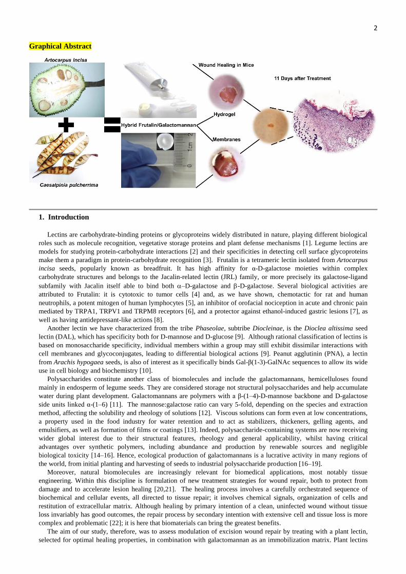
2 Graphical Abstract
1. Introduction
Lectins are carbohydrate-binding proteins or glycoproteins widely distributed in nature, playing different biological
roles such as molecule recognition, vegetative storage proteins and plant defense mechanisms [1]. Legume lectins are
models for studying protein-carbohydrate interactions [2] and their specificities in detecting cell surface glycoproteins
make them a paradigm in protein-carbohydrate recognition [3]. Frutalin is a tetrameric lectin isolated from Artocarpus
incisa seeds, popularly known as breadfruit. It has high affinity for α-D-galactose moieties within complex
carbohydrate structures and belongs to the Jacalin-related lectin (JRL) family, or more precisely its galactose-ligand
subfamily with Jacalin itself able to bind both –D-galactose and -D-galactose. Several biological activities are
attributed to Frutalin: it is cytotoxic to tumor cells [4] and, as we have shown, chemotactic for rat and human
neutrophils, a potent mitogen of human lymphocytes [5], an inhibitor of orofacial nociception in acute and chronic pain
mediated by TRPA1, TRPV1 and TRPM8 receptors [6], and a protector against ethanol-induced gastric lesions [7], as
well as having antidepressant-like actions [8].
Another lectin we have characterized from the tribe Phaseolae, subtribe Diocleinae, is the Dioclea altissima seed
lectin (DAL), which has specificity both for D-mannose and D-glucose [9]. Although rational classification of lectins is
based on monosaccharide specificity, individual members within a group may still exhibit dissimilar interactions with
cell membranes and glycoconjugates, leading to differential biological actions [9]. Peanut agglutinin (PNA), a lectin
from Arachis hypogaea seeds, is also of interest as it specifically binds Gal-β(1-3)-GalNAc sequences to allow its wide
use in cell biology and biochemistry [10].
Polysaccharides constitute another class of biomolecules and include the galactomannans, hemicelluloses found
mainly in endosperm of legume seeds. They are considered storage not structural polysaccharides and help accumulate
water during plant development. Galactomannans are polymers with a β-(1–4)-D-mannose backbone and D-galactose
side units linked α-(1–6) [11]. The mannose:galactose ratio can vary 5-fold, depending on the species and extraction
method, affecting the solubility and rheology of solutions [12]. Viscous solutions can form even at low concentrations,
a property used in the food industry for water retention and to act as stabilizers, thickeners, gelling agents, and
emulsifiers, as well as formation of films or coatings [13]. Indeed, polysaccharide-containing systems are now receiving
wider global interest due to their structural features, rheology and general applicability, whilst having critical
advantages over synthetic polymers, including abundance and production by renewable sources and negligible
biological toxicity [14–16]. Hence, ecological production of galactomannans is a lucrative activity in many regions of
the world, from initial planting and harvesting of seeds to industrial polysaccharide production [16–19].
Moreover, natural biomolecules are increasingly relevant for biomedical applications, most notably tissue
engineering. Within this discipline is formulation of new treatment strategies for wound repair, both to protect from
damage and to accelerate lesion healing [20,21]. The healing process involves a carefully orchestrated sequence of
biochemical and cellular events, all directed to tissue repair; it involves chemical signals, organization of cells and
restitution of extracellular matrix. Although healing by primary intention of a clean, uninfected wound without tissue
loss invariably has good outcomes, the repair process by secondary intention with extensive cell and tissue loss is more
complex and problematic [22]; it is here that biomaterials can bring the greatest benefits.
The aim of our study, therefore, was to assess modulation of excision wound repair by treating with a plant lectin,
selected for optimal healing properties, in combination with galactomannan as an immobilization matrix. Plant lectins

3 from Artocarpus integrifolia (Jacalin), Artocarpus incisa (Frutalin), Arachis hypogaea (PNA) and Dioclea altissima
(DAL) were chosen for the initial screen, as each has different carbohydrate-recognition specificity. They were
extracted from seeds, purified and assayed for bioactivity, including use of cultured fibroblasts. Frutalin proved most
effective in fibroblast migration assays and hence was combined with galactomannan to obtain hydrogels and also
membrane scaffolds; both formulations were then evaluated for dermal wound healing in mice bearing punch biopsy
excisional wounds.
2. Materials and methods
2.1. Materials
Artocarpus incisa, Artocarpus integrifolia and Dioclea altissima seeds were collected within metropolitan Fortaleza,
Ceará, while Arachis hypogaea seeds purchased at the local São Sebastião Market. All the seeds were selected and
peeled. Arachis hypogaea seeds were delipidated, and apart from Dioclea altissima, all the others were dehydrated with
acetone to obtain a flour of grain size ~0.4 mm. Caesalpinia pulcherrima seeds were also collected in metropolitan
Fortaleza, cleaned and stored at room temperature until further use. A representative specimen was deposited in
Herbarium Prisco Bezerra (EAC), Federal University of Ceará, under no. 56367.
2.2. Extraction and purification
2.2.1. Lectins
Crude extracts of A. incisa (breadfruit), A. integrifolia (jackfruit), A. hypogaea (peanuts) and D. altissima were
prepared by stirring for 30 min in 0.15 M NaCl (flour: saline, 1:10), centrifuging for 30 min at 10,000 xg (4 °C) and
filtered. The obtained extracts were submitted to the affinity chromatography: Frutalin and Jacalin were isolated on D-
Galactose-agarose affinity columns (Pierce®); PNA on a lactose-agarose column (Sigma); and DAL on a dextran
(Sephadex G50) column. The eluted lectin fractions were collected, dialysed against distilled water and lyophilized.
Purity was confirmed by SDS-PAGE, while functional activity was assessed by a routine hemagglutination assay to
measure Minimal Concentration for Agglutination (MCA) [23,24].
2.2.2. Galactomannan
C. pulcherrima seeds were milled, and after manually separating endosperms from cotyledon and husk they were
immersed in boiling 96% ethanol for 10 min to deactivate enzymes. The endosperms were kept in distilled water (w:v,
1:5) at 7 ˚C overnight, then water (10 vol) added before blending at 500 rpm to obtain a viscous solution. After filtration
in a nylon net filter (180 µm pore size), galactomannan was precipitated by addition of 96% ethanol (2 vol), collected
and dispersed in acetone (1:5, w:v) for 15 min, followed by airflow drying to produce a powder. This was suspended in
water (1:100, w:v) and ethanol precipitation repeated, before milling the purified polysaccharide and sieving (0.125 mm
mesh) [17].
The total protein content of galactomannan was estimated by the Kjeldahl method for total nitrogen content [25]
using the conversion factor of N × 5.7. For monosaccharide composition, 1H NMR analysis was performed on
galactomannan dissolved (10 mg/ml) in high quality D2O (99.96% D). Spectra were recorded at 85°C on an Avance
DRX-500 Bruker Spectrometer equipped with a process controller, and using a relaxation delay of 1 s and a pulse width
of 90 to reach conditions for quantitative analysis. The 1H anomeric proton signals for mannose (at 4.00 and 4.72 ppm
on 1H) and galactose (at 4.99 and 5.00 ppm on
1H) were integrated, using Varian VNMRJ 4 software, to estimate their
relative proportions in the sample.
2.3. Microorganism agglutination, MIC and MBC
Candida albicans (ATCC 10231), Escherichia coli (ATCC 8739), Klebsiella pneumoniae (ATCC 13883),
Pseudomonas aeruginosa (ATCC 9027), and Staphylococcus aureus (ATCC 6538) strains were supplied by the
Laboratory of Microbiological Quality Control, University of Fortaleza (UNIFOR) and cultured in brain heart infusion
(BHI) broth at 35±2 °C for 24 h. The microorganisms were collected by centrifugation (1000 xg) and suspended in BHI
broth with O.D. (λ620) = 0.080 to 0.100, (1.5 x 10⁸ CFU/ mL). Strain agglutination was performed on glass slides using 2
mg/mL lectin solutions and the microorganism suspensions (20 μL lectin + 20 μL microorganism). The slides were
shaken in circles and left for 5 min at room temperature, subjected to Gram staining and observed visually under 20x
magnification by optical microscopy.
Minimum Inhibitory Concentration (MIC) and Minimum Bactericidal Concentration (MBC) were determined by the
96-well plate microdilution method according to The Clinical & Laboratory Standards Institute [26]. Serial dilutions of
lectins (initially 1 mg/ mL) were made in saline and 50 μL added to each well, followed by 100 μL BHI broth, 50 μL
microorganism inoculums (1.5 x 10⁵ CFU/mL) and resazurin solution (10 μL). After incubation for 24 h at 37 °C,
sample growth was monitored by resazurin color change; the lowest concentration without visible growth was defined
as MIC. For MBC, 5 μL aliquots were aseptically removed from MIC wells without visible turbidity, seeded onto

4 Müeller Hinton agar (Merck®) and incubated for 24 h at 37 °C. MBC was defined as the lowest lectin concentration,
which inhibited 99.99% or more of the initial inoculum. Each measurement was performed in quadruplicate.
2.4. TLR4 reporter assay
TLR4 stimulation by the lectins was monitored using HEK-Blue™ hTLR4, obtained by co-transfection of the
hTLR4 gene, the MD-2/CD14 co-receptor genes and a secreted embryonic alkaline phosphatase (SEAP) reporter gene
into HEK293 cells (InvivoGen ®). The medium waks supplemented with 50 µg/mL of each lectin and levels of SEAP
were determined with HEK-Blue™ detection over the time of 24 h. 100 ng/mL of LPS was used as agonist.
2.5. In vitro assays using fibroblasts
Normal human skin fibroblasts (NHSF) were derived from dermal biopsies of healthy donors and cultured at the
Royal Free Campus, UCL (studies performed under Research Ethics Committee Protocol Approval Number:
REC/6398). Cells were grown, and experiments conducted, in Dulbecco's Modified Eagle's Medium (GIBCO, USA)
high-glucose medium supplemented with 10% fetal bovine serum (GIBCO, USA) and 1% penicillin-streptomycin by
incubation at 37 °C in 5% CO2. In experiments, fibroblasts were used between passages 2 and 6 to avoid phenotypic
changes during subculture.
2.5.1. Cell viability
NHSF cells were seeded at 5 x10³ per well in 96-well plates. After adhesion and recovery for 12 h, cells were treated
with each of the four lectins in the concentration range of 1-1000 μg/ mL for 72 h, following which cell viability was
measured by the PrestoBlue® resazurin-based reagent. Results were expressed as a percentage compared to untreated
cells. Statistical significance was assessed by ANOVA one-way analysis of variance, followed by the Bonferroni test
for multiple means. The level of significance was determined as p <0.05 using GraphPad Prism software, version 6.00,
La Jolla California USA.
2.5.2. Fibroblast migration assays
The ability of lectins to induce NHSF cell migration into a denuded and wounded cell monolayer area was assessed
using the scratch wound assay [24,27]. Fibroblasts were seeded into 6-well plates and grown overnight to confluence
before creating a 1 mm linear scratch in each well. After removal of growth media, triplicate test wells received 50
μg/mL lectin and 5 μg/mL mitomycin-C (to prevent cell proliferation), while control wells received vehicle alone.
Scratched areas were photographed at 0, 8, 12, 24 and 48 h post-treatment and data analyzed with ImageJ 1.42q
software (National Institutes for Health, USA) to measure the distance travelled by cells into the wound. The percentage
of cell migration at each time was calculated as follows:
Scratch closure rate = [(D0 – Dn / D0] x 100%
where D0 is the average initial distance between both sides of the scratch and Dn is the average distance between both
sides of the scratch at the measured time. Experiments were performed in triplicate in two independent assays.
Of the four lectins, Frutalin showed the greatest closure rate and so was selected for further evaluation by the
Boyden Chamber cell migration assay using 24-well plates with 8 μm inserts. Briefly, NHSF fibroblasts (3 x10⁵
per
well) were grown to confluence in the upper chamber and then Frutalin (50 μg/mL) added to the lower chamber;
controls were no Frutalin and also equal Frutalin concentrations in both lower and upper chambers. After 24 h exposure,
upper surface cells were removed by sterile swab, and those migrating to the lower compartment fixed with methanol
and stained with 0.1% crystal violet. Migrated cells were counted in 5 randomized fields per specimen under inverted
microscope and treatments compared statistically by Student's t-test, p <0.05 using GraphPad Prism software, version
6.00, La Jolla California USA.
2.5.3. Assessment of fibroblast physiology and IL-6 production
NHSF fibroblasts were grown overnight in 6-well plates and then medium containing Frutalin (50 μg/mL) added for
0, 0.5, 1, 2, 4 and 24 h; control wells received medium alone. At each time, cells were washed with PBS, then lysed
with RIPA buffer (Sigma-Aldrich) supplemented with cOmplete ™ Protease Inhibitor Cocktail (Roche) and
Phosphatase Inhibitor Cocktails (Sigma-Aldrich). Supernatants were collected by centrifugation (30 min at 10,000 xg, 4
°C) and, after protein measurement by BCA assay, 10 μg samples were separated on NuPAGE™ 4-12% Bis-Tris
Protein Gels (1.0 mm) gel before electrotransfer to polyvinylidene difluoride (PVDF) membranes.
Following incubation with casein blocking buffer for 1 h to reduce nonspecific interactions, the membranes were
incubated with diverse primary antibodies for 12 h at 4 °C to assess Frutalin-induced cell changes in cell signaling plus
collagen, cytokine and growth factor production. Primary antibodies used and respective dilutions were: anti-β-tubulin
(1: 10000, Abcam, ab6046); Anti-p-IkB-β antibody (1: 500, Abcam, ab 7547); Collagen Type I antibody (1: 1000,
Millipore, AB758); Anti-p-ERK 1/2(1: 1000, Abcam, 9101s), anti-MyD88 antibody (1: 1000, Abcam, ab2068), anti-
CTGF (1: 500, Santa Cruz Biotechnology, Sc-14939) and anti-NF-kB p65 antibody (1: 1000, Santa Cruz
Biotechnology, sc-8008). Detection was with HRP-conjugated secondary antibodies, the labeled proteins being
visualized with an ECL Western Blotting kit (ECL Plus, Amersham, UK) and quantified with Image J software
5 (National Institute of Health, USA) by normalization to β-tubulin. Results were analyzed by Student's t-test, p <0.05
using GraphPad Prism software, version 6.00, La Jolla California USA.
In parallel experiments, NHSF cells were exposed to Frutalin (50 μg /mL) for 24 h and the culture medium assayed
for IL-6 secretion by ELISA using a Quantikine ELISA kit (R&D Systems, Minneapolis, USA).
2.6. Molecular docking studies
We docked Frutalin (PDB id: 4WOG) to the TLR4/MD-2 receptor complex (PDB id: 3FX1) using HADDOCK 2.2
[28,29] to generate an interface model for the interaction. MD-2 is a soluble protein that non-covalently associates with
TLR4, but can directly form a complex with LPS in the absence of TLR4 [30]. The NetNGlyc 1.0 Server
(http://www.cbs.dtu.dk/services/NetNGlyc/) predicted two glycosylation sites for MD-2, one at Asn8 and the other
closer to the TLR4/MD-2 interface at Asn96. Thus, Frutalin may interact with the carbohydrate portion of MD-2 at
position 96 and activate TLR4. Residue Asn96 was considered the pivotal anchoring site and residues within a radius
<6.5 Å were considered to maximize the interaction with the lectin. Frutalin is a homotetramer and each monomer was
used for docking and evaluation taking into account its carbohydrate-recognition domain (CRD) (Gly25, Tyr146,
Trp147 and Asp149) and the Asn96 residue in MD-2. Our protocol undertook three steps of Haddock execution
followed by rigid-body docking, flexible-interface docking and the water layer around the TLR4/MD-2 complex
[28,29]. The best ligand conformation was selected based on the HADDOCK score and the models were clustered at 7.5
Å.
2.7. Hydrogel and membrane scaffold formulations
2.7.1. Hydrogels
A 1.5% (w/v) solution of C. pulcherrima galactomannan in 0.15 M NaCl was prepared in a Turratec TE102
homogenizer for 10 min at 18,000 rpm, following which 0.1% cetyltrimethylammonium bromide (CTAB) was added to
prevent microbiological growth. This stock solution was designated F0 (galactomannan hydrogel alone; control) and
used to prepare the Frutalin formulations, F01, F05 and F10, which contained 0.1, 0.5 and 1.0 mg Frutalin/ g
galactomannan hydrogel, respectively. Each hydrogel was then centrifuged for 30 min at 10,000 xg and 4 °C to remove
insoluble materials and refrigerated until use.
2.7.2. Microbiological examination
Quantitative enumeration of mesophilic bacteria and fungi that may grow under aerobic conditions in the
hydrogel preparations were evaluated according to the European Pharmacopoeia [31]. Samples of at least 3 subsequent
ten-fold dilutions were inoculated in nutrient and Sabouraud-dextrose agar following the pour-plate method. Then, the
total viable aerobic count was performed after 7 days of incubation at 30-35 °C for bacteria and 20-25 °C for fungi.
2.7.3. Membrane scaffolds
The same hydrogel solutions were prepared as described in 2.7.1., but without 0.1% CTAB. Each solution was
distributed in 24-well plates (1 g/ well) and lyophilized under temperature and pressure ramps (AdVantage 2.0 Bench
Top Freeze Dryer, VirTis, SP Scientific) to obtain a more uniform ‘spongy membrane’. The resulting membrane
scaffolds were designated: MF0 (galactomannan hydrogel alone; control) plus MF01, MF05 and MF10, containing 0.1,
0.5 and 1.0 mg Frutalin/ g galactomannan hydrogel, respectively.
2.7.4. Hemagglutination assay with the hydrogels and membrane scaffolds
To verify that our Frutalin-galactomannan formulations retained bioactivity, both hydrogels and membrane scaffolds
(dissolved in saline) were tested in routine hemagglutination assays. They also underwent thermostability testing in the
40-100°C range to determine the upper temperature at which Frutalin still remains active.
2.8. Rheology studies
Mechanical stability studies used a stress-controlled rheometer (Anton Paar MCR 302) housing a circular parallel
plate cell with internal diameter (i.d.) = 25 mm and height (h) = 1 mm gap between plates. All experiments were
conducted at 25 °C and included stress-strain tests, rotational flow, and oscillatory viscoelastic linear-response
measurements. The rheometer was operated in oscillatory mode for stress-strain tests: at a fixed frequency ω = 10 rad/s
with an externally applied shear strain from 0.01 to 100 %, while recording storage (G’) and loss (G’’) moduli. This
allows mechanical responses of samples to be assessed from the reversible linear response regime up to the nonlinear
region, where the sample structure collapses and flow starts.
The flow study was conducted in continuous steady-state shear rotational mode by varying the shear rate from 1 to
1000 s. Dynamic oscillatory mode studies were done by varying the applied torque/deformation angular frequency from
0.1 to 100 rad/s. The storage (G’) and loss (G’’) moduli sample responses were recorded during frequency sweeps at
5.0% fixed strain amplitude, enough to guarantee linear reversible responses.

6 2.9. Physical characterization of membrane scaffolds: FTIR, SEM and WVP
Membrane scaffolds were ground under liquid N2, mixed with KBr and their infra-red spectra determined using a
Fourier transform infrared spectrometer (IRTracer-100 spectrophotometer, Shimadzu, Japan). Spectra were recorded at
room temperature and, on average, 64 scans were made for each sample at a resolution of 4 cm-1
in the range of 400-
4000 cm-1
.
Scanning electron microscopy (SEM) for morphology investigations was carried out in the Central Analítica ,
Federal University of Ceara. Cross-sectional samples of membrane scaffolds were placed on aluminum stubs and
splutter coated with gold prior to analysis. Images were captured with a Phillips XL30 SEM operating at an acceleration
voltage of 15 keV.
Water Vapor Permeability (WVP) was determined gravimetrically according to ASTM E96-95 (1992) protocols
[32]. Eight samples were cut from each sponge membrane, each sealed in a permeation cell (a water-containing cup at
100 % RH; 2.337 × 10³ Pa vapor pressure at 20 °C), and placed in a desiccator with silica gel (0 % RH; 20 °C). The
water vapor transferred through the test samples was determined from cup weight loss over time. Steady-state weight
loss was reached after 24 h.
2.10. In vivo studies
2.10.1. Animals, surgical procedures and macroscopic analysis
Swiss albino mice (20-25 g) housed in the animal facility at the University of Fortaleza (UNIFOR) were kept in a
controlled environment with free access to water and standard pellet diet (Purina, São Paulo, Brazil). The experimental
protocols followed the ethical guidelines of CONCEA (Brazilian Council for the Control of Animal Experimentation)
and were approved by the Animal Ethics Committee of UNIFOR under entry number 004/2014.
Mice were anesthetized by intraperitoneal (i.p.) injection of ketamine (115 mg/ kg) and xylazine (10 mg/ kg)
hydrochlorides. The dorsal skin of the animals was shaved, cleaned with 1% iodopovidone and then subjected to a
punch biopsy excisional wound with a cutting blade diameter of 10 mm. After recovery from anesthesia, mice were
housed individually in disinfected cages and randomized into nine treatment groups (20 animals/each): Control (0.15 M
NaCl), F0, F01, F05 and F10 for the hydrogel formulations and MF0, MF01, MF05 and MF10 for the membrane
scaffolds. Administration of NaCl and hydrogels was done daily with 100 μL sample applied over the wound area,
whereas membrane scaffolds were implanted under the muscular fascia and left until the end of the experiment. All
groups were evaluated 3, 7, 11 and 14 days after excision (n = 5 mice/ day/ group). At each time point, the wound area
was measured using images with a metric reference by the ImageJ® program. In addition, wounds were assessed for
hyperemia, edema, bleeding, secretion and scab formation. The classification was performed semi-quantitatively
according to the intensity, with the following scores used for edema and hyperemia: Absence (0), Light (1), Moderate
(2), Intense (3). The presence of inflammatory exudate was classified as serous, bloody or purulent [33].
2.10.2. Histological microscopic analysis
For histological examination the wound area was removed surgically from individual anesthetized mice, which then
underwent euthanasia. Tissue samples were arranged in cassettes, fixed in 10% buffered formaldehyde pH 7.2 and then
dehydrated in increasing concentrations of ethanol, before diaphanization in xylol and embedding in paraffin wax.
Thick (5 µm) tissue sections were cut and stained with H&E as per routine histology. Stained sections were scanned
(Scanner panoramic desk, 3DHISTECH) observed using CaseViewer 1.4 software (3DHISTECH Ltd.) and the
histopathological properties of each tissue section were assessed by an experienced pathologist, blinded to the
experimental groups.
2.11. Statistical analysis
All statistics were performed using GraphPad Prism 6 (GraphPad Software Inc.). Statistical significance was
assessed by ANOVA one-way analysis of variance, followed by the Bonferroni’s test for multiple mean or by Kruskal-
Wallis comparisons for median, followed by the Dunns test. The level of significance was considered as p < 0.05.
3. Results
3.1. Extraction, purification and characterization of lectins and galactomannans
The yields of Frutalin, Jacalin, PNA and DAL based on the flour prepared from seeds were 1.56%, 1.96%, 0.01%
and 1.38%, respectively. Excellent purity was shown by electrophoresis (Supplementary Fig. S1A), while a routine
hemagglutination assay confirmed their post-extraction functionality with Jacalin and Frutalin the most potent showing
visible agglutination at the lowest concentrations (MCAs of 0.004 µg/ mL and 0.015 µg/ mL, respectively.
Additionally, each lectin was able to bind and agglutinate several pathogenic strains involved in many common
infections and impaired healing, namely Staphylococcus aureus, Klebsiella pneumoniae, Pseudomonas aeruginosa,
7 Escherichia coli and Candida albicans (Fig. 1). On a subjective scale, the degree of agglutination was Jacalin>Frutalin>
DAL> PNA.
Despite promoting agglutination of pathogens, no lectin showed bacteriostatic (MIC) or bactericidal (MBC)
activities when tested at ≤1mg/ mL. As TLR4 plays a key recognition role of LPS in bacteria outer membranes to
trigger the innate response, lectins were tested to see if they were capable of stimulating such a receptor. Dose-response
curves showed that all lectins activated the TLR4 receptor, with Frutalin and Jacalin having a comparable potency to
LPS, used as a control (Fig. 2). Next, we modelled molecular docking between a TLR4/MD-2 complex and the Frutalin
homotetramer (A, B, C and D), assembling them in over 180 complexes. The clusters of each monomer presented
negative Z-values and were grouped into 10. The 10 clusters with the lowest Z-values represented over 90% of the
water-refined HADDOCK models generated for the monomers. Except for few differences in Z-values for monomer A,
all clusters displayed similar orientation. As a tetramer, Frutalin occupies a molecular volume similar to that of TLR4,
which based on docking data suggests that the lectin will interact most favorably with the TLR4/MD-2 complex if
orientated as illustrated in Fig.3.
Protein contamination of purified C. pulcherrima galactomannan was negligible at 0.08% as judged by total nitrogen
content, reflecting the double ethanol precipitation which increased polysaccharide purity. The monosaccharide ratio
was determined by NMR and gave a mannose:galactose ratio of 2.36 in this hemicellulose.
3.2. In vitro assays
3.2.1. Cell viability
Human skin fibroblasts were used to check lectin cytotoxicity. Frutalin presented no toxicity at concentrations below
1000 μg/ mL. Proliferative effect was observed at concentrations higher than 500 μg/ mL for jacalin and 1000 μg / mL
for DAL. The opposite effect was observed in PNA, demonstrating reduction of cell viability at concentrations > 500
μg/ mL.
3.2. Fibroblast migration
The ability of each lectin to stimulate NHSF migration in vitro was tested at 50 μg/ mL, as none were toxic to the
cells at this concentration. Of the four lectins, only Frutalin significantly outperformed untreated (control) cells; after
48 h incubation there was 100% cell migration versus 30% (Fig. 4A).
To verify this Frutalin potency, NHSF cells were also cultured in Boyden chambers to assess its effect on fibroblast
directional migration. Addition of Frutalin (50 μg/ mL) to the lower chamber alone significantly stimulated NHSF
migration compared to untreated cells, or when compared to Frutalin addition to both chambers (Fig. 4B).
3.2.3. Fibroblast signal transduction, and matrix, cytokine and growth factor production
As for migration studies, NHSF cells were incubated with Frutalin (50 μg/ mL) and assayed 0.5, 1, 2, 4 and 24 h
later. Cell proteins were extracted, quantified and changes in cell signalling proteins and collagen, cytokine and growth
factor levels measured by Western blot and ELISA. Exposure to Frutalin produced significantly elevated levels of p-
ERK1/2 and MyD88 in NHSF cells, but no change in NFκB (Fig. 5). Cell collagen levels were decreased 24 h post-
treatment, but the amount of collagen secreted into the culture medium was unchanged at each time point.
Levels of IL-6 secreted into the NHSF culture were also assayed and increased five-fold (ng/mL) with addition of
Frutalin for 24 h (1.880.23 versus 10.510.10; p<0.0001). Although IL-6 has a key role in the acute phase response, it
is also involved in different signalling pathways, including those stimulating angiogenesis and cell migration. Hence, all
following experiments focussed on Frutalin and its healing potential in the development of topical formulations.
3.3. Hydrogel and spongy membrane scaffold characterization
Given the known drug delivery properties of galactomannan, we prepared two Frutalin-galactomannan formulations,
hydrogels and spongy membrane scaffolds, for in vivo studies to assess whether Frutalin bioactivity for wound healing
would be enhanced. Prior to such testing (see 3.4.), we carried out basic biological and physicochemical evaluations.
First, we checked both formulations for bacteria, yeast or and fungi contamination, which would be detrimental to in
vivo use, employing standard microbiological culture conditions. All plates with varying aliquots of the formulations
showed no growth of microorganisms (data not shown), and hence were deemed suitable for mouse wound healing
experiments. Second, use of the hemagglutination assay confirmed that Frutalin retained its activity when combined
with galactomannan; each formulation (F01, F05 and F10) retained the ability to agglutinate rabbit erythrocytes.
Similarly, solubilization of the membrane scaffolds in saline showed the same agglutination capacity. Moreover, both
formulations had good thermostability; heating for 5 min to a limit of 55 °C showed good retention of hemagglutinating
activity (data not shown).
Rheological tests were performed to evaluate how Frutalin influences the behaviour of its viscous galactomannan
vehicle. As shown in Figs. 6A and 6B, the flow curves of all samples had a characteristic profile of a pseudoplastic
fluid. By submitting the rheological behaviour of the F0, F01, F05 and F10 hydrogels to the power law model, it was
evident that increasing the amount of Frutalin led to an increase in fluid pseudoplasty as seen by an increasing behavior
index n (Fig. 6B). Indeed, Frutalin decreased the consistency index, which is directly associated to solution viscosity.

8 The hydrogels were further subjected to oscillatory tests. The oscillation frequency was kept constant (10 rad/ s),
while the amplitude ranged from 0.01 to 100% to vary angular frequency from 100 to 0.1 rad/ s (Fig. 6C). Increasing
Frutalin concentrations in the samples increased the G' moduli, considered the elastic part of the sample. At 25.1 rad/ s
angular frequency, G' = G" for all samples, while from this point onwards G' > G'' with the samples starting to present a
solid profile, acquiring gel structural characteristics.
Temperature sweeps ranging from 25 to 100 °C were used to investigate the viscosity behaviour of each hydrogel
with amplitude and frequency kept constant at 5% and 10 rad/ s, respectively. The modulus G " reached a plateau in the
range of 50 to 60 °C (Fig. 6D), at which point the samples began to undergo structural changes with the drastic decrease
in G'' directly related to higher concentrations of Frutalin in the galactomannan solution.
The membrane scaffolds MF0, MF01, MF05 and MF10 were analysed by infrared spectroscopy to identify the main
functional groups present in the structures through the absorbance peaks as a function of wave number (Fig. 7A). The
bands at 825 and 867 cm-1
are related to anomeric configurations (α and β) and glycosidic bonds, indicating the
presence of α-D-galactopyranose units and β-D-mannopyranose units, respectively, while the 1025 cm-1
band is due to –
CH2 vibrational torsion. Bands at 1056 and 1093 cm-1
were also noted and attributed to stretching of the primary alcohol
-CH2OH, whereas the 1141 cm-1
band corresponds to angular deformation of C-O due to the pyranose ring. Bands in
the range 1379 and 1438 cm-1
are due to symmetrical deformations of the CH2 and COH groups, while the amide I
(1652 cm−1
) and II (1542 cm−1
) bands mainly originate from C=O and N–H vibrations in proteins. The band around
2137 cm-1
, more predominant in membranes containing Frutalin, was attributed to the bond between the C and N atoms,
when involved in in peptide bonds. The bands at 2920 and 3350 cm-1
correspond to stretches of the CH2 and OH
groups, respectively, with the latter undergoing clear displacement to 3595 cm-1
for each of the Frutalin-containing
membrane scaffolds. This Frutalin-dependent displacement was also observed in photomicrographs of the MF0, MF1,
MF2 and MF3 membranes (Fig. 7B), presumably because Frutalin interacts to OH groups presented in the structure of
galactomannan matrix.
The Frutalin-galactomannan membrane scaffolds were also compared based on Water Vapor Permeability (WVP).
As shown in Fig. S4, there was a significant increase only between MF0 (Frutalin absent) and MF01 (0.1mg Frutalin / g
hydrogel) with higher concentrations of Frutalin having no significant effect on WVP.
3.4. In vivo assays
3.4.1. Hydrogel
Frutalin-galactomannan hydrogels were applied daily to deep excisional skin wounds on mice, who were monitored
at four different time points. Edema occurred mildly in all hydrogel-treated groups (F0, F01, F05 and F10) with a
median and variation of 1 (0-1) 3 days post-surgery compared to the saline-treated (control) group. Mild hyperemia
was seen in all groups (data no shown), but additional signs of exudation were not observed in treatment groups
compared to the control. A significant increase in wound contraction was noted 3 days after treatment for groups F0 and
F01; this was not observed for the F05 and F10 groups (Supplementary Fig. S5), or seen for the remaining days (7, 11
and 14).
The dynamics of the healing process was followed by histological analysis and was initially marked by an intense
inflammatory infiltrate on days 3 and 7, while fibroblast proliferation and the start of total re-epithelialization of the
previously ulcerated area was noted from the 11th
day. On day 14 it was possible to observe the beginnings of collagen
reorganization. For all the groups, the healing process was accompanied by mild congestion, but marked by intense
angiogenesis and proliferation of dermal fibroblast-like cells even 3 days post-surgery (Fig. 8).
In comparing individual groups, the application of hydrogels to F0, F01, F05 and F10 all resulted in an ability to
modulate and improve the healing process until the 7th
day. However, by day 11 only F01 (0.1mg of frutalin /g of
hydrogel) had promoted collagen organization. It appeared that higher concentrations of Frutalin contained within a
galactomannan matrix do not enhance or promote further healing as judged by relevant histopathological changes.
Although, as described above, there was no clear relation between wound contraction and treatment time or group, the
injured tissue did show healing characteristics. Thus, on the 14th
day a complete re-epithelization of the F01-treated
lesions was observed, increasing the proliferation of keratinocytes. Taken together these data strongly support the
potential of Frutalin to aid and accelerate repair processes within injured skin.
3.4.2. Spongy membrane scaffolds
The four groups (MF0, MF01, MF05 and MF10) treated with membrane scaffolds did not show significant amounts
of edema or hyperemia when compared to the control group. Measurements of wound contraction revealed a
significant increase only for the MF05 group on the 3rd
day compared to the untreated control group (Supplementary
Fig. S6).
When comparing the control mice to those receiving the galactamannan-only membrane (MF0), we observed no
relevant histopathological differences (edema, congestion, and inflammatory infiltrate) between the two groups.
However, for the Frutalin-containing membrane scaffolds (MF01, MF05 and MF10), the healing process was marked
by intense angiogenesis and absence of edema. Moreover, from the 11th
day reorganization of the conjunctive matrix
with fibroblasts and other fibroblast-like cells, such as fibrocytes was evident, with a mild to moderate inflammatory
infiltrate, diffuse, and predominantly lymphocytes (Fig. 9). Nevertheless, when comparing the different amounts of

9 Frutalin in the galactomannan scaffold membranes (MF01 vs. MF05 vs. MF10), we did not observe relevant differences
in histopathology. A similar finding was noted for the hydrogel-treated groups, suggesting that Frutalin levels higher
than those in MF01/ F01 do not further increase healing capacity of excised lesions. On the other hand, the benefits of
Frutalin to wound repair and regeneration were clearly evident.
4. Discussion
Plant lectins are relatively soluble in saline solutions, making their extraction and isolation by appropriate
carbohydrate-affinity chromatography a simple method. Lectin-carbohydrate interactions occur via hydrogen bonds,
Van der Waals forces and other hydrophobic forces, allowing easy elution by excess ligand or pH change. Lectin
carbohydrate specificities have important roles in biological recognition phenomena involving cells and proteins,
including immunomodulatory properties in different biomedical settings. In general, the different lectins can be grouped
into classes based mainly on the "architecture" of their carbohydrate binding site(s), including specificity and 3D
structure plus sequence similarities [34].
Mannose, for example, binds to lectins through diverse residues; ConA via Asn and Asp, Galanthus nivalus
agglutinin via Asn, Asp, Gln and Tyr, and mannose-binding protein A via Asn and Gln [34]. By contrast, Frutalin is a
breadfruit seed glycoprotein with four isoforms modifying a few amino acids. This gives Frutalin multiple-binding
character, able to interact with different ligands using the same carbohydrate binding site. However, its highest affinity
is for α-D-galactose, which we have shown to be anchored by a cavity of four residues (Gly25, Tyr146, Trp147 and
Asp149) near the N-terminal of the α chain [35].
Despite much research, biological roles of native lectins are often more speculative than proven. Many consider
their main function as species protection, as lectins such as WGA, PNA and SBA inhibit growth and sporulation of
fungi including Trichoderma viride, Penicilium notatum and Aspergillus niger [34]. Indeed, a minor breadfruit lectin,
Frutackin that binds chitin, has fungicidal activity [36]. In the microbial world, lectins are often termed as
hemagglutinins, adhesins and toxins, serving to link different cell types, and also viruses to cells, via surface
carbohydrates [34,37]. Although dietary plant lectins may cause allergies, trigger autoimmune diseases, or limit nutrient
uptake [32], their diverse biological activities have potential biomedical applications, including both diagnostics and
therapeutics.
Here, we chose four lectins, Jacalin, Frutalin, DAL and PNA based on their different carbohydrate specificities, for
wound healing studies. Each was extracted and isolated with high purity (SDS-PAGE analysis) and good bioactivity
(hemagglutination assay). Additionally, only PNA showed some toxicity to cultured human skin fibroblasts at levels
below 1.0 mg/ mL, allowing further evaluation of all lectins in fibroblast migration assays. This in vitro test, routinely
employed prior to animal investigations, identified Frutalin as the most potent lectin, an attractive candidate due to its
abundance in Artocarpus incisa seeds (our yield was 1.56% from seed flour) or potential for recombinant synthesis in
P. pastoris [38]. Our native preparation was pure, the two distinct 12- and 15.5-kDa protein bands consistent with
glycosylation differences between molecules, and performed strongly in the hemagglutination assay with an MCA of
0.15 pg/ mL.
Inflammatory, fibrogenic and regenerative responses are part of the sequence of events in an injury response system
that serves to contain damage and to restore tissue function through recruitment of several types of specialized cells
such as platelets and fibroblasts which provide mechanical stability to the wound in short and long terms; phagocytic
leukocytes that combat potential pathogens and clear the site of injury from dead cells and debris, and progenitor or
stem cells to replace functional epithelium [39]. Once skin is injured, opportunistic microorganisms obtain access to the
underlying tissues, causing invasive infections within a wound with subsequent host injury. Then, we studied
interaction of our lectins with five pathogenic microorganisms, all common species in opportunistic infections.
However, though we showed all four lectins interacted with surfaces of E. coli, S. aureus, K. pneumoniae, P.
aeruginosa and C. albicans, to promote microorganism agglutination, none was able to inhibit growth or kill a strain.
Nonetheless, all lectins activated the TLR-4 receptor, a key player in detection of invading pathogens through several
pattern-recognition mechanisms and regulation of the immune system [30]. There is substantial diversity in the TLR4
cellular expression pattern and tissue distribution in the different mammalian species, as humans, mice, rats, rabbits, and
other ones. In mice, TLR4 is expressed in myeloid subsets (monocytes, macrophages, granulocytes, dendritic cells) and
lymphoid subsets [40]. Specific TLRs activation and/ or conditions that affect TLR expression are key to whether TLR
promotes or inhibits wound healing processes [41]. Frutalin as a multiple-binding lectin is able to recognize most of
glycans on cell surface of those microorganisms, which might help control the infection at the wound bed either by
agglutinating those pathogens or activating the innate immune response. Through this perspective, a molecular
modelling of Frutalin interaction with the TLR4/MD-2 receptor complex was provided through docking studies.
Glycosylation on Asn8 and Asn96 residues of MD-2 molecule are pivotal for Frutalin’s anchoring and activation of the
complex TLR4/MD-2. In addition, TLR4 mediated the activation of neutrophils and it has a particular role to play in the
regulation of neutrophil life span, and TLR4 signalling can regulate neutrophil survival [42]
We selected Frutalin for further study, mainly based on its strong stimulation of fibroblast migration in both scratch
wound and Boyden two-chamber assays. However, our selection is endorsed by studies citing Frutalin’s therapeutic
potential in different areas, including anti-cancer activities in vitro [14], and by gastroprotection [16] or orofacial pain
reduction [15] in preclinical studies. First, we added modest amounts (50 μg/ mL) of Frutalin to fibroblast cultures and
10 found significantly increased levels of p-ERK and MyD88 at both short (0.5 h) and long (24 h) times. The former is
likely involved in fibroblast proliferation and the latter in TLR-4 receptor signaling when stimulated. TLR-4 stimulation
may activate either MyD88-dependent or TRIF-dependent pathways [43]. As we found an increased level of MyD88 we
assume that the fibroblast activation is mainly through MyD88 pathway. Additionally, we showed that the fibroblasts
had been hyperstimulated (5-fold) to produce IL-6, a cytokine considered pyrogenic and associated with cell
proliferation, as well as many innate immunity response pathways.
These diverse positive effects of Frutalin on fibroblasts, endorsed the next step of our work, namely to develop a
lectin-carrier combination to steadily release Frutalin into microenvironments to promote desired healing actions. We
chose galactomannan as the vehicle; it forms viscous hydrogel solutions at low concentrations, gives porous membrane
scaffolds on freeze drying and is reported to provide stabilizing matrices for lectins [44,45]. However, unlike these
earlier studies [44,45] we isolated the polysaccharide from Caesalpinia pulcherrima seeds. This species is readily
cultivated in tropical semiarid regions, produces seeds 6 months after planting and, as we have described [36], is well-
characterized and gave galactomannan yields of ~25% from dried seed mass.
Hydrogels should exhibit basic features, such as swelling capacity, drug compatibility, safety, low toxicity, shelf
stability and high purity; other requirements include biocompatibility, biodegradability, mechanical strength and
multiple biofunctionalities. For tissue engineering, hydrogel biodegradability is essential as they invariably function as
temporary extracellular matrix until replaced by new tissue, while bioactivity is essential for guiding cell behavior, such
as proliferation, differentiation and matrix production [46]. Natural polymers are known to protect skin from harmful
factors by such diverse hydrogel attributes and galactomannan, in particular, has cosmeceutical potential for its
antioxidant and anti-aging actions [47].
Physico-chemical characterization of our Frutalin-galactomannan combinations showed that higher Frutalin levels
increased galactomannan pseudoplasty, as viscosity was lost. The lectin interacts via H bonds with OH groups present
in branches of the galactomannan chain; this can allow "sliding" of the chains, reducing galactomannan-galactomannan
contact. It was evident by infrared spectroscopy as a band shift from 3350 cm-1
to 3595 cm-1
when Frutalin binds to
galactomannan OH groups.
Oscillatory tests, performed with amplitude and frequency sweeps, can separate and quantify elastic and viscous
parts of hydrogels by plotting graphs as a function of G' and G", respectively. Samples with G’ > G”' values are
classified as viscoelastic solid, while G'' > G' indicates a viscoelastic liquid. Such analysis also allows calculation of
sample Linear Viscoelastic Limit (LVE) i.e. the maximum deformation that the sample can undergo without permanent
alteration of its structure. For our studies, the LVE value was held at 10% during frequency and temperature sweeps;
this was the maximum deformation supported by our samples thus ensuring no structural changes occurred during the
tests. Moreover, by subjecting samples to a varying oscillation frequency we could check stability in the short- and
long-term (shelf life). Minor frequencies equate to long-term behaviour, while larger frequencies influence the short-
term. As we found G'' > G' for all samples, they are viscoelastic liquids; despite the high viscosity of a 1.5%
galactomannan solution, it does not have a gel-like or solid structure. Moreover, Frutalin additions did not alter this
property, as for all concentrations examined the viscous portion (G”) exceeded the elastic portion (G’), consistent with
liquid behaviour rather than that of a gel or solid.
Lyophilization of our hydrogels produced membrane scaffolds with a highly porous sponge-like structure containing
macropores (50-100 μm). The water vapor transmission rate (WVTR) is vital for the healing process. WVTR for normal
skin, first degree burns, and granulating wounds are (20±1) x10, (28±3) x10, and (51±2) x102 g/m
2 day, respectively. So
that, it is recommended that wound dressings should have WVTRs values in the range of (20–25)x102 g/m
2 day [48].
Such water vapor permeability is pertinent to our work, since our membranes presented values ranging within 29.6 ±6.6
g/m2 day. Wound dressings must allow damaged skin to breathe as failure to restore oxygenation impairs healing [49].
In normal wound healing, reactive oxygen species (ROS) such as hydrogen peroxide (H2O2) and superoxide (O2-) act as
cellular messengers to stimulate key processes, including cellular mobility, cytokine signaling and angiogenesis [50].
Such pathways are predicted to be stimulated by our Frutalin formulations, as in our previous pre-clinical studies we
showed that antinociceptive and gastroprotective effects are mediated by nitrergic mechanisms [6,7].
Noteworthy too is the direct influence that the mannose:galactose ratio has on water vapor permeability. In
galactamannan it is very low, resulting in many free mannose residues which in turn leads to dense packing of the
chains and reduced diffusion of water vapor through the matrix [51]. However, we only observed a significant
difference (p<0.05) between MF0 and MF01 (P <0.05), not for the membrane scaffolds with higher levels of Frutalin.
Most likely this reflects that when Frutalin is freeze-dried in contact with galactomannan intractable spaces remain in
the polymer, which are unaffected by further increases in Frutalin i.e. the extra lectin only fills spaces already generated
by initial Frutalin-galactomannan interactions and, of course, the volume occupied by a lectin molecule is much smaller
than the polysaccharide chairs.
In addition to the wound severity, multiple factors such as age, stress, sex hormones in aged individuals, diabetes,
medications, obesity, alcohol consumption, smoking, and nutrition can cause impaired wound healing by affecting one
or more phases of this complex process [50]. The excisional model in mice is an experimental model that may result in
many extrapolations of the current healing process, emphasizing once again that tissue repair is a very dynamic event.
Hydrogel liquid bandages, for example, are not recommended for stinging applications and/or open wound with
significant extension area. Although those water-based formulations are less traumatic when applied to an open wound,
the drying time in the wound bed can be long, and a polymer coating may be difficult to form because of flow of the

11 formulation away of the injured area [52]. On the other hand, efforts have been made to develop membranes that mimic
full-thickness skin wounds since those sort of dressings present suitable structure and properties for a better healing
process, as they protect the wound from moisture loss and infection. Both hydrogels and membranes of our hybrid plant
lectins/galactomannan formulations were tested in order to cover different ranges of topical applications. Hence, the
hydrogels were applied daily, whereas the membrane scaffold was implanted once only on the day of surgery.
Moreover, the membrane scaffolds carried 10 times more Frutalin than one 100 μL hydrogel application, since this was
sufficient to cover the entire injured area. Both treatments allowed the total recover of the wounded skin in 11 days. We
suggest that 100 µg of Frutalin/g of hydrogel or membrane as a helpful treatment in cutaneous wounds, especially in
cases of infected wounds, burns and disease-related ulcers.
5. Conclusions
Of the four lectins investigated in the initial phase of this work, Frutalin was identified as the best candidate to take
forward into in vivo wound healing studies: it substantially promoted migration of human skin fibroblast (at 50µg/mL),
was non-cytotoxic and stimulated TLR4. We then assessed various combinations of Frutalin and galactomannan
(isolated from C. pulcherrima) in hydrogels and, following their lyophilization, as membrane scaffolds for their efficacy
in excision wound healing. Hydrogels were applied daily to the dorsal wounds of mice, while membrane scaffolds were
implanted on the surgery day. Both formulations were effective with Frutalin at 100 µg/ g galactomannan enhancing
recovery from the dermal injuries after 11 days of treatment. We conclude that the breadfruit lectin, Frutalin, is a
potential therapeutic biomolecule for wound healing and therapeutic approach to restore skin integrity, and perhaps
other related skin diseases and injuries. Frutalin/galactomannan hybrids may represent an alternative to provide
improved health care for wound, burn and surgical patients in addition to reducing the high cost of current health care
treatment.
Disclosure
None
Acknowledgements
The authors would like to thank University of Fortaleza, Federal University of Ceará and University College London
(UCL) for infrastructure. This work was supported by a grant from Conselho Nacional de Desenvolvimento Científico e
Tecnológico (CNPq). Felipe Domingos de Sousa and Ayrles Fernanda Brandão da Silva were recipients of a PhD
sandwich fellowship and post-doctoral research fellowship, respectively, at UCL [Grants: 201016/2015-0 and
205991/2014-9]. This work was also supported by Fundação Cearense de Apoio ao Desenvolvimento Científico e
Tecnológico do Ceará (FUNCAP/SESA /MS/CNPq, Chamada 07/2013).
Appendix A. Supplementary data
Supplementary data associated with this article can be found in the online version.
References
[1] W.J. Peumans, E.J.M. Van Damme, Plant lectins: Versatile proteins with important perspectives in
biotechnology, Biotechnol. Genet. Eng. Rev. 15 (1998) 199–228. doi:10.1080/02648725.1998.10647956.
[2] J. Arnaud, A. Audfray, A. Imberty, Binding sugars: from natural lectins to synthetic receptors and engineered
neolectins., Chem. Soc. Rev. 42 (2013) 4798–4813. doi:10.1039/c2cs35435g.
[3] S. Fanayan, M. Hincapie, W.S. Hancock, Using lectins to harvest the plasma/serum glycoproteome,
Electrophoresis. 33 (2012) 1746–1754. doi:10.1002/elps.201100567.
[4] C. Oliveira, A. Nicolau, J.A. Teixeira, L. Domingues, Cytotoxic effects of native and recombinant frutalin, a
plant galactose-binding lectin, on Hela cervical cancer cells, J. Biomed. Biotechnol. 2011 (2011).
doi:10.1155/2011/568932.
[5] A.C. Brando-Lima, R.F. Saldanha-Gama, C.R. Pereira, C.G. Villela, A.L.F. Sampaio, A.C.O. Monteiro-
Moreira, M.D.G.M.O. Henriques, R.A. Moreira, C. Barja-Fidalgo, Involvement of phosphatidylinositol-3
kinase-Akt and nuclear factor kappa-B pathways in the effect of frutalin on human lymphocyte, Int.
Immunopharmacol. 6 (2006) 465–472. doi:10.1016/j.intimp.2005.09.008.
[6] M.B.M. V Damasceno, J.D.M.A. De Melo-Júnior, S.A.A.R. Santos, L.T.M. Melo, L.H.I. Leite, A.E. Vieira-

12
Neto, R.D.A. Moreira, A.C.D.O. Monteiro-Moreira, A.R. Campos, Frutalin reduces acute and neuropathic
nociceptive behaviours in rodent models of orofacial pain, Chem. Biol. Interact. 256 (2016) 9–15.
doi:10.1016/j.cbi.2016.06.016.
[7] A.P.D.V. Abdon, G.C. De Souza, L.N.C. De Souza, R.P. Vasconcelos, C.A. Castro, M.M. Guedes, R.C.P.
Lima-Júnior, R.D.A. Moreira, A.C.D.O. Monteiro-Moreira, A. Rolim-Campos, Gastroprotective potential of
frutalin, a D-galactose binding lectin, against ethanol-induced gastric lesions, Fitoterapia. 83 (2012) 604–608.
doi:10.1016/j.fitote.2012.01.005.
[8] J.R.C. Araújo, J. de M.A. de M. Júnior, M. de B.M.V. Damasceno, S.A.A.R. Santos, A.E. Vieira-Neto, M.D.P.
Lobo, A.R. Campos, R. de A. Moreira, A.C. de O. Monteiro-Moreira, Neuropharmacological characterization
of frutalin in mice: Evidence of an antidepressant-like effect mediated by the NMDA receptor/NO/cGMP
pathway, Int. J. Biol. Macromol. 112 (2018) 548–554. doi:10.1016/j.ijbiomac.2018.01.180.
[9] R.A. Moreira, A.C.O. Monteiro, A.C.G. Horta, J.T.A. Oliveira, B.S. Cavada, Isolation and characterization of
Dioclea altissima var. megacarpa seed lectin, Phytochemistry. 46 (1997) 139–144. doi:10.1016/S0031-
9422(97)00262-8.
[10] S.K. Natchiar, O. Srinivas, N. Mitra, A. Surolia, N. Jayaraman, M. Vijayan, Structural studies on peanut lectin
complexed with disaccharides involving different linkages: Further insights into the structure and interactions of
the lectin, Acta Crystallogr. Sect. D Biol. Crystallogr. 62 (2006) 1413–1421. doi:10.1107/S0907444906035712.
[11] L. Chaires-Martínez, J. A. Salazar-Montoya, E.G. Ramos-Ramírez, Physicochemical and functional
characterization of the galactomannan obtained from mesquite seeds (Prosopis pallida), Eur. Food Res.
Technol. 227 (2008) 1669–1676. doi:10.1007/s00217-008-0892-0.
[12] K.S. Mikkonen, H. Rita, H. Hele, R.A. Talja, L. Hyvo, Effect of Polysaccharide Structure on Mechanical and
Thermal Properties of Galactomannan-Based Films, Biomacromolecules. 8 (2007) 3198–3205.
doi:10.1021/bm700538c.
[13] M. Srivastava, V.P. Kapoor, Seed Galactomannans : An Overview, Chem. Biodivers. 2 (2005) 295–317.
doi:10.1002/cbdv.200590013.
[14] C.A. Finch, Book Review: Hydrocolloid applications—gum technology in the food and other industries,
Blackie Academic & Professional, London, 1998. doi:10.1002/(SICI)1097-0126(199804)45:4<428::AID-
PI962>3.0.CO;2-N.
[15] C. Cevoli, F. Balestra, L. Ragni, A. Fabbri, Rheological characterisation of selected food hydrocolloids by
traditional and simplified techniques, Food Hydrocoll. 33 (2013) 142–150. doi:10.1016/j.foodhyd.2013.02.022.
[16] V.D. Prajapati, G.K. Jani, N.G. Moradiya, N.P. Randeria, B.J. Nagar, N.N. Naikwadi, B.C. Variya,
Galactomannan: a versatile biodegradable seed polysaccharide., Int. J. Biol. Macromol. 60 (2013) 83–92.
doi:10.1016/j.ijbiomac.2013.05.017.
[17] M.A. Cerqueira, A.C. Pinheiro, B.W.S. Souza, A.M.P. Lima, C. Ribeiro, C. Miranda, J.A. Teixeira, R.A.
Moreira, M.A. Coimbra, M.P. Gonçalves, A.A. Vicente, Extraction, purification and characterization of
galactomannans from non-traditional sources, Carbohydr. Polym. 75 (2009) 408–414.
doi:10.1016/j.carbpol.2008.07.036.
[18] C.T. Andrade, E.G. Azero, L. Luciano, M.P. Gonc, Solution properties of the galactomannans extracted from
the seeds of Caesalpinia pulcherrima and Cassia javanica : comparison with locust bean gum, Int. J. Biol.
Macromol. 26 (1999) 181–185. doi:https://doi.org/10.1016/S0141-8130(99)00075-6.
[19] A. Grenha, M. Dionísio, Locust bean gum: Exploring its potential for biopharmaceutical applications, J. Pharm.
Bioallied Sci. 4 (2012) 175. doi:10.4103/0975-7406.99013.
[20] N.M. Siqueira, B. Paiva, M. Camassola, E.Q. Rosenthal-Kim, K.C. Garcia, F.P. Dos Santos, R.M.D. Soares,
Gelatin and galactomannan-based scaffolds: Characterization and potential for tissue engineering applications,
Carbohydr. Polym. 133 (2015) 8–18. doi:10.1016/j.carbpol.2015.06.039.
[21] L. Bedian, A.M.V. Rodríguez, G.H. Vargas, R. Parra-Saldivar, H.M.N. Iqbal, Bio-based materials with novel
characteristics for tissue engineering applications – A review, Int. J. Biol. Macromol. 98 (2017) 837–846.
doi:10.1016/j.ijbiomac.2017.02.048.
[22] D. Harper, A. Young, C.E. McNaught, The physiology of wound healing, Surg. (United Kingdom). 32 (2014)
445–450. doi:10.1016/j.mpsur.2014.06.010.
[23] R.A. Moreira, J.C. Perrone, Purification and partial characterization of a lectin from Phaseolus vulgaris., Plant
Physiol. 59 (1977) 783–7. doi:10.1104/pp.59.5.783.
[24] F.D. de Sousa, B.B. da Silva, G.P. Furtado, I. de S. Carneiro, M.D.P. Lobo, Y. Guan, J. Guo, A.R. Coker, M.R.

13
Lourenzoni, M.I.F. Guedes, J.S. Owen, D.J. Abraham, A.C. de O. Monteiro-Moreira, R. de A. Moreira,
Frutapin, a lectin from Artocarpus incisa (breadfruit): cloning, expression and molecular insights, Biosci. Rep.
37 (2017) BSR20170969. doi:10.1042/BSR20170969.
[25] AOAC, Official Methods of Analysis of AOAC International, Assoc. Off. Anal. Chem. Int. (2000) Method ce
2-66. doi:10.3109/15563657608988149.
[26] NCCLS, NCCLS document M7-A10, in: Methods Dilution Antimicrob. Susceptibility Tests Bact. That Grow
Aerob. Approv. Stand., 2015: pp. M7-A10. http://shop.clsi.org/microbiology-documents/M07-M100-PK.html.
[27] M. Fronza, B. Heinzmann, M. Hamburger, S. Laufer, I. Merfort, Determination of the wound healing effect of
Calendula extracts using the scratch assay with 3T3 fibroblasts, J. Ethnopharmacol. 126 (2009) 463–467.
doi:10.1016/j.jep.2009.09.014.
[28] G.C.P. Van Zundert, J.P.G.L.M. Rodrigues, M. Trellet, C. Schmitz, P.L. Kastritis, E. Karaca, A.S.J. Melquiond,
M. Van Dijk, S.J. De Vries, A.M.J.J. Bonvin, The HADDOCK2.2 Web Server: User-Friendly Integrative
Modeling of Biomolecular Complexes, J. Mol. Biol. 428 (2016) 720–725. doi:10.1016/j.jmb.2015.09.014.
[29] T.A. Wassenaar, M. van Dijk, N. Loureiro-Ferreira, G. van der Schot, S.J. de Vries, C. Schmitz, J. van der
Zwan, R. Boelens, A. Giachetti, L. Ferella, A. Rosato, I. Bertini, T. Herrmann, H.R.A. Jonker, A. Bagaria, V.
Jaravine, P. Güntert, H. Schwalbe, W.F. Vranken, J.F. Doreleijers, G. Vriend, G.W. Vuister, D. Franke, A.
Kikhney, D.I. Svergun, R.H. Fogh, J. Ionides, E.D. Laue, C. Spronk, S. Jurkša, M. Verlato, S. Badoer, S. Dal
Pra, M. Mazzucato, E. Frizziero, A.M.J.J. Bonvin, WeNMR: Structural Biology on the Grid, J. Grid Comput.
10 (2012) 743–767. doi:10.1007/s10723-012-9246-z.
[30] Y.C. Lu, W.C. Yeh, P.S. Ohashi, LPS/TLR4 signal transduction pathway, Cytokine. 42 (2008) 145–151.
doi:10.1016/j.cyto.2008.01.006.
[31] Council of Europe, The European Pharmacopoeia 8th (Ph.Eur.), 2014.
[32] ASTM, ASTM E96 Standard Test Methods for Water Vapor Transmission of Materials, ASTM Stand. 14
(2002) 1–10. doi:10.1520/E0096.
[33] I.S.T. De Figueiredo, M.V. Ramos, N.M.P.S. Ricardo, M.L.D.C. Gonzaga, R.S.P. Pinheiro, N.M.N. De
Alencar, Efficacy of a membrane composed of polyvinyl alcohol as a vehicle for releasing of wound healing
proteins belonging to latex of Calotropis procera, Process Biochem. 49 (2014) 512–519.
doi:10.1016/j.procbio.2013.12.015.
[34] F.C. John, K. Tabbasum, C.P. Rao, Chemico-biological aspects of plant lectins with a preference to legume
lectins, 1st ed., Copyright © 2013 Elsevier B.V. All rights reserved., 2013. doi:10.1016/B978-0-444-59603-
1.00010-2.
[35] A.C.O. Monteiro-Moreira, H. D’Muniz Pereira, A.E. Vieira-Neto, F.B.M.B. Moreno, M.D.P. Lobo, F.D. Sousa,
R.A. Moreira, Crystallization and preliminary X-ray diffraction studies of frutalin, an α-D-galactose-specific
lectin from Artocarpus incisa seeds, Acta Crystallogr. Sect. F Struct. Biol. Commun. F71 (2015) 1282–1285.
doi:10.1107/S2053230X15015186.
[36] M.B. Trindade, J.L.S. Lopes, A. Soares-Costa, A.C. Monteiro-Moreira, R.A. Moreira, M.L. V. Oliva, L.M.
Beltramini, Structural characterization of novel chitin-binding lectins from the genus Artocarpus and their
antifungal activity, Biochim. Biophys. Acta - Proteins Proteomics. 1764 (2006) 146–152.
doi:10.1016/j.bbapap.2005.09.011.
[37] H. Singh, S.P. Sarathi, Insight of Lectins- A review, Int. J. Sci. Eng. Res. 3 (2012) 1–9.
[38] C. Oliveira, J.A. Teixeira, L. Domingues, Recombinant production of plant lectins in microbial systems for
biomedical application - the frutalin case study., Front. Plant Sci. 5 (2014) 390. doi:10.3389/fpls.2014.00390.
[39] P. Huebener, R.F. Schwabe, Regulation of wound healing and organ fibrosis by toll-like receptors, Biochim.
Biophys. Acta - Mol. Basis Dis. 1832 (2013) 1005–1017. doi:10.1016/j.bbadis.2012.11.017.
[40] C. Vaure, Y. Liu, A comparative review of toll-like receptor 4 expression and functionality in different animal
species, Front. Immunol. 5 (2014) 1–15. doi:10.3389/fimmu.2014.00316.
[41] M.J. Portou, D. Baker, D. Abraham, J. Tsui, The innate immune system, toll-like receptors and dermal wound
healing: A review, Vascul. Pharmacol. 71 (2015) 31–36. doi:10.1016/j.vph.2015.02.007.
[42] I. Sabroe, L.R. Prince, E.C. Jones, M.J. Horsburgh, S.J. Foster, S.N. Vogel, S.K. Dower, M.K.B. Whyte,
Selective Roles for Toll-Like Receptor (TLR)2 and TLR4 in the Regulation of Neutrophil Activation and Life
Span, J. Immunol. 170 (2003) 5268–5275. doi:10.4049/jimmunol.170.10.5268.
[43] V. Premkumar, M. Dey, R. Dorn, I. Raskin, MyD88-dependent and independent pathways of toll-like receptors

14
are engaged in biological activity of triptolide in ligand-stimulated macrophages, BMC Chem. Biol. 10 (2010).
doi:10.1186/1472-6769-10-3.
[44] P.B.S. Albuquerque, C.S. Silva, P.A.G. Soares, W. Barros, M.T.S. Correia, L.C.B.B. Coelho, J.A. Teixeira,
M.G. Carneiro-da-Cunha, Investigating a galactomannan gel obtained from Cassia grandis seeds as
immobilizing matrix for Cramoll lectin, Int. J. Biol. Macromol. 86 (2016) 454–461.
doi:10.1016/j.ijbiomac.2016.01.107.
[45] P.A.G. Soares, J.R.P. C De Seixas, P.B.S. Albuquerque, G.R.C. Santos, P.A.S. Mourão, W. Barros, M.T.S.
Correia, M.G. Carneiro-Da-Cunha, Development and characterization of a new hydrogel based on
galactomannan and κ-carrageenan, Carbohydr. Polym. 134 (2015) 673–679. doi:10.1016/j.carbpol.2015.08.042.
[46] R. Jin, P.J. Dijkstra, Hydrogels for Tissue Engineering Applications, in: R.M. Ottenbrite, K. Park, T. Okano
(Eds.), Biomed. Appl. Hydrogels Handb., Springer International Publishing, New York, 2010: pp. 215–225.
doi:10.1007/978-1-4419-5919-5.
[47] M. Yanti, A. Soegianto, Cosmeceutical effects of galactomannan fraction from Arenga pinnata fruits in vitro,
Pharmacognosy Res. 9 (2017) 39–45. doi:10.4103/0974-8490.199773.
[48] P.I. Morgado, A. Aguiar-Ricardo, I.J. Correia, Asymmetric membranes as ideal wound dressings: An overview
on production methods, structure, properties and performance relationship, J. Memb. Sci. 490 (2015) 139–151.
doi:10.1016/j.memsci.2015.04.064.
[49] C.K. Sen, S. Roy, Oxygenation state as a driver of myofibroblast differentiation and wound contraction:
Hypoxia impairs wound closure, J. Invest. Dermatol. 130 (2010) 2701–2703. doi:10.1038/jid.2010.316.
[50] S. Guo, L.A. Dipietro, Factors Affecting Wound Healing, J. Dent. Res. 89 (2010) 219–223.
doi:10.1177/0022034509359125.
[51] V.R.F. Dos Santos, B.W.S. Souza, J.A. Teixeira, A.A. Vicente, M.A. Cerqueira, Relationship between
galactomannan structure and physicochemical properties of films produced thereof, J. Food Sci. Technol. 52
(2015) 8292–8299. doi:10.1007/s13197-015-1961-6.
[52] J.C. Salamone, A.B. Salamone, K. Swindle-Reilly, K.X.-C. Leung, R.E. McMahon, Grand challenge in
Biomaterials-wound healing, Regen. Biomater. (2016) 127–128. doi:10.1093/rb/rbw015.
Figure Legends

15 Fig. 1. Agglutination of pathogenic microorganisms induced by Frutalin, Jacalin, PNA or DAL. The control
was PBS.

16 Fig. 2. Lectins activate TLR4 signalling. HEK-Blue hTLR4 cells were used in a reporter assay to determine
whether Frutalin, Jacalin, DAL and PNA (50 µg/mL) were TLR4 agonists. LPS (100 ng/mL) was used as a
positive control.

17 Fig. 3. Modelling of TLR4-MD-2/Frutalin interactions. Frutalin (blue) is shown docked with a TLR4/MD-
2-complex in an apposite interaction. TLR4 (red) is attached to a mock cellular membrane (gray lines), while
the Asn8 and Asn96 residues of MD-2 (yellow) are shown glycosylated (magenta).

18 Fig.4. Frutalin stimulates fibroblast migration. (A) Scratch wound assay using normal human skin
fibroblasts (NHSF) incubated with Frutalin (50 µg/mL). (B) NHSF migration using Boyden chambers. A:
medium only. B: Frutalin (50 µg/mL) in lower chamber only. C: Frutalin (50 µg/mL) in both lower and
upper chambers. Scale bar = 100 µm. (****p<0.0001; ***p<0.001)

19 Fig. 5. Effect of Frutalin (50 µg/mL) on NHSF physiology over time. (*p<0.05; **p<0.01)

20 Fig. 6.
(A) Flow curves of Frutalin/galactomannan-based hydrogels. These were: F0, pure galactomannan hydrogel
at 1.5%, which was used to prepare the Frutalin formulations, F01, F05 and F10 containing 0.1, 0.5 and 1.0
mg Frutalin/ g galactomannan hydrogel, respectively. (B) Rheological parameters by the Power Law model,
indicating that Frutalin reduces the pseudoplasty of galactomannan hydrogels. (C) Stress-strain experiments.
The storage (G’) and loss (G’’) moduli are displayed as a function of applied shear stress for the
galactomannan-based hydrogel (F0) and hydrogels containing Frutalin (F01, F05 and F10); all data were
obtained at the linear viscoelastic region (LVR). (C) G’ and G’’ moduli as a function of an oscillatory stress
frequency and (D)temperature.

21 Fig.7.
FTIR and SEM of Frutalin/galactomannan-based membrane scaffolds. MF0, pure galactomannan-based
scaffold membrane hydrogel at 1.5%, which was used to prepare the Frutalin formulations, MF01, MF05 and
MF10 containing 0.1, 0.5 and 1.0 mg Frutalin/ g galactomannan hydrogel, respectively. (A) FTIR scans and
(B) SEM images (x2000 magnification, scale bar = 20 µm) of each porous membrane scaffold.

22 Fig.8.
Histological analysis of dermal wound healing with hydrogels. Light microphotographs of haematoxylin and
eosin stained skin sections after 3, 7, 11 and 14 days of treatment with saline solution (negative control) and
hydrogel-treated animals (F0, F01, F05 and F10). C: crust; gt: granulation tissue; ep: epithelization; v:
vascularization; fb: fibroblast; and k: keratin. (scale bar = 100 µm).

23 Fig.9.
Histological analysis of dermal wound healing with membrane scaffolds. Light microphotographs of
haematoxylin and eosin stained skin sections after 3, 7, 11 and 14 days of treatment with saline solution
(negative control) and membrane-treated animals (MF0, MF01, MF05 and MF10). C: crust; gt: granulation
tissue; ep: epithelization; v: vascularization; fb: fibroblast; and k: keratin. (scale bar = 100 µm).

24
Supplementary Figures
Supplementary Fig. S1: (A) Electrophoresis showing the high purity of the extracted and purified seed
lectins, Frutalin, Jacalin, PNA and DAL.
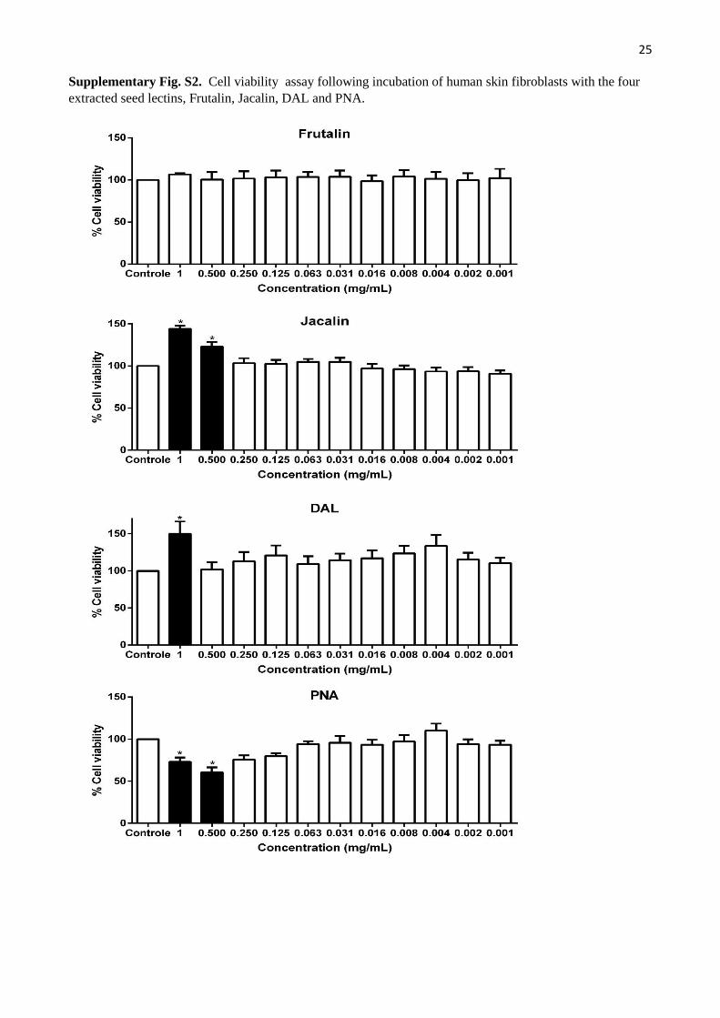
25 Supplementary Fig. S2. Cell viability assay following incubation of human skin fibroblasts with the four
extracted seed lectins, Frutalin, Jacalin, DAL and PNA.
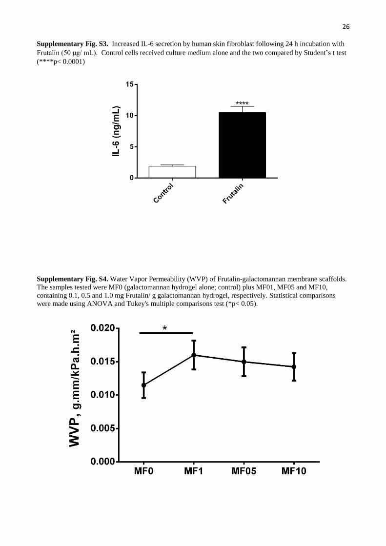
26 Supplementary Fig. S3. Increased IL-6 secretion by human skin fibroblast following 24 h incubation with
Frutalin (50 μg/ mL). Control cells received culture medium alone and the two compared by Student’s t test
(****p< 0.0001)
Supplementary Fig. S4. Water Vapor Permeability (WVP) of Frutalin-galactomannan membrane scaffolds.
The samples tested were MF0 (galactomannan hydrogel alone; control) plus MF01, MF05 and MF10,
containing 0.1, 0.5 and 1.0 mg Frutalin/ g galactomannan hydrogel, respectively. Statistical comparisons
were made using ANOVA and Tukey's multiple comparisons test (*p< 0.05).

27 Supplementary Fig. S5. Frutalin-galactomannan hydrogels accelerate in vivo wound closure. Contraction
of the excision wound 3 days after treatment with galactomannan-based hydrogel (F0) and the three different
Frutalin-galactomannan hydrogel formulations (F01, F05 and F10). **p< 0.01 compared to the saline control
(two-way ANOVA–Bonferroni test).
Supplementary Fig. S6.
In vivo wound closure with implanted spongy membrane scaffolds. Contraction of the excision wound 3 days
is shown after treatment with galactomannan (MF0) plus three Frutalin-galactomannan (MF01, MF05 and
MF10) membranes. **p< 0.01 compared to the saline control (two-way ANOVA–Bonferroni test).