Hydatid cyst disaese
33
By: Dr. Kailash Makhejani HYDATID CYST OF LIVER
-
Upload
kailash-raj -
Category
Health & Medicine
-
view
75 -
download
3
Transcript of Hydatid cyst disaese
By: Dr. Kailash Makhejani
HYDATID CYST OF LIVER
Hydatid cyst (HC), or hydatidosis, is a global parasitic zoonosis
Hippocrates recognized human hydatid over 2,000 years ago
The Arab physician, Al Rhazes, made reference to hydatid disease of the liver in AD 900.
Liver hydatid disease --- zoonosis --- caused by larva of the dog tapeworm, Echinococcus granulosus, with human being acting as an accidental intermediate host.
Introduction
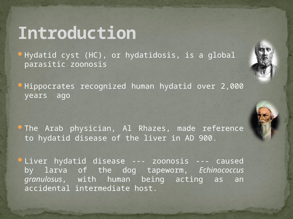
Causative agent Intermediate host Definative host Echinococcus granulosus(Cystic echonococcosis)
Sheep, Human dog
Echinococcus multilocurlaris (Alveolar echinococcosis)
Rodents ,Humans dog,fox
Etiology
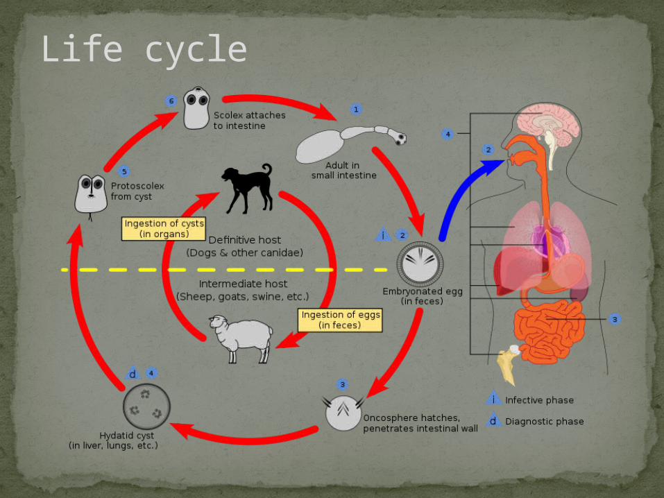
Life cycle
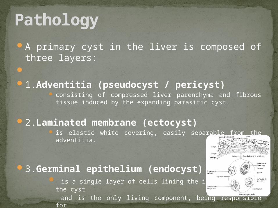
A primary cyst in the liver is composed of three layers: 1.Adventitia (pseudocyst / pericyst)
consisting of compressed liver parenchyma and fibrous tissue induced by the expanding parasitic cyst.
2.Laminated membrane (ectocyst) is elastic white covering, easily separable from the adventitia.
3.Germinal epithelium (endocyst) is a single layer of cells lining the inner aspects of the cyst
and is the only living component, being responsible for the formation of the other layers as well as the hydatid
fluid and brood capsules within the cyst.
Pathology
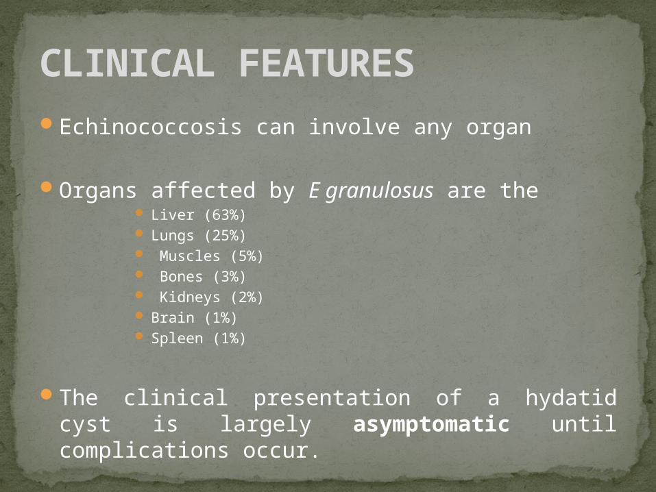
Echinococcosis can involve any organ
Organs affected by E granulosus are the Liver (63%) Lungs (25%) Muscles (5%) Bones (3%) Kidneys (2%) Brain (1%) Spleen (1%)
The clinical presentation of a hydatid cyst is largely asymptomatic until complications occur.
CLINICAL FEATURES
The most common presenting symptoms are abdominal pain, dyspepsia, and vomiting
The most frequent sign is hepatomegaly/palpable mass
Jaundice and fever are each present in about 8% of patients
Bacterial superinfection of a hydatid cyst can occur and present like a pyogenic abscess
Rupture of the cyst into the biliary tree
Free ruptures can result in disseminated echinococcosis and a potentially fatal anaphylactic reaction.
CLINICAL FEATURES ….. Cont….
Routine laboratory blood workup: Nonspecific
Liver involvement may be reflected in an
elevated bilirubin or alkaline phosphatase level.
Leukocytosis may suggest infection of the cyst.
Eosinophilia is present in 25% of all persons who are infected, while hypogammaglobinemia is present in 30%.
INVESTIGATIONS
Serodiagnostic techniques
Indirect hemagglutination(IHA) test and the enzyme-linked immunosorbent assay (ELISA)
sensitivity of 80% overall (90% in hepatic echinococcosis, 40% in pulmonary echinococcosis) and are the initial screening tests of choice.
Immunodiffusion and immunoelectrophoresis demonstrate antibodies to antigen 5 and provide specific confirmation of
reactivity
The ELISA test is useful in follow-up to detect recurrence.
INVESTIGATIONS … Cont…
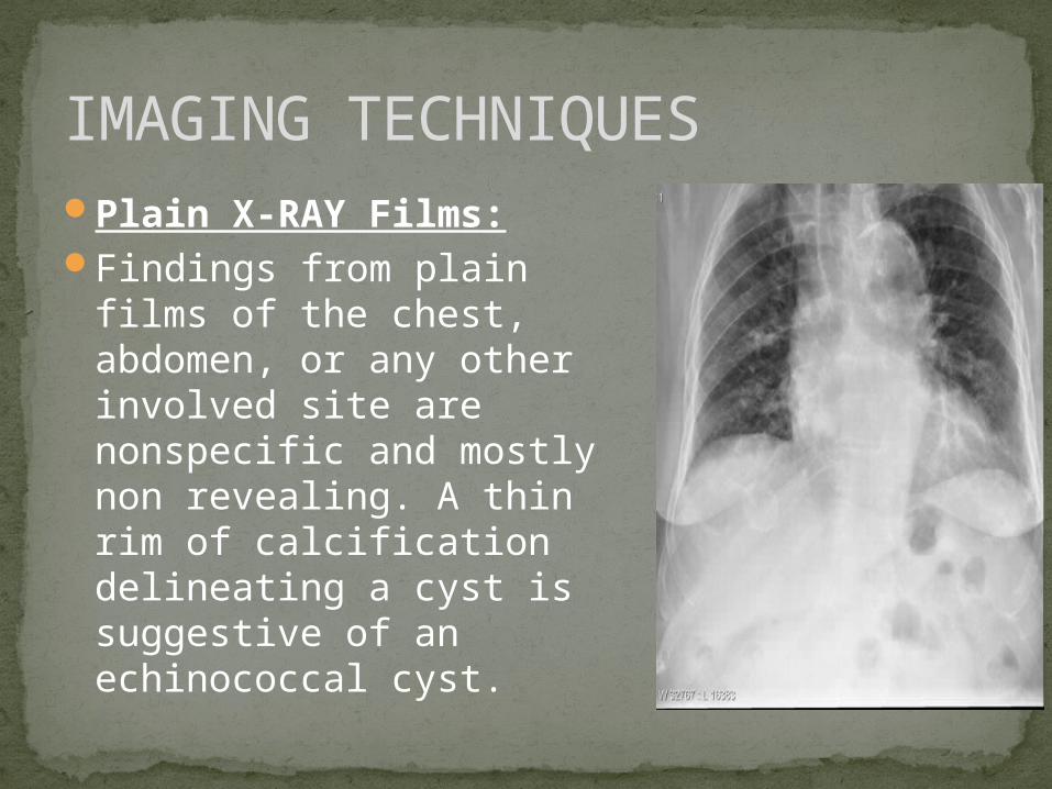
Plain X-RAY Films:Findings from plain films of
the chest, abdomen, or any other involved site are nonspecific and mostly non revealing. A thin rim of calcification delineating a cyst is suggestive of an echinococcal cyst.
IMAGING TECHNIQUES
Ultrasound: currently the primary diagnostic technique and has diagnostic accuracy of
90%.
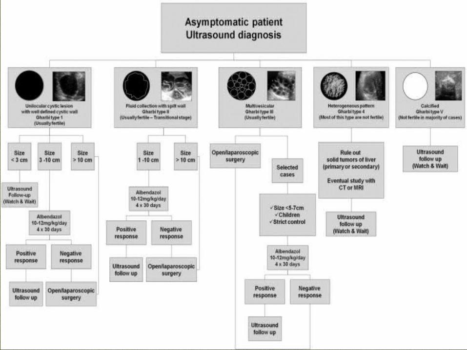
Findings usually seen are: a) Solitary Cyst –
anechoic univesicular cyst with well defined borders and enhancement of back wall echoes in a manner similar to simple or congenital cysts. Features are suggesting a hydatid etiology include dependent debris (hydatid sand) moving freely with change in position; presence of wall calcification or localized thickening in the wall corresponding to early daughter cysts.
b) Separation of membranes (ultrasonic water lily sign) due to collapse of germinal layer seen as an undulating linear collection of echoes.
c) Daughter cysts – probably the most characteristic sign with cysts within a cyst, producing a cartwheel
or honeycomb cyst.
d) Multiple cysts with normal intervening parenchyma (differential diagnosis are necrotic secondaries, Polycystic liver disease, abscess,
chronic hematoma and biliary cysts.
e) Complications may be evident such as echogenic cyst in infection or signs of biliary obstruction usually implying a biliary communication.
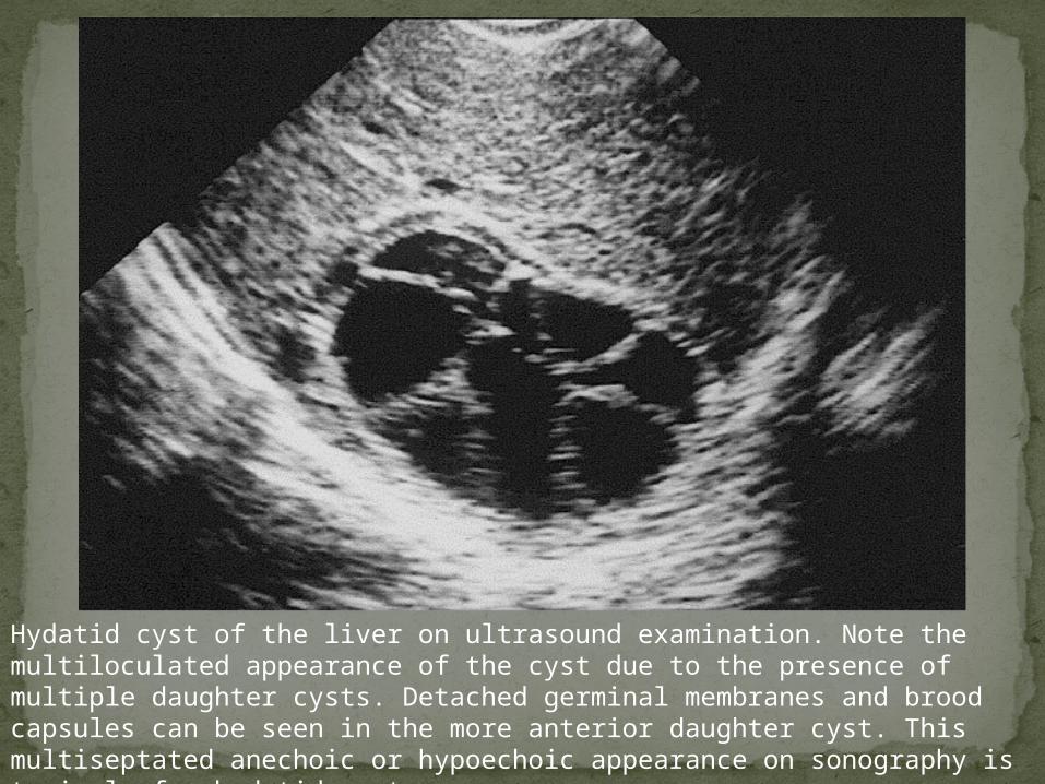
Hydatid cyst of the liver on ultrasound examination. Note the multiloculated appearance of the cyst due to the presence of multiple daughter cysts. Detached germinal membranes and brood capsules can be seen in the more anterior daughter cyst. This multiseptated anechoic or hypoechoic appearance on sonography is typical of a hydatid cyst.
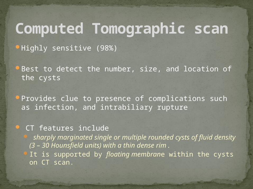
Highly sensitive (98%)
Best to detect the number, size, and location of the cysts
Provides clue to presence of complications such as infection, and intrabiliary rupture
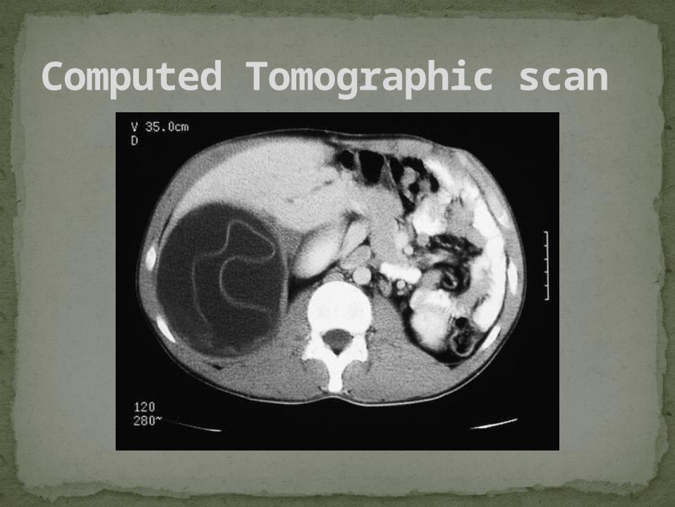
CT features include sharply marginated single or multiple rounded cysts of
fluid density (3 – 30 Hounsfield units) with a thin dense rim. It is supported by floating membrane within the cysts on CT
scan.
Computed Tomographic scan
Computed Tomographic scan
Angiography
of the liver is suggestive but due to lack of specificity and availability of lesser invasive techniques it is rarely required
It may be required in a differential diagnosis of suspected malignancy or vascular malformation
Typical features include an avascular lesion with vascular displacement and a thin peripheral halo of higher density
Direct cholangiography (Endoscopic or percutaneous)
May be required in suspected intrabiliary rupture and bile duct obstruction
ERCP is also a valuable method for detecting post-operative complications involving the biliary tree following surgical intervention.
Other Imaging Techniques
Radionuclide scan has largely replaced by ultrasound and CT scan. It
remains most accurate method of demonstration of a bronchobiliary fistula.
Immunoscintigraphy is an innovation using radiolabelled antibodies to
antigens in the parasite Magnetic resonance Imaging (MRI scan)
Images show the cysts adequately, but MRI offers no real advantage over CT scan.
Other Imaging Techniques …. Cont..
The treatment of choice is surgery
Available Options:Medical Per-cutaneous Endoscopic Surgical
TREATMENT
CHEMOTHERAPY FOR HYDATID DISEASE OF LIVER The compounds in common clinical use are Mebendazole and Albendazole
inhibit glucose uptake by the parasite and inhibit production of ATP
Indications:
Indicated in patients with primary liver or lung cysts that are inoperable (because of location or medical condition)
Patients with cysts in 2 or more organs Peritoneal cysts
Contraindications: Early pregnancy Bone marrow suppression Chronic hepatic disease Large cysts with the risk of rupture Inactive or calcified cysts A relative contraindication is bone cysts because of the significantly decreased response.
MEDICAL TREATMENT
Mebendazole: Disadvantages are that it is poorly absorbed from the gastrointestinal tract It is no longer used in hydatid disease
Albendazole administered in a dose of 10 – 15 mg/kg/day in adults or a fixed dose of 400 mg twice
daily. The treatment is given in cycles of 28 days with two weeks treatment free periods
between the cycles.
The different schedules for the treatment are: 1. Inoperable cases - as primary treatment - 3 cycles
2. Pre-operatively – to reduce the risk of recurrence 6 weeks continuous treatment
3. Post-operatively to prevent recurrence in cases of intraoperative cyst spillage – 3 cycles.
Side effects of Albendazole Mild abdominal pain, nausea, vomiting, pruritis, dizziness, alopecia, rash and headache. Occasionally leucopoenia, eosinophillia, icterus, and mild elevation in transaminase
levels.
MEDICAL TREATMENT
PAIR (Puncture, Aspiration, Injection, Re-aspiration)Proposed in 1986 by the Tunisian team that first used it
in a prospective study
Recent and minimally invasive therapeutic option, complements or replaces surgery which was long
considered as the only treatment.
If a catheter is temporarily left in the cyst after the procedure for drainage (D), the acronym PAIRD should be preferred
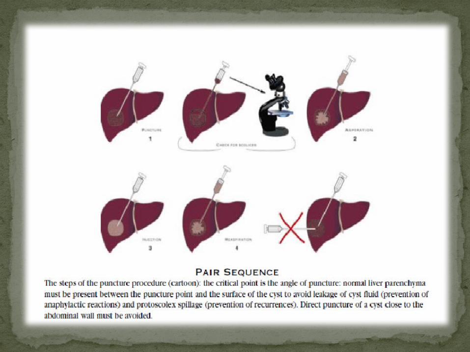
PERCUTANEOUS DRAINAGE OF HYDATID CYST (PAIR)
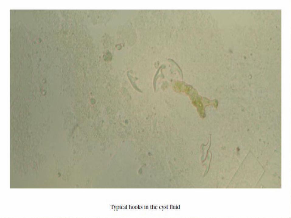
Performed using either ultrasound or CT guidance Involves aspiration of the contents via a special
cannulaFollowed by injection of a scolicidal agent for at least
15 minutesThen reaspiration of the cystic contentsThis is repeated until the return is clear The cyst is then filled with isotonic sodium chloride
solutionPerioperative treatment with a benzimidazole is
mandatory
The cysts should be larger than 5 cm in diameter
PAIR
Patients with: Non-echoic lesion ≥ 5 cm in diameter (TYPE 1)
Cysts with detachment of membranes (TYPE2) and/or with daughter cysts (TYPE 3)
Multiple cysts if accessible to puncture
Infected cysts
Also Pregnant women
Children >3 years old
Patients who fail to respond to chemotherapy alone
Patients in whom surgery is contraindicated
Patient who refuse surgery
Patients who relapse after surgery
INDICATIONS FOR PAIR
Non-cooperative patients and inaccessible or risky location of the cyst in the liver
Cyst in spine, brain and/or heart
Inactive or calcified lesion
Cysts communicating with the biliary tree
Cysts open into the abdominal cavity, bronchi and urinary tract
Contraindications for PAIR
Minimal invasiveness
Reduced risk compared with surgery
Confirmation of diagnosis
Removal of large numbers of protoscolices with the aspirated cyst fluid
Improved efficacy of chemotherapy given before and after puncture (probably because of an increased penetration of antihelminthic drugs into cysts re-filling with hydatid fluid )
Reduced hospitalization time
Cost of the puncture and chemotherapy usually less than that of surgery or chemotherapy alone
Benefits Of PAIR
Same risks as any puncture ( haemorrhage, mechanical lesions of other tissues, infections )
Anaphylactic shock or other allergic reactions
Secondary echinococcosis caused by spillage
Chemical ( sclerosing ) cholangitis if cysts communicate with the biliary tree
Sudden intracystic decompression, thus leading to biliary fistulas
Persistence of satellite daughter cysts
Systemic toxicity of alcohol or hypertonic saline in case of large cysts (total volume injected must be carefully calculated)
Risks Of PAIR:
ERCP Effective in diagnosing biliary tree involvement from the cyst
Useful in presence of intrabiliary rupture, which requires exploration and drainage of the biliary tract
Also useful in post surgerical cases with presence of residual hydatid material (membranes and daughter cyst) left in biliary tree
During the endoscopic exploration the biliary tree is cleared of any hydatid material with a balloon catheter or a dormia basket
The endoscopic sphinterotomy is also performed to facilitate drainage of the common bile duct.
ENDOSCOPIC MANAGEMENT OF HYDATID CYST
Indications: Large liver cysts with multiple daughter cysts
Superficially located single liver cysts that may rupture (traumatically or spontaneously)
Liver cysts with biliary tree communication or pressure effects on vital organs or structures
Infected cysts
Contraindications: General contraindications to surgical procedures (eg, extremes of age, pregnancy, severe preexisting
medical conditions)
Multiple cysts in multiple organs
Cysts that are difficult to access
Dead cysts
Calcified cysts
Very small cysts
SURGICAL TREATMENT
Biliary leakage is the most frequent postoperative complication following surgery for hydatid cyst of liver
It has been reported to occur in about 50% of cases because of the small-undetected communication between the cyst and the bile ducts
The surgical management of hydatid disease of liver carries Mortality rate of 0.9 to 3.6 %
Recurrence up to 11.3 % within 5 years
Multiple operations carry a progressively higher mortality – increasing from 6 % after second to 20% after third.
COMPLICATIONS OF SURGERY
Chemotherapy Postoperative treatment with benzimidazoles is continued for 1 month
in patients who have undergone complete resection or PAIR successfully
Laboratory tests Patients on benzimidazoles should have a
CBC count and liver enzyme evaluation performed at biweekly intervals for 3 months
Then every 4 weeks to monitor for toxicity
ELISA or indirect hemagglutination tests are usually performed at 3-, 6-, 12-, and 24-month intervals as screening for recurrence of resected disease or aggravation of existing disease
Imaging: Ultrasound and/or CT scan are used in follow-up at the same intervals
as the laboratory tests or as clinically indicated.
FOLLOW UP

THANKS