Humoral immune responses in mice to a mosquito salivary gland … · 2019-06-28 · mosquito...
57
Humoral immune responses in mice to a mosquito salivary gland antigen as a target for a diagnostic and immunotherapeutic agent Kyong Min Choi Department of Medicine The Graduate School, Yonsei University
Transcript of Humoral immune responses in mice to a mosquito salivary gland … · 2019-06-28 · mosquito...

Humoral immune responses in mice to a
mosquito salivary gland antigen as a
target for a diagnostic and
immunotherapeutic agent
Kyong Min Choi
Department of Medicine
The Graduate School, Yonsei University

Humoral immune responses in mice to a
mosquito salivary gland antigen as a
target for a diagnostic and
immunotherapeutic agent
Kyong Min Choi
Department of Medicine
The Graduate School, Yonsei University

Humoral immune responses in mice to a
mosquito salivary gland antigen as a
target for a diagnostic and
immunotherapeutic agent
Directed by Professor Dong Soo Kim
The Doctoral Dissertation
submitted to the Department of Medicine,
the Graduate School of Yonsei University
in partial fulfillment of the requirements for the degree
of Doctor of Philosophy
Kyong Min Choi
June 2014
This certifies that the Doctoral
Dissertation of Kyong Min Choi is
approved.

ACKNOWLEDGEMENTS
I would like to express my deepest appreciation for Prof. Dong
Soo Kim, my thesis supervisor, who encouraged me to work
towards my PhD degree. Also, I am indebted to Prof. Tai-Soon
Yong, Soung Hoo Jeon, Jung-Won Park and Myeong Heon
Shin for their kind and helpful advice.
Most of all, I would like to dedicate this paper to my loving
family. I would like to thank my dearest wife Su Jin Cho for
always standing by me, my son Sun Jae and my daughter
Sunwoo, my parents Sung Hyun Choi and Tai Yook Choi, and
my parents-in-law Young Ho Cho and Myung Jae Lee.
June 2014
Kyong Min Choi

<TABLE OF CONTENTS>
ABSTRACT ····································································· 1
I. INTRODUCTION ···························································· 3
II. MATERIALS AND METHODS ··········································· 9
1. Mosquitoes and salivary gland extract ································· 9
2. Production of antisera in mice ·········································· 11
3. Enzyme-linked immunosorbent assay ································· 11
4. SDS-PAGE and immunoblot analyses ······························· 14
5. Immunohistochemistry and confocal imaging ························ 14
6. Bacterial strains and phage ············································· 15
7. Total RNA and Poly(A)+ RNA ······································· 15
8. Construction of cDNA library ········································· 17
9. Immunoscreening of the cDNA and characterization of clones ·· 17
10. Sequence and 3D structure analysis ································· 20
III. RESULTS ·································································· 23
1. Fine structure of salivary gland in thorax of Aedes togoi ·········· 23
2. SDS-PAGE and immunoblot analyses ······························· 26
3. Enzyme-linked immunosorbent assay ································ 26
4. RNA extraction and construction of cDNA library················· 29
5. Identification of saliva antigens by immunoscreening ··········· 33
6. Analysis of the sequence and 3D modeling ··························· 36
IV. DISCUSSION ······························································ 41
V. CONCLUSION ····························································· 46
REFERENCES ································································· 47
ABSTRACT (IN KOREAN) ················································ 52

LIST OF FIGURES
Figure 1. Distribution of Aedes togoi ···························· 4
Figure 2. Allergenic protein model structures ··················· 8
Figure 3. Collection of mosquito larvae ························ 10
Figure 4. The isolation of salivary glands ······················ 12
Figure 5. Assay procedure summary ···························· 13
Figure 6. Lambda Uni-Zap XR insertion vector ················· 18
Figure 7 Cloning plasmid for genomic inserts ·················· 19
Figure 8. RNA polymerase II elongation complex ············· 22
Figure 9. Immunohistochemical staining of salivary gland ··· 24
Figure 10. Confocal imaging of mosquito salivary gland ····· 25
Figure 11. Protein electrophoretic & immunoblot analysis ··· 27
Figure 12. The mouse IgE levels in response to the salivary
gland extract ·························································· 28
Figure 13. Quality check of the cDNA library ·················· 30
Figure 14. Amplified cDNA library ······························ 31
Figure 15. Agarose gel showing the restriction digestion ······· 32
Figure 16. Demonstration of immunoreactive plaque ··········· 34
Figure 17. Alignment of deduced amino acid sequences ······· 37
Figure 18. 3D structure of protein of unknown function ········ 39

LIST OF TABLES
Table 1. The Character of E. coli strains used in this study · 16
Table 2. Proteins identified as saliva antigens··················· 35

1
ABSTRACT
Humoral immune responses in mice to a mosquito salivary gland antigen
as a target for a diagnostic and immunotherapeutic agent
Kyong Min Choi
Department of Medicine
The Graduate School, Yonsei University
(Directed by Professor Dong Soo Kim)
The mouthparts of mosquito are modified to pierce the tissues and
suck blood from humans or other mammals. Their bites can cause
immediate cutaneous reactions, such as wheal and flare, delayed
reactions and occasionally systemic reactions in humans. IgE-mediated
allergic reactions caused by mosquito bites are a common problem all
over the world. This study was undertaken to determine IgE levels in
anti-mouse serum, to elucidate mouse IgE binding patterns and to
investigate the immunogenicity of salivary gland antigens of Aedes
togoi. Mosquito larvae of Aedes togoi were collected and maintained in
the laboratory. The mosquito specific mouse IgE level was measured
using ELISA. Polypeptide patterns were analyzed by SDS-PAGE.
Western blot was performed with sensitized immune mouse sera, and
elucidated mouse IgE binding patterns to salivary gland extracts.

2
Protein band patterns of the salivary gland extracts (SGE) and whole
body extracts (WBE) of the specimens were different from one another.
Specific mouse IgE reacted to the protein in SGE of 18.0, 33.0, 35.0,
37.0, 45.0, 57.5, 72.0, 90.0 and 150.0 kDa from Aedes togoi. Molecular
biological techniques were used to study the genetic information and
functions of genes. The cDNA sequencing was carried out to elucidate
and compare the genome of Aedes togoi with other coded gene of the
allergen. Two previously unknown protein coding genes(DUF 1398,
DUF 2528) were identified among the 45 positive clones prepared from
the mosquito salivary gland by immunoscreening. Analysis of the 3D
structure of DUF1398 and DUF2528 was not similar with any other
allergens identified in plants or animals, despite low sequence identities
to their templates, the global folds of the 3D models of the cockroach
allergen Bla g 4 and the mosquito salivary protein antigen Aed a 2 had a
sizable fraction of structural overlap, suggesting that it would be a
potential target for therapeutic agents in specific mosquitoes bite
allergens.
-------------------------------------------------------------------------------------
Key words : allergen, mosquito, Aedes togoi, salivary gland, IgE

3
Humoral immune responses in mice to a mosquito salivary gland antigen
as a target for a diagnostic and immunotherapeutic agent
Kyong Min Choi
Department of Medicine
The Graduate School, Yonsei University
(Directed by Professor Dong Soo Kim)
I. INTRODUCTION
Mosquitoes transmit infectious diseases, such as malaria, filariasis,
Japanese encephalitis B. The life cycle of the mosquito is a complete
metamorphosis; the egg, larva, and pupa reside in water. Only female
mosquitoes suck blood and transmit pathogens. Three mosquito genera are of
medical importance1. They are Anopheles, Culex, and Aedes. They differ in
distribution, morphology, ecology, and diseases transmission (Fig. 1).
During an insect bite, the salivary glands release components that include
antihistamines, vasodilators like tachykinin, anticoagulants like thrombin and
factor IXa-directed molecules2 and immunomodulators, in order to facilitate
entry of inoculum containing pathogens. The salivary components of vectors
have been implicated to be of importance in transmission of pathogens (viral,

4
Figure 1. Distribution of Aedes togoi. This map denotes only the country or
general areas where Aedes togoi (red areas) has been recorded, not actual
distribution. (© 2006 M. Disbury SMS-NZB www.smsl.co.nz)
bacterial and protozoan) by ticks and mosquitoes3. Salivary glands of other
blood-sucking arthropods like a star tick bear prostaglandin E2 (PGE2)
receptor that stimulates secretion of an anticoagulant in order to facilitate
blood feeding4.
Reactions to mosquito bites are immunologic in nature. They are due to
specific sensitization to the mosquito salivary proteins, because initial
exposure to mosquito species to which an individual has not been previously
exposed causes no reaction. Mosquito bite-induced immediate wheals and
flares reaction correlate well with mosquito salivary gland-specific IgE levels.
The development of skin sensitization to mosquito bites also parallels the
levels of saliva-specific IgE antibodies.Mosquito saliva-specific IgG

5
antibodies, consisting mainly of the IgG1 and IgG4 subclasses, have been
found to be significantly elevated in individuals with positive mosquito bite
tests and in individuals with severe local reactions, but not systemic reactions,
to mosquito bites5.
IgE-mediated allergic reactions caused by mosquito bites are a common
problem all over the world. Mosquitoes inject saliva containing antigenic
proteins into the bite wound6,7
. Their bites can cause immediate cutaneous
reactions, such as wheal and flare, delayed reactions and occasionally
systemic reactions in humans8,9
.
It has been reported that mosquito saliva-specific IgE and IgG antibodies
are involved in mosquito bite allergy10
. In order to confirm these responses,
enzyme-linked immunosorbent assay (ELISA) 11
and immunoblot techniques12
have been carried out. Aedes togoi transmit infectious organisms including
filaria. The Aedes togoi is distributed on most islands and at the seaside in
Korea. However, the allergenicity of saliva antigen in humans and
experimental mice has not been reported until now. The saliva allergen was
prepared from salivary gland extracts and they probably mixed a large
admixture of protein. Mosquito larvae of Aedes togoi was collected and
maintained in the laboratory. Salivary gland extracts (SGE) and whole body
extracts (WBE) were prepared from female mosquitoes.
This study was undertaken to determine the IgE levels in anti-mouse
serum, to elucidate mouse IgE binding patterns and to investigate the
reactivity of salivary gland antigens of Aedes togoi. The mosquito specific
mouse IgE level was measured by using ELISA. Polypeptide patterns were
analyzed by SDS-PAGE. Western blot was performed with sensitized immune
mouse sera, and elucidated mouse IgE binding patterns to SGE. Protein band
patterns of the SGE and WBE of the specimens were different from one
another. The molecular biological techniques were used to study the genetic
information and functions of genes in a certain individual organism. The

6
number of important allergens has been expressed by using recombinant DNA
technology13
. The cDNA sequencing has been carried out to elucidate Aedes
togoi coded gene of the allergen. Utilization of this technique to produce pure
mosquito allergens in large quantities would be an important advance in
immunology.
The three-dimensional (3D) structures of macromolecules, as collected and
provided by the Protein Data Bank (PDB), offer tremendous insights into the
molecular function at the atomic level, and they often provide direct evidence for
aspects of that function by exemplifying molecular interactions between
individual macromolecules, or between macromolecules and small molecules14
.
We evaluated the similarities of the 3D folds, and found two novel proteins. Our
results from this study indicate that the examined models in two novel proteins
are useful for determining other characteristics of the allergenic proteins.
Structural similes of allergens are needed to supply a molecular
interpretation for clinically observed cross-reactivities between allergens from
different organisms15-25
and to predict whether new proteins or other
biotechnology products are potential allergens14,36
. However, there are only 86
experimental 3D structures in the Protein Data Bank (PDB) for allergens14
, a
small fraction of the 1499 allergen and isoallergens sequences collected in the
Structural Database of Allergenic Proteins (SDAP)26
. Thus reliable,
template-based models of allergens are needed to compare allergen features,
and to determine potential cross-reactive IgE binding surfaces27
.

7
Proteins. 2013;81:545–54.
Figure 2. Structural overlays of aligned regions of the structural database of
allergenic proteins model structures (red, yellow) with the corresponding
experimental structures (green). (A) 1,3-glucanase from latex rubber (Hev b 2),
(B) Stress-induced protein SAM22 from soybean (Gly m 4), (C) Venom allergen
III from fire ant (Sol i 3), (D) Pathogenesis-related protein PR-10 from carrot
(Dau c 1), (E) Cysteine protease from American dust mite (Der f 1.0106), (F)
Canine salivary lipocalin (Can f 2), (G) Conglutin from peanut (Ara h 2), (H)
Calycin from German cockroach (Bla g 4), (I) Salivary odorant binding protein
D7 from mosquito (Aed a 2), (J) Vicilin from peanut (Ara h 1).

8
II. MATERIALS AND METHODS
1. Mosquitoes and salivary gland extracts
Mosquito larvae are common in the coastal regions, usually occurring in
tidal pools or rock pools of saline or brackish water above the tidal zone, but
also occasionally in containers such as used tires with fresh water28
. Larvae of
Aedes togoi were brought from rock pools at the seaside to the laboratory and
reared in the cages (Fig. 3).
Figure 3. Collection of mosquito larvae. Mosquito larvae were collected from
rock pools at the seaside at Song-jong Dong, Pusan for Aedes togoi.
The collected larvae were maintained for emergence in the laboratory and
once the adults emerged, females were collected, anesthetized by CO2 gas and
20,000 female adult Aedes togoi were taken for the study. Dissection of
salivary gland tissues from mosquitoes requires prior preparation of 1X
phosphate buffered saline (1X PBS) solution and anesthetization of
mosquitoes by subjecting to a temperature of 4°C, until immobilized. The
mosquitoes remain anesthetized by placing in a Petri dish that is kept cold on

9
ice. Other materials required include: light microscope fitted with 10x
objective lens, pipette, fine-tipped forceps, glass slide, and needle-tip probes.
Salivary glands were obtained by dissection of 3 to 5 day old adults in 0.02 M
PBS, pH 7.2, under a stereomicroscope, and transferred to fresh PBS on ice
(Fig. 4). About 100 pairs of salivary glands were obtained and lyophilized.
The salivary gland extracts were reconstituted by dissolving the lyophilized
salivary gland proteins in PBS before use. This was centrifuged at 12,000 xg
for 20 min. The supernatant was filtered through a 0.22 μm Amicon filter and
the protein concentrations were measured with a Bio-Rad protein assay kit
(Bio-Rad Laboratories, Richmond, CA, USA).
2. Production of antisera in mice
The mice were immunized by the method of Brummer-Korven H with
some modifications29
. Briefly, 20 live female Aedes togoi per BALB/c mouse
were released to inject the antigens naturally, twice a week, for 12 weeks.
Serum samples were collected once after 12 weeks the immunization period.
All sera were stored at –80°C until analysis.
3. Enzyme-linked immunosorbent assay
The enzyme-linked immunosorbent assay (ELISA) is a test that uses
antibodies and color change to identify a substance. For the enzyme-linked
immunosorbent assay, mosquito-specific IgE levels in mouse serum samples
were first directly coated on 96-well polystyrene plate. Mosquito-specific IgE
level in sensitized mouse serum sample was measured by ELISA. To perform
the ELISA kit (BioLegend, Mouse IgE, CA, USA) (Fig. 5), dilute capture
antibody in coating buffer (0.05 M carbonate buffer, pH 9.6). Add 100 μl of
this capture antibody solution to all wells of a 96-well plate provided in this
set. Seal plate and incubate overnight (16-18 hrs) at 4°C. Wash plate 4 times
with 300 μl wash buffer (0.05% Tween 20 in 0.01 M PBS, pH 7.2) per well

10
and to block non-specific binding and reduce background, add 200 μl 1X
Assay Diluent A per well. Seal plate and incubate at RT for 1 hour with
shaking at 200 rpm on a plate shaker. Prepare 1,000 μL of top standard at 10
ng/ml in 1X Assay Diluent A (refer to Reagent Preparation). Perform six
two-fold serial dilutions of the 10 ng/ml top standard with Assay Diluent A in
separate tubes. After diluting, the mouse IgE standard concentrations are 10
ng/ml, 5 ng/ml, 2.5 ng/ml, 1.25 ng/ml, 0.625 ng/ml, 0.313 ng/ml and 0.156
ng/ml, respectively. Assay Diluent A serves as the zero standard (0 pg/ml)

11
Figure 4. The isolation of salivary glands. Place a drop of 1X PBS onto a glass
slide mounted under a light microscope. Pick up a mosquito by stabbing the
thorax with a needle-tip probe. Pull off the mosquito legs using your fingers.
Transfer the mosquito onto the slide. Remove the head of the mosquito using
forceps. While holding down the mosquito thorax with the probe, use another
probe to gently push down on the thorax. The salivary glands are located at the
anterior portion of the thorax and can be isolated by using a needle-tip probe
and severing the attachments that connect the gland to the thorax. Intact
salivary glands are comprised of three lobes: two lateral lobes and a medial
lobe.

12
Figure 5. Summary of Assay Procedure. Wash plate 4 times with wash buffer
and add 100 μL/well of standard and samples to the wells. Seal plate and
incubate at RT for 2 hours with shaking and then wash plate 4 times, add 100
μL of diluted Detection Antibody solution to each well, seal plate and incubate
at RT for 1 hour with shaking. Wash plate 4 times and add 100 μL of diluted
Avidin-HRP solution to each well. Seal plate and incubate at RT for 30
minutes with shaking. Wash plate 5 times with wash buffer, for this final wash,
soak wells in wash buffer for 30 seconds to 1 minute for each wash. Add 100
μl of freshly mixed TMB Substrate Solution and incubate in the dark for 20
minutes. Stop reaction by adding 100 μl of stop solution (1 N NaOH) to each
well. Read absorbance at 450 nm within 30 minutes (Dynatech, MR-5000,
Chantilly, VA., USA). All serum samples were analyzed in duplicate.

13
4. SDS-PAGE and Immunoblot Analyses
SDS-PAGE was performed by the method of Laemmli with 12%
acrylamide separating gels30
. SGE of mosquitoes separated by SDS-PAGE
were then electrophoretically transferred to a nitrocellulose membrane by the
method of Towbin et al. Membranes were incubated with pooled serum
samples with positive ELISA titers and sera of controls. Nitrocellulose
membrane was blocked for overnight at 4°C with 3% BSA. After several
washes for 45 min, the membrane was incubated with 1/10 diluted human
pooled antisera overnight at 4°C, and then with HRP-conjugated anti-mouse
IgG for 35 min at RT. After several washes for 45 min, immediately pour the
chemiluminescence reagent into the weigh dish with the membrane and
incubate for 1 min at room temperature. Use at least 1 mL per membrane and
incubate. Take the blot to the developing room and place the membrane
between the covers of a propylene sheet protector with the black interface
removed (plastic wrap works well also). Switch off the lights and place the
Kodak Scientific Imaging film on top of the membrane. Repeat the exposure,
varying the time as needed for optimal detection.
5. Immunohistochemistry and confocal imaging for analysis of fine
structure of salivary glands
To identify expression profiles, the salivary glands were analyzed for fine
structure in vivo.Adult females were used for the tissue specimens.The
tissue specimens were fixed in 3.7% PFA for 1 day at 4°C and decalcified with
10% ethylene di-amine tetra-acetic acid for 2 days. Making paraffin block and
then, 4 μm-thick serial sections were cut and processed for hematoxylin and
eosin staining. For fluorescence immunohistochemical staining, the tissue
sections were deparaffinized and hydrated with alcohol series. After antigen
retrieval in citrate buffer, the sections were treated with hydrogen peroxide.

14
After blocking with 3% BSA in PBS overnight at 4°C, the whole-mounted
specimens were incubated for 2 hrs at RT with sensitized mouse antibodies
and then incubated for 1 hr at RT with secondary antibodies. Salivary gland
proteins were detected as a red color using anti-mouse-Rhodamine RedTM.
For cytoskeleton, Phalloidin was detected as a green color using
anti-Phallotoxin-fluorescein isothiocyanate (v/v, 1:1000, Molecular Probes).
The nuclei were stained in 4',6-diamidino-2-phenylindole (DAPI, 2 μg/ml) for
5 min at RT. Specimens were imaged by a LSM 510 META confocal laser
scanning microscope (Carl Zeiss, Jena, Germany).
6. Bacterial strains and phage
The E. coli strains used in this study are XL1-Blue MRF’ and SOLR. The
characteristics of these microorganisms were described in Table 1. For
Mass-clone excision, f1 helper phage was used at the concentration of 1.2 ×
109 pfu/μl. The Uni-ZAP XR (Stratagene, CA, USA) was used for the
construction of cDNA library (Fig. 6).
7. Total RNA and Poly (A)+ RNA
Total RNA was isolated from 1g of 5,000 females of Aedes togoi using
Trizol reagent (Invitrogen, CA, USA). One microgram of frozen salivary gland
was incubated in 2ml Trizol reagent for 5 min at RT to permit the complete
dissociation of nucleoprotein complexes. After mixing, 400ul chloroform was
added and vortexed for 15 sec. The sample was centrifuged at 12,000xg for 15
min at 4°C. The supernatant was transferred to a new tube and mixed with 5 ml
isopropyl alcohol and then incubated at RT for 10 min. After centrifugation at
12,000xg for 10 min at 4°C, RNA pellet was resuspended in 1 ml of 0.1%
RNAse free water. Poly (A)+ RNA was isolated from 0.03mg of the total RNA
using oligo (dT) cellulose column. The quality and quantity of RNA were

15
checked by UV spectrophotometer (OD 260/280).
Table 1. The Character of E. coli strains used in this study.
Host Genotypes
XL1-Blue MRF’ Δ(mcrA)183 Δ(mcrCB-hsdSMR-mrr)173 endA1
supE44 thi-1 recA1 gyrA96 relA1 lac [F′ proAB
lacIqZΔM15 Tn10 (Tet
r)]
SOLR e14-(McrA-) Δ(mcrCB-hsdSMR-mrr)171 sbcC recB
recJ uvrC umuC::Tn5 (Kanr) lac gyrA96 relA1 thi-1
endA1 λR [F’ proAB lacI
qZ ΔM15]
C Su-
8. Construction of cDNA library
The cDNA library was constructed from mRNA using a ZAP-cDNA
synthesis kit (Stratagene, USA). Five micrograms of poly(A)+ RNAs were
primed with a 50-base oligo (dT) primer containing Xho I site at the 3’-end. First
strand cDNA was synthesized with the linker-primer (5’-GAG AGA GAG AGA
GAG AGA GAA CTA GTC TCG AGT TTT TTT TTT TTT TTT TTT-3’) and
is transcribed using Maloney Murine Leukemia Virus – Reverse Transcriptase
(MMLV – RT) and 5-methyl dCTP to create hemimethylated cDNA. RNAse H
and E. coli DNA polymerase synthesized double-stranded cDNA. After ligation
of EcoRI adapter, and phosphorylating the EcoRI ends onto the cDNA, it was
fractionated through sepharose CL-2B gel filtration medium in drip column.
After ligated cDNA into the Uni-ZAP XR vector (Fig. 6), it was packaged in
vitro using Gigapack III Gold kit. After tittering, the primary library were
amplified and stored prior be used.

16
9. Immunoscreening of the cDNA library and characterization of selected
immunoreactive clones
The Uni-ZAPTM premade ovarian carcinoma cDNA expression library was
purchased from Stratagene, La Jolla, CA, USA. cDNA expression library of the
mosquito MSQ452 and MSQ549 were screened with sensitized mouse serum.
The cDNA library was plated on NZY agar plates at a density of 400 clones/10
cm plate. The plates were incubated at 42°C for 4 hrs to allow plaques to
develop. Nitrocellulose filters soaked with isopropyl β-D-thiogalactopyranoside
(IPTG) were then laid on top of the plaques and incubated at 37°C for 4 hrs to
transfer the plaques onto the membranes. Subsequently, the filters were blocked
with 1% acetone powder solution/PBS-T (80 mM sodium orthophosphate, 20
mM sodium dihydrogen orthophosphate, 100 mM sodium chloride, and 0.1%
Figure 6. Lambda Uni-Zap XR insertion vector.

17
Figure 7. Cloning plasmid for genomic inserts. Lambda ZAP carries pBluescript
(-), which is excised in vivo upon infection with f1 or M13 helper phages. Inserts
are cloned within a polyliner located within lacZ. As with λgt11, a fusion protein
may be expressed if the insert DNA is in frame with the lacZ sequence; thus,
libraries made in this vector can be screened with antibodies. In λZAP, T7 and
T3 promoters flank the inserts, which allow RNA probes to be easily obtained.
pBluescript M13 (-), the excised plasmid is normally propagated as a
double-stranded circular DNA, but infection with a helper phage enables the
plasmid to be propagated as single-stranded DNA. DNA fragments up to 10 kb
can be inserted. Within the polylinker, unique Xho I and EcoR I cloning sites are
available.

18
Tween-20) overnight, at 4°C. To screen the library, the preabsorbed sensitized
mouse serum 1:200 in 5% bovine serum albumin/PBS-T. The diluted sensitized
mouse serum was first incubated with E. coli phage lysate (Stratagene, USA) for
2 hrs at room temperature to minimize the cross-reaction between the
autoantibodies and the bacterial/phage proteins. The screening procedure was
followed as described in the instruction manual (Stratagene, CA, USA). In brief,
serum samples diluted 1:10 were preabsorbed with lysate from E. coli and
bacteriophage-infected coupled to sepharose 4B (BioDynamics Lab Inc., Tokyo,
Japan). Nitrocellulose filters were then incubated with this preabsorbed lysate
from E. coli for 2 hrs at room temperature to identify cellular proteins that react
with the autoantibodies in the mouse serum. Following probing with the
sensitized mouse serum, the filters were washed with PBST for five times and
further treated with alkaline phosphatase-conjugated goat anti-mouse IgG
(Jackson Immunoresearch Laboratories, West Grove, PA, USA) diluted
2000-fold in 5% bovine serum albumin/PBST for 1 hr at room temperature. The
filters were washed again as above and then preceded to chemiluminescence
detection with a dioxetane-based substrate (Diagnostic Products Corporation,
Los Angeles, CA, USA). The plaques exhibiting immunoreactivities were
excised from the plates and the phages were converted into pBluescript
phagemids(Fig. 7) by in vivo excision with ExassistTM helper phage, following
the manufacturer’s instructions. The excised phagemids were purified and
subjected to automated DNA sequencing with M13 forward and reverse primers.
The insert sequences were compared to the known sequences in the GenBank
database with the BLASTN alignment algorithm31
.
10. Sequence and 3D structure analysis
The positive clones were subcloned to monoclonality, purified and excised in
vivo to pBK-CMV plasmid forms (Stratagene). Plasmid DNA was prepared
using a Quantum Prep Plasmid Mini- prep Kit (Bio-Rad, Hercules, CA). The

19
nucleotide sequence of cDNA inserts was determined by an ABI PRISM R310
Genetic Analyzer (PerkinElmer, Foster City, CA), and sequence alignments were
performed with BLAST software and compared to sequences in the GenBank III.
3D modeling for allergenic proteins used PubMed, Entrez/Protein,
Entrez/Nucleotide, PubChem, IBIS, VAST structure neighbors and the
Conserved Domain Database14,15
. 3D structures of biological units may be
visualized using the 3D viewer Cn3D, which has recently been released as a new
version v4.3, to support visualization of biologic- al units with macromolecules
generated via symmetry operations. Cn3D v4.3 also comes with a wider range of
features, such as side-by-side stereo, and is distributed as a helper application for
the web-browser. All 3D models and links to the NMR or X-ray crystal
structures in the PDB are available from the SDAP website (http://
fermi.utmb.edu/SDAP/). Our assessment of the quality of the 3D models using
the available native structures was also compared with results from three
assessment software tools, QMEAN32
(http:// swissmodel. expasy. org/ qmean/
cgi/index.cgi), ProSA33
(https:// prosa.services.came.sbg.ac.at/prosa.php) and
Verify_3D34
(http://nihserver.mbi.ucla.edu/ SAVES/) that provide quality scores
based only on the 3D structures of the protein models.

20
Nucleic Acids Research, 2012, Vol. 40, Database issue
Figure 8. The colors used to depict 3d Domains in the sequence annotation
graphic are independent of the colors used to depict molecules in the interactions
schematic. The page provides links to the source database (PDB), taxonomy,
PubMed, Entrez/Protein, Entrez/Nucleotide, PubChem, IBIS, VAST structure
neighbors and the Conserved Domain Database. The interaction schematic
shows that only the two largest protein subunits of the complex interact with the
nucleic acids and that the smaller protein subunits surround the core of the
complex and may interact with both or only one of the two large subunits.

21
III. RESULTS
1. Fine structure of salivary gland in the thorax of Aedes togoi
This study was undertaken to examine the morphology of collecting ducts
and secretary cells of salivary glands using polyclonal antibodies generated in
the mouse followed by confocal microscopy.
The paired salivary glands of mosquitoes are present in the thorax flanking
the esophagus. Each gland has three lobes, two laterals lobes and one median
lobe. In the female mosquito proximal, intermediate and distal regions form the
lateral lobes. Each lobe has a central duct constituted by a layer of epithelial cells
that are bound externally by a basal lamina (Fig. 9). The structure of the salivary
glands probably influences the adaption of their life cycle. The salivary gland
consists of a pair of glands, each with two identical long lateral lobes and one
short median lobe. All lobes are constituted by a single-layered epithelium of
predominantly secretory cells (acinus) surrounding the secretory cavity and the
salivary duct. The secretory cells from the distal part of female salivary glands
(Fig. 9) synthesize the apyrase involved in blood feeding35
. To identify the
expression profiles of the saliva antigens in vivo, secretory cells and their
surrounding tissues were immunostained. Adult female mosquitoes were used
for evaluation of the salivary gland tissues in adult thoraxes. Saliva antigens
were generally expressed in the surrounding cells near the endocytic vacuole
(Fig. 9). Interestingly, expression of saliva antigen was not detected in the
endocytic vacuole, but was only detected in the surrounding layer cells. Perhaps
the surrounding layer cells are the cells that secrete saliva of mosquitoes were
confirmed the fact. The endocytic vacuole layer was generally stained by
phalloidine antibody used in this study. These in vivo results supported our
findings on saliva antigen in vitro. Therefore it was suggested that anti-saliva
antibody could be useful as a marker for mosquito saliva allergen (Fig. 10).

22
Figure 9. Immunohistochemical staining of mosquito salivary gland. The panel
showed immunohistochemical staining samples. The specimens were cut in
thelongitudinal sections were stained with Hematoxylin and Eosin.

23
Figure 10. Confocal imaging of mosquito salivary gland. The tissue was treated
with anti-SGE and anti-Phalloidin antibodies. SG proteins were detected as a red
color using anti-mouse-Rhodamine (RedTM
), Phalloidin was detected as a green
color using anti-Phallotoxin-fluorescein isothiocyanate. The cell nuclei were
stained with DAPI. Samples were mounted for photography on a confocal
microscope.

24
2. SDS-PAGE and Immunoblot Analyses
The protein band patterns in whole body extracts and salivary gland extracts
of Aedes togoi is illustrated in figure 11. Up to 19 polypeptide fractions were
noted in the SGE and 26–30 polypeptide fractions were noted in the WBE. SGE
was analyzed with the sensitized serum by immunoblotting. There were 9
reactive protein bands in the SGE of Aedes togoi. Specific mouse sera reacted to
the proteins of 18.0, 33.0, 35.0, 37.0, 45.0, 57.5, 72.0, 90.0 and 150.0 kDa from
Aedes togoi. The extract of Dermatophagoides farinae (DEF) was used as
negative controls (Fig. 11). Immunoblot analysis disclosed that allergenic
proteins in SGE of mosquitoes and their patterns were essentially mouse sera to
the SGE of A. togoi.
3. Enzyme-linked immunosorbent assay
For the determination of cross-reactivity the SGE of mosquito, antisera from
the mice sensitized with Aedes togoi bites were used. The levels are shown in
Figure 12. The serological activity against Aedes togoi was 1 μg/ml IgE
concentration. Level check revealed a sufficient amount of IgE for
immunoscreening. All of the control sera showed very low IgE levels.

25
Figure 11. Protein electrophoretic profile of salivary glands of Aedes togoi and
Dermatophgoides farinae & immunoblot analysis of SGE of the Aedes togoi
using pooled mouse sera. Proteins were separated on a 12% SDS-PAGE gel and
stained with Coomassie Brilliant Blue and electrophoretically transferred to
nitrocellulose membrane. Transferred nitrocellulose strips were incubated with
pooled sera. Lane WBE, whole body extract of female mosquitoes at day 5 after
emergence; Lane SGE, salivary glands extracts of female mosquitoes; Lane DEF,
whole body extract of Dermatophagoides farinae; Lane left, Molecular weights
markers of sizes (kDa) indicated on the left side of the picture.

26
Figure 12. The mouse IgE levels in response to the SGE of Aedes togoi species
by ELISA.

27
4. RNA Extraction and construction of cDNA library
High quality of RNA extraction is a primary requirement for various
experiments in animal molecular biology such as gene cloning and
characterization. Aedes togoi RNAs of high qualities (OD 260/280 = 1.81-1.93)
yielding 0.56-0.78 mg/ml were isolated. Using 5 ug mRNA prepared, a primary
and secondary cDNA strand was constructed to check whether they represents
appropriate size of cDNA strands and cDNA was confirmed with agarose gel
(Fig. 13). Then cDNA library was constructed. After plaque amplification, a
cDNA expression library of 1.2 × 10¹⁰
pfu/ml and 1.0 × 10⁹ pfu/ml SM buffer
were prepared from the mosquito salivary gland MSQ452 and MSQ549,
respectively (Fig. 14). To check whether the library represents appropriate size
of cDNA, some of clones were digested with EcoR I and Xho I. Most of clones
are within the range of 0.2-0.8 kb, reflecting the abundance of cDNAs of this
size in the library (Fig. 15).

28
Figure 13. Quality check of the cDNA library from total RNA. Agarose gel
shows the salivary gland cDNA samples from Aedes togoi.

29
Figure 14. Amplified cDNA library. Lysis plaques of lambda phage on
XL1-Blue MRF’ E. coli bacteria.
Figure 15. Agarose gel showing the restriction digestion of 6 cDNA with EcoRI
and XhoI from Aedes togoi cDNA library. Left lane DNA marker.

30
5. Identification of saliva antigens by immunoscreening of mosquito salivary
gland cDNA expression library with sensitized serum from Aedes togoi
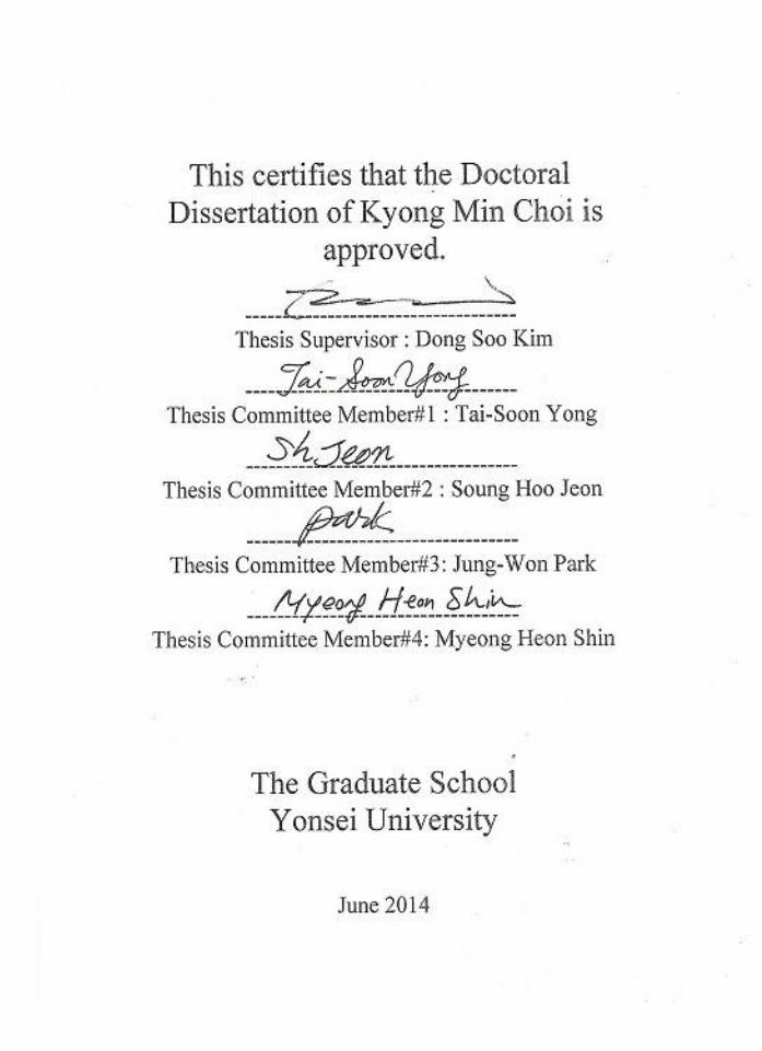
A recombinant mosquito salivary gland cDNA library was screened with
sensitized serum from Aedes togoi. In total, 4,000 clones were screened.
Forty-five clones were found to have immunoreactivity. An example of a
positive clone is shown in Figure 16. In order to confirm the identities of these
positive clones, they were isolated and converted to phagemids. The inserts were
then sequenced and compared to the GenBank database using the BLAST
program. Four selected sequenced clones, which had more than 400bp of the
insert, were analyzed. Four positive clones are known proteins (Table 2). Two of
these proteins are protein of unknown function (#006 and #039), an Aspartate
aminotransferase (AAT) superfamily of pyridoxal phosphate (#001), and
Aldo/keto reductases, related to diketogulonate reductase (#015).
Immunoscreening of 4×10³ clones with sensitized serum yielded a total of 45
positive clones (Fig. 16). As shown in Figure 17, the nucleotide sequences of the
cDNA inserts identified 4 different genes, designated as #001, #006, #015 and
#039.
Table 2. Proteins identified as saliva antigens by immunoscreening of the
Aedes togoi cDNA expression library with sensitized serum.
Clone Putative identification Functional significance
#001
#006
#015
#039
Aspartate aminotransferase Enzyme in amino acid metabolism
DUF2528 Protein of unknown function
Aldo/keto reductases NADPH-dependent oxidoreductases
DUF1398 Protein of unknown function
31
Figure 16. Demonstration of immunoreactive plaque. The arrow shows an
immunoreactive plaque. An example showing a positive clone identified by
immunoscreening of the Aedes togoi cDNA expression library. The black dot
indicated by an arrow is the positive clone. Saliva protein-specific IgG can be
detected in the sera of sensitized with mosquito bite. A λ phage cDNA
expression library constructed from normal female mosquito was used to
transduce E. coli for picoBlue immunoscreening. A representative primary
screen used to identify immunoreactive phage plaques is shown.
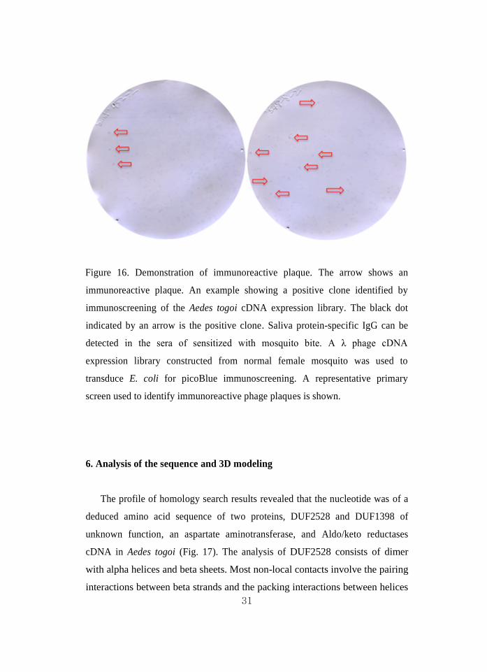
6. Analysis of the sequence and 3D modeling
The profile of homology search results revealed that the nucleotide was of a
deduced amino acid sequence of two proteins, DUF2528 and DUF1398 of
unknown function, an aspartate aminotransferase, and Aldo/keto reductases
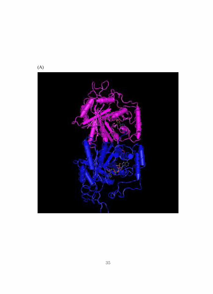
cDNA in Aedes togoi (Fig. 17). The analysis of DUF2528 consists of dimer
with alpha helices and beta sheets. Most non-local contacts involve the pairing
interactions between beta strands and the packing interactions between helices

32
and beta sheets. The analysis of DUF1398 consists of monomer with alpha
helices and beta sheets. Members of this family seem to be found exclusively in
Escherichia coli and Salmonella species. The function of this family is unknown
(Fig. 18 A and B).
The analysis of the sequence was performed using the NCBI/ BLAST
network service of the National Center for Biotechnology Information. In many
applications of 3D modeling for allergenic proteins, the goal is to determine the
solvent accessibility of side chains on the allergens, and possibly predict the
structure of conformational epitopes36
.

33
(A)
(B)
(C)
(D)

34
Figure 17. Alignment of deduced amino acid sequences of the partial # 001, #
006, # 015 and # 039 saliva antigen genes of the Aedes togoi. (A) Amino acid
sequence comparison of the # 001 immunoreactive plaque; aspartate
aminotransferase (AAT) superfamily (fold type I) of pyridoxal phosphate (PLP),
(B) The # 006 immunoreactive plaque; protein of unknown function
(DUF2528), (C) The # 015 immunoreactive plaque; aldo/keto reductases, related
to diketogulonate reductase, (D) The # 006 immunoreactive plaque; protein of
unknown function (DUF1398).
35
(A)

36
(B)
Figure 18. 3D structure of the protein of unknown function. (A) #006_Protein
of unknown function (DUF2528), crystal structure of saccharomyces
cerevisiae arabinose dehydrogenase Ara1 complexed with NADPH. (B)
#039_Protein of unknown function (DUF1398). This family consists of several
hypothetical Enterobacterial proteins of around 130 residues in length.

37
IV. DISCUSSION
Mosquitoes are very important from the standpoint of human health because
female mosquitoes suck blood from animals including human, and they serve as
vectors in the transmission of several important human diseases. Mosquito larvae
occur in a variety of aquatic habitats, such as ponds, pools of various sorts,
artificial containers, tree holes, streams, rice field, etc., and each species breeds
only in a particular type of habitat. In Korea, 9 genera and 48 species of
mosquitoes have been described37
. The Aedes togoi species is distributed on
most islands and at the seaside. The Aedes togoi breeds only in the rock pool of
seaside (Fig. 3). However, the allergenicity of salivary gland extracts of Korean
mosquitoes in humans and experimental mice has not been reported until now.
This species is thought to lay 30-80 eggs and overwinters in the egg stage,
however, during a warm winter they may also overwinter as larvae38
. The
allergic problems of Aedes togoi may occur mostly in coastal areas.
In the present study, many proteins in the Aedes togoi were detected in the
whole body extract and salivary gland extract, and some 20 proteins were shown
in the WBE and SGE by Coomassie brilliant blue staining (Fig. 11). These
results are somewhat different from previous studies, which have described high
molecular weight protein fractions of the Culex pipiens species. These
differences in the high molecular weight proteins between the Culex pipiens
species and Culex pipiens pallens may reflect a different subspecies of mosquito.
However, the low molecular weights of the proteins with 18 and 33 kDa were
similar to those in the previous study39
(Fig. 11).
Mosquitoes take blood meal for laying egg and their development. In this
study, the salivary glands consist of a pair of glands, each with two lateral lobes
and one median lobe in Aedes togoi (Fig. 9). The proximal part contains
enzymes such as α-glucosidase, α-amylase and maltase involved in the
metabolism of sugar meals, which are also found in the secretory cells of male

38
salivary glands. The median lobe, in addition to secretory cells, contains
nonsecretory cells implicated in fluid transport40,41
. The adult female appears to
have some control over the contents of released saliva (activities of maltase or
apyrase enzymes), depending on whether she is feeding on sugar or blood42
. The
salivary glands, like other organs in the hemocele, are covered by a basal lamina
that is external to the basal surface of the glandular cells. The apical surface of
the secretory cells is indented by a large secretory cavity that is a part of a
continuous cavity indenting all cells40
. The secretory duct where the saliva
comes from and the path from the secretory cavity to the proboscis differs
between Aedes and Anopheles. In Aedes, the chitinized duct continues to the end
of the distal portion of the gland40
; in Anopheles, it stops in the intermediate
zone40
. It is unknown if the end of the duct is open or closed. The duct wall in A.
aegypti is thinner in the distal than the proximal region of the lateral lobes40
.
The skin reactions to mosquito bites are derived from salivary secretions and
consist of an immediate and a delayed reaction with the involvement of IgE
mediated and lymphocyte-mediated hypersensitivities11,43
. In the saliva of Aedes
aegypti, a 68 kDa protein was identified as apyrase, an ATP diphosphohydrolase.
The role of this enzyme in mosquitoes seems to be related to the anti-platelet
aggregating activity found in their salivary secretion or salivary gland
homogenates42
.
It has been demonstrated on protein patterns of mosquitoes that there are
more than 20 protein fractions in the saliva of Aedes togoi using silver staining44
and 14- to 68-kDa protein fractions in the saliva of 4 Aedes and 3 Culex spp.
using colloidal gold staining39
.
In the present study, BALB/c mice bitten by the mosquito had significantly
higher anti-mosquito IgE antibody levels than those mice not bitten by the
mosquitoes (Fig. 12). These findings suggest that IgE-mediated hypersensitivity
is involved in the development of mosquito allergy. The results of
mosquito-specific IgE levels in mice show that five sera had positive titers

39
against only mosquito antigen when the serum was detected 0.625ng/ml. It is
suggested that a range of dose used for a species-specific IgE test is useful in
0.625ng/ml (Fig. 12).
Several investigations based mainly on Western blot analysis data have
described specific IgE-reactive proteins in mosquito saliva and salivary gland
extracts. Aedes togoi mosquito-sensitive individuals revealed IgE-binding bands
with apparent molecular weights of 18.5, 30.5, 33, 37 and 57.5 kDa13
.
In the present study, data obtained from Western blot with the pooled serum
of the sensitized to Aedes togoi were in agreement with the IgE-reactive protein
fractions from 18.0 to 57.5 kDa, but there was no reaction with 72.0, 90.0, and
150.0 kDa. In particular, the 18.0, 33.0, 37.0 and 57.0 kDa fractions elicited
strong IgE responses (Fig. 11). This suggests that these four protein fractions
may be major IgE-binding proteins in salivary gland extracts of Aedes togoi13
.
In our study, which was performed according to a method of Harlow and
Lane45
, there were no bands in the negative sera (Fig. 11). Harlow and Lane
stated that if background bands are generated due to a secondary reagent,
exchange of an alternative label or adsorption of the secondary reagent with liver
acetone powder is recommended. Liver acetone powder was used to reduce
background bands in this experiment.
In the mouse, interleukin-4 preferentially induces isotype switching to IgG1
and IgE46
. A previous study showed that human IgE and IgG4 antibodies of
mosquito bite-sensitive children bind to the same saliva proteins as antibodies
from the immune animals39
.
As shown in this study, the sensitized mice showed IgG1 antibody responses
to major salivary gland extracts of Aedes togoi, similar to IgE (Fig. 11, 12). Thus,
anti-mouse IgG1 reactivity to the salivary gland extracts of mosquitoes could be
useful to identify allergens by Western blot analysis.
Cross-reactivity with respect to bite reactions caused by different mosquito
species has been reported previously47
. In the present study, an ELISA inhibition

40
experiment with sera immune to Aedes togoi revealed that the mouse IgE level
decreases dose dependently with SGE of Aedes togoi (data not shown), whereas
it does not decrease in Dermatophagoides farinae. It is suggested that
species-shared allergens may not exist within these mosquitoes and house dust
mite.
The usual approach to isolate a recombinant DNA clone encoding a specific
allergen sequence is to screen a recombinant cDNA library. Serological
screening of recombinant cDNA expression libraries has been widely used for
the identification of allergens in various allergy types. Identification of allergens
in mosquito may facilitate the development of immunotherapies and biomarkers.
A recombinant cDNA library consists of a large number of mosquito salivary
gland, each one of which contains a different segment of saliva allergen DNA.
The purpose of our investigation is to identify allergens in mosquito salivary
glands by using the serological analysis of recombinant cDNA expression
libraries method. A recombinant mosquito salivary glands cDNA expression
library was screened with pooled sera from five sensitized mice (Fig. 13-16).
The examination of 3D structure often provides explanations for patterns of
sequence conservation observed in protein families, and 3D structure alignment
can serve as a guide for constructing accurate multiple sequence alignments such
as used in phylogenetic analysis. Recently the PDB has begun to provide
comprehensive data regarding the biologically relevant assemblies/ complexes or
quaternary structures of the macromolecules described in its records48
.
Four IgE response antigens encoded by two known and two unknown genes
were isolated, including aspartate aminotransferase (AAT) superfamily (fold
type I) of pyridoxal phosphate (PLP) and Aldo/keto reductases, related to
diketogulonate reductase. AAT is found in the liver, heart, skeletal muscle,
kidneys, brain, and red blood cells, and it is commonly measured clinically as a
marker for liver health.
A domain of unknown function (DUF) is a protein domain that has no

41
characterized function. A number has collected these families together in the
Pfam database using the prefix DUF followed, with examples being DUF2528
and DUF1398 (Fig. 17, 18). Accordingly, the NCBI Molecular Modeling
Database49
has been enhanced to emphasize the functional molecular complex
(i.e. quaternary structure) and the interactions between its molecular components
(Fig. 8).

42
V. CONCLUSION
The salivary glands of adult Aedes togoi are paired organs, located in the
thorax and composed of two identical lateral lobes and a shorter and wider
median lobe. Protein band patterns of the WBE and SGE were different from
one another. We confirmed the morphology of collecting ducts and secretary
cells of salivary glands. Specific mouse IgE reacted to the protein in SGE of
18.0, 33.0, 35.0, 37.0, 45.0, 57.5, 72.0, 90.0 and 150.0 kDa from Aedes togoi.
Two protein of unknown function was detected from this study. In summary,
we demonstrated that many candidate saliva antigens could be identified in the
cDNA library screening of Aedes togoi. Our findings warrant further
investigations on these proteins, aiming to elucidate their immunogenicity in
allergens. Analysis of the 3D structure of DUF1398 and DUF2528 was
not similar with any other allergens identified in plants or animals,
despite low sequence identities to their templates, the global folds of the
3D models of the cockroach allergen Bla g 4 and the mosquito salivary
protein antigen Aed a 2 had a sizable fraction of structural overlap,
suggesting that it would be a potential target for therapeutic agents in
specific mosquitoes bite allergens.
43
REFERENCES
1. Maekawa E, Aonuma H, Nelson B, Yoshimura A, Tokunaga F, Fukumoto S,
et al. The role of proboscis of the malaria vector mosquito Anopheles stephensi
in host-seeking behavior. Parasit Vectors 2011;4:10.
2. Isawa H, Yuda M, Yoneda K, Chinzei Y. The insect salivary protein,
prolixin-S, inhibits factor IXa generation and Xase complex formation in the
blood coagulation pathway. J Biol Chem 2000;275:6636-41.
3. Edwards JF, Higgs S, Beaty BJ. Mosquito feeding-induced enhancement of
Cache Valley Virus Bunyaviridae infection in mice. J Med Entomol
1998;35:261–5.
4. Yuan J, Bowman AS, Aljamali M, Payne MR, Tucker JS, Dillwith JW, et al.
Prostaglandin E(2)- stimulated secretion of protein in the salivary glands of
the lone star tick via a phosphoinositide signaling pathway. Insect Biochem
Mol Biol 2000;30:1099–106.
5. Simons FE, Peng Z. Skeeter syndrome. J Allergy Clin Immunol 1999;104(3
Pt 1):705-7.
6. Penneys NS, Nayar JK, Bernstein H, Knight JW, Leonardi C. Mosquito
salivary gland antigens identified by circulating human antibodies. Arch
Dermatol 1989;125:219–22.
7. Dhar R, Kumar N. Role of mosquito salivary glands. Current Science
2003;85:1308-13.
8. McCormack DR, Salata KF, Hershey JN, Carpenter GB, Engler RJ. Mosquito
bite anaphylaxis: Immunotherapy with whole body extracts. Ann Allergy
Asthma Immunol 1995;74:39–44.
9. Peng Z, Beckett AN, Engler RJ, Hoffman DR, Ott NL, Simons FE. Immune
responses to mosquito saliva in 14 individuals with acute systemic allergic
reactions to mosquito bite. J Allergy Clin Immunol 2004;114:1189-94.
10. Peng Z, Yang M, Simons FE. Immunological mechanisms in mosquito
allergy: Correlations of skin reactions with specific IgE and IgG antibodies and
lymphocyte proliferation response to mosquito antigens. Ann Allergy Asthma

44
Immunol 1996;77:238–44.
11. Engvall E, Perlman P. Enzyme-linked immunosorbent assay (ELISA).
Quantitative assay of immunoglobulin G. Immunochemistry. 1971:8:871-4.
12. Peng Z, Li H, Simons FE. Immunoblot analysis of salivary allergens in 10
mosquito species with worldwide distribution and the human IgE responses to
these allergens. J Allergy Clin Immunol 1998;101(4 Pt 1):498–505.
13. Tamborini E, Brandazza A, De Lalla C, Musco G, Siccardi AG, Arosio P, et
al. Recombinant allergen Lol p II: expression, purification and characterization.
Mol Immunol 1995:32:505-13.
14. Rose PW, Beran B, Bi C, Bluhm WF, Dimitropoulos D, Goodsell DS, et al.
The RCSB Protein Data Bank: redesigned web site and web services. Nucleic
Acids Res 2011;39(Database issus):D392–401.
15. Wang Y, Geer LY, Chappey C, Kans JA, Bryant SH. Cn3D: sequence and
structure views for Entrez. Trends Biochem Sci 2000;25:300–2.
16. Chapman MD, Pomés A, Breiteneder H, Ferreira F. Nomenclature and
structural biology of allergens. J Allergy Clin Immunol 2007;119:414–20.
17. Berkner H, Neudecker P, Mittag D, Ballmer-Weber BK, Schweimer K,
Vieths S, et al. Cross- reactivity of pollen and food allergens: soybean Gly m 4
is a member of the Bet v 1 superfamily and closely resembles yellow lupine
proteins. Biosci Rep 2009;29:183–92.
18. Robotham JM, Hoffman GG, Teuber SS, Beyer K, Sampson HA, Sathe SK,
et al. Linear IgE-epitope mapping and comparative structural homology
modeling of hazelnut and English walnut 11S globulins. Mol Immunol
2009;46:2975–84.
19. Cabanos C, Tandang-Silvas MR, Odijk V, Brostedt P, Tanaka A, Utsumi S,
et al. Expression, purification, cross-reactivity and homology modeling of
peanut profilin. Protein Expr Purif 2010;73:36–45.
20. Mattsson L, Lundgren T, Olsson P, Sundberg M, Lidholm J. Molecular and
immunological characterization of Can f 4: a dog dander allergen
cross-reactive with a 23 kDa odorant-binding protein in cow dander. Clin Exp
Allergy. 2010;40:1276-87.
21. Maleki SJ, Teuber SS, Cheng H, Chen D, Comstock SS, Ruan S, et al.
Computationally predicted IgE epitopes of walnut allergens contribute to

45
cross-reactivity with peanuts. Allergy 2011;66:1522–9.
22. Holm J, Ferreras M, Ipsen H, Würtzen PA, Gajhede M, Larsen JN, et al.
Epitope grafting, re-creating a conformational Bet v 1 antibody epitope on the
surface of the homologous apple allergen Mal d 1. J Biol Chem
2011;286:17569–78.
23. Hecker J, Diethers A, Etzold S, Seismann H, Michel Y, Plum M, et al.
Generation and epitope analysis of human monoclonal antibody isotypes with
specificity for the Timothy grass major allergen Phl p 5a. Mol Immunol
2011;48:1236–44.
24. Rougé P, Culerrier R, Granier C, Rancé F, Barre A. Characterization of
IgE-binding epitopes of peanut (Arachis hypogaea) PNA lectin allergen
cross-reacting with other structurally related legume lectins. Mol Immunol
2010;47:2359–66.
25. Dall’Antonia F, Gieras A, Devanaboyina SC, Valenta R, Keller W.
Prediction of IgE-binding epitopes by means of allergen surface comparison
and correlation to cross-reactivity. J Allergy Clin Immunol 2011;128:872–9.
26. Ivanciuc O, Schein CH, Braun W. SDAP: Database and computational tools
for allergenic proteins. Nucleic Acids Res 2003;31:359–62.
27. Gadermaier E. In shape--the art of mapping conformational epitopes. Int
Arch Allergy Immunol 2012;157:321–2.
28. Craven RB, Eliason DA, Francy DB, Reiter P, Campos EG, Jakob WL, et
al. Importation of Aedes albopictus and other exotic mosquito species into
the United States in used tires from Asia. J Am Mosq Control Assoc
1988;4:138-42.
29. Brummer-Korvenkontio H, Lappalainen P, Reunala T, Palosuo T.
Immunization of rabbits with mosquito bites: immunoblot analysis of IgG
antimosquito antibodies in rabbit and man. Int Arch Allergy Appl Immunol
1990:93:14–8.
30. Laemmli UK. Cleavage of structural proteins during the assembly of the
head of bacteriophage T4. Nature 1970;227:680–5.
31. Altschul SF, Madden TL, Schäffer AA, Zhang J, Miller W, Lipman DJ.
Gapped BLAST and PSI-BLAST: a new generation of protein database search
programs. Nucleic Acids Res 1997;25:3389–402.
http://www.ncbi.nlm.nih.gov/pubmed?term=Roug%C3%A9%20P%5BAuthor%5D&cauthor=true&cauthor_uid=20541807

46
32. Benkert P, Biasini M, Schwede T. Toward the estimation of the absolute
quality of individual protein structure models. Bioinformatics 2011;27:343–50.
33. Wiederstein M, Sippl MJ. ProSA-web: interactive web service for the
recognition of errors in three-dimensional structures of proteins. Nucleic Acids
Res 2007;35(Web Server issue):W407–10.
34. Bowie JU, Lüthy R, Eisenberg D. A method to identify protein sequences
that fold into a known three-dimensional structure. Science 1991;253:164–70.
35. James AA, Rossiqnol PA. Mosquito salivary glands: parasitological and
molecular aspects. Parasitol Today 1991;7:267-71.
36. Ivanciuc O, Schein CH, Garcia T, Oezguen N, Negi SS, Braun W. Structural
analysis of linear and conformational epitopes of allergens. Regul Toxicol
Pharmacol 2009;54(3 Suppl):S11–9.
37. Hong HK. Pictorial key to the species of mosquitoes in Korea. Rep NIH
Korea 1982;19:351–79(in Korean).
38. Riyong D, Choochote W, Jitpakdi A, Suvannadabba S, Leemingsawat S,
Chaithong U. Autogenous Aedes togoi sub-colony (Chanthaburi, Thailand
strain), an efficient laboratory vector in study of filariasis. Southeast Asian J
Trop Med Public Health 2000;31:246-51.
39. Brummer-Korvenkontio H, Palosuo T, Fran ois G, Reunala T.
Characterization of Aedes communis, Aedes aegypti and Anopheles stephensi
mosquito saliva antigens by immunobloting. Int Arch Allergy Immunol
1997;112:169-74.
40. Janzen HG, Wright KA. The salivary glands of Aedes aegypti (L.): an
electron microscope study. Can J Zool 1971;49:1343-6.
41. Warburg A, Touray M, Krettli AU, Miller LH. Plasmodium gallinaceum:
antibodies to circumsporozoite protein prevent sporozoites from invading the
salivary glands of Aedes aegvpti. Exp Parasitol 1992;75:303-7.
42. Ribeiro JM, Sarkis JJ, Rossiqnol PA, Spielman A. Salivary apyrase of
Aedes aegypti: characterization and secretory fate. Comp Biochem Physiol B
1984;79:81-6.

47
43. Oka K. Correlation of Aedes albopictus bite reaction with IgE antibody
assay and lymphocyte transformation test to mosquito salivary antigens. J
Dermatol 1989;16:341–7.
44. Racioppi JV, Spielman A. Secretory proteins from the salivary glands of
adult Aedes aegypti mosquitoes. Insect Biochem 1987;17:503–11.
45. Harlow E, Lane D. Immunoblotting. In: Antibodies: A laboratory manual.
New York: CSH Laboratory, Cold Spring Harbor; 1988. p.471-510.
46. Janeway Jr CA, Travers P. Immunobiology: the immune system in health
and disease. 2nd ed. Garland Publishing Inc. (New York, London) and
Current Biology, Ltd. (London, San Francisco, Philadelphia); 1996.
47. Peng Z, Simons FE. Cross-reactivity of skin and serum specific IgE
responses and allergen analysis for three mosquito species with worldwide
distribution. J Allergy Clin Immunol 1997;100:192–8.
48. Wang Y, Addess KJ, Chen J, Geer LY, He J, He S, et al. MMDB: annotating
protein sequences with Entrez’s 3D-structure database. Nucleic Acids Res
2007;35(Database issue):D298–300.
49. Bateman A, Coggill P, Finn RD. DUFs: families in search of function. Acta
Crystallogr Sect F Struct Biol Cryst Commun 2010;66(Pt 10):1148–52.

48
ABSTRACT (IN KOREAN)
진단 및 면역치료 시약 개발을 위한 모기 침샘 내 항원에
대한 생쥐의 체액성 면역 반응
<지도교수 김 동 수>
연세대학교 대학원 의학과
최 경 민
모기의 주둥이는 자흡형으로서 흡혈시에 숙주의 피부속으로
주둥이를 삽입하고 혈액응고 방지를 위하여 항 응고제가
포함된 타액을 주입한다. 이 경우 대부분의 사람에서
과민면역반응을 나타내어 소양감, 홍반, 구진, 궤양성구진, 및
수포형성등 다양한 국소 피부반응과 심한경우 전신반응을
일으킨다.
본 연구는 국내서식하는 토고숲모기(Aedes togoi)를
대상으로하여 타액선을 분리하고, 유전자 서고를 확립한 후,
모기에 노출된 실험 쥐로부터 그 혈청을 확보하고, 타액
알레르기 원인 항원의 유전자 특성을 규명하였으며, 재조합
특이 모기타액 단백질을 제조할 수 있는 기반 기술과
유전자를 획득하고자 하였다.
그 결과 토고숲모기 타액 항원은 18.0 kDa, 33.0 kDa, 35.0 kDa,
37.0 kDa, 45.0 kDa, 57.5 kDa, 72.0 kDa, 90.0 kDa 및 150.0 kDa의
분획이 모기 알레르겐의 주요 항원으로 추정되었다. 확보된
49
모기 침샘 유전자 서고와 모기 타액에 감작된 실험쥐 혈청을
이용한 면역스크린법 으로부터 양성반응을 보인 45개
플락중에서 2개의 알려지지 않은 단백질 코드 유전자를
확보하였다. 새로이 확인된 2개 DUF1398 와 DUF2528의
단백질을 3차원적 구조를 분석한 결과 식물성 알레르겐이나
동물성 알레르겐과 일치된 결과를 얻지 못하였으나, 3차원
모델의 구형 접힘구조에 있어서 그 유전자의 염기서열의
유사성과는 상관없이 이전에 알려진 바 있는 바퀴 알레르겐
Bla g 4 및 모기 침샘 알레르겐 Aed a 2와 부분적으로 그
구조가 일치함을 확인하였다. 이는 잠정적으로 알레르겐의
후보 물질로 간주되며, 지속적으로 연구해야할 표적 물질로
판단된다.
--------------------------------------------------------------------------------------
핵심되는 말 : 알레르겐, 모기, 토고숲모기, 침샘, 면역글로브린 E