Humanstanniocalcin: Apossiblehormonalregulatorof mineralmetabolism · ·...
5
Proc. Natl. Acad. Sci. USA Vol. 93, pp. 1792-1796, March 1996 Biochemistry Human stanniocalcin: A possible hormonal regulator of mineral metabolism HENRIK S. OLSEN*t, MARIO A. CEPEDA*, QING-QING ZHANG*, CRAIG A. RoSEN*, BENITO L. VOZZOLO*, AND GRAHAM F. WAGNERt *Human Genome Sciences, 9620 Medical Center Drive, Rockville, MD 20850-3338; and tDepartment of Physiology, Faculty of Medicine, University of Western Ontario, London ON, Canada N6A SC1 Communicated by Henry G. Friesen, Medical Research Council of Canada, Otawa, Canada, October 17, 1995 ABSTRACT We have isolated a human cDNA clone en- coding the mammalian homolog of stanniocalcin (STC), a calcium- and phosphate-regulating hormone that was first described in fishes where it functions in preventing hypercal- cemia. STC has a unique amino acid sequence and, until now, has remained one of the few polypeptide hormones never described in higher vertebrates. Human STC (hSTC) was found to be 247 amino acids long and to share 73% amino acid sequence similarity with fish STC. Polyclonal antibodies to recombinant hSTC localized to a distinct cell type in the nephron tubule, suggesting kidney as a possible site of syn- thesis. Recombinant hSTC inhibited the gill transport of calcium when administered to fish and stimulated renal phosphate reabsorption in the rat. The evidence suggests that mammalian STC, like its piscine counterpart, is a regulator of mineral homeostasis. Stanniocalcin (STC) is a calcium-regulating hormone in bony fishes that has never been described in higher vertebrates, including mammals (1). The hormone is synthesized by the corpuscles of stannius (CS), endocrine glands that are associ- ated with the kidneys of all fishes with a bony skeleton (2). The primary function of STC in fishes is the prevention of hyper- calcemia and a rise in serum calcium levels is the primary stimulus for secretion (3). Upon release into the circulation, STC lowers calcium transport by the gills thereby reducing its rate of influx from the environment into the extracellular compartment (1). A second equally important action of STC is stimulation of phosphate reabsorption by renal proximal tubules (4). The consequence of this renal effect is increased levels of plasma phosphate, the latter of which combines with excess calcium and promotes its disposal into bone and scales. Because the CS have never been identified in higher verte- brates, it has long been assumed that STC was unique to fishes. However, recent evidence of STC immunoreactivity in human kidney and serum argues for a more widespread existence of the hormone (5). By a process of random sequencing of human tissue cDNAs, we have isolated a lung-derived cDNA clone whose deduced protein sequence bears a strikingly high level of homology to salmon and eel STC (6, 7).§ Data indicating that human (h)STC inhibits calcium uptake in fish and phosphate excretion in rats suggest that hSTC is a hormonal regulator of mineral metabolism. MATERIALS AND METHODS cDNA Isolation and Analysis. cDNA isolation. The initial expressed sequence tag (EST) clones used in the study were discovered by scientists at The Institute for Genomic Research by using established EST methods (8, 9). These clones were The publication costs of this article were defrayed in part by page charge payment. This article must therefore be hereby marked "advertisement" in accordance with 18 U.S.C. §1734 solely to indicate this fact. part of a larger EST project (10). This clone was used for rescreening the same library and a full-length clone encoding hSTC was obtained. Southern blot analysis. Ten-microgram aliquots of human genomic DNA were cut with BamHI, EcoRI, and Xba I and then separated on an 0.8% agarose gel. After transfer to a nylon membrane, the blot was probed with 32P-labeled hSTC cDNA and washed under high stringency. Northern blot analysis. RNA was separated in a 0.8% aga- rose/formaldehyde gel in lx Mops buffer (10X Mops = 0.2 M Mops/50 mM NaOAc/10 mM EDTA). After transfer to a nylon membrane, the blot was probed with 32P-labeled hSTC cDNA and washed under high stringency, and the RNA expression pattern was analyzed by autoradiography. Expression of hSTC in Bacteria. hSTC was cloned into the bacterial expression (pQE) vector and a histidine-tagged pro- tein was purified by ion-affinity column chromotography ac- cording to the manufacturer (Quiagen, Chatsworth, CA). Briefly, the bacterial culture harboring the hSTC expression plasmid was grown to an OD60o of 0.4, induced with 1 mM isopropyl ,B-D-thiogalactoside, and grown for an additional 4 h. After induction, the cell pellet was dissolved in 6 M guanidine hydrochloride (pH 8.0) and applied to the ion-affinity column. After extensive washing, the protein was renatured by applying a gradient of urea (4 M to 0 M) in 150 mM NaCl/10% (vol/vol) glycerol. Protein was eluded in 250 mM imidazole/150 mM NaCl/25 mM Tris/10% glycerol and dialyzed. Fish Bioassay. For testing the effects of hSTC in fishes, an established fish STC bioassay was employed essentially as described (1). This bioassay monitors the inhibitory effects of STC on gill calcium transport. Goldfish (1.2 ± 0.1 g per 10 fish per group) were given two intraperitoneal injections, 1 h apart, of recombinant hSTC, purified salmon STC, or saline, and then placed in tanks of water containing 45Ca (100,000 dpm/ ml) for a 3-h period. The fish were then sacrificed and ashed overnight in a muffle furnace, and the isotope content of the resulting ash was determined by scintillation counting. The rate of gill calcium transport in each fish was determined on the basis of body isotope content and water-specific activity and was expressed as ,umol of Ca2+ per kg (body weight) per h. Individual treatment groups were considered significantly different than solvent-injected controls if P < 0.05 (ANOVA and Dunnet's test). Immunocytochemistry. Antibodies were prepared in rabbits after three monthly immunizations, each of 200 ,g of bacte- rially expressed recombinant hSTC dissolved in Freund's adjuvant/saline, 1:1 (vol/vol). The development of antibody titer was monitored by ELISA and the highest titer was obtained after the third immunization. Western blot analysis of Abbreviations: STC, stanniocalcin; h, human; ICC, immunocytochem- istry; CS, corpuscles of stannius; STCir, STC immunoreactive. tTo whom reprint requests should be addressed. §The sequence reported in this paper has been deposited in the GenBank data base (accession no. U46768). 1792
Transcript of Humanstanniocalcin: Apossiblehormonalregulatorof mineralmetabolism · ·...
Proc. Natl. Acad. Sci. USAVol. 93, pp. 1792-1796, March 1996Biochemistry
Human stanniocalcin: A possible hormonal regulator ofmineral metabolismHENRIK S. OLSEN*t, MARIO A. CEPEDA*, QING-QING ZHANG*, CRAIG A. RoSEN*, BENITO L. VOZZOLO*,AND GRAHAM F. WAGNERt*Human Genome Sciences, 9620 Medical Center Drive, Rockville, MD 20850-3338; and tDepartment of Physiology, Faculty of Medicine, University of WesternOntario, London ON, Canada N6A SC1
Communicated by Henry G. Friesen, Medical Research Council of Canada, Otawa, Canada, October 17, 1995
ABSTRACT We have isolated a human cDNA clone en-coding the mammalian homolog of stanniocalcin (STC), acalcium- and phosphate-regulating hormone that was firstdescribed in fishes where it functions in preventing hypercal-cemia. STC has a unique amino acid sequence and, until now,has remained one of the few polypeptide hormones neverdescribed in higher vertebrates. Human STC (hSTC) wasfound to be 247 amino acids long and to share 73% amino acidsequence similarity with fish STC. Polyclonal antibodies torecombinant hSTC localized to a distinct cell type in thenephron tubule, suggesting kidney as a possible site of syn-thesis. Recombinant hSTC inhibited the gill transport ofcalcium when administered to fish and stimulated renalphosphate reabsorption in the rat. The evidence suggests thatmammalian STC, like its piscine counterpart, is a regulator ofmineral homeostasis.
Stanniocalcin (STC) is a calcium-regulating hormone in bonyfishes that has never been described in higher vertebrates,including mammals (1). The hormone is synthesized by thecorpuscles of stannius (CS), endocrine glands that are associ-ated with the kidneys of all fishes with a bony skeleton (2). Theprimary function of STC in fishes is the prevention of hyper-calcemia and a rise in serum calcium levels is the primarystimulus for secretion (3). Upon release into the circulation,STC lowers calcium transport by the gills thereby reducing itsrate of influx from the environment into the extracellularcompartment (1). A second equally important action of STCis stimulation of phosphate reabsorption by renal proximaltubules (4). The consequence of this renal effect is increasedlevels of plasma phosphate, the latter of which combines withexcess calcium and promotes its disposal into bone and scales.Because the CS have never been identified in higher verte-brates, it has long been assumed that STC was unique to fishes.However, recent evidence of STC immunoreactivity in humankidney and serum argues for a more widespread existence ofthe hormone (5).By a process of random sequencing of human tissue cDNAs,
we have isolated a lung-derived cDNA clone whose deducedprotein sequence bears a strikingly high level of homology tosalmon and eel STC (6, 7).§ Data indicating that human(h)STC inhibits calcium uptake in fish and phosphate excretionin rats suggest that hSTC is a hormonal regulator of mineralmetabolism.
MATERIALS AND METHODScDNA Isolation and Analysis. cDNA isolation. The initial
expressed sequence tag (EST) clones used in the study werediscovered by scientists at The Institute for Genomic Researchby using established EST methods (8, 9). These clones were
The publication costs of this article were defrayed in part by page chargepayment. This article must therefore be hereby marked "advertisement" inaccordance with 18 U.S.C. §1734 solely to indicate this fact.
part of a larger EST project (10). This clone was used forrescreening the same library and a full-length clone encodinghSTC was obtained.
Southern blot analysis. Ten-microgram aliquots of humangenomic DNA were cut with BamHI, EcoRI, and Xba I andthen separated on an 0.8% agarose gel. After transfer to anylon membrane, the blot was probed with 32P-labeled hSTCcDNA and washed under high stringency.Northern blot analysis. RNA was separated in a 0.8% aga-
rose/formaldehyde gel in lx Mops buffer (10X Mops = 0.2M Mops/50 mM NaOAc/10 mM EDTA). After transfer to anylon membrane, the blot was probed with 32P-labeled hSTCcDNA and washed under high stringency, and the RNAexpression pattern was analyzed by autoradiography.
Expression of hSTC in Bacteria. hSTC was cloned into thebacterial expression (pQE) vector and a histidine-tagged pro-tein was purified by ion-affinity column chromotography ac-cording to the manufacturer (Quiagen, Chatsworth, CA).
Briefly, the bacterial culture harboring the hSTC expressionplasmid was grown to an OD60o of 0.4, induced with 1 mMisopropyl ,B-D-thiogalactoside, and grown for an additional 4 h.After induction, the cell pellet was dissolved in 6 M guanidinehydrochloride (pH 8.0) and applied to the ion-affinity column.After extensive washing, the protein was renatured by applyinga gradient of urea (4M to 0 M) in 150mM NaCl/10% (vol/vol)glycerol. Protein was eluded in 250 mM imidazole/150 mMNaCl/25 mM Tris/10% glycerol and dialyzed.
Fish Bioassay. For testing the effects of hSTC in fishes, anestablished fish STC bioassay was employed essentially asdescribed (1). This bioassay monitors the inhibitory effects ofSTC on gill calcium transport. Goldfish (1.2 ± 0.1 g per 10 fishper group) were given two intraperitoneal injections, 1 h apart,of recombinant hSTC, purified salmon STC, or saline, andthen placed in tanks of water containing 45Ca (100,000 dpm/ml) for a 3-h period. The fish were then sacrificed and ashedovernight in a muffle furnace, and the isotope content of theresulting ash was determined by scintillation counting. Therate of gill calcium transport in each fish was determined on thebasis of body isotope content and water-specific activity andwas expressed as ,umol of Ca2+ per kg (body weight) per h.Individual treatment groups were considered significantlydifferent than solvent-injected controls if P < 0.05 (ANOVAand Dunnet's test).Immunocytochemistry. Antibodies were prepared in rabbits
after three monthly immunizations, each of 200 ,g of bacte-rially expressed recombinant hSTC dissolved in Freund'sadjuvant/saline, 1:1 (vol/vol). The development of antibodytiter was monitored by ELISA and the highest titer wasobtained after the third immunization. Western blot analysis of
Abbreviations: STC, stanniocalcin; h, human; ICC, immunocytochem-istry; CS, corpuscles of stannius; STCir, STC immunoreactive.tTo whom reprint requests should be addressed.§The sequence reported in this paper has been deposited in theGenBank data base (accession no. U46768).
1792
Proc. Natl. Acad. Sci. USA 93 (1996) 1793
human kidney extracts and recombinant hSTC yielded twoclosely spaced bands in both instances. Fresh human kidneybiopsies were obtained from Surgical Pathology, UniversityHospital, London, with patient consent, and immediately fixedovernight in PBS containing 4% (wt/vol) paraformaldehyde(pH 7.4). Tissues were dehydrated, embedded in paraffin, and4-,tm serial sections were mounted on coated slides. Forimmunocytochemistry (ICC), tissue sections were dewaxed,rehydrated, and equilibrated in ICC diluent buffer (0.025 MTris, pH 7.5/0.15 M NaCl). Sections were then treated for 10min with undiluted normal goat serum to reduce backgroundstaining prior to an overnight incubation with a 1:1000 dilutionof hSTC antiserum. After an extensive wash in ICC buffer, thesections were incubated for 1 h in peroxidase-coupled goatanti-rabbit IgG. The sites of antibody binding were visualizedwith 0.025% diaminobenzidene prepared in ICC buffer con-taining 0.01% H202. After development, the sections werecounterstained in hematoxylin, dehydrated, and mounted.Control procedures included the application of normal rabbitserum or hSTC antiserum preabsorbed with hSTC in lieu ofantiserum alone.Rat Bioassay. Recombinant hSTC was tested for effects in
rats by using standard methods for assessment of renal func-tion. Male Wistar rats (300 g) were anesthetized with Inactin(100 mg/kg) and ketamine (10 mg/kg), and a tracheostomywas performed to facilitate breathing. Body temperature wasmaintained at 37 ± 0.5°C with a heating pad. A right jugularvein cannula was used for the infusion of saline and testsubstances. A right femoral artery cannula was used formonitoring mean arterial pressure and for blood sampling.Both ureters were cannulated near the renal pelvis for urinecollection. The animals were infused with saline for 1 h aftersurgery prior to commencing an experiment. To start anexperiment, urine was collected over the first 20-min interval,Cl, and a blood sample was taken at the midpoint of urinecollection. Recombinant hSTC or solvent was given as a 500-,lbolus 15 min into the second collection period, C2. Urine andblood were then collected over the next 80 min, C3 through C6.At the end of the experiment, the animals were sacrificed andthe kidneys were removed, decapsulated, and weighed. Urinevolume was calculated gravimetrically and expressed as ml permin per g (kidney weight). Mean arterial pressure was deter-mined for each clearance period by averaging the readings overeach clearance period. Plasma and urinary concentrations ofNa+ and K+ were determined by flame photometry, whereascalcium (11) and inorganic phosphate (12) were determinedcolorimetrically. On the basis of these analyses, electrolyteexcretion rates in clearance periods C3 through C6 wereexpressed as changes from Cl levels. The data were analyzedby repeated measures ANOVA and groups were consideredsignificantly different if P < 0.05.
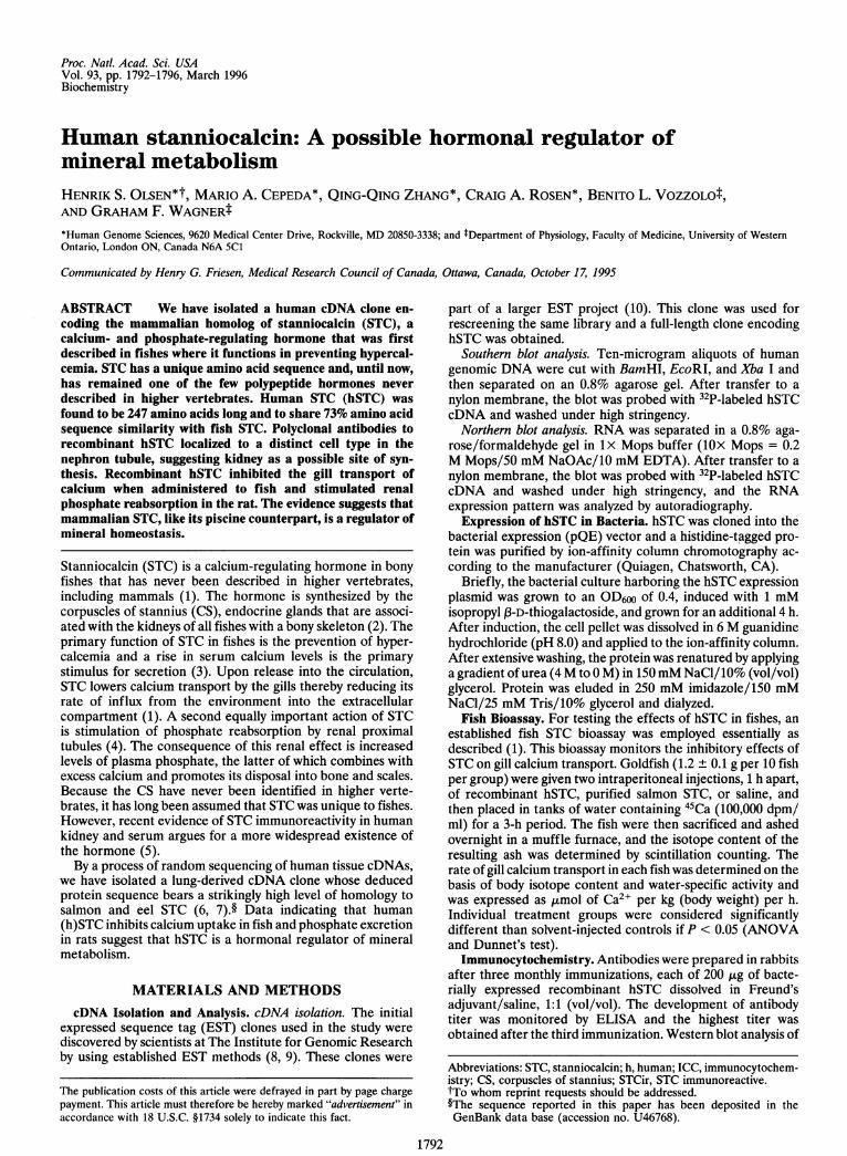
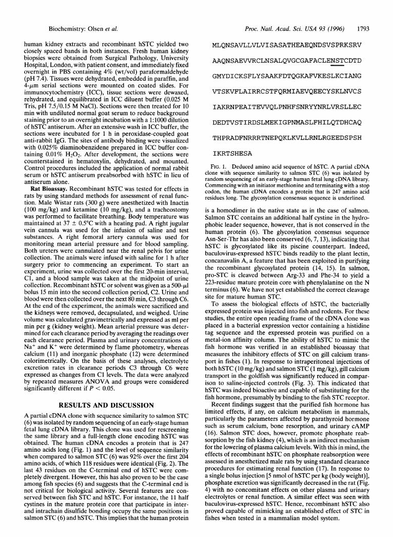
RESULTS AND DISCUSSIONA partial cDNA clone with sequence similarity to salmon STC(6) was isolated by random sequencing of an early-stage humanfetal lung cDNA library. This clone was used for rescreeningthe same library and a full-length clone encoding hSTC wasobtained. The human cDNA encodes a protein that is 247amino acids long (Fig. 1) and the level of sequence similaritywhen compared to salmon STC (6) was 92% over the first 204amino acids, of which 118 residues were identical (Fig. 2). Thelast 43 residues on the C-terminal end of hSTC were com-pletely divergent. However, this has also proven to be the caseamong fish species (6) and suggests that the C-terminal end isnot critical for biological activity. Several features are con-served between fish STC and hSTC. For instance, the 11 halfcystines in the mature protein core that participate in inter-and intrachain disulfide bonding occupy the same positions insalmon STC (6) and hSTC. This implies that the human protein
MLQNSAVLLVLVISASATHEAEQNDSVSPRKSRV
AAQNSAEVVRCLNSALQVGCGAFACLENSTCDTD
GMYDICKSFLYSAAKFDTQGKAFVKESLKCIANG
VTSKVFLAIRRCSTFQRMIAEVQEECYSKLNVCS
IAKRNPEAITEVVQLPNHFSNRYYNRLVRSLLEC
DEDTVSTIRDSLMEKIGPNMASLFHILQTDHCAQ
THPRADFNRRRTNEPQKLKVLLRNLRGEEDSPSH
IKRTSHESA
FIG. 1. Deduced amino acid sequence of hSTC. A partial cDNAclone with sequence similarity to salmon STC (6) was isolated byrandom sequencing of an early-stage human fetal lung cDNA library.Commencing with an initiator methionine and terminating with a stopcodon, the human cDNA encodes a protein that is 247 amino acidresidues long. The glycosylation consensus sequence is underlined.
is a homodimer in the native state as in the case of salmon.Salmon STC contains an additional half cystine in the hydro-phobic leader sequence, however, that is not conserved in thehuman protein (6). The glycosylation consensus sequenceAsn-Ser-Thr has also been conserved (6, 7, 13), indicating thathSTC is glycosylated like its piscine counterpart. Indeed,baculovirus-expressed hSTC binds readily to the plant lectin,concanavalin A, a feature that has been exploited in purifyingthe recombinant glycosylated protein (14, 15). In salmon,pro-STC is cleaved between Arg-33 and Phe-34 to yield a223-residue mature protein core with phenylalanine on the Nterminus (6). We have not yet established the correct cleavagesite for mature human STC.To assess the biological effects of hSTC, the bacterially
expressed protein was injected into fish and rodents. For thesestudies, the entire open reading frame of the cDNA clone wasplaced in a bacterial expression vector containing a histidinetag sequence and the expressed protein was purified on ametal-ion affinity column. The ability of hSTC to mimic thefish hormone was verified in an established bioassay thatmeasures the inhibitory effects of STC on gill calcium trans-port in fishes (1). In response to intraperitoneal injections ofboth hSTC (10 mg/kg) and salmon STC (1 mg/kg), gill calciumtransport in the goldfish was significantly reduced in compar-ison to saline-injected controls (Fig. 3). This indicated thathSTC was indeed bioactive and capable of substituting for thefish hormone, presumably by binding to the fish STC receptor.
Recent findings suggest that the purified fish hormone haslimited effects, if any, on calcium metabolism in mammals,particularly the parameters affected by parathyroid hormonesuch as serum calcium, bone resorption, and urinary cAMP(16). Salmon STC does, however, promote phosphate reab-sorption by the fish kidney (4), which is an indirect mechanismfor the lowering ofplasma calcium levels. With this in mind, theeffects of recombinant hSTC on phosphate reabsorption wereassessed in anesthetized male rats by using standard clearanceprocedures for estimating renal function (17). In response toa single bolus injection [5 nmol of hSTC per kg (body weight)],phosphate excretion was significantly decreased in the rat (Fig.4) with no concomitant effects on other plasma and urinaryelectrolytes or renal function. A similar effect was seen withbaculovirus-expressed hSTC. Hence, recombinant hSTC alsoproved capable of mimicking an established effect of STC infishes when tested in a mammalian model system.
Biochemistry: Olsen et al.

Proc. Natl. Acad. Sci. USA 93 (1996)
10 20 30Human MLQNSAVLLVLVISASATHEAEQNDSVSPRKSRVAAQN
1 : : 1:: : :1 1 :
Salmon MLAKFGLCAVFLVLGTAATFDTDPEEA-SPRRARFSSNS
40 50 60 70 80 90SAEVVRCLNSALQVGCGAFACLENSTCDTDGMYDICKSFLYSAAKFDTQGKAFVKESLKC
1 111111 111 1111111111111 111 11:1:1111:111111:1PSDVARCLNGALAVGCGTFACLENSTCDTDGMHDICQLFFHTAATFNTQGKTFVKESLRC
7 -100 110 120 130 140 150IANGVTSKVFLAIRRCSTFQRMIAEVQEECYSKLNVCSIAKRNPEAITEVVQLPNHFSNR
IANGVTSKVFQTIRRCGVFQRMISEVQEECYSRLDICGVARSNPEAIGEVVQVPAHFPNR
160 170 180 190 200 210YYNRLVRSLLECDEDTVSTIRDSLMEKIGPNMASLFHILQTDHCAQTHPRADFNRRRTNE1I I::I1 1: 1:11:1: 1:: 1 1:I 1:11YYSTLLQSLLACDEETVAVVRAGLVARLGPDMETLFQLLQNKHCPQGSNQGPNSAPAGWR
220 230 240PQKLKVLLRNLRGEEDSPSHIKRTSHESA
WPMGSPPSFKIQPSMRGRDPTHLFARKRSVEALERVME
FIG. 2. Amino acid sequence comparison of hSTC and salmon STC. A solid line between species denotes identity and double dots denotesimilarity. Note the high level of identity in the core of the molecule. The level of sequence similarity when compared to salmon STC (6) was 92%over the first 204 amino acids, of which 118 residues were identical. The underlined sequence denotes the glycosylation consensus sequence. Thelast 43 residues on the C-terminal end of hSTC were completely divergent from the salmon, as is the case between the different fish STCs (6, 7).Sequence alignment was done with the University of Wisconsin Genetics Computer Group TFASTA program (182).
The hSTC gene is present as a single copy according toSouthern blot analysis. To identify its chromosomal locus, agenomic clone was isolated and used in fluorescent in situhybridization as described (18). The analysis of chromosomalspreads indicated that the hSTC gene was localized to band8p2l. When the tissue distribution of STC gene expression wasexamined by Northern blot analysis, low levels of expressionwere detected in several tissues including kidney, bone mar-row, and thymic stromal cells (Fig. 5). ICC (19) was employedto identify cellular sources of the protein in human kidney byusing a polyclonal antiserum to recombinant hSTC. In the
hSTC
sSTC
- **
Control
0 1 2 3 4 5
Ca2+ uptake - ,umol/kg/hFIG. 3. hSTC inhibits gill calcium transport in fish. hSTC and
salmon STC (sSTC) were tested for effects on gill calcium transport asdescribed (1). Goldfish (1.2 + 0.1 g; 10 fish per group) were givenintraperitoneal injections of hSTC (10 mg/kg), salmon STC (1 mg/kg),or saline and placed in tanks of 45Ca-containing water (50,000dpm/ml) for 3 h. The fish were then sacrificed and individually ashedovernight at 600°C, and the isotope content of the ash was determinedby scintillation counting. Based on the body weight of the fish and thespecific activity of the water, gill calcium transport in each fish wasexpressed as ,imol of Ca2+ per kg (body weight) per h. Both hSTC (**,P < 0.01) and salmon STC (*, P < 0.05) had statistically significantinhibitory effects on gill calcium transport (two-tailed ANOVA andDunnet's test).
kidney, no immunoreactivity was detected in the glomeruli,vascular elements, or hematopoietic tissue surrounding thenephrons; STC-immunoreactive (STCir) cells were confined tothe nephron, specifically in distal convoluted tubule and thecollecting tubule. Some STCir cells in the collecting tubulewere phenotypically unique due to their large size and ten-
18
IS^I.
j:j12 [9
630
-3
4-9
-12-is.13
Control
STC
*
*
-20-0 0-20 20040-60 803100Minutes
FIG. 4. hSTC inhibits phosphate excretion in the rat. Rats (250 ±10 g; 5 rats per group) were maintained under Inactin anesthesia withcatheters in one jugular vein, one carotid artery, and both ureters andwere continuously infused with inulin and p-aminohippuric acid formeasurement of glomerular filtration and renal blood flow, respec-tively, as described (15). Bolus injections ofhSTC (5 nmol/kg) or salinewere given via the jugular catheter after the first urine collectionperiod (arrow) and renal function, plasma and urinary electrolytes(Na+, K+, Ca2+, Mg2+, and P04), and blood pressure were monitoredthroughout. Phosphate excretion was maximally inhibited by hSTCafter 60 min and remained significantly different thereafter (P < 0.01;repeated measures ANOVA). STC had no effect on any otherparameters.
I I
1794 Biochemistry: Olsen et al.
I

Proc. Natl. Acad. Sci. USA 93 (1996) 1795
1 2 3 4 5 6 7 8 9 10 11 12
FIG. 5. Northern blot analysis of hSTC mRNA expression. ThehSTC cDNA was radiolabeled and hybridized to 10 xg of total RNAunless otherwise indicated. A 1-week exposure of the blot is shown.Lanes: 1, stomach; 2, thymus; 3, spleen; 4, peripheral T cells; 5,leukocytes; 6, kidney; 7, lung; 8, liver; 9, heart; 10, pancreas; 11, thymicstromal cells (1.5 u±g); 12, bone marrow [1 ,ug of poly(A) RNA]. Thearrowheads at left denote the positions of 18S (bottom) and 28S (top)rRNAs.
dency to have multiple nuclei (Fig. 6A), suggesting that theywere functionally different than surrounding cells. Antibodystaining of STCir cells was abolished when the primary anti-serum was preabsorbed with hSTC (Fig. 6B). In fishes, the CSglands are derived from the kidneys. Individual STC cells budoff from the nephron tubules during embryogenesis, coalesceto form CS glands, and then migrate to the kidney surface priorto hatching (20). In the most primitive bony fishes, however,the glands remain deep within the kidneys associated withindividual nephrons (21). Therefore, the presence of STCircells in the mammalian nephron is entirely consistent whenviewed from an evolutionary perspective. The ICC resultsobtained in the present study with hSTC antiserum haveconfirmed and extended earlier ICC findings in human kidneyusing antibodies to the fish hormone. In the case of bothantisera, staining was confined to the nephron tubule, as nostaining was observed in glomeruli, proximal tubules, vascularelements, or hematopoietic tissue. However, whereas both
antiserum stained distal tubule cells, the hSTC antiserum alsostained cells in the thick ascending limb and collecting tubules.As our fish antiserum is highly crossreactive with recombinanthSTC, the different ICC staining patterns obtained with thetwo antiserum may be indicative of there being different STCcell types.The hormonal regulation of mineral homeostasis in mam-
mals is a complex process involving parathyroid hormone (22,23), calcitonin (24), and the active metabolite of vitamin D(25). Parathyroid hormone and vitamin D counteract hypocal-cemia by stimulating bone resorption, as well as intestinal andrenal calcium transport, and parathyroid hormone also en-hances renal phosphate excretion. Calcitonin, on the otherhand, counteracts hypercalcemia by inhibiting osteoclasticbone resorption, although its importance as a minute-to-minute regulator of calcium homeostasis remains controversial(26). The complexity of mineral homeostasis is reflected in thenumerous diseases associated with the impaired regulation ofcalcium and phosphate, which impact on renal function, andthe vascular, neuronal, and muscular systems, in addition tobone mineralization. Recent findings involving tumor-inducedosteomalacia in humans (27) and murine models of X chro-mosome-linked hypophosphatemia (28) imply there is roomfor additional humoral regulators of mineral homeostasis inmammals and STC warrants consideration as a candidate.Given the structural similarities between hSTC and fish STCand their comparable effects on renal function, STC may havethe same role in mammals and fishes: preventing hypercalce-mia, in part, through its stimulatory effects on phosphatereabsorption. The remarkable evolutionary conservation be-tween fish and mammalian STC suggests an important role forthis hormone in higher vertebrates.
We thank Human Genome Sciences and The Institute for GenomicResearch sequencing facilities for cDNA sequencing; K. Carter and B.Shell for the fluorescence in situ hybridization mapping. We also thankDr. R. L. Kline (Department of Physiology, University of WesternOntario) for assistance with the rat studies. Grant and Scholarship
> s. <._.Y
I
w.?'\ 4,
A-K
4jtJ~~~~~~~~~~~~~~~~~~~~~~~~~~~4
A~~~~~~~~~~ B
FIG. 6. STCir cells are present in human kidney nephron tubule. ICC was performed as described (19). Fresh biopsies of human kidney werefixed overnight in phosphate-buffered 4% (wt/vol) paraformaldehyde (pH 7.2), dehydrated, and embedded in paraffin. Dewaxed 5-,Lm serialsections were incubated overnight with a 1:1000 dilution of rabbit anti-hSTC serum in Tris-buffered saline (pH 7.5) (A) or the same antiserumdilution preabsorbed with recombinant hSTC (B). The slides were then washed for three 10-min periods in Tris-buffered saline, incubated for 30min with peroxidase-coupled goat anti-rabbit IgG, washed as before, and developed in 0.025% diaminobenzidene containing 0.01% hydrogenperoxide. (A) Large multinucleated cell in the collecting tubule portion of the nephron in outer medullary kidney stained by the antiserum. (B)Adjacent section treated with preabsorbed antiserum and showing no specific staining. (Bar = 15 ,um.)
Biochemistry: Olsen et al.
o. X
.
ik .:,
V.-I):-.-V a
.. a
A
# ...1
wof,.% A
.iv:...'..-4

Proc. Natl. Acad. Sci. USA 93 (1996)
support from the Medical Research Council of Canada awarded toG.F.W. is also gratefully acknowledged.
Note Added in Proof. While this manuscript was in review, a similarsequence was reported by Chang et al. (29).
1. Wagner, G. F., Hampong, M., Park, C. M. & Copp, D. H. (1986)Gen. Comp. Endocrinol. 63, 481-491.
2. Stannius, H. (1839) Arch. Anat. Physiol. 6, 97-101.3. Wagner, G. F., Milliken, C., Friesen, H. G. & Copp, D. H. (1991)
Mol. Cell. Endocrinol. 79,129-138.4. Lu, M., Wagner, G. F. & Renfro, J. L. (1994) Am. J. Physiol. 36,
R1356-R1362.5. Wagner, G. F., Guiraudon, C. C., Milliken, C. & Copp, D. H.
(1995) Proc Natl Acad Sci. 92, 1871-1875.6. Wagner, G. F., Dimattia, G. E., Davie, J. R., Copp, D. H. &
Friesen, H. G. (1992) Mol. Cell. Endocrinol. 90, 7-15.7. Butkus, H., Roche, P. J., Fernley, R. T., Haralambidis, J., Pen-
schow, J. D., Ryan, G. B., Trahair, J. F., Tregear, G. W. &Coughlin, J. P. (1987) Mol. Cell. Endocrinol. 54, 123-134.
8. Adams, M. D., Kelley, J. M., Gocayne, J. D., Dubnick, M., Poly-meropoulos, M. H., Xiao, H., Merril, C. R., Wu, A., Olde, B. &Moreno, R. F. (1991) Science 252, 1651-1656.
9. Adams, M. D., Dubnick, M., Kerlavage, A. R., Moreno, R.,Kelley, J. M. & et al., (1992) Nature (London) 355, 632-634.
10. Adams, M. D., Kerlavage, A. R., Fleischmann, R. D. & Fuldner,R. A. (1995) Nature (London) 377, Suppl., 3-174.
11. Baginski, E. S., Marie, S. S., Clark, W. L. & Zak, B. (1973) Clin.Chem. Acta 46, 46-54.
12. Chen, P. S., Toribara, T. Y. & Warner, H. (1956)Anal. Chem. 28,1756-1758.
13. Wagner, G. F. (1994) in Fish Physiology, eds. Sherwood, N. &Hew, C. (Academic, New York), Vol. 13, pp. 273-306.
14. Summers, M. D. & Smith, G. E. (1987) Tex. Agric. Exp. Stn. Bull.1555 (abstr.).
15. Luckow, V. A. & Summers, M. D. (1989) Virology 170, 31-39.16. Stern, P. H., Shanker, G. L., Fargher, R. C., Copp, D. H., Mil-
liken, C. E., Sato, K., Goltzman, D. & Herrmann-Erlee, M. P. M.(1991) J. Bone Miner. Res. 11, 1153-1159.
17. McLennan, G. P., Kline, R. L. & Mercer, P. F. (1991) Hyperten-sion 17, 54-62.
18. Papadopoulos, N., Nicolaides, N. C., Wei, Y.-F., Ruben, S. M.,Carter, K. C., Rosen, C. A., Haseltine, W. A., Fleischmann,R. D., Fraser, C. M., Adams, M. D., Venter, J. C., Hamilton,S. R., Petersen, G. M., Watson, P., Lynch, H. T., Peltomaki, P.,Mecklin, J.-P., de la Chapelle, A., Kinzler, K. W. & Vogelstein,B. (1994) Science 263, 1625-1629.
19. Wagner, G. F., Copp, D. H. & Friesen, H. G. (1988) Endocrinol-ogy 122, 2064-2070.
20. Garrett, F. D. (1942) J. Morphol. 70, 41-67.21. Youson, J. H. & Butler, D. G. (1976) Acta Zool. (Stockholm) 57,
217-238.22. Brown, E. M., LeBoff, M. S., Oetting, M., Possilico, J. T. & Chen,
C. (1987) Res. Prog. Horm. Res. 43, 337-382.23. Aurbach, G. D. (1988) Calcium in Human Biology (Springer,
London), p. 43.24. Breimer, L. H., Maclntyre, I. & Zaidi, M. (1988) Biochem. J. 255,
377-390.25. DeLuca, H. F., Krisinger, J. & Darwish, H. (1990) Kidney Int. 38,
S2-S8.26. Munson, P. L. & Hirsch, P. F. (1992) J. Bone Miner. Res. 16,
162-165.27. Cai, Q., Hodgson, S. F., Kao, P. C., Lennon, V. A., Klee, G. C.,
Zinsmiester, A. R. & Kumar, R. (1994) N. Engl. J. Med. 330,1645-1649.
28. Meyer, R. A., Meyer, M. H. & Gray, R. W. (1989) J. Bone Miner.Res. 4, 493-500.
29. Chang, A. C., Janosi, J., Hulsbeek, M., de Jong, D., Jeffrey, K. J.,Noble, J. R. & Reddel, R. R. (1995) Mol. Cell. Endocrinol. 112,241-247.
1796 Biochemistry: Olsen et aL