Human Physiology/Pregnancy and birth - Saylor
26
Human Physiology/Pregnancy and birth 1 Human Physiology/Pregnancy and birth ← The female reproductive system — Human Physiology — Genetics and inheritance → Homeostasis — Cells — Integumentary — Nervous — Senses — Muscular — Blood — Cardiovascular — Immune — Urinary — Respiratory — Gastrointestinal — Nutrition — Endocrine — Reproduction (male) — Reproduction (female) — Pregnancy — Genetics — Development — Answers Introduction In this chapter we will discuss the topics covering pregnancy, from conception to birth. The chapter will cover fertilization, implantation of the zygote, to becoming a fetus, the three trimesters, and the progressive development of the fetus through the weeks of pregnancy. It will cover the topic of birth and different birthing methods. Fertilization A sperm fertilizing an ovum Fertilization is the joining of a sperm and an egg. A sperm is a male gamete that is released into the vagina of a female during intercourse. In order for fertilization to occur there must be a mature ovum present. Every month one of the ovaries releases an egg which will meet one of the A 4 million sperm the male ejaculates into the vagina. The sperm swim through the cervix and into the uterus which lead to the fallopian tubes. This is where fertilization is most likely to take place. The high amount of sperm in the ejaculate is needed because only around 100 survive to enter reach the fertilization site. In order to penetrate the egg the sperm must first break through 2 barriers surrounding the ovum. The acrosome of sperm comes in contact with the corona radiata and releases digestive enzymes that break down a gelatinous layer around the egg called, the zona pellucida. Once a sperm reaches the plasma membrane of the egg it sets off a reaction that spreads across the membrane of the egg preventing other sperm from breaking through the egg membrane. Once the sperm reaches the inside of the egg it sheds its tail and the two nuclei fuse and now the 23 chromosomes from the egg and the 23 chromosomes of the sperm join and they become a zygote. Chromosomes contain all the information needed to determine the genetic structure of the new baby. Normally all human beings have two chromosomes that determine sex: A combination of X and Y makes a male or a combination of X and X makes a female. All ovum have X sex chromosomes where as sperm have both X or Y sex chromosomes. Therefore, the male gametes determine the sex of the baby.
Transcript of Human Physiology/Pregnancy and birth - Saylor

Human Physiology/Pregnancy and birth 1
Human Physiology/Pregnancy and birth← The female reproductive system — Human Physiology — Genetics and inheritance →
Homeostasis — Cells — Integumentary — Nervous — Senses — Muscular — Blood — Cardiovascular — Immune — Urinary — Respiratory
— Gastrointestinal — Nutrition — Endocrine — Reproduction (male) — Reproduction (female) — Pregnancy — Genetics — Development —Answers
IntroductionIn this chapter we will discuss the topics covering pregnancy, from conception to birth. The chapter will coverfertilization, implantation of the zygote, to becoming a fetus, the three trimesters, and the progressive developmentof the fetus through the weeks of pregnancy. It will cover the topic of birth and different birthing methods.
Fertilization
A sperm fertilizing an ovum
Fertilization is the joining of a sperm and anegg. A sperm is a male gamete that isreleased into the vagina of a female duringintercourse. In order for fertilization tooccur there must be a mature ovum present.Every month one of the ovaries releases anegg which will meet one of the A 4 millionsperm the male ejaculates into the vagina.The sperm swim through the cervix and intothe uterus which lead to the fallopian tubes.This is where fertilization is most likely totake place. The high amount of sperm in theejaculate is needed because only around 100survive to enter reach the fertilization site.In order to penetrate the egg the sperm mustfirst break through 2 barriers surrounding the ovum. The acrosome of sperm comes in contact with the corona radiataand releases digestive enzymes that break down a gelatinous layer around the egg called, the zona pellucida. Once asperm reaches the plasma membrane of the egg it sets off a reaction that spreads across the membrane of the eggpreventing other sperm from breaking through the egg membrane. Once the sperm reaches the inside of the egg itsheds its tail and the two nuclei fuse and now the 23 chromosomes from the egg and the 23 chromosomes of thesperm join and they become a zygote. Chromosomes contain all the information needed to determine the geneticstructure of the new baby. Normally all human beings have two chromosomes that determine sex: A combination ofX and Y makes a male or a combination of X and X makes a female. All ovum have X sex chromosomes where assperm have both X or Y sex chromosomes. Therefore, the male gametes determine the sex of the baby.

Human Physiology/Pregnancy and birth 2
An 8-cell embryo in the process of cleavage.
Pre-embryonic Period
After fertilization, the zygote begins aprocess of dividing by mitosis in a processcalled cleavage. It divides until it reaches 16cells. It is now referred to as a morula. Asthe morula floats freely within the uterus, itstarts to bring nutrients into the cells. Themorula fills with fluid and the cells insidestart to form two separate groups. At thisstage it is now a blastocyst. The inner layerof cells is called the embryoblast, and willbecome the fetus. The outer layer is called atrophoblast which will develop into part ofthe placenta. At this point the zona pellucidais disintegrating. The trophoblast contains
specialized cells that become extensions, like fingers, that grow into the endometrium once in contact with the wellthickened endometrium.
ImplantationThe blastocyst preserves itself by secreting a hormone that indirectly stops menstruation. The trophoblast cellssecrete hCG hormones that help maintain the corpus luteum that would normally regress. In turn, the corpus luteumcontinues to secrete progesterone, which maintains the endometrium of the uterus in the secretory phase. This helpsthe blastocyst to continue to grow and stay embedded within the endometrium. The fetal life support system and theplacenta begin to form, and eventually the placenta will take over the job of producing progesterone.• Gastrulation and FormationThe embryoblast within the blastocyst forms 3 primary germs layers: ectoderm, mesoderm, and endoderm.
Ectoderm
This forms the nervous tissue and the epithelium covering the outer body surface. Epidermis of skin, including hairand nails, glands of skin, linings of oral cavity, nasal cavity, anal canal, vagina, brain, spinal cord, sensory organs,lens of eye and epithelium of conjunctiva (a membrane that covers the sclera and lines the inside of the eyelids),pituitary gland, adrenal medulla, and enamel of teeth.
Mesoderm
This forms all of the muscle tissue and the connective tissue of the body, as well as the kidneys and the epithelium ofthe serous membranes and blood vessels. All muscle tissue (skeletal, smooth, cardiac), all connective tissue (fibrousconnective tissue, bone, blood, cartilage), dentin of teeth, adrenal cortex, kidneys and ureters, internal reproductiveviscera, epithelium lining vessels, joint cavities, and the serous body cavities.

Human Physiology/Pregnancy and birth 3
Endoderm
Forms the lining epithelium and glands of the visceral body systems. Lining epithelium and glands of digestive,respiratory, and parts of urogenital systems, thyroid and parathyroid glands, and thymus.
Formation of PlacentaAs changes to the endometrium occur, cellular growth and the accumulation of glycogen cause fetal and maternaltissue to come together. This formation makes the functional unit called the placenta. The placenta does not mixblood between mother and fetus, but allows nutrients and waste products to diffuse between the two blood systems.The placenta provides protection by filtering out many harmful substances that the mother comes in contact with.Unfortunately the placenta cannot protect against some teratogens including but not limited to:• Thalidomide• Heroin• Cocaine• Aspirin• Alcohol• Chemicals in cigarette smoke• Propecia, also known as Finasteride, which can cause birth defects simply by a woman handeling a broken pill
during pregnancy.
Amniotic FluidAttached to placenta is the membranous sac which surrounds and protects the embryo. This sac is called the amnion.It grows and begins to fill, mainly with water, around two weeks after fertilization. This liquid is called Amnioticfluid, it allows the fetus to move freely, without the walls of the uterus being too tight against its body. Buoyancy isalso provided here for comfort. After a further 10 weeks the liquid contains proteins, carbohydrates, lipids andphospholipids, urea and electrolytes, all which aid in the growth of the fetus. In the late stages of gestation much ofthe amniotic fluid consists of fetal urine. The fetus swallows the fluid and then voids it to prepare its digestive organsfor use after birth. The fetus also "breathes" the fluid to aid in lung growth and development.
A small part of the placenta is shown at thebottom, while the fluid-filled amnion surrounds it
Not enough amniontic fluid, or oligohydramnios, can be a concernduring pregnancy. Oligohydramnios can be caused by infection, kidneydysfunction or malformation (since much of the late amniotic fluidvolume is urine), procedures such as chorionic villus sampling (CVS),and preterm, premature rupture of membranes (PPROM). One possibleoutcome of oligohydramnios can cause is underdeveloped, orhypoplastic, lungs. This condition is potentially fatal and the baby candie shortly after birth. Babies with too little amniotic fluid can alsodevelop contractures of the limbs, including clubbing of the feet andhands.As with too little fluid, too much fluid or polyhydramnios, can be acause or an indicator of problems for the mother and baby.Polyhydramnios is a predisposing risk factor for cord prolapse and issometimes a side effect of a macrosomic pregnancy. In both cases,however, the majority of pregnancies proceed normally and the baby isborn healthy.Preterm, premature rupture of membranes (PPROM) is a condition where the amniotic sac leaks fluid before 38weeks of gestation. This can be caused by a bacterial infection or by a defect in the structure of the amniotic sac,

Human Physiology/Pregnancy and birth 4
uterus, or cervix. In some cases the leak can spontaneously heal, but in most cases of PPROM, labor begins within48 hours of membrane rupture. When this occurs, it is necessary that the mother receive treatment immediately topostpone labor if the fetus is not viable, for as long as is safe, and for antibiotic treatments to avoid possible infectionin the mother and baby. If rupture occures too early in pregnancy little can be done to save the fetus.A very rare and most often fatal obstetric complication is an amniotic fluid embolism, or leakage of amniotic fluidinto the mothers vascular systems causing an allergic reation. This allergic reaction results in cardiorespiratory (heartand lung) collapse, developing into a condition known as disseminated intravascular coagulation in which themothers blood looses it's ability to clot.Amniotic band syndrome, or ABS, occurs when the inner fetal membrane (amnion) ruptures without injury to theouter membrane (chorion). Fibrous bands from the ruptured amnion float in the amniotic fluid and can entangle thefetus, reducing blood supply and causing congenital limb abnormalities dysmelia. In some cases a complete "natural"amputation of a digit(s) or limb may occur before birth or the digit(s) or limbs may be necrotic (dead) requiringsurgical removal.
Endocrine Function of the PlacentaThere are pituitary like hormones and steroid hormones secreted from the placenta. The pituitary like hormones arehCG and hCS. HCG is similar to LH and helps maintain the mothers corpus luteum. HCS is like prolactin andgrowth hormone and help aid in increasing fat breakdown that spares the use of glucose from the mothers tissues.This effect leaves more glucose available to the placenta and the fetus for necessary growth. The steroid hormonesare progesterone and estrogen. Progesterone helps maintain the endrometrium and supports the growth of mammaryglands. Estrogen also helps maintain the endrometrium and growth of mammary glands as well as inhibits prolactinsecretion.
Developing BabyThe womb is expanding, the baby is growing and taking all the nourishment from the mother. What once started as amicroscopic two-celled egg, will be formed into a baby in just 12 weeks. The baby develops from conception toterm, in a month-to-month progress.
Overview of Developmental Milestones
WEEK CHANGES IN MOTHER DEVELOPMENT OF BABY
Pre-embryonic Development
1 week Ovulation Occurs Fertilization occurs, cell division begins and continues, chorion appears
Embryonic Development
2 weeks Symptoms of early pregnancy (nausea, breast swellingand tenderness, fatigue); blood pregnancy tests mayshow positive
Implantation occurs; amnion and yolk sac appear; embryo has tissue;placenta begins to form
3 weeks First period missed; urine pregnancy test may showpositive; early pregnancy symptoms continue
Nervous system begins to develop; allantois and blood vessels are presentand placenta is well formed
4 weeks Limb buds form; heart is beating; nervous system further develops; embryohas tail; other systems are forming
5 weeks Uterus is the size of a hen's egg; mother may need tourinate frequently
Embryo is curved, head is large, limb buds are showing division, nose, earsand eyes are noticeable
6 weeks Uterus is the size of an orange Fingers and toes are present and skeleton is cartilaginous
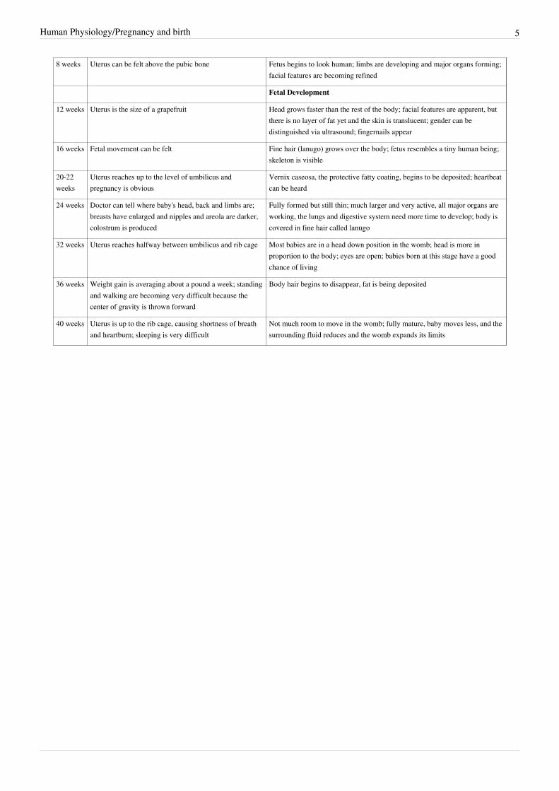
Human Physiology/Pregnancy and birth 5
8 weeks Uterus can be felt above the pubic bone Fetus begins to look human; limbs are developing and major organs forming;facial features are becoming refined
Fetal Development
12 weeks Uterus is the size of a grapefruit Head grows faster than the rest of the body; facial features are apparent, butthere is no layer of fat yet and the skin is translucent; gender can bedistinguished via ultrasound; fingernails appear
16 weeks Fetal movement can be felt Fine hair (lanugo) grows over the body; fetus resembles a tiny human being;skeleton is visible
20-22weeks
Uterus reaches up to the level of umbilicus andpregnancy is obvious
Vernix caseosa, the protective fatty coating, begins to be deposited; heartbeatcan be heard
24 weeks Doctor can tell where baby's head, back and limbs are;breasts have enlarged and nipples and areola are darker,colostrum is produced
Fully formed but still thin; much larger and very active, all major organs areworking, the lungs and digestive system need more time to develop; body iscovered in fine hair called lanugo
32 weeks Uterus reaches halfway between umbilicus and rib cage Most babies are in a head down position in the womb; head is more inproportion to the body; eyes are open; babies born at this stage have a goodchance of living
36 weeks Weight gain is averaging about a pound a week; standingand walking are becoming very difficult because thecenter of gravity is thrown forward
Body hair begins to disappear, fat is being deposited
40 weeks Uterus is up to the rib cage, causing shortness of breathand heartburn; sleeping is very difficult
Not much room to move in the womb; fully mature, baby moves less, and thesurrounding fluid reduces and the womb expands its limits

Human Physiology/Pregnancy and birth 6
Embryonic Development at Specific Stages
First trimester
An embryo this tiny shows very distinct anatomic features, including tail, limbbuds, heart (which actually protrudes from the chest), eye cups, cornea/lens, brain,and prominent segmentation into somites. The gestational sac is surrounded by a
myriad of chorionic villi resembling elongate party balloons. This embryo is aboutfive weeks old (or seven weeks in the biologically misleading but eminently
practical dating system used in obstetrics).
4 Weeks
• There are only the beginnings of facialfeatures. All the major organs are startingto form. Gill-like folds that develop intofacial features, beginnings of the spinalcord, skin is translucent, and rudimentary(basic; minimal) heart develops.
6 Weeks
• The length from crown to rump is aboutthe size of a finger tip, ¾ ". Thebeginnings of all the major organs willhave formed.
• The embryo floats in a fluid filled bubblethat will develop into the amniotic sac.The sac is covered by a protective layerof cells, called chorion. The yolk sacsupplies the embryo with all its nutrientsuntil the placenta is fully developed andtakes over at around the twelfth week.During the first 12 weeks, the embryowill develop features and major organs ofa human being. The embryo issusceptible to harmful environmentalinfluences. This is a vital time for theembryo to develop healthily; takingsupplements of folic acid, avoiding certain foods, and eliminating alcohol, cigarettes, and any unnecessary drugsor medicines.
9 Weeks
• The length from crown to rump approximately 1 1/4". The facial features are becoming more distinct, and the“tail” has disappeared. The muscles are also developing. Eyes are formed but eyelids are still closed over them.Arms now bend at the elbow and rudimentary hands and fingers develop. Knees will have formed and developingfeet with distinct toes.
• Heart- is now a four-chambered and fully formed organ; it beats about 180 times per minute.• Brain and nervous system- is four times the size it was at 6 weeks. Special glial cells are being formed within the
neural tube; they allow nerve cells to be joined so that messages can be transmitted from the brain to the body.• Digestive system- the mouth, intestine, and stomach are developing very rapidly, but do not function yet.• The fetal life-support system- the placental tissue that initially surrounds the fetus and the amniotic sac is
becoming concentrated in one circular area on the womb wall to form the placenta.

Human Physiology/Pregnancy and birth 7
Sonogram of a fetus at 14 weeks (Profile)
12 Weeks
• At twelve weeks the fetus looks like a tiny human. It is about 2 1/2"long and weighs 1/2 oz. Arms and legs are now beginning to move.Skin is red and translucent. Fingers and toes are more defined, andnails are starting to grow.
• Heart is complete and working, pumping blood to all parts of thebody. Digestive system has formed and is linked to the mouth andintestines. Sexual organs have formed inside the body, but cannotyet establish the sex of the baby.
Second Trimester
20 Weeks
• By 20 weeks the fetus will be about 6 1/3" long and weighs 12 oz.Movements are for more coordinated. The sexual organs are well
developed and are usually visible on ultra sound.• The fetus is growing very quickly. At this stage, the mother should feel the movements of the fetus. Movements
are more noticeable as the fetus's leg bones achieve their final relative proportions in a process called quickening.Quickening is the process of muscles contracting that cause movement at the fetus's synovial joints. The jointmovement enhances the nutrition of the articular cartilage and prevents the fusion of connective tissues within thejoint. It also promotes bone hardening.
• From now on, the fully developed placenta will provide all the fetus' needs until birth; oxygen, nutrients andprotective antibodies.
Fetus at 29 weeks gestation in 3D
Third Trimester
29 Weeks
• By 29 weeks the baby is about 10" long and weighs about 2 lbs. 7oz.
• The brain grows much larger, and fatty protective sheath covers thenerve fibers; this important development allows brain impulses totravel faster, enhancing the ability to learn. The lungs havedeveloped most of their airways and air sacs. The placenta is quiteselective in what it allows to pass from the mother to the baby'sblood, stopping some harmful substances, such as certain drugs,from crossing over.
40 Weeks
• The baby is now ready to be born. When the head of the baby moves down from high in the mother's abdomenand settles deeper into her pelvis in preparation for birth, it is called engagement. This can happen any timebetween 36 weeks and labor.
• In the last four weeks of pregnancy the baby puts on a lot of weight and develops a thick layer of fat. All organsare completely formed and functioning.

Human Physiology/Pregnancy and birth 8
Umbilical CordThis is the life support for a growing embryo. The umbilical cord stretches between the placenta and the fetus. Thiscord contains the umbilical arteries and vein. The umbilical cord forms by week 5 of conception. The average cord isclose to 22 inches long and may have the appearance of a coil. The umbilical cord is very rich in stem cells and isoften used for parents who choose to store their stem cells in a blood bank or donate it to a blood bank. These stemcells can be used to treat over 45 disorders and is an alternative from extracting the stem cells from a donor.
Human placenta shown a few minutes after birth. The side shown faces the baby with theumbilical cord top right. The unseen side connects to the uterine wall. The white fringe
surrounding the bottom is the remnants of the amniotic sac. You can see the differences inthe umbilical vein and arteries.
• Umbilical Arteries
The exchange of gases, nutrients andoxygen takes place between thematernal blood and fetal blood. Thereare 2 main arteries.• Umbilical Vein
Vein that carries nutrients and oxygenaway from the placenta to the growingfetus. It also carries oxygen andnutrient rich blood. There is only 1main vein.• Fetus doesn't use its lungs for gas
exchange, only a small amount ofblood is pumped to fetal lungs inorder to support their development.
Umbilical Abnormalities
• Single Umbilical Artery
One artery instead of two will result inchromosomal abnormalities. Some of these defects include poor fetal growth, preterm delivery, and still births. Thiscan be detected by a routine ultrasound. If an ultrasound is done and no other complications or abnormalities aredetected, the baby will usually be born healthy.• Umbilical Prolapse
This condition usually happens when a cord is too long. The baby may be born prematurely or will be breech.• Umbilical Nuchal Loops
This condition happens when the umbilical cord is wrapped around the baby's head at least one or more times. Thiscan be detected when a baby is in stress or by a simple ultrasound. In most cases the mother will have a cesareandelivery. In other cases the cord may be wrapped around the hands or feet.• Vasa Previa
This occurs in one in every 3,000 births, which can become life threatening for the unborn baby. This complicationhappens when the umbilical cord inserts abnormally in the fetal membranes of the placenta, which appearsabnormally shaped or positioned. Major risks include unprotected fetal blood vessels cross the cervix, oftentimesrupturing the membranes. Also, lack of blood pressure due from pressure, causes the loss of oxygen to the baby.Women who will be at risk for this would be those who already have experienced placenta previa or have used invitro fertilization.• Umbilical Cord Knots
About 1% of babies are born with one or more knots in their umbilical cord. Some knots happen during labor; othershappen from moving around in the womb. Most knots occur when the umbilical cord is too long. In some cases the

Human Physiology/Pregnancy and birth 9
knots can become tight, cutting off the oxygen supply to the baby. Cord knots result in miscarriages and stillbirth in5% and 10% of most cases. Most will require a cesarean delivery.• Umbilical Clotting
This is more common with genetic defects, such as Factor V Leiden. This complication will prevent blood flow toand from the baby and many times will cause the placenta to also clot and die. If this is not caught early enough, thebaby will die of starvation in the womb. A simple ultrasound can determine if there are problems with the bloodflow.
Pregnancy from the mother's perspective
Growth of the uterus in a pregnantfemale.
An inital sign of pregnancy is amenorreah, or the absence of menstration.Menses cease because the blastocyte begins the release of hCG or humanchorionic gonadotropin. Most pregnancy tests are specifically designed torecognize the presence of hCG, and hCG levels can be tested through themothers blood to learn whether or not a pregnancy is progressing normally.Human pregnancy lasts approximately 40 weeks from the time of the lastmenstrual cycle and childbirth (38 weeks from fertilization). The medicalterm for a pregnant woman is genetalian, just as the medical term for thepotential baby is embryo (early weeks) and then fetus (until birth). A womanwho is pregnant for the first time is known as a primigravida or gravida 1: awoman who has never been pregnant is known as a gravida 0; similarly, theterms para 0, para 1 and so on are used for the number of times a woman hasgiven birth.Image:Expecting mother.jpgIn many societies' medical and legal definitions, human pregnancy issomewhat arbitrarily divided into three trimester periods, as a means tosimplify reference to the different stages of fetal development. The firsttrimester period carries the highest risk of miscarriage (spontaneous death ofembryo or fetus). During the second trimester the development of the fetuscan start to be monitored and diagnosed. The third trimester marks thebeginning of viability, which means the fetus might survive if an early birthoccurs.

Human Physiology/Pregnancy and birth 10
Changing Body
(38 weeks) What a great change in the contents ofthe mothers abdomen
As soon as a woman becomes pregnant, her body begins to change sothat it can support both herself and the unborn baby. All of the bodyfunctions start to work much harder. The heart has to pump more bloodaround the body, in particular to the womb, placenta, and the fetus. Aswell as physical demands, pregnancy also causes a range of emotionalreactions.• The first trimester, the first twelve weeks, little is visible.• The second trimester, 13-27 Weeks, the waistline is rapidly
growing, the abdomen becomes noticeably pregnant.• The third trimester, 28-40 weeks, the body expands rapidly and the
womb enlarges and presses against the diaphragm.
First Trimester
In the early weeks the mother is likely to be more tired. Most expectantmothers are still in shock! As your uterus begins to grow your "bump" begins to be more noticeable. This is a goodtime to start looking into options on birthing and doctors.• Physical feelings: tiredness, nausea, constipation, frequent urination, food cravings, change in size of breasts,
fainting or dizziness, bloated stomach, and high emotions.
Second Trimester
The mother will probably be feeling full of energy and excitement.• Physical feelings: More energy, constipation, heartburn, and indigestion. The breasts continue to grow, as does
an increase in appetite. There is mild swelling in the feet, ankles, hands, and face. There is also more babymovement. There may be emotional ups and downs in the feeling of pregnancy, and short-term memory maybe poor.
• The hormones estrogen, progesterone, human placental lactogen, oxytocin, and prolactin prepare the body forfeeding the baby, and cause the breasts to enlarge, becoming painful and tender.
• The fetus, placenta, and amniotic fluid account for just over a third of the weight gain during pregnancy. Theremaining weight comes from increased blood volume, fluid retention, and extra body fat. The suggestedweight gain in most pregnancies is between 25-35 lbs.
Third Trimester
Physical feelingsShortness of breath, tiredness, difficulty in moving and sleeping, and frequent urination. The emotional moodswings ease off, but the mother begins to feel less enthusiastic about being pregnant. She may becomeimpatient and restless and just wants for the birth to be over.
• The body is changing to cope with the ever increasing size of the womb. The baby grows and pushes out thelower back of the mother. The breathing rate of the baby is growing very quickly. At this stage, the mothershould feel the movements of the fetus. Other signs may be the nipples secreting colostrum, Braxton-Hicks'contractions may begin, and blood flow to the womb has increased tenfold since conception.

Human Physiology/Pregnancy and birth 11
Prenatal CareOnce the female confirms her pregnancy, she will need to find out her physical condition and what to expect in thecoming months. Women typically begins pre-natal care at approximately 8-10 weeks gestation and prenancy careshould continue until approximately 6 week postpartum. The main purpose of the prenatal visits is to performpreventative medicine. Most complications in pregnancy are best treated if they are caught early on. A series of testswill be performed through out the pregnancy to judge the mother and fetus' well being including:• Mother's history• Urine tests for glucose, protein, and infection• The mother's weight• Blood tests such as a complete blood count, HIV test, or the triple screen which is test used most commonly to
look for neural tube defects and Downs Syndrome.• Physical examination• Blood pressure• Fetal heart monitoring• Ultrasound scans• Non-stress testsContinuous care is the best way to ensure a healthy mom and baby.
Labor and BirthLabor is defined as contractions and cervical change, contractions alone are not labor.• Pre-Labor Signs: as your body is preparing for labor, there are a few things that should be expected to happen
within four to six weeks of labor.1. Pressure on the pelvic area2. Occasional brownish discharge3. Energy level is noticeably increasing or decreasing4. Loss of the mucus plug (does not always exist)/increasing discharge5. Braxton Hicks contractions (painless contraction of the uterus)6. Movement of the baby into the pelvis• False Labor Signs: there are a few signs that indicate false labor.1. Timing of the contractions are irregular and do not become more frequent or more intense2. Contractions stop during rest, stopping what the mother is doing, walking, or changing position3. Inconsistent in strength (strong one minute then weak the next)4. Location of pain is in the front only• True Labor1. Pain in the lower back, radiating towards the front abdomen, possibly also the legs2. Contractions increase in strength and are closer together; coming now on a regular basis, 30 to 70 seconds apart3. The mucous plug is detached, showing bloody discharge4. The water breaks (usually this does not break until the doctor does it), when this happens, contractions become
much stronger5. Some women have the sudden need to go to the bathroom, diarrhea is common6. Contractions continue despite movement7. The cervix is thinning and dilatingWhen the contractions of labor begin, the walls of the uterus start to contract. They are stimulated by the release of the pituitary hormone oxytocin. The contractions cause the cervix to widen and begin to open. As labor progresses the amniotic sac can rupture causing a slow or a fast gush of fluids. Labor usually begins within a 24 hour period

Human Physiology/Pregnancy and birth 12
after the amniotic sac has ruptured. As contractions become closer and stronger the cervix will gradually start todilate. The first stage of labor is broken into three parts:• Early Phase First is the early phase of labor, when the cervix dilates from 1-4 centimeters, this can be the longest
and most exhausting part for the mother.• Active Phase The cervix dilates on average 1 cm per hour in the active phase of labor dilating from 4-7
centimeters. If an epidural is requested it is usually given in this phase.• Transition This is often considered the most intense part of labor with contractions lasting longer and having
shorter rest periods in between them. Dilation from 8-10 centimeters occurs during transition. Some womenexperience nausea and vomiting during this phase as well as rectal pressure and an urge to push.
At this point the labor enters the second stage, or the birth of the baby. The mother begins pushing to aid in the birthof the baby, this part of labor can last minutes, or even hours. A fetus usually delivered head first. 'Crowning' is theterm used when the fetus' head can be seen between the mothers labia as it emerges. At this point if necessary thebirth attendant may perform an episiotomy, which is a small surgical incision in the peritoneum. This procedure isusually done to deliver the baby more quickly in response to fetal distress.
Diagram showing an episiotomie
The third stage of labor is the delivery of the afterbirth (placenta).Oxytocin continues to be released to shrink the size of the uterus andaid in the limiting of blood loss from the site of the placenta. As theuterus shrinks the attachment site blood vessels, some of which can beas large as an adult finger, shrink also. The average blood loss in aroutine vaginal delivery is 400-500 cc.There are times when a mother may need outside aid in the delivery ofthe baby, some of these methods include:• Forceps, an instrument used to cradle the fetus' head and manipulate
the head under the pubic bone to more easily pass through the birthcanal.
• Vacuum Extraction, a suction cup is applied to the baby's head, and a plunger is used to suck any air frombetween the suction cup and the head to create a good seal. The babies head is then manipulated through the birthcanal. This usually leaves a baby's head bruised, but the mark fades within weeks after birth.
C-section Birth
• Cesarean section, or C-section, is the delivery of a baby through asurgical abdominal incision (Abdominal delivery - Abdominal birth- Cesarean section). A C-section delivery is performed when avaginal birth is not possible or is not safe for the mother or child.Surgery is usually done while the woman is awake but anesthetizedfrom the chest to the legs by epidural or spinal anesthesia. Anincision is made across the abdomen just above the pubic area. Theuterus is opened, and often brought through the incision afterdelivery for better visualization. The amniotic fluid is drained, andthe baby is delivered. The baby's mouth and nose are cleared offluids, and the umbilical cord is clamped and cut. After delivery anursery nurse or pediatrician check the make sure that the baby is breathing and responding. Due to a variety ofmedical and social factors, C-sections have become fairly common; around 25% of births are performed byC-section. C-sections carry some risks to mother and baby. Compared to a vaginal birth, the risks to motherinclude increased risk of death, surgical injury, infection, postpartum depression, and hemorrhage, although theseare rare. Babies born by c-section are more likely to be admitted to the ICU for breathing problems. Mothers areadvised to carefully weigh the risks of C-section versus vaginal birth.

Human Physiology/Pregnancy and birth 13
New baby !
Delivery Options
Hospital BirthsThe chances of having natural, uncomplicated birth areoptimized by carefully selecting your obstetrician and hospital.Doctors who work with midwives have lower cesarean sectionrates because midwives handle less complicated pregnancies.Delivering babies by abdominal surgery has been steadily risingin America over the past two decades, so that now 22-30% ofbirths in American hospitals are cesarean section. The U.S.,despite having the most advanced technology and highly trainedmedical personnel, ranks 23rd in infant mortality and 18th inperinatal mortality.Medical interventions such as epidural anesthesia, pitocinaugmentation of labor, vacuum extraction of fetus, episiotomyand separation of newborn and mother are common in Americanhospitals. There are circumstances where medical proceduressuch as these are necessary, but many parents and professionalsnow question the routine use of such interventions. In some cases, the routine use of these procedures havelead to further complications. For example, the epidural anesthetic, while providing pain relief, has shown toincrease the operative vaginal delivery rate (i.e. forceps and vacuum extraction rates slightly) especially in firsttime mothers. Epidurals have not been shown to increase the cesarean section rate in recent well documentedstudies.
Freestanding Birth Centers & Water Birth"Freestanding" Birth Centers are not inside of or affiliated with a hospital. They are run by collaboration ofmidwives or physicians. This is an alternative choice for the woman who does not wish to birth in a hospitalenvironment yet is not comfortable giving birth at home. Birth centers do not provide any additional measureof safety than most planned home births with qualified midwives; they may provide the expectant couple withthe physiological comfort necessary to enable the mother to relax.Out of hospital birth centers are designed for women having low-risk pregnancies who want drug-free birthwith minimal intervention in a home-like environment. Family members may participate in the birth.C-sections rates are lower than most hospitals because the pregnancies are low risk. Freestanding BirthCenters are an alternative choice for a woman who has had a previous cesarean and wishes to maximize herchances of a vaginal delivery. However, vaginal birth attempts after a prior cesarean section have a 1-2% riskof uterine rupture. Heath insurance may cover costs. Many birth centers offer birthing tubs where one can givebirth in water.
HomebirthBirth at home provides parents with intimacy, privacy, comfort and family-centered experience. Childbirth athome may be a safe option for healthy women having normal pregnancies. It is for those who have a verystrong desire for natural childbirth and who are willing to take high degree of responsibility for their healthcare and baby's birth. At home, the parents and midwife are in control of the birthing environment, and stricttime perimeters for length of labor are not imposed, or routine medical interventions such as IVs done.However, the World Health Organization (WHO) states that "giving birth in a health facility (not necessarily ahospital) with professional staff is safer by far than doing so at home." (The World Health Report 2005). Also,the American College of Obstetricians and Gynecologists (ACOG) opposes out of hospital births. In choosing

Human Physiology/Pregnancy and birth 14
the comfort of home parents are also choosing to be further away from lifesaving measures shouldcomplications arise.Homebirth midwives provide complete prenatal care including monthly visits, laboratory tests, screening forinfections. They provide nutritional counseling and support for psycho-social issues. There is a chance that arare, but critical emergency might occur during the birth where hospital services may not be able to beobtained quick enough. Again, the WHO states that "it is just before, during, and in the very first hours anddays after birth that life is most at risk," (The World Health Report 2005) and that "many of the complicationsthat result in maternal deaths and many that contribute to perinatal deaths are unpredictable, and their onsetcan be both sudden and severe." (WHO Birth and Emergency Preparedness in Antenatal Care, 2006) Homebirth midwives are trained to know when an emergency requires medical interface and can provide stabilizingmeasures until critical care can be obtained. While homebirth midwives generally have the training,equipment, and medicine to handle many complications, there is great variation in training and skill levelamong midwives. In choosing a homebirth midwife one should careful examine credentials and training.
A newborn with umbilical cord still attached (3minutes.)
Postpartum care
After the baby is born the umbilical cord is clamped and cut andthe baby is looked over by a doctor or nurse. The baby is given anAPGAR score at one and five minutes after birth. This is ananalysis of how well the baby is performing its vital functions.

Human Physiology/Pregnancy and birth 15
Score of 0 Score of 1 Score of 2 Acronym
Skin color blue all over blue at extremities normal Appearance
Heart rate absent <100 >100 Pulse
Reflex irritability no responseto stimulation
grimace/feeblecry
when stimulated
sneeze/cough/pullsaway
when stimulated
Grimace
Muscle tone none some flexion active movement Activity
Respiration absent weak or irregular strong + bgcolor=#abcdef|The five criteria of the Apgar score:
If tearing, or an episiotomy occurs the wound is closed with absorbable suture. The mother is closely watched forblood loss, infection, or any other possible complications. Breastfeeding should be initiated as soon as possible afterdelivery as the stimulation of oxytocin in the mother aids in hemostasis.
Risks in PregnancyPregnancies that warrant close attention usually come from an existing medical condition such as asthma, diabetes,epilepsy, or a condition developed because of pregnancy. Conditions that arise during pregnancy will require specialtreatment. The purpose of prenatal care is to detect these conditions, and to monitor and deal with them before theybecome serious.• Preeclampsia is the medical term for high blood pressure during pregnancy. It is also characterized by edema,
blurry vision, liver pain, and can progress into Eclampsia in which the mother can experience seizures, coma oreven death.
• Gestational Diabetes is diabetes mellitus that develops during pregnancy. All women should be tested for thecondition at about 28 weeks gestation. Gestational and pre-existing diabetes can cause large for gestational agebabies, a sudden drop in a neonates blood sugar after birth, and has a high risk for stillbirth
Other serious risks include:• Teratogens (substances that cause birth defects including alcohol and certain prescription and recreational drugs)• Infection (such as rubella or cytomegalovirus) An infection in the eleventh week is less likely to damage the
heart, but the baby may be born deaf.• Genetics (such as Factor V Leiden) Diabetes, blood conditions, etc.• Radiation (ionizing radiation such as X-rays, radiation therapy, or accidental exposure to radiation)• Nutritional deficiencies
• Fetal Alcohol Syndrome or FAS exposure is the leading known cause of mental retardation in the Westernworld. It is a disorder of permanent birth defects that occurs in the offspring of women who drink alcohol duringpregnancy, depending on the amount, frequency, and timing of alcohol consumption. Alcohol crosses theplacental barrier and can stunt fetal growth or weight, create distinctive facial stigmata, damage neurons and brainstructures, and cause other physical, mental, or behavioral problems. Drinking during pregnancy should beavoided. Women who drink more than 4 or 5 drinks per day may cause permanent damage to their fetus,including, behavioral problems, sight and hearing loss, deformed organs and central nervous system dysfunction.
• Smoking can cause low birth weight, still birth, birth defects, preterm births and immature lung development. Itcan also contribute to addiction in the child's later teen years.
• Illegal Drugs can be the most devastating. Risks include SIDS (Sudden Infants Death Syndrome), learningdisorders, birth defects, uncontrollable trembling, hyperactive, and drug dependency. Most drugs can be tested bya simple urine or blood test.

Human Physiology/Pregnancy and birth 16
• Medications. All medication use should be discussed with your doctor. Many over the counter and prescriptiondrugs have warning labels. Follow these precautions to help avoid birth defects or other related problems.
MiscarriageMiscarriage or spontaneous abortion is the natural or spontaneous end of a pregnancy at a stage where the embryo orthe fetus is incapable of surviving, generally defined in humans at a gestation of prior to 20 weeks. Miscarriages arethe most common complication of pregnancy. Basic Facts: 15-20% of pregnancies end in miscarriage, 70% of thetime there is a chromosomal abnormality with the fetus, and one miscarriage does not increase your risk in the nextpregnancy. Miscarriage is almost never the mother's fault.If the products of conception are not completely expelled after fetal death this is known as a missed abortion and isusually treated surgically by a procedure known as a D&C or dilation and curettage.
Bleeding During PregnancyVaginal bleeding at any stage should be taken seriously. Severe bleeding in the early weeks may be a sign ofmiscarriage. However, 25% of pregnant patient bleed in the first trimester. After 24 weeks the mother should seekmedical advice immediately. Third trimester bleeding in pregnancy is often one of the first signs of placenta previa;placenta is across the opening of the cervix. An ultrasound should be performed to establish the location. Othercauses of late term bleeding include:• Preterm Labor or labor that occurs before 38 weeks gestation that can have multiple causes• Placental Abruption is a condition in which the placenta is torn away from the uterine wall causing loss of
oxygen and nutrients to the baby, and hemorrhage of mother and baby from the large blood vessels in theplacenta. Most women, but not all experience heavy bleeding and abdominal pain. This is a life threateningemergency as a fetus can only survive as long as 50% of the placenta is still attached.
Blood ConditionsIndividuals either have, or do not have, the Rhesus factor (or Rh D antigen) on the surface of their red blood cells.This is usually indicated by 'RhD positive' (does have the RhD antigen) or 'RhD negative' (does not have the antigen)suffix to the ABO blood type i.e. A+ B- blood typing. This is a problem only when an Rh-negative woman has apartner who is Rh-positive resulting in an Rh-positive baby. If the mother's and the baby's blood come into contactduring the birth, her body produces antibodies against the baby's blood. This problem usually does not affect thecurrent pregnancy but can be dangerous for future pregnancies as the antibodies stay in the blood causing an immuneresponse against future Rh+ fetus. In essence the mother's body "rejects" the fetus as it would a foreign body. A drugcalled Rhogam is now given by injection given at 28-30 weeks gestation and given again if there is confirmation thatthe baby is Rh positive within 24 hours after birth to protect the future pregnancies. Rh isoimmunization is rare inour day. Rh- mothers should also be given the injection after miscarriage or abortion.If a mother is untreated they are at risk to subsequently deliver babies who suffer from hemolytic disease of thenewborn. Hemolytic disease of the newborn, also known as HDN, is an alloimmune condition that develops in afetus, when the IgG antibodies that have been produced by the mother and have passed through the placenta includeones which attack the red blood cells in the fetal circulation. The red cells are broken down and the fetus can developreticulocytosis and anemia. This fetal disease ranges from mild to very severe, and fetal death from heart failure(hydrops fetalis) can occur. When the disease is moderate or severe, many erythroblasts are present in the fetal bloodand so these forms of the disease can be called erythroblastosis fetalis (or erythroblastosis foetalis). Hemolysis leadsto elevated bilirubin levels. After delivery bilirubin is no longer cleared (via the placenta) from the neonate's bloodand the symptoms of jaundice (yellowish skin and yellow discoloration of the whites of the eyes) increase within 24hours after birth. Like any other severe neonatal jaundice, there is the possibility of acute or chronic kernicterus.Profound anemia can cause high-output heart failure, with pallor, enlarged liver and/or spleen, generalized swelling,

Human Physiology/Pregnancy and birth 17
and respiratory distress. The prenatal manifestations are known as hydrops fetalis; in severe forms this can includepetechiae and purpura. The infant may be stillborn or die shortly after birth.
Other AbnormalitiesPhysical and Genetic Defects: Physical anomalies are present at birth. Examples are; cardiac, facial (such as cleftpalate), club foot, etc. These do not always endanger the baby's life. 1-2% of babies are born with a significantcongenital abnormality. 4-6% with something relatively minor.• Chromosomal Abnormalities: Occur when there is a problem in the baby's genetic makeup; these include
conditions such as Down syndrome. Other genetic defects, such as cystic fibrosis, can be inherited from theparents.
Staying HealthyPregnancy and childbirth place great demands, it is important to keep healthy. The more healthy and relaxed themother is, the better it will be to cope with the demands of pregnancy. A healthy lifestyle combines many factors:Balanced Diet
A poor diet can cause a low birth weight. Excessive weight gain during pregnancy can cause back problems,varicose veins, or indicate preclampsia. Advice on diet often includes to eat foods that are high in nutritionalcontent. Sufficient protein, vitamins, carbohydrates, fats, and minerals, as well as fiber. Limit intake ofsaturated fats and sugar, and salt. Drink plenty of fluids.
Regular ExerciseMild exercise, such as walking or swimming, is beneficial and will help cope with the workload of pregnancyand the demands of labor. Mother's should listen to her body and stop exercising when it tells her to. Exerciseshould never be painful.
Baby's HealthSmoking reduces the oxygen and nutrients passing via the placenta to the baby. Avoid alcohol to avoid seriousbirth defects.
In vitro Fertilization and Artificial Implantation
Oocyte is injected with sperm outside of the womb.
An alternative when other methods ofachieving contraception have failed.
In vitro fertilization (IVF) is a technique inwhich egg cells are fertilized by spermoutside the woman's womb. IVF is a majortreatment in infertility when other methodsof achieving conception have failed. Theprocess involves hormonally controlling theovulatory process, removing ova (eggs)from the woman's ovaries and letting spermfertilize them in a fluid medium. Thefertilized egg (zygote) is then transferred tothe patient's uterus with the intent toestablish a successful pregnancy.

Human Physiology/Pregnancy and birth 18
The term in vitro, from the Latin root, is used, because early biological experiments involving cultivation of tissuesoutside the living organism from which they came, were carried out in glass containers such as beakers, test tubes, orpetri dishes.While the overall live birth rate via IVF in the U.S. is about 27% per cycle (33% pregnancy rate), the chances of asuccessful pregnancy via IVF vary widely based on the age of the woman (or, more precisely, on the age of the eggsinvolved). Where the woman's own eggs are used as opposed to those of a donor, for women under 35, thepregnancy rate is commonly approximately 43% per cycle (37% live birth), while for women over 40, the rate fallsdrastically - to only 4% for women over 42. Other factors that determine success rates include the quality of the eggsand sperm, the duration of the infertility, the health of the uterus, and the medical expertise. It is a common practicefor IVF programmes to boost the pregnancy rate by placing multiple embryos during embryo transfer. A flip side ofthis practice is a higher risk of multiple pregnancy, itself associated with obstetric complications.Embryo cryopreservation If multiple embryos are generated, patients may choose to freeze embryos that are nottransferred. Those embryos are placed in liquid nitrogen and can be preserved for a long time. There are currently500,000 frozen embryos in the United States. The advantage is that patients who fail to conceive may becomepregnant using such embryos without having to go through a full IVF cycle. Or, if pregnancy occurred, they couldreturn later for another pregnancy.
Embryonic stem cells
Pluripotent, embryonic stem cells originate as inner mass cells with in a blastocyst. Thestem cells can become any tissue in the body, excluding a placenta. Only the morula's
cells are totipotent, able to become all tissues and a placenta.
Embryonic celtic cell lines (ES celllines) are cultures of cells derived fromthe epiblast tissue of the inner cellmass (ICM) of a blastocyst. Ablastocyst is an early stage embryo -approximately 4 to 5 days old inhumans and consisting of 50-150 cells.ES cells are pluripotent, and give riseduring development to all derivativesof the three primary germ layers:ectoderm, endoderm and mesoderm. Inother words, they can develop intoeach of the more than 200 cell types ofthe adult body when given sufficientand necessary stimulation for a specificcell type. They do not contribute to theextra-embryonic membranes or theplacenta. This means they can becomeany kind of human tissue (ie. hearttissue, nerve tissue, etc.).
When given no stimuli fordifferentiation, ES cells will continue to divide in vitro and each daughter cell will remain pluripotent. Thepluripotency of ES cells has been rigorously demonstrated in vitro and in vivo, thus they can be indeed classified asstem cells.Because of their unique combined abilities of unlimited expansion and pluripotency, embryonic stem cells are apotential source for regenerative medicine and tissue replacement after injury or disease. To date, no approvedmedical treatments have been derived from embryonic stem cell research. This is not surprising considering that

Human Physiology/Pregnancy and birth 19
many nations currently have moratoria (suspension of practices) on either ES cell research or the production of newES cell lines.There exists a widespread controversy over stem cell research that emanates from the techniques used in the creationand usage of stem cells. Embryonic stem cell research is particularly controversial because, with the present state oftechnology, starting a stem cell line requires the destruction of a human embryo and/or therapeutic cloning.Opponents of the research argue that this practice is a slippery slope to reproductive cloning and tantamount to theinstrumentalization of a human being. Contrarily, some medical researchers in the field argue that it is necessary topursue embryonic stem cell research because the resultant technologies are expected to have significant medicalpotential, and that the embryos used for research are only those meant for destruction anyway (as a product of invitro fertilization). This in turn, conflicts with opponents in the pro-life movement, who argue that an embryo is ahuman being and therefore entitled to dignity even if legally slated for destruction. The ensuing debate has promptedauthorities around the world to seek regulatory frameworks and highlighted the fact that stem cell research representsa social and ethical challenge.• Reproductive Cloning
Reproductive Cloning is a technology used to generate an animal that contains the same nuclear DNA as anothercurrently or previously existing animal. Scientists transfer the genetic material from the nucleus of a donor adult cellto an egg whose nucleus, and thus its genetic material has been removed. The egg containing the DNA, nowreconstructed, has to be treated with chemicals or electric current in order to stimulate cell division. Once the clonedembryo reaches a suitable stage, it is transferred to the uterus of a female host to continue development until birth.Currently this is illegal to practice in the United States.• Therapeutic Cloning
Recent research by researchers led by Anthony Atala of Wake Forest University and a team from Harvard Universityhas found that amniotic fluid, in addition to its main functions of cushioning a growing fetus and providingbuoyancy, is also a plentiful source of non-embryonic stem cells. These cells have demonstrated the ability todifferentiate into a number of different cell-types, including brain, liver and bone.Therapeutic Cloning refers to a procedure that allows the cloning of specific body parts and organs to be used formedical purposes. Although this has not been realized, much research is being done on the subject.
Pregnancy and LactationA mother's milk is ideal because it meets the baby's specific needs. Lactation is a neuroendocrine response in milkproduction. Sucking stimulates the sensory nerve endings in the nipples and sends stimulus to the hypothalamus. Thehypothalamus stimulates anterior pituitary and prolactin is released. In milk let-down the sucking stimulates sensorynerves in the nipples. This stimulates the hypothalamus which then stimulates the posterior pituitary and releasesoxytocin. Sucking also stimulates contraction of the cells around the alveoli in the mammary cells. Milk then flowsinto the milk ducts causing milk let-down.Breast milk provides all the nutrients required for the first 4-6 months. It contains macronutrients like carbohydrateslike lactose, fat such as high linoleic acid and protein that is easily digestable and alpha-lactalbumin is absorbed.Breast milk also contains an adequate supply of vitamins and minerals, digestive enzymes, hormones andimmunological factors.The first milk produced after birth is called colostrum. This is synthesized during the end of pregnancy and 3-5 daysof postpartum. It is very high in protein and low in fat and carbohydrates, and contains immunoglobulins. This helpthe baby have a first bowel movement and prevent jaundice. The bowel movement that results from the colostrum isa different color and consistency than future bowel movements once the mother's milk comes in. In some cultures thecolostrum is discarded because of the difference, but what they do not know is that it is the best thing for the baby.

Human Physiology/Pregnancy and birth 20
The composition varies in breast milk during feeding, and over time with development of the baby. Whenbreastfeeding there are three names for the composition of the milk: the fore milk, present during the beginning ofbreastfeeding; mid is the middle of feeding; and hind which is toward the end of the feeding and contains acomposition high in fat.When breastfeeding the female should consider the types of food that will be consumed. If the mother is on a low fatdiet or if foods like garlic, broccoli, and onions are eaten, it may affect the baby's preference for breast feeding. Also,the mother should consider not breastfeeding after the consumption of alcohol, caffeine, smoking, and certainmedications.Barriers of breastfeeding are lack of professional and social support, misinformation, embarrassment, early dischargeform the hospital without instruction, and returning to work or school without adequate lactation rooms and if themother refuses to tend breastfed infant.When breastfeeding initiate as soon after delivery as possible, position the baby correctly, feed on demand from bothbreasts at each feeding and at least 10 minutes on each breast. Additionally there should be a good educator in thecase the infant is not latching on.A common problem that may happen when breastfeeding is mastitis, which is an inflammation of one or both breastsand is usually associated with the infection of a blocked milk duct during lactation. The symptoms include flu-likesymptoms, red streaks on the breast, and hot skin. Antibiotics may be necessary to clear the infection. Thrush mayalso happen and could be passed between mom and baby. A symptom of thrush includes white flecks on tongue, andthe baby and mother should be treated by a doctor.Breast milk is recommended through the first 12 months. Supplementation of cow's milk is not recommended due tothe high protein that would cause liver damage to the baby.Why breastfeed?• It is easily digested• Composition changes with infant needs• Changes during a feeding, high in fat at the end of feeding• Antibodies in milk• Breastfeeding moms miss less work because babies are sick less• Fewer allergies• Less spit-up• Less constipation and diarrhea• Better jaw development• Decreased risk of SIDS (Sudden Infant Death Syndrome)• Higher IQ• Decreased risk of diabetes, Crohn's Disease, Celiac Sprue• Bonding• Convenient, always at the correct temperature and ready to go• Less expensive• Helps the uterus return to normal size more quickly• Less incidence of postpartum “blues”• Lower risk of breast cancer• Lower risk of osteoporosis

Human Physiology/Pregnancy and birth 21
Postpartum DepressionHaving a baby is usually one of the happiest times in a woman's life, but for some women, it can include times ofsadness and depression. More women actually suffer from postpartum depression than we really know. Womenusually ignore the emotional and physical signs, dealing with their feelings on their own.Postpartum depression affects approximately 10 to 15 percent of new mothers. It often causes anxiety and obsessionabout caring for the baby or the cleanliness of the home. It may cause changes in sleep patterns and affectrelationships including the ability to form a bond with the baby and other family members. Some mothers withpostpartum depression have thoughts of wanting to die or of hurting the baby. If the symptoms are so severe thatthey keep the mother from being able to function, medical treatment is necessary. http:/ / www. siumed. edu/ news/Newsline%20TEXT05/ 8-03-04. htmBaby blues are common due to rapid hormonal changes but resolve after 1-2 weeks. Post-partum depression ischaracterized by persisting symptoms, and the mother should notify her provider immediately.
Testing Your KnowledgeAnswers for these questions can be found here [1]
1. Is at this stage that an egg implants in the uterine liningA) morulaB) zygoteC) blastocystD) embryoblast
2. Which part of the embryoblast will become the central nervous system in developmentA) ectogermB) mesodermC) endoderm
3. This hormone is only produced in the human body when a woman is pregnantA) estrogenB) HCGC) progesteroneD) FSHE) LH
4. By this week of pregnancy, the beginnings of all major organs have formedA) 4B) 7C) 5D) 6E) 8
5. Stem cells are found in the embryoblast and use of them is very controversial, another place to find stem cells thatare usable to treat leukemia and other disorders is the
A) morulaB) chorion
Human Physiology/Pregnancy and birth 22
C) amnionD) amniotic fluidE) umbilical cord
6. The cervix dilates on an average of ______ per hour in the active phase of laborA) 2 mmB) 2 cmC) 1mmD) 1 cm
7. The contractions of the uterus are stimulated by the release ofA) oytocinB) FSHC) LHD) prolactinE) estrogen
8. A sign of pre-labor isA) irregular contractionsB) pain in the front onlyC) loss of the mucas plugD) contractions stop during rest
9. This is the most common complication of pregnancyA) preclampciaB) miscarriageC) smokingD) Rh factorE) teratogens
10. Sue decides to breastfeed because she has been told that colostrum containsA) high proteinB) low fatC) immunoglobulinsD) all of the aboveE) none of the above
11. What is the first milk, after birth, called?A)ThrushB)MastitisC)ColostrumD)Milk let down

Human Physiology/Pregnancy and birth 23
GlossaryAbruption: Premature separation of the placenta from the wall of the wombAmnion: An embryonic membrane that encircles a developing fetus and contains amniotic fluid.Amniocentesis: A procedure in which a small sample of amniotic fluid is removed from around the fetusAmniotic fluid: The fluid surrounding the fetusAmniotomy: (artificial rupture of membranes, ARM) Breaking the membranes using a special plastic hookAnemia: Lack of hemoglobin in red blood cells, due to iron deficiency or diseaseAntepartum Hemorrhage: (APH) Vaginal bleeding that happens after 24 weeks of pregnancy and before deliveryBreech: The baby is lying bottom down in the wombCeliac sprue: Nutrient absorption impairment which is improved when gluten is removed form the diet.Characteristic mucosal lesion of the small intestine.Cephalic: The baby is lying head down in the wombChorion: The embryonic membrane that forms the outermost covering around the developing fetus.Chorion Villus Sampling: (CVS) A method for sampling placental tissue for genetic or chromosome studies.Colostrum the fluid that is made late in pregnancy and the first few days postpartum in the breast that containsimmunologic substances and essential nutrients.Cleavage: The early successive divisions of embryonic cells into smaller and smaller cells.Cilia: The fine hairs that line the fallopian tubes'Cordocentesis: The procedure for taking blood from the fetal umbilical cord via a needle through the mother’sabdomenCopulation: (Coitus, sexual intercourse) is the procreative act of a man's erect penis is inserted into a woman'svagina. At climax, semen is ejaculated from the penis at the cervix of the uterus. Sperm then propel themselves intothe uterine tubes where fertilization may occur if an egCrohn's disease: Skip lesions in the colon and is a malabsorptive disease.Cystitis: Infection of the bladderDizygous: Not identical (fraternal) twinsDoppler: A form of ultrasound used specially to investigate blood flow in the placenta or in the fetusDown Syndrome: (Trisomy 21) A disorder caused by the presence of an extra chromosome 21 in the cellsEctopic Pregnancy: A pregnancy that develops outside of the wombEdema: Swelling of the fingers, legs, toes, and face.Embryo: The medical term for the baby from conception to about six weeksEngagement: The process in which the head of the baby moves down from high in the mother's abdomen and settlesdeeper into her pelvis in preparation for birth. This can happen any time between 36 weeks and labor.Epidural Anesthesia: A method of numbing the nerves of the lower spinal cord to ensure a pain-free laborEpisiotomy: A cut of the perineum and vagina performed to make the delivery easierExternal Fetal Monitor: An electronic monitor used to record the fetal heartbeat and mother’s contractionsFallopian Tubes: (uterine tubes) Two tubular structures (one on each side of the womb) leading from the ovaries tothe uterusFertilization: The union of an egg cell and a sperm cell is present wherein 23 chromosomes from each parent come together to form a zygote. After sperm penetrates, the ovum undergoes a chemical change to prevent other sperm from entering. Multiple births can occur from complete division of the conceptus during early cleavage or from

Human Physiology/Pregnancy and birth 24
fertilization of multiple ova. Birth control techniques are designed to prevent ovulation or to prevent fertilization bybarriers, that keep sperm and ova separated.Fetus: Medical term for the baby from six weeks after conception until birthForceps: Metal instruments that fit on either side of the baby's head and are used to help deliver the babyFundus: The top of the wombGerm layer: Layers of cells within an embryo that form the body organd during development.Glial Cells (neuroglia; glia): Non-neuronal cells that provide support and nutrition, maintain homeostasis, formmyelin, and participate in signal transmission in the nervous system. In the human brain, glia are estimated tooutnumber neurons by about 10 to 1.Glial cells provide support and protection for neurons, the other main type of cell in the central nervous system. Theyare thus known as the "glue" of the nervous system. The four main functions of glial cells are to surround neuronsand hold them in place, to supply nutrients and oxygen to neurons, to insulate one neuron from another, and todestroy pathogens and remove dead neurons.Hemoglobin: (Hb)The oxygen carrying constituent of red blood cellsInduction of labor: (IOL) the procedure for initiating labor artificiallyIn utero death: (IUD)the death of the unborn fetus after 24 weeksIn vitro fertilization: (IVF) a method of assisted conception in which fertilization occurs outside the mother's andthe embryo is replaced in the wombLanugo: fine hair that covers the fetus in the wombLochia: blood loss after birthMastitis inflammation of the breast most frequently in lactation.Neonatal: baby less than 28 days oldNuchal scan: special ultrasound scan that gives an estimate of the risk of Down syndromeOocyte: one egg that is released from the ovary at each ovulationPlacenta: The structure by which an unborn child is attached to it's mother's uterine wall and through which it isnourished.Postnatal: After birthPrenatal: Before birthQuickening: The process that occurs between the seventeenth and twentieth weeks of fetal development, the fetus'sleg bones achieve their final relative proportions. In this process the muscles contract, causing movement at thefetus's sinovial joints. The joint movement enhances the nutrition of the articular cartilage and prevents the fusion ofconnective tissues within the joint. It also promotes bone hardening. It is this stage, where the fetus's bones becomemore developed and harder, that the mother begins to notice fetal movement.Rudimentary: Basic; minimal; with less than, or only the minimum, necessaryThrush: Creamy white flakes on a red papillae on tongue and tongue may be enlarged.Umbilical cord: The cord like structures that connects the fetus to the placenta.Zygote: A cell produced by the fusion of an egg and a sperm; a fertilized egg cell.

Human Physiology/Pregnancy and birth 25
Reference• "as your baby grows From Conception to Birth"published by American Baby
• http:/ / www. babybluesconnection. com• "Pregnancy and Birth" authors: Dr. Karina Reynolds, Dr. Christoph Lees, Grainne McCartan• "Fundamental Concepts of Human Anatomy" authors: M.J. Shively D.V.M., M.S., Ph.D. and D.P. Homan B.S.,
M.S.• "Essentials of Anatomy and Physiology" authors:Valerie C. Scanlon and Tina Sanders, fourth edition• http:/ / www. MERLOT. com Stanford Site• "The New Parent" author DR. Miriam Stoppard• www.marchofdimes.com• http:/ / health. allrefer. com/ health/ fetal-development-info. html• American Pregnancy Association
Internet groups: International Awareness Network: www.ican-online.org
References[1] http:/ / en. wikibooks. org/ wiki/ Human_Physiology/ Appendix_1:_answers_to_review_questions#Pregnancy_and_birth

Article Sources and Contributors 26
Article Sources and ContributorsHuman Physiology/Pregnancy and birth Source: http://en.wikibooks.org/w/index.php?oldid=1975241 Contributors: Adrignola, AmWengert, Amada44, Anne Robinson MD, BrendaJohnson,Brentwaldrop, Brittine Krmpotich, Cabeman, Carlosmoreno, Cde grey, Chaneldewitt, Danseyffert, DorothyD, ElizabethDurham, Herbythyme, Jandreakeller, Jennifermorley, Jomegat, Krstnplmr,Macp, Msol, Nataliehaveron, Never2late, Provophys, Pwoodson, Recent Runes, Shakah, Shaleneroberts, Shanannie, Stephanie greenwood, Sterlingsilver, Trevan5, Whiteknight, 104 anonymousedits
Image Sources, Licenses and ContributorsImage:Sperm-egg.jpg Source: http://en.wikibooks.org/w/index.php?title=File:Sperm-egg.jpg License: Public Domain Contributors: Blurpeace, Conscious, EugeneZelenko, Giggy, GrawpSock,Herbythyme, Ies, Jacklee, Kameraad Pjotr, Manuelt15, Mattes, Myself488, Noddy93, Pi@k, Platonides, The Evil IP address, Tronicum, 11 anonymous editsImage:Embryo, 8 cells.jpg Source: http://en.wikibooks.org/w/index.php?title=File:Embryo,_8_cells.jpg License: Public Domain Contributors: DO11.10, LlullImage:Fetus.jpg Source: http://en.wikibooks.org/w/index.php?title=File:Fetus.jpg License: Public Domain Contributors: Ferrylodge, Irigi, 1 anonymous editsImage:Tubal Pregnancy with embryo.jpg Source: http://en.wikibooks.org/w/index.php?title=File:Tubal_Pregnancy_with_embryo.jpg License: unknown Contributors: Ed Uthman, MD (Flickr, Wikipedia)Image:Embryo at 14 weeks profile.JPG Source: http://en.wikibooks.org/w/index.php?title=File:Embryo_at_14_weeks_profile.JPG License: Public Domain Contributors: User:Mirmillon,user:hidroImage:3dultrasound.png Source: http://en.wikibooks.org/w/index.php?title=File:3dultrasound.png License: Public Domain Contributors: Dar-Ape, LeonardoG, Nevit, Teebeutel, ThuressonImage:Human placenta baby side.jpg Source: http://en.wikibooks.org/w/index.php?title=File:Human_placenta_baby_side.jpg License: Public Domain Contributors: AirBa, Berillium, Glenn,Helcaf, Kajk, 2 anonymous editsImage:Grosse derGebarmutter in den einzelneMonaten derSchwangerschaft.gif Source:http://en.wikibooks.org/w/index.php?title=File:Grosse_derGebarmutter_in_den_einzelneMonaten_derSchwangerschaft.gif License: Public Domain Contributors: Haabet, Lennert B, TX55Image:Placenta.svg Source: http://en.wikibooks.org/w/index.php?title=File:Placenta.svg License: unknown Contributors: User:Amada44Image:Medio-lateral-episiotomy-blank.png Source: http://en.wikibooks.org/w/index.php?title=File:Medio-lateral-episiotomy-blank.png License: Public Domain Contributors: AirBaImage:Caesarian shown.jpg Source: http://en.wikibooks.org/w/index.php?title=File:Caesarian_shown.jpg License: Creative Commons Attribution-Sharealike 2.0 Contributors: Dbenbenn,FlickreviewR, Husky, Melimama, Schekinov Alexey Victorovich, 1 anonymous editsImage:Caesarian_shown_cropped.jpg Source: http://en.wikibooks.org/w/index.php?title=File:Caesarian_shown_cropped.jpg License: Creative Commons Attribution-Sharealike 2.5 Contributors: DO11.10, Ranveig, 1 anonymous editsImage:Umbilicalcord.jpg Source: http://en.wikibooks.org/w/index.php?title=File:Umbilicalcord.jpg License: unknown Contributors: Majorly, Nevit, The catImage:Icsi.JPG Source: http://en.wikibooks.org/w/index.php?title=File:Icsi.JPG License: Public Domain Contributors: DrKiernan, Lipothymia, Michiel1972Image:Stem cells diagram.png Source: http://en.wikibooks.org/w/index.php?title=File:Stem_cells_diagram.png License: Creative Commons Attribution-Sharealike 2.5 Contributors: MikeJones
LicenseCreative Commons Attribution-Share Alike 3.0 Unportedhttp:/ / creativecommons. org/ licenses/ by-sa/ 3. 0/